


Abstract
Silk contains a fibre forming protein, fibroin, which is biocompatible, particularly after removing the potentially immunogenic non-fibroin proteins. Silk can be engineered into a wide range of materials with diverse morphologies. Moreover, it is possible to regenerate fibroin with a desired amount of crystallinity, so that the biodegradation of silk materials can be controlled. These advantages have sparked new interest in the use of silk fibroin for biomedical applications, including tissue engineering scaffolds and carriers for sustained release of biologically active molecules. This article summarizes the current research related to the formation of silk materials with different morphologies, their biocompatibility, and examples of their biomedical applications. Recent work on the preparation of silk particles by mechanical milling and their applications in silk composite scaffolds is also discussed.
1. Introduction
Silk consists of fibrous proteins that are stored as a liquid in silk producing arthropods (such as silkworms, spiders, scorpions, mites and bees) and are spun into fibres at secretion [1]. Amongst various silk species, silkworm and spider silk fibres have been widely studied for their structure, processing, and functional properties. About 90% of commercial silk fibres used in the textile industry come from Lepidopteron silkworms from the family, Bombycidae [2]. In the textile industry, it is commonly referred to as Mulberry silk. Saturniidae is another class of Lepidopteron silkworms that produce commercial silk fibres such as Tasar, Eri and Muga.
Apart from their long history as luxury textiles, silk has also been used in the human body in the form of suture material [3]. A study by Minoura et al. [4,5] has suggested much wider application prospects for silk. Their study showed that silk fibroin of B.mori and A.pernyi was equal to or even better than collagen in supporting attachment and proliferation of murine L929 fibroblast cells. The results have generated enormous interest in the ensuing years to examine silk fibroin for various biomedical and healthcare applications through in-vitro or animal model studies.
Native silk fibre suture is a Food and Drug Administration (FDA, USA) approved biomaterial. Recently, regenerated silk has also been approved by the FDA for human clinical tests.
2. Different Forms of Silk Biomaterial
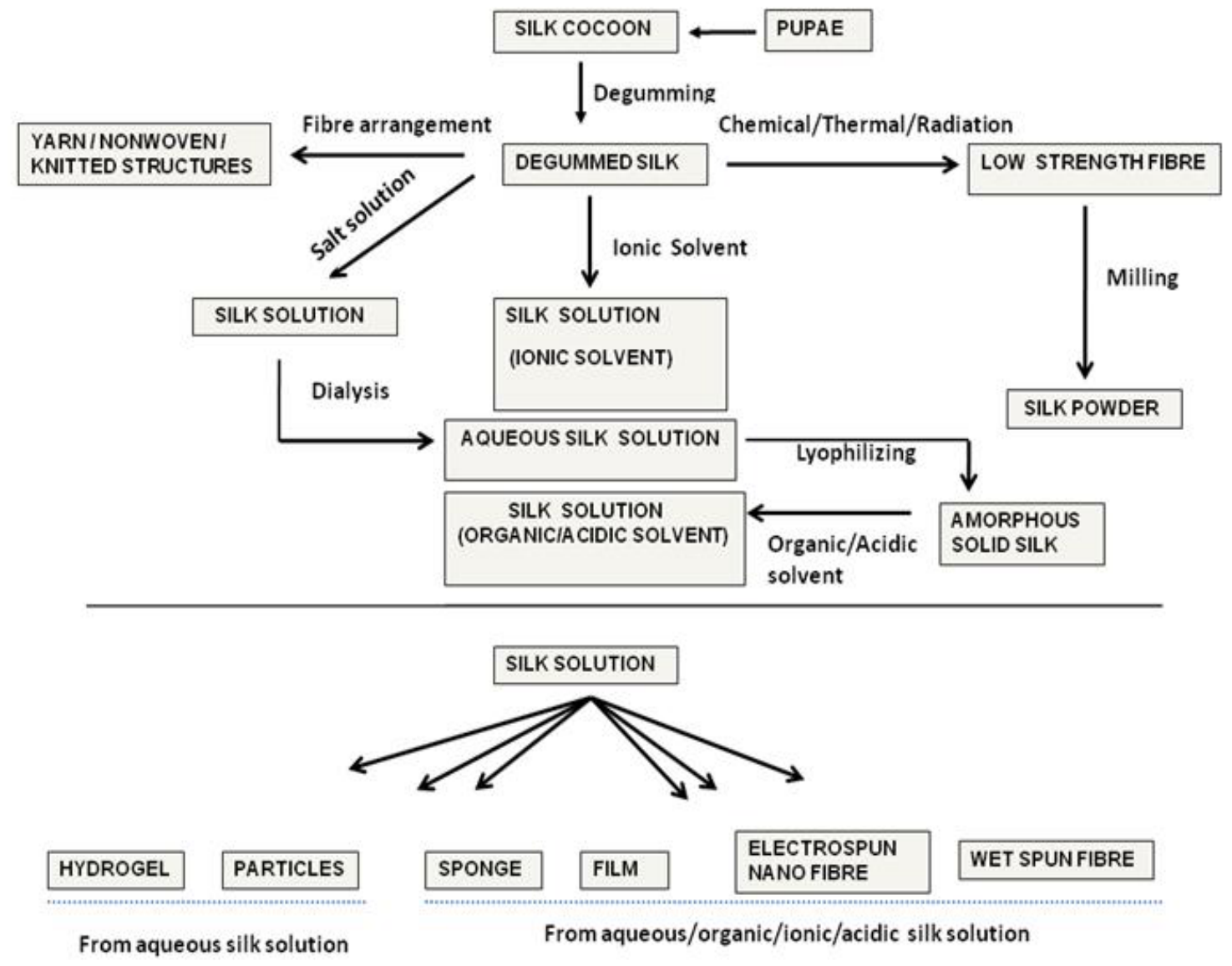
Silk biomaterials can be prepared directly from silk fibres, or reconstituted from silk fibroin solution. An alternate way is to convert silk fibres into ultrafine particles through milling. Figure 1 shows a schematic diagram of processing silk fibres into various forms of diverse morphologies [6].
2.1. Native Silk Fibre Based Structures
Degummed silk filaments in the form of twisted structures, such as wire-rope, cable, braided, and textured yarns have been analysed for potential biomedical applications [7]. Silk filaments can also be used to construct nonwoven structures as a cell supporting template, by partially dissolving silk fibres [8,9]. Random arrangements of filaments in a cocoon can be preserved during degumming to design a porous nonwoven silk mat for cell seeding [9]. An alternate way of using silk filaments directly in tissue engineering is making a knitted structure [10]. Such knitted structures have been used to reinforce 3-D porous tissue engineering scaffolds for improved mechanical properties [11].
2.2. Regenerated Silk Biomaterials
Silk fibres can be dissolved by using a highly concentrated solution of chaotropic salts followed by dialysis to prepare an aqueous silk solution. Alternatively, ionic solvents have been used to dissolve silk fibres [12,13,14]. Silk aqueous solutions form gels during prolonged storage (months), particularly when silk concentration is high [15]. Hence, silk aqueous solutions are normally lyophilized for the purpose of storage and further processing. Lyophilized silk is amorphous in nature and can be reconstituted into dope using different organic [16,17,18,19,20] or acidic solvents [21]. A number of different regenerated products can be formed using silk fibroin aqueous solutions or reconstituted dope. The following forms of regenerated silk have been examined in recent years:
Figure 1.
Process pathways for the different forms of silk biomaterial.
2.2.1. Silk Films
Silk fibroin films can be prepared by casting [13,21,22,23,24], spin coating [13], Langmuir-Blodgett (LB) [25] and layer by layer deposition [26,27,28]. Recently, patterned silk films have been developed as a cell supporting template for improved cell proliferation [13,29]. High oxygen and water vapour permeability of silk films is important for their wound healing applications [30,31].
2.2.2. Regenerated Silk Fibres
Electrospinning has been used to create silk nonwoven mats with a large surface area and a porous structure that are useful for cell seeding [32,33] and as separation membranes [34]. More recently, 3-D constructions of silk nanofibres have been used as blood vessel grafts and nerve guides [35,36]. Likewise, wet spinning of silk has been examined to produce silk fibres with physical and mechanical properties otherwise not available in its natural form. Producing regenerated silk with properties comparable to native silk fibres has been a major challenge. However, in recent years, it has been shown that by appropriate post spinning drafting, regenerated silk fibres having superior mechanical properties could be produced [37]. Likewise, it was also demonstrated that by optimising the rate of coagulation during extrusion and through post spinning stream annealing of extruded fibres, the fibre properties could be significantly improved. Fibres which are finer and tougher than native silk have also been prepared [38].
2.2.3. Silk Hydrogel
Hydrogels are three-dimensional polymer networks useful for encapsulation and delivery of biologically relevant agents. Silk hydrogels can be formed through sol–gel transition of aqueous silk fibroin solutions in the presence of acids, dehydrating agents, and ions [39,40,41,42]. Recently, silk hydrogels have been formed using ultrasonic energy [43].
2.2.4. 3-D Porous Scaffold
Porous 3-D sponges are ideal structures for tissue engineering scaffolds as they provide an environment quite similar to the in-vivo microenvironment for cells to grow into various tissues. Silk 3-D porous scaffolds have been prepared using freeze drying, porogen leaching, and solid freeform fabrication techniques [44,45,46,47,48]. Due to the success in achieving good control over porosity and pore size, the development of porogen leached 3-D silk scaffolds has continued during the last few years for a number of tissue engineering applications, predominantly for bone and cartilage applications [49,50,51,52,53,54,55,56,57].
2.2.5. Silk Particles
In the past, silk particles were prepared from aqueous silk solutions either by dehydration followed by pulverisation [58] or by spray drying [59,60]. In order to control the particle size and morphology and also to further reduce particle sizes, new techniques have been examined in recent years. These include self assembly [61,62,63], phase separation [64,65], or rapid expansion of supercritical fluid solution.
3. Biologically Relevant Properties of Silk Biomaterials
In addition to the ability to fabricate diverse morphologies, studies in recent years have confirmed that the following properties of silk biomaterials are important for their applications as advanced biomaterials.
3.1. Slow and Tuneable Biodegradation
Silk is a biodegradable material. The rate of biodegradation of silk depends on its structure and morphology. For example, Arai et al. [66] demonstrated that native silk fibres degrade slowly and the degradation depends on the diameter of the silk fibre [67]. Wang et al. [68] examined in-vivo degradation behaviour of silk porous scaffolds. Their studies revealed that by changing the processing method (aqueous vs. organic solvent) and processing variables (silk fibroin concentration and pore size in scaffolds), short-term (up to 2 months) and long-term (up to 1 year) in-vivo degradation could be altered. Other studies on the degradation of silk showed that processing solvents [24,46] and the annealing methods [69] could also influence biodegradation of silk. These studies indicate predictable degradation behaviour of silk which is highly desirable for biomedical applications.
3.2. Biocompatibility
All non-autologous biomaterials result in some foreign body response when interacting with living tissues. Despite their longstanding use as suture materials, including the successful embolization of cerebral arteriovenous malformations [70,71], some adverse immunological events have been associated with silk proteins. Such problems are now linked to the presence of silk gum (sericin) [72] or one or more non fibroin components, which can be removed while preparing silk based biomaterials [73]. There are still some concerns that fibroin itself may also cause delayed hypersensitivity, but admittedly only in rare cases [74]. Multiple studies with silk biomaterials, such as fibres [75], micro-fibrous meshes [8], nonwoven mats [8,76], films [25,77,78], hydrogels [42], and 3-D sponge [68], have shown only mild foreign body response in some cases when studied in-vitro [8,25,42,72,75,77,79]. Similarly, cytotoxicity was found to be very low in animal model studies [8,68,76,78]. Overall, there has been widespread acceptance that properly degummed and sterilised silk products are biocompatible and comparable with other commonly used biomaterials, such as polylactic acid (PLA) and collagen [72,80]. Some studies even suggest that, compared to collagen coatings, silk coatings have superior ability over synthetic biomaterials to reduce the expression of proinflammatory cytokines, like IL-1β, TNF-α, or TGF-β1 [81,82], and also reduce thrombogenesis [83].
4. Targeted Biomedical Applications
4.1. Wound Healing and Tissue Engineering
In 2000, Sugihara et al. [84] showed that wound healing was much faster with silk films than conventional hydrocolloid dressing (Duro Active). Their histological findings revealed greater collagen regeneration and less inflammatory response in the case of silk. Other silk materials, such as nonwoven mats [76], hydrogels [85], and electro-spun silk fibroin mats [86] have also shown promises as wound healing materials. In recent years, silk has been widely studied for tissue engineering applications. Host cells are preseeded in scaffolds followed by implantation in the damaged site of the body. Silk scaffolds have been extensively used for the regeneration of musculoskeletal tissues, such as bone and cartilage with promising results [87,88,89]. For example, Meinel et al. [53,55] showed that silk 3-D scaffolds with predifferentiated (osteogenic) hMSCs could support healing critical size femoral segment defects and calvarial critical sized defects in nude rats and mice, respectively. 3-D scaffolds exhibited good osteogenic potential, almost bridging the defects with new bone after 8 weeks and exhibited good load-bearing capabilities and torque when compared to their other experimental and control groups [53]. Incorporation of BMP-2 in scaffolds could further accelerate the healing of bone defects in animals with or without predifferentiated hMSCs [54]. In addition, 3-D porous scaffolds showed promising results for cartilage tissue engineering [9,56,57,90] and generation of bone and cartilage at the same time [91].
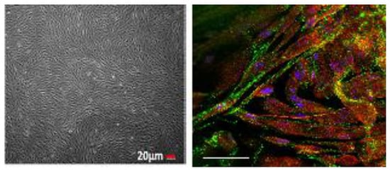
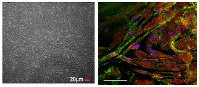
Other tissue engineering studies with silk for repair of specific defects include regeneration of ligament [7,11,92], peripheral nerve [79], skin [93,94], cornea [29,95], ear drum [96], and trachea [97]. Recently, Lovett et al. used silk fibroin for the construction of microvascular grafts [98]. Tissue engineered silk sling for the treatment of stress urinary incontinence has also been reported [10]. Silk membranes have been used for the growth of human tympanic membrane (TM) kerotinocytes [99]. Figure 2 presents images of TM keratinocytes grown on such silk films.
Figure 2.
Human Tympanic membrane keratinocytes growth on silk fibroin membrane: (i) Light microscopy image of day 18 [99]; (ii) Confocal microscopy image of cells immunofluorescently stained with occludin (green) and ZO-1 (red). Cell nuclei are counterstained with DAPI (blue) [100].
4.2. Immobilisation and Delivery of Drugs/Enzymes
Mild aqueous processing of silk is advantageous for loading sensitive drugs without affecting their functions. For example, Lu et al. studied glucose oxidase, lipase, and horseradish peroxidise entrapped in silk films and found that for over 10 months, enzymes retained their significant activity, even when stored at 370C, and in the case of glucose oxidase, they did not lose any activity [101]. Release kinetics of drugs and enzymes is reported to depend, to a large extent, on the content of crystalline β-sheets [28,61,102,103].
Silk has been used for the control release of specific drugs for targeted clinical needs. Uebersax et al. [104] and Szybala et al. [105] used silk matrices as adenosine-releasing bioincubators that may be useful in the management of epilepsy. Recently, Pritchard et al. demonstrated that slow (up to 2 weeks) and linear release of adenosine was possible by controlling the thickness and cystrallinity of silk coatings [105]. Bayraktar et al. [106] used aqueous silk solutions to coat theophylline tablets. It was shown that slow release by multilayered silk coatings could enhance the efficacy of delivering emodin, an anti breast cancer and anti tumour drug [107,108]. Numata and Kaplan have recently reviewed silk-based multi-block copolymer systems as vehicles for sustained release of small molecule drugs, proteins and genes [109].
Silk particles have also been examined for potential drug delivery applications. Lorenz Meinel’s group reported encapsulation and release of horseradish peroxidase (HRP) using self assembled silk microspheres [61]. They also reported encapsulation of salicylic acid, propranolol hydrochloride, and recombinant human insulin growth factor I (IGF-I) in silk microspheres. Encapsulation efficiency of IGF-I was close to 100%. They also used BMP-2 and IGF-I loaded silk microspheres in silk porous scaffolds and obtained enhanced osteochondral tissue engineering outcomes [91].
Silk’s ability to hold therapeutic enzymes can be combined with the change in optical response of silk to design biodegradable and chemically responsive implantable optofluidic devices for applications such as monitoring changes in blood glucose. Domachuk et al. incorporated hemoglobin into free-standing silk diffraction gratings and simultaneously immobilized enzymes to examine such prospects [110].
5. Recent Advances in Silk Milling and Applications of Milled Silk Particles
Developments of silk particles for biomedical applications have been mostly restricted to bottom up approaches as discussed already. However, top down approach of particle preparation through milling has comparative advantages as it can avoid the use of chemicals and the long dialysis time associated with the bottom up methods. The silk powder thus prepared should enhance the application scope of silk based materials also.
5.1. Silk Milling
Silk has a hierarchical structure with fibroin chains forming nano fibrils, bundles of which constitute the fibre [111,112,113,114,115,116,117]. This suggests an opportunity to defibrillate silk into nano fibrils and fine particles. However, silk being a strong viscoelastic material is difficult to mill into fine particles with a controlled morphology and a narrow size distribution. Hence, a very long milling time (up to 48 hours) is usually necessary to obtain maximum possible fineness of around 5 µm [118,119,120]. To overcome such problems, pre-treatments such as alkali degradation [118], exposure to high temperature [121], radiation [122,123], and steam explosion [124,125], have been used to improve the milling efficiency. New developments have been reported to improve fine silk powder production in recent years. Li et al. discussed preparation of nano silk particles through a multistep process which includes a special high pressure device and filtering large particles during the process [126,127].
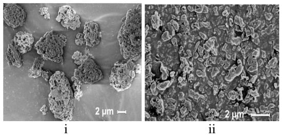
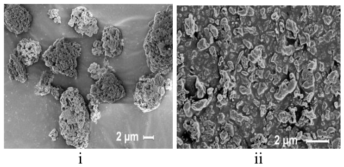
We have recently examined a number of standard milling devices to prepare ultrafine silk particles from Mulberry and non-Mulberry silk fibres. Milling sequences using a combination of milling systems such as a cutter mill, a rotary mill, different forms of ball mills and an air jet mill were examined [128,129]. We observed that non-Mulberry silk fibres which have a more porous structure and lower strength could be milled more efficiently than mulberry silk [129]. It was shown that by appropriate selection of milling sequence and milling parameters, milling time could be reduced to about 6 hours without any pre-treatment. A final step using air jet milling could achieve dry silk powder with a volume based median particle size of less than 1 μm. Figure 3 shows images of such particles.
Figure 3.
Wet milled and spray dried silk particles (i) before and (ii) after air jet milling.
5.2. Silk Composites with Silk Particles
Despite excellent biological outcomes, 3-D silk scaffolds are not strong enough for load-bearing tissue engineering applications. Kim et al. [49] reported that, usually, bone-like tissue ingrowth could be seen after 12 weeks of cell seeding in 3-D silk scaffolds. Such bonny tissue deposition could enhance the strength of scaffolds, but they were still much weaker (wet compressive modulus remained less than 200 kPa and yield strength less than 40 kPa) than the natural bone.
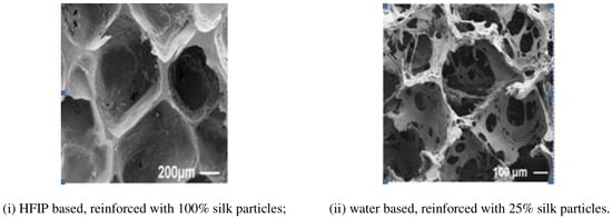
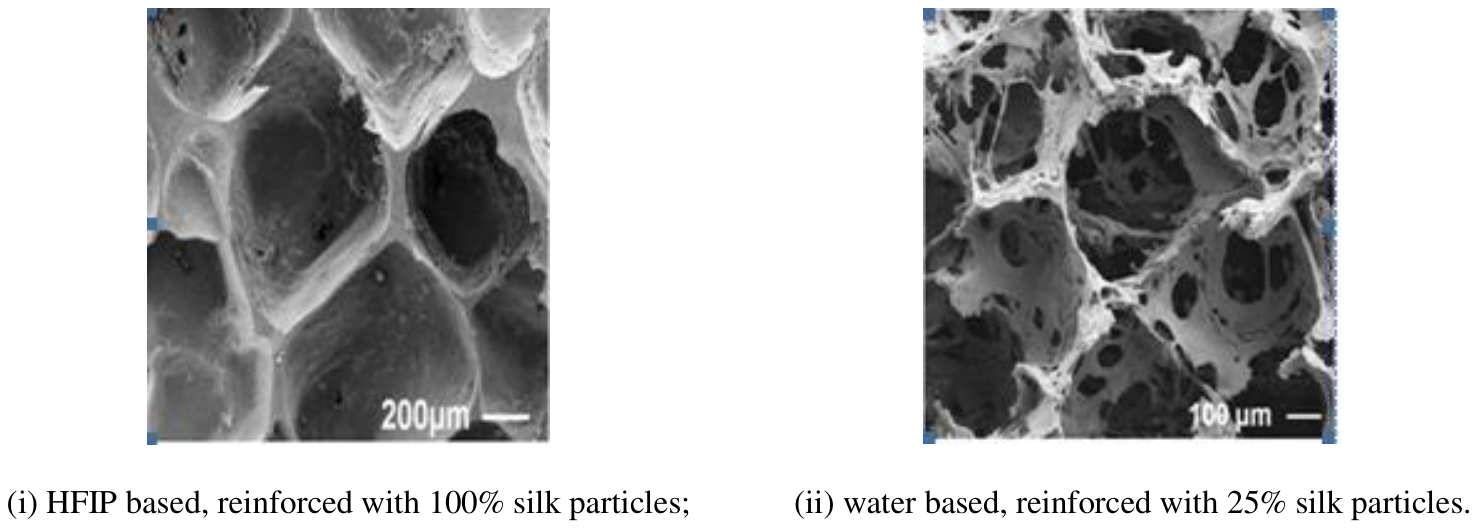
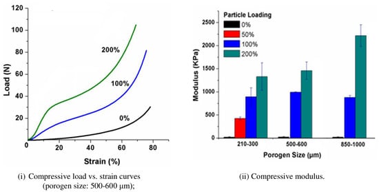
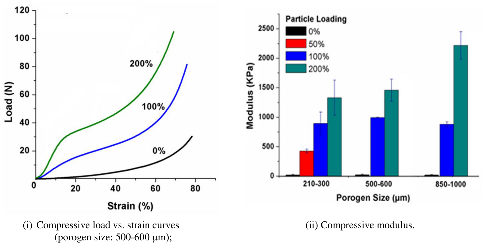
To increase the mechanical properties of silk scaffolds, milled silk particles were used as a reinforcing agent to fabricate a new silk-silk composite system [130]. SEM images of the composites are presented in Figure 4. The reinforcing particles maintained their crystalline structure in the composites [131], but unlike parent fibres, the particles are partially soluble in organic solvent, such as hexaflouroisopropanol (HFIP) [130]. This provides a high interfacial bonding between the particles and the bulk phase prepared using HFIP. This mechanism has resulted in a substantial increase in mechanical properties of silk particle reinforced silk composites (Figure 5). This novel silk processing method therefore provides a promising option for preparing new silk based biomaterials for hard tissue engineering applications. Further work is ongoing in this area in our laboratory in collaboration with Prof Kaplan’s group at Tufts University.
Figure 4.
SEM images of 3D silk composite scaffolds.
Figure 5.
Compressive properties of silk composite scaffolds prepared from 17% HFIP-based silk solution reinforced with different % of silk particles [130].
6. Conclusions
Research into silk materials and their in-vitro and invivo performance for targeted biomedical applications has gained much ground over the last few years. This has resulted in unique innovations in silk materials developed for specific applications. In addition to silk fibres and solutions, milled silk particles are now expanding the application scope for silk based biomaterials.
References
- Craig, C.L. Evolution of Arthopod silk. Annu. Rev. Entomol. 1997, 42(1), 231–267. [Google Scholar] [CrossRef] [Scilit] [PubMed]
- International Year of Natural Fibres. Available online: www.naturalfibres2009.org/en/fibres/silk.
- Moy, R.; Lee, A.; Zalka, A. Commonly used suture materials in skin surgery. Am. Fam. Physician 1991, 88(4), 2123–2128. [Google Scholar]
- Minoura, N.; Aiba, S.I.; Gotoh, Y.; TsukadaM; Imai, Y. Attachment and growth of cultured fibroblast cells on silk protein matrices. J. Biomed. Mater. Res. 1995, 29(10), 1215–1221. [Google Scholar] [CrossRef] [Scilit] [PubMed]
- Minoura, N.; Aiba, S.I.; Higuchi, M.; Gotoh, Y.; Tsukada, M.; Imai, Y. Attachment and Growth of Fibroblast Cells on Silk Fibroin. Biochem. Biophys. Res. Commun. 1995, 208(2), 511–516. [Google Scholar] [CrossRef] [Scilit] [PubMed]
- Rajkhowa, R. Fabricating and Characterising Silk Powder for Biomedical and Sorption Applications. Ph.D. Thesis, Deakin University, Geelong, 2009. [Google Scholar]
- Altman, G.H.; Horan, R.L.; Lu, H.H.; Moreau, J.; Martin, I.; Richmond, J.C.; Kaplan, D.L. Silk matrix for tissue engineered anterior cruciate ligaments. Biomaterials 2002, 23(20), 4131–4141. [Google Scholar] [CrossRef] [Scilit] [PubMed]
- Dal Pra, I.; Freddi, G.; Minic, J.; Chiarini, A.; Armato, U. De novo engineering of reticular connective tissue in vivo by silk fibroin nonwoven materials. Biomaterials 2005, 26(14), 1987–1999. [Google Scholar] [CrossRef] [Scilit] [PubMed]
- Gellynck, K.; Verdonk, P.C.M.; Nimmen, E.V.; Almqvist, K.F.; Gheysens, T.; Schokens, G.; Langenhove, L.V.; Kiekens, P.; Mertens, J.; Verbruggen, G. Silkworm and spider silk scaffolds for chondrocyte support. J. Mater. Sci. Mater. Med. 2008, 19, 33993409. [Google Scholar] [CrossRef] [Scilit] [PubMed]
- Zou, X.; Chen, X.; Jin, H.; Wang, L.; Jiang, Y.; Yin, Z.; Ouyang, H. Mesenchymal stem cell seeded knitted silk sling for the treatment of stress urinary incontinence. Biomaterials 2010, 31(18), 48724879. [Google Scholar] [CrossRef] [Scilit] [PubMed]
- Fan, H.; Liu, H.; Wong, E.J.W.; Toh, S.L.; Goh, J.C.H. In vivo study of anterior cruciate ligament regeneration using mesenchymal stem cells and silk scaffold. Biomaterials 2008, 29(23), 33243337. [Google Scholar] [CrossRef] [Scilit] [PubMed]
- Phillips, D.M.; Drummy, L.F.; Naik, R.R.; De Long, H.C.; Fox, D.M.; Trulove, P.C.; Mantz, R.A. Regenerated silk fiber wet spinning from an ionic liquid solution. J. Mater. Chem. 2005, 15(39), 4206–4208. [Google Scholar] [CrossRef] [Scilit]
- Gupta, M.K.; Khokhar, S.K.; Phillips, D.M.; Sowards, L.A.; Drummy, L.F.; Kadakia, M.P.; Naik, R.R. Patterned Silk Films Cast from Ionic Liquid Solubilized Fibroin as Scaffolds for Cell Growth. Langmuir 2007, 23, 1315–1319. [Google Scholar] [CrossRef] [Scilit] [PubMed]
- Phillips, D.M.; Drummy, L.F.; Conrady, D.G.; Fox, D.M.; Naik, R.R.; Stone, M.O.; Trulove, P.C.; De Long, H.C.; Mantz, R.A. Dissolution and Regeneration of Bombyx mori Silk Fibroin Using Ionic Liquids. J. Am. Chem. Soc. 2004, 126(44), 14350–14351. [Google Scholar] [CrossRef] [Scilit] [PubMed]
- Zainuddin Le, T.T.; Park, Y.; Chirila, T.V.; Halley, P.J.; Whittaker, A.K. The behavior of aged regenerated Bombyx mori silk fibroin solutions studied by 1H NMR and rheology. Biomaterials 2008, 29(32), 4268–4274. [Google Scholar] [CrossRef] [Scilit] [PubMed]
- Trabbic, K.A.; Yager, P. Comparative Structural Characterization of Naturally- and Synthetically-Spun Fibers of Bombyx mori Fibroin. Macromolecules 1998, 31(2), 462–471. [Google Scholar] [CrossRef] [Scilit]
- Nazarov, R.; Jin, H.J.; Kaplan, D.L. Porous 3-D Scaffolds from Regenerated Silk Fibroin. Biomacromolecules 2004, 5(3), 718–726. [Google Scholar] [CrossRef] [Scilit] [PubMed]
- Lock, R.L. Fiber-spinnable solutions of silkworm fibroin US 5252285, 1993.
- Jin, H.J.; Fridrikh, S.V.; Rutledge, G.C.; Kaplan, D.L. Electrospinning Bombyx mori Silk with Poly(ethylene oxide). Biomacromolecules 2002, 3(6), 1233–1239. [Google Scholar] [CrossRef] [Scilit] [PubMed]
- Zuo, B.; Dai, L.; Wu, Z. Analysis of structure and properties of biodegradable regenerated silk fibroin fibers. J. Mater. Sci. 2006, 41, 3357–3361. [Google Scholar] [CrossRef] [Scilit]
- Ha, S.W.; Tonelli, A.E.; Hudson, S.M. Structural Studies of Bombyx mori Silk Fibroin during Regeneration from Solutions and Wet Fiber Spinning. Biomacromolecules 2005, 6(3), 17221731. [Google Scholar] [CrossRef] [Scilit] [PubMed]
- Um, I.C.; Kweon, H.; Park, Y.H.; Hudson, S. Structural characteristics and properties of the regenerated silk fibroin prepared from formic acid. Int. J. Biol. Macromol. 2001, 29(2), 91–97. [Google Scholar] [CrossRef] [Scilit] [PubMed]
- Tretinnikov, O.N.; Tamada, Y. Influence of Casting Temperature on the Near-Surface Structure and Wettabiligy of Cast Silk Fibroin Films. Langmuir 2001, 17, 7406–7413. [Google Scholar] [CrossRef] [Scilit]
- Vasconcelos, A.; Freddi, G.; Cavaco-Paulo, A. Biodegradable Materials Based on Silk Fibroin and Keratin. Biomacromolecules 2008, 9, 1299–1305. [Google Scholar] [CrossRef] [Scilit] [PubMed]
- Higuchi, A.; Yoshida, M.; Ohno, T.; Asakura, T.; Hara, M. Production of interferon-β in a culture of fibroblast cells on some polymeric films. Cytotechnology 2000, 34, 165–173. [Google Scholar] [CrossRef] [Scilit] [PubMed]
- Wang, X.; Kim, H.J.; Peng, X.; Matsumoto, A.; Kaplan, D.L. Biomaterial Coatings by Stepwise Deposition of Silk Fibroin. Langmuir 2005, 21, 11335–11341. [Google Scholar] [CrossRef] [Scilit] [PubMed]
- Jiang, C.; Wang, X.; Gunawidjaja, R.; Lin, Y.H.; Gupta, M.K.; Kaplan, D.L.; Naik, R.R.; Tsukruk, V.V. Mechanical Properties of Robust Ultrathin Silk Fibroin Films. Adv. Funct. Mater. 2007, 17, 2229–2237. [Google Scholar] [CrossRef] [Scilit]
- Wang, X.; Hu, X.; Daley, A.; Rabotyagova, O.; Cebe, P.; Kaplan, D.L. Nanolayer biomaterial coatings of silk fibroin for controlled release. J. Control. Release 2007, 121(3), 190–199. [Google Scholar] [CrossRef] [Scilit] [PubMed]
- Gil, E.S.; Park, S.H.; Marchant, J.; Omenetto, F.; Kaplan, D.L. Response of Human Corneal Fibroblasts on Silk Film Surface Patterns. Macromol. Biosci. 2010, 10(6), 664–673. [Google Scholar] [CrossRef] [Scilit] [PubMed]
- Minoura, N.; Tsukada, M.; Nagura, M. Physicochemical properties of silk fibroin membrane as a biomaterial. Biomaterials 1990, 11(6), 430–434. [Google Scholar] [CrossRef] [Scilit] [PubMed]
- Minoura, N.; Tsukada, M.; Nagura, M. Fine structure and oxygen permeability of silk fibroin membrane treated with methanol. Polymer 1990, 31(2), 265269. [Google Scholar] [CrossRef] [Scilit]
- Bondar, B.; Fuchs, S.; Motta, A.; Migliaresi, C.; Kirkpatrick, C.J. Functionality of endothelial cells on silk fibroin nets: Comparative study of micro- and nanometric fibre size. Biomaterials 2008, 29(5), 561–572. [Google Scholar] [CrossRef] [Scilit] [PubMed]
- Ki, C.S.; Kim, J.W.; Hyun, J.H.; Lee, K.H.; Hattori, M.; Rah, D.K.; Park, Y.H. Electrospun Three-Dimensional Silk Fibroin Nanofibrous Scaffold. J. Appl. Polym. Sci. 2007, 106, 3922–3928. [Google Scholar] [CrossRef] [Scilit]
- Ki, C.S.; Gang, E.H.; Um, I.C.; Park, Y.H. Nanofibrous membrane of wool keratose/silk fibroin blend for heavy metal ion adsorption. J. Membr. Sci. 2007, 302(1-2), 20–26. [Google Scholar] [CrossRef] [Scilit]
- Lovett, M.L.; Cannizzaro, C.M.; Vunjak-Novakovic, G.; Kaplan, D.L. Gel spinning of silk tubes for tissue engineering. Biomaterials 2008, 29(35), 46504657. [Google Scholar] [CrossRef] [Scilit] [PubMed]
- Zhang, X.; Wang, X.; Keshav, V.; Wang, X.; Johanas, J.T.; Leisk, G.G.; Kaplan, D.L. Dynamic culture conditions to generate silk-based tissueengineered vascular grafts. Biomaterials 2009, 30(19), 3213–3223. [Google Scholar] [CrossRef] [Scilit] [PubMed]
- Ha, S.W.; Gracz, H.S.; Tonelli, A.E.; Hudson, S.M. Structural Study of Irregular Amino Acid Sequences in the Heavy Chain of Bombyx mori Silk Fibroin. Biomacromolecules 2005, 6, 25632569. [Google Scholar] [CrossRef] [Scilit] [PubMed]
- Zhou, G.; Shao, Z.; Knight, D.P.; Yan, J.; Chen, X. Silk Fibres Extruded Artificially from Aqueous Solutions of Regenerated Bombyx mori Silk Fibroin are Tougher than their Natural Counterpart. Adv. Mater. 2009, 21, 366370. [Google Scholar] [CrossRef] [Scilit]
- Chen, X.; Li, W.; Zhong, W.; Lu, Y.; Yu, T. pH sensitivity and ion sensitivity of hydrogels based on complex-forming chitosan/silk fibroin interpenetrating polymer network. J. Appl. Polym. Sci. 1997, 65(11), 22572262. [Google Scholar] [CrossRef] [Scilit]
- Hanawa, T.; Watanabe, A.; Tsuchiya, T.; Ikoma, R.; Hidaka, M.; Sugihara, M. New oral dosage form for elderly patients: Preparation and characterization of silk fibroin gel. Chem. Pharm. Bull. 1995, 43(2), 284–288. [Google Scholar] [CrossRef] [Scilit] [PubMed]
- Kim, U.J.; Park, J.; Li, C.; Jin, H.J.; Valluzzi, R.; Kaplan, D.L. Structure and Properties of Silk Hydrogels. Biomacromolecules 2004, 5(3), 786–792. [Google Scholar] [CrossRef] [Scilit] [PubMed]
- Motta, A.; Migliaresi, C.; Faccioni, F.; Torricelli, P.; Fini, M.; Giardino, R. Fibroin hydrogels for biomedical applications: preparation, characterization and in vitro cell culture studies. J. Biomater. Sci. Polym. Edn 2004, 15(7), 851–864. [Google Scholar] [CrossRef] [Scilit] [PubMed]
- Wang, X.; Kludge, J.A.; Leisk, G.G.; Kaplan, D.L. Sonication-induced geletation of silk fibroin for cell encapsulation. Biomaterials 2008, 29, 10511064. [Google Scholar] [CrossRef] [Scilit] [PubMed]
- Tsukada, M.; Freddi, G.; Minoura, N.; Allara, G. Preparation and application of porous silk fibroin materials. J. Appl. Polym. Sci. 1994, 54(4), 507–514. [Google Scholar] [CrossRef] [Scilit]
- Li, M.; Wu, Z.; Zhang, C.; Lu, S.; Yan, H.; Huang, D.; Ye, H. Study on Porous Silk Fibroin Materials. II. Preparation and Characteristics of Spongy Porous Silk Fibroin Materials. J. Appl. Polym. Sci. 2001, 79, 2129–2199. [Google Scholar] [CrossRef] [Scilit]
- Kim, U.J.; Park, J.; Joo Kim, H.; Wada, M.; Kaplan, D.L. Three-dimensional aqueous-derived biomaterial scaffolds from silk fibroin. Biomaterials 2005, 26(15), 2775–2785. [Google Scholar] [CrossRef] [Scilit] [PubMed]
- Harris, L.D.; Kim, B.S.; Moone, D.J. Open pore biodegradable matrices formed with gas foaming. J. Biomed. Mater. Res. 1998, 42(3), 396–402. [Google Scholar] [CrossRef]
- Lv, Q.; Feng, Q.L. Preparation of 3-D regenerated fibroin scaffolds with freeze drying method and freeze drying/foaming technique. J. Mater. Sci. Mater. Med. 2006, 17, 1349–1356. [Google Scholar] [CrossRef] [Scilit] [PubMed]
- Kim, H.J.; Kim, U.J.; Leisk, G.G.; Bayan, C.; Georgakoudi, I.; Kaplan, D.L. Bone Regeneration on Macroporous Aqueous-Derived Silk 3-D Scaffolds. Macromol. Biosci. 2007, 7(5), 643–655. [Google Scholar] [CrossRef] [Scilit] [PubMed]
- Meinel, L.; Hofmann, S.; Karageorgiou, V.; Zichner, L.; Langer, R.; Kaplan, D.; Vunjak-Novakovic, G. Engineering cartilage-like tissue using human mesenchymal stem cells and silk protein scaffolds. Biotechnol. Bioeng. 2004, 88(3), 379–391. [Google Scholar] [CrossRef] [Scilit] [PubMed]
- Kim, H.J.; Kim, U.J.; Vunjak-Novakovic, G.; Min, B.H.; Kaplan, D.L. Influence of macroporous protein scaffolds on bone tissue engineering from bone marrow stem cells. Biomaterials 2005, 26(21), 4442–4452. [Google Scholar] [CrossRef] [Scilit] [PubMed]
- Meinel, L.; Karageorgiou, V.; Fajardo, R.; Snyder, B.; Shinde-Patil, V.; Zichner, L.; Kaplan, D.; Langer, R.; Vunjak-Novakovic, G. Bone Tissue Engineering Using Human Mesenchymal Stem Cells: Effects of Scaffold Material and Medium Flow. Ann. Biomed. Eng. 2004, 32(1), 112–122. [Google Scholar] [CrossRef] [Scilit] [PubMed]
- Meinel, L.; Betz, O.; Fajardo, R.; Hofmann, S.; Nazarian, A.; Cory, E.; Hilbe, M.; McCool, J.; Langer, R.; Vunjak-Novakovic, G.; Merkle, H.P.; Rechenberg, B.; Kaplan, D.L.; Kirker-Head, C. Silk based biomaterials to heal critical sized femur defects. Bone 2006, 39(4), 922–931, Erratum in Bone 2008, 43, 1123. [Google Scholar] [CrossRef] [Scilit] [PubMed]
- Karageorgiou, V.; Tomkins, M.; Fajardo, R.; Meinel L. Snyder, B.; Wade, K.; Chen, J.; Vunjak-Novakovic, G.; Kaplan, D.L. Porous silk fibroin 3-D scaffolds for delivery of bone morphogenetic protein-2 in vitro and in vivo. J. Biomed. Mater. Res. Part A 2006, 78A(2), 324–334. [Google Scholar] [CrossRef] [Scilit] [PubMed]
- Meinel, L.; Fajardo, R.; Hofmann, S.; Langer, R.; Chen, J.; Snyder, B.; Vunjak-Novakovic, G.; Kaplan, D. Silk implants for the healing of critical size bone defects. Bone 2005, 37(5), 688–698, Erratum Bone 2008, 43, 1123. [Google Scholar] [CrossRef] [Scilit] [PubMed]
- Wang, Y.; Blasioli, D.J.; Kim, H.J.; Kim, H.S.; Kaplan, D.L. Cartilage tissue engineering with silk scaffolds and human articular chondrocytes. Biomaterials 2006, 27(25), 4434–4442. [Google Scholar] [CrossRef] [Scilit] [PubMed]
- Wang, Y.; Kim, U.J.; Blasioli, D.J.; Kim, H.J.; Kaplan, D.L. In vitro cartilage tissue engineering with 3D porous aqueous-derived silk scaffolds and mesenchymal stem cells. Biomaterials 2005, 26(34), 7082–7094. [Google Scholar] [CrossRef] [Scilit] [PubMed]
- Ohtomo, K.; Korikawa, Y.; Otoi, K. Finely powdered fibroin and process for producing same. EP0011161, 14.10.81, 1981. [Google Scholar]
- Tomoaki, H.; Masao, T.; Saburo, S. Change in secondary structure of silk fibroin during preparation of its microspheres by spray-drying and exposure to humid atmosphere. J. Colloid Interface Sci. 2003, 266(1), 68–73. [Google Scholar] [CrossRef] [Scilit] [PubMed]
- Yeo, J.H.; Lee, K.G.; Lee, Y.W.; Kim, S.Y. Simple preparation and characteristics of silk fibroin microsphere. Eur. Polym. J. 2003, 39(6), 1195–1199. [Google Scholar] [CrossRef] [Scilit]
- Wang, X.; Wenk, E.; Matsumoto, A.; Meinel, L.; Li, C.; Kaplan, D.L. Silk microspheres for encapsulation and controlled release. J. Control. Release 2007, 117(3), 360–370. [Google Scholar] [CrossRef] [Scilit] [PubMed]
- Zhang, Y.Q.; Wei-De, S.; Ru-Li, X.; Zhuge, L.J.; Gao, W.J.; Wang, W.B. Formation of silk nanoparticles in water-miscible organic solvent and their characterization. J. Nanoparticle Res. 2007, 9, 885–900. [Google Scholar] [CrossRef] [Scilit]
- Lammel, A.S.; Hu, X.; Park, S.H.; Kaplan, D.L.; Scheibel, T.R. Controlling silk fibroin particle features for drug delivery. Biomaterials 2010, 31(16), 4583–4591. [Google Scholar] [CrossRef] [Scilit] [PubMed]
- Nam, J.; Hwan, Y. Morphology of Regenerated silk Fibroin: Effect of Freezing Temperature, Alcohol addition, and Molecular Weight. J. Appl. Polym. Sci. 2001, 81, 3008–3021. [Google Scholar] [CrossRef] [Scilit]
- Cao, Z.; Chen, X.; Yao, J.; Huang, L.; Shao, Z. The preparation of regenerated silk fibroin microspheres. Soft Matter 2007, 3, 910–915. [Google Scholar] [CrossRef] [Scilit] [PubMed]
- Arai, T.; Freddi, G.; Innocenti, R.; Tsukada, M. Biodegradation of Bombyx mori silk fibroin fibers and films. J. Appl. Polym. Sci. 2004, 91(4), 2383–2390. [Google Scholar] [CrossRef] [Scilit]
- Horan, R.L.; Antle, K.; Collette, A.L.; Wang, Y.; Huang, J.; Moreau, J.E.; Volloch, V.; Kaplan, D.L.; Altman, G.H. In vitro degradation of silk fibroin. Biomaterials 2005, 26(17), 3385–3393. [Google Scholar] [CrossRef] [Scilit] [PubMed]
- Wang, Y.; Rudym, D.D.; Walsh, A.; Abrahamsen, L.; Kim, H.J.; Kim, H.S.; Kirker-Head, C.; Kaplan, D.L. In vivo degradation of three-dimensional silk fibroin scaffolds. Biomaterials 2008, 29(24-25), 34153428. [Google Scholar] [CrossRef] [Scilit] [PubMed]
- Jin, H.J.; Park, J.; Karageorgiou, V.; Kim, U.J.; Valluzzi, R.; Cebe, P.; Kaplan, D.L. Water-Stable Silk Films with Reduced β-sheet content. Adv. Funct. Mater. 2005, 15(8), 1241–1247. [Google Scholar] [CrossRef] [Scilit]
- Schmutz, F.; McAuliffe, W.; Anderson, D.M.; Elliott, J.P.; Eskridge, J.M.; Winn, H.R. Embolization of cerebral arteriovenous malformations with silk: histopathologic changes and hemorrhagic complications. Ajnr Am. J. Neuroradiol. 1997, 18(7), 1233–1237. [Google Scholar] [PubMed]
- Song, J.K.; Eskridge, J.M.; Chung, E.C.; Blake, L.C.; Elliott, J.P.; Finch, L.; Niakan, C.; Maravilla, K.R.; Winn, H.R. Preoperative embolization of cerebral arteriovenous malformations with silk sutures: analysis and clinical correlation of complications revealed on computerized tomography scanning. J. Neurosurg. 2000, 92(6), 955–960. [Google Scholar] [CrossRef] [Scilit] [PubMed]
- Altman, G.H.; Diaz, F.; Jakuba, C.; Calabro, T.; Horan, R.L.; Chen, J.; Lu, H.; Richmond, J.; Kaplan, D.L. Silkbased biomaterials. Biomaterials 2003, 24(3), 401–416. [Google Scholar] [CrossRef] [Scilit] [PubMed]
- Hakimi, O.; Gheysens, T.; Vollrath, F.; Grahn, M.F.; Knight, D.P.; Vadgama, P. Modulation of cell growth on exposure to silkworm and spider silk fibers. J. Biomed. Mater. Res. Part A 2010, 92(4), 1366–1372. [Google Scholar] [CrossRef] [Scilit] [PubMed]
- Kurosaki, S.; Otsuka, H.; Kunitomo, M.; Koyama, M.; Pawankar, R.; Matumoto, K. Fibroin allergy. IgE mediated hypersensitivity to silk suture materials. Nippon Ika Daigaku Zasshi 1999, 66(1), 41–44. [Google Scholar] [CrossRef] [Scilit] [PubMed]
- Panilaitis, B.; Altman, G.H.; Chen, J.; Jin, H.J.; Karageorgiou, V.; Kaplan, D.L. Macrophage responses to silk. Biomaterials 2003, 24(18), 3079–3085. [Google Scholar] [CrossRef] [Scilit] [PubMed]
- Kim, K.H.; Jeong, L.; Park, H.N.; Shin, S.Y.; Park, W.H.; Lee, S.C.; Kim, T.I.; Park, Y.J.; Seol, Y.J.; Lee, Y.M.; Ku, Y.; Rhyu, I.C.; Han, S.B.; Chung, C.P. Biological efficacy of silk fibroin nanofiber membranes for guided bone regeneration. J. Biotechnol. 2005, 120(3), 327–339. [Google Scholar] [CrossRef] [Scilit] [PubMed]
- Santin, M.; Motta, A.; Freddi, G.; Cannas, M. In-vitro evaluation of the inflammatory potential of the silk fibroin. J. Biomed. Mater. Res. 1999, 46(3), 382–389. [Google Scholar] [CrossRef] [Scilit]
- Meinel, L.; Hofmann, S.; Karageorgiou, V.; KirkerHead, C.; McCool, J.; Gronowicz, G.; Zichner, L.; Langer, R.; Vunjak-Novakovic, G.; Kaplan, D.L. The inflammatory responses to silk films in vitro and in vivo. Biomaterials 2005, 26(2), 147–155. [Google Scholar] [CrossRef] [Scilit] [PubMed]
- Yang, Y.; Chen, X.; Ding, F.; Zhang, P.; Liu, J.; Gu, X. Biocompatibility evaluation of silk fibroin with peripheral nerve tissues and cells in vitro. Biomaterials 2007, 28(9), 1643–1652. [Google Scholar] [CrossRef] [Scilit] [PubMed]
- Burcharth, F.; Hahn-Pedersen, J.; Andersen, B.; Andersen, J.R. Inguinal hernia repair with silk or polyglycolic acid sutures: A controlled trial with 5-years’ follow-up. World J. Surg. 1983, 7(3), 416–418. [Google Scholar] [CrossRef] [Scilit] [PubMed]
- Chiarini, A.; Petrini, P.; Bozzini, S.; Pra, I.D.; Armato, U. Silk fibroin/poly(carbonate)-urethane as a substrate for cell growth: in vitro interactions with human cells. Biomaterials 2003, 24(5), 789–799. [Google Scholar] [CrossRef] [Scilit] [PubMed]
- Dal Prà, I.; Petrini, P.; Charini, A.; Bozzini, S.; Farè, S.; Armato, U. Silk Fibroin-Coated Three-Dimensional Polyurethane Scaffolds for Tissue Engineering: Interactions with Normal Human Fibroblasts. Tissue Eng. 2003, 9(6), 11131121. [Google Scholar] [CrossRef] [Scilit] [PubMed]
- Huang, Fuhua; Sun, Lizhong; Zheng, Jun. In Vitro and In Vivo Characterization of a Silk FibroinCoated Polyester Vascular Prosthesis. Artif. Organs 2008, 32(12), 932–941. [Google Scholar] [CrossRef] [Scilit] [PubMed]
- Sugihara, A.; Sugiura, K.; Morita, H.; Ninagawa, T.; Tubouchi, K.; Tobe, R.; Izumiya, M.; Horio, T.; Abraham, N.G.; Ikehara, S. Promotive Effects of a Silk Film on Epidermal Recovery from FullThickness Skin Wounds. Proc. Soc. Exp. Biol. Med. 2000, 225(1), 58–64. [Google Scholar] [CrossRef] [Scilit]
- Fini, M.; Motta, A.; Torricelli, P.; Giavaresi, G.; Nicoli Aldini, N.; Tschon, M.; Giardino, R.; Migliaresi, C. The healing of confined critical size cancellous defects in the presence of silk fibroin hydrogel. Biomaterials 2005, 26(17), 3527–3536. [Google Scholar] [CrossRef] [Scilit] [PubMed]
- Schneider, A.; Wang, X.Y.; Kaplan, D.L.; Garlick, J.A.; Egles, C. Biofunctionalized electrospun silk mats as a topical bioactive dressing for accelerated wound healing. Acta Biomater. 2009, 5(7), 2570–2578. [Google Scholar] [CrossRef] [Scilit] [PubMed]
- Livingston, T.; Ducheyne, P.; Garino, J. In-vivo evaluation of a bioactive scaffold for bone tissue engineering. J. Biomed. Mater. Res. 2002, 62(1), 1–13. [Google Scholar] [CrossRef] [Scilit] [PubMed]
- Puelacher, W.C.; Vacanti, J.P.; Ferraro, N.F.; Schloo, B.; Vacanti, C.A. Femoral shaft reconstruction using tissue-engineered growth of bone. Int. J. Oral Maxillofac. Surg. 1996, 25(3), 223–228. [Google Scholar] [CrossRef] [Scilit] [PubMed]
- Kim, H.J.; Kim, U.J.; Kim, H.S.; Li, C.; Wada, M.; Leisk, G.G.; Kaplan, D.L. Bone tissue engineering with premineralized silk scaffolds. Bone 2008, 42(6), 1226–1234. [Google Scholar] [CrossRef] [Scilit] [PubMed]
- Morita, Y.; Tomita, N.; Aoki, H.; Sonobe, M.; Wakitani, S.; Tamada, Y.; Suguro, T.; Ikeuchi, K. Frictional properties of regenerated cartilage in vitro. J. Biomech. 2006, 39(1), 103109. [Google Scholar] [CrossRef] [Scilit] [PubMed]
- Wang, X.; Wenk, E.; Zhang, X.; Meinel, L.; VunjakNovakovic, G.; Kaplan, D.L. Growth factor gradients via microsphere delivery in biopolymer scaffolds for osteochondral tissue engineering. J. Control. Release 2009, 134(2), 81–90. [Google Scholar] [CrossRef] [Scilit] [PubMed]
- Chen, J.; Altman, G.H.; Karageorgiou, V.; Horan, R.L.; Collette, A.L.; Volloch, V.; Colabro, T.; Kaplan, D.L. Human bone marrow stromal cell and ligament fibroblast responses on RGD-modified silk fibers. J. Biomed. Mater. Res. Part A 2003, 67(A) (2), 559–570. [Google Scholar] [CrossRef] [Scilit] [PubMed]
- Sobajo, C.; Behzad, F.; Yuan, X.F.; Bayat, A. Silk: A Potential Medium for Tissue Engineering. e Plasty 2008. [Google Scholar] [CrossRef] [Scilit]
- Wang, Y.; Kim, H.J.; Vunjak-Novakovic, G.; Kaplan, D.L. Stem cell-based tissue engineering with silk biomaterials. Biomaterials 2006, 27(33), 60646082. [Google Scholar] [CrossRef] [Scilit] [PubMed]
- Chirila, T.V.; Barnard, Z.; Zainuddin; Harkin, D.G.; Schwab, I.R.; Hirst, L.W. Bombyx mori Silk Fibroin Membranes as Potential Substrata for Epithelial Constructs Used in the Management of Ocular Surface Disorders. Tissue Eng. Part A 2008, 14(7), 1203–1211. [Google Scholar] [CrossRef] [Scilit] [PubMed]
- Ghassemifar, R.; Redmond, S.; Zainuddin; Chirila, T.V. Advancing Towards a Tissue-engineered Tympanic Membrante: Silk Fibroin as a Substratum for Growing Human Eardrum Keratinocyte. J. Biomater. Appl. 2010, 24(7), 591–606. [Google Scholar] [CrossRef] [Scilit] [PubMed]
- Ni, Y.; Zhao, X.; Zhou, L. Radiological and histological Characterization of silk fibroin as scaffold coating for tracheal defect repair. Otolaryngol.-Head Neck Surg. 2008, 139(2), 256–261. [Google Scholar] [CrossRef] [Scilit] [PubMed]
- Lovett, M.; Cannizzaro, C.; Daheron, L.; Messmer, B.; Vunjak-Novakovic, G.; Kaplan, D.L. Silk fibroin microtubes for blood vessel engineering. Biomaterials 2007, 28(35), 5271–5279. [Google Scholar] [CrossRef] [Scilit] [PubMed]
- Levin, B.; Rajkhowa, R.; Redmond, S.L.; Atlas, M.D. Grafts in myringplasty:utilizing a silk fibroin scaffold as a novel decice. Expert Rev. Med. Devices 2009, 6, 653–664. [Google Scholar] [CrossRef] [Scilit] [PubMed]
- Levin, B.; Redmond, S.L.; Rajkhowa, R.; Eikelboom, R.H.; Marano, R.J.; Atlas, M.D. Preliminary results of the application of a silk fibroin scaffold to otology. Otolaryngol.-Head Neck Surg. 2010, 142(3), S33–S35. [Google Scholar] [CrossRef] [Scilit] [PubMed]
- Lu, S.; Wang, X.; Lu, Q.; Hu, X.; Uppal, N.; Omenetto, F.G.; Kaplan, D.L. Stabilization of Enzymes in Silk Films. Biomacromolecules 2009, 10(5), 10321042. [Google Scholar] [CrossRef] [Scilit] [PubMed]
- Wang, X.; Wenk, E.; Hu, X.; Cascardo, G.R.; Lorenz, M.; Wang, X.; Li, C.; Merkle, H.; Kaplan, D.L. Silk coatings on PLGA and alginate microspheres for protein delivery. Biomaterials 2007, 28, 4161–4169. [Google Scholar] [CrossRef] [Scilit] [PubMed]
- Hofmann, S.; Wong Po Foo, C.T.; Rossetti, F.; Textor, M.; Vunjak-Novakovic, G.; Kaplan, D.L.; Merkle, H.P.; Meinel, L. Silk fibroin as an organic polymer for controlled drug delivery. J. Control. Release 2006, 111(1-2), 219–227. [Google Scholar] [CrossRef] [Scilit] [PubMed]
- Uebersax, L.; Fedele, D.E.; Schumacher, C.; Kaplan, D.L.; Merkle, H.P.; Boison, D.; Meinel, L. The support of adenosine release from adenosine kinase deficient ES cells by silk substrates. Biomaterials 2006, 27, 4599–4607. [Google Scholar] [CrossRef] [Scilit] [PubMed]
- Pritchard, E.M.; Szybala, C.; Boison, D.; Kaplan, D.L. Silk fibroin encapsulated powder reservoirs for sustained release of adenosine. J. Control. Release 2010, 144(2), 159–167. [Google Scholar] [CrossRef] [Scilit] [PubMed]
- Bayraktar, O.; Malay, Ö.; Özgarip, Y.; BatIgün, A. Silk fibroin as a novel coating material for controlled release of theophylline. Eur. J. Pharm. Biopharm. 2005, 60(3), 373–381. [Google Scholar] [CrossRef] [Scilit] [PubMed]
- Cheema, S.K.; Gobin, A.S.; Rhea R. Lopez-Berestein, G.; Newman, R.A.; Mathur, A.B. Silk fibroin mediated delivery of liposomal emodin to breast cancer cells. Int. J. Pharm. 2007, 341(1-2), 221–229. [Google Scholar] [CrossRef] [Scilit] [PubMed]
- Gobin, A.S.; Rhea, R.; Newman, R.A.; Mathur, A.B. silk-fibroin-coated liposomes for long-term and targeted drug delivery. Int. J. Nanomed. 2006, 1(1), 81–87. [Google Scholar] [CrossRef] [Scilit] [PubMed]
- Numata, K.; Kaplan, D.L. Silk-based delivery systems of bioactive molecules. Advanced Drug Delivery Reviews. In Press. [Google Scholar] [CrossRef] [Scilit] [PubMed]
- Domachuk, P.; Perry, H.; Amsden, J.; Kaplan, D.; Omenetto, F. Bioactive "self-sensing" optical systems. Appl. Phys. Lett. 2009, 95, 253702. [Google Scholar] [CrossRef] [Scilit] [PubMed]
- Putthanarat, S.; Stribeck, N.; Fossey, S.A.; Eby, R.K.; Adams, W.W. Investigation on the nanofibrils of silk fibers. Polymer 2000, 41, 7735–7747. [Google Scholar] [CrossRef] [Scilit]
- Poza, P.; Perez-Rigueiro, J.; Elices, M.; Lorca, J. Fractographic analysis of silkworm and spider silk. Eng. Fract. Mech. 2002, 69, 10351048. [Google Scholar] [CrossRef] [Scilit]
- Hakimi, O.; Knight, D.P.; Knight, M.M.; Grahn, M.F.; Vadgama, P. Ultrastructure of Insect and Spider Cocoon Silks. Biomacromolecules 2006, 7(10), 2901–2908. [Google Scholar] [CrossRef] [Scilit] [PubMed]
- Putthanarat, S.; Eby, R.K.; Adams, W.W.; Liu, G.F. Aspects of the Morphology of Silk of Bombyx Mori. J. Macromol. Sci. Part A 1996, 33(7), 899–911. [Google Scholar] [CrossRef] [Scilit]
- Riekel, C.; Vollrath, F. Spider silk fibre extrusion: combined wide- and small-angle X-ray microdiffraction experiments. Int. J. Biol. Macromol. 2001, 29(3), 203–210. [Google Scholar] [CrossRef] [Scilit] [PubMed]
- Sapede, D.; Seydel, T.; Forsyth, V.T.; Koza, M.M.; Schweins, R.; Vollrath, F.; Riekel, C. Nanofibrillar Structure and Molecular Mobility in Spider Dragline Silk. Macromolecules 2005, 38(20), 8447–8453. [Google Scholar] [CrossRef] [Scilit]
- Dobb, M.G.; Fraser, R.D.B.; Macrae, T.P. The Fine Structure of silk Fibrion. J. Cell Biol. 1967, 32, 289–295. [Google Scholar] [CrossRef] [Scilit] [PubMed]
- Tsubouchi, K. Method for manufacturing crystalline superfine silk powder. US 6427933, 6 August 2002. [Google Scholar]
- Sano, M.; Mikami, S.; Sasaki, N.; Kusamoto, N.; Fukatsu, F.; Ubara, A.; Yasue, T.; Ohyama, S. Substance including natural organic substance fine powder. US 5718954, 1998. [Google Scholar]
- Oyama, S. Modified powder for liquid composition for molded article. WO 2006016506, 1998-02-17, 2006. [Google Scholar]
- Li, M.; Lu, S.; Wu, Z.; Yan, H.; Mo, J.; Wang, L. Study on porous silk fibroin materials. I. Fine structure of freeze dried silk fibroin. J. Appl. Polym. Sci. 2001, 79(12), 2185–2191. [Google Scholar] [CrossRef] [Scilit]
- Shida, K.; Hidefumi, T.; Kamiishi, Y. Change in Silk Protein by Radiation, Takasaki Symposium on Radiation Processing of Natural Polymers, takasaki, Japan, November 23 and 24, 2000, 2001; Japan Atomic Enerty Research Institute: Takasaki, Japan, 2001; pp. 85–93. [Google Scholar]
- Hidefumi, T.; Kazushige, I.; Youichi, K.; Fumio, Y.; Tamikazu, K. Production of fine powder from silk by radiation. Macromol. Mater. Eng. 2000, 283, 126–131. [Google Scholar] [CrossRef] [Scilit]
- Xu, W.; Ke, G.; Peng, X. Studies on the Effects of the Enzymatic Treatment on Silk Fine Powder. J. Appl. Polym. Sci. 2006, 101, 2967–2971. [Google Scholar] [CrossRef] [Scilit]
- Hamaoka, Y.; Katayama, T.; Yamazaki, M.; Nagasuna, O.; Yasui, M. Manufacture of white silk fibroin powders with minimal residue of alkali salts. JP 2005281332, 2005. [Google Scholar]
- Li, Y.; Hu, J. Method for Pulverizing Natural Organic Substance into Nano-scale Fibreus Material. WO 2004055250, 2004. [Google Scholar]
- Li, Y.; Yuen, C.W.; Hu, J.Y.; Cheng, Y.F. Analysis of the Structural Characteristics of Nanoscale Silk Particles. J. Appl. Polym. Sci. 2006, 100, 268–274. [Google Scholar] [CrossRef] [Scilit]
- Rajkhowa, R.; Wang, L.; Kanwar, J.; Wang, X. Fabrication of ultrafine powder from eri silk through attritor and jet milling. Powder Technol. 2009, 191(1-2), 155–163. [Google Scholar] [CrossRef] [Scilit]
- Rajkhowa, R.; Wang, L.; Wang, X. Ultra-fine silk powder preparation through rotary and ball milling. Powder Technol. 2008, 185(1), 87–95. [Google Scholar] [CrossRef] [Scilit]
- Rajkhowa, R.; Gil, E.S.; Kludge, J.A.; Numata, K.; Wang, L.; Wang, X.; Kaplan, D.L. Reinforcing Silk Scaffolds with Silk Particles. Macromol. Biosci. 2010, 10(6), 599–611. [Google Scholar] [CrossRef] [Scilit] [PubMed]
- Rajkhowa, R.; Wang, L.; Kanwar, J.; Wang, X. Molecular weight and secondary structure change in eri silk during alkali degumming and powdering. J. Appl. Polym. Sci. [Google Scholar] [CrossRef] [Scilit]
Disclaimer/Publisher’s Note: The statements, opinions and data contained in all publications are solely those of the individual author(s) and contributor(s) and not of MDPI and/or the editor(s). MDPI and/or the editor(s) disclaim responsibility for any injury to people or property resulting from any ideas, methods, instructions or products referred to in the content. |
© 2010 Textile Bioengineering and Informatics Society. This article is an open access article distributed under the terms and conditions of the Creative Commons Attribution (CC BY) license (https://creativecommons.org/licenses/by/4.0/).