


Abstract
In this study, poly(lactic acid) (PLA) and silk fibroin(SF)-gelatin were sequentially electrospun on the collector to prepare the PLA/SF-gelatin composite fiber membranes. Scanning electronic microscope (SEM) was used to observe the morphology of the membranes. The microstructure of the fiber membranes before and after treatment with methanol was characterized using Fourier transform infrared (FTIR) and differential thermal analysis (DTA), and then the effects of the microstructure change on the mechanical properties, dissolution rate and shrinkage were evaluated. Furthermore, SEM and MTT assays were preformed to assess the proliferation and adhesion of 3T3 mouse fibroblasts cultured on the PLA/SF-gelatin scaffolds. After chemical treatment with methanol, the findings suggested the microstructure of PLA fiber did not change obviously, whereas the β-sheets of SF and α-helix structures of gelatin in SF-gelatin blend nanofibers increased, consequently the diameter of electrospun PLA/SF- gelatin membranes, the dissolution rate declined by 64%, the strain at break electrospun PLA/SF-gelatin scaffolds, the shrinkage of the PLA/SF –gelatin composite scaffolds caused by chemical treatment decreased by 40%. The cells adhered on to the surface of blend layer fibers and formed a confluent monolayer after 12 days cultivation. The good mechanical properties and dimensional stability of the chemically treated PLA/SF -gelatin scaffolds, combined with the ability to support cell growth in vitro, suggested tremendous potential application in tissue engineering.
1. Introduction
SF and gelatin are both natural biopolymers and have good biocompatibility and biodegradation, containing ligand (Arg-Gly-Asp, which can be recognized by integrin receptor in the cell membrane) that can promote cell differentiation, proliferation and adhesion. Kim [1] and jonathan [2] fabricated SF nanofiber membranes. Min [3] and Jin [4] further cultured human keratinocytes, fibroblasts and human bone marrow stromal cells on SF Nanofibers and investigated the feasibility for the wound dressing and cell scaffolds. Zheng-Ming Huang [5] investigated the Electrospinning of gelatin. Zhang [6] further performed the cross-linking of the electrospun gelatin nanofibers to improve their water-resistant ability and thermal-mechanical performance.
Although SF and gelatin can be electrospun into nanofibers and have the capacity of mimicking native extracellular matrix, they were generally water soluble and mechanically weak. To overcome these limitations, cross-linking was considered to be the most effective method because it could change molecular conformation [4,7,8,9]. However, these methods often resulted in the formation of brittle matrices due to high content of crystalline structures and the dimensional stability became poor.
Synthetic biodegradable polymers such as PLA and poly(lactide-co-glycolide) (PLGA) have recently become the promising scaffold materials. Yang [10] produced an aligned PLA nanofibrous scaffold for the study of neurite outgrowth and differentiation of neural stem cells seeded onto scaffolds. However, these synthetic polymers which have poor hydrophilic properties are well known in lacking bioactivity and cell affinity, for example, PCL did not promote the proliferation and adhesion of smooth muscle cells [11].
To achieve a strong and pliable scaffold that was low scaffold that has low dissolvability and excellent biological properties, SF and gelatin were electrospun and deposited on the PLA fibrous layer firstly formed by multi-electrospinning to construct bi-layered scaffolds. PLA is responsible for the resistance to rupture and shrinkage and confers the necessary elasticity, SF-gelatin and nanofiber layer support cell growth. The microstructure of the PLA/SF-gelatin nanofiber scaffold was examined before and after the treatment with methanol, and then the effects of the microstructure change on the mechanical properties, shrinkage and dissolution rate were investigated. MTT assay and SEM evaluated the adhesion and proliferation of mouse fibroblasts seeded on fibrous scaffolds.
2. Experimental section
2.1. Preparation of the composite PLA/SF-gelatin membranes
The air dried SF membranes, or a mixture of the air-dried SF membranes and gelatin with the weight ratio of 70:30, 50:50, were dissolved in 98%(w/w) formic acids yielding a 13%(w/w) spinning solution. PLA were dissolved in chloroform and acetone mixed solvent volume ratio of 2:1 to obtain a concentrated of 5%.
PLA Electrospinning parameters were as follows: voltage 25kv, flow rate 0.1 ml/h, distance between nozzle and collector was kept 15cm; for SF and SF-gelatin blend nanofiberes with different ratios were deposited onto the as-spun PLA fibrous membranes that Electrospinning, pure PLA and SF-gelatin nanofiber membranes were also fabricated according to the above parameters. These membranes were immersed in 100% methanol for 10min and dried for 24h at room temperature to form the chemically treated membranes.
2.2. Morphology
SEM (Hitachi S-570, Japan) was used to observe the morphology of the scaffolds. Magnification for the PLA layer was 1000 times and the others were 10,000 times.
2.3. Characterization of microstructure
FTIR spectra were obtained by using a series spectrometer (Nicolet 570, PE Co. USA) in the spectral region of 4000-400cm-1; DTA curves were obtained through a Thermal Analysis Instrument (Diamond 5700, PE Co. USA) at heating rate of 10℃/min, scan range of 40-400℃ and nitrogen gas flow rate of 120ml/min.
2.4. Dissolvability and shrinkage test
The membranes were immersed in water with the bath ratio of 1:100(w/w) at 37±1℃ for 1h, then dried at 50℃ dissolution rates were calculated according the following formula:
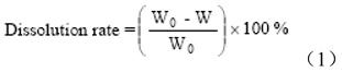
Where W0 and W were the weights of the scaffolds before and after dissolution respectively.
As-electrospun membranes of PLA, SF-gelatin and PLA/SF-gelatin were cut into a square shape with dimensions of 2x2cm for shrinkage test, three samples in each group. The sizes and chemically treated membranes were measured and compared with initial dimensions. The shrinkage percentage was defined as the rate of the surface dimensional change of the recovered membranes.
2.5. Mechanical property
The tensile testing was performed by using an Instron tester (Model 3365). The length of samples was 30mm and the width was 6mm. The cross-head speed was20mm/min, five samples in each group. The mechanical property was characterized according to the following formula:
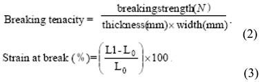
Where L0 was the length of sample, L1 was the length at break.
2.6. Cell structure
3T3 mouse fibroblasts (Chinese Academic of Science, Shanghai, China) were cultured in DMEM containing 10% fetal bovine serum at 37℃ with 5% CO the medium was changed three times a week. After reaching about 80% confluence, the cells were detached by 0.25% trypsin. After treatment, the scaffolds were cut out and sterilized by γ ray and placed in 24-well plates. 3T3 mouse fibroblasts were cultured at a density of 5x10⁴ per well in four different types of materials: (1) negative control well with no material; (2) PLA/SF; (3); PLA/SF-gelatin (50:50); (4) PLA/SF-gelatin (70:30) in particular, the cells were seeded on the SF or SF-gelatin layer of the composite membranes.
2.7. SEM observation
After 12 days of culture, cellular constructs were harvested, rinsed twice with PBS to remove non-adherent cells and subsequently fixed with 2.5% phosphate-buffered glutaraldehyde at 4℃ for 2hrs. After that, the samples were dehydrated through a series of graded ethanol solutions and air-dried overnight. Dry cellular constructs were sputtered with old and observed by SEM
Cell proliferation was tested using the mitochondrial metabolic (MTT) activity assay. After removal of medium, cell layers were rinsed with PBS twice. MTT solution (100μl) and phenol red-free RPM 11640 medium (1ml) (Sigma, USA) were added to each well and incubated for 4hrs. Thereafter, 1ml/well Dimethyl sulfoxide (DMSO) was added and shaken for 10min. Subsequently the absorbance at 540nm was measured using a spectrophotometer.
3. Results and discussion
3.1. Microstructure
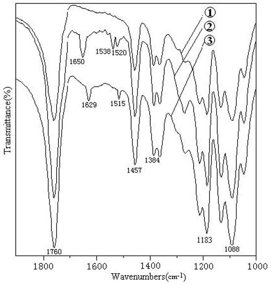
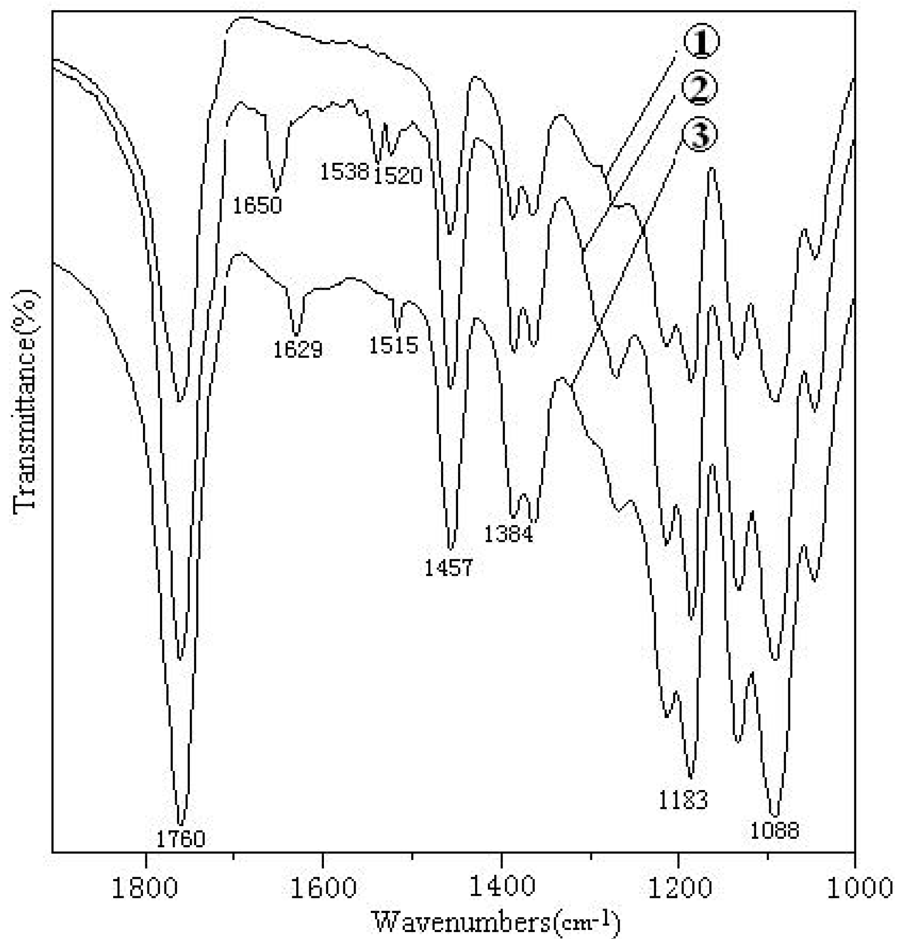
As shown in Figure 1, all samples illustrated common bands appeared at approximately 1760 cm-1, 1457 cm-1, 1384 cm-1, 1088 cm-1, corresponding stretching vibration of C=O bond and bending vibration of -CH3 and symmetrical bending vibration of C-H bonds and stretching vibration of C-O bonds respectively, which suggested that the conformation of PLA molecules did not change significantly after chemical treatment. For the secondary structure of SF molecules, we mainly differentiated random coils and β sheet structures, random coils show strong absorption bands at 1650-1665cm-1 of amide I (νc=o), 1535-1545cm-1 of amide II (δN-H) and β sheets show the absorption bands at 1625-1640cm of amide I, 1515-1525cm of amide II [2]. As shown in Figure 1(2) the two absorption bands at 1650cm (amide 1) and 1538cm (amide II), which can be attributed to the random coil of conformation of SF. Additionarlly the amide I band at 1650cm exhibited a mixture of random coils and α-helical conformation of gelatin [12].
Figure 1.
IR spectra of PLA membranes (1), PLA/SF-gelatin membranes (2), and chemically treated PLA/SF-gelatin membranes (3).
After treatment with methanol, Figure 1(3) showed the absorption bands of amide I and amide II shifted to the low wave-numbers, 1629, 1515cm-1, respectively, suggested loose random coils transformed to close β sheets due to the denaturation of SF proteins. Zhang [6] found the cross-linking with glutaraldehyde vapor enhanced the context of helix structure of the electrospun gelatin fibers, which is primarily dependent on the special features of the peptide bonds involving the imino acids, supplemented by interchain hydrogen bonds at the positions occupied by glycine. We speculated the confirmation of gelatin in the electrospun SF-gelatin nanofibers which also underwent the coil-helix transition caused by hydrogen bonds.
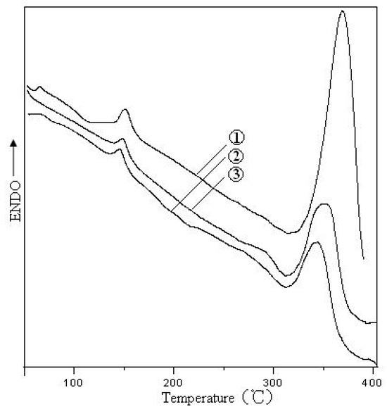
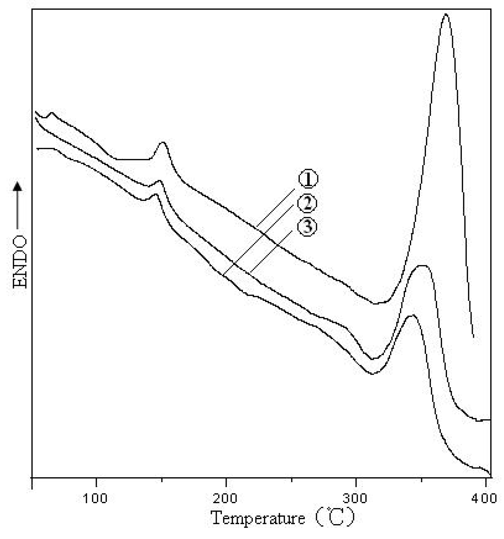
Compared with the electrospun PLA/SF-gelatin membranes, the thermal decomposition temperature of the chemically treated PLA/SF-gelatin membranes increased from 345℃ to 358℃, which was caused by the increase of SF beta-sheets and gelatin α-helix structures since the PLA conformation did not change significantly. In addition, Figure 2 shows that the thermal enthalpy corresponding to the decomposition temperature increased from 84.09J/g to 184.95J/g after the treatment, which also proved that the chemical treatment induced the conformational conversion of SF and gelatin molecules.
Figure 2.
DTA curves of PLA membranes (1) PLA/SF-gelatin membranes (2), and chemically treated SF-gelatin membranes (3).
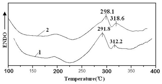
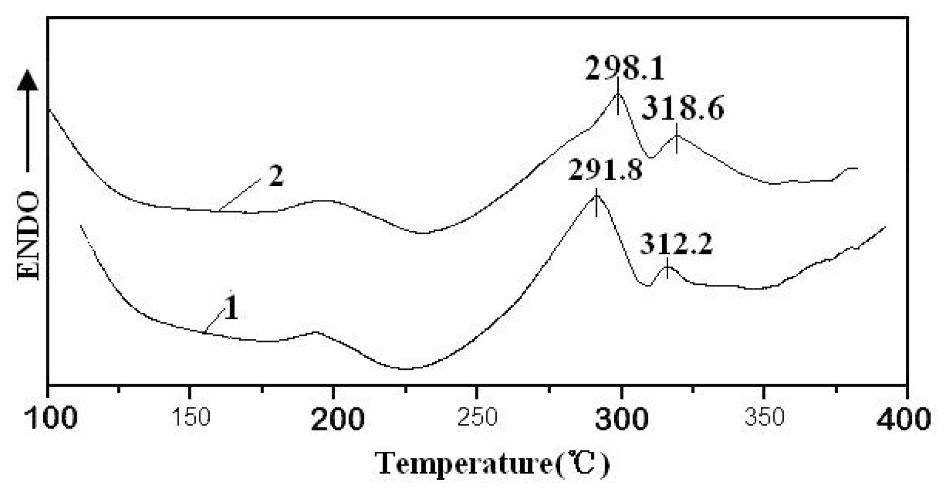
To further study the effects of the chemical treatment on the SF-gelatin fibers, the thermal properties of the electrospun membranes were examined by DTA. J. Magoshi [13] found that the thermal decomposition temeptarue of SF (random coil) was 290-300℃ Figure 3(1) showed that SF-gelatin nanofiber had two thermal absorption peaks at about 291.6℃ and 312.2℃, indicating the random ciols. The chemically treated SF-gelatin membranes showed the peaks at 298.1℃ and 318℃ particularly, the intensity of the peak at 318.6℃ drastically increased, indicative of higher β-sheets and α-helix content, which can also prove the chemical treatment resulted in relatively easy crystallization, this result was consistent with FTIR analysis.
Figure 3.
DTA curves of the electrospun SF-gelatin membranes (1), and chemically treated SF-gelatin membranes (2).
3.2. Morphology of the PLA/SF-gelatin
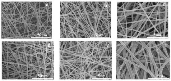
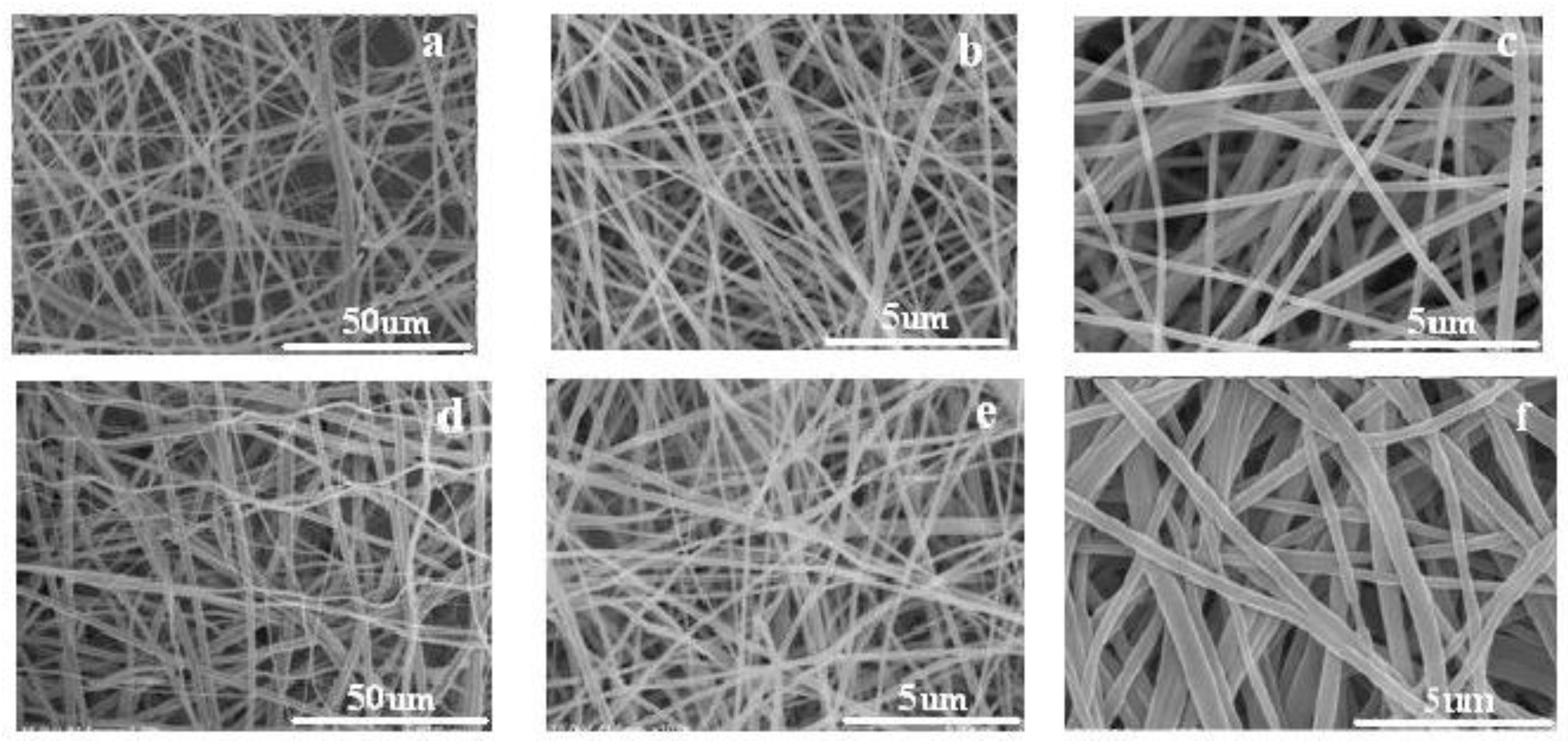
Figure 4 shows SEM of PLA and SF-gelatin layers of the composite membranes respectively, the diameters of PLA and SF-gelatin fibers are summarized in Table 1. The PLA layer was mainly composed of microscale fibers (Figure 4(a)), however, the SF-gelatin layer consisted of nanoscale fibers (Figure 4(b) and (c), which will contribute to mimicking the structure of ECMs consisting of nanoscale fibers. At the SF-gelatin ratio of 50:50 the electrospun fibers were homogenous and had lower diameters compared with the ratio of 70:30, which indicating the solution spinnability improved such that the splitting and elongation of the jet became easy as the gelatin content increased. In addition, SF and gelatin evenly dispersed in the fibers, no phase separation was found.
Figure 4.
SEM of PLAS-F/gelatin bil-ayered scaffold, ase-lectrospun scaffold: PLA layer(a), SF/gelatin layer(50:50)(b), SF/gelatin layer (70:30)(c), and chemically treated scaffold: PLA(d), SF/gelatin layer(50:50)(e), SF/gelatin (70:30)(f).
Table 1.
Fiber diameter of PLA-SF/gelatin composite fiber membrane.
When the SF-gelatin fibers were immersed in methanol, the hydrogen bonds formed between the polar methanol and the hydrophilic proteins and then weakened the intermolecular interaction, consequently, the fiber swelled and the diameters of the SF-gelatin fibers increased. However, compared with SF-gelatin Nanofibers, the morphology of PLA fibers did not exhibit the marked change, suggested the polar methanol had less effect on the PLA which is a hydrophobic synthetic polymer and thus PLA layers had higher dimensional stability.
3.3. Dissolution rate and Shrinkage
As shown in Table 2, the electrospun SF-gelatin membranes exhibited high dissolution rate whether the blend ratio was 70:30 or 50:50. In previous microstructure analysis, we found the electrospun SF-gelatin Nanofibers mainly consisted of random coil conformation, it is well-known that water molecules have easy access to amorphous regions. As a result, the electrospun SF-gelatin scaffolds had high dissolution rates. In comparison with SF-gelatin membranes, the dissolution rate of PLA/SF-gelatin membrane declined sharply. PLA molecular chains consist of ester bonds and thus it is a hydrophobic polymer. Hence the dissolution mainly occurred in the blend layer. The decrease of dissolution for the composite membrane was just a relative decline.
Table 2.
Dissolution rate of different scaffolds.
After treatment with methanol, the conformation of SF changed into a β sheet structure, and gelatin molecules were converted from random coils into α-helix structure. Consequently the dissolution rate of chemically treated PLA/SF-gelatin and SF-gelatin membranes decreased to 64% and 42%, respectively. Gelatin is a globular protein, but SF is composed of fibrous molecules. We all have known that the globular protein has higher dissolvability than the fibrous protein. Therefore, the dissolvability of blend membrane decreased as the content of gelatin increased.
Table 3 shows that the shrinkage of the fiber membranes consisting of natural protein polymer was higher than that of the electrospun PLA fiber membrane. The conformational transition changed from random coil to β sheet or α-helix structure corresponding to SF and gelatin respectively resulted in the shape contraction and the poor dimensional stability. In the previous studies, the methanol treatment had less effect on the PLA microstructure, therefore, PLA membrane was expected to have high dimensional stability and thus prevent the shrinkage of the SF-gelatin composite membranes decreased from 64.45±0.41% 74.13±3.2% to 38±8.21% 43.58±7.50% with the SF-gelatin ratio of 70:30 and 50:50. Additionally, after treatment, the shrinkage of the PLA/SF-gelatin and SF-gelatin membranes increased slightly with the gelatin content increased. In contrast with the fibrous SF protein, gelatin is a kind of globular protein and thus probably has higher shrinkage.
Table 3.
Dimensional stability of fiber membranes after treatment with methanol.
3.4. Mechanical properties
As shown in Table 4 after treatment, the breaking tenacity increased 3 times and preliminary modules enhanced 7 times than those of the electrospun PLA/SF-gelatin and SF-gelatin membranes, indicating that toughness declined and flexibility improved, which was also dependant on the change of SF and gelatin conformation. Additionally, it can be seen that compared to the treated SF-gelatin fiber membrane, preliminary modeules of the treated PLA/SF-gelatin decreased from 139.2±4.03 MPa to 76.3±3.42 MPa, but the strain at break was up to 7.3±2.05% and the breaking tenacity was still kept at 2.22 +/-0.24 MPa, demonstrating that PLA layer can help to increase pliability. Overall, the above results showed that the chemical treatment conferred strength to PLA/SF-gelatin scaffolds, and the PLA component provided them with pliability.
Table 4.
Mechanical properties of different membranes.
3.5. SEM observation
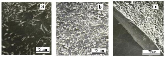
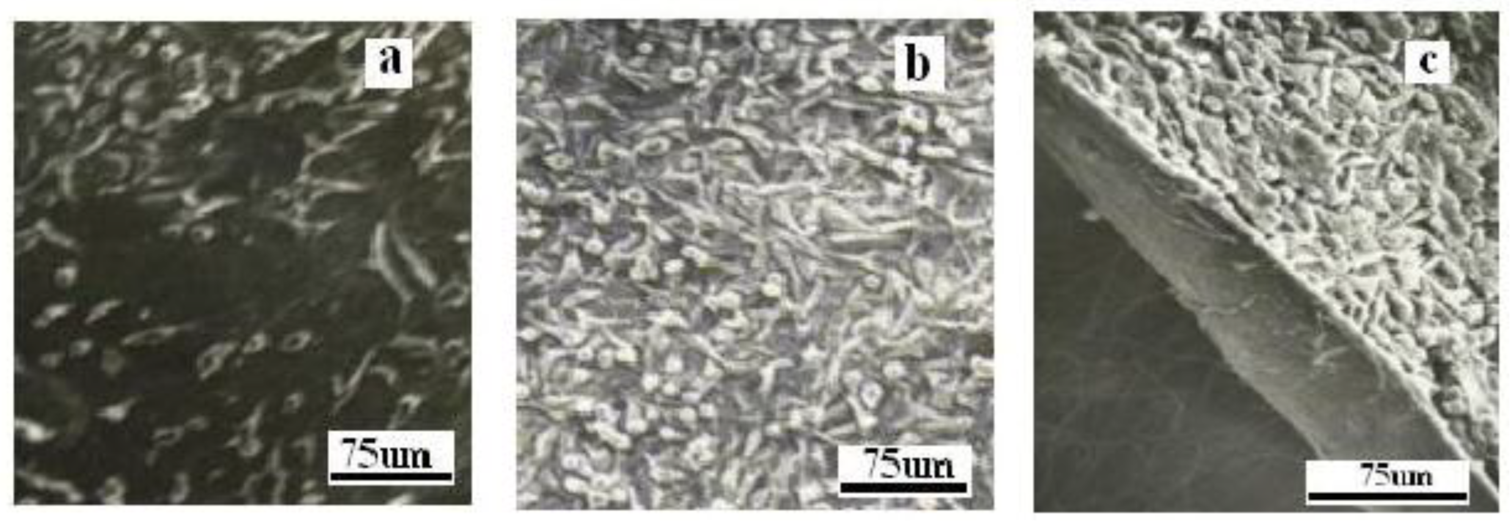
Figure 5 shows three kinds of fiber membranes viability of 3T3 mouse fibroblasts cultured on each could support cell growth; 3T3 cells adhered on membrane increased in a period of 12 days’ cell the surface of fiber membranes and demonstrated culture, then decreased. This is possibly because spindle shape, indicating that the PLA layer had no cells may have occupied all available spaces on the effect on the cell growth and the cells had good electrospun membranes adhesion and spreading on the fiber surface. In addition, it can be seen that cell proliferation micrographs revealed 3T3 mouse fibroblasts on the fiber membranes which were higher than controls, formed a confluent monolayer on the surface of the indicating nanofiber three-dimensional scaffolds, SF-gelatin layer and became integrated after having high surface area to volume ratio in 12 days. In addition, the density of cells on the porosity, were more suitable for cell growth than PLA/SF-gelatin composite membrane was TCPs whose surface was smooth.
Figure 5.
SEM of cells seed on different membranes after 12 days cultivation, PLAS-F (a), PLAS-F/gelatin (70:30) (b), and PLA-SF/gelatin (50:50) (c).
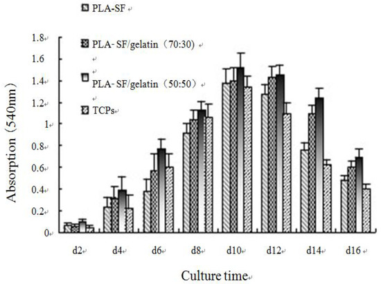
In addition, the significantly higher than that of PLA/SF fiber proliferation of 3T3 cells on the PLA/SF-gelatin membrane, demonstrating the gelatin, which had membrane was higher than that of PLA-SF fiber the same composition and structure as collagen membrane (p<0.05). Moreover, the proliferation (extracellular matrix components), could promote significantly (p<0.05) as the gelatin cell proliferation content increased, demonstrating the composition of scaffolds had an effect on the cell growth, and the gelatin could promote cell proliferation.
3.6. MTT assay
As shown in Figure 6, all the membranes could support 3T3 cell growth and proliferation. The viability of 3T3 mouse fibroblasts cultured on each membrane increased in a period of 12 days cell culture, then decreased. This is possibly because cells may have occupied all available spaces on the electrospun membranes.
Figure 6.
MTT results of 3T3 mouse fibroblasts cultured on different scaffolds.
In addition, it can be seen that cell proliferation on the fiber membranes were higher than controls, indicating nanofiber three-dimensional scaffolds having high surface area to volume ratio and porosity, were more suitable for cell growth than TCPs whose surface was smooth. In addition, the proliferation of 3T3 cells on the PLA-SF fiber membrane (p<0.05). Moreover, the proliferation increased significantly (p<0.05) as the gelatin content increased, demonstrating the composition of scaffolds had an effect on the cell growth, and the gelatin could promote cell proliferation.
4. Conclusions
In the electrospun SF-gelatin nanofibers, SF mainly shows random coils, and gelatin exhibited a mixture of random coil and α-helical conformation. The treatment with methanol did not significantly affect the structure of PLA molecules, however it was observed that the transition of SF from random coil to β sheets forms and gelatin underwent coil-helix transition, consequently, PLA fiber layer was expected to have high dimensional stability and thus prevent the shrinkage of the SF-gelatin membrane. Additionally the chemical treatment conferred strength to PLA/SF-gelatin layer and became integrated. The proliferation of 3T3 cells in fiber membranes was higher than that of controls. The addition of gelatin promoted cell proliferation, additionally the proliferation increased as the gelatin content increased.
This paper provided basic research of PLA-SF/gelatin fiber membranes used as scaffolds. Further research will form tubular scaffolds by a rotating mandrel-type collector and deeply explore the feasibility of those as blood vessel engineering materials.
References
- Kim, S.H.; Nam, Y.S.; Taek, S.; Lee. Polymer 2003, 35(2), 185–190.
- Ayutesedea, J.; Gandhib, M.; Sukigara, S. Polymer 2005, 46, 1625–1634.
- Min, B.M.; Gene, L. Biomaterials 2004, 5, 1039–1047.
- Huang, Z.M.; Zhang, Y.Z.; Ramakrishna, S. Polymer 2004, 45, 5361–5368.
- Zhang, Y.X.; Venugopal, J.; Huang, Z.M. Polymer 2006, 47, 2911–2917.
- Lim, J.; Yong, K.L.; Liu, J.Y. Int. J. Biol. Macromol. 2006, 38, 140–144.
- Joel; Stitzel; Liu, J.; Lee, S.J. Biomaterials 2006, 27, 1088–1094.
- Buttafoco, L.; Kolkman, N.G.; Buijtenhuijs, P. Biomaetrials 2006, 27, 724–734.
- Yang, F.; Murugan, R.; Wang, S. Biomaterials 2005, 26, 2603–2610.
- Venugopal, J.; Ma, L.L.; Yong, T. Cell Biol. Int. 2005, 29, 861–867.
- Ki, C.S.; Baek, D.H.; Gang, K.D. Polymer 2005, 46, 5094–5102.
- Magoshi, J.J. Polym. Sci. Polym. Phys. Ed. 1977, 15, 1675–1683.

© 2009 Textile Bioengineering and Informatics Society. This article is an open access article distributed under the terms and conditions of the Creative Commons Attribution (CC BY) license (https://creativecommons.org/licenses/by/4.0/).