


Abstract
Polyunsaturated fatty acids, particularly γ-linolenic acid, are recognized for their therapeutic and nutritional properties. Zygomycetes, such as Cunninghamella elegans, represent a promising microbial platform for sustainable gamma-linolenic acid (GLA) production as an alternative to conventional sources. Despite this potential, the immunomodulatory activity of metabolites from C. elegans has not been previously explored. In this study, C. elegans was cultivated on hydrolysates from discarded residues of Pleurotus spp. cultures (DRPC-HL), optimized to release assimilable compounds, promoting valorization of low-value biomass within a circular bioeconomy. Dry mycelial biomass, lipid-free biomass, and intracellular lipids from these cultures, alongside previously reported C. elegans cultures grown under nitrogen-excess (N-Xs) and nitrogen-limited (N-Lim) conditions, were tested on THP-1-derived macrophages, under lipopolysaccharide (LPS)-induced inflammatory conditions. Following in vitro gastrointestinal digestion, dry biomass and lipid-free dry biomass fractions upregulated the anti-inflammatory cytokine IL10 and downregulated IL1B and TNF, particularly from N-Xs and DRPC-HL cultures. Lipids mainly enhanced IL10 expression, especially when derived from N-Xs cultures. No changes were observed in upstream regulators (TLR2, TLR4, NFKB1, RELA), suggesting a feasible post-receptor immunomodulatory action. Overall, these findings highlight the dual value of fungal bioproducts derived from agro-industrial residues, combining sustainable bioprocessing with bioactive compound generation, supporting environmentally friendly microbial platforms for industrial applications.
1. Introduction
In recent years, scientific interest has grown significantly in the cultivation of Zygomycete fungi, primarily due to their ability to produce lipids rich in polyunsaturated fatty acids (PUFAs), especially gamma-linolenic acid (GLA) [1,2,3,4,5]. PUFAs, particularly omega-3 and omega-6, are essential biomolecules, acting both as structural components of cellular membranes and as precursors of bioactive mediators such as eicosanoids [6]. Among them, GLA, an omega-6 PUFA synthesized through the action of Δ6-, Δ9-, and Δ12-desaturases, plays a crucial role in supporting the immune system among others, while it also presents interesting anticancer properties, specifically after its transformation into fatty acid lithium or potassium salts [7]. Due to their anti-inflammatory properties, GLA-rich oils are considered beneficial for managing health conditions such as eczema, multiple sclerosis, or menstrual pain, underscoring the importance of their dietary intake [4,8]. Zygomycetes, particularly species from the genera Cunninghamella, Mortierella, Thamnidium, or Mucor, have shown promising PUFA and GLA production, especially when cultivated under nitrogen-rich conditions. These conditions promote cell proliferation over lipid accumulation, resulting in lipids being incorporated mainly into cellular membranes. This leads to higher PUFA yields due to the increased cellular biomass and, therefore, more membrane material [4,5,9,10].
Additionally, these filamentous fungi can thrive on a broad spectrum of commercial and low-cost agro-industrial substrates [4,11,12,13,14,15]. Utilizing such low-cost feedstocks presents a significant advantage, as it lowers bioprocessing costs and thereby improves the prospects for commercializing the high-value fatty acids. Among these putative substrates, lignocellulosic biomass is the most abundant resource on Earth [16,17], and mushroom residues represent a fraction of this category. Approximately 20% of the fruiting body biomass generated during commercial mushroom cultivation—typically including non-marketable stems and defective parts—is discarded in landfills due to failure to meet market quality standards [18]. With the global commercialization of mushrooms (Agaricus spp., Pleurotus spp., Lentinula spp., Tuber spp., etc.) reaching 50 million tons in 2023 [19], species of the genus Pleurotus rank among the most commercially acceptable. These mushrooms are rich sources of valuable secondary metabolites, including β-glucans, proteins, polysaccharides, antioxidants, or ergosterol [18,20,21,22,23,24,25,26]. As a result, the discarded residues of Pleurotus spp. cultures (DRPCs) represent both a large-scale agro-industrial waste and a promising nutrient-rich substrate for microbial cultivation, particularly after appropriate (physical, chemical, and/or enzymatic) pretreatment [27,28,29,30,31].
Despite increasing research on Zygomycete metabolism, their immunological activities remain poorly explored. Within the order Mucorales, species such as Rhizopus, Mucor, and Lichtheimia have been more extensively studied for their roles in host–pathogen interactions [32,33]. However, despite their close phylogenetic proximity, little is known about the host immune response specifically triggered by Cunninghamella species under different culture conditions. A recent study has demonstrated that C. elegans can be cultivated on agro-industrial wastes such as corn steep liquor and molasses, enabling a green metabolic conversion into biopolymers with defined physicochemical properties and notable antimicrobial activity of pharmaceutical relevance [34]. Prior reviews highlighted that macrophages and neutrophils play central roles in the recognition of Mucorales, leading to the release of key inflammatory mediators, including IL-1β, TNF-α, and IL-8 [35]. Conversely, anti-inflammatory mediators such as IL-10 are also secreted by T cells and NK cells during mucoralean infections, suggesting an immune-balancing effect that could be context- or strain-dependent [36]. Nonetheless, the majority of these studies have focused broadly on the Mucorales order without providing direct evidence of immunomodulatory activity by various cells, including dendritic cells, monocytes, and macrophages. These gaps highlight the need to investigate less-characterized Mucorales genera such as C. elegans within immunological contexts.
The innate immune system represents an immediate and non-specific barrier with macrophages playing a critical role by controlling both inflammatory and anti-inflammatory responses [37]. In particular, Toll-like receptors—TLR2 and TLR4—can detect exogenous molecules like lipopolysaccharide (LPS), a structural component of the Gram-negative bacterial outer membrane and a key indicator of Gram-negative infections [38]. While TLR4 is the main receptor mediating LPS recognition, TLR2 has also been reported previously to participate under certain conditions [39,40]. Binding of such ligands activates intracellular cascades, leading to the downstream triggering of the nuclear factor kappa-light-chain enhancer of activated B cells (NF-κB) pathway [41].
The NF-κB family of transcription factors can be activated via pathways—canonical and non-canonical—each characterized by specific signaling molecules and biological functions [42]. Both are important in regulating immune and inflammatory responses [43]. In the canonical pathway, activation leads to the phosphorylation and nuclear translocation of p65 (RELA) and p50 subunits, leading to the transcription of pro-inflammatory genes, including chemokines and cytokines [43]. Key targets, such as tumor necrosis factor-alpha (TNF-α), interleukin-1 beta (IL-1β), and interleukin-8 (CXCL8 or IL-8), drive immune cell recruitment and inflammatory amplification responses [44,45,46,47]. Also, the aforementioned mediators can activate NF-κB signaling in innate immune cells and fibroblasts, forming a feed-forward loop [48]. More specifically, TNF-α and IL-1β are early-response cytokines that initiate inflammation by activating endothelial cells and leukocytes and amplifying cytokine cascades [49]. In contrast, IL-8 primarily acts as a neutrophil chemoattractant and pro-inflammatory mediator, contributing to host defense and pathological inflammation [50]. Their expression is tightly regulated by NF-κB/RELA signaling, a hallmark of TLR-driven immune activation [51]. Meanwhile, interleukin-10 (IL-10) represents an essential component of inflammation resolution, primarily through the suppression of IL-1β and TNF-α expression [52,53].
Macrophages are therefore central regulators of immune homeostasis, exhibiting remarkable plasticity, allowing their cellular metabolism to shift between M1- and M2-like phenotypes. Thus, this adaptability enables them to continually switch between predominantly pro-inflammatory and anti-inflammatory states [54]. While the classical M1 (pro-inflammatory) and M2 (anti-inflammatory) classification remains useful, recent transcriptomic analyses have revealed numerous intermediate activation states, characterized by distinct gene expression profiles and context-dependent functions [55]. These dynamic properties render the THP-1 cell line a first-rate in vitro model for exploring macrophage inflammatory responses and assessing the immunomodulatory potential of dietary components, natural products, and microbial metabolites. In this context, macrophages were selected as the experimental model due to their role as primary responders to exogenous bioactive compounds, including fungal-derived lipid components. In particular, PUFAs, including GLA, have been reported to modulate macrophage function by regulating NF-κB activation, thereby influencing cytokine production [56].
To the best of our knowledge, no study has assessed whether bioproducts derived from C. elegans can modulate inflammatory signaling in human macrophages. In the present work, C. elegans NRRL Y-1392 was cultivated on enzymatic hydrolysates derived from discarded residues of Pleurotus spp. cultures (DRPC-HL). Prior to cultivations, extraction and hydrolysis treatment to recover assimilable compounds from DRPC were carried out. Three fractions, namely whole dry mycelial biomass (X), dry lipid-free biomass (Lf-X), and intracellular lipids (L), were recovered after cultures on DRPC-HL, as well as from previously studied and published C. elegans cultivations on semi-defined media under nitrogen-excess (N-Xs) and nitrogen-limited (N-Lim) conditions. Following simulated gastrointestinal digestion when appropriate, these fractions were evaluated for their immunomodulatory activity on PMA-differentiated, LPS-challenged THP-1 macrophages. This approach combines the valorization of agro-industrial residues with the first evaluation of C. elegans bioproducts as potential modulators of human inflammatory responses.
2. Materials and Methods
2.1. Recovery of Assimilable Compounds from DRPC Through Extraction and Hydrolysis
The dried discarded mushroom residues were obtained from commercial Pleurotus ostreatus cultures of the company MANITUS—KARAGIANNIS IOANNIS (Attica, Greece). The DRPCs were initially weighed and then dried at 105 °C until a constant mass was achieved. Following drying, the biomass was milled using a high-speed grinder and further pulverized to obtain particles smaller than 0.25 mm. The compositional analysis of the dried mushroom residues was performed according to NREL protocols and presented by Vasilakis et al. [5]. The residues consisted of 28.4% (w/w) cellulose, 16.6% proteins, 3.2% lignin, 1.0% hemicellulose, 1.4% lipophilic compounds, and 8.4% ash. Hot-water extraction resulted in the recovery of approximately 44.2 g of total carbohydrates per 100 g of processed residues, comprising 20.6 g of free carbohydrates (expressed as glucose eq.) and 23.6 g of polymeric carbohydrates per 100 g of biomass.
To recover free carbohydrate-type molecules from DRPC, a hot-water extraction approach, followed by enzymatic hydrolysis, was applied, as these are widely established, scientifically validated approaches for recovering fermentable sugars from lignocellulosic biomass [57,58,59]. For the extraction process, various extraction temperatures were tested—namely, 25 °C, 35 °C, 45 °C, 50 °C, 60 °C, and 70 °C. Extractions were performed using a 15% (w/v) suspension of dried DRPC in dH2O, at a final working volume of 60 mL in 100 mL Duran bottles, under gentle stirring for 24 h. The concentration of free reducing sugars in the extracts was determined using the dinitrosalicylic acid (DNS) method, as originally described by Sumner [60] and detailed in the section below (refer to Section 2.3). To quantify the total reducing sugars of the extracted carbohydrates—including those extracted in polymeric form—prior hydrolysis to their monomeric units was required before DNS analysis. This procedure was carried out via acid hydrolysis using 5 M HCl and heating at 100 °C for at least 30 min, followed by neutralization with 5 M NaOH prior to DNS analysis. In addition, the aqueous extracts were analyzed by HPLC to more accurately quantify individual molecules, based on calibration curves prepared from known standards. The HPLC procedure is described in detail in Section 2.3. Furthermore, the concentration of free amino nitrogen (FAN) was determined under the same temperatures mentioned above. The analysis was performed using a ninhydrin-based method, as will be described in Section 2.3. For each extraction condition, the yields of reducing sugars and FAN were calculated relative to the initial biomass substrate (%, w/w).
To recover additional sugars from cellulose and hemicellulose, as well as nitrogenous compounds from proteins, a 24 h enzymatic hydrolysis was performed following a 24 h hot-water extraction step, at the lowest temperature (25 °C) that yielded high carbohydrate concentrations in the previous stage. In parallel, a 24 h direct enzymatic hydrolysis was also tested—without prior extraction—at the enzyme’s optimal temperature (50 °C). The enzymatic hydrolysis experiments included cellulases, hemicellulases, and proteases. Specifically, the enzymes used were “BakeZyme® HSP 6000 BG” [cellulase (6.4 ECU/g) and endo-xylanase (6000 ECU/g) (DSM Food Specialties, Seclin, France) at 50 °C and pH = 6] and “Protease from Aspergillus oryzae” [protease (50,000 ECU/g) (Tokyo Chemical Industry, Tokyo, Japan) at 50 °C and pH = 7]. All experiments were carried out using 15% (w/v) substrate suspensions in dH2O and an enzyme-to-solid-substrate ratio of 10% (w/w), in Duran bottles, as described previously. Free and total reducing sugars were determined using the DNS assay, following acid hydrolysis in the latter case, as previously described, and FAN through the ninhydrin-based assay, as both assays will be described in Section 2.3. Yields of reducing sugars and FAN to initial substrate suspension (g/g) were also calculated. The extraction and hydrolysis processes were performed in triplicate. The presented results correspond to the mean values of three independent technical replicates, accompanied by the standard error (SEM) for each experimental point.
2.2. Microorganism
The fungal strain Cunninghamella elegans NRRL Y-1392 was kindly provided by the ARS Culture Collection—NRRL (Peoria, IL, USA). Spores are preserved in cryovials containing a 50% (v/v) glycerol solution under freezing conditions (−18 °C). The strain was cultivated monthly on PDA medium (Potato Dextrose Agar, Condalab, Madrid, Spain) for maintenance.
2.3. Culture Medium and Analytical Methods
The strain C. elegans NRRL Y-1392 was cultivated on DRPC hydrolysate (DRPC-HL), which was obtained through enzymatic hydrolysis under the optimal conditions identified during the aforementioned process optimization. Following hydrolysis, the mixture was centrifuged and filtered using fiber glass filters (Whatman™, Cytiva, Marlborough, MA, USA) to remove insoluble solids. The clarified liquid was transferred to 2 L Erlenmeyer flasks (Sigma-Aldrich/Merck KGaA, Darmstadt, Germany) containing 400 mL of working volume (20%, v/v) and sterilized without the addition of salts or other nutrients. Inoculation was performed using spores harvested from PDA cultures (as previously described), suspended in sterile deionized water containing 0.1% (v/v) Tween 80, under aseptic conditions in a laminar flow hood. The initial fungal spore concentration in the liquid culture medium was approximately 60,000 spores/mL. The cultures were incubated on a rotary shaker (New Brunswick Scientific, Edison, NJ, USA) at 28 ± 1 °C and 180 ± 5 rpm. At regular intervals, culture broth was aseptically sampled to monitor sugar concentration (expressed as glucose equivalents) using the DNS method, as well as the concentration of FAN.
The methods applied during extractions, hydrolyses, and microbial cultivations are described in detail below. The determination of reducing sugars via the DNS (3,5-dinitrosalicylic acid) assay involved mixing 0.5 mL of sample with 0.5 mL of DNS reagent, followed by boiling at 100 °C for 5 min. After cooling, 5 mL of deionized water was added, and absorbance was measured at 540 nm against a blank. Quantification was performed using a standard glucose calibration curve and results were expressed as glucose equivalents (g/L). The identification and quantification of molecules during the extraction process was carried out using HPLC on a Waters Alliance 2695 system (Waters Corporation, Milford, MA, USA) equipped with refractive index detector (2414 Refractive Index Detector, Waters Corporation, MA, USA). The separation column used was an Aminex HPX-87H (30.0 cm × 7.8 mm; Bio-Rad Laboratories, Hercules, CA, USA), with 0.005 M H2SO4 as the mobile phase at a flow rate of 0.5 mL/min (isocratic elution) and a column temperature of 60 °C. Identification and quantification were performed based on calibration curves generated for each molecule (glucose, trehalose, mannitol), constructed through serial dilutions of solutions of known concentrations, using high-purity standards (>99.8%, w/w). The determination of free amino nitrogen was carried out by mixing 1.0 mL of the sample with 0.5 mL of color reagent (containing ninhydrin), followed by boiling at 100 °C for 16 min. The mixture was then cooled in an ice bath for 20 min, after which 2.5 mL of dilution reagent was added. Absorbance was measured at 570 nm against a blank. Quantification was based on a standard calibration curve, and results were expressed as FAN (mg/L). More detailed information on the above protocols is provided in Vasilakis et al. [61].
The total mycelial biomass obtained after cultivation in DRPC-HL was recovered by filtration using Whatman No. 1 filter paper (Whatman™, Cytiva, MA, USA), rinsed with distilled water, and then dried to a constant weight. Biomass was determined gravimetrically and expressed as dry biomass (X, g/L). Total intracellular lipids (L, g/L) were extracted using a mixture of chloroform and methanol (2:1, v/v) and quantified gravimetrically after solvent evaporation [5,62]. The fatty acid profile was analyzed via gas chromatography (GC) following the conversion of lipids to their corresponding fatty acid methyl esters (FAMEs), as described by Vasilakis et al. [61]. Fatty acid content was expressed as a percentage of total lipids by weight (g/100 g total lipids or %, w/w). The collected filtrate was analyzed for pH (Hanna Instruments Hellas, Athens, Greece), residual sugar concentration (SR, g/L) using the DNS assay, and residual free amino nitrogen (FANR) using the ninhydrin-based protocol. The consumed sugars (SCON, g/L) and free amino nitrogen (FANCON, mg/L) concentrations were calculated as the difference between the initial and residual values, SCON = S0 − SR and FANCON = FAN0 − FANR, respectively. For more detailed information on the above procedures, refer to Vasilakis et al. [4,61]. Cultivations were performed in triplicate, and all analytical measurements were conducted in triplicate; thus, the results are presented as mean values ± SEM.
Based on the above, the biomass yield coefficient on substrate (YX/SCON, g of total dry biomass produced per g of substrate consumed), the lipid yield coefficient on substrate (YL/SCON, g of lipids per g of substrate consumed), the lipid content of the biomass (KL/X, g of lipids per g of dry biomass), and the productivities of dry biomass (PX, dry biomass concentration per hour, mg/L/h), lipids (PL, lipid concentration per hour, mg/L/h), polyunsaturated fatty acids (PPUFA, PUFA concentration per hour, mg/L/h), and γ-linolenic acid (PGLA, γ-linolenic acid concentration per hour, mg/L/h) were calculated according to Papanikolaou et al. [63].
2.4. Simulated Gastrointestinal Digestion (SGID) Static Model and Digestates’ Fractionation
Zygomycete-derived bioproducts were obtained from cultivations on DRPC-HL (present study), as well as after growth on commercial semi-defined synthetic media under nitrogen-excess (N-Xs) and nitrogen-limited (N-Lim) conditions (previously published details by Vasilakis et al. [5]). Samples of the Ζygomycete bioproducts, specifically biomass and lipid-free biomass (X and Lf-X, respectively), were resuspended in Milli-Q H2O and incubated at 4 °C for 12 h to ensure complete and homogeneous hydration [64]. Following hydration, the protein concentration of each sample was adjusted to 10 mg/mL to standardize the input for digestion. Due to the negligible levels of starch and fat in the samples, amylase and pancreatic lipase were omitted from the digestion protocol, and digestion was performed solely based on equal protein amounts. The rehydrated samples were subjected to SGID according to the detailed protocol described by Dalaka et al. [65] based on the standardized and optimized INFOGEST 2.0 method with minor modifications [66,67]. The conditions mimic those occurring during the physiological digestion within the human body, aiming to release sample components with potential biofunctional properties. Electrolyte stock solutions for the simulated digestive fluids, namely simulated salivary fluid, gastric fluid, and intestinal fluid, were prepared, with pH adjustments performed using 5 M HCl and 6 M NaOH. Fresh solutions of pepsin, pancreatin, and bile salts were prepared immediately before use.
Once the intestinal digestion phase was completed, the digestates (Ds) were heated at 85 °C for 10 min and then immediately cooled on ice. Subsequently, the Ds were centrifuged at 1200× g for 5 min and the supernatants were filtered through sterile 0.22 μm PVDF syringe filters. To isolate fractions with molecular weights below 3 kDa (D-P3), ultrafiltration was performed using membrane filters with a 3 kDa molecular weight cut-off. All samples were stored at −20 °C until further analysis. Digestions were conducted in duplicate. In parallel, four replicates of blank digestates (BL-Ds) were prepared using water in place of samples, following the same in vitro digestion protocol. The resulting blank fraction is referred to as BL-D-P3 in the text and as “Blank” in the corresponding figures. The Ds contained a protein concentration of 0.125% w/v, as previously described [68].
2.5. THP-1 Cell Culture, Cell Viability, Differentiation, and Activation
The human THP-1 cell line is widely used as an in vitro model to study the immune response of monocytes, monocyte-derived macrophages and activated macrophages due to its similarities with peripheral blood mononuclear cells in vivo [69]. THP-1 cells were grown in RPMI 1640 medium (Biosera, Cholet, France) supplemented with 10% (v/v) fetal bovine serum (Gibco ThermoFisher Scientific, Waltham, MA, USA), 100 μg/mL penicillin–streptomycin (Biosera, Cholet, France), 1 mM sodium pyruvate (Biosera, Cholet, France), 10 U/mL L-glutamine (Biosera, Cholet, France), and 100 μM non-essential amino acid solution (Biosera, Cholet, France). Cultures were kept in a humidified atmosphere of 5% CO2 at 37 °C with cell passaging every 2–3 days. In detail, 2 × 10⁶ monocytes (0.8 × 10⁶ cells/mL) were seeded in 6-well culture plates and were incubated with supplemented RPMI 1640 and 100 ng/mL phorbol 12-myristate 13-acetate (PMA; Sigma-Aldrich, Saint Louis, MO, USA) for 2 days to induce differentiation into adherent macrophage-like cells. During PMA stimulation, THP-1 cells undergo phenotypic changes, including adherence to culture plates and a morphological transition to a more spread, amoeboid-like shape, consistent with macrophage differentiation [70]. After this period, the RPMI 1640 medium was discarded, the adherent cells were washed, followed by an incubation with supplemented RPMI 1640 for an additional day (resting phase). The resting phase following PMA treatment is critical to minimize residual activation and prevent PMA-induced bias in downstream gene expression, ensuring a more physiologically relevant macrophage phenotype [71]. Subsequently, cells were exposed to the D-P3 fraction (refer to Section 2.4.—X and Lf-X interventions at 15%) along with 100 ng/mL lipopolysaccharide (LPS from Escherichia coli O111:B4; Sigma-Aldrich, Saint Louis, MO, USA) to induce mild inflammation for an additional 3 h. Each experimental condition was performed in triplicate.
For the isolated lipid interventions (L), THP-1 cells were exposed to 50 μg fat/mL in the form of triglycerides dissolved in ethanol (<0.1%, v/v, to avoid cytotoxicity) [72,73]. Of note, lipid was not subjected to SGID prior to exposure to THP-1-activated macrophages, in order to evaluate the direct immunomodulatory effects of native fatty acids. This approach aligns with previous studies where native n-3 and n-6 PUFAs were added directly to THP-1 cultures, resulting in significant modulation of cytokine secretion and NF-κB signaling [72,74]. THP-1 cells stimulated with PMA and LPS were concurrently used as control conditions for comparison with the different L treatments. The resulting control fraction is referred to as “Control” in the text and as “LPS” in the corresponding figures. Each experimental condition was performed in triplicate.
2.6. Quantification of Gene Expression in THP-1 Cells
Following treatment, total RNA (derived from THP-1 cells pretreated with X, Lf-X, L, Blank, or LPS preparations) was isolated from adherent cells using the NucleoZOL reagent (Macherey-Nagel, Düren, Germany), in accordance with the manufacturer’s instructions. Genomic DNA was removed using DNase I (20,000 Units/mg; New Englands Biolabs, Ipswich, MA, USA), following the supplier’s protocol, and purified RNA was obtained via ethanol precipitation [75]. RNA concentration and purity were calculated using a Q5000 spectrophotometer (Quawell Technology Inc., San Jose, CA, USA). Reverse transcription was performed with the PrimeScript RT reagent kit (Takara Bio, Shiga, Japan), according to the manufacturer’s guidelines. Quantitative PCR (qPCR) was conducted on a thermal cycler (SaCycle96, Sacace Biotechnologies, Como, Italy) using the FastGene 2× IC Green qPCR Universal Mix (Nippon Genetics, Tokyo, Japan). All reactions were run in duplicate. Primers for both target and reference genes (B2M, RPS18, and RPL37A) were designed in-house, with an annealing temperature set at 60 °C. Relative gene expression levels were calculated using the method described by Hellemans et al. [76], normalized to three aforementioned housekeeping genes. The primer details are listed in Table 1.
Table 1.
Oligonucleotide primer sequences, reaction efficiency, and amplicon size in qPCR.
2.7. Statistical Analysis
Statistical analyses were performed using the SPSS software (version 22.0.0) and graphs were visualized with the GraphPad Prism 8 program. Data are presented as means ± SEM from a minimum of two technical replicates. Normality was assessed using the Kolmogorov–Smirnov test. Where necessary, data were log-transformed or normalized [77] to achieve a normal distribution. Group comparisons were conducted using one-way analysis of variance (ANOVA), followed by Duncan’s post hoc test. Statistical significance was defined as p < 0.05.
3. Results
3.1. Recovered Compounds from DRPC Through Aqueous Thermal Extraction
Extractions were conducted using suspensions of dried and ground DRPC biomass (15% w/v) at various temperatures for 24 h. In the final extracts, the concentrations of free reducing sugars, total extracted polymeric carbohydrates (measured as total reducing sugars after acid hydrolysis), and FAN were determined. The yields of reducing sugars and FAN were calculated based on the initial biomass substrate, and the results are presented in Table 2.
Table 2.
Concentrations and yields of free (FRS, g/L) and total reducing sugars (TRSs, g/L), as well as of free amino nitrogen (FAN, mg/L), after 24 h of extraction at the various temperatures tested.
The highest concentrations of free (37.9–41.1 g/L) and total reducing sugars (52.8–56.4 g/L—following acid hydrolysis of the liquid extract) were observed at the lower extraction temperatures of 25 and 35 °C. In all other tested temperatures, the concentrations significantly decreased, with the lowest values recorded at 70 °C: 6.8 g/L for free reducing sugars and 32.6 g/L for total reducing sugars. The yield coefficients of free and total reducing sugars were 0.25 and 0.35 g/g, respectively, at 25 °C, and 0.27 and 0.37 g/g, respectively, at 35 °C. As expected, the yields declined with increasing temperature, likely due to the reduced extraction efficiency of reducing compounds. At 70 °C, the yield coefficients were 0.05 g/g for free and 0.22 g/g for total reducing sugars. HPLC analysis of the 24 h extracts (prior to acid hydrolysis) showed that glucose was the most abundant compound, followed by the non-reducing compounds trehalose and mannitol. The highest glucose concentration (27.0 g/L) was observed at the lower temperatures (25–35 °C), whereas the highest concentration of the non-reducing disaccharide trehalose (9.3 g/L) was found at 45 °C. Mannitol levels were approximately 3.2 g/L across the 25–50 °C range, with no statistically significant differences. In addition to carbohydrates, the concentration of free amino nitrogen (FAN) was also quantified. The highest FAN concentration (515.1 mg/L, corresponding to a yield of 0.34%) was observed at 45 °C, though this did not differ significantly from the values obtained at 25 and 35 °C. At higher temperatures, a decrease in FAN concentration was also observed, which consequently led to a reduction in its yield.
3.2. Recovered Compounds from DRPC Through Enzymatic Treatment
For the enzymatic hydrolysis, the commercial formulations “BakeZyme® HSP 6000 BG” and “Protease from Aspergillus oryzae” were used, containing the necessary cellulases, hemicellulases, and proteases, at an enzyme-to-solid-substrate ratio of 10% (w/w). Hydrolysis was carried out at the optimal temperature and pH conditions for the enzymatic activity, as specified by the manufacturer, for 24 h (second stage). Prior to hydrolysis, a 24 h hot-water extraction step was tested at the lowest temperature (25 °C) that previously yielded high carbohydrate concentrations (first stage). In parallel, a direct 24 h enzymatic hydrolysis—single-stage, without prior extraction—was also tried at the enzyme’s optimal temperature (50 °C). In the resulting hydrolysates, the concentrations of free reducing sugars, total extracted polymeric carbohydrates (measured as total reducing sugars after acid hydrolysis), and free amino nitrogen (FAN) were determined. The yields of reducing sugars and FAN were calculated based on the initial biomass substrate, and the results are presented in Table 3.
Table 3.
Concentrations and yields of free (FRS, g/L) and total reducing sugars (TRSs, g/L), as well as of free amino nitrogen (FAN, mg/L)), after aqueous thermal and/or enzymatic treatment.
The 24 h extraction at 25 °C followed by enzymatic hydrolysis using the commercial product “BakeZyme® HSP 6000 BG” at 50 °C yielded the highest concentration of free reducing sugars, reaching 105.2 g/L, with a corresponding yield of 0.70 g per g of initial substrate. The total sugars, measured after subsequent acid hydrolysis of the hydrolysate, amounted to 109.5 g/L, with a yield of 0.73 g/g—approaching the maximum based on the sugar content of the initial substrate (approximately 74%, w/w, when summing the water-soluble components, cellulose, and hemicellulose contents reported in Table 3 by Vasilakis et al. [5]). In contrast, the simultaneous extraction and enzymatic hydrolysis process carried out at 50 °C for 24 h resulted in only 43.2 g/L and 53.3 g/L of free and total reducing sugars, respectively, corresponding to yields of 0.29 and 0.36 g/g. According to the results obtained using the enzymatic commercial product “Protease from Aspergillus oryzae”, the sequential process of prior extraction followed by enzymatic hydrolysis led to the release of 937.2 mg/L of FAN, corresponding to a yield of 0.62 mg/g. In contrast, the simultaneous extraction–hydrolysis process yielded only 501.7 mg/L of FAN, with a yield of 0.34 mg/g.
3.3. Cultivation of C. elegans on DRPC Hydrolysate
The strain C. elegans NRRL Y-1392 was cultivated on the enzymatic hydrolysate of DRPC, after the two-stage pretreatment, following appropriate filtration, dilution, and sterilization steps, without further supplementation of nutrients. The aim was to promote fungal growth and the production of metabolites of interest, such as PUFAs and GLA. The initial sugar concentration was adjusted to 30 g/L through appropriate dilution, as higher concentrations were previously shown to inhibit growth despite sufficient nitrogen availability, according to [4]. The end-point results of the cultivation are presented in Table 4, and the lipid composition is shown in Table 5.
Table 4.
Results from the cultivation of C. elegans on an enzymatic hydrolysate of DRPC.
Table 5.
Fatty acid composition of intracellular lipids isolated from the cultivation of C. elegans on an enzymatic hydrolysate of DRPC.
The microorganism was able to grow on this complex substrate, assimilating a portion of the available sugars (21.8 g/L) and yielding 9.4 g/L of dry biomass, with a biomass yield coefficient of 0.43 g/g after 88 h of cultivation. The concentration of FAN stabilized after 24 h, with a total consumption of approximately 190 mg/L. Under these conditions, lipid production reached 3.3 g/L (YL/S = 0.15 g/g), corresponding to a lipid accumulation of 35% (w/w). The production and productivity values of PUFAs were 614 mg/L and 7.0 mg/L/h, respectively, while the corresponding values for GLA were 112 mg/L and 1.3 mg/L/h. Based on the fatty acid profile, oleic acid was predominant, accounting for 58.6% (w/w), followed by linoleic acid (15.2%, w/w), palmitic acid (13.6%, w/w), and stearic acid (7.2%, w/w). GLA was present at 3.4% (w/w), while the total PUFA content reached 18.6% (w/w).
Then, to assess the impact of C. elegans bioproducts on the inflammatory cascade, THP-1 cells were differentiated and activated into macrophages using PMA and LPS. To provide a complete understanding of the biological effects of C. elegans metabolites, cells were treated with three distinct preparations: the whole dry biomass (X), representing the complete matrix of bioactive compounds, the defatted dry biomass (Lf-X), obtained after removal of the lipid fraction, representing the non-lipid constituents, and the isolated lipid fraction (L), containing the extracted lipophilic components. The metabolites were derived from the cultivation of the fungus on DRPC-HL, as well as from the cultivation of the same strain under nitrogen-excess (N-Xs) and nitrogen-limited (N-Lim) conditions (for further details, refer to Section 3.3.1 in the relevant publication by Vasilakis et al. [5]). Among these, the X and Lf-X fractions were subjected to SGID prior to cell exposure, following the protocol described in Section 2.4, whereas the isolated lipid intervention (L) was tested in its native, undigested form.
Recent studies have demonstrated that macrophages are not fixed in a singular pro-inflammatory state, as traditionally believed, but are instead dynamic regulators of immune responses. They exhibit remarkable plasticity, contributing not only to inflammation but also to its resolution through anti-inflammatory functions, underscoring their pivotal role in maintaining immune homeostasis [78,79]. Therefore, the expression levels of various genes were evaluated, including transcription factors (NFKB1 and RELA), receptors (TLR2 and TLR4), pro- and anti-inflammatory cytokines (IL1B, IL10, and TNF), and chemokines (CXCL8).
3.4. Effect of Biomass Digestates on mRNA Expression of Inflammation-Related Genes in LPS-Challenged PMA-Induced THP-1-Derived Macrophages
The effect of biomass (X) digestates obtained under the three different aforementioned culture conditions on the expression of inflammation-related genes was evaluated in LPS-stimulated, PMA-induced THP-1-derived macrophages (Figure 1). No statistically significant differences were observed in the expression of NFKB1, RELA, TLR2, and TLR4 (p > 0.05; Figure 1a–d respectively) across treatments, indicating that the upstream regulators of immune signaling remained unaffected by the dietary interventions. In contrast, significant modulation was observed in several cytokine genes. The expression of IL1B was significantly downregulated (p < 0.05) in the presence of N-Xs-derived X digestate, compared to the one derived from N-Lim conditions, while no significant differences were observed compared to the Blank (which included digestion with water and represents the <3 kDa peptide fraction of the digestate; for further details, refer to Section 2.4) and DRPC-HL-derived X preparations (Figure 1e). In contrast, the anti-inflammatory cytokine IL10 (Figure 1f) was consistently upregulated (p < 0.05) across all treatments compared to the Blank, indicating an overall anti-inflammatory response associated with the presence of C. elegans-derived bioproducts. In line with this finding, the pro-inflammatory cytokine TNF (Figure 1h) was significantly downregulated (p < 0.05) across all treatments compared to the Blank, reinforcing the evidence for an anti-inflammatory effect. A significant decrease in CXCL8 transcription levels was observed in cells treated with X produced under DRPC-HL conditions compared to those produced under nitrogen-limited conditions (p < 0.05; Figure 1g). In contrast, no significant differences in CXCL8 expression were found between the treatments with the mycelial preparations and the Blank. Together, these findings suggest that although transcription of TLR and NF-κB signaling components remained unchanged, a pattern of modulation was observed in downstream cytokine responses, influenced by the different dietary culture conditions.
Figure 1.
Effect of X (produced under different culture conditions) on the mRNA expression in LPS-challenged, PMA-induced THP-1-derived macrophages. THP-1 cells were pretreated with PMA (100 ng/mL, 48 h), rested (24 h), and then stimulated with LPS (100 ng/mL) in the presence of X-D-P3 (0.125% w/v) or BL-D-P3 for 3 h. Expression of (a) NFKB1, (b) RELA, (c) TLR2, (d) TLR4, (e) IL1B, (f) IL10, (g) CXCL8, and (h) TNF was quantified by qPCR. Data are means ± SEM of three technical replicates. Columns with different letters within the same panel indicate significant differences (p < 0.05).
3.5. Effect of Lipid-Free Biomass Digestates on mRNA Expression of Inflammation-Related Genes in LPS-Challenged PMA-Induced THP-1-Derived Macrophages
TLRs, particularly TLR2 and TLR4, can activate the NF-κB signaling cascade via MyD88- and TRIF-dependent pathways, resulting in the transcriptional upregulation of pro-inflammatory cytokines, including TNF-α and IL-1β [80,81,82]. In our study, Lf-X digestates induced significantly different effects on TLR2, as well as on the expression of specific pro- and anti-inflammatory genes, depending on the culture conditions under which the biomass was produced. Specifically, TLR2 expression was significantly lower (p < 0.05; Figure 2c) in cells treated with Lf-X produced under N-Lim conditions compared to those treated under N-Xs conditions and the Blank. Furthermore, cell treatment with all Lf-X digestates led to downregulation of TNF expression compared to the Blank, regardless of culture conditions, with no significant differences observed among them (p < 0.05; Figure 2h). A significant decrease in IL1B transcription was also observed in cells treated with Lf-X produced under N-Xs and DRPC-HL conditions (p < 0.05; Figure 2e), while Lf-X derived under N-Lim conditions did not exert a similar effect. In the context of anti-inflammatory response, cells treated with Lf-X produced under either N-Xs conditions or DRPC-HL exhibited significantly increased IL10 mRNA levels (p < 0.05; Figure 2f), an effect not observed under N-Lim conditions. As for CXCL8, significant differences were observed among treatments depending on culture conditions (p < 0.05; Figure 2g). Treatment with Lf-X produced under N-Lim conditions resulted in downregulation of CXCL8 expression compared to the Blank, whereas cells treated with Lf-X derived from culture on DRPC-HL showed an increase in CXCL8 expression compared to both N-Xs and N-Lim. Finally, no statistically significant differences were observed in the expression of NFKB1, RELA, and TLR4 across treatments (p > 0.05; Figure 2a,b,d).
Figure 2.
Effect of Lf-X (produced under different culture conditions) on the mRNA expression in LPS-challenged, PMA-induced THP-1-derived macrophages. THP-1 cells were pretreated with PMA (100 ng/mL, 48 h), rested (24 h), and then stimulated with LPS (100 ng/mL) in the presence of Lf-X-D-P3 (0.125% w/v) or BL-D-P3 for 3 h. Expression of (a) NFKB1, (b) RELA, (c) TLR2, (d) TLR4, (e) IL1B, (f) IL10, (g) CXCL8, and (h) TNF was quantified by qPCR. Data are means ± SEM of three technical replicates. Columns with different letters within the same panel indicate significant differences (p < 0.05).
3.6. Effect of Lipids on mRNA Expression of Inflammation-Related Genes in LPS-Challenged PMA-Induced THP-1-Derived Macrophages
The impact of lipid fractions (L) derived from the different culture conditions on the expression of inflammation-related genes was assessed in LPS-stimulated, PMA-induced THP-1-derived macrophages (Figure 3). Overall, no statistically significant differences were observed in the mRNA levels of transcription factors (NFKB1 and RELA), Toll-like receptors (TLR2 and TLR4), pro-inflammatory cytokines (IL1B and TNF), and chemokine (CXCL8) between cells treated with the L fraction and those exposed only to LPS (p > 0.05; Figure 3a–e,g,h). However, a significant increase in IL10 mRNA expression was observed in cells treated with L derived from N-Xs, N-Lim, and DRPC-HL culture conditions compared to the LPS control, with the N-Xs treatment eliciting the most pronounced upregulation (p < 0.05; Figure 3f). This suggests that the nitrogen-supplemented condition may enhance anti-inflammatory response more effectively than the other culture conditions, potentially contributing to a stronger immunomodulatory effect. In general, these results suggest that, under the tested conditions, fatty acids do not modulate pro-inflammatory gene expression in this model, regardless of the culture condition from which the lipids were derived.
Figure 3.
Effect of L (produced under different culture conditions) on mRNA expression in LPS-challenged, PMA-induced THP-1-derived macrophages. THP-1 cells were pretreated with PMA (100 ng/mL, 48 h), rested (24 h), and then stimulated with LPS (100 ng/mL) in the presence of L or Control for 3 h. Expression of (a) NFKB1, (b) RELA, (c) TLR2, (d) TLR4, (e) IL1B, (f) IL10, (g) CXCL8, and (h) TNF was quantified by qPCR. Data are means ± SEM of three technical replicates. Columns with different letters within the same panel indicate significant differences (p < 0.05).
4. Discussion
Filamentous fungi are particularly known for their efficient decomposition abilities through evolution [83]. They possess unique metabolic ability and enzymatic activity to transform a wide range of organic materials (including waste) into valuable products with industrial potential, in the context of circular bioeconomy [84]. Within this group, Zygomycetes exhibit remarkable metabolic versatility, enabling them to assimilate a wide range of organic substrates and produce lipid-, polysaccharide-, and protein-rich biomass. These characteristics support their promising applications in the development of functional food/feed and nutraceuticals for the respective industries [85]. A representative genus of this class, Cunninghamella, has been a prominent tool for its diverse biotechnological applications, serving as a microbial model for mammalian drug metabolism [86,87] and exhibiting strong potential for the biotransformation and degradation of various chemical compounds, including pigments, pharmaceuticals, and agrochemicals [88,89,90]. Recent studies have also highlighted the potential of Cunninghamella spp., including C. elegans, in generating metabolites with significant biological activities such as anticancer and anti-inflammatory effects. In detail, several studies have focused on the biotransformation of drugs by C. elegans, revealing promising outcomes regarding its cytotoxicity and potential therapeutic benefits [91,92,93]. In parallel, its capacity to assimilate lignocellulosic sugars, derived either from acid-pretreated wheat straw or hot-water extracts from mushroom stalks (named as DRPC in the current study), as alternative substrates for metabolite production has also been demonstrated, further supporting its versatility in bioconversion processes [5,94].
In the present study, the immunomodulatory effects of C. elegans metabolites were investigated, when the fungus was grown on commercial semi-defined substrates, under different nitrogen conditions (N-Xs and N-Lim) (as described by Vasilakis et al. [5]) or after cultivation on DRPC-HL, prior to proper pretreatment procedures. Additionally, the DRPC represents a lignocellulosic matrix rich in bioactive compounds, including polysaccharides, proteins, and minor metabolites. Following enzymatic hydrolysis (DRPC-HL), these components are released into assimilable forms (e.g., oligosaccharides and peptides), which may influence fungal metabolism and, consequently, the bioactive profile of the derived biomass. The first stage of the study involved the extraction of assimilable compounds from DRPC through aqueous thermal treatment at different temperatures. The highest concentrations of free and total reducing sugars—mainly glucose, according to the HPLC results—were obtained at the lowest temperatures tested, namely 25 and 35 °C. Although, in theory, increasing temperature should enhance the release of reducing carbohydrates, such an effect is typically observed at much higher temperatures—above 100 °C—and over significantly shorter extraction times, as reported in the literature [59,95,96]. Instead, in our case, such high temperatures were not used (<70 °C), to minimize energy consumption and avoid potential Maillard reactions and the formation of undesirable derivatives such as furfurals, hydroxy-methyl-furfurals, etc. Thus, sugar concentrations and, consequently, extraction yields decreased with increasing temperature. A possible explanation for this observation is the possible presence of endogenous enzymes in the discarded residues of mushrooms. Higher fungi of the genus Pleurotus sp. are known producers of various enzyme types capable of degrading lignocellulosic biomass, including peroxidases, laccases, cellulases, xylanases, amylases, and proteases [17,97,98,99]. It is therefore hypothesized that during the extraction process, endogenous enzymes may be released and contribute to sugar extraction. However, elevated temperatures could limit or even inhibit their activity. This hypothesis is partially supported by calculating the ratios of free to total reducing sugars presented in Table 2, where it is evident that the ratio decreases as the temperature increases. This suggests that the extracted polymers are not being hydrolyzed to the same extent as at lower temperatures, potentially due to enzyme inactivation. Another possible explanation for lower concentrations as temperature increases involves the non-enzymatic browning (caramelization or Maillard-type reactions) of sugars at higher tested temperatures, which could lead to a decrease in their measurable concentrations. In any case, this observation is novel and reported here for the first time. It warrants further investigation to identify the underlying mechanisms responsible for these results.
Additionally, enzymatic hydrolysis, in the presence of commercial preparations of cellulases, hemicellulases, and proteases, was carried out to increase assimilable compound recovery. The enzymatic activity led to liquid rich in sugars and FAN. Among the two approaches studied (a two-stage sequential process versus a single-stage simultaneous aqueous thermal and enzymatic treatment), higher results were observed when the two-stage process was conducted. Furthermore, a comparison between the results of the single-stage pretreatment and those of the aqueous thermal extraction process under the corresponding conditions (50 °C for 24 h—see Section 3.1.) highlights the effect of the enzymes on both hydrolysis and extraction. The addition of enzymes resulted in a 2.4-fold increase in free reducing sugars, while total reducing sugars increased by 1.5-fold.
The hydrolysate was diluted to a final sugar concentration of 30 g/L, since, according to Vasilakis et al. [4], higher concentrations could inhibit growth—likely due to constrained transport and dispersion of nutrients and/or oxygen caused by the high concentration of mycelial biomass—and subsequently lead to autolysis. The fungus was able to assimilate a significant portion of the available sugars and grow, accumulating lipids; however, the lipid fraction presented low GLA content. It appears that the nutrient medium was poorer in nitrogen than required to promote sufficient proliferation. Increased proliferation would lead to more cellular membranes, which are the primary sources of PUFAs and GLA [4,9,10]. In the study by Vasilakis et al. [5], a higher biomass production was observed (14.5 g/L vs. 9.4 g/L in the present study), but not lipid production (1.8 g/L vs. 3.3 g/L in the present study), during the cultivation of the same strain in a water extract derived from mushroom stalks under nitrogen-excess conditions. The high nitrogen availability favored cell growth in that case, resulting in a lower lipid yield compared to the present study, but with a substantially higher GLA content (15.0% vs. 3.4%, w/w, in the present study).
To provide a comprehensive understanding of the biological effects of C. elegans metabolites, three fractions (i.e., X, Lf-X, and L) were recovered following the three distinct cultivation conditions: one in DRPC-HL medium, and two others in commercial substrates under nitrogen-excess (N-Xs) and nitrogen-limited (N-Lim) conditions [5]. These specific conditions were chosen because, under nitrogen-excess conditions, biomass rich in polysaccharides (including β-glucans) and proteins was produced, with a low lipid content; however, these lipids were particularly rich in PUFAs and GLA. On the other hand, nitrogen limitation led to higher lipid accumulation at the expense of other biomass fractions, but the lipids were richer in oleic acid, resulting in lower PUFA content [5]. Τhe biomass and lipid-free biomass fractions were subjected to static in vitro digestion, while the lipids were not, and subsequently, all were tested in THP-1 cells, which were differentiated and activated into macrophages using PMA and LPS, simulating a low-grade inflammatory environment. To our knowledge, C. elegans biomass, lipid-free biomass and isolated lipids were administered for the first time to a human cancer-derived immune model.
The observed immunomodulatory responses, particularly the upregulation of IL10 and downregulation of IL1B and TNF following treatment with total dry biomass and lipid-free dry biomass preparations, may be attributed, at least in part, to structural polysaccharides. These molecules are recognized by pattern recognition receptors (e.g., Dectin-1 and Toll-like receptors) on macrophages [100]. Their engagement may modulate anti-inflammatory signaling pathways, contributing to increased IL-10 and IL-1Ra production and suppression of pro-inflammatory mediators such as TNF-α and IL-1β [101,102]. These effects are potentially mediated through downstream signaling cascades, such as NF-κB and mitogen-activated protein kinase kinase (MAPK) pathways, providing a plausible mechanistic basis for the observed immunomodulatory phenotype in our THP-1-derived macrophage model. However, as these findings are based on transcriptional data, validation at the protein level (e.g., cytokine secretion via ELISA or Western blot analysis) is required to confirm the functional immunomodulatory activity. Direct mechanistic validation remains necessary to establish causal structure–activity relationships underlying the observed changes in IL10, TNF, and IL1B mRNA levels.
The quantity, branching, and overall architecture of β-glucans can be modulated by the cultivation substrate. In this study, the DRPC-HL substrate, naturally enriched in β-glucans, may have enhanced glucan content or altered the β-(1,3)/(1,6) branching in the cell wall, contributing to the anti-inflammatory phenotype observed in THP-1-derived macrophages. Similar substrate-driven modifications in β-glucan structure and downstream immunological responses have been reported in other filamentous fungi grown on lignocellulosic or agro-industrial residues [103,104,105].
In this study, the lipid fraction was evaluated in its native, undigested form to assess its direct immunomodulatory effects on macrophages, since lipids can interact directly with cell membranes and pattern recognition receptors such as TLR2 and TLR4 without prior enzymatic hydrolysis, thereby modulating downstream signaling pathways including NF-κB [106]. To date, studies have mainly focused on fatty acid lithium salts (FALSs) derived from total lipids of Cunninghamella species, evaluating their effects on various human cell models. Among them, GLA salts have demonstrated significant therapeutic potential, exhibiting strong antiproliferative activity and enhancing the efficacy of several chemotherapeutic agents across diverse cancer models [107]. A previous study by Kalampounias et al. [8] evaluated the antiproliferative, anti-migratory, stress-inducing, and cell death-inducing effects of lipids derived from C. elegans NRRL-1393 on human cancer models. Specifically, the study included a normal thyrocyte model (Nthy-Ori 3-1), papillary thyroid carcinoma cell lines (TPC-1 and K1), and prostate cancer cell lines (PC-3 and DU-145), and a wide range of FALSs were tested. FALSs induced cytotoxicity in a dose-dependent manner, resulting in decreased cell viability across all cell lines following incubation.
Similarly, Alakhras et al. [108] investigated the effects of FALSs derived from C. echinulata lipids on HL-60 leukemic cells and demonstrated that FALSs exhibit a dose-dependent cytotoxicity, with higher concentrations (15 and 20 μg/mL) significantly reducing cell viability. Specifically, relative cell viability dropped from 94% at 5 μg/mL to 70% at 20 μg/mL. Moreover, when combined with H2O2 (100 μM), FALSs exerted a synergistic cytotoxic effect, further reducing cell viability to as little as 56% at the highest concentration of 20 μg/mL. In contrast, concurrent treatment of HL-60 cells with both H2O2 and low concentrations (5 and 10 μg/mL) revealed a protective effect of FALSs against H2O2 genotoxicity. While the lipid fraction was evaluated in its native form, rather than after SGID, this approach provides direct insight into its immunomodulatory effects and allows comparison with previous studies; including a digested lipid fraction in future work could further clarify the role of bioaccessible lipid metabolites.
Interestingly, these findings underscore the dual role of fatty acids, which can exert either protective or detrimental effects depending on their dosage and the cellular microenvironment. This duality is consistent with our observations in LPS-challenged THP-1-derived macrophages, where the lipid fraction (L) of C. elegans cultivated under all tested conditions, and particularly under nitrogen-limited conditions, significantly upregulated IL10 gene expression, a key marker of anti-inflammatory response. This suggests that, at controlled concentrations, FALSs or native lipids may modulate macrophage responses toward a more tolerogenic or resolution-phase phenotype, possibly through mechanisms involving oxidative stress modulation and NF-κB pathway balancing. However, it should be noted that the present findings are based on a well-established in vitro model and transcriptional responses, which do not fully capture the complexity of in vivo systems or account for parameters such as bioaccessibility, bioavailability, and absorption kinetics of the fractions. These considerations underline the critical importance of both concentration and immune context when evaluating the immunomodulatory properties of fungal-derived metabolites.
The selection of such agro-industrial residues as substrates for process development is driven by the abundance of the raw material and triggered by the industry’s need to identify viable routes for their valorization. The proposed approach may, in the future, encounter certain barriers, such as the cost of enzymatic hydrolysis and challenges associated with the recovery and purification of the final product, and upscaling engineering-related issues may also arise. However, these can be effectively addressed with the support of a suitable multidisciplinary team to conduct appropriate scale-up, process optimization, techno-economical, and sustainability assessments. Future studies should aim to address the aforementioned challenges, and also to define concentration-dependent thresholds across different cell models, and incorporate more advanced in vitro systems such as co-culture cell systems and dynamic gastrointestinal models. Additionally, flow cytometry analysis of macrophage polarization markers (e.g., CD86 for M1 and CD206 for M2 phenotypes) would help to validate the observed gene expression patterns. Targeted approaches, including proteomics and signaling pathway analyses, coupled with in vivo validation, will be essential to further elucidate the underlying mechanisms and support the potential therapeutic applications of these fractions.
5. Conclusions
This study demonstrates that Zygomycetes, such as C. elegans, can efficiently grow on hydrolysates derived from discarded Pleurotus sp. residues (DRPC-HL), supporting fungal growth and producing significant amounts of mycelial biomass and intracellular lipids. The hypotheses regarding the higher recovery of assimilable reducing sugars (mainly glucose) through extraction at lower temperatures remain unresolved and require further investigation. Moreover, enriching the substrate with additional nitrogen could potentially lead to even higher biomass yields and potentially increase the PUFA and GLA contents. C. elegans-derived bioproducts appear, based on these preliminary tests, to exhibit notable immunomodulatory properties; however, further complementary studies will be required to validate these findings. Both total and lipid-free biomass digestates significantly upregulated anti-inflammatory IL10 expression and suppressed pro-inflammatory cytokines IL1B and TNF in THP-1-derived macrophages, while the lipid fraction alone enhanced IL10 gene expression, suggesting a mild anti-inflammatory effect. Overall, DRPC-HL constitutes a sustainable and effective substrate for fungal cultivation, enabling the production of bioactive compounds with promising anti-inflammatory potential. Future studies should address techno-economic feasibility, life cycle assessment, and mechanistic in vivo validation to fully explore the therapeutic applicability of these fungal bioproducts.
Author Contributions
Conceptualization, E.D., G.V., D.K., S.P., and G.T.; methodology, E.D., G.V., D.K., M.S., and S.P.; software, E.D., G.V., and D.K.; validation, D.K., M.S., and S.P.; formal analysis, E.D., G.V., and D.K.; investigation, E.D., G.V., M.B., and M.S.; data curation, E.D., G.V., M.B., and M.S.; writing—original draft preparation, E.D. and G.V.; writing—review and editing, D.K., I.P., P.D., S.P., and G.T.; visualization, E.D., G.V., and M.B.; supervision, S.P. and G.T.; project administration, S.P. and G.T.; funding acquisition, I.P., P.D., S.P., and G.T. All authors have read and agreed to the published version of the manuscript.
Funding
This research was funded within the framework of the Project Operational Program “Research and Innovation synergies in the Attica region”, project code ATTP4-0339570, MIS 5185063, Residues2value, by the Hellenic State and the European Union.
Institutional Review Board Statement
Not applicable.
Informed Consent Statement
Not applicable.
Data Availability Statement
The original contributions presented in this study are included in the article. Further inquiries can be directed to the corresponding author.
Acknowledgments
The THP-1 (TIB-202) human monocyte cell line was purchased from the American Type Culture Collection (ATCC) and kindly provided to us by Dimitrios Kletsas (Institute of Biosciences and Applications, NCSR “Demokritos,” Greece).
Conflicts of Interest
The authors declare no conflicts of interest. The funders had no role in the design of the study; in the collection, analyses, or interpretation of data; in the writing of the manuscript; or in the decision to publish the results.
Abbreviations
The following abbreviations are used in this manuscript:
| DNS | 3,5-Dinitrosalicylic Acid |
| DRPC | Discarded Residues of Pleurotus spp. Cultures |
| DRPC-HL | Discarded Residues of Pleurotus spp. Cultures Hydrolysate |
| FAMEs | Fatty Acid Methyl Esters |
| FAN | Free Amino Nitrogen |
| FANR | Residual Free Amino Nitrogen |
| FAN0 | Initial Free Amino Nitrogen |
| FANCON | Consumed Free Amino Nitrogen |
| GC | Gas Chromatography |
| GLA | Gamma-Linolenic Acid |
| KL/X | Lipid Content on Dry Biomass |
| Lf-X | Lipids-Free Biomass |
| LPS | Lipopolysaccharide |
| NF-κB | Nuclear Factor Kappa-Light-Chain Enhancer of Activated B Cells |
| N-Lim | Nitrogen-Limited |
| N-Xs | Nitrogen-Excess |
| NREL | National Renewable Energy Laboratory |
| PGLA | Productivity Value for GLA |
| PL | Productivity Value for Lipids |
| PPUFA | Productivity Value for PUFAs |
| PX | Productivity Value for Dry Biomass |
| PMA | Phorbol 12-myristate 13-acetate |
| PUFAs | Polyunsaturated Fatty Acids |
| SCON | Consumed Sugar Concentration |
| S0 | Initial Concentration |
| SR | Residual Sugar Concentration |
| SGID | Simulated Gastrointestinal Digestion |
| TLRs | Toll-like Receptors |
| YL/SCON | Consumed Substrate-to-Lipid Conversion Coefficient |
| YX/SCON | Consumed Substrate-to-Biomass Conversion Coefficient |
| X | Dry Mycelial Biomass |
References
- Papanikolaou, S.; Galiotou-Panayotou, M.; Fakas, S.; Komaitis, M.; Aggelis, G. Lipid Production by Oleaginous Mucorales Cultivated on Renewable Carbon Sources. Eur. J. Lipid Sci. Technol. 2007, 109, 1060–1070. [Google Scholar] [CrossRef] [Scilit]
- Passoth, V. Lipids of Yeasts and Filamentous Fungi and Their Importance for Biotechnology. In Biotechnology of Yeasts and Filamentous Fungi; Springer: Cham, Switzerland, 2017; pp. 149–204. [Google Scholar] [CrossRef] [Scilit]
- Ratledge, C.; Hopkins, S. Lipids from Microbial Sources; Woodhead Publishing Limited: Cambridge, UK, 2006. [Google Scholar]
- Vasilakis, G.; Roidouli, C.; Karayannis, D.; Giannakis, N.; Rondags, E.; Chevalot, I.; Papanikolaou, S. Study of Different Parameters Affecting Production and Productivity of Polyunsaturated Fatty Acids (PUFAs) and γ-Linolenic Acid (GLA) by Cunninghamella elegans Through Glycerol Conversion in Shake Flasks and Bioreactors. Microorganisms 2024, 12, 2097. [Google Scholar] [CrossRef] [Scilit] [PubMed]
- Vasilakis, G.; Gamraoui, A.; Karayannis, D.; Giannakis, N.; Chatti, A.; Politis, I.; Diamantopoulou, P.; Papanikolaou, S. Mycelial Mass, Microbial Lipids and γ-Linolenic Acid (GLA) by Cunninghamella elegans Cultivated on Agro-Industrial Residues. Resour. Chem. Mater. 2025, 4, 100082. [Google Scholar] [CrossRef] [Scilit]
- Sergeant, S.; Rahbar, E.; Chilton, F.H. Gamma-Linolenic Acid, Dihommo-Gamma Linolenic, Eicosanoids and Inflammatory Processes. Eur. J. Pharmacol. 2016, 785, 77–86. [Google Scholar] [CrossRef] [Scilit] [PubMed]
- Kalampounias, G.; Katsoris, P. Alkali Salts of Microbial Lipids with Anticancer Potential. Lipidology 2025, 2, 12. [Google Scholar] [CrossRef] [Scilit]
- Kalampounias, G.; Gardeli, C.; Alexis, S.; Anagnostopoulou, E.; Androutsopoulou, T.; Dritsas, P.; Aggelis, G.; Papanikolaou, S.; Katsoris, P. Poly-Unsaturated Fatty Acids (PUFAs) from Cunninghamella elegans Grown on Glycerol Induce Cell Death and Increase Intracellular Reactive Oxygen Species. J. Fungi 2024, 10, 130. [Google Scholar] [CrossRef] [Scilit]
- Fakas, S.; Papanikolaou, S.; Galiotou-Panayotou, M.; Komaitis, M.; Aggelis, G. Lipids of Cunninghamella echinulata with emphasis to γ-linolenic acid distribution among lipid classes. Appl. Microbiol. Biotechnol. 2006, 73, 676–683. [Google Scholar] [CrossRef] [Scilit]
- Bellou, S.; Moustogianni, A.; Makri, A.; Aggelis, G. Lipids Containing Polyunsaturated Fatty Acids Synthesized by Zygomycetes Grown on Glycerol. Appl. Biochem. Biotechnol. 2012, 166, 146–158. [Google Scholar] [CrossRef] [Scilit]
- Bellou, S.; Makri, A.; Sarris, D.; Michos, K.; Rentoumi, P.; Celik, A.; Papanikolaou, S.; Aggelis, G. The Olive Mill Wastewater as Substrate for Single Cell Oil Production by Zygomycetes. J. Biotechnol. 2014, 170, 50–59. [Google Scholar] [CrossRef] [Scilit]
- Gardeli, C.; Athenaki, M.; Xenopoulos, E.; Mallouchos, A.; Koutinas, A.A.; Aggelis, G.; Papanikolaou, S. Lipid Production and Characterization by Mortierella (Umbelopsis) isabellina Cultivated on Lignocellulosic Sugars. J. Appl. Microbiol. 2017, 123, 1461–1477. [Google Scholar] [CrossRef] [Scilit]
- Carota, E.; Crognale, S.; D’Annibale, A.; Petruccioli, M. Bioconversion of Agro-Industrial Waste into Microbial Oils by Filamentous Fungi. Process Saf. Environ. Prot. 2018, 117, 143–151. [Google Scholar] [CrossRef] [Scilit]
- Papanikolaou, S.; Aggelis, G. Sources of Microbial Oils with Emphasis to Mortierella (Umbelopsis) isabellina Fungus. World J. Microbiol. Biotechnol. 2019, 35, 63. [Google Scholar] [CrossRef] [Scilit] [PubMed]
- Dritsas, P.; Aggelis, G. Studies on the Co-Metabolism of Glucose and Glycerol in the Fungus Umbelopsis osabellina. Carbon Resour. Convers. 2023, 6, 326–333. [Google Scholar] [CrossRef] [Scilit]
- Haq, I.U.; Qaisar, K.; Nawaz, A.; Akram, F.; Mukhtar, H.; Zohu, X.; Xu, Y.; Mumtaz, M.W.; Rashid, U.; Ghani, W.A.W.A.K.; et al. Advances in Valorization of Lignocellulosic Biomass towards Energy Generation. Catalysts 2021, 11, 309. [Google Scholar] [CrossRef] [Scilit]
- Diamantopoulou, P.; Aggelis, G.; Papanikolaou, S. Renewable Carbon Sources as Microbial Substrates for the Production of Amylases and Lignocellulases. Carbon Resour. Convers. 2025, 9, 100356. [Google Scholar] [CrossRef] [Scilit]
- Hamza, A.; Mandari, V.; Santhosh Kumar, D. Efficient Production of Biomass and Exopolysaccharide from P. ostreatus and Physio-Chemical Characterization of Biomass Powder. Food Biosci. 2023, 55, 103073. [Google Scholar] [CrossRef] [Scilit]
- FAOSTAT. Available online: https://www.fao.org/faostat/en/#data/QCL/visualize (accessed on 28 July 2025).
- Bekiaris, G.; Tagkouli, D.; Koutrotsios, G.; Kalogeropoulos, N.; Zervakis, G.I. Pleurotus Mushrooms Content in Glucans and Ergosterol Assessed by ATR-FTIR Spectroscopy and Multivariate Analysis. Foods 2020, 9, 535. [Google Scholar] [CrossRef] [Scilit]
- Dedousi, M.; Gardeli, C.; Papanikolaou, S.; Diamantopoulou, P. Combined Effect of Spent Mushroom Substrate and Agro-Industrial Residues on Pleurotus columbinus Production and Intra-Cellular Polysaccharide Synthesis. BioTech 2025, 14, 34. [Google Scholar] [CrossRef] [Scilit]
- Diamantis, I.; Dedousi, M.; Melanouri, E.-M.; Dalaka, E.; Antonopoulou, P.; Adelfopoulou, A.; Papanikolaou, S.; Politis, I.; Theodorou, G.; Diamantopoulou, P. Impact of Spent Mushroom Substrate Combined with Hydroponic Leafy Vegetable Roots on Pleurotus citrinopileatus Productivity and Fruit Bodies Biological Properties. Microorganisms 2024, 12, 1807. [Google Scholar] [CrossRef] [Scilit]
- Hamza, A.; Khalad, A.; Kumar, D.S. Enhanced Production of Mycelium Biomass and Exopolysaccharides of Pleurotus ostreatus by Integrating Response Surface Methodology and Artificial Neural Network. Bioresour. Technol. 2024, 399, 130577. [Google Scholar] [CrossRef] [Scilit]
- Gardeli, C.; Mela, N.; Dedousi, M.; Kandyliari, A.; Kaparakou, E.; Diamantopoulou, P.; Pappas, C.; Mallouchos, A. The Influence of Substrate and Strain on Protein Quality of Pleurotus ostreatus. Appl. Sci. 2024, 14, 4040. [Google Scholar] [CrossRef] [Scilit]
- Melanouri, E.M.; Diamantis, I.; Dedousi, M.; Dalaka, E.; Antonopoulou, P.; Papanikolaou, S.; Politis, I.; Theodorou, G.; Diamantopoulou, P. Pleurotus ostreatus: Nutritional Enhancement and Antioxidant Activity Improvement Through Cultivation on Spent Mushroom Substrate and Roots of Leafy Vegetables. Fermentation 2025, 11, 20. [Google Scholar] [CrossRef] [Scilit]
- Dedousi, M.; Melanouri, E.M.; Panagopoulou, I.; Gardeli, C.; Papanikolaou, S.; Diamantopoulou, P. Biochemical, Functional and Antioxidant Dynamics Potential of Higher Fungi Cultivated on Agro-Industrial Residues. Part I: Cultures on Media Supplemented with Yeast Extract, Gypsum and Commodity Vegetable Oils. Resour. Chem. Mater. 2024, 3, 258–269. [Google Scholar] [CrossRef] [Scilit]
- Singh Nee Nigam, P.; Gupta, N.; Anthwal, A. Pre-Treatment of Agro-Industrial Residues. In Biotechnology for Agro-Industrial Residues Utilisation; Utilisation of Agro-Residues; Springer: Dordrecht, The Netherlands, 2009; pp. 13–33. [Google Scholar] [CrossRef] [Scilit]
- Karimi, N.; Pourahmad, R.; Taheri, S.; Eyvazzadeh, O. Isolation and Purification of Bioactive Peptides from Yogurt Whey: Application as a Natural Preservative in a Model Food System. J. Food Process. Preserv. 2021, 45, e16086. [Google Scholar] [CrossRef] [Scilit]
- Vasilakis, G.; Rigos, E.M.; Giannakis, N.; Diamantopoulou, P.; Papanikolaou, S. Spent Mushroom Substrate Hydrolysis and Utilization as Potential Alternative Feedstock for Anaerobic Co-Digestion. Microorganisms 2023, 11, 532. [Google Scholar] [CrossRef] [Scilit] [PubMed]
- Dedousi, M.; Melanouri, E.M.; Karayannis, D.; Kaminarides, E.I.; Diamantopoulou, P. Utilization of Spent Substrates and Waste Products of Mushroom Cultivation to Produce New Crops of Pleurotus ostreatus, Pleurotus eryngii and Agaricus bisporus. Carbon Resour. Convers. 2024, 7, 100196. [Google Scholar] [CrossRef] [Scilit]
- Dedousi, M.; Melanouri, E.M.; Diamantis, I.; Papanikolaou, S.; Diamantopoulou, P. Biochemical, Functional and Antioxidant Potential of Higher Fungi Cultivated on Agro-Industrial Residues. Part II: Cultures on Mixtures of Spent Mushroom Substrates and Mushroom Cropping by-Products. Resour. Chem. Mater. 2024, 3, 175–187. [Google Scholar] [CrossRef] [Scilit]
- Montaño, D.E.; Voigt, K. Host Immune Defense upon Fungal Infections with Mucorales: Pathogen-Immune Cell Interactions as Drivers of Inflammatory Responses. J. Fungi 2020, 6, 173. [Google Scholar] [CrossRef] [Scilit]
- Andrianaki, A.M.; Kyrmizi, I.; Thanopoulou, K.; Baldin, C.; Drakos, E.; Soliman, S.S.M.; Shetty, A.C.; McCracken, C.; Akoumianaki, T.; Stylianou, K.; et al. Iron Restriction inside Macrophages Regulates Pulmonary Host Defense against Rhizopus Species. Nat. Commun. 2018, 9, 3333. [Google Scholar] [CrossRef] [Scilit]
- Berger, L.R.R.; Stamford, T.C.M.; Stamford-Arnaud, T.M.; de Alcântara, S.R.C.; da Silva, A.C.; da Silva, A.M.; do Nascimento, A.E.; de Campos-Takaki, G.M. Green Conversion of Agroindustrial Wastes into Chitin and Chitosan by Rhizopus arrhizus and Cunninghamella ilegans Strains. Int. J. Mol. Sci. 2014, 15, 9082–9102. [Google Scholar] [CrossRef] [Scilit]
- Wurster, S.; Thielen, V.; Weis, P.; Walther, P.; Elias, J.; Waaga-Gasser, A.M.; Dragan, M.; Dandekar, T.; Einsele, H.; Löffler, J.; et al. Mucorales Spores Induce a Proinflammatory Cytokine Response in Human Mononuclear Phagocytes and Harbor No Rodlet Hydrophobins. Virulence 2017, 8, 1708–1718. [Google Scholar] [CrossRef] [Scilit]
- Castillo, P.; Wright, K.E.; Kontoyiannis, D.P.; Walsh, T.; Patel, S.; Chorvinsky, E.; Bose, S.; Hazrat, Y.; Omer, B.; Albert, N.; et al. A New Method for Reactivating and Expanding T Cells Specific for Rhizopus oryzae. Mol. Ther. Methods Clin. Dev. 2018, 9, 305–312. [Google Scholar] [CrossRef] [Scilit]
- Marshall, J.S.; Upton, J.E.M.; Vliagoftis, H.; Hildebrand, K.J.; Byrne, A.; Watson, W. Introduction to Immunology and Immune Disorders. Allergy Asthma Clin. Immunol. 2024, 20, 69. [Google Scholar] [CrossRef] [Scilit] [PubMed]
- Amoroso, M.; Rossol, M.; Heine, H.; Meusch, U.; Quandt, D.; Klein, C.; Sweet, M.J.; Hauschildt, S. LPS-Induced Cytokine Production in Human Monocytes and Macrophages. Crit. Rev. Immunol. 2012, 31, 379–446. [Google Scholar]
- Kawasaki, T.; Kawai, T. Toll-like Receptor Signaling Pathways. Front. Immunol. 2014, 5, 461. [Google Scholar] [CrossRef] [Scilit]
- Kawai, T.; Akira, S. The Role of Pattern-Recognition Receptors in Innate Immunity: Update on Toll-like Receptors. Nat. Immunol. 2010, 11, 373–384. [Google Scholar] [CrossRef] [Scilit]
- Ciesielska, A.; Matyjek, M.; Kwiatkowska, K. TLR4 and CD14 Trafficking and Its Influence on LPS-Induced pro-Inflammatory Signaling. Cell. Mol. Life Sci. 2021, 78, 1233–1261. [Google Scholar] [CrossRef] [Scilit]
- Lawrence, T.; Natoli, G. Transcriptional Regulation of Macrophage Polarization: Enabling Diversity with Identity. Nat. Rev. Immunol. 2011, 11, 750–761. [Google Scholar] [CrossRef] [Scilit]
- Liu, T.; Zhang, L.; Joo, D.; Sun, S.C. NF-ΚB Signaling in Inflammation. Signal Transduct. Target. Ther. 2017, 2, 17023. [Google Scholar] [CrossRef] [Scilit]
- Dinarello, C.A. Proinflammatory Cytokines. Chest 2000, 118, 503–508. [Google Scholar] [CrossRef] [Scilit]
- Dinarello, C.A. Interleukin-1 in the Pathogenesis and Treatment of Inflammatory Diseases. Blood 2011, 117, 3720–3732. [Google Scholar] [CrossRef] [Scilit] [PubMed]
- Sedger, L.M.; McDermott, M.F. TNF and TNF-Receptors: From Mediators of Cell Death and Inflammation to Therapeutic Giants—Past, Present and Future. Cytokine Growth Factor Rev. 2014, 25, 453–472. [Google Scholar] [CrossRef] [Scilit] [PubMed]
- Russo, R.C.; Garcia, C.C.; Teixeira, M.M.; Amaral, F.A. The CXCL8/IL-8 Chemokine Family and Its Receptors in Inflammatory Diseases. Expert Rev. Clin. Immunol. 2014, 10, 593–619. [Google Scholar] [CrossRef] [Scilit] [PubMed]
- Simmonds, R.E.; Foxwell, B.M. Signalling, Inflammation and Arthritis: NF-ΚB and Its Relevance to Arthritis and Inflammation. Rheumatology 2008, 47, 584–590. [Google Scholar] [CrossRef] [Scilit]
- Kany, S.; Vollrath, J.T.; Relja, B. Cytokines in Inflammatory Disease. Int. J. Mol. Sci. 2019, 20, 6008. [Google Scholar] [CrossRef] [Scilit]
- Baggiolini, M.; Clark-Lewis, I. Interleukin-8, a Chemotactic and Inflammatory Cytokine. FEBS Lett. 1992, 307, 97–101. [Google Scholar] [CrossRef] [Scilit]
- Oeckinghaus, A.; Hayden, M.S.; Ghosh, S. Crosstalk in NF-ΚB Signaling Pathways. Nat. Immunol. 2011, 12, 695–708. [Google Scholar] [CrossRef] [Scilit]
- Ma, J.; Chen, T.; Mandelin, J.; Ceponis, A.; Miller, N.E.; Hukkanen, M.; Ma, G.F.; Konttinen, Y.T. Regulation of Macrophage Activation. Cell. Mol. Life Sci. 2003, 60, 2334–2346. [Google Scholar] [CrossRef] [Scilit]
- Ipseiz, N.; Pickering, R.J.; Rosas, M.; Tyrrell, V.J.; Davies, L.C.; Orr, S.J.; Czubala, M.A.; Fathalla, D.; Robertson, A.A.; Bryant, C.E.; et al. Tissue-resident Macrophages Actively Suppress IL-1beta Release via a Reactive Prostanoid/IL-10 Pathway. EMBO J. 2020, 39, e103454. [Google Scholar] [CrossRef] [Scilit]
- Scheurlen, K.M.; Snook, D.L.; Littlefield, A.B.; George, J.B.; Parks, M.A.; Beal, R.J.; MacLeod, A.; Riggs, D.W.; Gaskins, J.T.; Chariker, J.; et al. Anti-Inflammatory Mechanisms in Cancer Research: Characterization of a Distinct M2-like Macrophage Model Derived from the THP-1 Cell Line. Cancer Med. 2023, 12, 21172–21187. [Google Scholar] [CrossRef] [Scilit]
- Stout, R.D.; Suttles, J. Functional Plasticity of Macrophages: Reversible Adaptation to Changing Microenvironments. J. Leukoc. Biol. 2004, 76, 509–513. [Google Scholar] [CrossRef] [Scilit] [PubMed]
- Rueda-Munguía, M.; Luévano-Martínez, L.A.; García-Rivas, G.; Castillo, E.C.; Lozano, O. Macrophages: Their Role in Immunity and Their Relationship with Fatty Acids in Health and Disease. Front. Immunol. 2025, 16, 1694892. [Google Scholar] [CrossRef] [Scilit] [PubMed]
- Xu, C.; Xiong, Y.; Zhang, J.; Li, K.; Zhong, S.; Huang, S.; Xie, C.; Gong, W.; Zhu, Z.; Zhou, Y.; et al. Liquid Hot Water Pretreatment Combined with High-Solids Enzymatic Hydrolysis and Fed-Batch Fermentation for Succinic Acid Sustainable Processed from Sugarcane Bagasse. Bioresour. Technol. 2023, 369, 128389. [Google Scholar] [CrossRef] [Scilit] [PubMed]
- Martín-Lara, M.A.; Chica-Redecillas, L.; Pérez, A.; Blázquez, G.; Garcia-Garcia, G.; Calero, M. Liquid Hot Water Pretreatment and Enzymatic Hydrolysis as a Valorization Route of Italian Green Pepper Waste to Delivery Free Sugars. Foods 2020, 9, 1640. [Google Scholar] [CrossRef] [Scilit]
- Wells, J.M.; Drielak, E.; Surendra, K.C.; Kumar Khanal, S. Hot Water Pretreatment of Lignocellulosic Biomass: Modeling the Effects of Temperature, Enzyme and Biomass Loadings on Sugar Yield. Bioresour. Technol. 2020, 300, 122593. [Google Scholar] [CrossRef] [Scilit]
- Sumner, J.B.; Graham, V.A. Dinitrosalicylic Acid: A Reagent for the Estimation of Sugar in Normal and Diabetic Urine. J. Biol. Chem. 1921, 47, 5–9. [Google Scholar] [CrossRef] [Scilit]
- Vasilakis, G.; Karayannis, D.; Massouras, T.; Politis, I.; Papanikolaou, S. Biotechnological Conversions of Mizithra Second Cheese Whey by Wild-Type Non-Conventional Yeast Strains: Production of Yeast Cell Biomass, Single-Cell Oil and Polysaccharides. Appl. Sci. 2022, 12, 11471. [Google Scholar] [CrossRef] [Scilit]
- Sarantou, S.; Stoforos, N.G.; Kalantzi, O.; Papanikolaou, S. Biotechnological Valorization of Biodiesel-Derived Glycerol: Trials with the Non-Conventional Yeasts Yarrowia lipolytica and Rhodosporidium sp. Carbon Resour. Convers. 2021, 4, 61–75. [Google Scholar] [CrossRef] [Scilit]
- Papanikolaou, S.; Chevalot, I.; Komaitis, M.; Aggelis, G.; Marc, I. Kinetic Profile of the Cellular Lipid Composition in an Oleaginous Yarrowia lipolytica Capable of Producing a Cocoa-Butter Substitute from Industrial Fats. Antonie Leeuwenhoek 2001, 80, 215–224. [Google Scholar] [CrossRef] [Scilit]
- Arranz, E.; Segat, A.; Velayos, G.; Flynn, C.; Brodkorb, A.; Giblin, L. Dairy and Plant Based Protein Beverages: In Vitro Digestion Behaviour and Effect on Intestinal Barrier Biomarkers. Food Res. Int. 2023, 169, 112815. [Google Scholar] [CrossRef] [Scilit]
- Dalaka, E.; Politis, I.; Theodorou, G. Antioxidant Activity of Sweet Whey Derived from Bovine, Ovine and Caprine Milk Obtained from Various Small-Scale Cheese Plants in Greece before and after In Vitro Simulated Gastrointestinal Digestion. Antioxidants 2023, 12, 1676. [Google Scholar] [CrossRef] [Scilit] [PubMed]
- Minekus, M.; Alminger, M.; Alvito, P.; Ballance, S.; Bohn, T.; Bourlieu, C.; Carrière, F.; Boutrou, R.; Corredig, M.; Dupont, D.; et al. A Standardised Static in Vitro Digestion Method Suitable for Food-an International Consensus. Food Funct. 2014, 5, 1113–1124. [Google Scholar] [CrossRef] [Scilit] [PubMed]
- Brodkorb, A.; Egger, L.; Alminger, M.; Alvito, P.; Assunção, R.; Ballance, S.; Bohn, T.; Bourlieu-Lacanal, C.; Boutrou, R.; Carrière, F.; et al. INFOGEST Static in Vitro Simulation of Gastrointestinal Food Digestion. Nat. Protoc. 2019, 14, 991–1014. [Google Scholar] [CrossRef] [Scilit] [PubMed]
- Dalaka, E.; Stefos, G.C.; Politis, I.; Theodorou, G. Evaluation of In Vitro Antihypertensive and Anti-Inflammatory Properties of Dairy By-Products. Appl. Sci. 2024, 14, 6885. [Google Scholar] [CrossRef] [Scilit]
- Genin, M.; Clement, F.; Fattaccioli, A.; Raes, M.; Michiels, C. M1 and M2 Macrophages Derived from THP-1 Cells Differentially Modulate the Response of Cancer Cells to Etoposide. BMC Cancer 2015, 15, 577. [Google Scholar] [CrossRef] [Scilit]
- Chanput, W.; Mes, J.J.; Wichers, H.J. THP-1 Cell Line: An in Vitro Cell Model for Immune Modulation Approach. Int. Immunopharmacol. 2014, 23, 37–45. [Google Scholar] [CrossRef] [Scilit]
- Daigneault, M.; Preston, J.A.; Marriott, H.M.; Whyte, M.K.B.; Dockrell, D.H. The Identification of Markers of Macrophage Differentiation in PMA-Stimulated THP-1 Cells and Monocyte-Derived Macrophages. PLoS ONE 2010, 5, e8668. [Google Scholar] [CrossRef] [Scilit]
- Hung, H.C.; Tsai, S.F.; Chou, H.W.; Tsai, M.J.; Hsu, P.L.; Kuo, Y.M. Dietary Fatty Acids Differentially Affect Secretion of Pro-Inflammatory Cytokines in Human THP-1 Monocytes. Sci. Rep. 2023, 13, 5511. [Google Scholar] [CrossRef] [Scilit]
- Salehipour, M.; Javadi, E.; Reza, J.Z.; Doosti, M.; Rezaei, S.; Paknejad, M.; Nejadi, N.; Heidari, M. Polyunsaturated Fatty Acids and Modulation of Cholesterol Homeostasis in THP-1 Macrophage-Derived Foam Cells. Int. J. Mol. Sci. 2010, 11, 4660–4672. [Google Scholar] [CrossRef] [Scilit]
- Zhao, G.; Etherton, T.D.; Martin, K.R.; Vanden Heuvel, J.P.; Gillies, P.J.; West, S.G.; Kris-Etherton, P.M. Anti-Inflammatory Effects of Polyunsaturated Fatty Acids in THP-1 Cells. Biochem. Biophys. Res. Commun. 2005, 336, 909–917. [Google Scholar] [CrossRef] [Scilit]
- Walker, S.E.; Lorsch, J. RNA Purification—Precipitation Methods, 1st ed.; Elsevier Inc.: Amsterdam, The Netherlands, 2013; Volume 530. [Google Scholar]
- Hellemans, J.; Mortier, G.; De Paepe, A.; Speleman, F.; Vandesompele, J. QBase Relative Quantification Framework and Software for Management and Automated Analysis of Real-Time Quantitative PCR Data. Genome Biol. 2007, 8, R19. [Google Scholar] [CrossRef] [Scilit] [PubMed]
- Templeton, G.F.; Templeton, G.F. A Two-Step Approach for Transforming Continuous Variables to Normal: Implications and Recommendations for IS Research. Commun. Assoc. Inf. Syst. 2011, 28, 41–58. [Google Scholar] [CrossRef] [Scilit]
- Benoit, M.; Desnues, B.; Mege, J.-L. Macrophage Polarization in Bacterial Infections. J. Immunol. 2008, 181, 3733–3739. [Google Scholar] [CrossRef] [Scilit] [PubMed]
- Ross, E.A.; Devitt, A.; Johnson, J.R. Macrophages: The Good, the Bad, and the Gluttony. Front. Immunol. 2021, 12, 708186. [Google Scholar] [CrossRef] [Scilit]
- Paik, Y.H.; Schwabe, R.F.; Bataller, R.; Russo, M.P.; Jobin, C.; Brenner, D.A. Toll-Like Receptor 4 Mediates Inflammatory Signaling by Bacterial Lipopolysaccharide in Human Hepatic Stellate Cells. Hepatology 2003, 37, 1043–1055. [Google Scholar] [CrossRef] [Scilit]
- Opitz, B.; Schröder, N.W.J.; Spreitzer, I.; Michelsen, K.S.; Kirschning, C.J.; Hallatschek, W.; Zähringer, U.; Hartung, T.; Göbel, U.B.; Schumann, R.R. Toll-like Receptor-2 Mediates Treponema Glycolipid and Lipoteichoic Acid-Induced NF-ΚB Translocation. J. Biol. Chem. 2001, 276, 22041–22047. [Google Scholar] [CrossRef] [Scilit]
- Mukherjee, S.; Karmakar, S.; Babu, S.P.S. TLR2 and TLR4 Mediated Host Immune Responses in Major Infectious Diseases: A Review. Braz. J. Infect. Dis. 2016, 20, 193–204. [Google Scholar] [CrossRef] [Scilit]
- Spatafora, J.W.; Aime, M.C.; Gregoriev, I.V.; Martin, F.; Stajich, J.E.; Blackwell, M. The Fungal Tree of Life: From Molecular Systematics to Genome-Scale Phylogenies. In The Fungal Kingdom; ASM Press: Washington, DC, USA, 2017; pp. 3–34. [Google Scholar] [CrossRef] [Scilit]
- Meyer, V.; Basenko, E.Y.; Benz, J.P.; Braus, G.H.; Caddick, M.X.; Csukai, M.; De Vries, R.P.; Endy, D.; Frisvad, J.C.; Gunde-Cimerman, N.; et al. Growing a Circular Economy with Fungal Biotechnology: A White Paper. Fungal Biol. Biotechnol. 2020, 7, 5. [Google Scholar] [CrossRef] [Scilit]
- Takahashi, J.A.; Barbosa, B.V.R.; Martins, B.d.A.; Guirlanda, C.P.; Moura, M.A.F. Use of the Versatility of Fungal Metabolism to Meet Modern Demands for Healthy Aging, Functional Foods, and Sustainability. J. Fungi 2020, 6, 223. [Google Scholar] [CrossRef] [Scilit]
- Amadio, J.; Murphy, C.D. Production of Human Metabolites of the Anti-Cancer Drug Flutamide via Biotransformation in Cunninghamella Species. Biotechnol. Lett. 2011, 33, 321–326. [Google Scholar] [CrossRef] [Scilit]
- Zhong, D.F.; Sun, L.; Liu, L.; Huang, H.H. Microbial Transformation of Naproxen by Cunninghamella Species. Acta Pharmacol. Sin. 2003, 24, 442–447+479. [Google Scholar]
- Dulf, F.V.; Vodnar, D.C.; Toşa, M.I.; Dulf, E.H. Simultaneous Enrichment of Grape Pomace with γ-Linolenic Acid and Carotenoids by Solid-State Fermentation with Zygomycetes Fungi and Antioxidant Potential of the Bioprocessed Substrates. Food Chem. 2020, 310, 125927. [Google Scholar] [CrossRef] [Scilit] [PubMed]
- Elkhateeb, W.A.; AL Kolaibe, A.G.; Daba, G.M. Bioactive Metabolites of Cunninghamella, Biodiversity to Biotechnology. Pharm. Pharmacol. Res. 2021, 4, 1–5. [Google Scholar] [CrossRef] [Scilit] [PubMed]
- El-Seadawy, H.M.; El-Shabasy, R.M.; Zayed, A. Rediscovering the Chemistry of the Cunninghamella Species: Potential Fungi for Metabolites and Enzymes of Biological, Industrial, and Environmental Values. RSC Adv. 2024, 14, 38311–38334. [Google Scholar] [CrossRef] [Scilit] [PubMed]
- Aamer, M.; Siddiqui, M.; Jabeen, A.; Irshad, R.; Khan, F.A.; Atia-Tul-Wahab, N.; Choudhary, M.I.; Wang, Y. Structural Transformation of Methasterone with Cunninghamella blakesleeana and Macrophomina phaseolina. RSC Adv. 2022, 12, 9494–9500. [Google Scholar] [CrossRef] [Scilit]
- Ibrahim, I.; Atia-tul-Wahab; Khan, N.; Siddiqui, M.; Hassan Ajandouz, E.; Jabeen, A.; Mesmar, J.; Baydoun, E.; Iqbal Choudhary, M. Biotransformation of Contraceptive Drug Desogestrel with Cunninghamella elegans, and Anti-Inflammatory Activity of Its Metabolites. Steroids 2020, 162, 108694. [Google Scholar] [CrossRef] [Scilit]
- He, X.; Wang, X.; Liu, B.; Su, L.; Wang, G.; Qu, G.; Yao, Z.; Rui, H.L.; Yao, X. Microbial Transformation of Methyl Protodioscin by Cunninghamella elegans. J. Mol. Catal. B Enzym. 2005, 35, 33–40. [Google Scholar] [CrossRef] [Scilit]
- Zheng, Y.; Yu, X.; Zeng, J.; Chen, S. Feasibility of Filamentous Fungi for Biofuel Production Using Hydrolysate from Dilute Sulfuric Acid Pretreatment of Wheat Straw. Biotechnol. Biofuels 2012, 5, 50. [Google Scholar] [CrossRef] [Scilit]
- Wolf, M.; Berger, F.; Hanstein, S.; Weidenkaff, A.; Endreß, H.U.; Oestreich, A.M.; Ebrahimi, M.; Czermak, P. Hot-Water Hemicellulose Extraction from Fruit Processing Residues. ACS Omega 2022, 7, 13436–13447. [Google Scholar] [CrossRef] [Scilit]
- Timung, R.; Mohan, M.; Chilukoti, B.; Sasmal, S.; Banerjee, T.; Goud, V.V. Optimization of Dilute Acid and Hot Water Pretreatment of Different Lignocellulosic Biomass: A Comparative Study. Biomass Bioenergy 2015, 81, 9–18. [Google Scholar] [CrossRef] [Scilit]
- Elisashvili, V.I.; Chichua, D.; Kachlishvili, E.; Tsiklauri, N.; Khardziani, T. Lignocellulolytic Enzyme Activity During Growth and Fruiting of the Edible and Medicinal Mushroom Pleurotus Ostreatus (Jacq.:Fr.) Kumm. (Agaricomycetideae). Int. J. Med. Mushrooms 2003, 5, 1–6. [Google Scholar] [CrossRef] [Scilit]
- Elkhateeb, W.A.; El-Ghwas, D.E.; Daba, G.M. Mushrooms as Efficient Enzymatic Machinery. J. Biomed. Res. Environ. Sci. 2022, 3, 423–428. [Google Scholar] [CrossRef] [Scilit]
- Melanouri, E.M.; Dedousi, M.; Diamantopoulou, P. Cultivating Pleurotus ostreatus and Pleurotus eryngii Mushroom Strains on Agro-Industrial Residues in Solid-State Fermentation. Part I: Screening for Growth, Endoglucanase, Laccase and Biomass Production in the Colonization Phase. Carbon Resour. Convers. 2022, 5, 61–70. [Google Scholar] [CrossRef] [Scilit]
- Goodridge, H.S.; Wolf, A.J.; Underhill, D.M. β-Glucan Recognition by the Innate Immune System. Immunol. Rev. 2009, 230, 38–50. [Google Scholar] [CrossRef] [Scilit] [PubMed]
- Batbayar, S.; Lee, D.H.; Kim, H.W. Immunomodulation of Fungal β-Glucan in Host Defense Signaling by Dectin-1. Biomol. Ther. 2012, 20, 433–445. [Google Scholar] [CrossRef] [Scilit]
- Zhong, X.; Wang, G.; Li, F.; Fang, S.; Zhou, S.; Ishiwata, A.; Tonevitsky, A.G.; Shkurnikov, M.; Cai, H.; Ding, F. Immunomodulatory Effect and Biological Significance of β-Glucans. Pharmaceutics 2023, 15, 1615. [Google Scholar] [CrossRef] [Scilit]
- Antunes, F.; Marçal, S.; Taofiq, O.; Morais, A.M.M.B.; Freitas, A.C.; Ferreira, I.C.F.R.; Pintado, M. Valorization of Mushroom By-Products as a Source of Value-Added Compounds and Potential Applications. Molecules 2020, 25, 2672. [Google Scholar] [CrossRef] [Scilit]
- Dedousi, M.; Melanouri, E.M.; Diamantopoulou, P. Carposome Productivity of Pleurotus ostreatus and Pleurotus eryngii Growing on Agro-Industrial Residues Enriched with Nitrogen, Calcium Salts and Oils. Carbon Resour. Convers. 2023, 6, 150–165. [Google Scholar] [CrossRef] [Scilit]
- Baeva, E.; Bleha, R.; Lavrova, E.; Sushytskyi, L.; Čopíková, J.; Jablonsky, I.; Klouček, P.; Synytsya, A. Polysaccharides from Basidiocarps of Cultivating Mushroom Pleurotus ostreatus: Isolation and Structural Characterization. Molecules 2019, 24, 2740. [Google Scholar] [CrossRef] [Scilit]
- Chang, C.S.; Sun, H.L.; Lii, C.K.; Chen, H.W.; Chen, P.Y.; Liu, K.L. Gamma-Linolenic Acid Inhibits Inflammatory Responses by Regulating NF-ΚB and AP-1 Activation in Lipopolysaccharide-Induced RAW 264.7 Macrophages. Inflammation 2009, 33, 46–57. [Google Scholar] [CrossRef] [Scilit]
- Ilc, K.; Ferrero, J.-M.; Fischel, J.-L.; Formento, P.; Bryce, R.; Etienne, M.-C.; Milano, G. Cytotoxic Effects of Two Gamma Linoleic Salts (Lithium Gammalinolenate or Meglumine Gammalinolenate) Alone or Associated with a Nitrosourea an Experimental Study on Human Glioblastoma Cell Lines. Anticancer Drugs 1999, 10, 413–417. [Google Scholar] [CrossRef] [Scilit]
- Alakhras, R.; Bellou, S.; Fotaki, G.; Stephanou, G.; Demopoulos, N.A.; Papanikolaou, S.; Aggelis, G. Fatty Acid Lithium Salts from Cunninghamella echinulata Have Cytotoxic and Genotoxic Effects on HL-60 Human Leukemia Cells. Eng. Life Sci. 2015, 15, 243–253. [Google Scholar] [CrossRef] [Scilit]
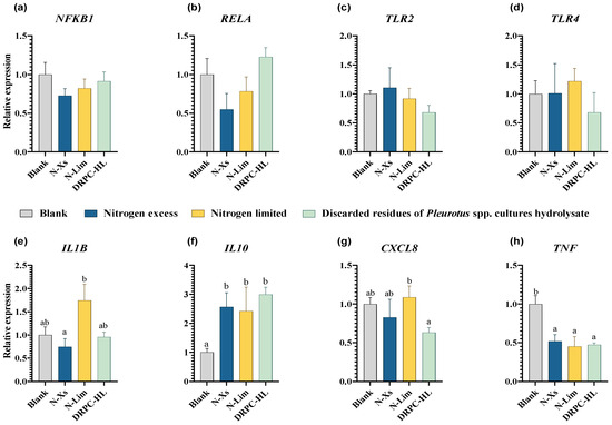
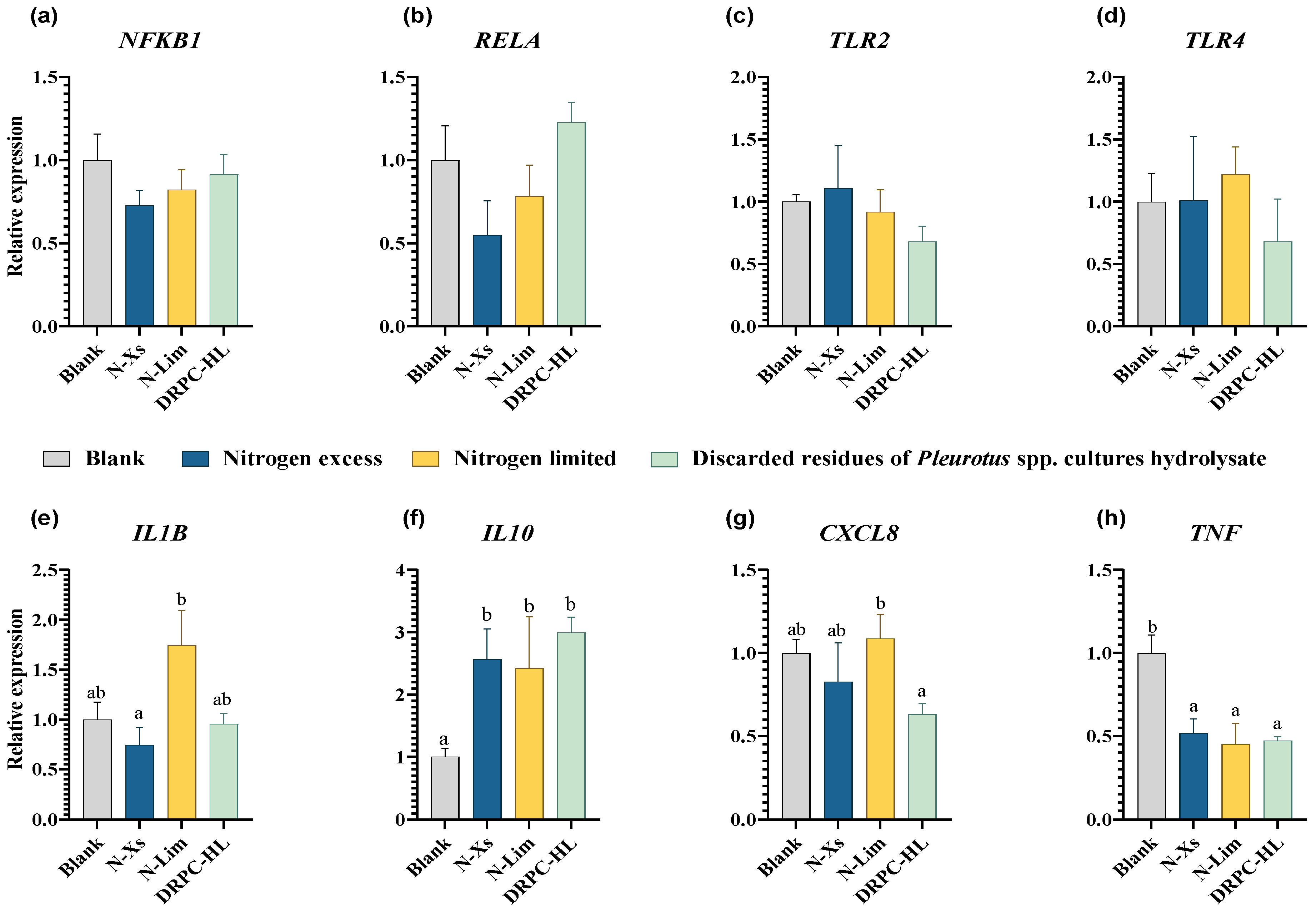
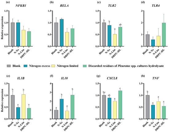
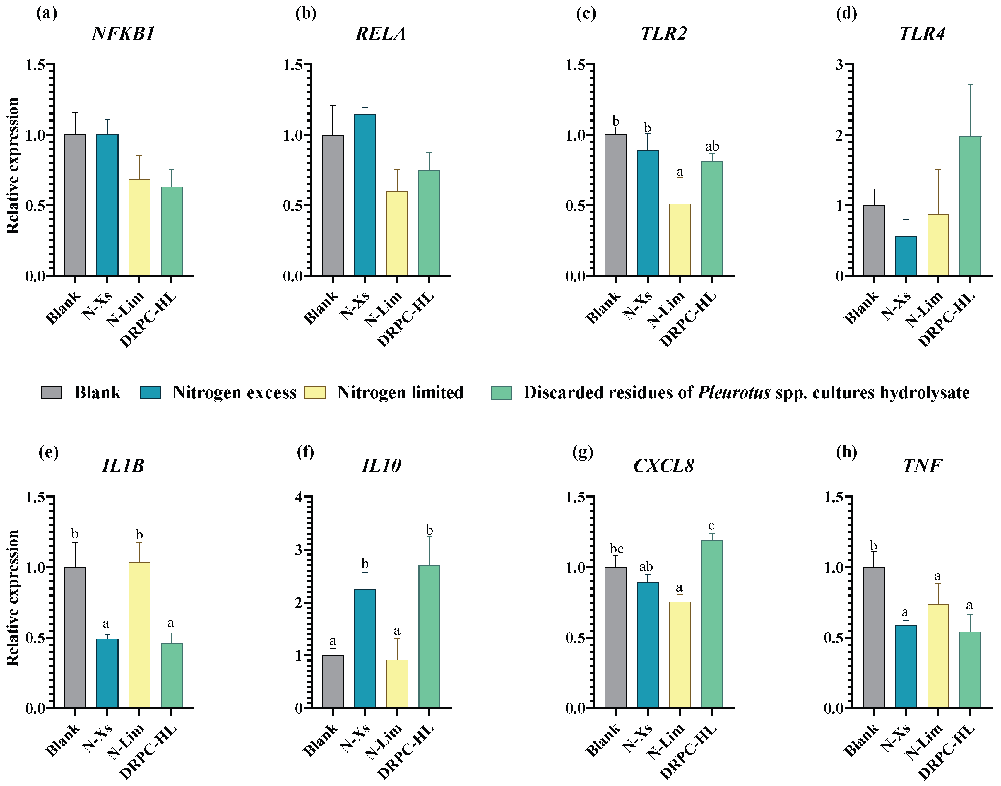
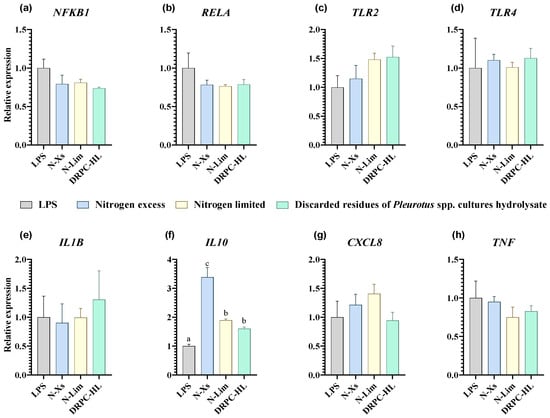
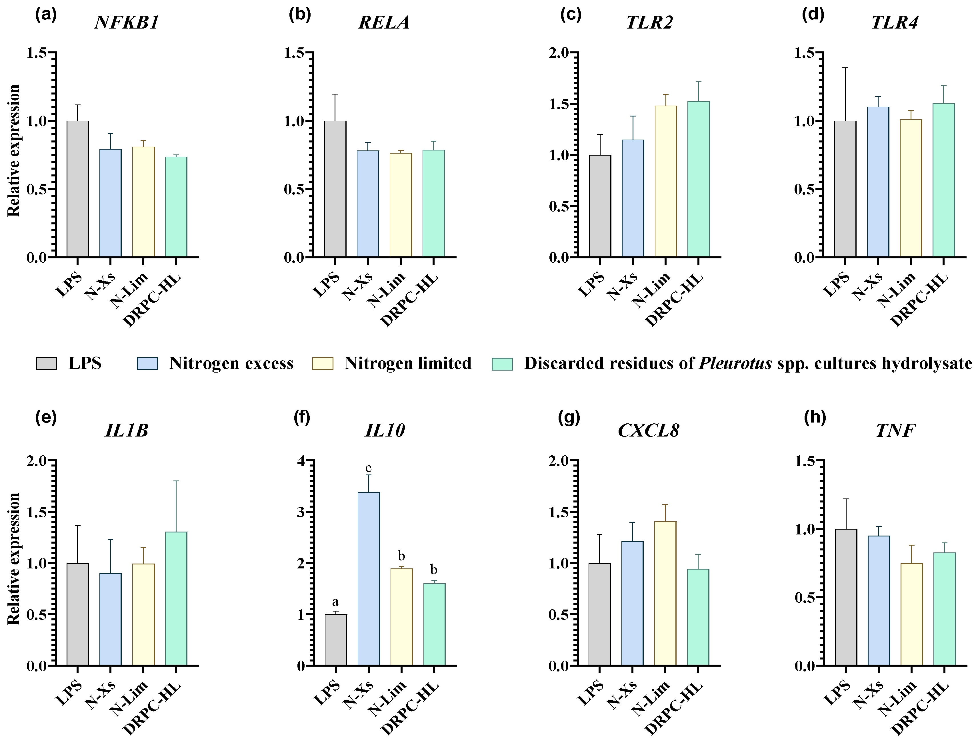

Disclaimer/Publisher’s Note: The statements, opinions and data contained in all publications are solely those of the individual author(s) and contributor(s) and not of MDPI and/or the editor(s). MDPI and/or the editor(s) disclaim responsibility for any injury to people or property resulting from any ideas, methods, instructions or products referred to in the content. |
© 2026 by the authors. Licensee MDPI, Basel, Switzerland. This article is an open access article distributed under the terms and conditions of the Creative Commons Attribution (CC BY) license.