


Abstract
Background and Clinical Significance: Acquired arteriovenous fistula (AVF) following deep vein thrombosis (DVT) is an uncommon condition, typically reported in association with persistent venous obstruction. Case Presentation: We report the case of an 18-year-old male with pulmonary embolism (PE) and extensive left iliofemoral–popliteal DVT treated with anticoagulation and compression therapy. Follow-up duplex ultrasound demonstrated progressive venous recanalization, with complete restoration of popliteal vein patency and persistent external iliac vein occlusion. During routine surveillance, in the absence of post-thrombotic symptoms, a popliteal AVF was incidentally detected. This anatomical localization is exceptionally rare, and the delayed appearance of the lesion after documented venous patency makes an early recanalization-related mechanism less likely. Although the absence of overt post-thrombotic syndrome limits definitive conclusions, a delayed hemodynamic alteration secondary to persistent proximal venous outflow obstruction was hypothesized as a possible contributing factor. Given the asymptomatic presentation and recent detection, conservative management comprising anticoagulation, elastic compression, venoactive therapy, and close ultrasound follow-up was adopted. Conclusions: This case highlights that post-thrombotic AVFs may manifest as late incidental findings and supports a tailored management approach based on clinical and hemodynamic features.
1. Introduction and Clinical Significance
Deep vein thrombosis (DVT) of the lower extremities is a common vascular disorder that may be followed by post-thrombotic syndrome (PTS) and chronic venous insufficiency (CVI). After the acute phase, the thrombus does not remain static but undergoes a dynamic remodeling process characterized by organization, recanalization, and neovascularization. This process is driven by inflammation and involves monocytes, neutrophils, vascular endothelial growth factor (VEGF), angiogenic chemokines, and matrix metalloproteinases, leading to the formation of new vascular channels within the thrombus and partial restoration of venous flow [1,2,3].
Experimental and clinical studies suggest that venous thrombus recanalization is not simply the result of clot retraction or lysis but rather is closely related to angiogenesis and neovascularization. Wakefield et al. proposed that neovascularization plays a major role in thrombus organization, while Waltham et al. further demonstrated that VEGF enhances venous thrombus recanalization and organization [1,3]. In addition, Labropoulos et al. reported the presence of small intrathrombotic vascular channels with flow patterns consistent with AVF within acutely thrombosed veins, suggesting that inflammation-mediated neovascularization may contribute to the development of intra-thrombotic arteriovenous communications [2]. A second possible pathophysiological mechanism may involve chronic venous hypertension secondary to persistent proximal venous outflow obstruction. In particular, residual iliac vein occlusion or severe stenosis after DVT may maintain elevated venous pressure and favor the development or persistence of acquired AVFs.
In a series of patients with acquired AVF after lower-extremity DVT, Yuan et al. identified iliac venous obstruction in all cases, suggesting that chronic venous hypertension may promote recruitment or retrograde opening of physiological arteriovenous shunts or sustain newly formed micro-arteriovenous connections [4]. Experimental studies on venous hypertension further support the concept that elevated venous pressure alone may induce acquired arteriovenous communications [5,6]. Although rare, acquired AVFs associated with DVT may be underdiagnosed because their clinical presentation frequently overlaps with that of PTS, including swelling, pain, skin changes, and venous ulceration [2,4]. Therefore, in patients with persistent or disproportionate symptoms following DVT, an acquired AVF should be considered in the differential diagnosis, particularly when Doppler ultrasound (DUS) demonstrates arterialized venous flow or focal high-velocity turbulent signals.
Herein, we report a case of popliteal AVF diagnosed after DVT and discuss the potential underlying mechanisms, with a focus on thrombus recanalization, neovascularization, and the role of chronic venous hypertension. Awareness of these mechanisms may improve diagnostic suspicion and help avoid attributing persistent symptoms exclusively to conventional PTS.
2. Case Presentation
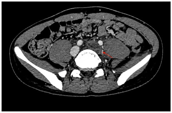
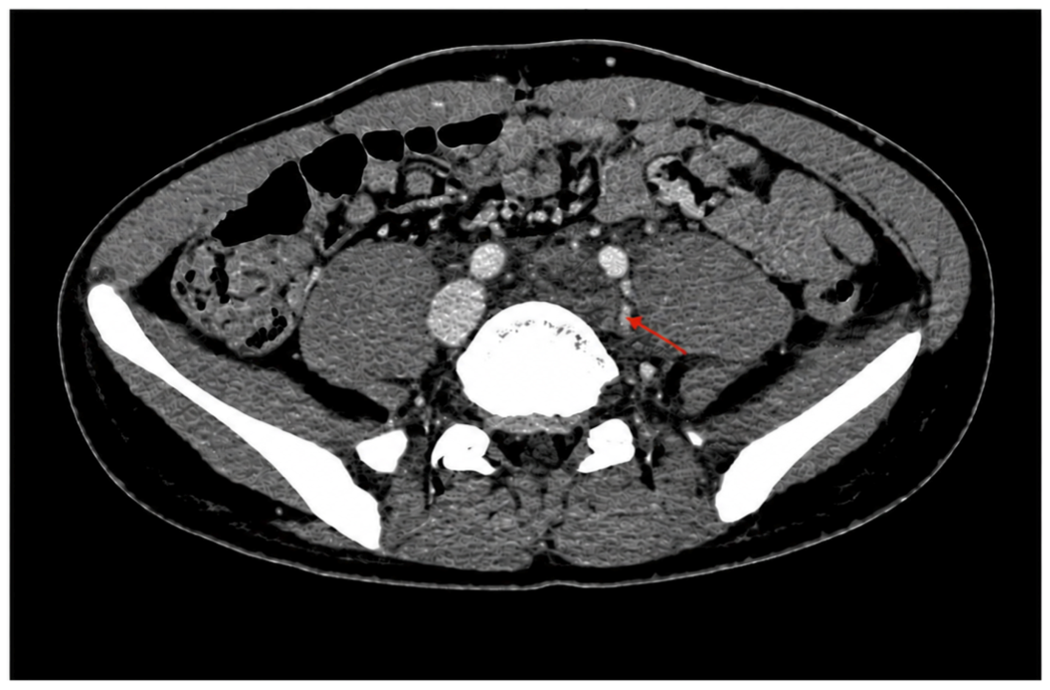
In December 2023, an 18-year-old male, with no history of congenital vascular malformations, trauma, or invasive vascular procedures, presented to the emergency department with a 10-day history of respiratory symptoms suggestive of an acute respiratory tract infection, accompanied by dyspnea and cough. Notably, this prolonged acute respiratory infection had led to a recent period of reduced mobility prior to admission. Admission laboratory investigations revealed a marked increase in inflammatory markers (C-reactive protein 148 mg/L, reference range <5 mg/L) and, notably, a significantly elevated D-dimer level (8432 µg/L, reference range 0–500 µg/L). Given the respiratory manifestations and marked edema of the left lower limb, computed tomography angiography (CTA) was performed, demonstrating complete thrombosis of the left iliofemoral veins extending into the proximal inferior vena cava (Figure 1), together with a pulmonary embolism (PE) involving a lobar branch of the right lower lobe. Baseline CTA and vascular evaluations did not reveal extrinsic venous compression syndromes, such as May–Thurner syndrome. Subsequent color DUS also demonstrated complete occlusion of the left popliteal vein. Anticoagulant therapy was immediately initiated with fondaparinux (7.5 mg once daily subcutaneous injection). Following clinical stabilization, the patient was transitioned to rivaroxaban according to standard therapeutic protocols and discharged with a prescription for a class II thigh-length elastic compression stocking.
Figure 1.
Venous-phase CTA showing thrombosis of the common iliac vein.
2.1. The Three-Month Follow
At evaluation at our outpatient clinic three months after the acute event, the patient was in excellent general condition and completely free of signs or symptoms suggestive of PTS. Follow-up DUS demonstrated a favorable yet complex pattern of evolution: partial recanalization of the left popliteal vein, with a small residual thrombus measuring 3.0 mm in maximum thickness. Of note, color Doppler ultrasonography evaluation revealed no evidence of an AVF. Partial recanalization was also observed in the left common femoral vein, with persistent patency and continuous flow at the saphenofemoral junction (Figure 2). The external iliac vein remained occluded, whereas complete recanalization of the left common iliac vein was maintained through internal iliac collateral pathways and associated suprapubic venous collaterals. During thrombophilia screening, the patient demonstrated markedly reduced antithrombin (AT) III activity (52%). Given the possibility of hereditary AT III deficiency, repeat testing was performed during follow-up, after the acute phase, and confirmed persistently reduced AT III activity. Furthermore, cascade screening was extended to first-degree relatives, revealing AT III deficiency in the patient’s father and thus further supporting a hereditary pattern of the defect. Considering these findings, the therapeutic plan was adjusted. Long-term therapeutic-dose anticoagulation with rivaroxaban (20 mg once daily) was maintained. The class II thigh-length compression stocking was replaced with a class II knee-high compression stocking, and a micronized purified flavonoid fraction (MPFF, 500 mg twice daily) was added to support venous circulation, given its pleiotropic effects, including the reduction of venous remodeling, inhibition of inflammatory cascades, and improvement in venous tone and stasis [7].
Figure 2.
Poor recanalization was also observed in the common femoral vein, while continuous flow was noted at the saphenofemoral junction.
2.2. Long-Term Evolution and Last Follow-Up
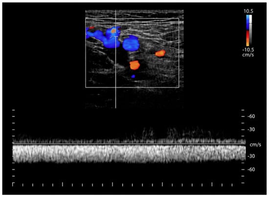
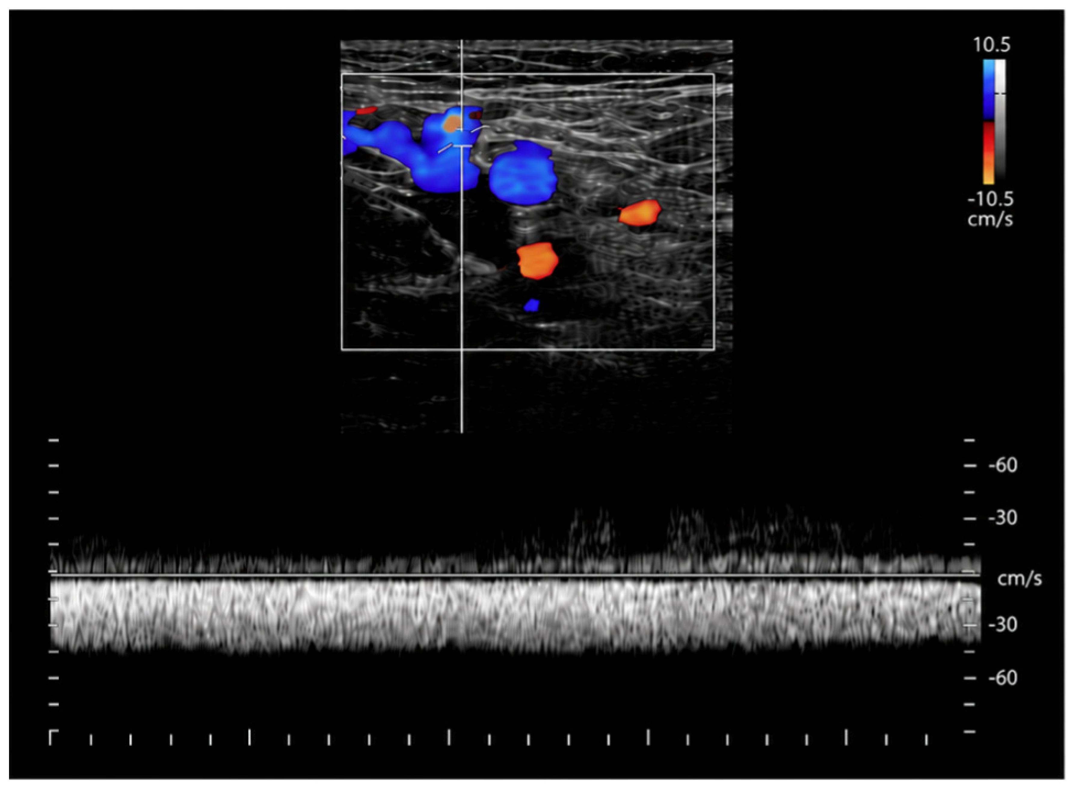
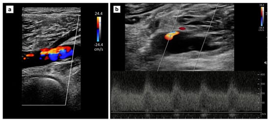
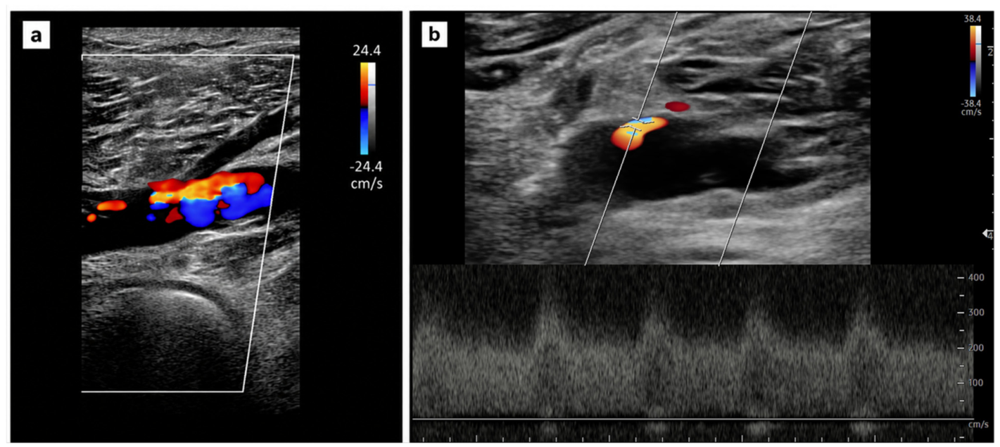
During subsequent scheduled six-monthly follow-up visits, the patient demonstrated excellent adherence to both pharmacological and compression therapies, which remained unchanged over time. Clinically, no signs or symptoms of PTS were recorded (Villalta score = 0). Periodic DUS examinations documented progressive and encouraging recanalization of the deep venous system: complete restoration of the lumen was achieved in the left popliteal vein, alongside partial recanalization of the femoral and common femoral veins, whereas occlusion of the external iliac vein persisted. During the routine evaluation performed in July 2026, approximately 31 months after the onset of DVT, the patient remained in excellent clinical status and remained free of post-thrombotic complications. However, the follow-up DUS revealed the new onset of a popliteal AVF (Figure 3, Video S1 in Supplementary Material), with no evidence of a direct communication with the main popliteal artery trunk. The AVF was considered likely to originate from adventitial neovessels, as the main popliteal artery consistently demonstrated normal high-resistance, triphasic arterial waveforms, without a reduction in the resistive index. The patient also underwent a cardiological evaluation with echocardiography, which revealed no signs of cardiac overload or alterations in the pulmonary-to-systemic flow ratio (Qp/Qs). Given the absence of cardiac alterations and symptoms suggestive of venous hypertension, significant deep venous reflux on color DUS, or limb edema, a conservative management strategy consisting of close DUS surveillance without further diagnostic procedures was adopted.
Figure 3.
(a) Color duplex ultrasound image of the arteriovenous fistula at the popliteal venous level. (b) Spectral Doppler tracing demonstrating flow within the arteriovenous fistula at the popliteal venous level.
3. Discussion
The present case highlights a rare yet clinically relevant vascular entity: the development of an acquired popliteal AVF following DVT. Although a direct causal relationship cannot be established with certainty, the temporal sequence between the acute DVT and the subsequent AVF detection suggests that post-thrombotic vascular remodeling contributed to its pathogenesis. One plausible hypothesis centers on the biological evolution of the thrombus itself. Venous thrombus organization is an active, inflammation-driven process characterized by leukocyte recruitment, angiogenic mediator release, and the formation of new vascular channels. Experimental evidence demonstrates that recanalization is closely linked to active neovascularization rather than simple fibrinolysis or thrombus recanalization [1,3]. Labropoulos et al. described intrathrombotic microvascular channels supplied by perivenous arterial branches exhibiting AVF flow, supporting the concept that thrombus neovascularization may occasionally evolve into functional microarteriovenous communications [2]. This provides a plausible rationale for AVF development in patients lacking a history of penetrating trauma, vascular access, or surgical intervention. A second, complementary mechanism involves chronic venous hypertension secondary to persistent venous outflow obstruction. In patients with post-thrombotic iliac vein occlusion, sustained elevation of venous pressure may promote the recruitment and enlargement of pre-existing physiologic arteriovenous shunts or sustain newly formed micro-fistulous channels. Indeed, Yuan et al. observed that all patients in their series of acquired AVF after DVT exhibited severe iliac venous obstruction, suggesting that venous hypertension may have contributed substantially to clinical presentation and disease evolution [4]. These observations align with experimental models demonstrating that venous hypertension alone can induce acquired arteriovenous communications [5,6]. To explicate the pathophysiological nuances of this case, a clear distinction must be made between subclinical hemodynamic venous outflow obstruction and overt clinical venous hypertension. While persistent external iliac vein occlusion created a structural substrate for altered distal hemodynamics, our patient remained entirely asymptomatic (Villalta score = 0) without edema, venous skin changes, or marked deep reflux on DUS. Invasive venous pressure measurements were not performed. Therefore, while residual proximal outflow obstruction likely established a local hemodynamic milieu predisposing to micro-fistula persistence, it represents a subclinical contributing factor rather than a cause of symptomatic clinical venous hypertension in this specific instance. In line with this, MPFF was introduced not to treat acute clinical hypertension, but as a supportive microcirculatory agent.
From a clinical perspective, an acquired AVF following DVT may easily be overlooked because its manifestations closely mimic those of PTS. Swelling, pain, skin hyperpigmentation, and venous ulceration may all be attributed solely to chronic venous disease, thereby delaying recognition of the underlying fistulous component [2,4]. For this reason, DUS plays a pivotal role, as it can detect arterialized venous waveforms, focal high-velocity turbulent flow, and abnormal early venous filling, prompting further diagnostic evaluation with CTA or conventional angiography. In our patient, serial DUS sampling of the main popliteal artery trunk demonstrated persistently normal high-resistance, triphasic arterial waveforms without a decrease in resistive index, arguing against a direct high-flow communication involving the major arterial axis and supporting the possibility of a low-flow communication originating from small adventitial or perivenous neovessels.
Importantly, the presence of AVF does not necessarily imply that the fistula itself is the primary driver of clinical symptoms. Available evidence suggests that, in many patients, the hemodynamic burden of persistent venous obstruction and ambulatory venous hypertension represents the dominant pathophysiology, whereas the AVF may act as a secondary or aggravating factor [4]. This distinction carries crucial therapeutic implications, as relieving proximal venous outflow obstruction may alleviate symptoms and reduce fistulous flow without requiring direct intervention on every micro-arteriovenous communication.
Currently, there is no standardized consensus regarding the optimal management of post-thrombotic AVFs, and treatment strategies must be tailored to clinical severity and hemodynamic features. For low-flow, asymptomatic, or minimally symptomatic cases, conservative management with compression therapy serves as the first-line intervention, counteracting distal venous stasis and optimizing microvascular dynamics without invasive risks. When symptomatic post-thrombotic syndrome or severe proximal outflow obstruction is present, venous outflow restoration via stenting, angioplasty, or surgical bypass is required to relieve the proximal pressure gradient and promote spontaneous fistulous regression [8]. Direct interventions, such as transcatheter arterial embolization, covered stent placement, or surgical ligation, should be reserved for high-flow or rapidly expanding lesions, as well as cases complicated by secondary aneurysms, high-output cardiac strain, or refractory ulceration.
Our case differs from most reports available in the literature [4,9] in two relevant aspects. First, the arteriovenous fistula was located in the popliteal district, an exceptionally rare site compared with the more frequently reported iliac or femoral locations. Second, an intra-thrombotic recanalization and neovascularization-driven mechanism appears less plausible in our patient, as serial ultrasound evaluations had documented complete popliteal vein patency prior to the emergence of the fistula, with no evidence of arteriovenous communications at earlier check-ups over approximately 31 months after acute DVT, and a completely normal DUS examination had been performed 6 months before the AVF was detected. These findings suggest that, in this instance, fistula formation may have been related to intrinsic vascular remodeling and a delayed hemodynamic process rather than early thrombus revascularization alone.
In managing this patient, it is essential to distinguish the treatment of the underlying thromboembolic/post-thrombotic condition from the management of the AVF itself.
In our patient, the popliteal anatomical localization was a critical determinant in choosing conservative surveillance. The popliteal space is subject to continuous mechanical joint flexion and extension; deploying covered stents or metallic coils in this segment carries significant risks of stent fracture, migration, or early thrombosis [10]. Furthermore, given the diffuse, low-flow adventitial nature of the micro-vessels, targeted embolization carried a risk of non-target peripheral ischemia. Combined with complete popliteal vein recanalization and the total absence of symptoms, active DUS monitoring represented the safest approach.
Regarding anticoagulant therapy management, the patient has a confirmed hereditary antithrombin III deficiency—classified as a high-risk thrombophilia—following an extensive unprovoked venous thromboembolic event (VTE). International guidelines strictly mandate lifelong therapeutic anticoagulation in such cases to prevent life-threatening recurrences [11]. Although dose reduction to a prophylactic regimen might be considered over time, current evidence remains limited. Consequently, full-dose anticoagulation and class II compression therapy were maintained to treat the VTE and mitigate post-thrombotic progression, while the asymptomatic AVF was managed via serial DUS surveillance.
Although persistent iliac venous obstruction was considered a possible hemodynamic substrate for AVF development in our patient, iliac vein recanalization was not undertaken. In a completely asymptomatic young patient, prophylactic stenting carries procedural risks (such as in-stent restenosis or acute stent thrombosis) that outweigh potential benefits [12], especially since lifelong anticoagulation is independently required due to AT III deficiency. Invasive endovascular options remain preserved should clinical or hemodynamic deterioration occur during follow-up. A conservative approach was therefore adopted, consisting of active imaging surveillance in combination with anticoagulant therapy, elastic compression, and venoactive treatment. In our view, this strategy was justified by the lack of clinically significant venous hypertension and allowed careful monitoring of the natural evolution of the lesion, while preserving the option of endovascular intervention in the event of clinical or hemodynamic progression. Reduced-dose anticoagulation appears safe regarding bleeding, thrombotic recurrence and PTS progression [13]; however, we preferred to maintain the full-dose anticoagulation.
From a practical perspective, this case emphasizes the clinical value of structured duplex ultrasound surveillance in patients with extensive proximal DVT and persistent iliac vein occlusion. Even in the absence of overt clinical PTS symptoms, a routine colour DUS protocol should include careful hemodynamic assessment along the recanalized venous segments to identify high-velocity turbulent flow or arterialized venous waveforms early. While prophylactic invasive revascularization of an asymptomatic iliac occlusion is not recommended solely to prevent secondary AVF formation, strict adherence to tailored compression therapy, effective anticoagulation, and serial non-invasive imaging may represent important components of long-term management to prevent hemodynamic worsening and detect late vascular complications.
Overall, this case supports the concept that acquired AVFs following DVT may arise through at least two non-mutually exclusive mechanisms: first, inflammation-driven recanalization and neovascularization within the thrombus; and second, potential hemodynamic alterations related to residual proximal venous obstruction. Awareness of these mechanisms may improve diagnostic suspicion and help avoid attributing persistent symptoms exclusively to conventional PTS.
4. Conclusions
Late-onset acquired popliteal AVF should be considered among the uncommon vascular sequelae of extensive lower-limb DVT. In our case, the delayed appearance of the fistula after documented venous patency argues against a mechanism solely related to early thrombus revascularization, supporting a possible contribution of persistent post-thrombotic hemodynamic alterations. DUS was crucial for detection, while the absence of symptoms supported a conservative management strategy. Overall, this case highlights the importance of individualized clinical and hemodynamic assessment in guiding both the pathophysiological interpretation and clinical management of post-thrombotic AVF.
5. Limitations
A limitation of the present report is that advanced imaging modalities, such as CTA, magnetic resonance angiography, or conventional invasive catheter angiography, were not performed at the time of AVF detection. Although serial duplex ultrasound provided detailed longitudinal hemodynamic evaluation, cross-sectional or catheter angiography would have offered superior anatomical definition of the micro-fistulous architecture and could have helped confirm the absence of subtle arterial feeder branches. However, given the patient’s young age, total lack of symptoms, and low-flow DUS features without high-risk hemodynamic markers, subjecting the patient to ionizing radiation and iodinated contrast media or invasive arterial puncture was deemed clinically unjustified in the absence of an immediate plan for invasive intervention.
Supplementary Materials
The following supporting information can be downloaded at https://www.mdpi.com/article/10.3390/reports9040390/s1: Video S1: Color duplex ultrasound video demonstrating the popliteal arteriovenous fistula.
Author Contributions
Conceptualization, L.C.; investigation, L.C., F.L.D.P. and M.M.; writing—original draft preparation, L.C.; writing—review and editing, M.M. and L.C.; visualization, F.L.D.P., L.C. and M.M. All authors have read and agreed to the published version of the manuscript.
Funding
This research received no external funding.
Institutional Review Board Statement
Ethics approval was not required by the Comitato Etico Catania 1 (Catania 1 Ethics Committee), as this manuscript describes a single retrospective case report arising from standard clinical practice and does not constitute formal human subjects research.
Informed Consent Statement
Written informed consent was obtained from the individual for the publication of any potentially identifiable images or data included in this article.
Data Availability Statement
The original contributions presented in the study are included in the article; further inquiries can be directed to the corresponding author.
Conflicts of Interest
The authors declare no conflicts of interest.
Abbreviations
The following abbreviations are used in this manuscript:
| AVF | arteriovenous fistula |
| DVT | deep vein thrombosis |
| PTS | post-thrombotic syndrome |
| CVI | chronic venous insufficiency |
| VEGF | vascular endothelial growth factor |
| DUS | Doppler ultrasound |
| CTA | computed tomography angiography |
| PE | pulmonary embolism |
| AT | antithrombin |
| MPFF | micronized purified flavonoid fraction |
References
- Wakefield, T.W.; Linn, M.J.; Henke, P.K.; Kadell, A.M.; Wilke, C.A.; Wrobleski, S.K.; Sarkar, M.; Burdick, M.D.; Myers, D.D.; Strieter, R.M. Neovascularization during venous thrombosis organization: A preliminary study. J. Vasc. Surg. 1999, 30, 885–892. [Google Scholar] [CrossRef] [Scilit] [PubMed]
- Labropoulos, N.; Bhatti, A.F.; Amaral, S.; Leon, L.; Borge, M.; Rodriguez, H.; Kalman, P. Neovascularization in acute venous thrombosis. J. Vasc. Surg. 2005, 42, 515–518. [Google Scholar] [CrossRef] [Scilit] [PubMed]
- Waltham, M.; Burnand, K.G.; Collins, M.; McGuinness, C.L.; Singh, I.; Smith, A. Vascular endothelial growth factor enhances venous thrombus recanalisation and organisation. Thromb. Haemost. 2003, 89, 169–176. [Google Scholar] [CrossRef] [Scilit] [PubMed]
- Yuan, H.; Sun, J.; Zhou, Z.; Qi, H.; Wang, M.; Dong, D.; Wu, X. Diagnosis and treatment of acquired arteriovenous fistula after lower extremity deep vein thrombosis. Int. Angiol. 2019, 38, 10–16. [Google Scholar] [CrossRef] [Scilit] [PubMed]
- Terada, T.; Higashida, R.T.; Halbach, V.V.; Dowd, C.F.; Tsuura, M.; Komai, N.; Wilson, C.B.; Hieshima, G.B. Development of acquired arteriovenous fistulas in rats due to venous hypertension. J. Neurosurg. 1994, 80, 884–889. [Google Scholar] [CrossRef] [Scilit] [PubMed]
- Herman, J.M.; Spetzler, R.F.; Bederson, J.B.; Kurbat, J.M.; Zabramski, J.M. Genesis of a dural arteriovenous malformation in a rat model. J. Neurosurg. 1995, 83, 539–545. [Google Scholar] [CrossRef] [Scilit] [PubMed]
- Li, K.X.; Diendéré, G.; Galanaud, J.P.; Mahjoub, N.; Kahn, S.R. Micronized purified flavonoid fraction for the treatment of chronic venous insufficiency, with a focus on postthrombotic syndrome: A narrative review. Res. Pract. Thromb. Haemost. 2021, 5, e12527. [Google Scholar] [CrossRef] [Scilit] [PubMed]
- Hoshino, Y.; Yokoi, H. Post-thrombotic syndrome of the lower extremity associated with arteriovenous fistula: Three case reports. J. Vasc. Surg. Cases Innov. Tech. 2022, 8, 629–633. [Google Scholar] [CrossRef] [Scilit] [PubMed]
- Link, D.P.; Garza, A.S.; Monsky, W. Acquired peripheral arteriovenous malformations in patients with venous thrombosis: Report of two cases. J. Vasc. Interv. Radiol. 2010, 21, 387–391. [Google Scholar] [CrossRef] [Scilit] [PubMed]
- Bloch, R.A.; Caron, E.; Pomposelli, F.B.; Prushik, S.G.; Shean, K.E.; Conrad, M.F. Outcomes of endovascular intervention for atherosclerotic lesions confined to the popliteal artery. J. Vasc. Surg. 2024, 80, 498–504. [Google Scholar] [CrossRef] [Scilit] [PubMed]
- Nicolaides, A.N.; Fareed, J.; Spyropoulos, A.C.; Kakkar, R.H.L.; Antignani, P.L.; Avgerinos, E.; Baekgaard, N.; Barber, E.; Bush, R.L.; Caprini, J.A.; et al. Prevention and management of venous thromboembolism. International Consensus Statement. Guidelines according to scientific evidence. Int. Angiol. 2024, 43, 1–222. [Google Scholar] [CrossRef] [Scilit] [PubMed]
- Morris, R.I.; Khan, T.; Black, S.A. Complications of Deep Venous Stenting and Their Management. Cardiovasc Interv. Radiol. 2024, 47, 1669–1676. [Google Scholar] [CrossRef] [Scilit] [PubMed]
- Costanzo, L.; Di Paola, F.; Pedi, A.M.; Failla, G.; Mangiafico, M. Long-Term Efficacy and Safety of Direct Oral Anticoagulants at Reduced Doses in the Secondary Prevention of Venous Thromboembolism and Post-Thrombotic Syndrome. J. Clin. Med. 2024, 13, 2394. [Google Scholar] [CrossRef] [Scilit] [PubMed] [PubMed Central]
Disclaimer/Publisher’s Note: The statements, opinions and data contained in all publications are solely those of the individual author(s) and contributor(s) and not of MDPI and/or the editor(s). MDPI and/or the editor(s) disclaim responsibility for any injury to people or property resulting from any ideas, methods, instructions or products referred to in the content. |
© 2026 by the authors. Licensee MDPI, Basel, Switzerland. This article is an open access article distributed under the terms and conditions of the Creative Commons Attribution (CC BY) license.