


Abstract
Background and Clinical Significance: Extensive rectal ischemia is exceptionally rare due to the rectum’s robust vascular network, with segmental ischemia being more common. Case Presentation: We report the case of a 69-year-old female who presented with whole-segment rectal ischemia, encompassing the upper, mid, and lower rectum. This severe local ischemic event culminated in full-thickness perforation and extensive fecal peritonitis, which subsequently precipitated postoperative septic shock. The patient underwent emergency low anterior resection with Hartmann’s procedure and received intensive multidisciplinary postoperative care. Conclusions: In this case, we aimed to highlight the importance of early recognition, decisive surgical intervention, and the pathophysiological and diagnostic challenges in managing rare cases of whole-segment rectal ischemia.
1. Introduction and Clinical Significance
Rectal ischemia is a rare but serious condition, distinct from ischemic colitis, which commonly affects the left colon. The rectum’s extensive collateral blood supply usually protects it from ischemic injury [1,2,3]. However, systemic factors such as severe hypotension, thromboembolism, hypercoagulable states, and cardiovascular comorbidities can overwhelm this vascular network, leading to ischemic changes and, in severe cases, perforation [4,5,6,7]. In this case report, we discuss the diagnostic and management complexities of rectal ischemia in an older patient with comorbidities, including hypertension and cardiovascular disease. Unlike typical cases where systemic shock precedes bowel necrosis, this patient developed severe, spontaneous whole-segment rectal ischemia that led to full-thickness perforation and fecal peritonitis. This massive intra-abdominal infection then precipitated severe postoperative septic shock. While segmental ischemia, particularly of the upper rectum, is more commonly observed in systemic hypoperfusion scenarios, this case describes a rare instance of whole-segment rectal ischemia, challenging the conventional understanding of the rectum’s vascular resilience.
2. Case Presentation
2.1. Patient History
The patient, a 69-year-old female, had a medical history significant for hypertension managed with antihypertensive medication. Although the specific pre-admission antihypertensive agents were not distinctly itemized in the initial emergency records beyond confirming that she was under regular medication control, her hypertension was a chronic, long-standing condition. On preoperative day 1, she presented to the emergency department with severe lower abdominal pain, difficulty with defecation, and episodes of bloody stool. She reported occasional black, tarry stools but denied fever, chills, nausea, vomiting, or weight loss. She also had a history of controlled cardiovascular disease but no recent surgeries or thromboembolic events.
2.2. Initial Clinical Examination
On arrival, the patient was alert (Glasgow Coma Scale: E4M6V5) but hypertensive, with systolic blood pressure readings between 160 and 190 mmHg. Physical examination revealed mild tenderness in the lower abdomen. A digital rectal examination demonstrated blood on the glove without signs of hemorrhoids or any palpable mucosal defects or perforations. The rest of the physical examination, including neurological and cardiopulmonary assessments, was unremarkable.
2.3. Laboratory and Imaging Findings
Initial laboratory investigations showed significant leukocytosis (white blood cell count: 16.97 × 10³/μL), indicating an acute inflammatory response. Hemoglobin levels were initially within the normal range (14.1 g/dL) but subsequently declined rapidly to 7.7 g/dL on POD 3, suggesting active gastrointestinal bleeding. Platelet count decreased to 115 × 103/μL, and C-reactive protein spiked to 42.77 mg/dL by POD 3, consistent with systemic inflammation. The patient’s N-terminal pro-B-type natriuretic peptide was mildly elevated at 106.6 pg/mL, suggesting cardiac strain likely due to underlying hypertension and sepsis.
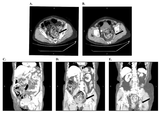
An abdominal and pelvic CT scan was performed in the emergency department prior to hospital admission on preoperative day 1. The images revealed free air surrounding the rectum (pneumoperitoneum), rectal wall thickening, and signs of ischemic changes (Figure 1). No evidence of mesenteric vessel thrombosis was observed. Given the severity of these findings, an emergency colorectal surgical consultation was requested.
Figure 1.
(A–E) Contrast-enhanced CT imaging demonstrating extraluminal gas (arrows) in the perirectal space, indicating full-thickness ischemic rectal perforation.
2.4. Surgical Intervention
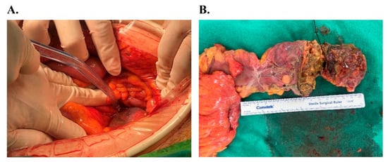
On POD 0, the patient underwent an exploratory laparotomy. Intraoperative findings revealed severe ischemic necrosis of the rectum with full-thickness perforation and extensive fecal peritonitis. The surgical team performed a low anterior resection (LAR) with Hartmann’s procedure, excising three segments of the rectum measuring 9.0 cm, 9.5 cm, and 3.0 cm. Gross examination of the resected specimen showed dark, edematous mucosa with ischemic changes extending to the serosal surface (Figure 2).
Figure 2.
Intraoperative and macroscopic findings: (A) Low anterior resection (LAR) with Hartmann’s procedure. (B) Resected rectal segments displaying dark, edematous mucosa and transmural ischemic necrosis.
2.5. Pathological Findings
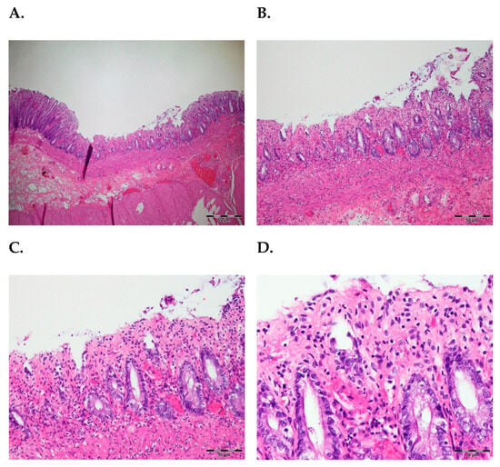
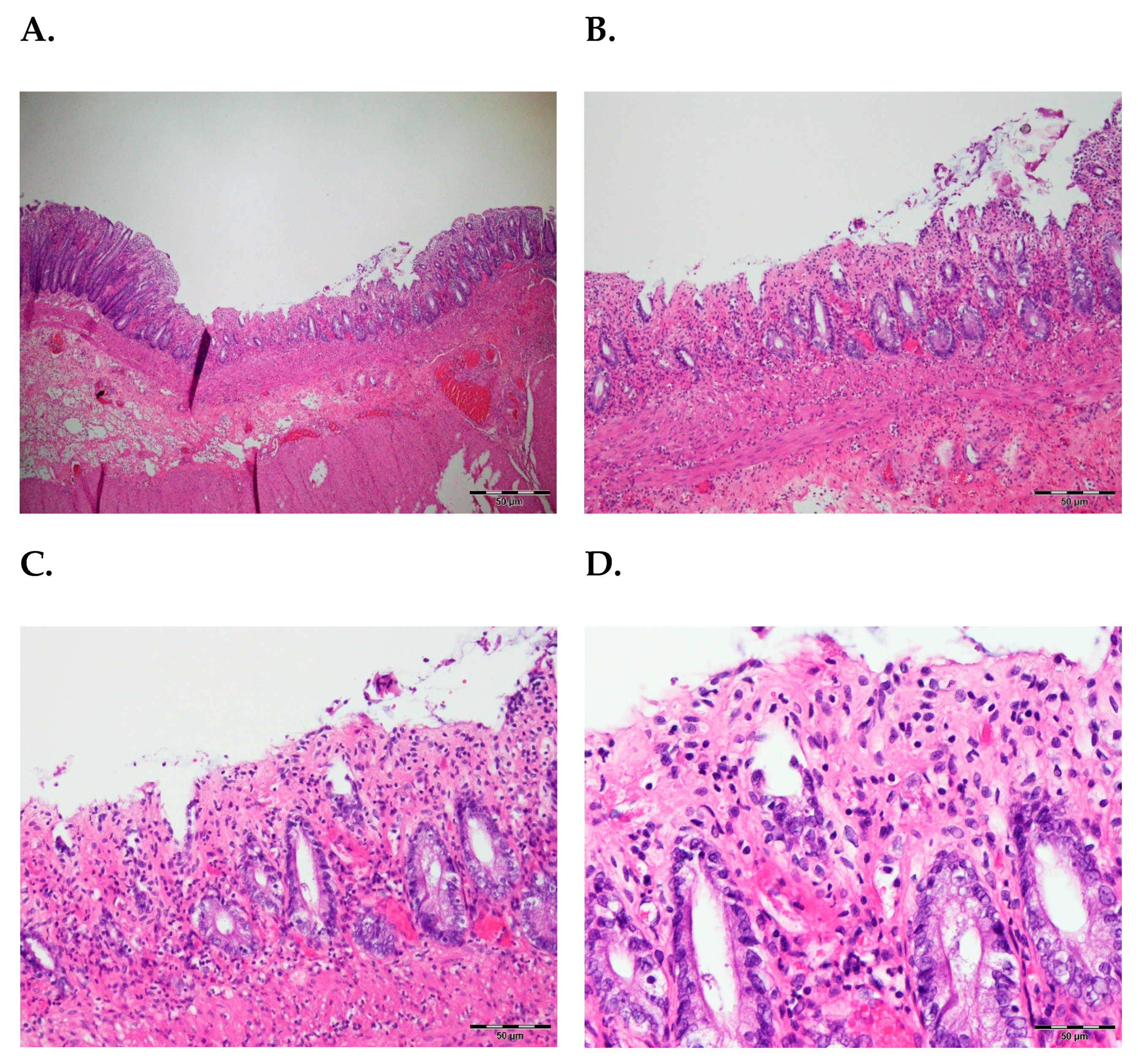
Histopathological analysis confirmed severe rectal ischemia characterized by mucosal sloughing, congestion, and extensive edema. Acute inflammation was evident, with infiltration of neutrophils extending to the serosal layer, consistent with peritonitis. No thrombus was identified in the mesenteric arteries, suggesting a non-occlusive ischemic process likely secondary to chronic microvascular compromise from her underlying hypertensive cardiovascular disease (Figure 3).
Figure 3.
Histopathology of non-occlusive ischemia: (A,B) Rectal mucosa exhibiting sloughing, congestion, edema, and acute inflammation. (C,D) Intact arterial walls without thrombus.
2.6. Postoperative Course
The patient was admitted to the intensive care unit (ICU) due to septic shock following surgery. She received aggressive intravenous fluid resuscitation and albumin supplementation. Because she remained hypertensive (systolic blood pressure of 160–190 mmHg) despite her critical condition, a Nicardipine pump was utilized to actively and strictly control her blood pressure while maintaining adequate perfusion. Extubation was attempted on POD 1, followed by respiratory support with a Venturi mask, which was gradually weaned to a nasal cannula by POD 6.
Intraoperative hematuria was noted, prompting urological consultation. The hematuria was suspected to be related to sepsis-induced coagulopathy. A three-way Foley catheter with saline irrigation was placed, and the hematuria resolved by POD 3. Dietary progression was cautious, starting with nothing by mouth status and nasogastric decompression, transitioning to a clear liquid diet on POD 3, and advancing to a full liquid diet by POD 7. During the postoperative period, the patient developed a skin rash with bullae formation over her right armpit; a dermatology consultation diagnosed a herpes zoster infection, which was treated with topical medications and Medovir.
On POD 14, the Jackson–Pratt (J-P) drain output became brownish, suggesting a possible intra-abdominal infection. While the ascitic fluid culture obtained on POD 15 showed no bacterial growth, an earlier anaerobic wound culture obtained on POD 8 grew Peptoniphilus asaccharolyticus and unidentified anaerobic Gram-negative bacilli, confirming the presence of infection. The laparotomy wound exhibited signs of dehiscence with sloughy discharge and a foul odor, requiring debridement and a rectus local flap closure on POD 15. Following this procedure, the patient maintained acceptable hemodynamics, good wound healing, and well-tolerated oral intake. She was subsequently discharged in a stable condition with planned outpatient department (OPD) follow-up. Because this is a recent case, long-term follow-up regarding stoma reversal, functional recovery, and overall quality of life is not yet available.
3. Discussion
3.1. Unique Insights from This Case
In this case, we present a rare occurrence of whole-segment rectal ischemia, involving the upper, mid, and lower rectum, which is typically protected by an extensive vascular supply. Rectal ischemia is usually segmental; therefore, the rarity of lower rectal ischemia makes this case particularly notable [8,9]. The widespread ischemia observed in our patient initially developed prior to any hemodynamic instability, likely driven by her underlying hypertensive cardiovascular disease rather than an initial state of shock. This extensive ischemic injury overwhelmed the vascular defenses of the lower rectum, leading to full-thickness perforation and severe intra-abdominal fecal peritonitis, which subsequently precipitated the postoperative septic shock.
Pathophysiologically, rectal ischemia can be occlusive or non-occlusive. In this case, the absence of a thrombus points to non-occlusive mesenteric ischemia (NOMI) [5,6,10]. While NOMI is most frequently caused by systemic hypoperfusion or shock in critically ill patients, our patient presented to the emergency department alert and severely hypertensive. This chronic microvascular compromise likely overwhelmed the vascular defenses of the lower rectum, leading to spontaneous whole-segment ischemia and subsequent full-thickness perforation. The profound systemic hypotension and septic shock observed in this patient developed only after the extensive intra-abdominal fecal peritonitis occurred, acting as a secondary catastrophic complication rather than the primary cause of the ischemia [11].
3.2. Diagnostic Challenges and Advances
The diagnosis of rectal ischemia is challenging, as its clinical presentation often mimics other gastrointestinal disorders, such as diverticulitis or ulcerative colitis [12]. The diagnosis of rectal ischemia relies on a triad of radiologic, intraoperative, and histologic criteria. Radiologically, contrast-enhanced CT demonstrates rectal wall thickening, pneumoperitoneum, and decreased mucosal enhancement [13,14]. In this case, embolic disease was definitively ruled out because histopathological analysis of the resected specimen confirmed the absence of any thrombus in the mesenteric arteries. Radiation proctitis was excluded, as the patient had no prior history of pelvic radiation therapy. Additionally, stercoral perforation was considered but dismissed, as there was no evidence of severe fecal impaction or stercoral ulcers noted during the initial digital rectal examination or during the intraoperative exploratory laparotomy. Finally, vasculitis was deemed highly unlikely given the patient’s lack of prior autoimmune history and the complete absence of systemic rheumatologic symptoms, such as arthralgia or joint swelling, throughout her clinical course. CT findings were crucial in identifying the extent of ischemic injury and guiding the decision for emergency surgery [13,14]. Intraoperatively, findings include full-thickness ischemic necrosis and edematous, dark mucosa extending to the serosa. Histologically, it is confirmed by mucosal sloughing, acute inflammatory infiltration, and the absence of arterial thrombi, which distinguishes it from occlusive disease [15].
Recent advances, including endoscopic Doppler ultrasound and perfusion CT, have shown promise in assessing mesenteric blood flow in real-time. These techniques may facilitate earlier diagnosis and improve outcomes by enabling timely intervention. Despite their potential, these technologies are not yet widely available and require further clinical validation [16].
3.3. Multidisciplinary Management Approach
Effective management of severe rectal ischemia requires a multidisciplinary approach, particularly in cases complicated by perforation and peritonitis [17]. In our patient, the decision to perform an LAR with Hartmann’s procedure was driven by the need to excise necrotic tissue and control peritoneal contamination. This surgical approach is supported by recent systematic reviews, which have shown improved survival rates when early resection is performed before the onset of multi-organ failure [18].
Postoperative care is complex, involving close collaboration among critical care, surgical, urological, and dermatological teams. In our case, complications such as septic shock, coagulopathy, and wound dehiscence required a coordinated effort [19]. The occurrence of herpes zoster in the postoperative period may indicate an immunocompromised state, highlighting the need for vigilant monitoring and prompt treatment of secondary infections in critically ill patients [20].
3.4. Emerging Therapeutic Strategies
Emerging therapeutic strategies for rectal ischemia focus on optimizing systemic perfusion and addressing coagulopathic states. Aggressive fluid resuscitation, vasopressor support, and albumin supplementation are critical in restoring hemodynamic stability and enhancing mesenteric blood flow [16,21]. In our patient, Nicardipine effectively controlled blood pressure while maintaining adequate perfusion, aligning with guidelines recommending hemodynamic optimization to prevent further ischemic damage [22].
3.5. Prognostic Considerations
The prognosis of rectal ischemia is significantly influenced by the speed of diagnosis and intervention. Delayed recognition can lead to irreversible tissue damage, perforation, and multi-organ failure. Early surgical resection, as in this case, improves survival outcomes by removing necrotic tissue and preventing further sepsis [23,24]. However, postoperative complication risks remain high, particularly in patients with extensive ischemic injury and underlying cardiovascular diseases [24,25]. This case highlights the importance of early recognition, timely surgical intervention, and a coordinated, multidisciplinary approach to managing severe rectal ischemia, particularly in older patients with cardiovascular risk factors. However, as a single retrospective case report, this study is limited by its inherent lack of generalizability, absence of long-term follow-up, and lack of advanced vascular imaging.
4. Conclusions
In summary, this case highlights the rare presentation of whole-segment rectal ischemia. The involvement of all rectal segments underscores the profound impact that chronic microvascular compromise and hypertensive cardiovascular disease can have in overwhelming the rectum’s robust vascular resilience. Early diagnosis via contrast-enhanced CT and prompt surgical intervention with a low anterior resection and Hartmann’s procedure were critical in managing this life-threatening perforation. Following a complex postoperative course complicated by septic shock, sepsis-induced coagulopathy, wound dehiscence, and secondary infections, the patient was successfully stabilized and discharged, highlighting the critical importance of a coordinated, multidisciplinary approach in optimizing outcomes. Further research is needed to better understand the pathophysiological thresholds that disrupt the rectum’s vascular defenses and to explore innovative strategies for managing severe rectal ischemia.
Author Contributions
Conceptualization: J.-R.J. and Y.-C.C.; Data curation: J.-R.J.; Formal analysis: J.-R.J.; Funding acquisition: Y.-C.C.; Investigation: T.-W.P.; Methodology: J.-C.K. and Y.-C.C.; Project administration: J.-C.K. and Y.-C.C.; Resources: T.-W.P. and R.-Y.S.; Supervision: J.-C.K. and Y.-C.C.; Validation: R.-Y.S. and T.-W.P.; Visualization: J.-R.J.; Writing—original draft: J.-R.J.; Writing—review and editing: All authors. All authors have read and agreed to the published version of the manuscript.
Funding
This research received no external funding.
Institutional Review Board Statement
Ethical approval was waived for this retrospective single case report by the Institutional Review Board of Songshan Branch, Tri-Service General Hospital, National Defense Medical Center, in accordance with institutional policy and national regulations. The study was conducted in compliance with the Declaration of Helsinki.
Informed Consent Statement
Written informed consent for publication was obtained from the patient.
Data Availability Statement
The data presented in this study are available upon request from the corresponding author. The data are not publicly available due to privacy or ethical restrictions.
Conflicts of Interest
The authors declare no conflicts of interest.
Abbreviations
The following abbreviations are used in this manuscript:
| CT | computed tomography |
| LAR | low anterior resection |
| ICU | intensive care unit |
| POD | postoperative day |
| J-P | Jackson–Pratt |
References
- Sharif, S.; Hyser, M. Ischemic proctitis: Case series and literature review. Am. Surg. 2006, 72, 1241–1247. [Google Scholar] [CrossRef] [Scilit]
- Maimone, A.; De Ceglie, A.; Siersema, P.D.; Baron, T.H.; Conio, M. Colon ischemia: A comprehensive review. Clin. Res. Hepatol. Gastroenterol. 2021, 45, 101592. [Google Scholar] [CrossRef] [Scilit] [PubMed]
- Azimuddin, K.; Raphaeli, T. Acute ischemic gangrene of the rectum: Report of 3 cases and review of literature. Int. J. Surg. Case Rep. 2013, 4, 1120–1123. [Google Scholar] [CrossRef] [Scilit] [PubMed]
- Iacobellis, F.; Narese, D.; Berritto, D.; Brillantino, A.; Di Serafino, M.; Guerrini, S.; Grassi, R.; Scaglione, M.; Mazzei, M.A.; Romano, L. Large bowel ischemia/infarction: How to recognize it and make differential diagnosis? A Review. Diagnostics 2021, 11, 998. [Google Scholar] [CrossRef] [Scilit] [PubMed]
- Ebrahimi, M.; Arabi, A.; Dabiri, S.; Razavinasab, S.A.; Pasandi, A.P.; Zeidabadi, A. A case report of transmural rectosigmoid ischemia in an elderly patient. Int. J. Surg. Case Rep. 2023, 107, 108372. [Google Scholar] [CrossRef] [Scilit]
- Rozner, R.; Gisriel, S.; Damianos, J.; Grimshaw, A.A.; Rizwan, R.; Nawaz, A.; Chan, K.; Wan, D.; Pantel, H.; Bhutta, A.Q.; et al. Idiopathic myointimal hyperplasia of the mesenteric veins: A systematic review and individual patient data regression analysis. J. Gastroenterol. Hepatol. 2023, 38, 1040–1046. [Google Scholar] [CrossRef] [Scilit]
- Kennedy, G.D.; Heise, C.P. Radiation colitis and proctitis. Clin. Colon Rectal Surg. 2007, 20, 64–72. [Google Scholar] [CrossRef] [Scilit]
- Kishikawa, H.; Nishida, J.; Hirano, E.; Nakano, M.; Arakawa, K.; Morishita, T.; Kawashima, J.; Koide, O.; Tanaka, Y.; Ishii, H. Chronic ischemic proctitis: Case report and review. Gastrointest. Endosc. 2004, 60, 304–308. [Google Scholar] [CrossRef] [Scilit]
- Perry, R.J.; Martin, M.J.; Eckert, M.J.; Sohn, V.Y.; Steele, S.R. Colonic ischemia complicating open vs endovascular abdominal aortic aneurysm repair. J. Vasc. Surg. 2008, 48, 272–277. [Google Scholar] [CrossRef] [Scilit]
- Chen, Z.; Liu, X.; Shou, C.; Yang, W.; Yu, J. Advances in the diagnosis of non-occlusive mesenteric ischemia and challenges in intra-abdominal sepsis patients: A narrative review. PeerJ 2023, 11, e15307. [Google Scholar] [CrossRef] [Scilit]
- Jarczak, D.; Kluge, S.; Nierhaus, A. Sepsis—Pathophysiology and therapeutic concepts. Front. Med. 2021, 8, 628302. [Google Scholar] [CrossRef] [Scilit] [PubMed]
- Kucejko, R.J.; Poggio, J.L. Considerations and changes in the evaluation, management, and outcomes in the management of diverticular disease: The diagnosis, pathology, and treatment of diverticular colitis. Clin. Colon Rectal Surg. 2018, 31, 221–225. [Google Scholar] [CrossRef] [Scilit] [PubMed]
- Nakamura, Y.; Kondo, S.; Narita, K.; Maeda, S.; Fonseca, D.; Honda, Y.; Tani, C.; Fukumoto, W.; Mitani, H.; Ishibashi, M.; et al. Understanding CT imaging findings based on the underlying pathophysiology in patients with small bowel ischemia. Jpn. J. Radiol. 2023, 41, 353–366. [Google Scholar] [CrossRef] [Scilit] [PubMed]
- Olson, M.C.; Bach, C.R.; Wells, M.L.; Andrews, J.C.; Khandelwal, A.; Welle, C.L.; Fidler, J.L. Imaging of bowel ischemia: An update, from the AJR special series on emergency radiology. AJR Am. J. Roentgenol. 2023, 220, 173–185. [Google Scholar] [CrossRef] [Scilit]
- Theodoropoulou, A.; Koutroubakis, I.E. Ischemic colitis: Clinical practice in diagnosis and treatment. World J. Gastroenterol. 2008, 14, 7302–7308. [Google Scholar] [CrossRef] [Scilit]
- Bala, M.; Catena, F.; Kashuk, J.; De Simone, B.; Gomes, C.A.; Weber, D.; Sartelli, M.; Coccolini, F.; Kluger, Y.; Abu-Zidan, F.M.; et al. Acute mesenteric ischemia: Updated guidelines of the World Society of Emergency Surgery. World J. Emerg. Surg. 2022, 17, 54. [Google Scholar] [CrossRef] [Scilit]
- Tarasconi, A.; Perrone, G.; Davies, J.; Coimbra, R.; Moore, E.; Azzaroli, F.; Abongwa, H.; De Simone, B.; Gallo, G.; Rossi, G.; et al. Anorectal emergencies: WSES-AAST guidelines. World J. Emerg. Surg. 2021, 16, 48. [Google Scholar] [CrossRef] [Scilit]
- Qian, W.; Bruinsma, J.; Mac Curtain, B.; Ibraheem, M.; Temperley, H.C.; Ng, Z.Q. Surgical prevalence and outcomes in ischemic colitis: A systematic review and meta-analysis. World J. Surg. 2024, 48, 943–953. [Google Scholar] [CrossRef] [Scilit]
- Iba, T.; Helms, J.; Levy, J.H. Sepsis-induced coagulopathy (SIC) in the management of sepsis. Ann. Intensive Care 2024, 14, 148. [Google Scholar] [CrossRef] [Scilit]
- Calabrese, C.; Kirchner, E.; Fernandez, J.; Calabrese, L.H. Preventing herpes zoster in immunocompromised patients: Current concepts. Clevel. Clin. J. Med. 2024, 91, 437–445. [Google Scholar] [CrossRef] [Scilit]
- Arabi, Y.M.; Belley-Cote, E.; Carsetti, A.; De Backer, D.; Donadello, K.; Juffermans, N.P.; Hammond, N.; Laake, J.H.; Liu, D.; Maitland, K.; et al. European Society of Intensive Care Medicine clinical practice guideline on fluid therapy in adult critically ill patients. Part 1: The choice of resuscitation fluids. Intensive Care Med. 2024, 50, 813–831. [Google Scholar] [CrossRef] [Scilit]
- Chi, N.F.; Chung, C.P.; Cheng, H.M.; Liu, C.H.; Lin, C.J.; Hsu, L.C.; Tang, S.C.; Lee, J.T.; Po, H.L.; Jeng, J.S.; et al. 2021 Taiwan Stroke Society Guidelines of blood pressure control for ischemic stroke prevention. J. Chin. Med. Assoc. 2022, 85, 651–664. [Google Scholar] [CrossRef] [Scilit]
- Genstorfer, J.; Schäfer, J.; Kettelhack, C.; Oertli, D.; Rosenthal, R. Surgery for ischemic colitis: Outcome and risk factors for in-hospital mortality. Int. J. Color. Dis. 2014, 29, 493–503. [Google Scholar] [CrossRef] [Scilit]
- Beghdadi, N.; Reitano, E.; Cochennec, F.; Desgranges, P.; Amiot, A.; Sobhani, I.; Mongardon, N.; Langeron, O.; Notarnicola, M.; Mulé, S.; et al. Predictors of mortality following emergency open colectomy for ischemic colitis: A single-center experience. World J. Emerg. Surg. 2020, 15, 40. [Google Scholar] [CrossRef] [Scilit]
- Lorusso, R.; Mariscalco, G.; Vizzardi, E.; Bonadei, I.; Renzulli, A.; Gelsomino, S. Acute bowel ischemia after heart operations. Ann. Thorac. Surg. 2014, 97, 2219–2227. [Google Scholar] [CrossRef] [Scilit]
Disclaimer/Publisher’s Note: The statements, opinions and data contained in all publications are solely those of the individual author(s) and contributor(s) and not of MDPI and/or the editor(s). MDPI and/or the editor(s) disclaim responsibility for any injury to people or property resulting from any ideas, methods, instructions or products referred to in the content. |
© 2026 by the authors. Licensee MDPI, Basel, Switzerland. This article is an open access article distributed under the terms and conditions of the Creative Commons Attribution (CC BY) license.