


Abstract
Brain aging is characterized by heterogeneous microglial states and altered mitochondrial, metabolic, and lysosomal homeostasis. Mitophagy is central to mitochondrial quality control, but its contribution to age-associated microglial dysfunction remains incompletely understood. This review examines how microglial mitophagy interacts with mitochondrial and lysosomal quality control, immunometabolic adaptation, and inflammatory signaling during brain aging, while distinguishing direct microglial evidence from findings derived from other experimental systems. Aging remodels microglial mitochondrial, metabolic, and lysosomal functions in a state- and context-dependent manner, including reactive, disease-associated, lipid-associated, and senescence-like states. Mitophagy may preserve mitochondrial quality control by limiting the accumulation of damaged mitochondria and associated oxidative and danger signals. However, static mitophagy-associated markers do not establish completed mitophagic flux, which requires mitochondrial delivery to lysosomes and subsequent degradation within them. Human postmortem studies reveal age- and disease-associated alterations in mitochondrial quality control and mitophagy-related pathways, but direct measurements of mitophagic flux in defined human microglial populations remain limited. Age-related changes in lysosomal capacity, acidification, metabolic stress, mitochondrial dynamics, and coordination between mitochondrial clearance and biogenesis may collectively influence microglial resilience. Whether phagocytic and mitophagic cargo directly compete for lysosomal capacity remains unproven. We therefore propose a Microglial Mitophagy Threshold Model as a conceptual, testable framework in which mitochondrial damage, mitophagic clearance, lysosomal capacity, mitochondrial renewal, and microglial state collectively determine mitochondrial homeostasis or persistent inflammatory dysfunction. Microglial mitophagy should be viewed as part of an interconnected mitochondrial–lysosomal quality control network. Direct assessment of mitophagic flux alongside microglial state, mitochondrial dynamics and function, bioenergetics, and lysosomal capacity will be essential to validate this framework.
1. Introduction
Microglia are the resident immune cells of the central nervous system (CNS) and are essential for maintaining tissue surveillance, synaptic homeostasis, debris clearance, and immune regulation throughout life. Microglia were historically categorized as either “resting” or “activated”; however, this binary framework is now recognized as insufficient to capture their dynamic functional states. These states may overlap according to age, brain region, disease, and the local microenvironment [1]. Advances in live-cell imaging, transcriptomics, spatial profiling, and metabolic profiling have demonstrated the functional heterogeneity of microglia [2,3,4,5,6]. Rather than existing in a dormant state, homeostatic microglia actively patrol the neural parenchyma by constantly extending and retracting their processes. Through this dynamic surveillance, they monitor their local environment and respond to subtle changes, even minor alterations in neuronal activity, extracellular metabolites, and tissue integrity. This active surveillance enables microglia to survey the parenchyma, clear cellular material, and participate in synaptic remodeling and tissue homeostasis [7,8].
An important function of homeostatic microglia is synaptic refinement. During development and throughout adulthood, microglia help eliminate unnecessary or weak synaptic connections, preserving efficient neuronal circuitry. This process requires a delicate balance between phagocytic activity and immune restraint, ensuring that tissue remodeling occurs without provoking excessive inflammatory responses [8]. Thus, the homeostatic state reflects actively regulated immune competence rather than cellular inactivity.
Maintaining this homeostatic state depends heavily on mitochondrial fitness. Beyond their classical role as bioenergetic organelles, mitochondria function as critical signaling hubs. They regulate cellular metabolism, redox balance, calcium homeostasis, and innate immune responses [9,10,11,12]. In microglia, mitochondrial integrity is increasingly recognized as a prerequisite for immune quiescence. Healthy mitochondria support oxidative phosphorylation (OXPHOS), efficient adenosine triphosphate (ATP) generation, and controlled reactive oxygen species (ROS) production [13]. These processes provide the energy required to sustain the high demands of continuous surveillance and phagocytic activity. However, mitochondrial dysfunction can disturb intracellular signaling, increase oxidative stress, and lower the threshold for inflammatory activation [14].
The classical (M1-like) and alternative (M2-like) nomenclature remains useful for explaining defined experimental responses, but primary human microglia can express features of both states and can change their phenotype depending on the stimulus [15,16]. Human single-cell studies have also identified multiple homeostatic, inflammatory, lipid-processing, phagocytic, and disease-associated states that cannot be reduced to an M1/M2 classification [3,4,5,6]. Mitochondrial quality control should therefore be considered state-dependent rather than a uniform property of all microglia.
Aging introduces additional complexity to this relationship. Aged microglia display subtle molecular and functional alterations that separate them from their younger counterparts. These changes include increased basal expression of inflammatory genes, altered lysosomal activity, impaired phagocytic efficiency, and shifts in cellular metabolism. Importantly, aged microglia often exhibit a “primed” phenotype, characterized by an exaggerated response to subsequent inflammatory stimuli despite appearing morphologically quiescent under basal conditions [17]. Such cells are frequently described as being in a “ready-to-react” state, requiring a smaller trigger to initiate robust inflammatory responses. Age-associated mitochondrial dysfunction may contribute significantly to this primed phenotype. Accumulation of damaged mitochondria, altered mitochondrial turnover, and reduced metabolic flexibility can gradually reshape microglial behavior during aging and may contribute to increased inflammatory responsiveness [18,19]. Thus, aging may not simply activate microglia; rather, it progressively modifies the metabolic landscape within which microglia operate, lowering the threshold for inflammatory reprogramming.
In this review, inflammaging refers to the chronic, low-grade inflammatory state associated with aging without an acute inflammatory trigger. This age-associated inflammatory phenotype is distinct from acute neuroinflammation elicited by defined injury or infection and from inflammation associated with established neurodegenerative disease [20,21,22]. Although these processes may share molecular pathways, inflammaging is used here primarily to describe age-associated immune dysregulation rather than the inflammatory response to a single acute pathological trigger.
Mitochondrial health is an important regulator of microglial function and contributes to tissue homeostasis. Human studies have shown heterogeneity among microglial populations during aging and Alzheimer’s disease (AD), with differences in inflammatory, lipid-processing, phagocytic, and disease-associated programs [4,5,6]. Recent analysis of primary microglia isolated from 189 human postmortem brains further identified disease-associated transcriptional changes across the clinical and pathological spectrum of AD [6]. Human postmortem studies have also demonstrated age-related changes in mitochondrial quality control pathways, including increased phosphorylated ubiquitin at Ser65 (pS65-Ub), a marker associated with PTEN-induced kinase 1 (PINK1)-Parkin-mediated mitophagy, in several brain regions [23]. In AD, increased pS65-Ub was associated with mitochondria, lysosomal structures, and early tau pathology, indicating altered mitochondrial quality control signaling, although these findings do not establish completed mitophagic flux [24]. Human postmortem studies have also identified senescence-associated microglia with increased p16-related signatures, reduced phagocytic pathways, and evidence of mitochondrial and cellular stress [25,26]. Thus, mitochondrial quality control, particularly mitophagy, may help balance microglial homeostasis with inflammatory or senescence-associated states. However, direct measurement of mitophagic flux in specific human microglial populations, including disease-associated microglia (DAM) and senescence-associated microglia (SAM), remains lacking.
Previous reviews have considered microglial autophagy, mitophagy, mitochondrial dysfunction, or neuroinflammation primarily as individual components of aging and neurodegeneration. The present review takes a more integrated approach by examining how mitochondrial clearance, lysosomal capacity, mitochondrial biogenesis, immunometabolic adaptation, and microglial state heterogeneity may interact during brain aging. A central emphasis is the distinction between pathway activation and completed mitophagic flux, particularly in human microglia, together with explicit separation of experimentally established mechanisms from the proposed conceptual model. Three unresolved questions motivate this framework: whether age-associated changes in mitophagy reflect increased pathway initiation or impaired completion; whether lysosomal dysfunction can constrain mitochondrial clearance under increased phagocytic burden; and whether these relationships differ across reactive, disease-associated, lipid-processing, and senescence-associated microglial states.
2. Mitochondria as Integrators of Metabolism and Innate Immunity
Mitochondria in microglia extend beyond ATP production. Mitochondria function as integrated metabolic and signaling platforms that coordinate energy metabolism, redox balance, calcium homeostasis, and cellular stress responses, thereby influencing inflammatory responsiveness and adaptation to changes in the tissue environment [27]. Mitochondria function not only as bioenergetic organelles but also as signaling platforms that integrate metabolic status, redox balance, calcium homeostasis, and cellular stress responses. In microglia, these functions can influence inflammatory responsiveness and adaptation to changes in the tissue environment [27,28,29].
When mitochondrial homeostasis is disrupted, mitochondrial-derived signals can contribute to sterile inflammatory signaling. Increased mitochondrial reactive oxygen species (mtROS) production, altered cardiolipin biology, and cytosolic release of mitochondrial nucleic acids can generate endogenous danger signals [30,31,32]. These signals engage innate immune pathways that originally evolved to detect microbial infection, effectively converting mitochondrial stress into inflammatory signaling [31].
Mitophagy is an important component of mitochondrial quality control that limits the persistence of damaged mitochondria and may thereby constrain mitochondrial danger signaling. By promoting the removal of damaged mitochondria, mitophagy has the potential to limit mitochondrial stress and thereby modulate inflammatory pathways, including cyclic GMP-AMP synthase–stimulator of interferon genes (cGAS-STING) and NOD-like receptor family pyrin domain containing 3 (NLRP3) signaling [32,33,34]. In healthy microglia, this resilience system maintains immune quiescence despite continuous metabolic stress. With aging, altered mitochondrial and lysosomal homeostasis may increase microglia’s susceptibility to persistent inflammatory activation.
3. Aging-Associated Changes in Lysosomal Function and Microglial Mitochondrial Quality Control
Microglia continuously process extracellular material, including myelin debris, apoptotic cells, protein aggregates, and damaged synaptic material. This places a substantial demand on the microglial lysosomal system during aging [35,36,37]. Age-related myelin fragmentation has been shown to increase the accumulation of poorly degradable material within microglial lysosomes, while aged microglia also show increased lysosomal storage bodies and altered clearance function [35,36]. Age-related impairment of microglial phagocytosis has also been demonstrated, including reduced clearance of myelin debris and other extracellular material [37].
The available evidence suggests that aging increases the burden on the lysosomal system and may reduce its ability to process cellular material efficiently. However, no direct evidence shows that phagocytic cargo competes with damaged mitochondria for lysosomal processing. One possibility is that, when microglia face a sustained phagocytic burden, limited lysosomal capacity could make it harder to fully degrade damaged mitochondria. This remains a hypothesis that needs testing. Studies that combine measurements of extracellular cargo handling, mitochondrial delivery to lysosomes, lysosomal function, and mitophagic flux within the same microglial population would help determine whether such a relationship exists. Although mitophagy and phagocytosis are distinct pathways, both ultimately depend on functional lysosomes for cargo degradation. Damaged mitochondria are selectively targeted for autophagic removal and subsequently delivered to lysosomes, whereas extracellular material is processed through phagolysosomal pathways [35,36,37]. Thus, age-related changes in lysosomal acidification or degradative capacity could potentially affect both processes. However, whether these two cargo types directly compete within lysosomes remains unknown.
Recent studies also indicate that mitochondria and lysosomes are functionally connected, suggesting that changes in one system may influence the other. Studies in non-microglial cellular and aging models indicate mitochondria–lysosome contact sites can contribute to lysosomal acidification, and disruption of these contacts during cellular aging can reduce lysosomal acidification and autophagic flux [38,39]. In a tauopathy model, experimental disruption of transcription factor EB (TFEB)-vacuolar-ATPase signaling impaired lysosomal acidification and activity and altered microglial responses, providing direct evidence that lysosomal functional capacity can influence microglial state [40]. These findings provide a possible explanation for the close relationship between mitochondrial and lysosomal dysfunction, but they were not established specifically in aged human microglia.
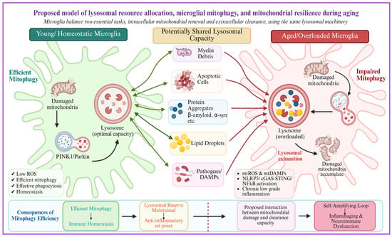
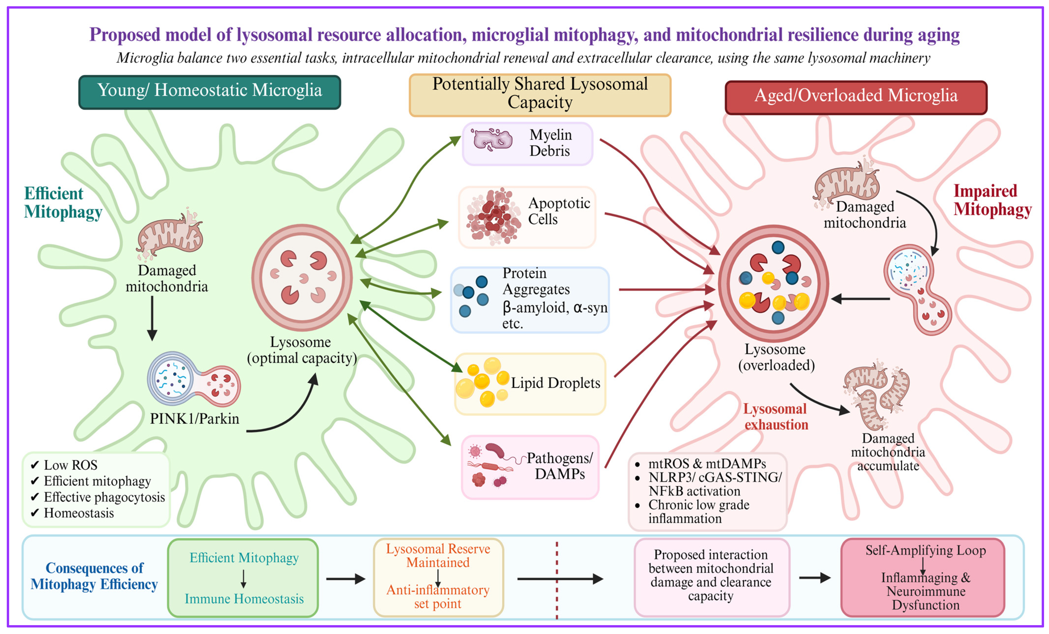
Thus, as illustrated in Figure 1, the present evidence supports a model in which increased extracellular cargo, lysosomal storage, and reduced lysosomal efficiency could contribute to incomplete mitochondrial degradation during aging. The concept of direct competition for lysosomal processing capacity remains a testable hypothesis rather than an established mechanism.
Figure 1.
Proposed relationship between lysosomal burden, mitochondrial clearance, and microglial mitochondrial resilience during aging. Conceptual model illustrating a proposed relationship among extracellular cargo burden, lysosomal function, mitochondrial clearance, and microglial mitochondrial resilience during aging. Created in BioRender. Kumar, A. (2026) https://BioRender.com/eo94hxq.
Experimental studies demonstrate age-associated increases in extracellular and phagocytic cargo, lysosomal storage, and impaired microglial clearance [35,36,41]. The interaction shown between these processes therefore represents a testable hypothesis rather than an experimentally demonstrated mechanism.
4. Mitophagy and Microglial Mitochondrial Resilience
Microglia remain under continuous metabolic and oxidative stress and maintain mitochondrial quality while responding to changes in the extracellular environment. Mitophagy is a key pathway for selectively removing damaged mitochondria and may thereby limit their persistence as sources of mitochondrial stress and danger signaling. In primary microglia, mitochondrial dysfunction and defective mitophagy have been associated with increased inflammatory cytokine production and altered mitochondrial homeostasis [42]. Inhibition of microglial autophagy has also been shown to increase NLRP3 inflammasome activation in experimental neurodegeneration [43].
Commonly used terms “mitochondrial threshold” and “buffering capacity” represent a conceptual framework. Rather than representing a defined biological threshold, they describe the proposed balance between the rate of mitochondrial damage and microglias’ capacity to remove and replace damaged mitochondria.
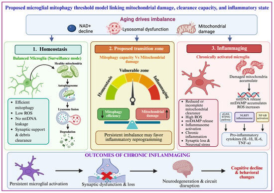
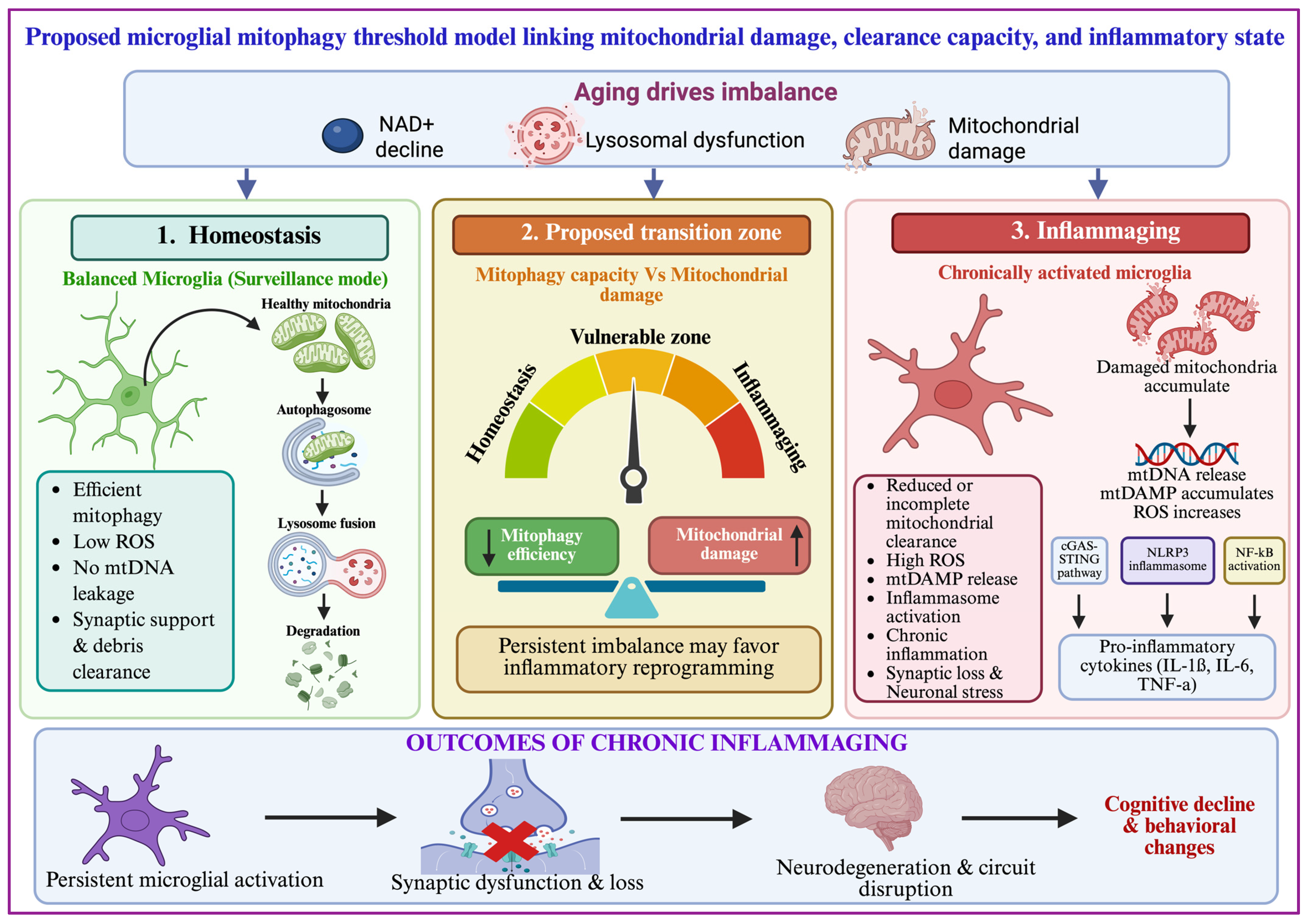
Mitochondrial danger signals can activate several inflammatory pathways. mtROS and oxidized mitochondrial components can promote NLRP3 inflammasome activation, while cytosolic mitochondrial DNA (mtDNA) can activate cGAS-STING signaling [32,44,45]. In non-microglial senescent-cell models, mitochondrial dsRNA can also activate retinoic acid-inducible gene I (RIG-I), melanoma differentiation-associated protein 5 (MDA5), and mitochondrial antiviral signaling (MAVS) and contribute to the senescence-associated secretory phenotype [46,47]. These pathways may contribute to sustained inflammatory signaling and senescence-associated phenotypes. However, the complete sequence linking impaired mitophagic flux to mtDNA or mtRNA release and then to inflammatory activation has not been directly demonstrated in aging human microglia [45,47]. Thus, mitochondrial nucleic acids should be viewed as candidate mediators within a bidirectional network rather than as obligatory downstream consequences of impaired mitophagy. Therefore, as shown in Figure 2, mitophagy is best considered an important regulator of microglial inflammatory tone rather than a single switch. Persistent mitochondrial stress, defective mitochondrial clearance, and inflammatory signaling may reinforce one another and contribute to chronic microglial dysfunction during aging.
Figure 2.
Proposed microglial mitophagy threshold model linking mitochondrial damage, clearance capacity, and inflammatory state. Conceptual framework illustrating a proposed relationship among mitochondrial damage, mitochondrial clearance, lysosomal function, and microglial inflammatory state during aging. In this conceptual framework, inflammaging may become more likely when cumulative mitochondrial stress exceeds the capacity of compensatory quality control and renewal mechanisms. Whether such a functional threshold exists in microglia, and which variables determine it, remain to be established experimentally. Created in BioRender. Kumar, A. (2026) https://BioRender.com/rbtkpq1.
5. Aging and the Breakdown of Microglial Homeostasis
Human microglial studies suggest that aging is accompanied by mitochondrial, metabolic, and cellular stress. Primary human microglia isolated from aged postmortem brains show changes in inflammatory and senescence-associated pathways [48], while dystrophic microglia in human white matter exhibit lipid accumulation, oxidative stress, lipid peroxidation, increased mitochondrial markers, and cellular damage [49]. These findings support an association between aging-related mitochondrial stress and altered microglial homeostasis but do not establish that impaired mitophagic flux drives these changes. Human postmortem and ex vivo studies further demonstrate substantial molecular heterogeneity across microglial states during normal aging and AD [3,4,6]. Importantly, longitudinal analysis of mitophagy in the mammalian brain has shown that mitophagic activity can be sustained during healthy aging, with substantial cell-type and region-specific variation, arguing against a uniform age-related collapse of mitophagy [50].
Senescence-associated microglia show changes in mitochondrial, lysosomal, oxidative-stress, and cellular-stress pathways, along with reduced functional capacity, in several human and experimental datasets. Experimental studies in microglial cells have shown that quinolinic acid exposure impairs mitolysosome formation, reduces mitochondrial turnover, and promotes senescence-associated phenotypes [51]. In the C. elegans model, quinolinic acid-induced impairment of neuronal mitophagy was associated with reduced health span [51]. The relevance of these findings to physiological human brain aging remains to be established. Senescence-associated programs may arise within several microglial states, including DAM-like populations, emphasizing that these categories overlap rather than being mutually exclusive. Consistent with this overlap, experimental studies on mice have identified a senescent triggering receptor expressed on myeloid cells 2 (TREM2)-expressing microglial population that can be distinguished from conventional DAM-like populations, indicating that TREM2 expression alone does not define a single functional microglial state [52]. Whether these distinct states differ systematically in mitophagic flux remains unresolved. Lipid accumulation is another important feature of aged microglia. Lipid-droplet-accumulating microglia (LDAM) have been described as a dysfunctional and pro-inflammatory population with impaired phagocytosis and altered lipid metabolism [36,53]. Mitochondrial stress may contribute to these age-associated changes. Recent work in human microglia has shown that mitochondrial stress responses can promote lipid accumulation, altered cellular communication, and a senescence-associated phenotype [23]. However, these findings do not establish that impaired mitophagic flux is the initial event.
The relationship between DAM and senescent microglia is also not identical. DAM are characterized by changes in TREM2, apolipoprotein E (APOE), lipoprotein lipase (LPL), glycoprotein NMB (GPNMB), and other genes associated with lipid metabolism, phagocytosis, and lysosomal function [4,6,16]. In patients with AD who received amyloid-β immunization, spatial transcriptomic studies identified distinct microglial states associated with amyloid clearance, showing that DAM can remain functionally active and participate in phagocytic clearance [54]. In contrast, senescence-associated microglia show more persistent changes in cellular homeostasis and may have reduced functional capacity.
Thus, aging should not be described simply as a progressive loss of mitochondrial function. Instead, mitochondrial damage, lysosomal function, metabolic adaptation, mitophagy, and inflammatory signaling change together, and their relative contribution may differ between microglial populations.
Experiments using human microglia derived from induced pluripotent stem cells have provided additional evidence that mitochondrial stress can influence microglial function. Mitochondrial proteotoxic stress was associated with metabolic remodeling, lipid accumulation, and a senescence-associated phenotype, suggesting that persistent mitochondrial stress may contribute to microglial dysfunction and senescence [55]. The available evidence therefore supports state-dependent remodeling of mitochondrial quality control rather than a uniform loss of mitophagy with age. An important unresolved question is whether reactive, DAM-like, lipid-processing, and senescence-associated microglia differ primarily in mitophagy initiation, mitochondrial delivery to lysosomes, lysosomal degradation, or compensatory mitochondrial biogenesis.
6. Immunometabolic Reprogramming of Aged Microglia
Aging changes the metabolic state of microglia, but these changes should be distinguished from mitophagy itself. Similarly, altered mitochondrial respiration, glycolytic reliance, mitochondrial dynamics, and mitochondrial mass are not direct measurements of mitophagic flux. Their relationship with mitophagy should therefore be inferred only when the experimental design measures mitochondrial turnover or lysosomal delivery directly [3,4,5,6]. Mitochondrial respiratory activity can directly influence inflammatory microglial activation. Peruzzotti-Jametti et al. demonstrated that mitochondrial complex I activity in microglia sustains neuroinflammation, supporting a direct link between mitochondrial metabolism and inflammatory function [56].
Aging-associated microglial metabolic remodeling can include altered oxidative metabolism, substrate utilization, and glycolytic reliance, although the magnitude and direction of these changes vary across microglial states and experimental systems [53,57]. Microglia also exhibit substantial metabolic flexibility under changing energetic conditions; experimental studies show that alternative substrate utilization can sustain surveillance functions and involve mechanistic target of rapamycin (mTOR)-dependent signaling [58].
Persistent mitochondrial damage from insufficient mitochondrial quality control could contribute to metabolic stress, while altered cellular metabolism may, in turn, influence mitochondrial turnover. Thus, the relationship between mitophagy and immunometabolic remodeling is likely bidirectional. TREM2-dependent pathways are important for adaptation to chronic lipid and phagocytic stress, and TREM2 loss alters microglial metabolic fitness and cholesterol handling [59,60].
These findings support an interaction among lipid burden, lysosomal function, and mitochondrial metabolism. However, the direct effect of lipid accumulation on mitophagic flux in aged human microglia remains insufficiently defined.
7. Microglia–Neuron–Astrocyte Crosstalk
Microglial mitochondrial dysfunction can affect neighboring neural cells through changes in cytokine production, extracellular metabolites, and phagocytic activity. Microglia with altered mitochondrial function can therefore influence neuronal and astrocytic responses, but the extent of these effects depends on the experimental model and the microglial state.
Activated microglia can induce inflammatory responses in astrocytes, and astrocyte-derived mediators can in turn modify microglial function [44]. However, these findings do not establish that impaired mitophagy in microglia is the sole cause of this crosstalk. The available evidence is more consistent with a bidirectional network in which mitochondrial stress, inflammatory signaling, and intercellular communication can influence one another.
In experimental microglial systems, rapamycin and trehalose have been shown to induce autophagy and reduce inflammatory mediator production; however, these findings demonstrate modulation of general autophagy rather than direct evidence of increased mitophagic flux [61]. The direct contribution of mitophagic flux to these effects remains less well-established than the effects of general mitochondrial or autophagic dysfunction. Accordingly, the role of microglial mitophagy in neuron–glia communication should be viewed as a potential contributor rather than a demonstrated single upstream mechanism.
8. Disease and Experimental Contexts Informing Microglial Mitochondrial Quality Control During Aging
Neurodegenerative and infectious disorders provide experimentally informative contexts for studying microglial mitochondrial quality control, but these conditions should not be considered direct extensions of physiological brain aging. AD, PD, and viral neuroinflammatory disorders differ in their initiating mechanisms, tissue environments, disease trajectories, and inflammatory stimuli [16,62,63]. Accordingly, evidence from these conditions is considered here as complementary mechanistic context rather than direct evidence of normal aging.
Recent experimental evidence further supports a close relationship between mitochondrial dynamics, mitochondrial quality control, and inflammatory microglial responses. In a mouse model of Japanese encephalitis virus (JEV) infection, mitochondrial dysfunction was associated with altered mitochondrial fusion–fission signaling and increased NADPH oxidase 2 (NOX2)-mediated oxidative stress and neuronal injury [64]. Although this study did not directly establish microglia-specific mitophagic dysfunction, it demonstrates that infectious stress can substantially perturb mitochondrial homeostasis. In contrast, chronic lipopolysaccharide (LPS) exposure in young and aged mice produced increased expression of the fission-associated gene FIS1 and decreased expression of the fusion-associated genes mitofusin1 (MFN1) and mitofusin2 (MFN2), accompanied by enhanced proinflammatory microglial responses, with more pronounced alterations in aged animals [31]. These findings provide direct experimental evidence linking age-associated mitochondrial remodeling to microglial inflammatory activation. Furthermore, JEV infection was associated with impaired autophagic flux, characterized by increased microtubule-associated protein 1 light chain 3-II/3-I (LC3-II/I) and sequestosome 1 (SQSTM1)/p62 and reduced cathepsin-D activity, together with increased microglial activation and proinflammatory cytokine expression [65,66]. Importantly, these findings demonstrate impaired autophagy and lysosomal processing rather than defective mitophagic flux per se. Collectively, these studies indicate that mitochondrial dynamics and intracellular quality control pathways are closely associated with inflammatory microglial states and may influence mitochondrial energetic resilience; however, whether these alterations culminate in defective mitophagic flux during physiological brain aging remains unresolved.
Mitophagy-modulating compounds such as kaempferol and rhapontigenin have improved disease-related phenotypes in experimental models, supporting the possibility that mitochondrial quality control can influence pathological trajectories [67,68]. However, these findings do not establish that defective microglial mitophagy is a conserved initiating mechanism across neurological disorders, nor do they demonstrate that the observed benefits are mediated specifically through microglia. In experimental PD models, impaired microglial autophagy has been linked to NLRP3 activation and dopaminergic neurodegeneration [43]. These findings support a role for microglial degradative pathways, but they should not be interpreted as direct evidence of impaired mitophagic flux. In human immunodeficiency virus type 1 (HIV-1) Tat-exposed microglia, mitochondrial dysfunction and defective mitophagy have been associated with increased inflammatory activation [42]. In a herpes simplex virus-1 (HSV-1) microglial infection model, pharmacological modulation of mitophagy-related pathways improved inflammatory and phagocytic phenotypes [63]. This provides direct microglial evidence that mitochondrial quality control can influence inflammatory responses under viral stress, but the finding may not be extrapolated directly to physiological brain aging. Human studies further emphasize heterogeneity. Ex vivo human microglia from AD brains show disease-related transcriptional changes, and recent studies of treated patients have identified distinct microglial states associated with amyloid clearance [4,6,54]. Therefore, DAM changes should be interpreted in the context of the specific microglial population, disease stage, and tissue environment. In AD, postmortem studies have also demonstrated alterations in mitophagy-associated pathways. Increased pS65-Ub has been observed in AD brain and is associated with granulovacuolar degeneration and early tau pathology [24]. Experimental studies have also shown that enhancing mitophagy can improve microglial amyloid-β clearance and reduce Alzheimer’s pathology [34]. However, these findings should be distinguished from direct measurements of mitophagic flux in human microglia. Across these disease contexts, mitochondrial quality control repeatedly emerges as a relevant component of microglial adaptation to cellular stress. The specific contribution of mitophagy, however, varies with disease context and remains incompletely resolved.
Collectively, these experimental and disease-associated findings support an interconnected relationship among mitochondrial dynamics, autophagy/lysosomal function, mitophagy, and microglial inflammatory adaptation. Nevertheless, altered mitochondrial dynamics or autophagy-associated markers should not be equated with defective mitophagy without direct measurement of mitochondrial-specific flux. Defining how mitophagic clearance interacts with mitochondrial dynamics, cellular bioenergetics, lysosomal capacity, and microglial state will be critical to determining whether impaired mitochondrial quality control drives, follows, or reflects an adaptive response to age-associated microglial dysfunction.
9. Evidence-Graded Therapeutic Strategies for Microglial Mitochondrial Resilience
Therapeutic strategies targeting mitochondrial quality control differ substantially in their level of evidence. To avoid equating experimental modulation of mitochondrial metabolism with direct activation of microglial mitophagy, interventions are considered according to three evidence categories: (i) direct evidence in microglia; (ii) evidence from in vivo models of neurological disease; and (iii) evidence derived primarily from other cell types or indirect mechanisms. Researchers have investigated potential therapeutic strategies targeting mitochondrial resilience, autophagy, lysosomal function, immunometabolism, and inflammatory signaling in experimental microglial and neurological disease models. However, these approaches differ substantially in their biological targets and levels of evidence, and most have not been demonstrated to directly alter mitophagic flux in aged human microglia. To distinguish direct microglial evidence from disease-model and indirect evidence, Table 1 summarizes the principal strategies and their current limitations.
Table 1.
Therapeutic strategies targeting microglial mitochondrial resilience and quality control: evidence level and limitations.
Evidence categories distinguish direct microglial studies, neurological disease models, and indirect/non-microglial evidence. Changes in mitophagy-associated markers, mitochondrial morphology, mitochondrial mass, or general autophagy are not considered evidence of altered mitophagic flux. “Not established” indicates that direct evidence in aged human microglia is lacking. Most strategies remain preclinical.
Direct microglial evidence includes interventions that modify mitochondrial metabolism or microglial mitochondrial quality control [45]. Peruzzotti-Jametti et al. showed that mitochondrial complex I activity contributes to inflammatory activation in microglia, providing direct evidence that mitochondrial metabolism can be therapeutically targeted in microglia [56]. Peroxisome proliferator-activated receptor gamma coactivator 1-alpha (PGC-1α) has also been studied directly in microglia and was associated with improved mitochondrial function and reduced inflammatory responses after cerebral ischemia [69]. Ubiquinone-10 (CoQ10) may mitigate mitochondrial oxidative stress and inflammatory activation in an experimental microglial model [80].
Researchers have investigated other approaches mainly in animal models of neurological disease. Urolithin A has been reported to improve mitophagy-related and lysosomal phenotypes and reduce AD pathology in experimental models, but these effects have not been established as microglia-specific or as evidence of increased mitophagic flux in aged microglia [70]. Similarly, pharmacological enhancement of mitophagy improved amyloid-β and tau pathology in AD models, with evidence that improved microglial phagocytosis contributed to the benefit [71]. These findings are promising but remain preclinical. Some interventions act indirectly on microglial function rather than directly on mitophagy. Cluster of differentiation 22 (CD22) blockade restored phagocytic and homeostatic functions of aged microglia, but it is not a direct mitophagy-targeting treatment [37]. Interventions targeting NAD+ metabolism [73,74], AMPK [76,77], mTOR [78], or sirtuins [72,79] may influence mitochondrial function, autophagy, or immune metabolism in experimental systems, but their specific effects on mitophagic flux in aged human microglia remain unclear.
Thus, therapeutic strategies should not be presented as equivalent mitophagy treatments. Their effects should be described according to whether they directly alter mitophagic flux in microglia, indirectly improve microglial mitochondrial function, or act mainly through other cell types or systemic metabolic pathways.
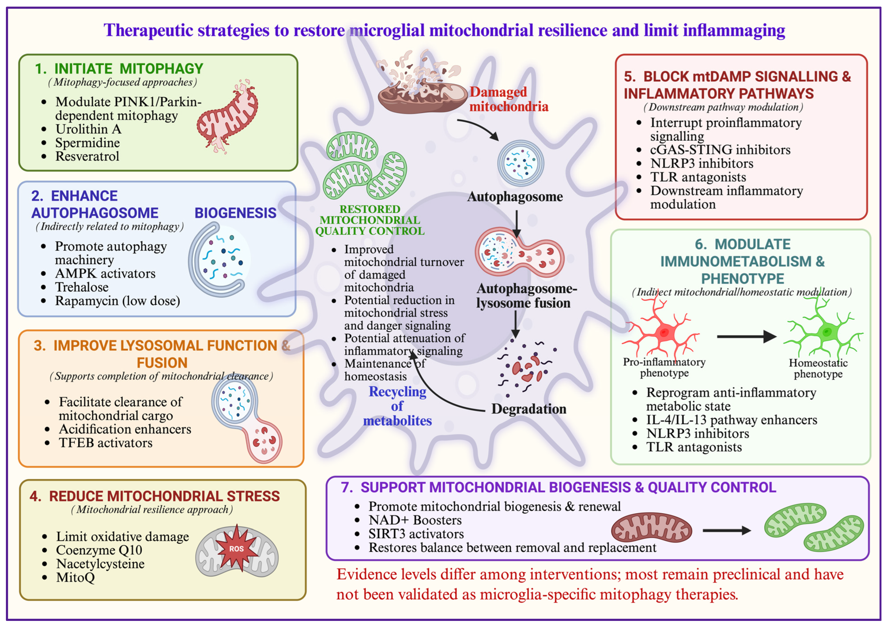
Persistent mitochondrial degradation without adequate mitochondrial biogenesis could reduce ATP availability, compromise metabolic flexibility, and impair essential microglial functions such as surveillance and phagocytosis [31,81]. As illustrated in Figure 3, the therapeutic objective may therefore be to restore coordinated mitochondrial turnover rather than indiscriminately enhance mitophagy. Increasing mitochondrial clearance without adequate mitochondrial biogenesis or lysosomal degradation may be ineffective or potentially disrupt cellular bioenergetic balance.
Figure 3.
Conceptual therapeutic strategies to restore microglial mitochondrial resilience and limit inflammaging during brain aging. The approaches shown include mitophagy-focused, lysosomal, mitochondrial stress, inflammatory, immunometabolic, and mitochondrial biogenesis strategies. Evidence levels vary among interventions, ranging from direct studies in microglia to preclinical disease models and indirect evidence from other experimental systems. Most approaches remain preclinical, and their ability to specifically modify mitophagic flux in aged human microglia remains to be established. Created in BioRender. Kumar, A. (2026) https://BioRender.com/gilbfzo.
10. Mitophagy-Associated Biomarkers and Experimental Gaps
Despite growing evidence that altered mitochondrial quality control signaling contributes to inflammaging, a major obstacle to clinical translation is the limited ability to measure this process accurately in living systems. Studies using human postmortem brain tissue indicate that mitochondrial quality control pathways change during normal aging, although these changes do not reflect a simple reduction in mitophagy. Hou et al. found an age-related increase in p-S65-Ub in several brain regions of neurologically normal individuals [23]. The increase in p-S65-Ub may reflect increased mitochondrial damage and activation of the mitophagy pathway, but may also reflect incomplete downstream clearance. Therefore, increased mitophagy-associated markers in aged human brain should not be interpreted as direct evidence of increased completed mitophagic flux. Age-related changes in Parkin solubility have also been reported in human postmortem brain, suggesting that aging may alter Parkin availability and function in mitochondrial quality control [82].
A major barrier to clinical translation is the lack of validated tools to quantify mitophagic flux specifically in human microglia in vivo. PINK1, Parkin, BNIP3, FUNDC1, LC3-II, mitochondrial mass, and lysosomal proteins can show that pathway components have changed, but these are static measurements and do not indicate whether damaged mitochondria are ultimately delivered to lysosomes and degraded.
This distinction is important because accumulation of PINK1, Parkin, LC3-positive structures, or damaged mitochondria can result from either increased pathway activation or impaired downstream degradation. Therefore, static markers should not be interpreted as direct evidence of increased or decreased mitophagic flux.
Direct assessment of mitophagic flux requires measurement of mitochondrial delivery to the lysosomal compartment and, ideally, the kinetics of subsequent degradation. The mitochondrial quality control (mito-QC) reporter uses a pH-sensitive mitochondrial GFP/mCherry system to identify mitochondria that have reached acidic lysosomes [83]. Mitochondria-targeted Keima (mt-Keima) is another mitochondrial reporter that changes its fluorescence according to lysosomal pH and can be used to quantify mitochondrial delivery to lysosomes [84]. Comparison studies indicate that mt-Keima can detect PINK1-Parkin-dependent mitophagy with greater sensitivity than the mito-QC reporter in some experimental conditions [85]. Lysosomal inhibitors such as bafilomycin A1 can also distinguish increased delivery to lysosomes from downstream degradation defects.
Human postmortem studies provide useful information but do not directly measure flux. Hou et al. demonstrated an age- and disease-associated increase in pS65-Ub in human postmortem brain, indicating increased activation or accumulation of the PINK1-Parkin mitochondrial quality control pathway [23]. Because this marker is measured in fixed tissue, it cannot determine whether mitochondrial degradation was completed. Therefore, human postmortem evidence should be interpreted as evidence of altered mitochondrial quality control signaling rather than direct evidence of mitophagic flux.
Recent human studies also show age- and disease-associated changes in microglial states. Postmortem AD studies identified senescence-associated microglial signatures, while large human cohorts showed that multiple microglial states can acquire senescence-related changes [25,26]. These studies provide important cellular context, but direct mitophagic flux measurements in phenotypically defined human DAM or SAM are still lacking. As shown in Table 2, ATP/OXPHOS, mtROS, mitochondrial morphology, PINK1/Parkin, LC3-II, TOM20/mitochondrial mass, and pS65-Ub indicate mitochondrial function, stress, morphology, abundance, or pathway activity but do not independently demonstrate mitophagic flux. Mito-QC reporter and mt-Keima provide more direct flux-related evidence of mitochondrial delivery to acidic lysosomal compartments, although this does not necessarily confirm complete mitochondrial degradation. Interpretation should consider the experimental context, particularly when evidence derives from disease or injury models rather than physiological aging.
Table 2.
Common mitochondrial and mitophagy-associated readouts and their interpretive limitations.
Importantly, longitudinal reporter-based studies in the mammalian brain demonstrate that mitophagy can remain active across healthy aging, emphasizing that aging should not be equated with a uniform reduction in mitophagic activity [50]. Future studies should combine microglial state identification with direct or surrogate measurements of mitochondrial turnover, lysosomal function, mitochondrial respiration, and inflammatory activity at single-cell or spatial resolution. Ideally, experimental designs should distinguish mitochondrial delivery to lysosomes from lysosomal degradation itself and should compare homeostatic, reactive, DAM-like, lipid-processing, and senescence-associated populations. Such studies would directly test whether mitochondrial damage, lysosomal capacity, and mitochondrial clearance interact as proposed by the conceptual framework.
11. Conclusions
Mitophagy represents an important component of mitochondrial quality control in microglia, but current evidence does not support a simple model in which aging uniformly suppresses mitophagy or in which mitophagy acts as a single molecular switch controlling inflammaging. Instead, aging is associated with heterogeneous changes in mitochondrial stress, lysosomal function, metabolism, and microglial state. Human postmortem studies and experimental mouse models provide evidence of altered mitophagy-associated signaling, whereas longitudinal reporter-based studies in the mammalian brain indicate that mitophagy can remain sustained during healthy aging [23,24,50]. Taken together, these findings suggest that aging does not necessarily lead to a uniform decline in mitophagy, although it remains unclear whether the same pattern occurs in defined populations of human microglia.
We propose the Microglial Mitophagy Threshold Model as a conceptual framework in which mitochondrial damage, mitochondrial clearance, lysosomal capacity, mitochondrial biogenesis, and microglial state interact to influence cellular function. Similarly, the possibility that increased phagocytic burden affects lysosomal processing of damaged mitochondria should be considered a testable hypothesis rather than an established mechanism.
An important unanswered question is whether incomplete mitochondrial clearance is an early driver of age-related microglial dysfunction, a consequence of it, or both. Addressing this will require direct measurements of mitophagic flux alongside spatial and single-cell analyses of microglial states in human-relevant models. From a translational perspective, therapeutic approaches may be more effective if they aim to restore the overall balance between mitochondrial turnover, lysosomal function, and mitochondrial renewal rather than simply increasing mitophagy.
Author Contributions
D.G.: conceptualization, methodology, literature search, writing the original manuscript draft, illustrations and tables preparation; A.K.: editing, reviewing, and finalizing the manuscript. All authors have read and agreed to the published version of the manuscript.
Funding
The following study was supported by a research grant from the Anusandhan National Research Foundation (ANRF), SERB, Core Research Grant (File No. CRG/2023/006869) and Indian Council of Medical Research (ICMR), Govt. of India, New Delhi (F.No.R. 5/4-4/18/mh/2022-NCD-11) to A.K.
Institutional Review Board Statement
Not applicable.
Informed Consent Statement
Not applicable.
Data Availability Statement
No new data were created or analyzed in this study. Data sharing is not applicable to this article.
Acknowledgments
The figures have been prepared using https://BioRender.com (accessed on 15th September 2026). After completing the manuscript, the authors used Grammarly (v1.2.298.1959; https://www.grammarly.com) accessed on 15 September 2026; solely to improve grammar, language clarity, and readability.
Conflicts of Interest
The authors declare no conflicts of interest.
Abbreviations
| AD | Alzheimer’s disease |
| AMPK | AMP-activated protein kinase |
| APOE | Apolipoprotein E |
| ATP | Adenosine triphosphate |
| BNIP3 | BCL2 interacting protein 3 |
| C1q | Complement component 1q |
| CD22 | Cluster of differentiation 22 |
| cGAS-STING | cyclic GMP-AMP synthase-stimulator of interferon genes |
| CNS | Central nervous system |
| CoQ10 | Ubiquinone-10 |
| DAM | Disease-associated microglia |
| DAMPs | Damage-associated molecular patterns |
| FIS1 | Fission 1 |
| FUNDC1 | FUN14 domain-containing protein 1 |
| GPNMB | Glycoprotein NMB (non-metastatic melanoma protein B) |
| HIV-1 | Human immunodeficiency virus type 1 |
| HSV-1 | Herpes simplex virus 1 |
| IL-1β | Interleukin-1β |
| IL-18 | Interleukin-18 |
| JEV | Japanese encephalitis virus |
| LC3-II/I | Microtubule-associated protein 1 light chain 3-II/3-I |
| LDAM | Lipid-droplet-accumulating microglia |
| LPL | Lipoprotein lipase |
| LPS | Lipopolysaccharide |
| MAVS | Mitochondrial antiviral signaling protein |
| MCL-1 | Myeloid cell leukemia 1 |
| MDA5 | Melanoma differentiation-associated protein 5 |
| MFN1 | Mitofusin 1 |
| MFN2 | Mitofusin 2 |
| mito-QC | Mitochondrial quality control |
| mt-Keima | Mitochondria-targeted Keima |
| mTOR | Mechanistic target of rapamycin |
| mtDNA | Mitochondrial DNA |
| mtROS | Mitochondrial reactive oxygen species |
| NAD+ | Nicotinamide adenine dinucleotide (oxidized form) |
| NF-κB | Nuclear factor kappa B |
| NLRP3 | NOD-like receptor family pyrin domain containing 3 |
| NOX2 | NADPH oxidase 2 |
| OXPHOS | Oxidative phosphorylation |
| PD | Parkinson’s disease |
| PGC-1α | Peroxisome proliferator-activated receptor gamma coactivator 1-alpha |
| PINK1 | PTEN-induced kinase 1 |
| pS65-Ub | Phosphorylated ubiquitin at serine 65 |
| RIG-I | Retinoic acid-inducible gene I |
| ROS | Reactive oxygen species |
| SAM | Senescence-associated microglia |
| SIRT3 | NAD-dependent deacetylase sirtuin-3 |
| SQSTM1/p62 | Sequestosome 1 (SQSTM1)/p62 |
| TFEB | Transcription factor EB |
| TNF-α | Tumor necrosis factor-α |
| TREM2 | Triggering receptor expressed on myeloid cells 2 |
References
- Hammond, T.R.; Dufort, C.; Dissing-Olesen, L.; Giera, S.; Young, A.; Wysoker, A.; Walker, A.J.; Gergits, F.; Segel, M.; Nemesh, J.; et al. Single-Cell RNA Sequencing of Microglia throughout the Mouse Lifespan and in the Injured Brain Reveals Complex Cell-State Changes. Immunity 2019, 50, 253–271.e6. [Google Scholar] [CrossRef] [Scilit]
- Sankowski, R.; Böttcher, C.; Masuda, T.; Geirsdottir, L.; Sagar; Sindram, E.; Seredenina, T.; Muhs, A.; Scheiwe, C.; Shah, M.J.; et al. Mapping microglia states in the human brain through the integration of high-dimensional techniques. Nat. Neurosci. 2019, 22, 2098–2110, Correction in Nat. Neurosci. 2019, 25, 975. https://doi.org/10.1038/s41593-022-01089-3. [Google Scholar] [CrossRef] [Scilit]
- Olah, M.; Patrick, E.; Villani, A.-C.; Xu, J.; White, C.C.; Ryan, K.J.; Piehowski, P.; Kapasi, A.; Nejad, P.; Cimpean, M.; et al. A transcriptomic atlas of aged human microglia. Nat. Commun. 2018, 9, 539. [Google Scholar] [CrossRef] [Scilit]
- Prater, K.E.; Green, K.J.; Mamde, S.; Sun, W.; Cochoit, A.; Smith, C.L.; Chiou, K.L.; Heath, L.; Rose, S.E.; Wiley, J.; et al. Human microglia show unique transcriptional changes in Alzheimer’s disease. Nat. Aging 2023, 3, 894–907. [Google Scholar] [CrossRef] [Scilit]
- Tuddenham, J.F.; Taga, M.; Haage, V.; Marshe, V.S.; Roostaei, T.; White, C.; Lee, A.J.; Fujita, M.; Khairallah, A.; Zhang, Y.; et al. A cross-disease resource of living human microglia identifies disease-enriched subsets and tool compounds recapitulating microglial states. Nat. Neurosci. 2024, 27, 2521–2537. [Google Scholar] [CrossRef] [Scilit]
- Kosoy, R.; Fullard, J.F.; Bendl, J.; Kleopoulos, S.P.; Shao, Z.; Argyriou, S.; Mathur, D.; Psychogyiou, K.; Malakates, P.; Vicari, J.; et al. Alzheimer’s disease transcriptional landscape in ex vivo human microglia. Nat. Neurosci. 2025, 28, 1830–1843. [Google Scholar] [CrossRef] [Scilit]
- Gao, C.; Jiang, J.; Tan, Y.; Chen, S. Microglia in neurodegenerative diseases: Mechanism and potential therapeutic targets. Signal Transduct. Target. Ther. 2023, 8, 359. [Google Scholar] [CrossRef] [Scilit]
- Paolicelli, R.C.; Bolasco, G.; Pagani, F.; Maggi, L.; Scianni, M.; Panzanelli, P.; Giustetto, M.; Ferreira, T.A.; Guiducci, E.; Dumas, L.; et al. Synaptic Pruning by Microglia Is Necessary for Normal Brain Development. Science 2011, 333, 1456–1458. [Google Scholar] [CrossRef] [Scilit]
- Hanada, Y.; Ishihara, N.; Wang, L.; Otera, H.; Ishihara, T.; Koshiba, T.; Mihara, K.; Ogawa, Y.; Nomura, M. MAVS is energized by Mff which senses mito-chondrial metabolism via AMPK for acute antiviral immunity. Nat. Commun. 2020, 11, 5711. [Google Scholar] [CrossRef] [Scilit]
- West, A.P.; Brodsky, I.E.; Rahner, C.; Woo, D.K.; Erdjument-Bromage, H.; Tempst, P.; Walsh, M.C.; Choi, Y.; Shadel, G.S.; Ghosh, S. TLR signalling augments macrophage bactericidal activity through mitochondrial ROS. Nature 2011, 472, 476–480. [Google Scholar] [CrossRef] [Scilit]
- Mills, E.L.; Kelly, B.; Logan, A.; Costa, A.S.H.; Varma, M.; Bryant, C.E.; Tourlomousis, P.; Däbritz, J.H.M.; Gottlieb, E.; Latorre, I.; et al. Succinate Dehydrogenase Supports Metabolic Repurposing of Mitochondria to Drive Inflammatory Macrophages. Cell 2016, 167, 457–470.e13. [Google Scholar] [CrossRef] [Scilit]
- Zhang, D.; Wang, F.; Li, P.; Gao, Y. Mitochondrial Ca2+ Homeostasis: Emerging Roles and Clinical Significance in Cardiac Remodeling. Int. J. Mol. Sci. 2022, 23, 3025. [Google Scholar] [CrossRef] [Scilit]
- Lu, Y.; Gao, L.; Yang, Y.; Shi, D.; Zhang, Z.; Wang, X.; Huang, Y.; Wu, J.; Meng, J.; Li, H.; et al. Protective role of mitophagy on microglia-mediated neuroin-flammatory injury through mtDNA-STING signaling in manganese-induced parkinsonism. J. Neuroinflammation 2025, 22, 55. [Google Scholar] [CrossRef] [Scilit]
- Nair, S.; Sobotka, K.S.; Joshi, P.; Gressens, P.; Fleiss, B.; Thornton, C.; Mallard, C.; Hagberg, H. Lipopolysaccharide-induced alteration of mito-chondrial morphology induces a metabolic shift in microglia modulating the inflammatory response in vitro and in vivo. Glia 2019, 67, 1047–1061. [Google Scholar] [CrossRef] [Scilit]
- Peferoen, L.A.N.; Vogel, D.Y.S.; Ummenthum, K.; Breur, M.; Heijnen, P.D.A.M.; Gerritsen, W.H.; Peferoen-Baert, R.M.; van der Valk, P.; Dijkstra, C.D.; Amor, S. Activation Status of Human Microglia Is Dependent on Lesion Formation Stage and Remyelination in Multiple Sclerosis. J. Neuropathol. Exp. Neurol. 2015, 74, 48–63. [Google Scholar] [CrossRef] [Scilit]
- Keren-Shaul, H.; Spinrad, A.; Weiner, A.; Matcovitch-Natan, O.; Dvir-Szternfeld, R.; Ulland, T.K.; David, E.; Baruch, K.; Lara-Astaiso, D.; Toth, B.; et al. A Unique Microglia Type Associated with Restricting Development of Alzheimer’s Disease. Cell 2017, 169, 1276–1290.e17. [Google Scholar] [CrossRef] [Scilit]
- Fonken, L.K.; Frank, M.G.; Kitt, M.M.; D’Angelo, H.M.; Norden, D.M.; Weber, M.D.; Barrientos, R.M.; Godbout, J.P.; Watkins, L.R.; Maier, S.F. The Alarmin HMGB1 Mediates Age-Induced Neuroinflammatory Priming. J. Neurosci. 2016, 36, 7946–7956. [Google Scholar] [CrossRef] [Scilit]
- Wang, X.; Jia, Y.; Yan, X.; Wang, Y. Metabolic and epigenetic regulation of microglia memory in neurological disorders. J. Leukoc. Biol. 2026, 118, qiag085. [Google Scholar] [CrossRef] [Scilit]
- Škandík, M.; Friess, L.; Vázquez-Cabrera, G.; Keane, L.; Grabert, K.; Cruz De Los Santos, M.; Posada-Pérez, M.; Baleviciute, A.; Cheray, M.; Joseph, B. Age-associated microglial transcriptome leads to diminished immunogenicity and dysregulation of MCT4 and P2RY12/P2RY13 related functions. Cell Death Discov. 2025, 11, 16. [Google Scholar] [CrossRef] [Scilit]
- Ferrucci, L.; Harris, T.B.; Guralnik, J.M.; Tracy, R.P.; Corti, M.; Cohen, H.J.; Penninx, B.; Pahor, M.; Wallace, R.; Havlik, R.J. Serum IL-6 Level and the Development of Disability in Older Persons. J. Am. Geriatr. Soc. 1999, 47, 639–646. [Google Scholar] [CrossRef] [Scilit]
- Harris, T.B.; Ferrucci, L.; Tracy, R.P.; Corti, M.C.; Wacholder, S.; Ettinger, W.H.; Heimovitz, H.; Cohen, H.J.; Wallace, R. Associations of elevated Interleukin-6 and C-Reactive protein levels with mortality in the elderly. Am. J. Med. 1999, 106, 506–512. [Google Scholar] [CrossRef] [Scilit]
- Puzianowska-Kuźnicka, M.; Owczarz, M.; Wieczorowska-Tobis, K.; Nadrowski, P.; Chudek, J.; Slusarczyk, P.; Skalska, A.; Jonas, M.; Franek, E.; Mossakowska, M. Interleukin-6 and C-reactive protein, successful aging, and mortality: The PolSenior study. Immun. Ageing 2016, 13, 21. [Google Scholar] [CrossRef] [Scilit]
- Hou, X.; Fiesel, F.C.; Truban, D.; Castanedes Casey, M.; Lin, W.; Soto, A.I.; Tacik, P.; Rousseau, L.G.; Diehl, N.N.; Heckman, M.G.; et al. Age- and disease-dependent increase of the mitophagy marker phospho-ubiquitin in normal aging and Lewy body disease. Autophagy 2018, 14, 1404–1418. [Google Scholar] [CrossRef] [Scilit]
- Hou, X.; Watzlawik, J.O.; Cook, C.; Liu, C.; Kang, S.S.; Lin, W.; DeTure, M.; Heckman, M.G.; Diehl, N.N.; Al-Shaikh, F.S.H.; et al. Mitophagy alterations in Alzheimer’s disease are associated with granulovacuolar degeneration and early tau pathology. Alzheimer’s Dement. 2021, 17, 417–430. [Google Scholar] [CrossRef] [Scilit]
- Fancy, N.N.; Smith, A.M.; Caramello, A.; Tsartsalis, S.; Davey, K.; Muirhead, R.C.J.; McGarry, A.; Jenkyns, M.H.; Schneegans, E.; Chau, V.; et al. Characterisation of premature cell senescence in Alzheimer’s disease using single nuclear transcriptomics. Acta Neuropathol. 2024, 147, 78. [Google Scholar] [CrossRef] [Scilit]
- Li, B.; Wang, S.; Kerman, B.; Hugo, C.; Shwab, E.K.; Shu, C.; Chiba-Falek, O.; Arvanitakis, Z.; Yassine, H.N. Microglial States Are Susceptible to Senescence and Cholesterol Dysregulation in Alzheimer’s Disease. Aging Cell 2025, 24, e70189. [Google Scholar] [CrossRef] [Scilit]
- Ghosh, D.; Kumar, A. Harnessing Mitophagy for Therapeutic Advances in Aging and Chronic Neurodegenerative Diseases. Neuroglia 2024, 5, 391–409. [Google Scholar] [CrossRef] [Scilit]
- Zhang, Q.; Raoof, M.; Chen, Y.; Sumi, Y.; Sursal, T.; Junger, W.; Brohi, K.; Itagaki, K.; Hauser, C.J. Circulating mitochondrial DAMPs cause inflammatory responses to injury. Nature 2010, 464, 104–107. [Google Scholar] [CrossRef] [Scilit]
- West, A.P.; Khoury-Hanold, W.; Staron, M.; Tal, M.C.; Pineda, C.M.; Lang, S.M.; Bestwick, M.; Duguay, B.A.; Raimundo, N.; MacDuff, D.A.; et al. Mitochondrial DNA stress primes the antiviral innate immune response. Nature 2015, 520, 553–557. [Google Scholar] [CrossRef] [Scilit]
- Nakahira, K.; Haspel, J.A.; Rathinam, V.A.K.; Lee, S.-J.; Dolinay, T.; Lam, H.C.; Englert, J.A.; Rabinovitch, M.; Cernadas, M.; Kim, H.P.; et al. Autophagy proteins regulate innate immune responses by inhibiting the release of mitochondrial DNA mediated by the NALP3 inflammasome. Nat. Immunol. 2011, 12, 222–230. [Google Scholar] [CrossRef] [Scilit]
- Ghosh, D.; Singh, G.; Mishra, P.; Singh, A.; Kumar, A.; Sinha, N. Alteration in mitochondrial dynamics promotes the proinflammatory response of microglia and is involved in cerebellar dysfunction of young and aged mice following LPS exposure. Neurosci. Lett. 2023, 807, 137262. [Google Scholar] [CrossRef] [Scilit]
- Iyer, S.S.; He, Q.; Janczy, J.R.; Elliott, E.I.; Zhong, Z.; Olivier, A.K.; Sadler, J.J.; Knepper-Adrian, V.; Han, R.; Qiao, L.; et al. Mitochondrial Cardiolipin Is Required for Nlrp3 Inflammasome Activation. Immunity 2013, 39, 311–323. [Google Scholar] [CrossRef] [Scilit]
- Zhong, Z.; Liang, S.; Sanchez-Lopez, E.; He, F.; Shalapour, S.; Lin, X.-J.; Wong, J.; Ding, S.; Seki, E.; Schnabl, B.; et al. New mitochondrial DNA synthesis enables NLRP3 inflammasome activation. Nature 2018, 560, 198–203. [Google Scholar] [CrossRef] [Scilit]
- Fang, E.F.; Hou, Y.; Palikaras, K.; Adriaanse, B.A.; Kerr, J.S.; Yang, B.; Lautrup, S.; Hasan-Olive, M.M.; Caponio, D.; Dan, X.; et al. Mitophagy inhibits amyloid-β and tau pathology and reverses cognitive deficits in models of Alzheimer’s disease. Nat. Neurosci. 2019, 22, 401–412. [Google Scholar] [CrossRef] [Scilit]
- Safaiyan, S.; Kannaiyan, N.; Snaidero, N.; Brioschi, S.; Biber, K.; Yona, S.; Edinger, A.L.; Jung, S.; Rossner, M.J.; Simons, M. Age-related myelin degradation burdens the clearance function of microglia during aging. Nat. Neurosci. 2016, 19, 995–998. [Google Scholar] [CrossRef] [Scilit]
- Burns, J.C.; Cotleur, B.; Walther, D.M.; Bajrami, B.; Rubino, S.J.; Wei, R.; Franchimont, N.; Cotman, S.L.; Ransohoff, R.M.; Mingueneau, M. Differential accumulation of storage bodies with aging defines discrete subsets of microglia in the healthy brain. eLife 2020, 9, e57495. [Google Scholar] [CrossRef] [Scilit]
- Pluvinage, J.V.; Haney, M.S.; Smith, B.A.H.; Sun, J.; Iram, T.; Bonanno, L.; Li, L.; Lee, D.P.; Morgens, D.W.; Yang, A.C.; et al. CD22 blockade restores homeostatic microglial phagocytosis in ageing brains. Nature 2019, 568, 187–192. [Google Scholar] [CrossRef] [Scilit]
- Liu, Q.; Yoo, S.; Zhang, Z.A.; Li, L.; Su, H.; Vannur, L.; Wooldredge, A.C.; Hughes, J.-W.B.; Desprez, P.-Y.; Hao, N.; et al. Mitochondria-lysosome coupling contributes to lysosome acidification and aging. Mol. Cell 2026, 86, 2425–2442.e10. [Google Scholar] [CrossRef] [Scilit]
- Tian, Z.; Chen, R.; Fang, G.; Qiu, K.; Wu, W.; Shao, X.; Liu, D.; Que, H.; Wang, X.; Gao, J.; et al. Mitochondria acidify lysosomes through membrane contacts. Cell Rep. 2026, 45, 117112. [Google Scholar] [CrossRef] [Scilit]
- Wang, B.; Martini-Stoica, H.; Qi, C.; Lu, T.-C.; Wang, S.; Xiong, W.; Qi, Y.; Xu, Y.; Sardiello, M.; Li, H.; et al. TFEB–vacuolar ATPase signaling regulates lysosomal function and microglial activation in tauopathy. Nat. Neurosci. 2024, 27, 48–62. [Google Scholar] [CrossRef] [Scilit]
- Cantuti-Castelvetri, L.; Fitzner, D.; Bosch-Queralt, M.; Weil, M.-T.; Su, M.; Sen, P.; Ruhwedel, T.; Mitkovski, M.; Trendelenburg, G.; Lütjohann, D.; et al. Defective cholesterol clearance limits remyelination in the aged central nervous system. Science 2018, 359, 684–688. [Google Scholar] [CrossRef] [Scilit]
- Thangaraj, A.; Periyasamy, P.; Liao, K.; Bendi, V.S.; Callen, S.; Pendyala, G.; Buch, S. HIV-1 TAT-mediated microglial activation: Role of mitochondrial dysfunction and defective mitophagy. Autophagy 2018, 14, 1596–1619, Correction in Autophagy 2018, 17, 831–832. https://doi.org/10.1080/15548627.2020.1870318. [Google Scholar] [CrossRef] [Scilit]
- Qin, Y.; Qiu, J.; Wang, P.; Liu, J.; Zhao, Y.; Jiang, F.; Lou, H. Impaired autophagy in microglia aggravates dopaminergic neurodegeneration by regulating NLRP3 inflammasome activation in experimental models of Parkinson’s disease. Brain Behav. Immun. 2021, 91, 324–338. [Google Scholar] [CrossRef] [Scilit]
- Gulen, M.F.; Samson, N.; Keller, A.; Schwabenland, M.; Liu, C.; Glück, S.; Thacker, V.V.; Favre, L.; Mangeat, B.; Kroese, L.J.; et al. CGAS–STING drives ageing-related inflammation and neurodegeneration. Nature 2023, 620, 374–380. [Google Scholar] [CrossRef] [Scilit]
- Hou, X.; Wang, M.; Deng, R.; Lu, Y.; Zhang, J.; Ren, L.; Chen, J.; Liu, C.-F.; Yang, Y.-P.; Hu, L.-F. ADT-OH promotes mitophagy and suppresses the cytosolic mtDNA-cGAS-STING inflammatory cascade in microglia. Acta Pharmacol. Sin. 2026, 47, 2209–2223. [Google Scholar] [CrossRef] [Scilit]
- López-Polo, V.; Maus, M.; Zacharioudakis, E.; Lafarga, M.; Attolini, C.S.-O.; Marques, F.D.M.; Kovatcheva, M.; Gavathiotis, E.; Serrano, M. Release of mitochondrial dsRNA into the cytosol is a key driver of the inflammatory phenotype of senescent cells. Nat. Commun. 2024, 15, 7378. [Google Scholar] [CrossRef] [Scilit]
- Victorelli, S.; Eppard, M.; Martini, H.; Woo, S.-H.; Everts, S.P.A.; Lee, G.; Pirius, N.; Han, N.; Liang, E.Y.; Franco, A.C.; et al. Mitochondrial RNA cytosolic leakage drives the SASP. Nat. Commun. 2025, 16, 10992. [Google Scholar] [CrossRef] [Scilit]
- Olah, M.; Menon, V.; Habib, N.; Taga, M.F.; Ma, Y.; Yung, C.J.; Cimpean, M.; Khairallah, A.; Coronas-Samano, G.; Sankowski, R.; et al. Single cell RNA sequencing of human microglia uncovers a subset associated with Alzheimer’s disease. Nat. Commun. 2020, 11, 6129. [Google Scholar] [CrossRef] [Scilit]
- Streit, W.J.; Sammons, N.W.; Kuhns, A.J.; Sparks, D.L. Dystrophic microglia in the aging human brain. Glia 2004, 45, 208–212. [Google Scholar] [CrossRef] [Scilit]
- Rappe, A.; Vihinen, H.A.; Suomi, F.; Hassinen, A.J.; Ehsan, H.; Jokitalo, E.S.; McWilliams, T.G. Longitudinal autophagy profiling of the mammalian brain reveals sustained mitophagy throughout healthy aging. EMBO J. 2024, 43, 21. [Google Scholar] [CrossRef] [Scilit]
- Dongol, A.; Chen, X.; Zheng, P.; Seyhan, Z.B.; Huang, X.-F. Quinolinic acid impairs mitophagy promoting microglia senescence and poor healthspan in C. elegans: A mechanism of impaired aging process. Biol. Direct 2023, 18, 86. [Google Scholar] [CrossRef] [Scilit]
- Rachmian, N.; Medina, S.; Cherqui, U.; Akiva, H.; Deitch, D.; Edilbi, D.; Croese, T.; Salame, T.M.; Ramos, J.M.P.; Cahalon, L.; et al. Identification of senescent, TREM2-expressing microglia in aging and Alzheimer’s disease model mouse brain. Nat. Neurosci. 2024, 27, 1116–1124. [Google Scholar] [CrossRef] [Scilit]
- Marschallinger, J.; Iram, T.; Zardeneta, M.; Lee, S.E.; Lehallier, B.; Haney, M.S.; Pluvinage, J.V.; Mathur, V.; Hahn, O.; Morgens, D.W.; et al. Lipid-droplet-accumulating microglia represent a dysfunctional and proinflammatory state in the aging brain. Nat. Neurosci. 2020, 23, 194–208, Correction in Nat. Neurosci. 2020, 23, 1308. https://doi.org/10.1038/s41593-020-0682-y. [Google Scholar] [CrossRef] [Scilit]
- van Olst, L.; Simonton, B.; Edwards, A.J.; Forsyth, A.V.; Boles, J.; Jamshidi, P.; Watson, T.; Shepard, N.; Krainc, T.; Argue, B.M.; et al. Microglial mechanisms drive amyloid-β clearance in immunized patients with Alzheimer’s disease. Nat. Med. 2025, 31, 1604–1616, Correction in Nat. Med. 2025, 31, 1712. https://doi.org/10.1038/s41591-025-03664-0. [Google Scholar] [CrossRef] [Scilit]
- Perez, J.M.J.; Lam, A.; Weissleder, C.; Bertoli, F.; Raji, H.; Bosch, M.; Nemazanyy, I.; Kalb, S.; Kehili, M.; Hirschberg, I.; et al. The mitochondrial unfolded protein response in human microglia disrupts neuronal–glial communication and promotes senescence. Nat. Neurosci. 2026, 29, 1858–1872. [Google Scholar] [CrossRef] [Scilit]
- Peruzzotti-Jametti, L.; Willis, C.M.; Krzak, G.; Hamel, R.; Pirvan, L.; Ionescu, R.-B.; Reisz, J.A.; Prag, H.A.; Garcia-Segura, M.E.; Wu, V.; et al. Mitochondrial complex I activity in microglia sustains neuroinflammation. Nature 2024, 628, 195–203. [Google Scholar] [CrossRef] [Scilit]
- Baik, S.H.; Kang, S.; Lee, W.; Choi, H.; Chung, S.; Kim, J.-I.; Mook-Jung, I. A Breakdown in Metabolic Reprogramming Causes Microglia Dysfunction in Alzheimer’s Disease. Cell Metab. 2019, 30, 493–507.e6. [Google Scholar] [CrossRef] [Scilit]
- Bernier, L.-P.; York, E.M.; Kamyabi, A.; Choi, H.B.; Weilinger, N.L.; MacVicar, B.A. Microglial metabolic flexibility supports immune surveillance of the brain parenchyma. Nat. Commun. 2020, 11, 1559. [Google Scholar] [CrossRef] [Scilit]
- Nugent, A.A.; Lin, K.; van Lengerich, B.; Lianoglou, S.; Przybyla, L.; Davis, S.S.; Llapashtica, C.; Wang, J.; Kim, D.J.; Xia, D.; et al. TREM2 Regulates Microglial Cholesterol Metabolism upon Chronic Phagocytic Challenge. Neuron 2020, 105, 837–854.e9. [Google Scholar] [CrossRef] [Scilit]
- Ulland, T.K.; Song, W.M.; Huang, S.C.-C.; Ulrich, J.D.; Sergushichev, A.; Beatty, W.L.; Loboda, A.A.; Zhou, Y.; Cairns, N.J.; Kambal, A.; et al. TREM2 Maintains Microglial Metabolic Fitness in Alzheimer’s Disease. Cell 2017, 170, 649–663.e13. [Google Scholar] [CrossRef] [Scilit]
- Bussi, C.; Peralta Ramos, J.M.; Arroyo, D.S.; Gaviglio, E.A.; Gallea, J.I.; Wang, J.M.; Celej, M.S.; Iribarren, P. Autophagy down regulates pro-inflammatory mediators in BV2 microglial cells and rescues both LPS and alpha-synuclein induced neuronal cell death. Sci. Rep. 2017, 7, 43153. [Google Scholar] [CrossRef] [Scilit]
- Krasemann, S.; Madore, C.; Cialic, R.; Baufeld, C.; Calcagno, N.; El Fatimy, R.; Beckers, L.; O’Loughlin, E.; Xu, Y.; Fanek, Z.; et al. The TREM2-APOE Pathway Drives the Transcriptional Phenotype of Dysfunctional Microglia in Neurodegenerative Diseases. Immunity 2017, 47, 566–581.e9. [Google Scholar] [CrossRef] [Scilit]
- Oh, S.-J.; Kim, Y.Y.; Ma, R.; Choi, S.T.; Choi, S.M.; Cho, J.H.; Hur, J.-Y.; Yoo, Y.; Han, K.; Park, H.; et al. Pharmacological targeting of mitophagy via ALT001 improves herpes simplex virus 1 (HSV1)-mediated microglial inflammation and promotes amyloid β phagocytosis by restricting HSV1 infection. Theranostics 2025, 15, 4890–4908. [Google Scholar] [CrossRef] [Scilit]
- Singh, G.; Kumar, A. Japanese Encephalitis Virus Infection Causes an Imbalance in the Activation of Mitochondrial Fusion/Fission Genes and Triggers the Activation of NOX2-mediated Oxidative Stress and Neuronal Cell Death. Neurochem. Res. 2023, 48, 2196–2205. [Google Scholar] [CrossRef] [Scilit]
- Kumar, A.; Kalita, J.; Sinha, R.A.; Singh, G.; B, A.; Shukla, M.; Tiwari, S.; Dhole, T.N.; Misra, U.K. Impaired Autophagy Flux is Associated with Proinflammatory Microglia Activation Following Japanese Encephalitis Virus Infection. Neurochem. Res. 2020, 45, 2184–2195. [Google Scholar] [CrossRef] [Scilit]
- Kumar, A. Divergent Kinetics and Cellular Determinants of Neuroinflammation Across Traumatic, Endotoxin, and Viral Brain Injury. Clin. Exp. Neuroimmunol. 2026, 17, e70051. [Google Scholar] [CrossRef] [Scilit]
- Xie, C.; Zhuang, X.-X.; Niu, Z.; Ai, R.; Lautrup, S.; Zheng, S.; Jiang, Y.; Han, R.; Gupta, T.S.; Cao, S.; et al. Amelioration of Alzheimer’s disease pathology by mitophagy inducers identified via machine learning and a cross-species workflow. Nat. Biomed. Eng. 2022, 6, 76–93. [Google Scholar] [CrossRef] [Scilit]
- Xiao, L.; Liang, L.; Zhou, S.; Peng, F.; Yuan, C. Kaempferol attenuated LPS-induced microglial neurotoxicity by promoting mitophagy to inhibit mtDNA-mediated NLRP3 inflammasome activation. Phytomedicine 2025, 148, 157486. [Google Scholar] [CrossRef] [Scilit]
- Han, B.; Jiang, W.; Cui, P.; Zheng, K.; Dang, C.; Wang, J.; Li, H.; Chen, L.; Zhang, R.; Wang, Q.M.; et al. Microglial PGC-1α protects against ischemic brain injury by suppressing neuroinflammation. Genome Med. 2021, 13, 47. [Google Scholar] [CrossRef] [Scilit]
- Hou, Y.; Chu, X.; Park, J.; Zhu, Q.; Hussain, M.; Li, Z.; Madsen, H.B.; Yang, B.; Wei, Y.; Wang, Y.; et al. Urolithin A improves Alzheimer’s disease cognition and restores mitophagy and lysosomal functions. Alzheimer’s Dement. 2024, 20, 4212–4233. [Google Scholar] [CrossRef] [Scilit]
- Cen, X.; Chen, Y.; Xu, X.; Wu, R.; He, F.; Zhao, Q.; Sun, Q.; Yi, C.; Wu, J.; Najafov, A.; et al. Pharmacological targeting of MCL-1 promotes mitophagy and improves disease pathologies in an Alzheimer’s disease mouse model. Nat. Commun. 2020, 11, 5731. [Google Scholar] [CrossRef] [Scilit]
- Moon, C.Y.; Park, J.; Shin, E.J.; Kang, H. Nicotinamide riboside abrogates lipopolysaccharide-induced inflammation and mitochondrial dysfunction in microglia through the AMP-activated protein kinase-sirtuin 3 signaling pathway. Food Biosci. 2025, 63, 105799. [Google Scholar] [CrossRef] [Scilit]
- Mills, K.F.; Yoshida, S.; Stein, L.R.; Grozio, A.; Kubota, S.; Sasaki, Y.; Redpath, P.; Migaud, M.E.; Apte, R.S.; Uchida, K.; et al. Long-Term Administration of Nicotinamide Mononucleotide Mitigates Age-Associated Physiological Decline in Mice. Cell Metab. 2016, 24, 795–806. [Google Scholar] [CrossRef] [Scilit]
- Cerutti, R.; Pirinen, E.; Lamperti, C.; Marchet, S.; Sauve, A.A.; Li, W.; Leoni, V.; Schon, E.A.; Dantzer, F.; Auwerx, J.; et al. NAD+-Dependent Activation of Sirt1 Corrects the Phenotype in a Mouse Model of Mitochondrial Disease. Cell Metab. 2014, 19, 1042–1049. [Google Scholar] [CrossRef] [Scilit]
- Gao, Y.; Li, J.; Li, J.; Hu, C.; Zhang, L.; Yan, J.; Li, L.; Zhang, L. Tetrahydroxy stilbene glycoside alleviated inflammatory damage by mitophagy via AMPK related PINK1/Parkin signaling pathway. Biochem. Pharmacol. 2020, 177, 113997. [Google Scholar] [CrossRef] [Scilit]
- Drake, J.C.; Wilson, R.J.; Laker, R.C.; Guan, Y.; Spaulding, H.R.; Nichenko, A.S.; Shen, W.; Shang, H.; Dorn, M.V.; Huang, K.; et al. Mitochondria-localized AMPK responds to local energetics and contributes to exercise and energetic stress-induced mitophagy. Proc. Natl. Acad. Sci. USA 2021, 118, e2025932118. [Google Scholar] [CrossRef] [Scilit]
- Lantier, L.; Fentz, J.; Mounier, R.; Leclerc, J.; Treebak, J.T.; Pehmøller, C.; Sanz, N.; Sakakibara, I.; Saint-Amand, E.; Rimbaud, S.; et al. AMPK controls exercise endurance, mitochondrial oxidative capacity, and skeletal muscle integrity. FASEB J. 2014, 28, 3211–3224. [Google Scholar] [CrossRef] [Scilit]
- Hurez, V.; Dao, V.; Liu, A.; Pandeswara, S.; Gelfond, J.; Sun, L.; Bergman, M.; Orihuela, C.J.; Galvan, V.; Padrón, Á.; et al. Chronic mTOR inhibition in mice with rapamycin alters T, B, myeloid, and innate lymphoid cells and gut flora and prolongs life of immune-deficient mice. Aging Cell 2015, 14, 945–956. [Google Scholar] [CrossRef] [Scilit]
- Zhou, D.; Jiang, Y. Sirtuin 3 attenuates neuroinflammation-induced apoptosis in BV-2 microglia. Aging 2019, 11, 9075–9089. [Google Scholar] [CrossRef] [Scilit]
- Singh, A.; Kumar, A. Microglial Inhibitory Mechanism of Coenzyme Q10 Against Aβ (1-42) Induced Cognitive Dysfunctions: Possible Behavioral, Biochemical, Cellular, and Histopathological Alterations. Front. Pharmacol. 2015, 6, 268. [Google Scholar] [CrossRef] [Scilit]
- Lin, M.; Yu, H.; Xie, Q.; Xu, Z.; Shang, P. Role of microglia autophagy and mitophagy in age-related neurodegenerative diseases. Front. Aging Neurosci. 2023, 14, 1100133. [Google Scholar] [CrossRef] [Scilit]
- Tokarew, J.M.; El-Kodsi, D.N.; Lengacher, N.A.; Fehr, T.K.; Nguyen, A.P.; Shutinoski, B.; O’nUallain, B.; Jin, M.; Khan, J.M.; Ng, A.C.H.; et al. Age-associated insolubility of parkin in human midbrain is linked to redox balance and sequestration of reactive dopamine metabolites. Acta Neuropathol. 2021, 141, 725–754. [Google Scholar] [CrossRef] [Scilit]
- McWilliams, T.G.; Prescott, A.R.; Allen, G.F.G.; Tamjar, J.; Munson, M.J.; Thomson, C.; Muqit, M.M.; Ganley, I.G. Mito-QC illuminates mitophagy and mitochondrial architecture in vivo. J. Cell Biol. 2016, 214, 333–345. [Google Scholar] [CrossRef] [Scilit]
- Sun, N.; Malide, D.; Liu, J.; Rovira, I.I.; Combs, C.A.; Finkel, T. A fluorescence-based imaging method to measure in vitro and in vivo mitophagy using mt-Keima. Nat. Protoc. 2017, 12, 1576–1587. [Google Scholar] [CrossRef] [Scilit]
- Liu, Y.-T.; Sliter, D.A.; Shammas, M.K.; Huang, X.; Wang, C.; Calvelli, H.; Maric, D.S.; Narendra, D.P. Mt-Keima detects PINK1-PRKN mitophagy in vivo with greater sensitivity than mito-QC. Autophagy 2021, 17, 3753–3762. [Google Scholar] [CrossRef] [Scilit]
- Tang, X.; Huang, L.; Ma, W.; Huang, M.; Zeng, Z.; Yu, Y.; Qin, N.; Zhou, F.; Li, F.; Gong, S.; et al. Intestinal 8 gingerol attenuates TBI-induced neuroinflammation by inhibiting microglia NLRP3 inflammasome activation in a PINK1/Parkin-dependent manner. Phytomedicine 2025, 140, 156580. [Google Scholar] [CrossRef] [Scilit]
- Shibuya, Y.; Chang, C.C.Y.; Huang, L.-H.; Bryleva, E.Y.; Chang, T.-Y. Inhibiting ACAT1/SOAT1 in Microglia Stimulates Autophagy-Mediated Lysosomal Proteolysis and Increases Aβ1–42 Clearance. J. Neurosci. 2014, 34, 14484–14501. [Google Scholar] [CrossRef] [Scilit]
- Tsuboyama, K.; Koyama-Honda, I.; Sakamaki, Y.; Koike, M.; Morishita, H.; Mizushima, N. The ATG conjugation systems are important for degradation of the inner autophagosomal membrane. Science 2016, 354, 1036–1041. [Google Scholar] [CrossRef] [Scilit]
- Hou, Y.; Wei, Y.; Lautrup, S.; Yang, B.; Wang, Y.; Cordonnier, S.; Mattson, M.P.; Croteau, D.L.; Bohr, V.A. NAD+ supplementation reduces neuroinflammation and cell senescence in a transgenic mouse model of Alzheimer’s disease via cGAS–STING. Proc. Natl. Acad. Sci. USA 2021, 118, e2011226118. [Google Scholar] [CrossRef] [Scilit]

Disclaimer/Publisher’s Note: The statements, opinions and data contained in all publications are solely those of the individual author(s) and contributor(s) and not of MDPI and/or the editor(s). MDPI and/or the editor(s) disclaim responsibility for any injury to people or property resulting from any ideas, methods, instructions or products referred to in the content. |
© 2026 by the authors. Licensee MDPI, Basel, Switzerland. This article is an open access article distributed under the terms and conditions of the Creative Commons Attribution (CC BY) license.