


Abstract
Autism spectrum disorder is increasingly linked to altered microglial biology. However, current research models are limited by outdated descriptions of microglial “activation”. Here, we propose that microglial involvement in ASD is best understood as a problem of state mismatch, in which temporally programmed and regionally specialized microglial states fail to align with local developmental demands. We synthesize evidence across genetic models, human transcriptomics, and experimental systems to examine three axes of misalignment: developmental timing, circuit specificity, and functional phenotype. These mismatches produce divergent outcomes, including both excessive and insufficient synaptic pruning, and reflect a decoupling between microglial activation markers and effector capacity. We further evaluate molecular recognition systems governing microglia–synapse interactions, with emphasis on complement signaling and glycan-mediated pathways such as sialic acid–Siglec signaling and polysialylation. While glycosylation is not a universal driver of ASD pathology, it represents a plausible regulatory layer controlling synapse visibility and microglial engagement. This framework reconciles conflicting findings in the literature and positions microglia as dynamic developmental effectors whose misaligned state trajectories contribute to circuit-level dysfunction in ASD.
1. Introduction
Autism spectrum disorder (ASD) encompasses a heterogeneous group of neurodevelopmental conditions characterized by persistent deficits in social communication and social interaction, alongside restricted and repetitive patterns of behavior, interests, or activities [1]. Although clinically unified, ASD arises from extensive polygenic and environmental complexity converging on shared biological processes, including altered neural circuit formation, atypical synaptic maturation, disrupted excitatory/inhibitory balance, and dysregulated gene-environment interactions [1,2]. While these mechanisms were historically framed as neuron-centered phenomena, growing evidence demonstrates that proper circuit assembly depends critically on non-neuronal cells, particularly microglia, which have emerged as fundamental regulators of neurodevelopment and key contributors to ASD pathophysiology [3,4].
Microglia are highly dynamic developmental effectors that orchestrate multiple aspects of brain maturation. During embryonic and early postnatal stages, they regulate synaptic pruning, neural progenitor survival, trophic support, vascular stabilization, and oligodendrocyte-mediated myelination [5,6]. Microglial maturation is governed by tightly coordinated transcriptional and epigenomic programs shaped by local neuronal activity, tissue-specific signals, and narrow developmental windows, not by simple transitions between resting and activated states [3,7].
Despite this increasingly sophisticated developmental framework, the role of microglia in ASD remains deeply controversial. Early post-mortem studies described increased microglial density, altered morphology, and signatures interpreted as chronic neuroinflammation [8,9]. Subsequent transcriptomic analyses reinforced the association between ASD and immune-related microglial gene networks [10]. However, functional studies and animal models have produced divergent findings. ASD-associated mutations may drive either excessive synaptic engulfment [11] or impaired synaptic refinement [12,13]. Likewise, Positron Emission Tomography (PET) images studies using TSPO (Translocator Protein) as a marker of microglial activation have yielded inconsistent and region-dependent results across sexes and age groups [14].
These contradictions expose the limitations of conventional concepts such as “microglial activation” and “neuroinflammation”. Binary models, including the obsolete M1/M2 framework, reduce a multidimensional cellular landscape into biologically uninformative categories. Describing microglia as merely “activated” fails to distinguish critical functional states, such as whether cells are over-pruning synapses, lacking trophic support, undergoing metabolic exhaustion, or reacting secondarily to neuronal dysfunction [15]. To reconcile these inconsistencies, this review proposes that microglial involvement in ASD is best understood as a problem of state mismatch: microglial developmental trajectories fail to align temporally, spatially, and functionally with the requirements of local neural circuits. This mismatch operates across three intersecting axes. Developmentally, microglial programs may mature prematurely, remain immature, or progress toward senescence, thus disrupting critical windows of synaptic refinement [16]. Spatially, strong regional microglial heterogeneity means that identical genetic or environmental insults can generate distinct circuit-level outcomes [17]. Functionally, microglial sensory signaling may become dissociated from effector competence, producing reactive yet bioenergetically impaired states incapable of proper synaptic processing [18,19].
This framework also requires examining the molecular recognition systems that translate microglial states into synaptic outcomes. Although complement signaling pathways such as C1q, C3, and CR3 remain central regulators of microglia-synapse interactions [20,21], they are modulated by glycan-dependent recognition systems. Glycoconjugates such as sialic acids and PSA-NCAM act as molecular checkpoints that regulate synaptic accessibility through inhibitory receptors including Siglec-11 [22,23]. This review has no goal of positioning glycosylation as a universal cause of ASD, but to integrate it as a regulatory layer controlling microglial effector thresholds within the broader state-mismatch model.
By joining developmental trajectories, circuit heterogeneity, molecular recognition systems, and effector function, this review constructs a coherent framework for ASD microglial biology. We argue that apparent contradictions in the literature reflect distinct combinations of timing, anatomical context, and cellular competence. Furthermore, we propose that the state-mismatch model provides a more productive foundation for future mechanistic and therapeutic investigation than the activation paradigm it replaces.
2. Evidence of Microglial State Trajectories in ASD
Microglial maturation is regionally specialized via distinct, non-interchangeable CSF1R ligands, with IL-34 predominating in the forebrain and CSF1 maintaining cerebellar identity [24,25]. Notably, disrupting cerebellar CSF1 signaling in mice impairs Purkinje-associated circuitry and produces motor and social abnormalities [25]. This spatial heterogeneity is highly relevant to ASD, as distinct developmental demands across altered cortical, limbic, and cerebellar networks may create unequal regional microglial vulnerabilities [26].
During neurodevelopment, microglial functions extend beyond complement-mediated synaptic pruning [5,27]. They also contribute to trophic support, metabolic exchange, and neuronal maturation. Human iPSC-derived microglia, for example, promote organoid maturation through cholesterol transfer [28], while microglial IGF1 signaling supports prenatal GABAergic neurogenesis [29]. Together, these findings position microglia as developmental effectors that continuously adapt their functional state to the requirements of maturing neural circuits.
Consistent with their developmental roles, altered microglial trajectories are associated with changes in neural circuit maturation in ASD. Early post-mortem studies reported increased microglial density, altered morphology, and elevated inflammatory markers in ASD brains [30,31,32]. Transcriptomic studies have since refined this view. Bulk RNA-seq shows upregulation of neuroimmune pathways alongside downregulation of synaptic gene programs [26], while single-nucleus and multi-omic analyses point to specific involvement of defined glial and neuronal populations [17,33]. These findings support microglial involvement in ASD but do not support a uniform inflammatory activation state.
Experimental models demonstrate that ASD-associated genetic perturbations, such as in sodium channels (SCN2A), transcription factors (ADNP), or cytoskeletal modulators (CYFIP1)—distinctly alter microglial effector behaviors; for example, SCN2A deficiency drives excessive complement-mediated postsynaptic elimination in both mouse models and human organoids [11]. Similarly, ADNP acts as a critical regulator of synaptic pruning, with its deficiency disrupting endocytic trafficking and motility [12]. Furthermore, the ASD-associated gene CYFIP1 is highly expressed in microglia, where it regulates actin dynamics. Conditional deletion of Cyfip1 reduces microglial morphological complexity and tissue surveillance, while paradoxically increasing lysosomal capacity and presynaptic engulfment, ultimately shifting the cells away from a homeostatic state [34]. These findings indicate that ASD-relevant mutations influence how microglia recognize, process, and remodel synaptic material; the mechanistic underpinnings of these divergent perturbations are examined in Section 3.
Environmental and biological contexts critically shape microglial trajectories. Maternal immune activation persistently alters offspring microglial transcription, motility, and phagocytic behavior [35,36]. Furthermore, microglial maturation exhibits early sex-dependent divergence [37], driving distinct synaptic and behavioral outcomes; for instance, CX3CR1 deficiency disrupts pruning and social behavior [18], while Cntnap2-null models display male-specific increases in cortical phagocytosis linked to juvenile social deficits [38].
Neuroimaging studies similarly argue against a single inflammatory phenotype in ASD. Positron emission tomography (PET) targeting the translocator protein TSPO has yielded divergent results, including reduced regional signals in young males [39], no uniform increases across adult cohorts [40], and localized elevations in adult females [41]. Furthermore, TSPO broadly reflects mitochondrial and glial biology rather than serving as an exclusive marker of microglial activation [14]. These inconsistencies therefore likely reflect variation in sex, age, regional context, cellular source, and metabolic state. These are precisely the dimensions that the state-mismatch model, developed in the next section, is designed to accommodate.
3. Microglial State Mismatch in ASD
The microglial state mismatch hypothesis proposes that genetic and environmental perturbations disrupt normal microglial maturation trajectories [42], causing microglia to express transcriptomic and proteomic profiles that are inappropriate for the developmental stage or neural circuit they occupy. Such misalignment dysregulates synaptic refinement across critical windows of plasticity and contributes to autism-related neurobiology [15]. As established in Section 2, fetal and neonatal microglial programs are dominated by genes involved in axon guidance, morphogenesis, angiogenesis, and neurogenesis [3], progressively shifting through childhood and adolescence toward programs associated with synaptic pruning, myelination, and homeostatic surveillance [43,44]. In ASD, disruption of this developmental timeline may manifest as premature activation, accelerated functional aging, or persistence of immature states. Alterations that are especially consequential during the restricted windows in which synaptic architecture is most amenable to refinement. This framework is summarized in Figure 1, which organizes microglial state mismatch across developmental, regional, and functional axes.
Figure 1.
Overview of ASD-associated perturbations converging on microglial state mismatch through three axes: developmental timing, circuit specificity, and functional phenotype, leading to circuit-level outcomes including excessive pruning, insufficient pruning, E/I imbalance, local hyperconnectivity, and long-range hypoconnectivity.
Maternal immune activation is one of the best-studied environmental contributors to timing mismatch. Viral or bacterial infections during pregnancy increase circulating pro-inflammatory cytokines, particularly interleukin-6, interleukin-17A, and tumor necrosis factor alpha. These mediators may cross the placenta during sensitive developmental periods and induce sterile fetal neuroinflammation [45,46]. In animal models, maternal immune activation triggers premature downregulation of microglial genes required for early development, including the Spi1 proto-oncogene and interferon regulatory factor 8 [47,48]. This shift may drive microglia toward prematurely mature transcriptional states, reducing trophic support and phagocytic functions required for early circuit assembly, with downstream consequences including abnormal synaptogenesis and persistent behavioral alterations. Direct extrapolation from animal models to humans, however, should be made cautiously. Severe perinatal insults and extreme prematurity may also engage related immune pathways by exposing the immature brain to extrauterine antigens and destabilizing microglial developmental trajectories [49].
High-risk genetic variants can impose intrinsic vulnerabilities on microglial developmental timing. As previously mentioned, a key example is SCN2A, which encodes a neuronal voltage-gated sodium channel. Since it is a significant monogenic cause of ASD and intellectual disability, SCN2A provides the most mechanistically detailed illustration of how neuronal genetic anomalies affect microglial development. In Scn2a-deficient mice and human cortical organoids, microglia display morphofunctional hyperactivation during specific developmental windows and excessive complement-dependent phagocytosis of postsynaptic structures, contributing to synaptic loss and impaired circuit function [11]. This complement-mediated phenotype depends primarily on C3 signaling [50], and transient pharmacological depletion of microglia via CSF1R inhibition partially restored dendritic spine density and rescued synaptic transmission in hippocampal CA1 [11]. Importantly, temporal mismatch dynamics derived from rodent models require cautious extrapolation to humans, given substantial interspecies differences in neurodevelopmental timing and circuit maturation [43]. Studies using human cortico-striatal assembloids have shown that SCN2A haploinsufficiency induces network hyperexcitability and disrupts axon initial segment integrity, vulnerabilities that are incompletely reproduced in murine models [51,52,53]. Microglia exposed to this human-specific trajectory of network dysfunction may therefore exhibit distinct activation states, underscoring the value of evaluating temporal mismatch hypotheses within developmentally relevant human systems [54]. These findings nonetheless suggest that disrupted microglial ontogeny contributes to potentially reversible abnormalities in excitatory connectivity, even as organoid systems remain limited by incomplete vascularization and absent systemic complexity.
Within the state-mismatch framework, an important mechanistic question is whether the misalignment originates from intrinsic microglial dysfunction or emerges as a response to altered neuronal activity. This framework integrates both scenarios into a bidirectional causality loop instead of viewing them as mutually exclusive possibilities. In the cell-autonomous scenario, microglial dysfunction arises from myeloid-expressed genetic variants (e.g., ADNP or CYFIP1) [12,34] or early environmental insults such as maternal immune activation, leading to altered transcriptional programs and impaired endolysosomal function [35,46]. As a result, microglia may fail to support normal synaptic refinement despite receiving physiological neuronal signals. In the non-cell-autonomous scenario, primary neuronal abnormalities drive the mismatch. For example, SCN2A haploinsufficiency can alter network activity and reshape local signaling cues, promoting reactive microglial states secondary to neuronal dysfunction [11,55]. Although the initiating event differs, both pathways can converge through reciprocal interactions in which disrupted synaptic development influences microglial behavior, while maladaptive microglial responses further affect circuit maturation. In this model, ASD-related pathology arises from a progressive loss of coordination between neuronal and myeloid developmental states, regardless of which compartment is affected first.
Temporal mismatch may also affect myelination. During normal development, microglia closely interact with oligodendrocyte precursor cells and secrete trophic factors such as insulin-like growth factor 1 to support maturation and myelin deposition. Under chronic perinatal inflammation or genetic disruption, delayed or maladaptive microglial responses may lead to hypomyelination or disorganized myelin patterns. Such white matter abnormalities may provide a mechanistic basis for the long-range hypoconnectivity frequently reported in diffusion tensor imaging studies of ASD cohorts [45]. Thus, altered developmental timing is unlikely to be a mere epiphenomenon-inappropriate microglial activity during sensitive periods may durably reshape large-scale neural networks involved in higher cognition.
Circuit specificity constitutes a second central dimension of the state-mismatch model. For many years, microglia were viewed as a relatively homogeneous population whose functions varied mainly according to proximity to injury. Contemporary neuroscience has revised this view: spatial microglial heterogeneity is an integral feature of brain organization [56,57], and in ASD, aberrant molecular and immune signatures are not uniformly distributed across the brain. Instead, they preferentially affect regions linked to core symptoms, these being the prefrontal cortex, amygdala, striatum, thalamus and cerebellum. Single-cell and single-nucleus genomics support this regional asymmetry. Analyses of more than 100,000 post-mortem cortical nuclei from individuals with ASD and matched controls showed that pathological gene-expression changes are enriched in superficial-layer excitatory projection neurons and in microglia residing within the same layers, with correlations to clinical severity [33]. These findings support the idea of localized selective vulnerability: disrupted signaling between specific neuronal subtypes and neighboring microglial populations may preferentially compromise sociocognitive circuits. Nevertheless, post-mortem studies primarily capture late disease stages amid substantial clinical heterogeneity.
Subcortical regions also display marked microglial diversity. Human regional organoids and assembloid models identified a striatal-like microglial subpopulation enriched for genes related to GABA receptor pathways, clearly distinct from cortical or midbrain-associated microglia [58]. Given the proposed role of excitatory-inhibitory imbalance and chronic GABAergic dysfunction in ASD, disturbances affecting these striatal microglial populations may contribute to hyperconnectivity within repetitive motor circuits [55,59,60]. The hippocampus likewise exhibits region-dependent microglial interactions that are altered in neuroinflammatory models, leading to impaired consolidation through complement-dependent excessive pruning [11]. High-resolution spatial transcriptomics has become essential for mapping these changes while preserving anatomical context. Large-scale in situ hybridization studies indicate that ASD risk-gene networks converge in neuronal populations of the subplate, a transient embryonic zone critical for early cortical wiring [61,62]. Microglia colonizing these territories responds to local chemokine cues and feedback signals. Consequently, if the prefrontal cortex exhibits molecular and epigenetic environments distinct from those of the amygdala or cerebellum, systemic genetic risk may generate region-specific microglial responses. Selective vulnerability in ASD may therefore arise from impaired synchronization between local glial states and circuit-specific metabolic, synaptic, and excitability demands [6].
Functional phenotype represents the third dimension of state mismatch. The traditional binary classification of microglia as pro- or anti-inflammatory is insufficient to explain neurodevelopmental disorders [63,64]. Current evidence indicates that microglia occupy a dynamic spectrum of states shaped by immunometabolic flexibility and by inhibitory or activating interactions at synapses. In ASD, functional mismatch may emerge when microglial effector programs become uncoupled from local environmental signals-producing a dissociation between peripheral inflammatory markers and the actual tissue-level state of the brain. Systemic inflammation does not necessarily mirror cerebral immune function, and this distinction has direct implications for how peripheral biomarker studies should be interpreted. Microglial immunometabolism is central to this process. In healthy tissue, surveillant microglia maintain ramified morphology through mitochondrial oxidative phosphorylation supported by fatty acid oxidation. Under ASD-related susceptibility states, microglia may shift toward aerobic glycolysis [65], supporting rapid inflammatory responses but also reducing trophic support, impairing neuroplasticity, compromising lysosomal clearance, and favoring intracellular lipid droplet accumulation. When sustained, such metabolic reprogramming can reduce synaptic refinement efficiency and affect cognitive and social functions commonly altered in ASD [66]. Critically, this immunometabolic decoupling means that the same transcriptional signature of “reactive microglia” can be associated with fundamentally different effector capacities.
Single-cell transcriptomic studies indicate that microglial states are defined by dynamic developmental programs rather than by a simple activated-versus-resting dichotomy. During brain development, microglia transiently acquire phenotypes specialized for synaptic remodeling, phagocytosis, and apoptotic clearance. These populations include developmental microglia and DAM (Disease-Associated Microglia)-like states enriched for genes such as APOE, SPP1, and LPL [67,68]. A mismatch may occur when these transient programs persist beyond their normal developmental window or when the transition to mature homeostatic states marked by P2RY12 and TMEM119 is delayed or incomplete [3]. Maternal immune activation and other inflammatory exposures can also induce interferon-responsive microglial states that become uncoupled from local circuit maturation [45]. Recent single-cell analyses of ASD cortex identified shifts from homeostatic to reactive glial states, including microglia, indicating that altered state transitions are a feature of ASD pathology [17]. This perspective suggests that the critical defect is not microglial activation itself, but the timing, persistence, and resolution of specific microglial programs.
4. Molecular Recognition and Glycan Regulation
Having established the states microglia occupy and how those states can become misaligned with developmental demands, we now turn to a complementary question: which synapses become visible, accessible, or protected from microglial engagement? The answer lies in the molecular recognition interface that governs microglia–synapse interactions. During neurodevelopment, selective synaptic refinement requires precise communication between neurons and microglia, mediated by molecular cues that distinguish synapses to be preserved from those targeted for elimination. Complement proteins, phospholipids, glycoproteins, glycans, and glycolipids collectively shape this interface and determine how microglia interpret local synaptic signals within developing circuits.
The best-characterized recognition pathway is the classical complement cascade. During development, complement component 1q (C1q) localizes to subsets of synapses and initiates deposition of complement component 3 (C3) fragments on synaptic surfaces [20]. Microglia expressing complement receptor 3 (CR3; CD11b/CD18) recognize C3-tagged synapses and engulf synaptic material in an activity-dependent manner [5]. Thus, the C1q/C3/CR3 axis functions as a molecular tagging system that converts differences in synaptic activity or maturation into differential probability of microglial removal. In ASD, altered timing or intensity of complement-dependent pruning could therefore shift the balance between synaptic retention and elimination. Complement-mediated recognition, however, does not operate in isolation. Phosphatidylserine (PS), normally restricted to the inner leaflet of the plasma membrane, can become locally externalized during developmental pruning and function as an “eat me” signal recognized by receptors such as GPR56 [69,70]. Conversely, neuronal CD47 interacting with microglial SIRα protects synapses from excessive engulfment [71]. Together, these systems demonstrate that synaptic pruning depends on the integration of permissive and inhibitory recognition cues rather than complement alone.
Glycans constitute an additional regulatory layer controlling synaptic visibility. Sialic acid residues present on neuronal glycoproteins and glycolipids can reduce complement binding and CR3-dependent phagocytosis [72]. Sialylation therefore functions as an inhibitory checkpoint that limits microglial engagement with synaptic surfaces, whereas desialylation increases susceptibility to engulfment. This regulatory system is reinforced by sialic acid-binding immunoglobulin-like lectins (Siglecs), particularly Siglec-11, a human microglial receptor that recognizes polysialylated neuronal ligands and attenuates inflammatory and neurotoxic responses [73]. Activated microglia can also secrete sialidases that desialylate surrounding surfaces, thereby enhancing CR3-mediated phagocytosis. Importantly, in a mixed glial culture model inflammatory glial states dynamically remodel glycosylation patterns, including residues containing mannose, fucose, GlcNAc, GalNAc, and sialic acid [74]. Therefore, all these findings indicate that the glial and synaptic glycan landscape are itself state-dependent. This glycan-dependent regulation of synaptic visibility and microglial engagement is schematized in Figure 2.
Figure 2.
Schematic representation of glycan-mediated regulation of microglia–synapse recognition, contrasting a protected synapse with sialylated glycans, PSA-NCAM, Siglec-11 signaling, reduced complement binding, and less pruning, with a visible synapse showing C1q/C3 tagging, CR3-mediated recognition, enhanced engulfment, and more pruning. Dashed lines indicate recognition.
A specialized form of sialylation with developmental relevance is polysialic acid (PSA), primarily associated with neural cell adhesion molecule (NCAM). PSA-NCAM reduces membrane adhesion and promotes structural plasticity, cellular mobility, hippocampal long-term potentiation, and spatial learning [75,76]. Indirect evidence also links this pathway to ASD. In children with ASD, lower expression of the ST8SIA2 gene is associated with more severe stereotyped behaviors. Conversely, higher methylation levels at the Chr. 15: 92984625 site of this gene are positively correlated with stereotyped behavior scores [77]. Although these findings do not establish polysialylation as a universal ASD mechanism, they support the inclusion of PSA-mediated signaling among molecular systems capable of influencing developmental plasticity and synaptic refinement.
Several ASD-relevant findings further connect glycan regulation to microglial effector function. Transmembrane protein 59 (TMEM59), a Golgi-associated protein involved in N- and O-glycosylation [78], regulates microglial synapse engulfment through direct interaction with the complement-associated receptor CD93. In mice, Tmem59 deficiency promotes CD93 protein degradation in microglia. This impairs synapse engulfment, leading to increased dendritic spine density, enhanced excitatory synaptic transmission, and autism-like behaviors [79]. Glycosylation also regulates microglial receptor competence directly. N-glycans are required for proper trafficking and signaling of TREM2, a microglial receptor involved in lipid sensing, survival, and phagocytosis [80]. Thus, glycan-dependent regulation operates at two interconnected levels: it modifies the neuronal surface presented to microglia and simultaneously shapes the functional competence of microglial receptors themselves.
Glycolipids provide an additional layer of regional synaptic identity. Gangliosides such as GM1, GD1a, GD1b, and GT1b are abundant sialylated glycosphingolipids enriched in the nervous system and distributed heterogeneously across gray and white matter [81]. Broader glycomic analyses likewise demonstrate substantial spatial and developmental diversity in brain glycan composition [82]. This regional heterogeneity suggests that the glycan landscape encountered by microglia differs across cortical, cerebellar, limbic, and white matter circuits, potentially contributing to circuit-specific vulnerability in ASD. Peripheral studies in children with ASD have also reported altered sialic acid levels and increased anti-GM1 antibodies [83,84]. Although these findings derive from peripheral samples rather than brain tissue, they suggest that ganglioside-associated immune recognition may represent an additional layer of neurodevelopmental vulnerability.
Human genetic evidence further supports a relationship between glycan maturation and neurodevelopmental phenotypes. MAN2A2 encodes a Golgi α-mannosidase required for N-glycan maturation. Compound heterozygous MAN2A2 variants have been identified in a patient with ASD and cognitive impairment, accompanied by accumulation of immature N-glycans [85]. Earlier work identified a homozygous truncating MAN2A2 mutation causing a congenital glycosylation disorder with neurological involvement [86]. Although MAN2A2 is unlikely to represent a common ASD mechanism, these findings reinforce the principle that impaired glycan maturation can converge on neurodevelopmental and behavioral phenotypes.
These molecular recognition systems define the interface through which microglial state trajectories are translated into circuit-level outcomes. Complement provides a tagging mechanism for synaptic removal; glycan-mediated pathways regulate whether those synapses become permissive or resistant to microglial engagement. Within the state-mismatch framework developed in Section 3, alterations in complement signaling, sialylation, Siglec signaling, PSA-NCAM dynamics, receptor glycosylation, or glycan maturation each shift the threshold for microglial pruning. A given microglial state therefore does not operate on a fixed synaptic substrate, it encounters a recognition landscape that is itself variable across developmental stages, brain regions, and disease contexts. This interdependence between cellular state and molecular recognition provides the mechanistic foundation for reconciling the divergent circuit phenotypes examined in Section 5.
5. Reconciling Divergent Phenotypes in ASD
A central challenge in the neuroimmunology of ASD is reconciling a body of research characterized by seemingly contradictory data. Under traditional frameworks centered on “microglial activation,” these findings appear fundamentally incompatible. Within the state-mismatch framework, however, they reflect distinct outcomes generated by altered microglial states interacting with different developmental windows, regional circuit architectures, metabolic conditions, and molecular recognition landscapes, a logic developed in Section 2, Section 3 and Section 4 and applied here to specific empirical tensions.
A major limitation of the activation paradigm is the assumption that inflammatory transcriptional signatures directly predict microglial effector behavior. In ASD, these processes can become profoundly decoupled. Although bulk and single-cell transcriptomic studies frequently identify upregulation of immune-related gene networks in ASD cortices [17,26], increased inflammatory signaling does not necessarily imply increased functional competence. Recent immunometabolic analyses suggest that some ASD-associated microglial states may instead represent forms of “energy-starved inflammation,” in which inflammatory pathways remain active despite insufficient glycolytic compensation to sustain energetically demanding cellular functions [87]. In this context, microglia may retain reactive transcriptional profiles while becoming functionally impaired in process motility, endosomal trafficking, or large-scale synaptic engulfment [12]. Thus, transcriptomic “activation” and effective pruning capacity cannot be assumed to occupy the same biological axis.
This framework also reconciles one of the most persistent paradoxes in ASD models: the coexistence of excessive and insufficient synaptic pruning. As detailed in Section 3, distinct ASD-associated perturbations disrupt different mechanistic components of microglial function rather than producing a single inflammatory phenotype. Excessive complement-mediated engulfment in SCN2A-deficient models [11] and cytoskeletal dysregulation in CYFIP1-deleted microglia [34] illustrate one end of this spectrum. At the other, ADNP perturbation impairs endocytic trafficking and synaptic uptake despite increased microglial motility [12]. Together, these findings demonstrate that ASD-associated mutations affect distinct mechanistic layers, complement recognition, cytoskeletal dynamics, and endosomal processing, thereby producing opposite synaptic outcomes under the same broad category of “microglial dysfunction.” The common denominator is not the direction of synaptic change, but the misalignment between microglial state and circuit demand. Representative examples of these divergent microglial alterations and their mechanistic interpretation within the state-mismatch framework are summarized in Table 1.
Table 1.
Representative evidence supporting the microglial state-mismatch framework in ASD. The table summarizes how distinct genetic, developmental, metabolic, circuit-specific, and glycan-related perturbations alter microglial function and contribute to divergent synaptic and circuit-level outcomes.
The same logic extends to large-scale circuit organization. Functional neuroimaging studies in ASD frequently report local hyperconnectivity accompanied by long-range hypoconnectivity [90,91]. Within a state-mismatch framework, these phenotypes can emerge from distinct but parallel failures of microglial developmental coordination. Local hyperconnectivity may result from insufficient refinement of excitatory synapses or from excessive removal of inhibitory inputs, generating regional excitatory/inhibitory imbalance [92]. Conversely, long-range hypoconnectivity may reflect impaired microglial support of oligodendrocyte differentiation and myelination. Transcriptomic analyses from ASD-related mouse models demonstrate that altered microglial states are associated with disrupted oligodendrocyte programs and predicted deficits in long-range axonal connectivity [93]. Thus, the same developmental mismatch can simultaneously impair local synaptic refinement and long-range circuit stabilization.
Methodological differences further contribute to apparent inconsistencies across the ASD microglia literature. Human post-mortem transcriptomics capture terminal tissue states shaped by decades of environmental exposure, therapeutic intervention, compensation, and disease progression rather than the initiating developmental insult itself [10,17,26]. Similarly, TSPO PET imaging does not directly measure microglial activation, but instead reflects mitochondrial and immunometabolic activity more broadly. Consistent with this limitation, PET studies have reported reduced regional TSPO signal in young adult males with ASD [39], but elevated signal in specific regions of adult females [41]. These findings argue against a uniform brain-wide inflammatory process and instead support strong regional, metabolic, and sex-dependent heterogeneity.
Sex differences in microglial development may contribute to the higher prevalence of ASD in males. Male and female microglia follow distinct developmental trajectories and differ in their immune reactivity [94], which may influence the timing of complement-mediated synaptic pruning, a process essential for neural circuit refinement [5,20]. Within the state mismatch framework, sex-specific developmental programs could therefore affect the coordination between microglial maturation and circuit remodeling, generating distinct mismatch trajectories. Hormonal regulation may contribute to these differences, as sex hormones influence microglial function and neuroimmune responses [95], while sex-dependent biological pathways have been implicated in ASD susceptibility and the female protective effect [96,97]. Glycan-mediated signaling may also be relevant, since Siglec receptors regulate microglial activation and phagocytosis [73,98], and glycan-related pathways have been associated with ASD [99]. Although direct evidence linking sex-dependent glycan signaling to ASD is currently lacking, interactions among hormonal regulation, glycobiology, and microglial function represent a plausible mechanism that warrants further investigation within the state mismatch framework.
Experimental systems also possess inherent limitations. Rodent microglia lack components of the human glycan-recognition repertoire, including Siglec-11 [73], whereas cerebral organoids lack the vascular, endocrine, and systemic immune interactions necessary to fully model long-term developmental trajectories [100]. Divergent findings across systems therefore likely reflect partial sampling of a multidimensional biological state space rather than mutually exclusive mechanisms.
Molecular recognition systems further specify how altered microglial states are translated into divergent synaptic outcomes. As established in Section 4, sialylated neuronal surfaces and PSA-NCAM normally provide inhibitory signals through receptors such as Siglec-11, limiting complement-mediated engulfment [73]. Disruption of glycan maturation pathways, through ST8SIA2 or MAN2A2 alterations, modifies synaptic accessibility by shifting the balance between inhibitory and permissive recognition cues. Within a state-mismatch framework, the same synaptic target may therefore be over-pruned in the context of a hyper-phagocytic microglial state paired with premature desialylation, or under-refined when microglia are metabolically impaired regardless of the recognition landscape [101,102]. This interaction between recognition state and cellular competence explains why parallel perturbations can produce opposite circuit-level outcomes.
Taken together, the divergent phenotypes reported across transcriptomic studies, imaging datasets, animal models, and human cellular systems are more coherently interpreted as distinct coordinates within a multidimensional microglial state space rather than as evidence for mutually exclusive pathologies. The critical question in ASD is therefore not whether microglia are broadly “activated,” but whether the specific microglial state present within a given developmental window and neural circuit possesses the appropriate metabolic competence, recognition machinery, and effector capacity to meet local circuit demands.
6. Integrated Model and Outlook
Contemporary understanding of the etiopathogenesis of ASD requires moving beyond linear genetic models toward dynamic, multilevel frameworks. One influential perspective is the multiple-hit model of neurodevelopment, which places microglia at the intersection of genomic susceptibility and environmental exposure across critical developmental windows [46,103]. An early hit may occur during embryogenesis through genetic risk variants that alter synaptic organization, neuronal excitability, and epigenetic regulation. Genes such as NRXN1, SHANK3, CHD8, PTEN, and SCN2A have been repeatedly implicated in large-scale genomic studies [104,105]. In this context, cortical and limbic circuits may follow atypical maturation trajectories while thresholds for microglial responsiveness are altered. A later hit may arise from gestational environmental exposures, among which maternal immune activation remains one of the most consistently associated non-genetic risk factors for ASD [45]. Under these conditions, inflammatory mediators such as interleukin-6 and interleukin-17A may reshape the fetal milieu and influence neuronal and glial transcriptional programs [45,106]. The interaction between genetic liability and gestational inflammation therefore provides a plausible framework for disruptions in the timing of neurogenesis, synaptic pruning, and circuit assembly contributing to patterns of long-range underconnectivity and local hyperconnectivity reported in subsets of individuals with autism [107,108].
Support for this framework comes from convergent evidence across molecular, cellular, and systems-level studies. Postmortem analyses and single-cell transcriptomic datasets indicate that ASD-associated variants converge on molecular networks enriched in upper-layer excitatory neurons and selected glial populations, including microglia [26,33]. Human organoid and assembloid models further suggest that SCN2A/Nav1.2 disruption impairs synaptic maturation and alters neuroimmune signaling, including complement pathways involving C1q and C3 [54,93]. Complementary evidence comes from animal models of maternal immune activation induced by viral mimetics such as polyinosinic:polycytidylic acid, which reproduce behavioral and neurobiological phenotypes relevant to core ASD domains, including reduced sociability and impaired cognitive flexibility [54,103].
Several important questions nevertheless remain unresolved. It is difficult to determine whether microgliosis observed in postmortem brain tissue reflects a primary pathogenic mechanism, a compensatory response, or the cumulative consequences of neurological comorbidities and long-term pharmacological exposure. Similarly, in vivo assessment of neuroinflammation remains limited by the constraints of TSPO PET radioligands, including low cellular specificity, substantial biological noise, and sensitivity to common genetic polymorphisms. Experimental interventions, based on CSF1R blockade, have shown that microglial depletion can remodel synaptic architecture in rodents; however, their safety, therapeutic window, and translational relevance to the mature human brain remain uncertain [40,109].
Targeted microglial repopulation has been proposed as a strategy to restore homeostatic microglial states. Single-cell transcriptomic studies show that repopulated microglia reacquire homeostatic gene expression programs, with reduced inflammatory signaling and re-establishment of surveillance-associated phenotypes [51,53]. These findings suggest that microglial identity is plastic, even after depletion and repopulation. The relevance of this approach to ASD must be considered in light of developmental timing. Major features of circuit organization, including synaptic refinement and connectivity patterns, are established during early postnatal critical periods characterized by high plasticity [110]. After these windows close, structural features such as regional connectivity patterns become comparatively stable. There is currently no strong evidence that microglial repopulation in late childhood or adulthood can reverse established macro-scale alterations such as hyperconnectivity. Within this constraint, post-developmental microglial repopulation is more plausibly associated with functional modulation rather than structural rewiring. This may include reduction in chronic neuroimmune activation, adjustment of synaptic plasticity thresholds, and restoration of a more balanced neuroimmune environment [48], rather than reversal of established circuit architecture.
These translational findings point to specific molecular pathways as therapeutic candidates. One promising target is the glycobiological axis mediated by polysialic acid, which suppresses excessive inflammatory signaling by modulating inhibitory Siglec receptors. Notably, variations in the ST8SIA2 gene are associated with neuropsychiatric vulnerability and altered neurodevelopment [89,111,112]. In parallel, complement, TREM2, and CX3CR1 pathways remain strong candidates for regulating synaptic pruning, immune surveillance, and neuron–microglia communication [113]. Advances in immunometabolic profiling may further refine which biological subgroups are most likely to benefit from targeted neuroimmune intervention.
This framework generates testable predictions. If maladaptive microglial states contribute causally to ASD in defined subgroups, early interventions aimed at normalizing complement signaling or glial metabolic pathways should improve synaptic maturation markers in human cerebral organoid models and reduce behavioral abnormalities in vivo [114,115]. Current clinical trials targeting neuroimmune pathways in ASD have largely relied on broad immunomodulatory approaches, including agents such as minocycline. Results have been inconsistent, which may reflect the absence of stratification based on underlying neuroimmune states. More precise approaches will likely depend on markers that better capture microglial state dynamics. Longitudinal biomarker strategies integrating genetic risk, maternal cytokine exposure, neuroimaging, and peripheral transcriptomic profiles may help define developmental windows of higher therapeutic sensitivity [26,45]. Candidate fluid biomarkers, including soluble TREM2 (sTREM2) in cerebrospinal fluid, peripheral glycan signatures, and cytokine profiles, may offer indirect readouts of microglial function. These measures could support patient stratification and align interventions with distinct neuroimmune profiles, moving toward biologically informed rather than syndrome-level definitions of ASD.
7. Conclusions
The growing complexity of genetic, transcriptomic, and functional evidence in autism spectrum disorder has exposed the limitations of describing microglial involvement through static concepts such as “activation” or generalized “neuroinflammation.” Microglial dysfunction in ASD is better understood as a problem of state mismatch. This being a failure of temporally programmed and regionally specialized microglial states to align with the developmental demands of local neural circuits. Within this framework, microglial impact depends on the intersection of developmental timing, circuit context, metabolic competence, and molecular recognition capacity rather than on the presence or absence of a unitary inflammatory state.
This viewpoint reconciles the diverse findings and varied research models found in the ASD literature. Reactive transcriptional signatures do not necessarily imply effective synaptic pruning; impaired surveillance does not exclude excessive engulfment of selected targets; and similar labels of “microglial alteration” can produce opposite circuit-level outcomes depending on the specific mechanistic lesion and developmental context. Molecular recognition systems further refine these outcomes: complement signaling, phosphatidylserine exposure, and glycan-mediated pathways collectively determine whether synapses become permissive or resistant to pruning. These pathways are not universal causes of ASD, but rather context-dependent regulatory layers that shape how altered microglial states interact with developing circuits.
The state-mismatch framework necessitates a paradigm shift in experimental design and therapeutic strategy for ASD. Studies restricted to broad inflammatory markers fail to capture the multidimensional nature of microglial dysfunction. Moving forward, research requires integrated, multimodal analyses of microglial spatial organization, developmental timing, immunometabolic states, and real-time effector behaviors. Therapeutically, instead of applying generalized anti-inflammatory treatments, future strategies must stratify neuroimmune interventions to target specific functional microglial states. Ultimately, recognizing microglia as dynamic developmental effectors provides a robust mechanistic model for understanding how altered microglial trajectories drive circuit instability and neurodevelopmental dysfunction in autism.
Author Contributions
Conceptualization, V.J.S.O.; literature investigation and analysis, V.J.S.O., M.V.O. and V.R.P.-J.; writing—original draft preparation, V.J.S.O., M.V.O. and V.R.P.-J.; writing—review and editing, V.J.S.O., M.V.O., V.R.P.-J., R.B.L., F.S.B.M., B.S.C. and K.S.N.; visualization, V.J.S.O.; supervision, R.B.L., F.S.B.M., B.S.C. and K.S.N.; project administration, V.J.S.O., B.S.C. and K.S.N. All authors have read and agreed to the published version of the manuscript.
Funding
This research was funded by Conselho Nacional de Desenvolvimento Científico e Tecnológico (CNPq), Coordenação de Aperfeiçoamento de Pessoal de Nível Superior (CAPES), and Fundação Cearense de Apoio ao Desenvolvimento Científico e Tecnológico (FUNCAP).
Institutional Review Board Statement
Not applicable.
Informed Consent Statement
Not applicable.
Data Availability Statement
No new data were created or analyzed in this study.
Acknowledgments
The authors gratefully acknowledge the Instituto da Primeira Infância (IPREDE) as a Science, Technology, and Innovation Institution (ICT) for the institutional support and professional expertise contributed to this study. R.B.L., B.S.C., and K.S.N. are senior research fellows of CNPq. Gabriela L. Leal assisted with the preparation of the figures for the manuscript.
Conflicts of Interest
The authors declare no competing financial interest.
References
- Lord, C.; Brugha, T.S.; Charman, T.; Cusack, J.; Dumas, G.; Frazier, T.; Jones, E.J.H.; Jones, R.M.; Pickles, A.; State, M.W.; et al. Autism Spectrum Disorder. Nat. Rev. Dis. Prim. 2020, 6, 5. [Google Scholar] [CrossRef] [Scilit] [PubMed]
- Choi, T.-Y.; Gunawan, A.; Seo, D.; Park, J.; Ahn, E.H.; Suh, S.W.; Fuccillo, M.V.; Choi, K. Applying Biologically Anchored Subtypes to Advance Precision Medicine in Autism Spectrum Disorder. Neurobiol. Dis. 2026, 218, 107219. [Google Scholar] [CrossRef] [Scilit] [PubMed]
- Matcovitch-Natan, O.; Winter, D.R.; Giladi, A.; Vargas Aguilar, S.; Spinrad, A.; Sarrazin, S.; Ben-Yehuda, H.; David, E.; Zelada González, F.; Perrin, P.; et al. Microglia Development Follows a Stepwise Program to Regulate Brain Homeostasis. Science 2016, 353, aad8670. [Google Scholar] [CrossRef] [Scilit] [PubMed]
- Paolicelli, R.C.; Ferretti, M.T. Function and Dysfunction of Microglia during Brain Development: Consequences for Synapses and Neural Circuits. Front. Synaptic Neurosci. 2017, 9, 9. [Google Scholar] [CrossRef] [Scilit] [PubMed]
- Schafer, D.P.; Lehrman, E.K.; Kautzman, A.G.; Koyama, R.; Mardinly, A.R.; Yamasaki, R.; Ransohoff, R.M.; Greenberg, M.E.; Barres, B.A.; Stevens, B. Microglia Sculpt Postnatal Neural Circuits in an Activity and Complement-Dependent Manner. Neuron 2012, 74, 691–705. [Google Scholar] [CrossRef] [Scilit] [PubMed]
- Khodabakhsh, P.; Garaschuk, O. Microglial Maturation and Functional Heterogeneity: Mechanistic Links to Neurodevelopmental Disorders. Int. J. Mol. Sci. 2026, 27, 1185. [Google Scholar] [CrossRef] [Scilit] [PubMed]
- Han, C.Z.; Li, R.Z.; Hansen, E.; Trescott, S.; Fixsen, B.R.; Nguyen, C.T.; Mora, C.M.; Spann, N.J.; Bennett, H.R.; Poirion, O.; et al. Human Microglia Maturation Is Underpinned by Specific Gene Regulatory Networks. Immunity 2023, 56, 2152–2171.e13. [Google Scholar] [CrossRef] [Scilit] [PubMed]
- Xiong, Y.; Chen, J.; Li, Y. Microglia and Astrocytes Underlie Neuroinflammation and Synaptic Susceptibility in Autism Spectrum Disorder. Front. Neurosci. 2023, 17, 1125428. [Google Scholar] [CrossRef] [Scilit] [PubMed]
- Rodriguez, J.I.; Kern, J.K. Evidence of Microglial Activation in Autism and Its Possible Role in Brain Underconnectivity. Neuron Glia Biol. 2011, 7, 205–213. [Google Scholar] [CrossRef] [Scilit] [PubMed]
- Voineagu, I.; Wang, X.; Johnston, P.; Lowe, J.K.; Tian, Y.; Horvath, S.; Mill, J.; Cantor, R.M.; Blencowe, B.J.; Geschwind, D.H. Transcriptomic Analysis of Autistic Brain Reveals Convergent Molecular Pathology. Nature 2011, 474, 380–384. [Google Scholar] [CrossRef] [Scilit] [PubMed]
- Wu, J.; Zhang, J.; Chen, X.; Wettschurack, K.; Que, Z.; Deming, B.A.; Olivero-Acosta, M.I.; Cui, N.; Eaton, M.; Zhao, Y.; et al. Microglial over-Pruning of Synapses during Development in Autism-Associated SCN2A-Deficient Mice and Human Cerebral Organoids. Mol. Psychiatry 2024, 29, 2424–2437. [Google Scholar] [CrossRef] [Scilit] [PubMed]
- Teter, O.M.; McQuade, A.; Hagan, V.; Liang, W.; Dräger, N.M.; Sattler, S.M.; Holmes, B.B.; Castillo, V.C.; Papakis, V.; Leng, K.; et al. CRISPRi-Based Screen of Autism Spectrum Disorder Risk Genes in Microglia Uncovers Roles of ADNP in Microglia Endocytosis and Synaptic Pruning. Mol. Psychiatry 2025, 30, 4176–4193. [Google Scholar] [CrossRef] [Scilit] [PubMed]
- Calcagno, N.; Baufeld, C.; Madore, C.; Butovsky, O. TREMendous 2 Be Social. Immunity 2018, 48, 842–843. [Google Scholar] [CrossRef] [Scilit] [PubMed]
- Nutma, E.; Fancy, N.; Weinert, M.; Tsartsalis, S.; Marzin, M.C.; Muirhead, R.C.J.; Falk, I.; Breur, M.; de Bruin, J.; Hollaus, D.; et al. Translocator Protein Is a Marker of Activated Microglia in Rodent Models but Not Human Neurodegenerative Diseases. Nat. Commun. 2023, 14, 5247. [Google Scholar] [CrossRef] [Scilit] [PubMed]
- Mordelt, A.; de Witte, L.D. Microglia-Mediated Synaptic Pruning as a Key Deficit in Neurodevelopmental Disorders: Hype or Hope? Curr. Opin. Neurobiol. 2023, 79, 102674. [Google Scholar] [CrossRef] [Scilit] [PubMed]
- Anderson, S.R.; Vetter, M.L. Developmental Roles of Microglia: A Window into Mechanisms of Disease. Dev. Dyn. 2019, 248, 98–117. [Google Scholar] [CrossRef] [Scilit] [PubMed]
- Wamsley, B.; Bicks, L.; Cheng, Y.; Kawaguchi, R.; Quintero, D.; Margolis, M.; Grundman, J.; Liu, J.; Xiao, S.; Hawken, N.; et al. Molecular Cascades and Cell Type-Specific Signatures in ASD Revealed by Single-Cell Genomics. Science 2024, 384, eadh2602. [Google Scholar] [CrossRef] [Scilit] [PubMed]
- Zhan, Y.; Paolicelli, R.C.; Sforazzini, F.; Weinhard, L.; Bolasco, G.; Pagani, F.; Vyssotski, A.L.; Bifone, A.; Gozzi, A.; Ragozzino, D.; et al. Deficient Neuron-Microglia Signaling Results in Impaired Functional Brain Connectivity and Social Behavior. Nat. Neurosci. 2014, 17, 400–406. [Google Scholar] [CrossRef] [Scilit] [PubMed]
- Ghosh, S.; Castillo, E.; Frias, E.S.; Swanson, R.A. Bioenergetic Regulation of Microglia. Glia 2018, 66, 1200–1212. [Google Scholar] [CrossRef] [Scilit] [PubMed]
- Stevens, B.; Allen, N.J.; Vazquez, L.E.; Howell, G.R.; Christopherson, K.S.; Nouri, N.; Micheva, K.D.; Mehalow, A.K.; Huberman, A.D.; Stafford, B.; et al. The Classical Complement Cascade Mediates CNS Synapse Elimination. Cell 2007, 131, 1164–1178. [Google Scholar] [CrossRef] [Scilit] [PubMed]
- Cheadle, L.; Rivera, S.A.; Phelps, J.S.; Ennis, K.A.; Stevens, B.; Burkly, L.C.; Lee, W.-C.A.; Greenberg, M.E. Sensory Experience Engages Microglia to Shape Neural Connectivity through a Non-Phagocytic Mechanism. Neuron 2020, 108, 451–468.e9. [Google Scholar] [CrossRef] [Scilit] [PubMed]
- Gretenkort, L.; Thiesler, H.; Hildebrandt, H. Neuroimmunomodulatory Properties of Polysialic Acid. Glycoconj. J. 2023, 40, 277–294. [Google Scholar] [CrossRef] [Scilit] [PubMed]
- Wielgat, P.; Rogowski, K.; Niemirowicz-Laskowska, K.; Car, H. Sialic Acid-Siglec Axis as Molecular Checkpoints Targeting of Immune System: Smart Players in Pathology and Conventional Therapy. Int. J. Mol. Sci. 2020, 21, 4361. [Google Scholar] [CrossRef] [Scilit] [PubMed]
- Wang, Y.; Szretter, K.J.; Vermi, W.; Gilfillan, S.; Rossini, C.; Cella, M.; Barrow, A.D.; Diamond, M.S.; Colonna, M. IL-34 Is a Tissue-Restricted Ligand of CSF1R Required for the Development of Langerhans Cells and Microglia. Nat. Immunol. 2012, 13, 753–760. [Google Scholar] [CrossRef] [Scilit] [PubMed]
- Kana, V.; Desland, F.A.; Casanova-Acebes, M.; Ayata, P.; Badimon, A.; Nabel, E.; Yamamuro, K.; Sneeboer, M.; Tan, I.-L.; Flanigan, M.E.; et al. CSF-1 Controls Cerebellar Microglia and Is Required for Motor Function and Social Interaction. J. Exp. Med. 2019, 216, 2265–2281. [Google Scholar] [CrossRef] [Scilit] [PubMed]
- Gandal, M.J.; Haney, J.R.; Wamsley, B.; Yap, C.X.; Parhami, S.; Emani, P.S.; Chang, N.; Chen, G.T.; Hoftman, G.D.; de Alba, D.; et al. Broad Transcriptomic Dysregulation Occurs across the Cerebral Cortex in ASD. Nature 2022, 611, 532–539. [Google Scholar] [CrossRef] [Scilit] [PubMed]
- Paolicelli, R.C.; Sierra, A.; Stevens, B.; Tremblay, M.-E.; Aguzzi, A.; Ajami, B.; Amit, I.; Audinat, E.; Bechmann, I.; Bennett, M.; et al. Microglia States and Nomenclature: A Field at Its Crossroads. Neuron 2022, 110, 3458–3483. [Google Scholar] [CrossRef] [Scilit] [PubMed]
- Park, D.S.; Kozaki, T.; Tiwari, S.K.; Moreira, M.; Khalilnezhad, A.; Torta, F.; Olivié, N.; Thiam, C.H.; Liani, O.; Silvin, A.; et al. iPS-Cell-Derived Microglia Promote Brain Organoid Maturation via Cholesterol Transfer. Nature 2023, 623, 397–405. [Google Scholar] [CrossRef] [Scilit] [PubMed]
- Yu, D.; Jain, S.; Wangzhou, A.; Zhu, B.; Shao, W.; Coley-O’Rourke, E.J.; De Florencio, S.; Kim, J.; Choi, J.J.-Y.; Paredes, M.F.; et al. Microglia Regulate GABAergic Neurogenesis in Prenatal Human Brain through IGF1. Nature 2025, 646, 676–686. [Google Scholar] [CrossRef] [Scilit] [PubMed]
- Vargas, D.L.; Nascimbene, C.; Krishnan, C.; Zimmerman, A.W.; Pardo, C.A. Neuroglial Activation and Neuroinflammation in the Brain of Patients with Autism. Ann. Neurol. 2005, 57, 67–81. [Google Scholar] [CrossRef] [Scilit] [PubMed]
- Morgan, J.T.; Chana, G.; Pardo, C.A.; Achim, C.; Semendeferi, K.; Buckwalter, J.; Courchesne, E.; Everall, I.P. Microglial Activation and Increased Microglial Density Observed in the Dorsolateral Prefrontal Cortex in Autism. Biol. Psychiatry 2010, 68, 368–376. [Google Scholar] [CrossRef] [Scilit] [PubMed]
- Tetreault, N.A.; Hakeem, A.Y.; Jiang, S.; Williams, B.A.; Allman, E.; Wold, B.J.; Allman, J.M. Microglia in the Cerebral Cortex in Autism. J. Autism Dev. Disord. 2012, 42, 2569–2584. [Google Scholar] [CrossRef] [Scilit] [PubMed]
- Velmeshev, D.; Schirmer, L.; Jung, D.; Haeussler, M.; Perez, Y.; Mayer, S.; Bhaduri, A.; Goyal, N.; Rowitch, D.H.; Kriegstein, A.R. Single-Cell Genomics Identifies Cell Type-Specific Molecular Changes in Autism. Science 2019, 364, 685–689. [Google Scholar] [CrossRef] [Scilit] [PubMed]
- Scott-Solache, J.; Pei, J.; Drew, J.; López-Doménech, G.; Jolivet, R.B.; Nieto-Rostro, M.; Davenport, E.C.; Arancibia-Cárcamo, I.L.; Attwell, D.; Kittler, J.T. Control of Microglial Dynamics by the Arp2/3 Complex and the Autism- and Schizophrenia-Associated Protein CYFIP1. Proc. Natl. Acad. Sci. USA 2026, 123, e2532488123. [Google Scholar] [CrossRef] [Scilit] [PubMed]
- Mattei, D.; Ivanov, A.; Ferrai, C.; Jordan, P.; Guneykaya, D.; Buonfiglioli, A.; Schaafsma, W.; Przanowski, P.; Deuther-Conrad, W.; Brust, P.; et al. Maternal Immune Activation Results in Complex Microglial Transcriptome Signature in the Adult Offspring That Is Reversed by Minocycline Treatment. Transl. Psychiatry 2017, 7, e1120. [Google Scholar] [CrossRef] [Scilit] [PubMed]
- Ozaki, K.; Kato, D.; Ikegami, A.; Hashimoto, A.; Sugio, S.; Guo, Z.; Shibushita, M.; Tatematsu, T.; Haruwaka, K.; Moorhouse, A.J.; et al. Maternal Immune Activation Induces Sustained Changes in Fetal Microglia Motility. Sci. Rep. 2020, 10, 21378. [Google Scholar] [CrossRef] [Scilit] [PubMed]
- Thion, M.S.; Low, D.; Silvin, A.; Chen, J.; Grisel, P.; Schulte-Schrepping, J.; Blecher, R.; Ulas, T.; Squarzoni, P.; Hoeffel, G.; et al. Microbiome Influences Prenatal and Adult Microglia in a Sex-Specific Manner. Cell 2018, 172, 500–516.e16. [Google Scholar] [CrossRef] [Scilit] [PubMed]
- Dawson, M.S.; Gordon-Fleet, K.; Yan, L.; Tardos, V.; He, H.; Mui, K.; Nawani, S.; Asgarian, Z.; Catani, M.; Fernandes, C.; et al. Sexual Dimorphism in the Social Behaviour of Cntnap2-Null Mice Correlates with Disrupted Synaptic Connectivity and Increased Microglial Activity in the Anterior Cingulate Cortex. Commun. Biol. 2023, 6, 846. [Google Scholar] [CrossRef] [Scilit] [PubMed]
- Zürcher, N.R.; Loggia, M.L.; Mullett, J.E.; Tseng, C.; Bhanot, A.; Richey, L.; Hightower, B.G.; Wu, C.; Parmar, A.J.; Butterfield, R.I.; et al. [11C]PBR28 MR-PET Imaging Reveals Lower Regional Brain Expression of Translocator Protein (TSPO) in Young Adult Males with Autism Spectrum Disorder. Mol. Psychiatry 2021, 26, 1659–1669. [Google Scholar] [CrossRef] [Scilit] [PubMed]
- Simpson, D.; Gharehgazlou, A.; Da Silva, T.; Labrie-Cleary, C.; Wilson, A.A.; Meyer, J.H.; Mizrahi, R.; Rusjan, P.M. In Vivo Imaging Translocator Protein (TSPO) in Autism Spectrum Disorder. Neuropsychopharmacology 2022, 47, 1421–1427. [Google Scholar] [CrossRef] [Scilit] [PubMed]
- Tseng, C.-E.J.; Canales, C.; Marcus, R.E.; Parmar, A.J.; Hightower, B.G.; Mullett, J.E.; Makary, M.M.; Tassone, A.U.; Saro, H.K.; Townsend, P.H.; et al. In Vivo Translocator Protein in Females with Autism Spectrum Disorder: A Pilot Study. Neuropsychopharmacology 2024, 49, 1193–1201. [Google Scholar] [CrossRef] [Scilit] [PubMed]
- Prinz, M.; Jung, S.; Priller, J. Microglia Biology: One Century of Evolving Concepts. Cell 2019, 179, 292–311. [Google Scholar] [CrossRef] [Scilit] [PubMed]
- Thion, M.S.; Ginhoux, F.; Garel, S. Microglia and Early Brain Development: An Intimate Journey. Science 2018, 362, 185–189. [Google Scholar] [CrossRef] [Scilit] [PubMed]
- Neniskyte, U.; Gross, C.T. Errant Gardeners: Glial-Cell-Dependent Synaptic Pruning and Neurodevelopmental Disorders. Nat. Rev. Neurosci. 2017, 18, 658–670. [Google Scholar] [CrossRef] [Scilit] [PubMed]
- Han, V.X.; Patel, S.; Jones, H.F.; Dale, R.C. Maternal Immune Activation and Neuroinflammation in Human Neurodevelopmental Disorders. Nat. Rev. Neurol. 2021, 17, 564–579. [Google Scholar] [CrossRef] [Scilit] [PubMed]
- Estes, M.L.; McAllister, A.K. Maternal Immune Activation: Implications for Neuropsychiatric Disorders. Science 2016, 353, 772–777. [Google Scholar] [CrossRef] [Scilit] [PubMed]
- Andoh, M.; Koyama, R. Microglia Regulate Synaptic Development and Plasticity. Dev. Neurobiol. 2021, 81, 568–590. [Google Scholar] [CrossRef] [Scilit] [PubMed]
- Fan, G.; Ma, J.; Ma, R.; Suo, M.; Chen, Y.; Zhang, S.; Zeng, Y.; Chen, Y. Microglia Modulate Neurodevelopment in Autism Spectrum Disorder and Schizophrenia. Int. J. Mol. Sci. 2023, 24, 17297. [Google Scholar] [CrossRef] [Scilit] [PubMed]
- Supramaniam, V.; Vontell, R.; Srinivasan, L.; Wyatt-Ashmead, J.; Hagberg, H.; Rutherford, M. Microglia Activation in the Extremely Preterm Human Brain. Pediatr. Res. 2013, 73, 301–309. [Google Scholar] [CrossRef] [Scilit] [PubMed]
- Soteros, B.M.; Sia, G.M. Complement and Microglia Dependent Synapse Elimination in Brain Development. WIREs Mech. Dis. 2022, 14, e1545. [Google Scholar] [CrossRef] [Scilit] [PubMed]
- Chen, C.; Qiao, G.; Tang, D.; Xu, F.; Dong, J.; Zhang, J.; Jin, B. Repopulation of Microglia and Its Implications for CNS Disorders: Insights from the Single-Cell Era. Neurobiol. Dis. 2025, 215, 107066. [Google Scholar] [CrossRef] [Scilit] [PubMed]
- Chen, X.; Zhang, J.; Wu, J.; Robinson, M.J.; Kothandaraman, H.; Yoo, Y.-E.; Dopeso-Reyes, I.M.G.; Buffenoir, T.D.; Halurkar, M.S.; Zhang, Z.; et al. Autism-Associated SCN2A Deficiency Disrupts Cortico-Striatal Circuitry in Human Brain Assembloids. bioRxiv 2025. [Google Scholar] [CrossRef] [Scilit] [PubMed]
- Tahmasebi, F.; Barati, S. The Role of Microglial Depletion Approaches in Pathological Condition of CNS. Cell. Mol. Neurobiol. 2023, 43, 2459–2471. [Google Scholar] [CrossRef] [Scilit] [PubMed]
- Vacharasin, J.M.; Ward, J.A.; McCord, M.M.; Cox, K.; Imitola, J.; Lizarraga, S.B. Neuroimmune Mechanisms in Autism Etiology—Untangling a Complex Problem Using Human Cellular Models. Oxf. Open Neurosci. 2024, 3, kvae003. [Google Scholar] [CrossRef] [Scilit] [PubMed]
- Wu, J.; Chen, X.; Zhang, J.; Wettschurack, K.; Robinson, M.; Li, W.; Zhao, Y.; Yoo, Y.-E.; Deming, B.A.; Shu, Y.; et al. Human Microglia in Brain Assembloids Display Region-Specific Diversity and Respond to Hyperexcitable Neurons Carrying SCN2A Mutation. Sci. Adv. 2026, 12, eady2977. [Google Scholar] [CrossRef] [Scilit] [PubMed]
- Grabert, K.; Michoel, T.; Karavolos, M.H.; Clohisey, S.; Baillie, J.K.; Stevens, M.P.; Freeman, T.C.; Summers, K.M.; McColl, B.W. Microglial Brain Region-Dependent Diversity and Selective Regional Sensitivities to Aging. Nat. Neurosci. 2016, 19, 504–516. [Google Scholar] [CrossRef] [Scilit] [PubMed]
- De Biase, L.M.; Schuebel, K.E.; Fusfeld, Z.H.; Jair, K.; Hawes, I.A.; Cimbro, R.; Zhang, H.-Y.; Liu, Q.-R.; Shen, H.; Xi, Z.-X.; et al. Local Cues Establish and Maintain Region-Specific Phenotypes of Basal Ganglia Microglia. Neuron 2017, 95, 341–356.e6. [Google Scholar] [CrossRef] [Scilit] [PubMed]
- Favuzzi, E.; Huang, S.; Saldi, G.A.; Binan, L.; Ibrahim, L.A.; Fernández-Otero, M.; Cao, Y.; Zeine, A.; Sefah, A.; Zheng, K.; et al. GABA-Receptive Microglia Selectively Sculpt Developing Inhibitory Circuits. Cell 2021, 184, 5686. [Google Scholar] [CrossRef] [Scilit] [PubMed]
- Fatemi, S.H.; Reutiman, T.J.; Folsom, T.D.; Thuras, P.D. GABA(A) Receptor Downregulation in Brains of Subjects with Autism. J. Autism Dev. Disord. 2009, 39, 223–230. [Google Scholar] [CrossRef] [Scilit] [PubMed]
- Abbott, A.E.; Linke, A.C.; Nair, A.; Jahedi, A.; Alba, L.A.; Keown, C.L.; Fishman, I.; Müller, R.-A. Repetitive Behaviors in Autism Are Linked to Imbalance of Corticostriatal Connectivity: A Functional Connectivity MRI Study. Soc. Cogn. Affect. Neurosci. 2018, 13, 32–42. [Google Scholar] [CrossRef] [Scilit] [PubMed]
- Verkhratsky, A.; Lee, C.J.; Chun, H.; Göritz, C.; Harkany, T.; Lee, J.-H.; Lee, S.; Lindskog, M.; Koh, W.; Mulder, J.; et al. Curing the Brain: In Search for New Astrocyte-Specific Therapies. Exp. Mol. Med. 2026, 58, 1086–1127. [Google Scholar] [CrossRef] [Scilit] [PubMed]
- Roberts, K.; Aivazidis, A.; Kleshchevnikov, V.; Li, T.; Fropf, R.; Rhodes, M.; Beechem, J.; Hemberg, M.; Bayraktar, O.A. Transcriptome-Wide Spatial RNA Profiling Maps the Cellular Architecture of the Developing Human Neocortex. bioRxiv 2021. [Google Scholar] [CrossRef] [Scilit]
- Ransohoff, R.M. A Polarizing Question: Do M1 and M2 Microglia Exist? Nat. Neurosci. 2016, 19, 987–991. [Google Scholar] [CrossRef] [Scilit] [PubMed]
- Lian, A.; He, M.; Zhang, H.; Yang, Y. Microglia in Autism Spectrum Disorder: Heterogeneity, Immunometabolism, and Synapse-Related Pathways. Front. Immunol. 2026, 17, 1783755. [Google Scholar] [CrossRef] [Scilit] [PubMed]
- Lauro, C.; Limatola, C. Metabolic Reprograming of Microglia in the Regulation of the Innate Inflammatory Response. Front. Immunol. 2020, 11, 493. [Google Scholar] [CrossRef] [Scilit] [PubMed]
- Kim, H.-J.; Cho, M.-H.; Shim, W.H.; Kim, J.K.; Jeon, E.-Y.; Kim, D.-H.; Yoon, S.-Y. Deficient Autophagy in Microglia Impairs Synaptic Pruning and Causes Social Behavioral Defects. Mol. Psychiatry 2017, 22, 1576–1584. [Google Scholar] [CrossRef] [Scilit] [PubMed]
- Hammond, T.R.; Dufort, C.; Dissing-Olesen, L.; Giera, S.; Young, A.; Wysoker, A.; Walker, A.J.; Gergits, F.; Segel, M.; Nemesh, J.; et al. Single-Cell RNA Sequencing of Microglia throughout the Mouse Lifespan and in the Injured Brain Reveals Complex Cell-State Changes. Immunity 2019, 50, 253–271.e6. [Google Scholar] [CrossRef] [Scilit] [PubMed]
- Li, Q.; Cheng, Z.; Zhou, L.; Darmanis, S.; Neff, N.F.; Okamoto, J.; Gulati, G.; Bennett, M.L.; Sun, L.O.; Clarke, L.E.; et al. Developmental Heterogeneity of Microglia and Brain Myeloid Cells Revealed by Deep Single-Cell RNA Sequencing. Neuron 2019, 101, 207–223.e10. [Google Scholar] [CrossRef] [Scilit] [PubMed]
- Scott-Hewitt, N.; Perrucci, F.; Morini, R.; Erreni, M.; Mahoney, M.; Witkowska, A.; Carey, A.; Faggiani, E.; Schuetz, L.T.; Mason, S.; et al. Local Externalization of Phosphatidylserine Mediates Developmental Synaptic Pruning by Microglia. EMBO J. 2020, 39, e105380. [Google Scholar] [CrossRef] [Scilit] [PubMed]
- Li, T.; Chiou, B.; Gilman, C.K.; Luo, R.; Koshi, T.; Yu, D.; Oak, H.C.; Giera, S.; Johnson-Venkatesh, E.; Muthukumar, A.K.; et al. A Splicing Isoform of GPR56 Mediates Microglial Synaptic Refinement via Phosphatidylserine Binding. EMBO J. 2020, 39, e104136. [Google Scholar] [CrossRef] [Scilit] [PubMed]
- Lehrman, E.K.; Wilton, D.K.; Litvina, E.Y.; Welsh, C.A.; Chang, S.T.; Frouin, A.; Walker, A.J.; Heller, M.D.; Umemori, H.; Chen, C.; et al. CD47 Protects Synapses from Excess Microglia-Mediated Pruning during Development. Neuron 2018, 100, 120–134.e6. [Google Scholar] [CrossRef] [Scilit] [PubMed]
- Linnartz-Gerlach, B.; Schuy, C.; Shahraz, A.; Tenner, A.J.; Neumann, H. Sialylation of Neurites Inhibits Complement-Mediated Macrophage Removal in a Human Macrophage-Neuron Co-Culture System. Glia 2016, 64, 35–47. [Google Scholar] [CrossRef] [Scilit] [PubMed]
- Wang, Y.; Neumann, H. Alleviation of Neurotoxicity by Microglial Human Siglec-11. J. Neurosci. 2010, 30, 3482–3488. [Google Scholar] [CrossRef] [Scilit] [PubMed]
- Patil, V.; Bohara, R.; Winter, C.; Kilcoyne, M.; McMahon, S.; Pandit, A. An Insight into New Glycotherapeutics in Glial Inflammation: Understanding the Role of Glycosylation in Mitochondrial Function and Acute to the Chronic Phases of Inflammation. CNS Neurosci. Ther. 2023, 29, 429–444. [Google Scholar] [CrossRef] [Scilit] [PubMed]
- Johnson, C.P.; Fujimoto, I.; Rutishauser, U.; Leckband, D.E. Direct Evidence That Neural Cell Adhesion Molecule (NCAM) Polysialylation Increases Intermembrane Repulsion and Abrogates Adhesion. J. Biol. Chem. 2005, 280, 137–145. [Google Scholar] [CrossRef] [Scilit] [PubMed]
- Becker, C.G.; Artola, A.; Gerardy-Schahn, R.; Becker, T.; Welzl, H.; Schachner, M. The Polysialic Acid Modification of the Neural Cell Adhesion Molecule Is Involved in Spatial Learning and Hippocampal Long-Term Potentiation. J. Neurosci. Res. 1996, 45, 143–152. [Google Scholar] [CrossRef] [Scilit]
- Yang, X.; Li, L.; Chai, X.; Liu, J. The Association between ST8SIA2 Gene and Behavioral Phenotypes in Children with Autism Spectrum Disorder. Front. Behav. Neurosci. 2022, 16, 929878. [Google Scholar] [CrossRef] [Scilit] [PubMed]
- Ullrich, S.; Münch, A.; Neumann, S.; Kremmer, E.; Tatzelt, J.; Lichtenthaler, S.F. The Novel Membrane Protein TMEM59 Modulates Complex Glycosylation, Cell Surface Expression, and Secretion of the Amyloid Precursor Protein. J. Biol. Chem. 2010, 285, 20664–20674. [Google Scholar] [CrossRef] [Scilit] [PubMed]
- Meng, J.; Han, L.; Zheng, N.; Wang, T.; Xu, H.; Jiang, Y.; Wang, Z.; Liu, Z.; Zheng, Q.; Zhang, X.; et al. Microglial Tmem59 Deficiency Impairs Phagocytosis of Synapse and Leads to Autism-like Behaviors in Mice. J. Neurosci. 2022, 42, 4958–4979. [Google Scholar] [CrossRef] [Scilit] [PubMed]
- Shirotani, K.; Hatta, D.; Wakita, N.; Watanabe, K.; Iwata, N. The Role of TREM2 N-Glycans in Trafficking to the Cell Surface and Signal Transduction of TREM2. J. Biochem. 2022, 172, 347–353. [Google Scholar] [CrossRef] [Scilit] [PubMed]
- Vajn, K.; Viljetić, B.; Degmečić, I.V.; Schnaar, R.L.; Heffer, M. Differential Distribution of Major Brain Gangliosides in the Adult Mouse Central Nervous System. PLoS ONE 2013, 8, e75720. [Google Scholar] [CrossRef] [Scilit] [PubMed]
- Lee, J.; Ha, S.; Kim, M.; Kim, S.-W.; Yun, J.; Ozcan, S.; Hwang, H.; Ji, I.J.; Yin, D.; Webster, M.J.; et al. Spatial and Temporal Diversity of Glycome Expression in Mammalian Brain. Proc. Natl. Acad. Sci. USA 2020, 117, 28743–28753. [Google Scholar] [CrossRef] [Scilit] [PubMed]
- Yang, X.; Liang, S.; Wang, L.; Han, P.; Jiang, X.; Wang, J.; Hao, Y.; Wu, L. Sialic Acid and Anti-Ganglioside Antibody Levels in Children with Autism Spectrum Disorders. Brain Res. 2018, 1678, 273–277. [Google Scholar] [CrossRef] [Scilit] [PubMed]
- Ashaat, E.A.; Sabry, S.; Zaki, M.E.; Mohamed, R.; Abdelsattar, H.A.; Bawady, S.A.; Ashaat, N.A.; Elnaggar, W.; Ganem, M.M.F.; El-Hariri, H.M.; et al. Sialic Acid and Anti-Ganglioside M1 Antibodies Are Invaluable Biomarkers Correlated with the Severity of Autism Spectrum Disorder. Brain Dev. 2023, 45, 212–219. [Google Scholar] [CrossRef] [Scilit] [PubMed]
- Treccarichi, S.; Vinci, M.; Cirnigliaro, L.; Messina, A.; Palmigiano, A.; Pettinato, F.; Musumeci, A.; Chiavetta, V.; Saccone, S.; Sturiale, L.; et al. MAN2A2-Related Glycosylation Defects in Autism and Cognitive Delay. Sci. Rep. 2025, 15, 24471. [Google Scholar] [CrossRef] [Scilit] [PubMed]
- Mahajan, S.; Ng, B.G.; AlAbdi, L.; Earnest, P.D.J.; Sosicka, P.; Patel, N.; Helaby, R.; Abdulwahab, F.; He, M.; Alkuraya, F.S.; et al. Homozygous Truncating Variant in MAN2A2 Causes a Novel Congenital Disorder of Glycosylation with Neurological Involvement. J. Med. Genet. 2023, 60, 627–635. [Google Scholar] [CrossRef] [Scilit] [PubMed]
- Féron, F.; Caillol, D.; Fourel, L.; Leimkuhler, S.; Iranzo, O.; Gepner, B.; Guiraudie-Capraz, G. An Impaired Glycolysis Induces ATP Deficiency and Reduced Cell Respiration in Stem Cells of Patients with Autism Spectrum Disorders. Sci. Rep. 2025, 15, 38353. [Google Scholar] [CrossRef] [Scilit] [PubMed]
- Dervishi, A. Energy-Starved Inflammation in Autism: Failure of Glycolytic Compensation under IL-10–driven Metabolic Tolerance. Res. Sq. 2025. [Google Scholar] [CrossRef] [Scilit] [PubMed]
- Wißfeld, J.; Abou Assale, T.; Cuevas-Rios, G.; Liao, H.; Neumann, H. Therapeutic Potential to Target Sialylation and SIGLECs in Neurodegenerative and Psychiatric Diseases. Front. Neurol. 2024, 15, 1330874. [Google Scholar] [CrossRef] [Scilit] [PubMed]
- Supekar, K.; Uddin, L.Q.; Khouzam, A.; Phillips, J.; Gaillard, W.D.; Kenworthy, L.E.; Yerys, B.E.; Vaidya, C.J.; Menon, V. Brain Hyperconnectivity in Children with Autism and Its Links to Social Deficits. Cell Rep. 2013, 5, 738–747. [Google Scholar] [CrossRef] [Scilit] [PubMed]
- O’Reilly, C.; Lewis, J.D.; Elsabbagh, M. Is Functional Brain Connectivity Atypical in Autism? A Systematic Review of EEG and MEG Studies. PLoS ONE 2017, 12, e0175870. [Google Scholar] [CrossRef] [Scilit] [PubMed]
- Xu, Z.-X.; Kim, G.H.; Tan, J.-W.; Riso, A.E.; Sun, Y.; Xu, E.Y.; Liao, G.-Y.; Xu, H.; Lee, S.-H.; Do, N.-Y.; et al. Elevated Protein Synthesis in Microglia Causes Autism-like Synaptic and Behavioral Aberrations. Nat. Commun. 2020, 11, 1797. [Google Scholar] [CrossRef] [Scilit] [PubMed]
- Kozhemiako, N.; Nunes, A.S.; Vakorin, V.; Iarocci, G.; Ribary, U.; Doesburg, S.M. Alterations in Local Connectivity and Their Developmental Trajectories in Autism Spectrum Disorder: Does Being Female Matter? Cereb. Cortex 2020, 30, 5166–5179. [Google Scholar] [CrossRef] [Scilit] [PubMed]
- Hanamsagar, R.; Alter, M.D.; Block, C.S.; Sullivan, H.; Bolton, J.L.; Bilbo, S.D. Generation of a Microglial Developmental Index in Mice and in Humans Reveals a Sex Difference in Maturation and Immune Reactivity. Glia 2017, 65, 1504–1520. [Google Scholar] [CrossRef] [Scilit] [PubMed]
- Acosta-Martínez, M. Shaping Microglial Phenotypes through Estrogen Receptors: Relevance to Sex-Specific Neuroinflammatory Responses to Brain Injury and Disease. J. Pharmacol. Exp. Ther. 2020, 375, 223–236. [Google Scholar] [CrossRef] [Scilit] [PubMed]
- Werling, D.M.; Parikshak, N.N.; Geschwind, D.H. Gene Expression in Human Brain Implicates Sexually Dimorphic Pathways in Autism Spectrum Disorders. Nat. Commun. 2016, 7, 10717. [Google Scholar] [CrossRef] [Scilit] [PubMed]
- Wigdor, E.M.; Weiner, D.J.; Grove, J.; Fu, J.M.; Thompson, W.K.; Carey, C.E.; Baya, N.; van der Merwe, C.; Walters, R.K.; Satterstrom, F.K.; et al. The Female Protective Effect against Autism Spectrum Disorder. Cell Genom. 2022, 2, 100134. [Google Scholar] [CrossRef] [Scilit] [PubMed]
- Claude, J.; Linnartz-Gerlach, B.; Kudin, A.P.; Kunz, W.S.; Neumann, H. Microglial CD33-Related Siglec-E Inhibits Neurotoxicity by Preventing the Phagocytosis-Associated Oxidative Burst. J. Neurosci. 2013, 33, 18270–18276. [Google Scholar] [CrossRef] [Scilit] [PubMed]
- Hours, C.M.; Gil, S.; Gressens, P. Molecular and Cellular Insights: A Focus on Glycans and the HNK1 Epitope in Autism Spectrum Disorder. Int. J. Mol. Sci. 2023, 24, 15139. [Google Scholar] [CrossRef] [Scilit] [PubMed]
- Michels, S.; Mali, A.; Jäntti, H.; Rezaie, M.; Malm, T. Microglial Involvement in Autism Spectrum Disorder: Insights from Human Data and iPSC Models. Brain Behav. Immun. 2025, 130, 106071. [Google Scholar] [CrossRef] [Scilit] [PubMed]
- Jäntti, H.; Michels, S.; Cherubini, E.; Giniatullin, R.; Malm, T. Non-Neuronal Mechanisms in Autism Spectrum Disorder (ASD): Microglia and Astrocytes at the Crossroads with Neurons. Brain Behav. Immun. 2026, 133, 106275. [Google Scholar] [CrossRef] [Scilit] [PubMed]
- Osterne, V.J.S.; Oliveira, M.V.; Pinto-Junior, V.R.; Mota, F.S.B.; Cavada, B.S.; Nascimento, K.S. Does Altered Membrane Glycosylation Contribute to Neurodevelopmental Dysfunction in Autism Spectrum Disorder? Membranes 2026, 16, 18. [Google Scholar] [CrossRef] [Scilit] [PubMed]
- Meyer, U. Neurodevelopmental Resilience and Susceptibility to Maternal Immune Activation. Trends Neurosci. 2019, 42, 793–806. [Google Scholar] [CrossRef] [Scilit] [PubMed]
- Satterstrom, F.K.; Kosmicki, J.A.; Wang, J.; Breen, M.S.; De Rubeis, S.; An, J.-Y.; Peng, M.; Collins, R.; Grove, J.; Klei, L.; et al. Large-Scale Exome Sequencing Study Implicates Both Developmental and Functional Changes in the Neurobiology of Autism. Cell 2020, 180, 568–584.e23. [Google Scholar] [CrossRef] [Scilit] [PubMed]
- Fu, J.M.; Satterstrom, F.K.; Peng, M.; Brand, H.; Collins, R.L.; Dong, S.; Wamsley, B.; Klei, L.; Wang, L.; Hao, S.P.; et al. Rare Coding Variation Provides Insight into the Genetic Architecture and Phenotypic Context of Autism. Nat. Genet. 2022, 54, 1320–1331. [Google Scholar] [CrossRef] [Scilit] [PubMed]
- Choi, G.B.; Yim, Y.S.; Wong, H.; Kim, S.; Kim, H.; Kim, S.V.; Hoeffer, C.A.; Littman, D.R.; Huh, J.R. The Maternal Interleukin-17a Pathway in Mice Promotes Autism-like Phenotypes in Offspring. Science 2016, 351, 933–939. [Google Scholar] [CrossRef] [Scilit] [PubMed]
- Hong, S.-J.; Vos de Wael, R.; Bethlehem, R.A.I.; Lariviere, S.; Paquola, C.; Valk, S.L.; Milham, M.P.; Di Martino, A.; Margulies, D.S.; Smallwood, J.; et al. Atypical Functional Connectome Hierarchy in Autism. Nat. Commun. 2019, 10, 1022. [Google Scholar] [CrossRef] [Scilit] [PubMed]
- Lee, J.-E.; Kim, S.; Park, S.; Choi, H.; Park, B.-Y.; Park, H. Atypical Maturation of the Functional Connectome Hierarchy in Autism. Mol. Autism 2025, 16, 21. [Google Scholar] [CrossRef] [Scilit] [PubMed]
- Notter, T.; Coughlin, J.M.; Gschwind, T.; Weber-Stadlbauer, U.; Wang, Y.; Kassiou, M.; Vernon, A.C.; Benke, D.; Pomper, M.G.; Sawa, A.; et al. Translational Evaluation of Translocator Protein as a Marker of Neuroinflammation in Schizophrenia. Mol. Psychiatry 2018, 23, 323–334. [Google Scholar] [CrossRef] [Scilit] [PubMed]
- Hensch, T.K. Critical Period Plasticity in Local Cortical Circuits. Nat. Rev. Neurosci. 2005, 6, 877–888. [Google Scholar] [CrossRef] [Scilit] [PubMed]
- Liao, H.; Winkler, J.; Wißfeld, J.; Shahraz, A.; Klaus, C.; Neumann, H. Low Molecular Weight Polysialic Acid Prevents Lipopolysaccharide-Induced Inflammatory Dopaminergic Neurodegeneration in Humanized SIGLEC11 Transgenic Mice. Glia 2021, 69, 2845–2862. [Google Scholar] [CrossRef] [Scilit] [PubMed]
- Treder, N.; Bączek, T. Sialic Acid in Neurodegenerative and Psychiatric Disorders: From Molecular Regulation to Targeted Nanocarrier-Based Therapy. Pharmaceutics 2025, 17, 1593. [Google Scholar] [CrossRef] [Scilit] [PubMed]
- Filipello, F.; Morini, R.; Corradini, I.; Zerbi, V.; Canzi, A.; Michalski, B.; Erreni, M.; Markicevic, M.; Starvaggi-Cucuzza, C.; Otero, K.; et al. The Microglial Innate Immune Receptor TREM2 Is Required for Synapse Elimination and Normal Brain Connectivity. Immunity 2018, 48, 979–991.e8. [Google Scholar] [CrossRef] [Scilit] [PubMed]
- Sellgren, C.M.; Gracias, J.; Watmuff, B.; Biag, J.D.; Thanos, J.M.; Whittredge, P.B.; Fu, T.; Worringer, K.; Brown, H.E.; Wang, J.; et al. Increased Synapse Elimination by Microglia in Schizophrenia Patient-Derived Models of Synaptic Pruning. Nat. Neurosci. 2019, 22, 374–385. [Google Scholar] [CrossRef] [Scilit] [PubMed]
- Pagani, M.; Barsotti, N.; Bertero, A.; Trakoshis, S.; Ulysse, L.; Locarno, A.; Miseviciute, I.; De Felice, A.; Canella, C.; Supekar, K.; et al. mTOR-Related Synaptic Pathology Causes Autism Spectrum Disorder-Associated Functional Hyperconnectivity. Nat. Commun. 2021, 12, 6084. [Google Scholar] [CrossRef] [Scilit] [PubMed]
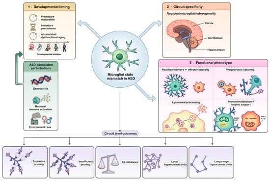
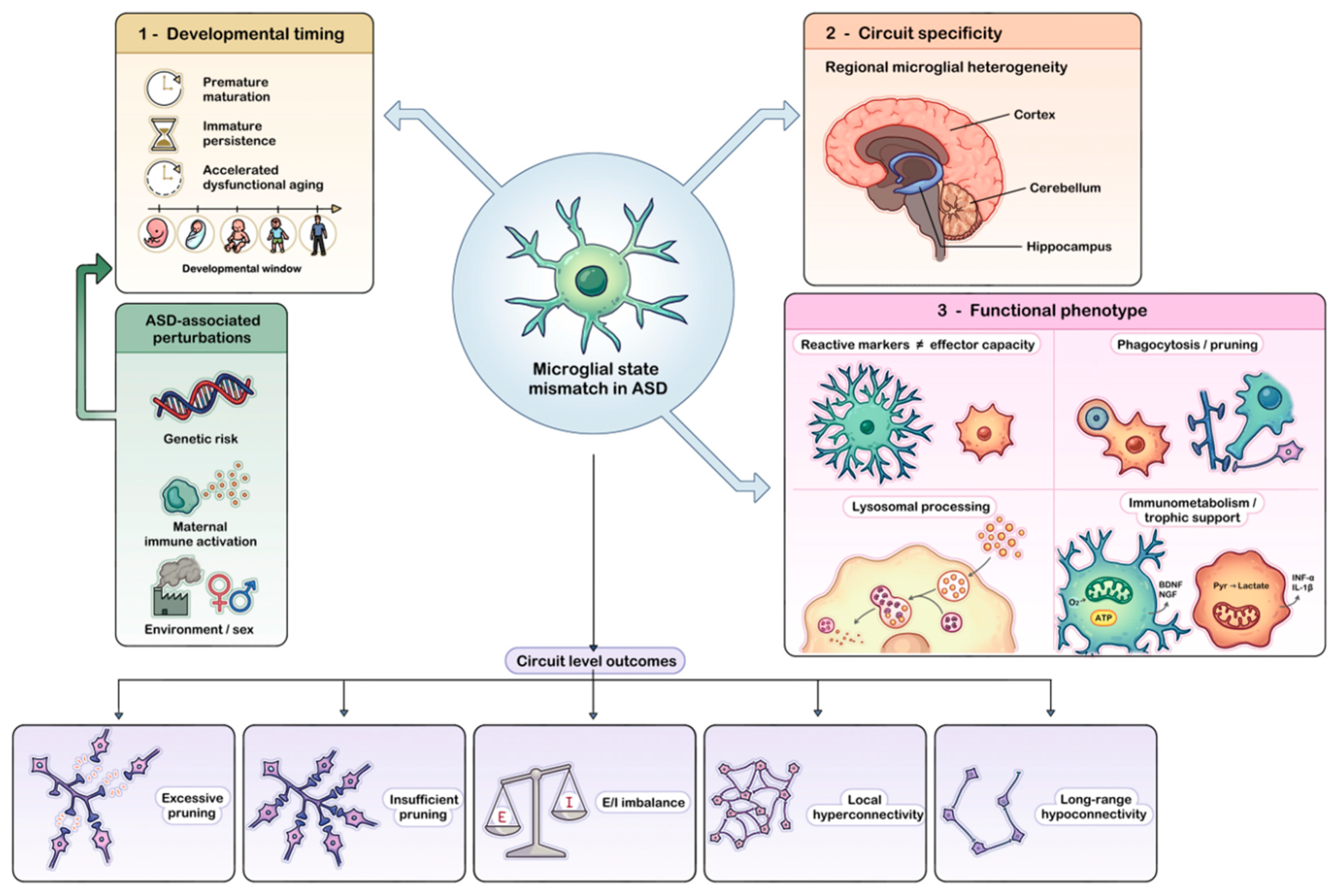
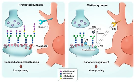
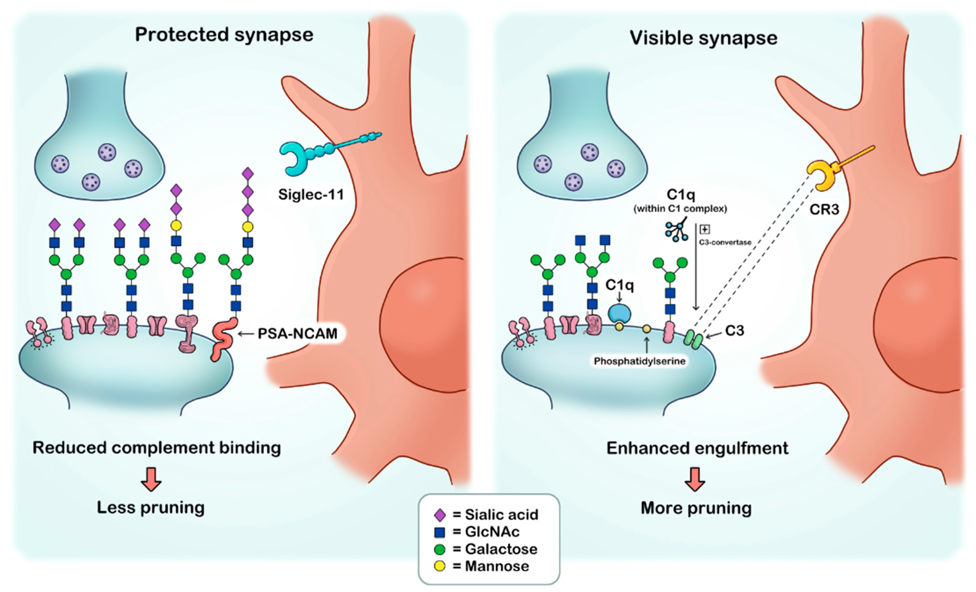

Disclaimer/Publisher’s Note: The statements, opinions and data contained in all publications are solely those of the individual author(s) and contributor(s) and not of MDPI and/or the editor(s). MDPI and/or the editor(s) disclaim responsibility for any injury to people or property resulting from any ideas, methods, instructions or products referred to in the content. |
© 2026 by the authors. Licensee MDPI, Basel, Switzerland. This article is an open access article distributed under the terms and conditions of the Creative Commons Attribution (CC BY) license.