


Abstract
Background/Objectives: Injuries to spinal ventral roots induce complex retrograde reactions that compromise motoneuron survival, synaptic organization, and glial responses, ultimately limiting the potential for regeneration. The endocannabinoid system (ECS) has emerged as a crucial modulator of neuroprotective processes, primarily through the activation of CB1 and CB2. However, the individual and combined contributions of these receptors to post-injury spinal responses remain poorly understood. Here, we examined the effects of selective blockade of CB1 and CB2 receptors in a murine model of ventral root crush (VRC). Methods: Female C57BL/6JUnib mice received daily intraperitoneal injections of the CB1 antagonist AM-251 and/or the CB2 antagonist AM-630 (1 mg/kg) for 14 days post-lesion. At 28 days after injury, spinal cords were analyzed for motoneuron survival (Nissl staining), glial responses (immunohistochemistry for GFAP and Iba-1), and synaptic coverage (immunohistochemistry for synaptophysin). Results: Selective blockade of CB2 receptors led to a marked reduction in motoneuron survival, enhanced microglial activation-associated morphology (morphological classification and Sholl analysis), and decreased synaptic coverage. CB1 blockade produced milder, context-dependent effects. Dual blockade exacerbated all outcomes, indicating complementary CB1/CB2 functions in the spinal microenvironment. Conclusions: Under the conditions tested, CB2 signaling is necessary for motoneuron preservation, limiting microglial activation-associated morphology, and maintaining synaptic coverage after VRC. The knowledge of specific actions of CB1 and CB2 provides mechanistic insight into the neuroprotective potential of endocannabinoid signaling and reinforces its therapeutic relevance for motoneuron preservation and functional recovery after axotomy.
1. Introduction
Spinal motoneurons (MNs) are the final common pathway for motor output, integrating multiple inputs to generate coordinated movements. These neurons are located in the ventral horn of the spinal cord and extend long axons through the ventral roots toward peripheral targets. Due to this anatomical organization, MNs are particularly vulnerable to axonal injuries, which can occur distally at the spinal nerves, or proximally at the ventral roots [1,2].
Although axonal regeneration and functional recovery are generally favorable following peripheral nerve injuries, regeneration after damage to the central nervous system (CNS)–peripheral nervous system (PNS) interface remains limited [3,4,5]. Lesions such as ventral root avulsion or crush result in significant motoneuron degeneration and synaptic detachment from the soma and proximal dendrites of surviving neurons. These events alter the spinal cord’s neurochemical environment, triggering robust glial activation that critically influence neuronal survival and repair [6,7,8].
Importantly, these pathological processes unfold in a defined temporal sequence following injury. The initial phase is characterized by early microglial activation accompanied by modest motoneuron loss and significant reduction in synaptic coverage. As injury progresses, both motoneuron degeneration and synaptic stripping intensify markedly. By 28 days, strong astrogliosis, sustained motoneuron loss and extensive synaptic detachment are well-established [7,8,9]. Therefore, the early post injury period, particularly the first two weeks, represents a critical therapeutic window for intervention, before these long-term deficits are permanently established.
Among the modulatory systems implicated in the regulation of neuroprotection, the endocannabinoid system (ECS) has emerged as a major homeostatic network. This lipid signaling system comprises the endocannabinoids anandamide (AEA) and 2-arachidonoylglycerol (2-AG), their metabolic enzymes, and the cannabinoid receptors CB1 and CB2 [10]. CB1 receptors are widely distributed across the CNS, with high densities in regions involved in motor and sensory processing, such as the cortex, basal ganglia, cerebellum, and spinal cord [11,12]. Notably, recent evidence indicated sex-dependent differences in CB1 expression and signaling, with females potentially exhibiting higher receptor density or sensitivity in specific CNS regions [13].
In contrast, CB2 receptors are sparsely expressed under physiological conditions but are rapidly upregulated on microglia following injury [14,15], which allows the ECS to regulate dynamic neuro-glial interactions. Activation of CB2 receptors typically promotes expression of anti-inflammatory factors by microglia, whereas CB1 signaling integrates metabolic and synaptic stability [16]. Consequently, the pharmacological modulation of these receptors represents a promising strategy to fine-tune the neuron-glia crosstalk and promote neuroprotection after axotomy [17,18,19].
In the present study, we employed a ventral root crush (VRC) model to investigate the effects of selectively blocking CB1 and CB2 receptors (individually or in combination) on cellular and molecular responses following motoneuron axotomy. Analyses were performed at 28 days post-lesion to evaluate the impact of this modulation on the consolidated glial and synaptic outcomes. This approach aimed to elucidate the distinct and potentially complementary roles of CB1- and CB2-mediated signaling in the spinal microenvironment after injury.
2. Materials and Methods
2.1. Animals and Experimental Groups
A total of 25 female wild-type C57BL/6JUnib naïve mice, (6–8 weeks, ~20 g) were obtained from the Multidisciplinary Center for Biological Research (CEMIB/UNICAMP). The mice were housed at the Laboratory of Nerve Regeneration (Institute of Biology—UNICAMP), under controlled lighting conditions (12/12 h light/dark cycle), humidity, and temperature (23 °C). The mice had free access to water and standard rodent chow. Environmental enrichment was provided by placing autoclavable polysulfone igloos inside the isolators and supplying paper-based nesting material.
Female mice were used in this study to maintain consistency with previous work from our group using the ventral root crush model [7,8]. Although male mice were not included, prior experiments in this model have shown that key outcome measures (including glial activation, motoneuron survival, and synaptic detachment) are comparable between males and females [9]. To minimize potential bias related to hormonal fluctuations, animals were housed together and arbitrary assigned to experimental groups using unique numerical identifiers [20]. This strategy enables unbiased data acquisition and analysis.
All mice underwent the ventral root crush procedure and were placed into one of the four experimental groups (n = 5/group): Vehicle (VE), AM-251, AM-630, and AM-251 + AM-630.
Sample size was estimated for a two-tailed comparison of two independent means with equal variances, using: . Assuming s = 0.2, α = 0.05, , 80% power () and a minimum detectable difference Δ ≈ 0.43, the formula yielded n = 3.36. We rounded up to 4 animals per group, with 5 animals allocated to account for potential experimental losses.
All experimental procedures were approved by the Institutional Committee for Ethics in Animal Experimentation (CEUA/IB/UNICAMP, protocol 5851-1/2021) and were carried out according to the guidelines of the Brazilian College of Animal Experimentation.
2.2. Surgical Procedure
The mice were anesthetized via an intraperitoneal injection of ketamine (100 mg/kg) and xylazine (10 mg/kg). Then, Liposic lubricating ophthalmic gel was applied to prevent dryness resulting from the loss of the eyelid reflex. Next, the dorsal fur was shaved, and the animals were placed on a heated platform. Anesthesia was maintained throughout the surgical procedure via the inhalation of Isoflurane (1% air volume). After confirming that the animal had reached a surgical plane of anesthesia through the absence of the paw withdrawal reflex, a dorsal incision was made parallel to the spine at the level of the lower thoracic and upper lumbar vertebrae. Hemilaminectomy was performed, and the dura-mater was opened to expose the L4, L5, and L6 ventral roots. The roots were crushed in three-10 s intervals using a # 4 Dumont forceps. After the procedure, the muscles were repositioned, and the skin was sutured with 8-0 mononylon thread. Animals were monitored in a heated, controlled environment until recovery from anesthesia. Immediately after the injury and for the following two days, the analgesic tramadol (Neoquimica, Anápolis, Brazil, 5 mg/kg) was administered.
The success of the surgical procedure was confirmed through histological analysis of spinal cord cross sections, as well as observation of animals’ motor behavior. Since the procedure was performed on the L4–L6 ventral roots of the spinal cord, the crushing significantly affected the somatic motor fibers of the sciatic nerve. This resulted in the paralysis of the ipsilateral pelvic limb of the injured animals [7,8,9]. In the present study, all the animals met the inclusion criteria, and no exclusions were made.
2.3. Preparation and Administration of Antagonists
A phosphate-buffered saline (PBS) solution containing 2% Tween 80 was used as the vehicle of dilution. The AM-251 (Abcam, Cambridge, UK—ab120088) and AM-630 (Cayman Chemical, An Harbor, MI, USA—10006974) antagonists were diluted at a dose of 1 mg/kg [21,22]. Administration began immediately after the surgery, via intraperitoneal injection, and was repeated every 24 h for 14 days.
The animals were examined for clinical signs of pain and distress including changes in body weight, decreased food or water intake, reduced locomotor activity, hunched posture, ruffled coat, abnormal vocalizations, and wound inflammation. Mice were evaluated daily during the first 14 days post-surgery and twice a week from days 15 to 28. Humane endpoints included intense body weight loss, severe persistent pain unresponsive to analgesia, severe wound complications, and inability to eat or drink. No adverse events meeting these criteria were observed during the study.
2.4. Perfusion and Tissue Processing
Twenty-eight days after the injury, the animals were euthanized with an anesthetic overdose (ketamine at 300 mg/kg and xylazine at 30 mg/kg) followed by transcardiac perfusion. First, a saline solution (0.9% NaCl in 0.1 M PB; pH 7.38) was injected to flush the organs and vessels. Then, a fixative solution (4% paraformaldehyde in 0.1 M PB; pH 7.38) was injected to preserve the tissue. The lumbar enlargements were then dissected out and immersed in the same fixative solution at 4 °C for 12 h. To cryopreserve the specimens, they were placed in solutions with increasing concentrations of sucrose (10%, 20%, and 30% in 0.1 M PB) for 12 h. The specimens were then embedded in Tissue-Tek and frozen at −33 °C. A cryostat (HM505, Microm, Walldorf, Germany) was used to obtain 12-μm-thick cross sections, which were then transferred to gelatin-coated slides. The slides were stored at −22 °C until analysis.
2.5. Motoneuron Counting
For staining, alternate slides containing spinal cord cross sections were immersed in a solution of 0.05% Toluidine Blue for 30 s. The sections were then washed and dehydrated in a series of alcohol solutions with increasing concentrations (70%, 80%, 95%, and 100%), before being diaphanized with xylene. After mounting the slides with Entellan (Merck, Darmstadt, Germany) and a coverslip, they were placed on wooden boards and left to dry for 24 h. Then, an optical microscope (DM5005, Leica, Wetzlar, Germany) was used to count and document motoneurons with visible nuclei and nucleoli on the ipsilateral and contralateral sides of the spinal cord. To avoid double-counting of neurons, the Abercrombie and Johnson formula was used as follows: N = (n · t) ÷ (t + d), where N = corrected neuron count; n = number of cells counted; t = distance between sections counted; d = mean cell diameter. For comparative purposes, the sum of all cells counted on each side was determined. Then, the ipsilateral/contralateral ratios were obtained for each animal within the experimental groups.
2.6. Immunohistochemistry
For immunohistochemistry, the obtained slides were acclimated, and the sections were outlined with a hydrophobic pen (PAP Pen Liquid Blocker, Ted Pella, Redding, CA, USA, Z377821). Then, the slides were placed in a dark humid chamber and washed three times with phosphate buffer (PB 0.01 M) for five minutes each. After washing, a blocking solution (3% bovine serum albumin, BSA, in PB 0.1 M) was added, and the slides were incubated for 45 min. Next, the slides were incubated with the primary antibodies: anti-GFAP (Abcam, Cambridge, UK, ab7260, 1/750); anti-Iba-1 (FUJIFILM Wako, Osaka, Japan, 019-19741, 1/750); or anti-synaptophysin (Novus Biologicals, Centennial, CO, USA, NBP2-25170, 1/1000) diluted in an incubation solution (1.5% BSA and 0.1% Tween in 0.1 M PB). The slides were kept in a closed humid chamber at room temperature for 4 h. After three five-minute washes with 0.01 PB, the slides were incubated with the corresponding secondary antibody (Alexa 488 anti-rabbit, Jackson ImmunoResearch, West Grove, PA, USA, 111.545.008, 1/500) and DAPI (D9542, Sigma-Aldrich, St. Louis, MO, USA, 1/1000) for 45 min. Then, the sections were washed again, dried, and mounted with a solution of glycerol/0.1 M PB (3:1) and coverslips.
Spinal cord cross sections from lumbar levels L4–L6 were analyzed using a Leica DM5500B fluorescence microscope equipped with a high-sensitivity digital camera (DFC345-FX, Leica, Wetzlar, Germany). Representative images were captured from both the ipsilateral and contralateral dorsolateral nuclei, across three nonconsecutive slides (one per lumbar level) for each animal and antibody. Images were acquired at 20× magnification with identical exposure and gain settings for each antibody.
2.7. Quantification of the Integrated Density of Pixels
The integrated density of pixels (IntDen) was quantified using ImageJ software (v. 1.8.0, National Institutes of Health, NIH, Bethesda, MD, USA). The images were converted to 8-bit grayscale, thresholded based on the respective RGB references, and then measured. For GFAP and Iba-1, IntDen covered the entire image. For synaptophysin, eight equidistant circular regions were placed around two axotomized motoneurons per section [7]. For analysis, the ipsilateral/contralateral ratios were computed per slide. Data-points represent the mean from three sections per animal.
2.8. Microglia Morphology
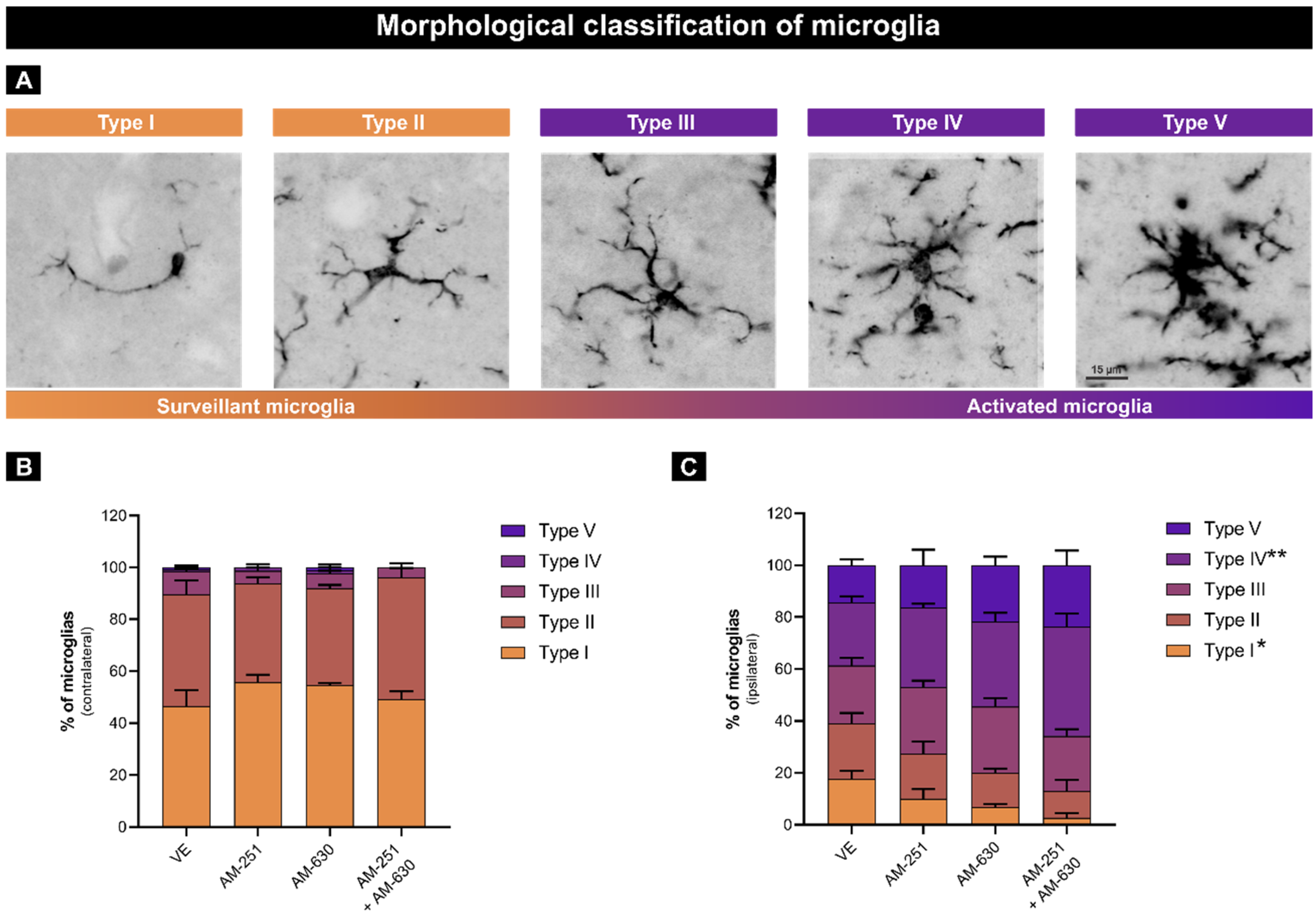
Microglial morphology was assessed in Iba-1-immunolabeled images using ImageJ software (NIH, USA). The cells were categorized into five types based on morphology: type I (two or fewer processes), type II (three to five short processes), type III (small soma with more than five long processes), type IV (enlarged soma with thick, retracted processes), and type V (amoeboid soma with numerous short processes). Types I and II are considered surveillant, and Types III–V are considered activated [23,24].
2.9. Sholl Analysis
Maximum intensity projection images (z-stacks with a total thickness ~ 8.84 μm total, post-deconvolution) of Iba-1-labeled sections were acquired under identical acquisition parameters. Nine clearly delineated microglia (defined cell body and processes) were analyzed per image from each side.
The images were processed in FIJI/ImageJ (version 1.54f): first, microglias were isolated and the images were thresholded, soma-centered, then analyzed via Sholl plugin with 5 μm concentric radii. The number and distribution of intersections were taken as indicators of microglial morphology. Surveillant microglia typically exhibit fewer, elongated branches resulting in a lower number of intersections distributed across larger radii. In contrast, activated microglia display shorter, thicker, and more numerous processes, yielding higher intersection counts within proximal radii [7].
2.10. Statistical Analyses
All data were first tested for normality using the Shapiro–Wilk test. Parametric data were analyzed using either one-way ANOVA followed by Tukey’s multiple comparisons test (for motoneuron survival, astrogliosis, microglial activation and synaptic coverage), or two-way ANOVA followed by Tukey’s multiple comparisons test (for microglial morphological classification and Sholl analysis). The level of statistical significance was set at p < 0.05. Data are presented as mean ± standard error of the mean (SEM). All statistical analyses were performed using GraphPad Prism (v.9.0.0).
3. Results
3.1. Motoneuron Survival
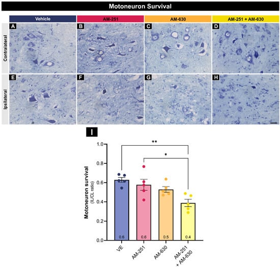
Twenty-eight days after the lesion, motoneuron survival was evaluated by the counting large motoneurons (more than 35 µm soma diameter) within the dorsolateral pool on the ipsilateral and contralateral sides. The ipsilateral-to-contralateral ratio was calculated and compared among each experimental group (Figure 1).
Figure 1.
Motoneuron survival 28 days after injury. Representative images from the contralateral (A–D) and ipsilateral (E–H) dorsolateral nuclei for Vehicle, AM-251, AM-630, and AM-251 + AM-630 groups, respectively (n = 5 mice/group). (I) The ipsilateral/contralateral ratios were compared using one-way ANOVA and Tukey post-test (* p < 0.05, ** p < 0.01). Data represent mean ± SEM. Scale bar = 50 µm.
The ipsilateral side exhibited a notable decrease in motoneuron number across all experimental groups. A one-way ANOVA revealed a significant effect of treatment on ipsilateral/contralateral ratios (F(3,16) = 6.77, p = 0.004). Treatment with AM 251 + AM630 significantly reduced motoneuron survival compared with the Vehicle (** p < 0.01), and AM-251 (* p < 0.05) groups (Vehicle = 0.63 ± 0.03; AM-251 = 0.58 ± 0.05, AM-630 = 0.53 ± 0.03, and AM-251 + AM-630 = 0.39 ± 0.04).
3.2. Glial Activation
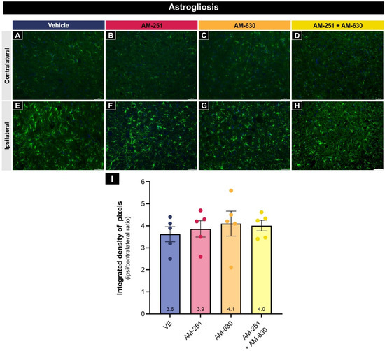
Twenty-eight days after ventral root crush, analysis of anti-GFAP immunostaining revealed a marked increase in the integrated density of pixels on the ipsilateral sides of all experimental groups (Figure 2). However, a one-way ANOVA revealed no significant differences among the experimental groups under the conditions tested (F(3,16) = 0.28, p = 0.84) (Vehicle = 3.62 ± 0.34; AM-251 = 3.86 ± 0.37; AM-630 = 4.10 ± 0.57; AM-251 + AM-630 = 4.01 ± 0.24).
Figure 2.
Astrogliosis (GFAP immunoreactivity) 28 days after injury. Representative fluorescence images showing contralateral (A–D) and ipsilateral (E–H) dorsolateral nuclei for Vehicle, AM-251, AM-630 and AM-251 + AM-630 groups (n = 5 mice/group). GFAP immunolabeling is depicted in green, and DAPI in blue. (I) Ipsilateral/contralateral IntDen ratios analyzed by one-way ANOVA and Tukey post-test (no significant intergroup differences in the conditions tested). Data show mean ± SEM. Scale bar = 50 µm.
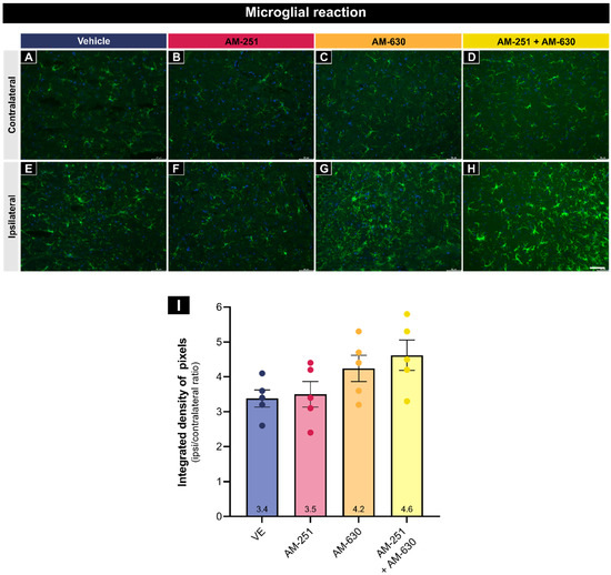
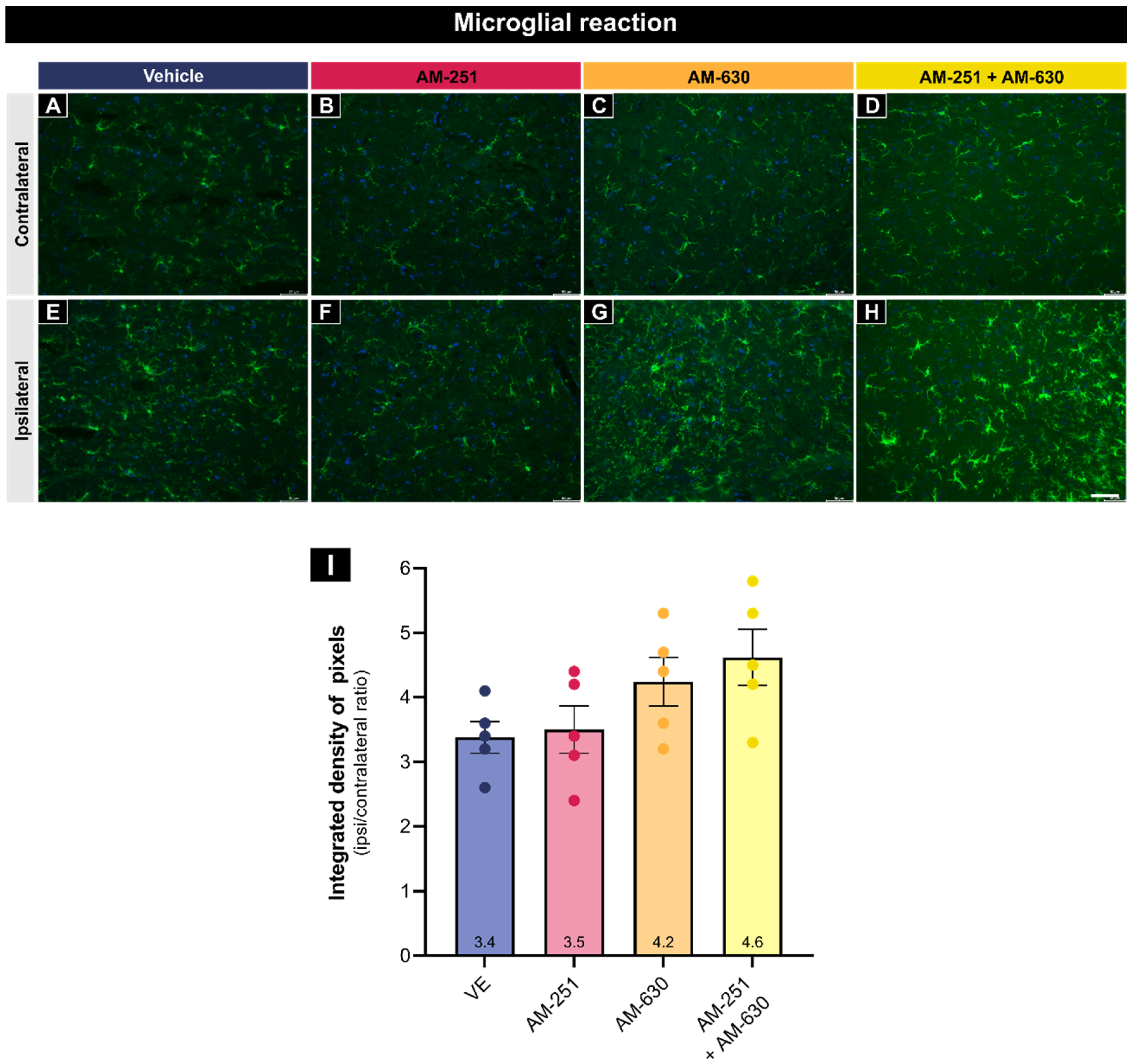
Similar results were obtained when analyzing Iba-1 immunolabeling. There was an increase in Iba-1 IntDen on the ipsilateral sides (Figure 3), and a one-way ANOVA revealed no detectable significant differences among the experimental groups under the conditions tested (F(3,16) = 2.68, p = 0.08) (Vehicle = 3.38 ± 0.25; AM-251 = 3.50 ± 0.37; AM-630 = 4.24 ± 0.38; AM-251 + AM-630 = 4.62 ± 0.44).
Figure 3.
Microglial activation (Iba-1 immunoreactivity) 28 days after injury. Representative fluorescence images showing contralateral (A–D) and ipsilateral (E–H) dorsolateral nuclei for Vehicle, AM-251, AM-630 and AM-251 + AM-630 groups (n = 5 mice/group). Iba-1 immunolabeling is depicted in green, and DAPI in blue. (I) Ipsilateral/contralateral IntDen ratios analyzed by one-way ANOVA and Tukey post-test (no significant intergroup differences in the conditions tested). Data show mean ± SEM. Scale bar = 50 µm.
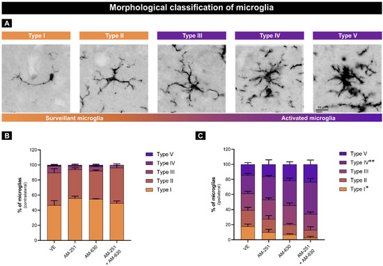
Since Iba-1 staining labels microglia regardless of their activation state, a morphological classification of Iba-1-positive cells into surveillant or activated was performed to better characterize the response after ventral root crush. The Iba-1-positive cells were classified into five types based on their morphological characteristics (Types I–II: surveillant; Types III–V: activated) (Figure 4).
Figure 4.
Morphological classification of microglia 28 days after injury. (A) Representative images of surveillant (I and II) and activated (III, IV, and V) types. Percentage distribution of microglia types on contralateral (B) and ipsilateral (C) sides across Vehicle, AM-251, AM-630 and AM-251 + AM-630 groups (n = 5 mice/group). Two-way ANOVA followed by Tukey post-test (* p < 0.05, ** p < 0.01). Data show mean ± SEM. Scale bar = 15 µm.
On the contralateral side, nearly all (89–96%) of the microglia were surveillant. Two-way ANOVA (treatment × type) revealed no main effect of treatment (F(3,80) ≈ 0, p > 0.99), dominant type effect (F(4,80) = 421.3, p < 0.0001), and interaction (F(12,80) = 1.93, p < 0.05). Tukey’s post hoc revealed that AM-251 treatment reduced Type I (* p < 0.05 vs. VE) and AM-251 + AM-630 decreased Type II (* p < 0.05 vs. AM-630).
On the ipsilateral side, 61–87% of the microglia surrounding axotomized motoneurons were classified as activated. Likewise, two-way ANOVA indicated no treatment effect (F(3,80) ≈ 0, p > 0.99), strong type effect (F(4,80) = 24.45, p < 0.0001), and interaction (F(12,80) = 2.88, p < 0.01). Tukey’s tests indicated that AM-251 + AM-630 treatment decreased surveillant-like Type I (* p < 0.05 vs. Vehicle) and increased reactive Type IV (** p < 0.01 vs. Vehicle).
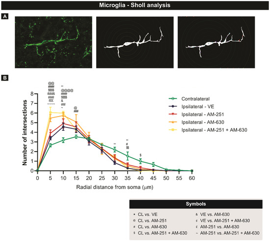
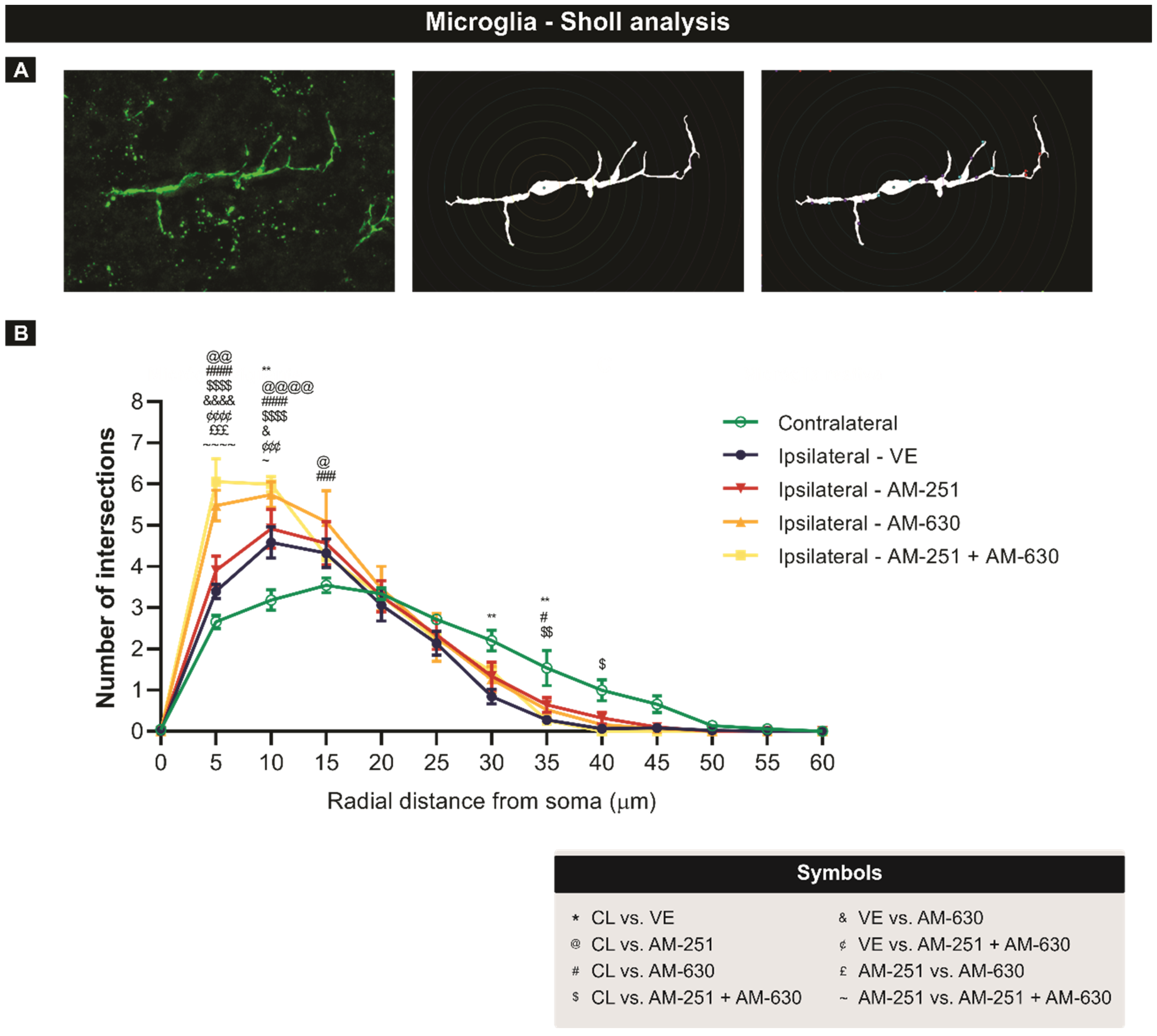
The influence of treatments on microglial activation was further quantified by Sholl analysis (Figure 5), which measures branching complexity by counting the number of intersections between microglial processes and concentric circles drawn at increasing distances from the soma. Two-way ANOVA (treatment × distance) revealed significant effects of treatment (F(12,260) = 280.50, p < 0.0001) and distance from soma (F(4,260) = 5.29, p < 0.001), and a highly significant interaction (F(48,260) = 5.25, p < 0.0001). Tukey’s post hoc tests confirmed a pronounced increase in ramification compared to the contralateral side, primarily at the proximal radii (5–15 µm) in the ipsilateral AM-630 and AM-251 + AM-630 groups (**** p < 0.0001). Thus, microglia on the ipsilateral side exhibited higher branch intersections within shorter radii compared to the contralateral side, particularly in the AM-630 and AM-251 + AM-630 groups. This profile indicates a shift from surveillant (long, fine processes) to reactive microglia morphology (compact, complex branching).
Figure 5.
Sholl analysis of microglial morphology 28 days after injury. (A) Example of cropped and thresholded Iba-1-stained microglia for Sholl analysis. (B) Number of processes intersections per 5 µm radial distance from the soma across the experimental groups. Two-way ANOVA and Tukey’s multiple comparisons test (~, &, @, #, $ p < 0.05, **, @@, $$ p < 0.01, ###, £££, ¢¢¢ p < 0.001, ####, &&&&, ¢¢¢¢,~~~~, @@@@, $$$$ p < 0.0001). Data show mean ± SEM.
3.3. Synaptic Preservation
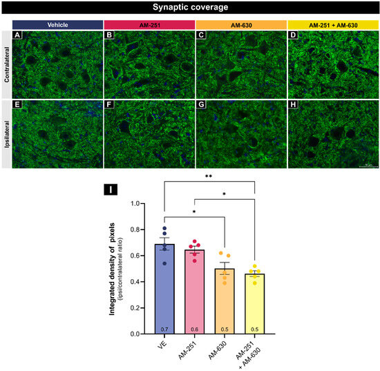
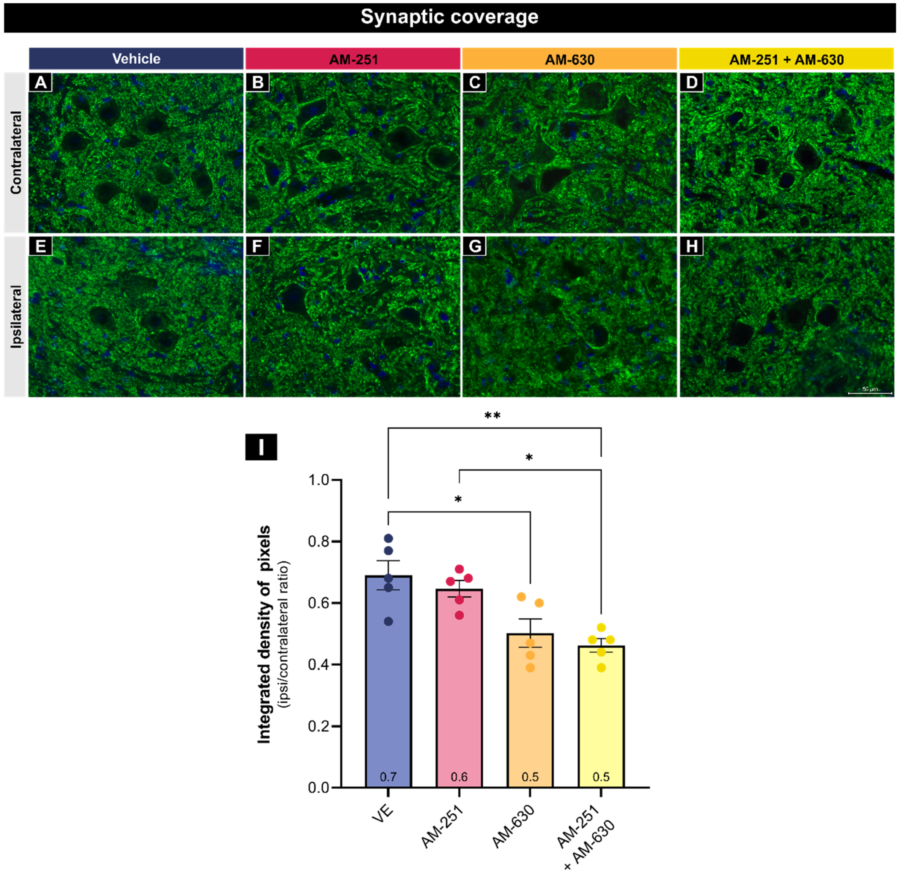
Another consequence of axotomy is the loss of synaptic inputs from the axotomized motoneuron. Synaptophysin, a protein found in the presynaptic neuronal vesicles, is a sensitive marker that can be used to evaluate the synaptic coverage after injury. Due to synaptic loss, the ipsilateral side of the lesion is expected to exhibit lower immunofluorescence than the contralateral side, as observed in all experimental groups (Figure 6).
Figure 6.
Synaptic coverage (synaptophysin immunoreactivity) 28 days after injury. Representative fluorescence images showing contralateral (A–D) and ipsilateral (E–H) dorsolateral nuclei for Vehicle, AM-251, AM-630 and AM-251 + AM-630 groups (n = 5 mice/group). Synaptophysin immunolabeling is depicted in green, and DAPI in blue. (I) Ipsilateral/contralateral IntDen ratios analyzed by one-way ANOVA and Tukey post-test (* p < 0.05, ** p < 0.01). Data show mean ± SEM. Scale bar = 50 µm.
A one-way ANOVA revealed a significant effect of treatment on ipsilateral/contralateral ratios (F(3,16) = 8.70, p = 0.001). Tukey’s post hoc test revealed that AM-630 treatment led to significant decrease in the synaptic coverage compared with the Vehicle (** p < 0.01). AM-251 + AM-630 treatment also led to a significant decrease in synaptic coverage compared to the Vehicle (** p < 0.01) and AM-251 (* p < 0.05) groups (Vehicle = 0.69 ± 0.05; AM-251 = 0.64 ± 0.03, AM-630 = 0.50 ± 0.05, and AM-251 + AM-630 = 0.46 ± 0.02).
4. Discussion
The nerve supply to the limbs is characterized by a complex intertwining of fibers originating from different spinal segments, which makes functional recovery after injury particularly challenging. Peripheral nerves are composed of relatively delicate structures that are highly susceptible to trauma from diverse causes and in multiple regions of the body [25,26]. Their regeneration is limited because it requires the regrowth of the injured axon, and the coordination of numerous cellular and molecular mechanisms. Thus, despite the intrinsic regenerative capacity of the PNS, the outcomes are often incomplete, and full recovery remains rare [27]. The endocannabinoid system has long been associated with processes related to neuroprotection and neuroregeneration. Its receptors, CB1 and CB2, are widely distributed in neural and glial cells and are known to modulate pathways involved in neuronal survival, synaptic plasticity, and responses to injury or inflammation [28,29,30]. These characteristics make them particularly attractive as therapeutic targets for neuroprotective interventions.
Following ventral root crush, the injured microenvironment often fails to sustain an effective pro-regenerative response. This typically results in the death of approximately 40% of affected motoneurons [7,9], a finding that was also observed in the Vehicle group in the motoneuron survival analysis described herein. A key finding of this study is that the motoneuron loss was significantly greater when the CB2 receptor was pharmacologically blocked by AM-630. Dual blockade of CB1 and CB2 exacerbated motoneuron loss, which highlights the critical contribution of the ECS to neuroprotection.
Previous findings from our group had already pointed to the involvement of the ECS in neuroprotection of spinal motoneurons. In a model of peripheral nerve injury in neonatal rats, blocking CB1 or CB1 + CB2 receptors was found to enhance motoneuron survival, while decreasing microglial activation and astrogliosis [31]. These results differ from those described above, indicating that the effect of blocking CB1 or CB2 receptors depends on the type of injury and varies between the neonatal phase and adulthood. Such differences are supported by developmental differences in ECS functions. During the neonatal phase, the ECS is highly active, and plays essential roles in neurogenesis and protecting against hypoxic–ischemic and excitotoxic damage, primarily via CB1 receptors [32]. In adults, the ECS continues to regulate homeostasis, but shifts toward neuroprotection and immunomodulation. After injury or inflammation, CB2 receptors become more involved [33].
Effects of CB2 on glial cells were observed in our study. Although general glial activation analyses using IntDen quantification of immunofluorescence images did not reveal a significant difference among the experimental groups, the quantification of the microglial morphological types and Sholl analyses indicated that the AM-251 + AM-630 group had a higher rate of microglia with activation-associated morphology. These results suggest that ECS plays an important role in controlling chronic glial activation after VRC, consistent with previous reports [24,32,33]. Although microglial morphology and Sholl analysis provide robust indicators of activation, they do not directly define specific functional or molecular phenotypes. Similarly, GFAP immunoreactivity alone may not fully capture the functional diversity of astrocyte phenotypes after injury. Complementary approaches such as RNA sequencing or assessments of phagocytic capacity would strengthen these conclusions. Likewise, several other studies support this interpretation. Adhikary et al. [34] reported improvements in motor and autonomic function associated with reduced inflammatory responses in mice following spinal cord injury and treatment with O-1966, a selective CB2 agonist. Baty, Zhang, Li, Erb, Adler, Ganea, Loftus, Jallo and Tuma [17] demonstrated that CB2 activation through O-1966 enhances motor recovery after peripheral nerve contusion. Similarly, Arevalo-Martin et al. [35] showed that CB2 blockade increased the number pro-inflammatory microglia and macrophages in the spinal cord, further supporting the receptor’s role in regulating glial activation.
In addition to regulating inflammatory tone, ECS may influence the structural remodeling of spinal circuits following axotomy. Following motoneuron axotomy, microglia typically become activated and participate in the selective removal of afferent terminals, in a process known as synaptic stripping [1,36]. However, the extent and specificity of this process are critically governed by endocannabinoid signaling. Under physiological conditions, endocannabinoid-mediated CB2 receptor engagement acts as a brake on excessive microglial phagocytic activity [14]. Nevertheless, unregulated microglial activity can deplete the synaptic inputs necessary for motoneuron reinnervation and restoration of synaptic connectivity, which is essential for functional recovery, including spinal reflexes [8,37,38]. Thus, our findings demonstrate that the pharmacological modulation of cannabinoid receptor signaling decreases synaptic coverage around axotomized motoneurons, particularly via CB2 or dual CB1 + CB2 antagonism. These results suggest that the endogenous endocannabinoid response after VRC supports not only neuronal survival but also the maintenance of essential synaptic circuitry.
Notably, in our work, simultaneous blockade of both cannabinoid receptors resulted in the worst outcomes. Previous studies have shown that CB2 exhibits consistent and robust upregulation, primarily on activated glial cells, whereas CB1 exhibits more complex patterns, including upregulation in certain regions and downregulation in others. This pattern likely reflects context-dependent plasticity rather than a uniform adaptive response [39,40]. It is important to note that, although clinically relevant, systemic pharmacological blockade lacks cellular specificity and cannot fully exclude off-target effects on non-neural tissues. Cell-type-specific genetic approaches would help clarify CB2’s cell-autonomous contributions to neuroprotection.
Additionally, there are other significant limitations of the study warrant discussion. While female mice were used to maintain consistency with previous studies from our group, recent evidence suggests that there are sex-dependent differences in cannabinoid receptor density, particularly for CB1 [13]. Although our previous studies in this injury model found comparable glial and neuronal outcomes between sexes [7,8,9], explicitly investigating of male versus female responses to ECS modulation within the VRC context would be valuable for understanding potential dimorphic mechanisms.
Taken together, our findings suggest that CB2 receptor signaling is necessary for optimal neuronal survival and glial activation responses following VRC. By preserving microglial activation-associated morphology and maintaining synaptic coverage, the ECS contributes to a permissive environment for recovery. These results not only advance our understanding of ECS-mediated neuroprotection in adult motoneurons but also suggest that pharmacological targeting of CB2 may represent a promising strategy to enhance regeneration after axotomy. Future studies should explore the temporal dynamics of CB2 activation and its interaction with other neuroimmune pathways to better define its therapeutic potential.
5. Conclusions
This study demonstrates that CB2 receptor activity is necessary for the survival of motoneurons and for the regulation of microglial responses after VRC. Pharmacological blockade of CB2, or CB1 + CB2, exacerbated neuronal loss and microglial activation, indicating that, under the conditions tested, endogenous CB2 signaling contributes to neuroprotection and maintenance of synaptic integrity. These findings highlight CB2 as a potential therapeutic target to promote repair and functional recovery following peripheral nerve trauma.
Author Contributions
C.M.T.: Methodology, Investigation, Writing—original draft, Data Visualization; A.L.R.d.O.: Conceptualization, Methodology, Writing—review and editing, Supervision and Funding; L.P.C.: Conceptualization, Methodology, Writing—review and editing, Data Visualization, Supervision and Funding. All authors have read and agreed to the published version of the manuscript.
Funding
This work was supported by the São Paulo Research Foundation—FAPESP (grant numbers, 2022/06609-6, 2022/05240-9, 2023/16415-7, 2023/02615-4) and the National Council for Scientific and Technological Development—CNPq (grant number 303050/2021-7).
Institutional Review Board Statement
The animal study protocol was approved by the National Council for Animal Experimentation Control (CONCEA—Brazil) and the Institutional Ethics Committee on the Use of Animals (CEUA/IB/UNICAMP—protocol number 5851-1/2021) on 19 August 2021.
Informed Consent Statement
Not applicable.
Data Availability Statement
Dataset available on request from the authors.
Acknowledgments
We would like to thank Francisco da Silveira Guimarães of the University of São Paulo in Ribeirão Preto for his invaluable partnership and insightful suggestions during the initial conception of this work.
Conflicts of Interest
The authors declare no conflicts of interest.
References
- Alvarez, F.J.; Rotterman, T.M.; Akhter, E.T.; Lane, A.R.; English, A.W.; Cope, T.C. Synaptic Plasticity on Motoneurons After Axotomy: A Necessary Change in Paradigm. Front. Mol. Neurosci. 2020, 13, 68. [Google Scholar] [CrossRef] [Scilit]
- Carlstedt, T. Central Nerve Plexus Injury; Imperial College Press: London, UK, 2007; p. 191. [Google Scholar]
- Alvarez, F.J.; Bullinger, K.L.; Titus, H.E.; Nardelli, P.; Cope, T.C. Permanent reorganization of Ia afferent synapses on motoneurons after peripheral nerve injuries. Ann. N. Y. Acad. Sci. 2010, 1198, 231–241. [Google Scholar] [CrossRef] [Scilit]
- Carlstedt, T.; Risling, M.; Linda, H.; Cullheim, S.; Ulfhake, B.; Sjogren, A.M. Regeneration after spinal nerve root injury. Restor. Neurol. Neurosci. 1990, 1, 289–295. [Google Scholar] [CrossRef] [Scilit]
- Gaudet, A.D.; Popovich, P.G.; Ramer, M.S. Wallerian degeneration: Gaining perspective on inflammatory events after peripheral nerve injury. J. Neuroinflamm. 2011, 8, 110. [Google Scholar] [CrossRef] [Scilit]
- Aldskogius, H.; Liu, L.; Svensson, M. Glial responses to synaptic damage and plasticity. J. Neurosci. Res. 1999, 58, 33–41. [Google Scholar] [CrossRef]
- Cartarozzi, L.P.; Perez, M.; Fernandes, G.G.; Chiarotto, G.B.; Luzo, A.C.M.; Campos, A.C.; Kirchhoff, F.; de Oliveira, A.L.R. Neuroprotection and gliosis attenuation by intravenous application of human mesenchymal stem cells (hMSC) following ventral root crush in mice. Mol. Cell. Neurosci. 2022, 118, 103694. [Google Scholar] [CrossRef] [Scilit]
- Cartarozzi, L.P.; Perez, M.; Kirchhoff, F.; Oliveira, A.L.R. Role of MHC-I Expression on Spinal Motoneuron Survival and Glial Reactions Following Ventral Root Crush in Mice. Cells 2019, 8, 483. [Google Scholar] [CrossRef] [Scilit] [PubMed]
- Cartarozzi, L.P.; de Melo Lima, B.H.; Tomiyama, A.; Santos, F.R.; de Oliveira, A.L.R. Toll-like receptors 2 and 4 influence neuronal survival and glial reactions following ventral root crush injury in mice. J. Neuroimmunol. 2025, 406, 578655. [Google Scholar] [CrossRef] [Scilit] [PubMed]
- Bernal-Chico, A.; Tepavcevic, V.; Manterola, A.; Utrilla, C.; Matute, C.; Mato, S. Endocannabinoid signaling in brain diseases: Emerging relevance of glial cells. Glia 2023, 71, 103–126. [Google Scholar] [CrossRef] [Scilit] [PubMed]
- Mackie, K. Distribution of cannabinoid receptors in the central and peripheral nervous system. In Cannabinoids; Handbook of Experimental Pharmacology (HEP, Volume 168); Springer: Berlin/Heidelberg, Germany, 2005; pp. 299–325. [Google Scholar] [CrossRef] [Scilit]
- Salio, C.; Doly, S.; Fischer, J.; Franzoni, M.F.; Conrath, M. Neuronal and astrocytic localization of the cannabinoid receptor-1 in the dorsal horn of the rat spinal cord. Neurosci. Lett. 2002, 329, 13–16. [Google Scholar] [CrossRef] [Scilit]
- Corli, G.; Roda, E.; Tirri, M.; Bilel, S.; De Luca, F.; Strano-Rossi, S.; Gaudio, R.M.; De-Giorgio, F.; Fattore, L.; Locatelli, C.A.; et al. Sex-specific behavioural, metabolic, and immunohistochemical changes after repeated administration of the synthetic cannabinoid AKB48 in mice. Br. J. Pharmacol. 2024, 181, 1361–1382. [Google Scholar] [CrossRef] [Scilit]
- Duffy, S.S.; Hayes, J.P.; Fiore, N.T.; Moalem-Taylor, G. The cannabinoid system and microglia in health and disease. Neuropharmacology 2021, 190, 108555. [Google Scholar] [CrossRef] [Scilit]
- Lopez, A.; Aparicio, N.; Pazos, M.R.; Grande, M.T.; Barreda-Manso, M.A.; Benito-Cuesta, I.; Vazquez, C.; Amores, M.; Ruiz-Perez, G.; Garcia-Garcia, E.; et al. Cannabinoid CB(2) receptors in the mouse brain: Relevance for Alzheimer’s disease. J. Neuroinflamm. 2018, 15, 158. [Google Scholar] [CrossRef] [Scilit] [PubMed]
- Martinez Ramirez, C.E.; Ruiz-Perez, G.; Stollenwerk, T.M.; Behlke, C.; Doherty, A.; Hillard, C.J. Endocannabinoid signaling in the central nervous system. Glia 2023, 71, 5–35. [Google Scholar] [CrossRef] [Scilit] [PubMed]
- Baty, D.E.; Zhang, M.; Li, H.; Erb, C.J.; Adler, M.W.; Ganea, D.; Loftus, C.M.; Jallo, J.I.; Tuma, R.F. Cannabinoid CB2 receptor activation attenuates motor and autonomic function deficits in a mouse model of spinal cord injury. Clin. Neurosurg. 2008, 55, 172–177. [Google Scholar] [CrossRef] [Scilit] [PubMed]
- Grundy, R.I.; Rabuffetti, M.; Beltramo, M. Cannabinoids and neuroprotection. Mol. Neurobiol. 2001, 24, 29–51. [Google Scholar] [CrossRef] [Scilit]
- Sarne, Y.; Mechoulam, R. Cannabinoids: Between neuroprotection and neurotoxicity. Curr. Drug Targets CNS Neurol. Disord. 2005, 4, 677–684. [Google Scholar] [CrossRef] [Scilit]
- Prendergast, B.J.; Onishi, K.G.; Zucker, I. Female mice liberated for inclusion in neuroscience and biomedical research. Neurosci. Biobehav. Rev. 2014, 40, 1–5. [Google Scholar] [CrossRef] [Scilit]
- Fogaca, M.V.; Campos, A.C.; Coelho, L.D.; Duman, R.S.; Guimaraes, F.S. The anxiolytic effects of cannabidiol in chronically stressed mice are mediated by the endocannabinoid system: Role of neurogenesis and dendritic remodeling. Neuropharmacology 2018, 135, 22–33. [Google Scholar] [CrossRef] [Scilit]
- Perez, M.; Cartarozzi, L.P.; Chiarotto, G.B.; Guimaraes, F.S.; Oliveira, A.L.R. Short and long-term neuroprotective effects of cannabidiol after neonatal peripheral nerve axotomy. Neuropharmacology 2021, 197, 108726. [Google Scholar] [CrossRef] [Scilit]
- Diz-Chaves, Y.; Pernia, O.; Carrero, P.; Garcia-Segura, L.M. Prenatal stress causes alterations in the morphology of microglia and the inflammatory response of the hippocampus of adult female mice. J. Neuroinflamm. 2012, 9, 71. [Google Scholar] [CrossRef] [Scilit] [PubMed]
- Lopez-Rodriguez, A.B.; Siopi, E.; Finn, D.P.; Marchand-Leroux, C.; Garcia-Segura, L.M.; Jafarian-Tehrani, M.; Viveros, M.P. CB1 and CB2 cannabinoid receptor antagonists prevent minocycline-induced neuroprotection following traumatic brain injury in mice. Cereb. Cortex 2015, 25, 35–45. [Google Scholar] [CrossRef] [Scilit] [PubMed]
- Hussain, G.; Wang, J.; Rasul, A.; Anwar, H.; Qasim, M.; Zafar, S.; Aziz, N.; Razzaq, A.; Hussain, R.; de Aguilar, J.G.; et al. Current Status of Therapeutic Approaches against Peripheral Nerve Injuries: A Detailed Story from Injury to Recovery. Int. J. Biol. Sci. 2020, 16, 116–134. [Google Scholar] [CrossRef] [Scilit] [PubMed]
- Lundborg, G.; Dahlin, L.B. Anatomy, function, and pathophysiology of peripheral nerves and nerve compression. Hand Clin. 1996, 12, 185–193. [Google Scholar] [CrossRef] [Scilit]
- Huebner, E.A.; Strittmatter, S.M. Axon regeneration in the peripheral and central nervous systems. In Cell Biology of the Axon; Results and Problems in Cell Differentiation (RESULTS, Volume 48); Springer: Berlin/Heidelberg, Germany, 2009; pp. 339–351. [Google Scholar] [CrossRef] [Scilit]
- Di Marzo, V.; Bifulco, M.; De Petrocellis, L. The endocannabinoid system and its therapeutic exploitation. Nat. Rev. Drug Discov. 2004, 3, 771–784. [Google Scholar] [CrossRef] [Scilit]
- Grundy, R.I. The therapeutic potential of the cannabinoids in neuroprotection. Expert Opin. Investig. Drugs 2002, 11, 1365–1374. [Google Scholar] [CrossRef] [Scilit]
- Lu, H.C.; Mackie, K. An Introduction to the Endogenous Cannabinoid System. Biol. Psychiatry 2016, 79, 516–525. [Google Scholar] [CrossRef] [Scilit]
- Perez, M.; Barroso Spejo, A.; Bortolanca Chiarotto, G.; Silveira Guimaraes, F.; Leite Rodrigues de Oliveira, A.; Politti Cartarozzi, L. Selective blockade of cannabinoid receptors influences motoneuron survival and glial responses after neonatal axotomy. Neuroscience 2025, 565, 265–276. [Google Scholar] [CrossRef] [Scilit]
- Shouman, B.; Fontaine, R.H.; Baud, O.; Schwendimann, L.; Keller, M.; Spedding, M.; Lelievre, V.; Gressens, P. Endocannabinoids potently protect the newborn brain against AMPA-kainate receptor-mediated excitotoxic damage. Br. J. Pharmacol. 2006, 148, 442–451. [Google Scholar] [CrossRef] [Scilit]
- Duranti, A.; Beldarrain, G.; Alvarez, A.; Sbriscia, M.; Carloni, S.; Balduini, W.; Alonso-Alconada, D. The Endocannabinoid System as a Target for Neuroprotection/Neuroregeneration in Perinatal Hypoxic-Ischemic Brain Injury. Biomedicines 2022, 11, 28. [Google Scholar] [CrossRef] [Scilit]
- Adhikary, S.; Li, H.; Heller, J.; Skarica, M.; Zhang, M.; Ganea, D.; Tuma, R.F. Modulation of inflammatory responses by a cannabinoid-2-selective agonist after spinal cord injury. J. Neurotrauma 2011, 28, 2417–2427. [Google Scholar] [CrossRef] [Scilit]
- Arevalo-Martin, A.; Molina-Holgado, E.; Garcia-Ovejero, D. Cannabinoids to treat spinal cord injury. Prog. Neuropsychopharmacol. Biol. Psychiatry 2016, 64, 190–199. [Google Scholar] [CrossRef] [Scilit]
- Oliveira, A.L.; Thams, S.; Lidman, O.; Piehl, F.; Hokfelt, T.; Karre, K.; Linda, H.; Cullheim, S. A role for MHC class I molecules in synaptic plasticity and regeneration of neurons after axotomy. Proc. Natl. Acad. Sci. USA 2004, 101, 17843–17848. [Google Scholar] [CrossRef] [Scilit]
- Rotterman, T.M.; Akhter, E.T.; Lane, A.R.; MacPherson, K.P.; Garcia, V.V.; Tansey, M.G.; Alvarez, F.J. Spinal Motor Circuit Synaptic Plasticity after Peripheral Nerve Injury Depends on Microglia Activation and a CCR2 Mechanism. J. Neurosci. 2019, 39, 3412–3433. [Google Scholar] [CrossRef] [Scilit]
- Rotterman, T.M.; Alvarez, F.J. Microglia Dynamics and Interactions with Motoneurons Axotomized After Nerve Injuries Revealed By Two-Photon Imaging. Sci. Rep. 2020, 10, 8648. [Google Scholar] [CrossRef] [Scilit]
- Arevalo-Martin, A.; Garcia-Ovejero, D.; Sierra-Palomares, Y.; Paniagua-Torija, B.; Gonzalez-Gil, I.; Ortega-Gutierrez, S.; Molina-Holgado, E. Early endogenous activation of CB1 and CB2 receptors after spinal cord injury is a protective response involved in spontaneous recovery. PLoS ONE 2012, 7, e49057. [Google Scholar] [CrossRef] [Scilit]
- Braun, M.; Khan, Z.T.; Khan, M.B.; Kumar, M.; Ward, A.; Achyut, B.R.; Arbab, A.S.; Hess, D.C.; Hoda, M.N.; Baban, B.; et al. Selective activation of cannabinoid receptor-2 reduces neuroinflammation after traumatic brain injury via alternative macrophage polarization. Brain Behav. Immun. 2018, 68, 224–237. [Google Scholar] [CrossRef] [Scilit]
Disclaimer/Publisher’s Note: The statements, opinions and data contained in all publications are solely those of the individual author(s) and contributor(s) and not of MDPI and/or the editor(s). MDPI and/or the editor(s) disclaim responsibility for any injury to people or property resulting from any ideas, methods, instructions or products referred to in the content. |
© 2026 by the authors. Licensee MDPI, Basel, Switzerland. This article is an open access article distributed under the terms and conditions of the Creative Commons Attribution (CC BY) license.