


Abstract
Recently, an anti-inflammatory effect of no-ozone cold plasma (NCP) has been reported, but the direct use of NCP for treating muscle inflammation is very difficult since NCP is a form of gas. In this study, we tested whether the anti-inflammatory effect of the NCP could be delivered through conductive metal needles to reduce muscle inflammation. C2C12 mouse muscle cells were treated with TNFα to induce muscle inflammation and then treated with NCP and a conductive metal needle separately or in combination. The effects of NCP and a needle were monitored by performing RT-PCR and Western blot analysis. As a result, NCP effectively suppressed the TNFα-mediated expression of the TNFα, IL1β, and FasL genes, but this effect weakened as the distance between the cells and the NCP increased. On the other hand, treatment of cells with a plasma-needle (PN) had an anti-inflammatory effect regardless of distance, and the anti-inflammatory effect of the PN was maintained under conditions where the gas flow of NCP was not delivered to the cells. It is believed that the PN-mediated activation of media plays a pivotal role in the anti-inflammatory effect of the PN. Finally, this study also showed that electroacupuncture can inhibit TNFα-induced inflammatory gene expression in a manner like a PN. Taken together, the results of this study demonstrate that the anti-inflammatory effect of NCP can be delivered through metal needles, suggesting that PN may be useful for treating inflammatory muscle pain.
1. Introduction
Muscle pain is one of the most common symptoms that occur in adults in various areas, such as the neck and shoulders [1,2]. Diseases related to muscle pain include polymyositis caused by an inflammatory response [3]; fibromyalgia, which causes chronic pain in soft tissues [4]; and delayed onset muscle soreness caused by damage to muscle connective tissue due to high-intensity exercise activities [5]. Various mechanisms have been identified as the cause of muscle pain, but in general, when muscles are overused or strained, inflammation within the muscle is induced. If this occurs repeatedly, muscle pain can occur due to an accumulated inflammatory response [6]. Muscle nociceptors can be stimulated under inflammatory conditions, which can cause muscle pain [7]. Proinflammatory cytokines such as TNFα, IL1β, and IL6 are involved in the nociceptor response [8]. Muscle pain can be reduced through treatment methods that can improve the recovery of muscle damage and reduce the inflammatory response. Common treatment methods for muscle pain include the use of steroids, immunosuppressants, and immunomodulators to reduce inflammation [9] and nondrug treatments such as acupuncture [10], electrical stimulation, and surgery. Physical therapy and exercise are also known to help relieve pain.
Plasma is a physical term meaning ionized gas, and various studies have recently been conducted on the use of plasma in the medical field [11,12]. To date, many biomedical functions of plasma, such as antimicrobial [13], wound healing [14], anticancer [15] and transdermal drug delivery [16], have been reported. We developed a unique plasma-generation technique called no-ozone cold plasma (NCP), which can minimize the production of harmful substances, such as ozone and nitrogen oxides, and can deliver high-density plasma flow at 27 °C. In our previous studies, the antimicrobial, anti-inflammatory, and tissue-renewal activities of NCP were reported [17,18,19,20]. However, although there are many biological functions of NCP, the direct effect of NCP on treating the tissue surface is limited since NCP is present in the gas phase. To treat muscle pain using NCP, the anti-inflammatory activity of the NCP must be delivered to muscle tissues. Therefore, there is a need to develop new methods for delivering several biological functions of NCP to deeper tissues of the body.
In our previous study, the transdermal drug delivery activity of NCP was removed by the presence of electronically grounded mesh between the NCP device and the cells [21]. These results suggest that some of the biological effects of NCP might be delivered through conductive materials. Therefore, in the present study, we tested whether the anti-inflammatory activity of NCP can be delivered through a conductive metal needle, which is used for electroacupuncture, in an in vitro muscle inflammation model. For this, we developed a new cell treatment method that combines NCP with a needle, called plasma-needle (PN), and tested its cell toxicity and anti-inflammatory activity in C2C12 mice muscle cells. Furthermore, we performed several experiments to elucidate the mechanism of action of the PN and compared its effectiveness with that of electroacupuncture.
2. Materials and Methods
2.1. Reagents
All chemicals were purchased from Sigma-Aldrich (St. Louis, MO, USA) unless otherwise indicated.
2.2. NCP Device, Needle and Lids for Plasma-Needle Treatment
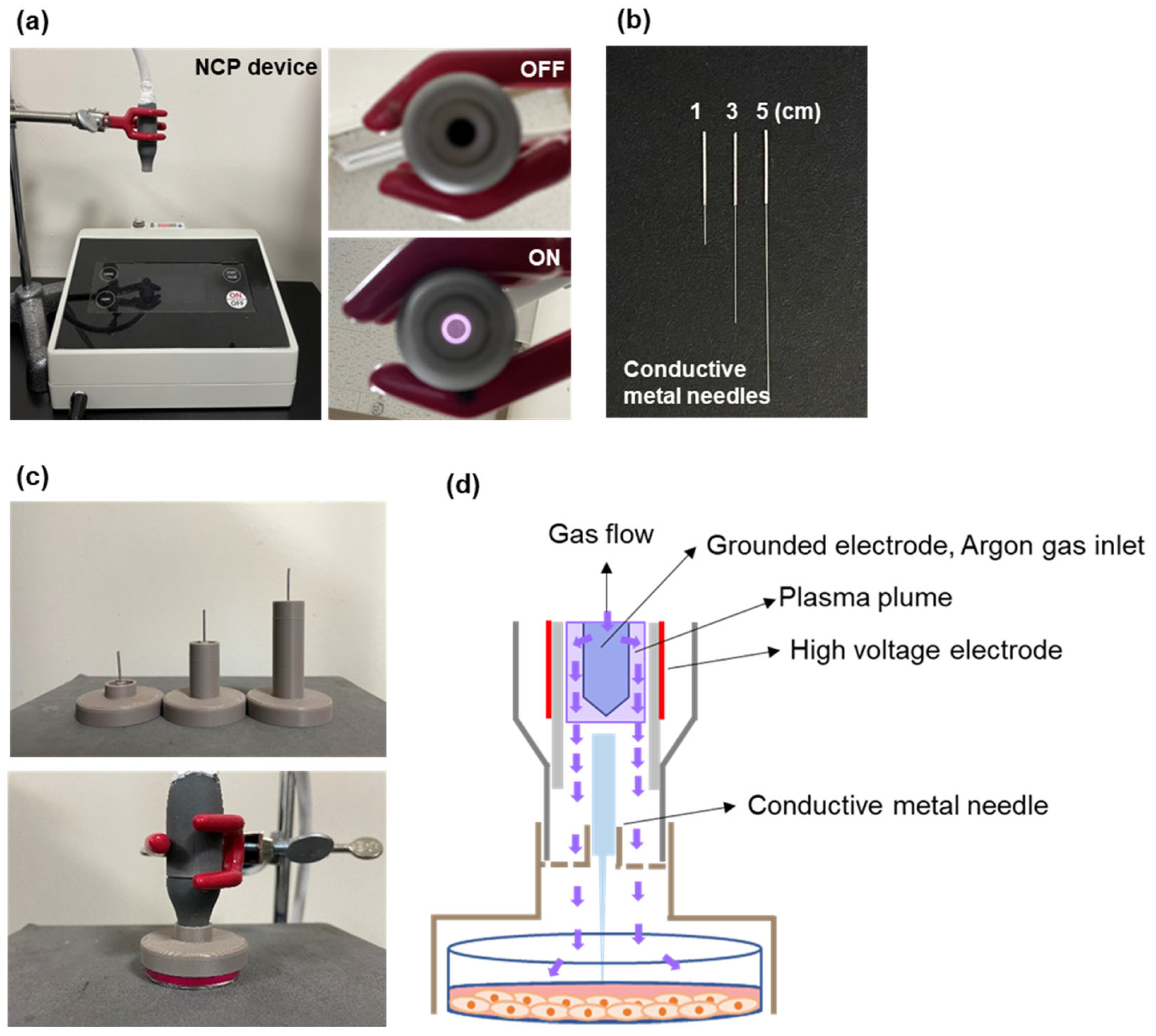
For this study, the NCP device was slightly modified from that used in our previous study [19]. Briefly, it has a coaxial dielectric barrier discharge (DBD)-type plasma-generating part consisting of two electrodes and one dielectric: an inner electrode made of stainless steel and an outer electrode coated on a ceramic tube. The outer electrode was connected to a sinusoidal high-voltage circuit generating a peak-to-peak voltage of 3 kV with a frequency of 20 kHz. The inner electrode was grounded to prevent the transition from glow discharge to arc discharge between the inner electrode and the cell culture medium during the plasma treatment. The working gas was argon, which independently flows between the inner electrode and ceramic tube at a flow rate of 1 slm (standard liters per minute). The plasma generated in this device does not extend outward like a plasma jet but is generated between the inner electrode and ceramic tube. When the device was turned on, a bright plasma plume could be easily observed (Figure 1a). To treat NCP more appropriately with a conductive metal needle, the NCP device in this study had an open path for needle entry in the plasma-generating part.
To test whether the anti-inflammatory effect of NCP can be delivered by the conductive metal needles, the acupuncture needles for EA treatment were adopted. The acupuncture needle (Dongbang Needle, Seongnam, Republic of Korea) had a handle with a diameter of 0.25 mm, and the length of the needle part was cut to 1, 3, or 5 cm (Figure 1b). To effectively treat cells in a 35 mm cell culture dish with NCP, needle, and plasma-needle, various types of cell treatment leads were manufactured using a 3D printer and used in experiments (Figure 1c). To minimize the cell transfer of the gas-phase working elements of the NCP, a lid with a tiny hole, which only a needle could pass through, was also manufactured (Figure 4a) and compared to the lid with larger pores. As shown in the schematic diagram in Figure 1d, the plasma plume cannot contact the needle, but afterglow of the argon plasma can be delivered to the needle and the cells. The NCP treatment time was fixed at 5 min, because the sufficient anti-inflammatory activity was confirmed in our previous studies.
Figure 1.
NCP device, acupuncture needle and lids used in this study. (a) The photographs showing the no-ozone cold plasma (NCP) device used in this study. (b) Acupuncture needles used in this study. The acupuncture needles with 1, 3 and 5 cm length were prepared. (c) The PN lids used for this study. The lids were produced using a 3D printer for treating NCP and acupuncture on cells in a 35 mm cell culture dish. (d) A schematic diagram for NCP device and PN treatment on cells.
2.3. Cell Culture
C2C12 cells, a mice muscle cell line, were purchased from the American Type Culture Collection (Manassas, VA, USA), and the cells were cultured in a 37 °C incubator with 5% CO2 using Dulbecco’s modified Eagle’s medium (Invitrogen Gibco, Grand Island, NY, USA) supplemented with 10% heat-inactivated fetal bovine serum (Invitrogen Gibco, Grand Island, NY, USA), 100 U/mL penicillin, and 100 µg/mL streptomycin.
2.4. Setting TNFα-Induced Inflammation Conditions Using C2C12 Cells
For setting TNFα-induced inflammation in C2C12 cells, the cells were treated with 0.1 μg of TNFα for 0.5, 1, 2, 4, 6, and 24 h and then the inflammatory reactions in the cells were monitored through RT-PCR and Western blot analysis (Figure 3a,b). Based on the result of this experiment, most of the experiments were performed at 2 h of TNFα exposure conditions.
2.5. Electroacupuncture Treatment
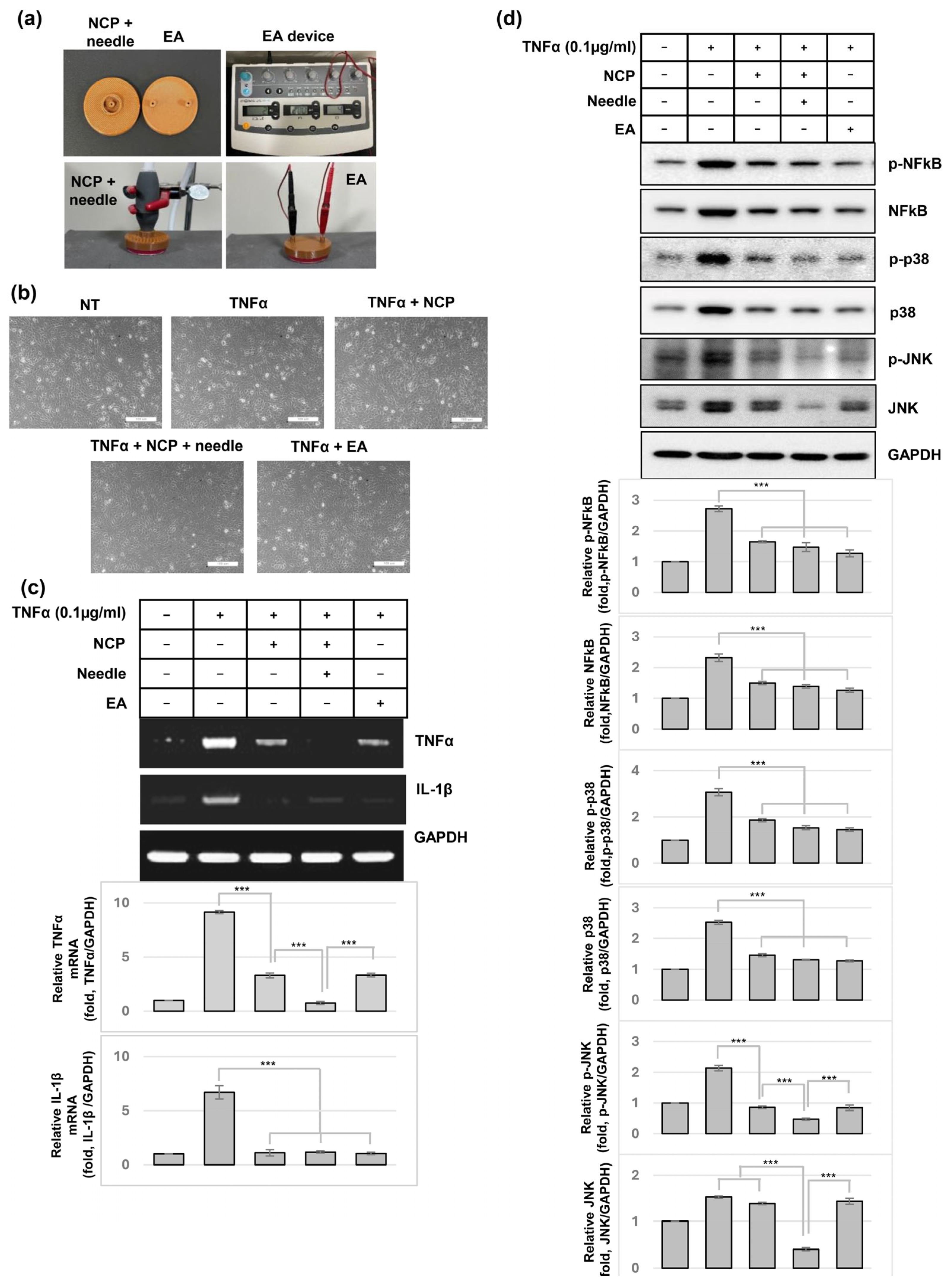
To compare the differences between an electroacupuncture device (ES-160, ITO Co., Ltd., Tokyo, Japan) and the plasma-needle, a lid was manufactured to apply an anode and a cathode to a cell culture dish (Figure 6a). For all cell treatments, C2C12 cells were treated with 0.1 μg/mL TNFα, the lids were covered, the NCP-generating part was mounted to the lids, and then the NCP was treated for 5 min in the presence or absence of needles. Electroacupuncture treatment was performed at 2 Hz, 200 μs for 5 min.
2.6. Sulforhodamine B (SRB) Assay
C2C12 cells were treated with NCP for 5 min using 3 kinds of needles (1, 3, and 5 cm) and cultured in an incubator for 24 h. After the medium was removed, the cells were treated with 4% paraformaldehyde and left at room temperature for 30 min, after which the cells were washed five times with PBS and dried. Each cell culture dish was treated with 1 mL of 0.4% SRB solution and reacted at room temperature for 30 min. The SRB solution was removed, the sample was washed 5 times with 1% acetic acid, and the sample was completely dried. After observing the cell morphology under a microscope, the reagent-stained cells were dissolved by treatment with 10 mM Tris solution and transferred to a 96-well microplate, and the absorbance was measured at 515 nm.
2.7. Amplex Red Assay
After treating C2C12 cells with NCP alone or with a needle at 1, 3, and 5 cm for 5 min, the amount of hydrogen peroxide produced in the medium was measured using an Amplex Red hydrogen peroxide/peroxidase assay kit (Invitrogen, Carlsbad, CA, USA). A mixture of 100 μM Amplex Red reagent and 0.2 U/mL HRP was mixed with an equal amount of NCP- or PN-treated media and reacted in a 96-well plate. A color reaction was induced at room temperature for 30 min in the dark, after which the absorbance was measured at 560 nm using a microplate reader.
2.8. Reverse Transcription-Polymerase Chain Reaction (RT-PCR)
Total RNA was isolated using TRIzol reagent (Invitrogen Life Technologies, Carlsbad, CA, USA). Complementary DNA (cDNA) was synthesized from 2 μg of total RNA using Maxime RT PreMix (iNtRON Biotechnology, Seongnam, Republic of Korea). PCR was performed using MG 2X HOT Mastermix with dye (MGmed, Seoul, Republic of Korea), and the primers used are listed in Table 1. PCR results were confirmed by electrophoresis on a 2% agarose gel and observed with an AI680 instrument (GE Healthcare, Uppsala, Sweden). The relative expression levels of genes were measured using ImageJ software (ver.1.54g).
Table 1.
Primer sequences for RT-PCR.
2.9. Western Blot Analysis
After a final incubation, the cells were washed with PBS and harvested in an ice-cold lysis buffer containing 50 mM Tris/HCl (pH 7.5), 150 mM NaCl, 1% (v/v) Nonidet P40, 10% (v/v) glycerol, 1 mM PMSF, 1 mM dithiothreitol, 20 mM NaF, 1 mM EDTA, and a protease inhibitor cocktail (Roche). Equal amounts of cell lysate (30 μg) were resolved by SDS/PAGE (8–12% gel) and transferred to PVDF membranes. Upon the completion of transfer, the membranes were probed with antibodies. The antibodies used for this study were phospho-p38, p38, phospho-JNK, JNK, and GAPDH (Santa Cruz Biotechnology, Santa Cruz, CA, USA) and phosphorylated-NFkB and NFkB (Cell Signaling Technology, Beverly, MA, USA). The specific bands were detected with advanced ECL Western blotting reagents (Merck Millipore, Darmstadt, Germany).
2.10. Direct–Indirect Treatment
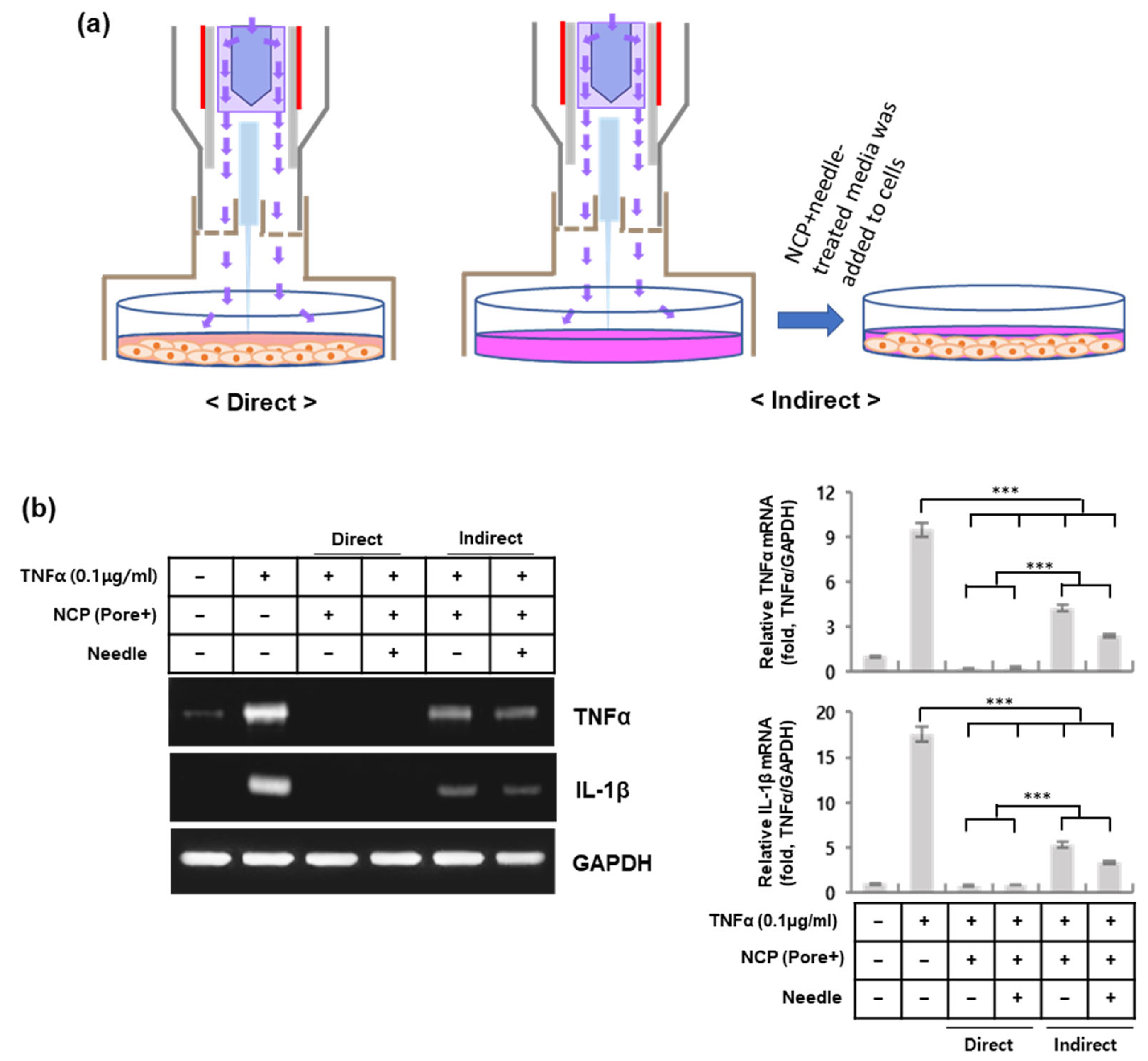
To elucidate how NCP and the PN exert anti-inflammatory effects, two types of treatments, direct and indirect treatments, were used (Figure 5a). The direct treatment is a method for treating NCP or the PN with TNFα-containing media in the presence of cells. On the other hand, indirect treatment is a method in which NCP or PN treatment is performed in the absence of cells, and then NCP- or PN-treated media are added to the cells. At 2 h after each treatment, the cells were harvested and subjected to RT-PCR.
2.11. Statistical Analysis
The data are presented as the means ± SDs of at least 3 independent experiments. Two-tailed Student’s t tests were used to assess the statistical significance of differences between means, and the threshold for significance was set at p < 0.05.
3. Results
3.1. The Effects of NCP and the PN on the Proliferation of C2C12 Cells and H2O2 Production in the Media
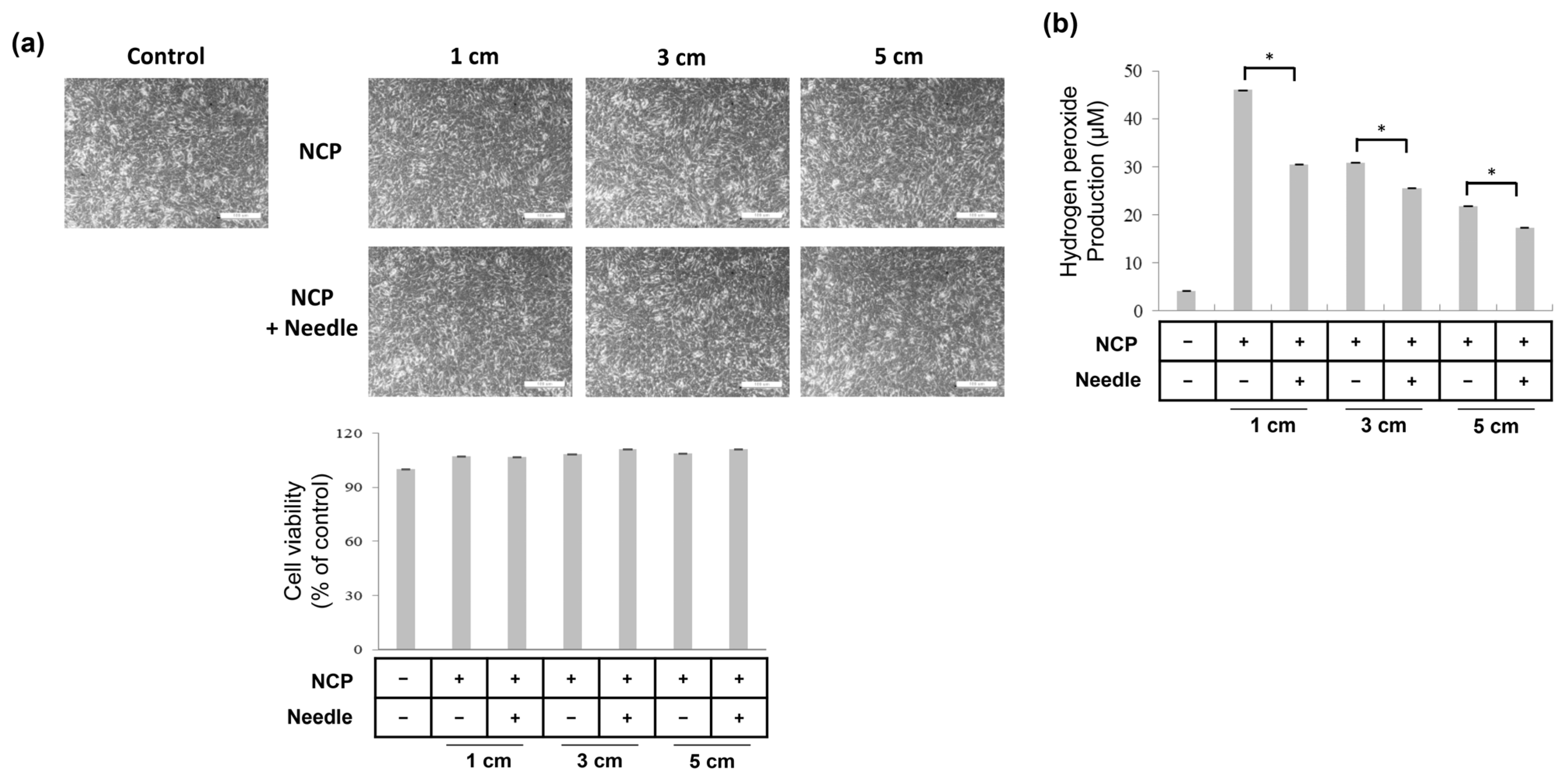
Prior to testing the anti-inflammatory effect of the PN, the effects of NCP and the PN on the proliferation of C2C12 mouse muscle cells were tested. For this purpose, C2C12 cells were subjected to NCP or the PN for 5 min using three kinds (1, 3, or 5 cm) of lids and needles. Twenty-four hours after treatment, the cells were subjected to an SRB assay. As shown in Figure 2a, the mere treatment of cells with NCP did not reduce the cell density but slightly increased the density. Likewise, PN-treated cells also showed a slight increase in cell density.
Since H2O2 production in plasma-treated media is a common phenomenon in plasma medicine, the effects of NCP and the PN on H2O2 production were tested (Figure 2b). For this, 2 mL of cell growth media was placed in 35 mm cell culture dish and treated with NCP and the PN for 5 min. Immediately after the treatment, samples were collected and used for hydrogen peroxide detection. Our results show that the mere treatment of media with NCP for 5 min at 1 cm can increase the H2O2 concentration from 3 μM to 47 μM, and the H2O2 production activity of NCP decreased when the NCP was treated with longer lids. Interestingly, the H2O2-producing activity of the NCP decreased when the needle was mounted.
Figure 2.
Effect of NCP and the PN on C2C12 cell proliferation and H2O2 level in the media. (a) Effects of NCP and PN (plasma-needle) on C2C12 cell proliferation. A total of 24 h after the 5 min NCP and PN treatment at a distance of 1, 3, 5 cm using the lids, cells were subjected to SRB assay, and photographs showing cell morphology were taken (upper panel) and the cell viability was measured (bottom panel). The photographs were taken at 200× magnification, and the size of the scale bar is 100 μm. (b) The effect of NCP and PN treatment on H2O2 level in the media. Data shown are representatives of three independent experiments, * p < 0.05.
3.2. NCP and PN Treatment Can Hinder the TNFα-Mediated Expression of Proinflammatory Cytokines in C2C12 Cells
To set the muscle-inflammation-mimicking condition in an in vitro cell culture system, C2C12 cells were treated with TNFα, and the expression of proinflammatory cytokines, including TNFα, IL-1β, and IL-6, was monitored at 0.5, 1, 2, 4, 6 and 24 h after treatment. As shown in Figure 3a, TNFα treatment induced the expression of TNFα mRNA at 30 min after treatment, but a slight reduction was detected at 2 h after treatment. On the other hand, in the case of IL-1α, the expression began to increase at 30 min after treatment with TNFα, and the mRNA level gradually increased until 24 h. The expression of IL-6 was also induced by TNFα treatment at 30 min after treatment; the maximum level was observed at 4 h, but its expression was completely diminished at 24 h after treatment. When monitoring the activity of the NFκB protein (Figure 3b), the TNFα-mediated increase in phosphorylated NFκB and the protein level of NFκB started to increase at 30 min after treatment, and this increase was maintained for 24 h after treatment. Based on these results, 2 h after TNFα treatment was used as the test condition to verify the anti-inflammatory efficacy of NCP and the PN against inflammatory muscle pain.
Figure 3.
Anti-inflammatory activity of NCP and the PN in TNFα-treated C2C12 cells. (a) The effect of TNFα treatment on the expression of TNFα, IL-1β and IL-6 genes in C2C12 cells. The cells were treated with 0.1 μg/mL of TNFα and then at 0.5, 1, 2, 4, 6, 24 h after the treatment, total RNA was extracted, and RT-PCR was performed. (b) At 0.5, 1, 2, 4, 6, 24 h after the TNFα treatment, the cell protein lysates were gathered, and used for Western blot analysis. (c) Immediately after TNFα treatment, cells were subjected to NCP treatment in the presence (PN) and absence (NCP) of an acupuncture needle using the 1 cm and 5 cm lids. A total of 2 h after the treatment, cells were used for RT-PCR analysis. Data shown are representatives of three independent experiments, *** p < 0.001.
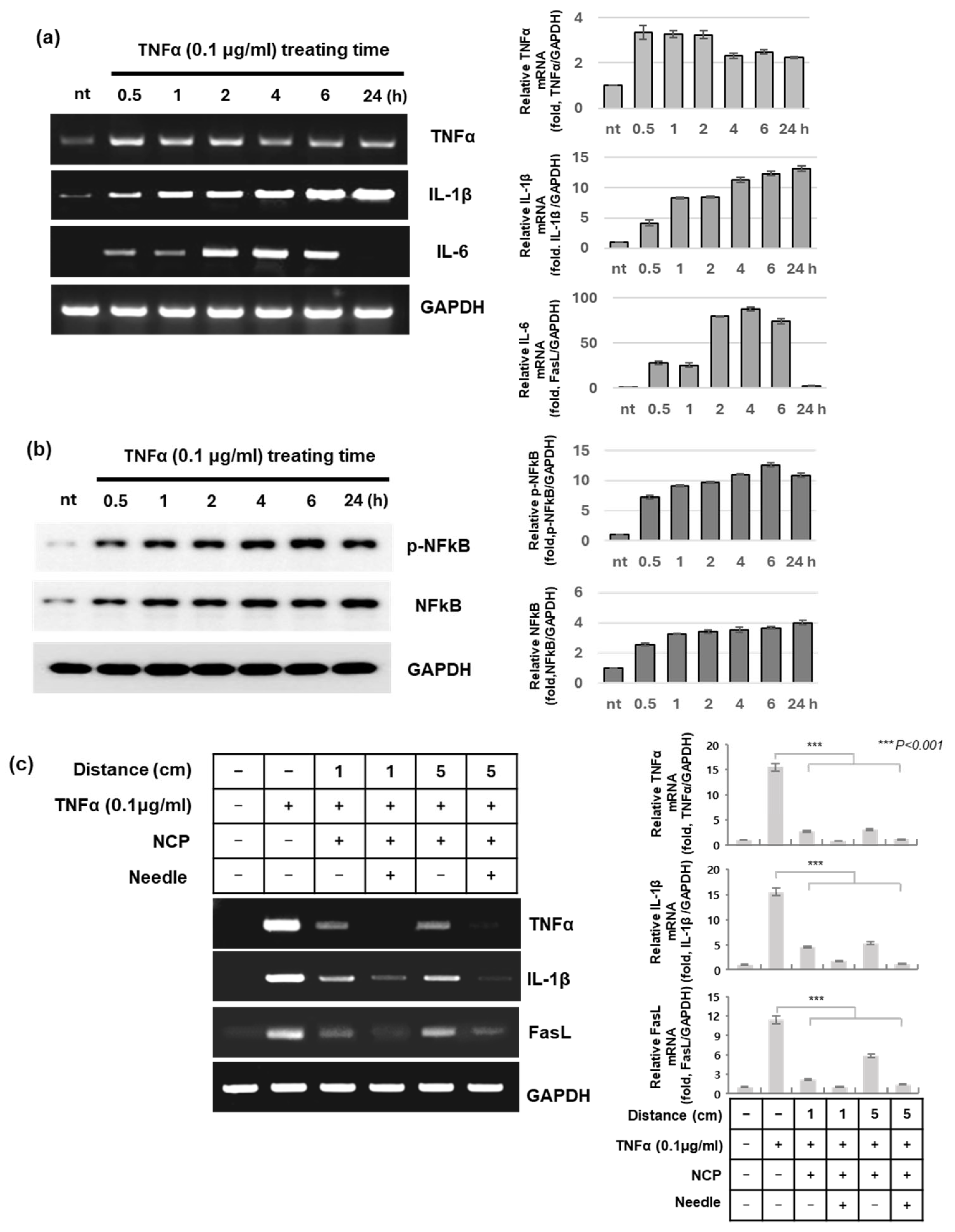
To confirm the anti-inflammatory effect of NCP and the PN, immediately after TNFα treatment, the C2C12 cells were subjected to NCP and PN treatment for 5 min using 1 cm and 5 cm needles. At 2 h after treatment, changes in the expression of proinflammatory cytokines were observed. As shown in Figure 3c, NCP treatment decreased the TNFα-mediated expression of TNFα, IL-1β, and FasL. This anti-inflammatory effect of NCP was stronger when the NCP was treated at a distance of 1 cm than at a distance of 5 cm. Interestingly, the PN treatment, which involves the mere addition of a needle to the NCP, had stronger anti-inflammatory effects than NCP. Furthermore, the effect of PN treatment at 5 cm from the cells showed more powerful anti-inflammatory effects than that of the PN treatment at 1 cm.
3.3. The Plasma Gas Flow of NCP Is Not Essential for the Anti-Inflammatory Effect of PN
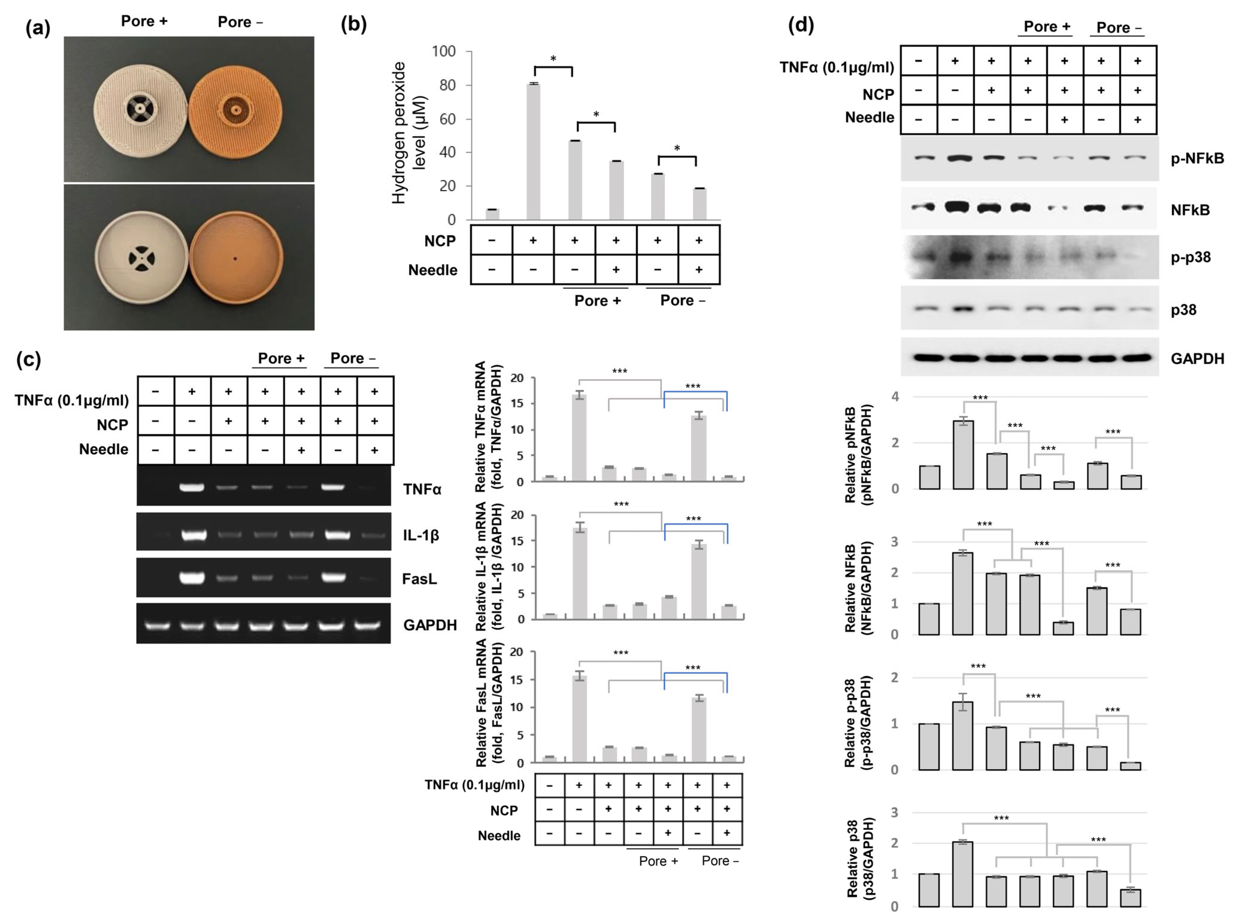
Many reports have shown the importance of plasma gas flow for the biological function of plasma. However, since the target tissue of inflammatory muscle pain is covered by skin tissue, plasma gas flow cannot be delivered. Since our previous results in this study used lids with several pores (Figure 4b, warm gray), which allow plasma gas to flow to muscle cells, the anti-inflammatory activity of the PN might be caused by plasma gas flow. To test whether the anti-inflammatory activity of the PN can be maintained under plasma gas flow-free conditions, we produced another type of lid (Figure 4a, brown). Since this lid has only one tiny pore in the center for mounting the acupuncture needle, the NCP gas flow can be minimized. Before testing the effect of the differences in plasma gas flow depending on the lid type on the anti-inflammatory effect, the effects on H2O2 production in the media were monitored. As Figure 4b shows, the mere treatment of NCP without the lid increased the H2O2 level to approximately 78 μM, but the NCP treatment using the lid with pores increased the H2O2 level to 48 μM, and the H2O2 level in the media treated with NCP using the lid without pores was only approximately 25 μM. Interestingly, the media treated with the PN using the lid with the pore has about 35 μM of H2O2, and the level of H2O2 in the media treated with the PN using the lid without the pore was about 18 μM.
To test whether the anti-inflammatory effect of NCP and the PN can be maintained even when the plasma gas flow is blocked, TNFα-treated C2C12 cells were treated with NCP and the PN using lids with or without pores for 5 min. Two hours after treatment, total RNA and protein lysates were harvested and subjected to RT-PCR and Western blotting, respectively. As shown in Figure 4c, the cells treated with NCP without lids and the cells treated with NCP using the lids with pores showed similar decreases in TNFα, IL-1β and FasL gene expression. Treatment of the cells with PN via lids with pores also blocked the TNFα-mediated induction of the three proinflammatory cytokines. Interestingly, the NCP treatment using a lid without pores showed the lowest anti-inflammatory activity among all treatment methods, whereas the strongest anti-inflammatory activity was monitored in the cells treated with the PN using a lid without pores. The Western blot results (Figure 4d) showed that the protein activity of NFκB and p38 was significantly decreased by NCP and PN treatment using the lids with pores. The cells treated with NCP using lids without pores showed slightly increased NFkB protein activity, but PN treatment using lids without pores blocked the TNFα-mediated increase in NFkB and p38 protein activity.
3.4. The Activation of the Media After NCP and PN Treatment Has an Important Role in the Anti-Inflammatory Effect
To verify whether the anti-inflammatory effects of NCP and the PN occur by directly affecting cells or by causing changes in cell media, two types of cell treatment methods (direct and indirect) were used (Figure 5a). In the direct treatment group, the cells were treated with NCP and the PN in the presence of TNFα-containing media; therefore, NCP and the PN can modulate cell function directly and indirectly through affecting media. On the other hand, in the indirect treatment group, NCP and PN treatment were performed in TNFα-containing media without cells, and then this treated media was added to the cells. Therefore, the cells in the indirect group can be affected by the modification of media triggered by NCP and PN treatment only. As shown in Figure 5b, both direct and indirect treatment with NCP and PN significantly reduced the TNFα-mediated increase in TNFα and IL-1β gene expression, but direct treatment had stronger anti-inflammatory effects than indirect treatment.
Figure 4.
NCP gas flow is not essential for anti-inflammatory activity of the PN. (a) Photographs showing the lids with (warm gray) and without (brown) pores for NCP gas flow. (b) A total of 2 mL of cell growth media was subjected to NCP treatment without a lid, NCP and PN treatment using the lids with or without pores for 5 min. The H2O2 level in the media was detected. (c) The result of RT-PCR analysis. The cells treated with TNFα were subjected to NCP and PN treatment as described. At 2 h after the NCP and PN treatment, the total RNA was used for RT-PCR. (d) At 2 h after NCP and PN treatment, cell lysate was used for Western blot analysis. Data shown are the representatives of three independent experiments, * p < 0.05, *** p < 0.001.
3.5. Electroacupuncture Treatment Also Showed Anti-Inflammatory Activity in TNFα-Treated C2C12 Cells, Like NCP and PN Treatment
Since PN treatment of TNFα-treated cells using lids without pores showed strong anti-inflammatory activity, the anti-inflammatory activity of the PN might be caused by the transfer of electrons from the NCP through the needle. To test whether the mere treatment of cells with an electron current can have anti-inflammatory effects, a lid for treating electroacupuncture (EA) on cells was produced (Figure 6a), and the effect of EA treatment on TNFα-mediated inflammatory reactions in C2C12 cells was tested.
Before testing the anti-inflammatory effect of EA, the effects of EA and the PN on the morphology and density of C2C12 cells were monitored. As shown in Figure 6b, treatment of TNFα-treated cells with NCP, PN or EA did not induce significant morphological or density changes.
Figure 5.
NCP and PN-mediated changes in the media are important for anti-inflammatory effect in TNFα-treated C2C12 cells. (a) A schematic diagram explaining the direct and indirect PN treatment methods. (b) The result of RT-PCR analysis. The cells were subjected to NCP and PN treatment in a direct and indirect manner. For direct treatment, the cells were treated with TNFα and then treated with NCP and PN. For indirect treatment, the media containing TNFα was treated with NCP and PN for 5 min, and this media was transferred to the cells immediately after the treatment. A total of 2 h after the treatment, total RNA was prepared and used for RT-PCR analysis. Data shown are the representatives of three independent experiments, *** p < 0.001.
According to the RT-PCR results showing the anti-inflammatory effect (Figure 6c), NCP treatment without lids and PN treatment with lids without pores reduced the TNFα-mediated induction of TNFα and IL-1β gene expression, as expected. Surprisingly, EA treatment strongly reduced TNFα and IL-1β gene expression in cells, but PN treatment had the strongest anti-inflammatory activity among NCP, EA, and the PN. Western blot analysis revealed that the NCP, PN and EA treatment had similar effects on blocking TNFα-mediated NFκB and p38 protein activation, but PN treatment had the strongest inhibitory effect on the JNK protein (Figure 6d).
Figure 6.
The electroacupuncture treatment also has anti-inflammatory activity as NCP and PN do. (a) Photographs showing the lids for PN and electroacupuncture (EA) treatment and electroacupuncture device used in this study. (b) The photographs showing morphology and density of C2C12 cells at 24 h after TNFα, NCP, PN and EA treatment. The photographs were taken at 200× magnification, and the size of the scale bar is 100 μm. (c) The result of RT-PCR analysis. TNFα-treated cells were subjected to NCP, PN, EA treatment as described and at 2 h after the treatment, total RNA was extracted and used for RT-PCR analysis. (d) The results of Western blot analysis. Data shown are the representatives of three independent experiments, *** p < 0.001.
4. Discussion
In this study, physical plasma, which is one of most anticipated upcoming medical technologies [12,22], was combined with an acupuncture needle to develop PN technology. In our previous studies, we developed a unique plasma-generating system called ‘No-ozone cold plasma’, NCP [19]. NCP is a type of argon-based cold plasma, which is designed to deliver after-glow plasma only at a temperature of 27 °C. Furthermore, since the NCP device generates plasma plume only in the argon-gas-filled inlet, it does not generate ozone or nitric oxides. Although NCP had shown strong anti-inflammatory activity on several inflammatory reactions [17,23], it was limited to the surface of tissues because NCP is a form of gas. In our previous studies, the two of NCP’s biological effects, inhibition of E-cadherin protein function [21] and oral bacteria-killing effect [19], were significantly reduced by placing grounded metal mesh between cells and the outlet of the NCP device. Furthermore, the anti-inflammatory activity of NCP was also reduced by placing a grounded metal mesh on top of a culture dish which contained inflamed human gingival cells [23]. These data suggest the possibility that many beneficial roles of NCP can be delivered through conductive metal. In this study, we adopted NCP for PN development, because NCP is an afterglow plasma, it might be the best way for minimizing the risk from electric shock which can be caused by the direct contact of the metal needle and plasma plume.
In this study, we tested whether the PN has direct anti-inflammatory activity like that of NCP. Since acupuncture is widely used for treating inflammatory muscle pain [10], we used TNFα-treated C2C12 cells to test the anti-inflammatory activity of the PN. First, the results of this study showed that PN treatment did not induce cell morphology changes or a reduction in the cell density of C2C12 cells; rather, PN treatment slightly increased the cell density, as did NCP (Figure 2a). This result was in line with our previous report, which showed that NCP treatment can enhance the proliferation of HaCaT human keratinocytes [18]. Many studies have elucidated the fact that argon-based plasma treatment on liquid media can increase the H2O2 level in the media [19,24,25]. In our study, the H2O2-producing activity of the NCP was affected by the distance between the cells and the NCP device, and the presence of an acupuncture needle reduced NCP-mediated H2O2 production. On the other hand, the use of acupuncture needles for PN treatment enhanced the anti-inflammatory activity of NCP in TNFα-treated C2C12 cells (Figure 3). Furthermore, although the anti-inflammatory activity of NCP was dependent on the distance between cells and the device, the anti-inflammatory activity of the PN was not affected. These data indicate that NCP- and PN-mediated anti-inflammatory activity is not dependent on H2O2 production and that the key elements involved in the anti-inflammatory activity of NCP can be delivered through an acupuncture needle. According to Gunawardena et al., H2O2 can mediate proinflammatory cell-to-cell signals and induce the expression of TNFα and IL-1β by activating NFκB protein activity [26]. Therefore, the increase in H2O2 in the NCP- and PN-treated media cannot be the reason for the observed anti-inflammatory activity. The results shown in Figure 4 also suggest that rather than H2O2 and other working elements of NCP, including ions and reactive gas-phase radicals such as OH, the electrons of NCP, which can be delivered through acupuncture needles, are key players in NCP- and PN-mediated anti-inflammatory activity. The observation that PN-mediated anti-inflammatory activity was detected in the cells treated with the PN under conditions in which the NCP gas flow was blocked suggests that the anti-inflammatory activity of the PN can be delivered to inflammatory muscle tissue even when the NCP is treated topically. Although more detailed analyses of media changes after NCP and PN treatment are yet to be performed, the results shown in Figure 5 indicate that changes in NCP- and PN-mediated media seem to play important roles in anti-inflammatory activity. Several studies showed that high-energy electrons are the key for cold plasma chemistry at the gas phase and gas–liquid interphase [12,27]. Through these electron-mediated chemical reactions, when cold plasma is treated on the media, several kinds of reactive species, such as •OH, •O2−, 1O2, O, H2O2, •NO2, ONOO−, •NO, NO2−, NO3− and others can be generated in the media. Among these reactive species, H2O2 is the weakest candidate among those considered to be the important because of PN’s anti-inflammatory efficacy. H2O2 is well known for inducing oxidative stress within cells, thereby increasing inflammation further [28]. Rather than H2O2, we think that ONOO− can be a reason for PN- and NCP-mediated anti-inflammatory activity. Levrand et al. showed that ONOO− can inhibit the NFkB protein, and this phenomenon can be directly linked to reduction in inflammatory reactions [29]. Likewise, in this study, PN treatment on TNFα-treated C2C12 cells can inhibit inflammatory reactions through inhibiting the NFkB protein. Although we did not test whether NCP and PN treatment on the media can increase ONOO− in the media, the increase in ONOO− in the media can be one of the reasons for NCP- and PN-mediated anti-inflammatory activity.
In the last part of this study, the anti-inflammatory activity of PN was compared to that of EA to test whether the mere treatment of cells with an electron current can inhibit TNFα-mediated inflammatory reactions in C2C12 cells as the PN does (Figure 6). According to our research, the anti-inflammatory activity of EA has been reported in several in-vivo models, but there are no studies testing its direct effect in an in vitro system. These studies showed that EA treatment reduced oxidative stress within tissues through activating the Nrf2 protein, and this was linked to anti-inflammatory activity [30,31]. Interestingly, the treatment of EA on TNFα-treated C2C12 cells directly inhibited the expression of TNFα and IL-1β gene expression as NCP and the PN do. This result also suggested that the presence of electrons in the NCP is a key factor for the anti-inflammatory activity of the PN. Kang et al. elucidated the fact that ONOO− can activate the Nrf2 protein [32], and Woedtke et al. showed that non-thermal plasma treatment can activate the Nrf2 protein [33]. Since Nrf2 protein activation is directly linked to anti-inflammatory activity [34], the electron-mediated media change in PN- and EA-treated media, possibly including increase in ONOO− in the media, can be the reason for PN- and EA-mediated anti-inflammatory reaction. Does NCP have an advantage over EA? For the EA procedure, at least twp acupuncture needles must be inserted into the acupoint for linking to the anode and cathode of the device. However, for PN treatment, the use of only one acupuncture needle is sufficient. Furthermore, it is expected that PN can have antimicrobial and tissue-regenerating effects on NCP, although more detailed studies are needed.
5. Conclusions
In this study, for the first time, we developed a plasma-needle technique by combining NCP technology with an acupuncture needle. This study confirms that the anti-inflammatory activity of the NCP can be delivered up to 5 cm through a needle. We also elucidated that the electrons in the NCP, which can be delivered through needles, are key players in NCP- and PN-mediated anti-inflammatory activity. We also showed the direct anti-inflammatory activity of EA using an in vitro system and compared the anti-inflammatory activity of EA with that of the PN and NCP. Since the anti-inflammatory activity was monitored even when the cells were treated with the PN in an NCP gas flow-free condition, it is expected that PN treatment can be used to overcome the main limitation of physical plasma, namely, the working depth. In our next study, the effect of the PN on inflammatory muscle pain will be tested using an animal model.
Author Contributions
J.-H.C., J.-W.H. and G.-C.K. designed the study and J.K. and W.H. advised it. The study was supervised by J.-H.C., J.-W.H. and G.-C.K. Most of the experiments and the data analyzing were performed by S.-A.P., J.K., W.H. and J.-H.C. advised it. H.-Y.L. and J.-H.C. developed the PN device module for this study and advised the device operation setting. S.-A.P., J.-H.C., J.K., W.H. and H.-Y.L. wrote most of the manuscript, G.-C.K. and J.-W.H. advised it. All authors have read and agreed to the published version of the manuscript.
Funding
This research was supported by a grant of the Korea Health Technology R&D Project through the Korea Health Industry Development Institute (KHIDI), funded by the Ministry of Health & Welfare, Republic of Korea (grant number: HI20C0252).
Data Availability Statement
All data generated or analyzed during this study are included in this published article.
Conflicts of Interest
Authors Jeong-Hae Choi, Seung-Ah Park, Hyun-Young Lee and Gyoo-Cheon Kim were employed by the company FEAGLE Corporations. The remaining authors declare that the research was conducted in the absence of any commercial or financial relationships that could be construed as a potential conflict of interest. The funders had no role in the design of the study; in the collection, analyses, or interpretation of data; in the writing of the manuscript; or in the decision to publish the results.
Abbreviations
| NCP | No-ozone Cold Plasma |
| PN | Plasma-needle |
| EA | Electroacupunctures |
| RT-PCR | Reverse transcription- polymerase chain reaction |
| TNFα | Tumor necrosis factor alpha |
| IL-1β | Interleukin-1beta |
| FasL | Fas ligand |
| NF-kB | Nuclear factor kappa-light-chain-enhancer of activated B cells |
| JNK | c-Jun N-terminal kinase |
| GAPDH | Glyceraldehyde 3-phosphate dehydrogenase |
| SRB | Sulforhodamine B |
| SDS-PAGE | Sodium Dodecyl Sulfate- Polyacrylamide Gel Electrophoresis |
References
- Glaubitz, S.; Schmidt, K.; Zschuntzsch, J.; Schmidt, J. Myalgia in myositis and myopathies. Best practice & research. Clin. Rheumatol. 2019, 33, 101433. [Google Scholar] [CrossRef] [Scilit]
- Ranchordas, M.K.; Rogerson, D.; Soltani, H.; Costello, J.T. Antioxidants for preventing and reducing muscle soreness after exercise: A Cochrane systematic review. Br. J. Sports Med. 2020, 54, 74–78. [Google Scholar] [CrossRef] [Scilit] [PubMed]
- Hunter, K.; Lyon, M.G. Evaluation and management of polymyositis. Indian J. Dermatol. 2012, 57, 371–374. [Google Scholar] [CrossRef] [Scilit]
- Jurado-Priego, L.N.; Cueto-Urena, C.; Ramirez-Exposito, M.J.; Martinez-Martos, J.M. Fibromyalgia: A Review of the Pathophysiological Mechanisms and Multidisciplinary Treatment Strategies. Biomedicines 2024, 12, 1543. [Google Scholar] [CrossRef] [Scilit]
- Wiecha, S.; Posadzki, P.; Prill, R.; Plaszewski, M. Physical Therapies for Delayed Onset Muscle Soreness: A Protocol for an Umbrella and Mapping Systematic Review with Meta-Meta-Analysis. J. Clin. Med. 2024, 13, 2006. [Google Scholar] [CrossRef] [Scilit]
- Pyne, D.B. Exercise-induced muscle damage and inflammation: A review. Aust. J. Sci. Med. Sport 1994, 26, 49–58. [Google Scholar] [PubMed]
- Navratilova, E.; Porreca, F. Substance P and Inflammatory Pain: Getting It Wrong and Right Simultaneously. Neuron 2019, 101, 353–355. [Google Scholar] [CrossRef] [Scilit]
- Matsuda, M.; Huh, Y.; Ji, R.R. Roles of inflammation, neurogenic inflammation, and neuroinflammation in pain. J. Anesth. 2019, 33, 131–139. [Google Scholar] [CrossRef] [Scilit]
- Desai, M.J.; Saini, V.; Saini, S. Myofascial pain syndrome: A treatment review. Pain Ther. 2013, 2, 21–36. [Google Scholar] [CrossRef] [Scilit]
- Ko, G.W.Y.; Clarkson, C. The effectiveness of acupuncture for pain reduction in delayed-onset muscle soreness: A systematic review. Acupunct. Med. 2020, 38, 63–74. [Google Scholar] [CrossRef] [Scilit]
- Brany, D.; Dvorska, D.; Halasova, E.; Skovierova, H. Cold Atmospheric Plasma: A Powerful Tool for Modern Medicine. Int. J. Mol. Sci. 2020, 21, 2932. [Google Scholar] [CrossRef] [Scilit] [PubMed]
- Von Woedtke, T.; Schmidt, A.; Bekeschus, S.; Wende, K.; Weltmann, K.D. Plasma Medicine: A Field of Applied Redox Biology. Vivo 2019, 33, 1011–1026. [Google Scholar] [CrossRef] [Scilit] [PubMed]
- Sakudo, A.; Yagyu, Y.; Onodera, T. Disinfection and Sterilization Using Plasma Technology: Fundamentals and Future Perspectives for Biological Applications. Int. J. Mol. Sci. 2019, 20, 5216. [Google Scholar] [CrossRef] [Scilit]
- Bolgeo, T.; Maconi, A.; Gardalini, M.; Gatti, D.; Di Matteo, R.; Lapidari, M.; Longhitano, Y.; Savioli, G.; Piccioni, A.; Zanza, C. The Role of Cold Atmospheric Plasma in Wound Healing Processes in Critically Ill Patients. J. Pers. Med. 2023, 13, 736. [Google Scholar] [CrossRef] [Scilit]
- Faramarzi, F.; Zafari, P.; Alimohammadi, M.; Moonesi, M.; Rafiei, A.; Bekeschus, S. Cold Physical Plasma in Cancer Therapy: Mechanisms, Signaling, and Immunity. Oxidative Med. Cell. Longev. 2021, 2021, 9916796. [Google Scholar] [CrossRef] [Scilit]
- Wen, X.; Xin, Y.; Hamblin, M.R.; Jiang, X. Applications of cold atmospheric plasma for transdermal drug delivery: A review. Drug Deliv. Transl. Res. 2021, 11, 741–747. [Google Scholar] [CrossRef] [Scilit]
- Choi, J.H.; Song, Y.S.; Lee, H.J.; Hong, J.W.; Kim, G.C. Inhibition of inflammatory reactions in 2,4-Dinitrochlorobenzene induced Nc/Nga atopic dermatitis mice by non-thermal plasma. Sci. Rep. 2016, 6, 27376. [Google Scholar] [CrossRef] [Scilit]
- Choi, J.H.; Song, Y.S.; Song, K.; Lee, H.J.; Hong, J.W.; Kim, G.C. Skin renewal activity of non-thermal plasma through the activation of beta-catenin in keratinocytes. Sci. Rep. 2017, 7, 6146. [Google Scholar] [CrossRef] [Scilit]
- Park, N.S.; Yun, S.E.; Lee, H.Y.; Lee, H.J.; Choi, J.H.; Kim, G.C. No-ozone cold plasma can kill oral pathogenic microbes in H2O2-dependent and independent manner. Sci. Rep. 2022, 12, 7597. [Google Scholar] [CrossRef] [Scilit]
- Park, S.Y.; Jaiswal, M.S.; Jang, Y.S.; Choi, J.H.; Kim, G.C.; Hong, J.W.; Hwang, D.S. Effect of No-ozone Cold Plasma on Regeneration after Crushed Mental Nerve Injury in rats. Int. J. Med. Sci. 2022, 19, 1732–1742. [Google Scholar] [CrossRef] [Scilit] [PubMed]
- Lee, H.Y.; Choi, J.H.; Hong, J.W.; Kim, G.C.; Lee, H.J. Comparative study of the Ar and He atmospheric pressure plasmas on E-cadherin protein regulation for plasma-mediated transdermal drug delivery. J. Phys. D Appl. Phys. 2018, 51, 215401. [Google Scholar] [CrossRef] [Scilit]
- Moszczynska, J.; Roszek, K.; Wisniewski, M. Non-Thermal Plasma Application in Medicine-Focus on Reactive Species Involvement. Int. J. Mol. Sci. 2023, 24, 12667. [Google Scholar] [CrossRef] [Scilit]
- Park, K.H.; Jang, Y.S.; Joo, J.Y.; Kim, G.C.; Choi, J.H. Anti-Inflammatory Activity of No-Ozone Cold Plasma in Porphyromonas gingivalis Lipopolysaccharide-Induced Periodontitis Rats. Int. J. Mol. Sci. 2024, 25, 6161. [Google Scholar] [CrossRef] [Scilit]
- Boonmee, T.; Sinpoo, C.; Laokulsiri, K.; Piyaphonsakon, G.; Panngom, K.; Disayathanoowat, T.; Pettis, J.S.; Chaimanee, V. The in vitro potential of non-thermal atmospheric pressure plasma against Nosema ceranae infection in honeybees (Apis mellifera). Sci. Rep. 2025, 15, 26975. [Google Scholar] [CrossRef] [Scilit]
- Milhan, N.V.M.; Chiappim, W.; Sampaio, A.D.G.; Vegian, M.; Pessoa, R.S.; Koga-Ito, C.Y. Applications of Plasma-Activated Water in Dentistry: A Review. Int. J. Mol. Sci. 2022, 23, 4131. [Google Scholar] [CrossRef] [Scilit] [PubMed]
- Gunawardena, D.; Raju, R.; Munch, G. Hydrogen peroxide mediates pro-inflammatory cell-to-cell signaling: A new therapeutic target for inflammation? Neural Regen. Res. 2019, 14, 1430–1437. [Google Scholar] [CrossRef] [Scilit] [PubMed]
- Privat-Maldonado, A.; Schmidt, A.; Lin, A.; Weltmann, K.D.; Wende, K.; Bogaerts, A.; Bekeschus, S. ROS from Physical Plasmas: Redox Chemistry for Biomedical Therapy. Oxidative Med. Cell. Longev. 2019, 2019, 9062098. [Google Scholar] [CrossRef] [Scilit] [PubMed]
- Jiang, F.; Xu, X.R.; Li, W.M.; Xia, K.; Wang, L.F.; Yang, X.C. Monotropein alleviates H2O2-induced inflammation, oxidative stress and apoptosis via NF-kappaB/AP-1 signaling. Mol. Med. Rep. 2020, 22, 4828–4836. [Google Scholar] [CrossRef] [Scilit] [PubMed]
- Levrand, S.; Pesse, B.; Feihl, F.; Waeber, B.; Pacher, P.; Rolli, J.; Schaller, M.D.; Liaudet, L. Peroxynitrite is a potent inhibitor of NF-kappaB activation triggered by inflammatory stimuli in cardiac and endothelial cell lines. J. Biol. Chem. 2005, 280, 34878–34887. [Google Scholar] [CrossRef] [Scilit]
- Dai, N.; Tang, C.; Liu, H.; Huang, S. Effect of electroacupuncture on inhibition of inflammatory response and oxidative stress through activating ApoE and Nrf2 in a mouse model of spinal cord injury. Brain Behav. 2021, 11, e2328. [Google Scholar] [CrossRef] [Scilit]
- Pu, Y.; Cheng, J.; Wang, Z.; Zhang, J.; Liang, F.; Zhang, X.; Zheng, Z.; Yin, M.; Wang, Z. Electroacupuncture pretreatment inhibits ferroptosis and inflammation after middle cerebral artery occlusion in rats by activating Nrf2. Histol. Histopathol. 2025, 40, 357–367. [Google Scholar] [CrossRef] [Scilit]
- Kang, K.W.; Choi, S.H.; Kim, S.G. Peroxynitrite activates NF-E2-related factor 2/antioxidant response element through the pathway of phosphatidylinositol 3-kinase: The role of nitric oxide synthase in rat glutathione S-transferase A2 induction. Nitric Oxide 2002, 7, 244–253. [Google Scholar] [CrossRef] [Scilit]
- Schmidt, A.; Dietrich, S.; Steuer, A.; Weltmann, K.D.; von Woedtke, T.; Masur, K.; Wende, K. Non-thermal plasma activates human keratinocytes by stimulation of antioxidant and phase II pathways. J. Biol. Chem. 2015, 290, 6731–6750. [Google Scholar] [CrossRef] [Scilit]
- Wang, L.; He, C. Nrf2-mediated anti-inflammatory polarization of macrophages as therapeutic targets for osteoarthritis. Front. Immunol. 2022, 13, 967193. [Google Scholar] [CrossRef] [Scilit]

Disclaimer/Publisher’s Note: The statements, opinions and data contained in all publications are solely those of the individual author(s) and contributor(s) and not of MDPI and/or the editor(s). MDPI and/or the editor(s) disclaim responsibility for any injury to people or property resulting from any ideas, methods, instructions or products referred to in the content. |
© 2026 by the authors. Licensee MDPI, Basel, Switzerland. This article is an open access article distributed under the terms and conditions of the Creative Commons Attribution (CC BY) license.