


A 43-year-old man underwent left ultrasound-guided percutaneous nephrolithotomy. Post-stone removal, massive renal vein bleeding occurred; we opted to control the hemorrhage by inserting a Foley catheter as a nephrostomy tube and inflating the balloon. The catheter was placed under ultrasound guidance, but the path was lost after 5–6 cm, necessitating blind insertion for the remainder.
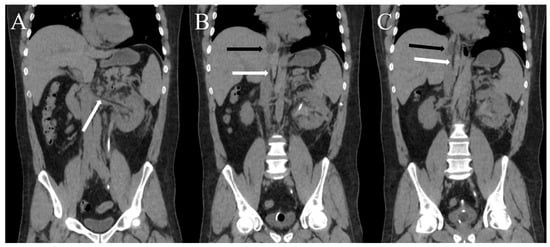
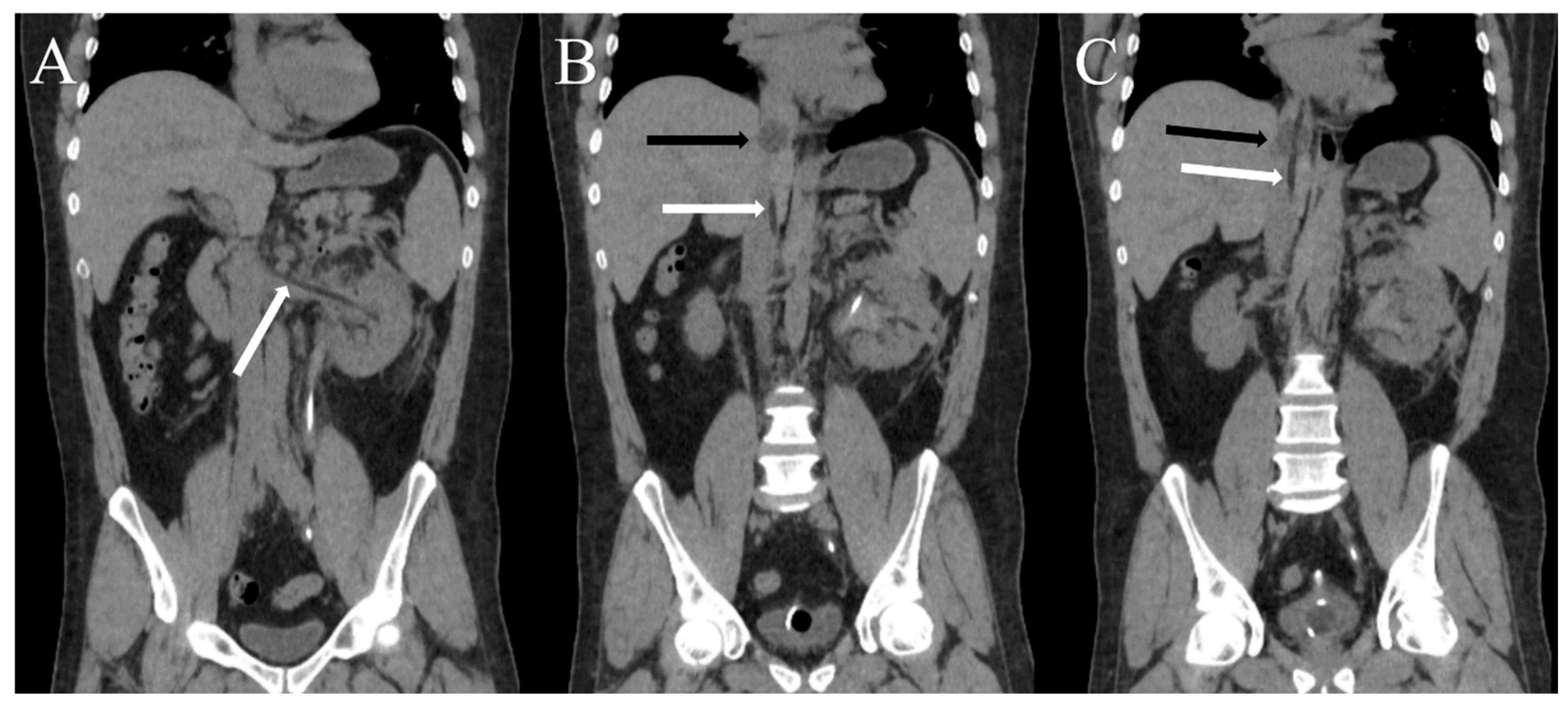
Subsequently, the patient experienced tachycardia and hypotension, requiring resuscitation with intravenous fluids and two units of packed cells. He was then stabilized and moved to recovery. However, he developed severe back and flank pain, unresponsive to analgesics. An abdominopelvic computed tomography (CT) scan was done (Figure 1).
Figure 1.
Computed tomography images; (A–C) images show the Foley catheter (used as a nephrostomy tube) and its balloon. White arrows show the tube, and black arrows show the balloon.
The balloon of the Foley obstructed the inferior vena cava (IVC) lumen. So, immediately, we returned the patient to the operative room, called the vascular surgeon to be in the operative room (if necessary), deflated the balloon, and repositioned the nephrostomy tube under fluoroscopy. The patient recovered uneventfully and was discharged after three days, following a color Doppler ultrasound to exclude renal vein thrombosis.
Nephrostomy tube misplacement into the IVC is a rare complication of nephrostomy tube insertion. According to the paper published in 2020 by Liu et al. [1], including their case, only 14 patients with nephrostomy tube misplacement into the renal vein or IVC were reported in the literature. Therefore, its optimal management could be challenging. In the literature, nephrostomy tube withdrawal under fluoroscopy or CT guidance followed by open procedure as an alternative choice has been preferred [2,3,4]. In addition, two important complications critical for evaluation in this condition are vein thrombosis and IVC syndrome [5]. To prevent vein thrombosis, which could be fatal, we consider antithrombotic therapy essential in these cases [2].
Author Contributions
Data collection, H.G.J. and M.V.; writing—original draft preparation, A.H.; writing—review and editing, H.K.; supervision, A.H. and H.G.J. All authors have read and agreed to the published version of the manuscript.
Funding
This research received no external funding.
Institutional Review Board Statement
The patient’s information was kept anonymous, and ethical review and approval were waived for this study.
Informed Consent Statement
Written informed consent has been obtained from the patient to publish this paper.
Data Availability Statement
Not applicable.
Conflicts of Interest
The authors declare no conflict of interest.
References
- Liu, J.; Jiang, B.; Mao, J.; Zeng, Z.; Gong, L.; Huang, C. Intravenous misplacement of the nephrostomy catheter following percutaneous nephrostolithotomy: A case report and literature review. J. Int. Med. Res. 2020, 48, 300060520979447. [Google Scholar] [CrossRef] [Scilit] [PubMed]
- Li, D.; Xiao, L.; Tang, Z.; Qi, L.; Luo, K.; Huang, L.; Huang, K.; Zhou, J.; Tang, Y.; Li, J. Management of intravenous migration of urologic catheter. Urology 2013, 82, 248–252. [Google Scholar] [CrossRef] [Scilit] [PubMed]
- Fu, W.; Yang, Z.; Xie, Z.; Yan, H. Intravenous misplacement of the nephrostomy catheter following percutaneous nephrostolithotomy: Two case reports and literature review. BMC Urol. 2017, 17, 43. [Google Scholar] [CrossRef] [Scilit] [PubMed]
- Skolarikos, A.; Alivizatos, G.; Papatsoris, A.; Constantinides, K.; Zerbas, A.; Deliveliotis, C. Ultrasound-guided percutaneous nephrostomy performed by urologists: 10-year experience. Urology 2006, 68, 495–499. [Google Scholar] [CrossRef] [Scilit] [PubMed]
- Lawrensia, S.; Khan, Y.S. Inferior Vena Cava Syndrome. In StatPearls; StatPearls Publishing LLC.: Treasure Island, FL, USA, 2023. [Google Scholar]
Disclaimer/Publisher’s Note: The statements, opinions and data contained in all publications are solely those of the individual author(s) and contributor(s) and not of MDPI and/or the editor(s). MDPI and/or the editor(s) disclaim responsibility for any injury to people or property resulting from any ideas, methods, instructions or products referred to in the content. |
© 2024 by the authors. Licensee MDPI, Basel, Switzerland. This article is an open access article distributed under the terms and conditions of the Creative Commons Attribution (CC BY) license (https://creativecommons.org/licenses/by/4.0/).