


Abstract
Distinguishing infectious collections from underlying malignancies may represent a significant diagnostic challenge, particularly when tumors present with necrotic, cystic, perforated, or abscess-like components. Here, we introduce the descriptive concept of “cancer–abscess” as a practical imaging-oriented framework for recognizing underlying malignancies presenting as presumed infectious collections. The first case involves an appendiceal-region malignancy initially interpreted as a postoperative pericecal collection after appendectomy; the second concerns a locally advanced rectal carcinoma complicated by abscess formation and fistulization into the gluteal region. Ultrasound, computed tomography, and magnetic resonance imaging findings are reviewed to highlight recurrent diagnostic red flags, including persistent mass-like architecture, enhancing solid components, irregular soft-tissue proliferation, necrotic-colliquative areas associated with pathological enhancement, suspicious nodal or implant-like disease, and atypical extension patterns. Recognition of these features may support earlier suspicion of malignancy, guide further diagnostic work-up, and reduce delays in oncologic treatment. The “cancer–abscess” concept should be considered whenever presumed infectious collections show atypical or persistent imaging characteristics.
1. Introduction
Abdominal and pelvic abscesses are common findings in emergency, postoperative, and oncologic imaging. They may arise in several clinical contexts, including complicated appendicitis, diverticulitis, inflammatory bowel disease, recent surgery, trauma, immunosuppression, or tumor-related perforation and superinfection [1,2]. In many cases, the integration of clinical presentation, laboratory markers of inflammation, and imaging findings allows a confident diagnosis and guides appropriate treatment with antibiotics, percutaneous drainage, surgery, or combined strategies [3]. However, a subset of malignant tumors may present with necrotic, cystic, perforated, mucinous, fistulizing, or secondarily infected components, thereby mimicking uncomplicated abscesses and creating a clinically relevant diagnostic overlap [4].
This issue is particularly important in the gastrointestinal tract. Appendiceal neoplasms are uncommon and may be difficult to recognize preoperatively because they can manifest with nonspecific symptoms or imaging findings resembling acute appendicitis, appendiceal mass, localized abscess formation, mucocele, or pseudomyxoma peritonei [5]. In selected patients initially treated for periappendiceal abscess, underlying appendiceal malignancy may be discovered only during follow-up or interval surgery, supporting the need for careful reassessment when clinical or imaging findings are atypical or persistent [6,7]. Similarly, colorectal and rectal cancers may rarely present with abscess formation, retroperitoneal extension, perirectal infection, fistulization, or gluteal involvement, leading to delayed recognition of the underlying neoplastic process [8,9].
The term ‘cancer-abscess’ is used in this manuscript as a descriptive concept to describe this diagnostic scenario: an underlying malignancy presenting as, or hidden within, a presumed infectious collection. Importantly, it is not intended to define a new diagnostic entity or pathological category. Rather, it represents a practical imaging-oriented framework that may help radiologists avoid premature diagnostic closure when viable tumor and abscess-like components coexist. In this manuscript, the term “cancer–abscess” is therefore proposed as a concise descriptive term to describe a recurring diagnostic pitfall in which malignant tissue and abscess-like imaging components coexist or overlap, particularly in gastrointestinal and pelvic tumors.
Recognizing this pattern is clinically important because the management pathway differs substantially from that of an uncomplicated abscess. While a simple abscess may resolve with antibiotics and drainage, a cancer-associated abscess requires further diagnostic work-up, multidisciplinary discussion, histopathological confirmation when appropriate, and timely oncologic planning.
Multimodality imaging has a central role in identifying features that should raise suspicion for malignancy. Ultrasound may represent the first-line examination in urgent or postoperative settings and can identify complex collections, internal echogenic components, adjacent inflammatory changes, or suspicious nodular tissue. Contrast-enhanced computed tomography provides a comprehensive evaluation of lesion morphology, enhancement pattern, regional extension, lymph nodes, peritoneal implants, fistulous tracts, and distant disease. Magnetic resonance imaging offers superior tissue characterization, particularly in pelvic malignancies, by helping differentiate fluid, necrosis, viable tumor, fibrosis, and inflammatory changes, while improving assessment of local invasion and fistulization [10,11].
Several imaging red flags may suggest an underlying neoplastic process rather than a simple infectious collection. These include persistent mass-like architecture, irregular or nodular wall thickening, enhancing solid components, heterogeneous soft-tissue proliferation, necrotic-colliquative areas associated with viable enhancing tissue, suspicious lymphadenopathy or implant-like nodules, atypical routes of extension, fistulization into unusual anatomical compartments, and poor radiological response despite adequate treatment. Among these, the coexistence of fluid-like areas and solid-enhancing tissue is particularly important, as it may indicate tumor necrosis or a neoplasm complicated by secondary infection rather than a purely inflammatory abscess.
This article aims to highlight the diagnostic value of multimodality imaging in the early recognition of gastrointestinal malignancies presenting as presumed infectious collections. Through two illustrative cases—an appendiceal-region malignancy initially suspected as a postoperative pericecal abscess and a locally advanced rectal carcinoma complicated by abscess formation and gluteal fistulization—we propose a practical imaging-oriented framework for recognizing “cancer–abscess” presentations. The main message is that presumed abscesses with atypical or persistent imaging features should prompt an active search for an underlying malignancy, second-level imaging when appropriate, and multidisciplinary assessment to avoid delays in oncologic diagnosis and treatment.
2. Materials and Methods
This manuscript was designed as a case report describing two illustrative cases, rather than as a systematic review, scoping review, or formal evidence synthesis. It was developed from two representative clinical cases selected from routine abdominal and pelvic oncologic imaging practice. A narrative literature search was performed to support the proposed imaging-oriented framework and contextualize the imaging findings. PubMed/MEDLINE was searched for articles addressing malignancies presenting as abscess-like lesions, gastrointestinal cancers mimicking infectious collections, appendiceal or colorectal malignancies associated with abscess formation, and imaging differential diagnosis between infectious and neoplastic collections. Search terms included combinations of “abscess”, “malignancy”, “cancer”, “appendiceal neoplasm”, “colorectal cancer”, “rectal cancer”, “perirectal abscess”, “periappendiceal abscess”, “fistulization”, “computed tomography”, “magnetic resonance imaging”, and “differential diagnosis”. Relevant original articles, reviews, pictorial reviews, and case-based reports were selected according to their clinical and diagnostic relevance to gastrointestinal and pelvic abscess-like tumor presentations. This literature search was narrative in purpose and was not designed to provide a systematic review, meta-analysis, prevalence estimate, or diagnostic accuracy assessment.
The cases were chosen because they illustrate a clinically relevant diagnostic scenario in which an underlying malignancy presented with imaging and clinical features initially suggestive of an infectious collection. The aim was not to provide an epidemiological analysis, but to use these cases as a practical framework for discussing multimodality imaging red flags that may help distinguish uncomplicated abscesses from neoplasms with abscess-like presentation.
All available imaging examinations were retrospectively reviewed, including ultrasound, contrast-enhanced computed tomography (CT), and magnetic resonance imaging (MRI), according to the clinical context of each case. Imaging findings were analyzed with particular attention to lesion morphology, internal architecture, contrast enhancement, solid components, necrotic-colliquative areas, regional extension, lymph nodes, implant-like disease, fistulous tracts, and concordance with a purely infectious process. The cases were then used to derive practical imaging clues and a diagnostic approach applicable to patients presenting with presumed infectious collections in the gastrointestinal and pelvic regions.
3. Case-Based Imaging Findings
3.1. Case 1: Appendiceal-Region Malignancy Presenting as a Presumed Postoperative Abscess
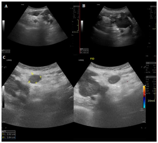
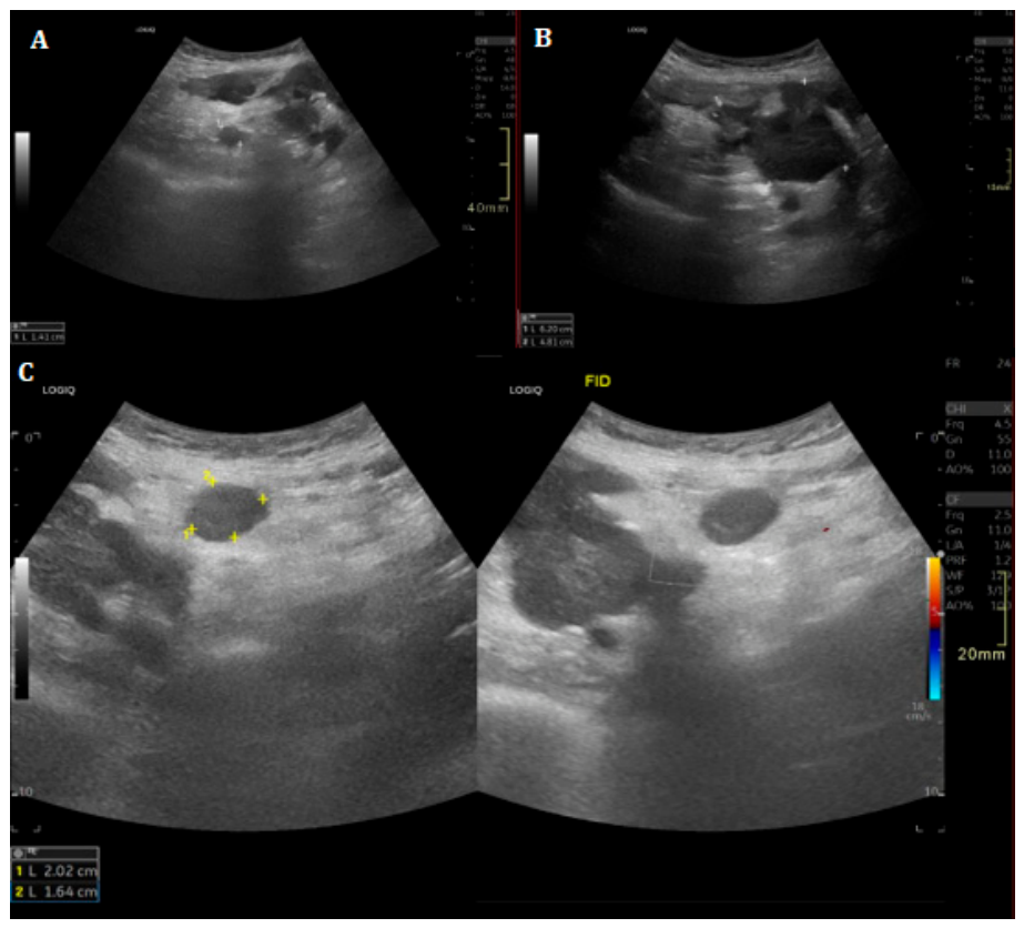
The first case involved a patient evaluated urgently approximately one month after appendectomy because of abdominal pain and fever. Ultrasound demonstrated a heterogeneous right pericecal collection measuring approximately 7 cm, with mixed anechoic and hyperechoic components, initially compatible with an organized postoperative fluid collection. However, the examination also showed adjacent mesenteric fat edema and hypoechoic nodular structures, including nodules up to approximately 20 × 16 mm, suspicious for lymphadenopathy, with additional similar nodules in the infrahepatic region and minimal perihepatic free fluid. Because of these atypical features, second-level imaging was recommended (Figure 1).
Figure 1.
Ultrasound evaluation in Case 1. Urgent post-appendectomy ultrasound performed for abdominal pain and fever demonstrates a heterogeneous right pericecal collection with mixed anechoic and hyperechoic components (A,B). Adjacent mesenteric fat edema and hypoechoic nodular structures suspicious for lymphadenopathy are also visible (C). These atypical findings prompted second-level imaging for further characterization.
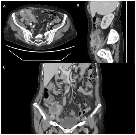
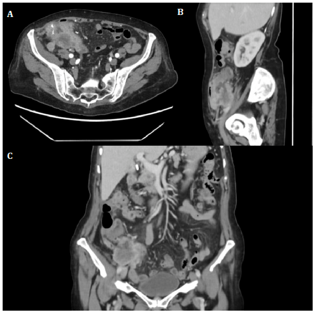
Contrast-enhanced CT performed in the arterial phase demonstrated a large, branched, heterogeneous mass in the right lower quadrant at the previous appendectomy site, measuring approximately 6 × 4 × 6 cm. The lesion showed a peripheral solid component with intense contrast enhancement and central necrotic-colliquative areas. This combination of viable enhancing tissue and fluid-like necrotic components was considered suspicious for an appendiceal-region neoplasm with abscess-like features rather than a simple postoperative collection. Multiple lymph nodes or implant-like nodular lesions with similar characteristics were detected in the perivisceral fat, mesenteric root, and along the right paracolic gutter, with additional right inguinal nodes. Pericecal disease implants were also observed (Figure 2).
Figure 2.
Initial contrast-enhanced CT findings in Case 1. Arterial-phase CT ((A) axial; (B) sagittal; (C) coronal) demonstrates a large branched heterogeneous mass in the right lower quadrant, at the previous appendectomy site, measuring approximately 6 × 4 × 6 cm. The lesion shows an intensely enhancing peripheral solid component and central necrotic-colliquative areas, suggesting an appendiceal-region neoplasm with abscess-like features. Associated nodal and implant-like disease is visible in the perivisceral fat and along the right paracolic gutter.
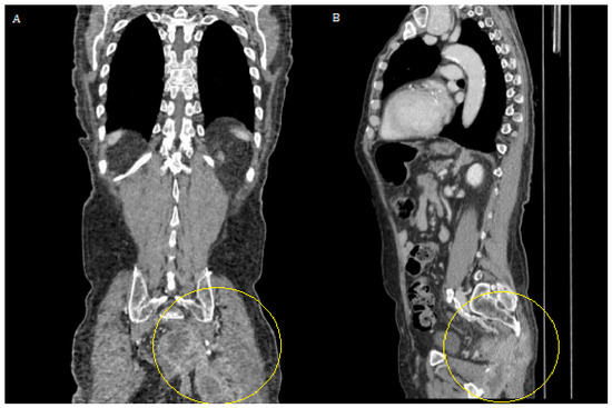
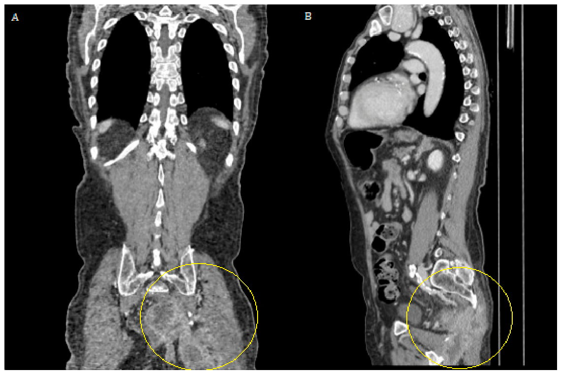
Follow-up CT showed a slight reduction in pelvic free fluid but no substantial change in the right lower quadrant mass, which remained stable in size and morphology, with persistent enhancing solid components and necrotic-colliquative areas (Figure 3).
Figure 3.
Follow-up CT in Case 1: (A) Coronal and (B) sagittal contrast-enhanced CT show persistence of the heterogeneous right lower quadrant mass at the previous appendectomy site. The circled region highlights the persistent mass-like component with enhancing solid tissue and necrotic-colliquative areas, supporting suspicion of an appendiceal-region neoplasm with abscess-like features rather than an uncomplicated postoperative collection.
The persistence of a mass-like structure despite the clinical context of presumed infection further supported the suspicion of malignancy. In this case, the relevant imaging red flags were the persistent mass-like architecture, enhancing solid peripheral component, necrotic-colliquative core, nodal or implant-like disease, and incomplete concordance with a simple postoperative abscess. The imaging suspicion of an underlying appendiceal-region malignancy was subsequently correlated with the final diagnostic work-up/pathological diagnosis, confirming that the abscess-like appearance was related to an underlying neoplastic process rather than to an uncomplicated postoperative collection.
3.2. Case 2: Rectal Carcinoma Complicated by Abscess Formation and Gluteal Fistulization
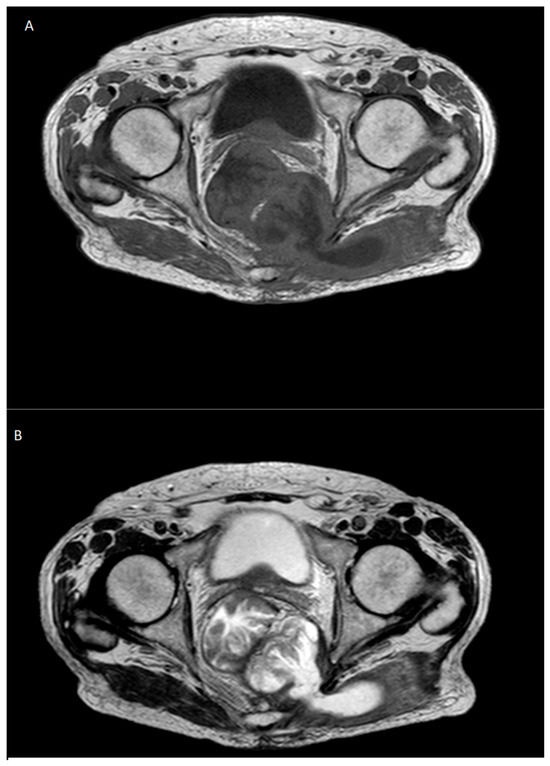
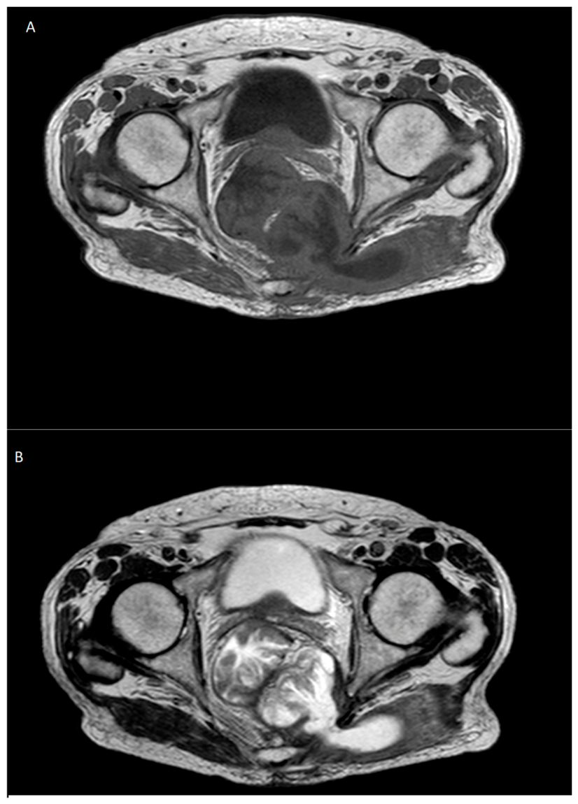
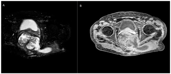
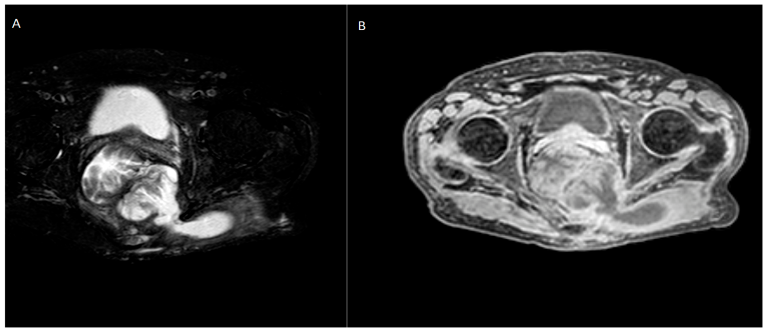
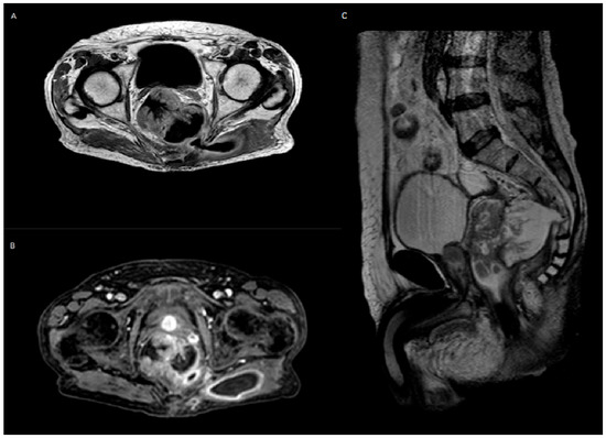
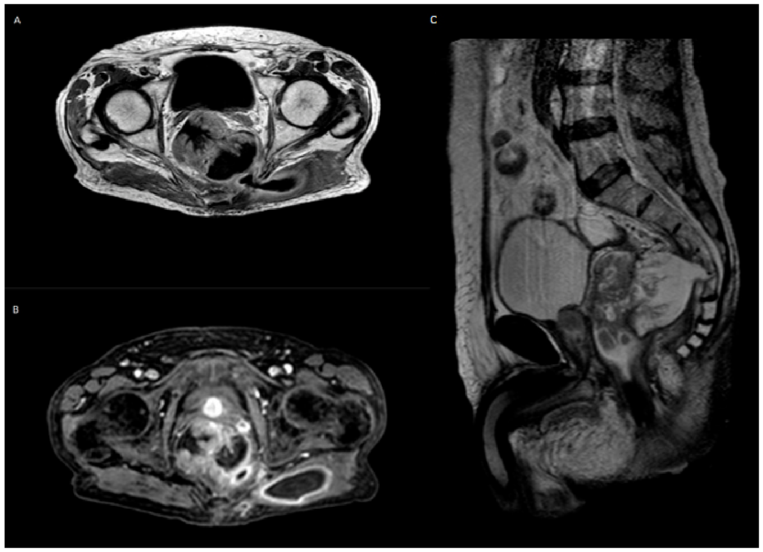
The second case involved a patient with a prior left colostomy and a drainage catheter placed in the left gluteal region. Pelvic MRI performed without and with intravenous contrast demonstrated a large expansile lesion involving the left rectal, pelvic, and gluteal regions. The lesion was partially cystic and partially solid, with contrast enhancement of the solid component. The sigmoid colon was fluid-distended and showed concentric irregular wall thickening of approximately 5 cm, suggesting functional stenosis and possible heteroplastic tissue with pathological enhancement. MRI findings were consistent with a rectal malignancy complicated by abscess formation, posterior extension into the adjacent gluteal musculature, and associated fistulization (Figure 4, Figure 5 and Figure 6).
Figure 4.
Pelvic MRI findings in Case 2. Axial T1- (A) and T2-weighted (B) images, fluid-sensitive sequences, and contrast-enhanced images show a large rectal–pelvic and gluteal lesion with mixed cystic and solid components. Enhancement of the solid tissue, irregular rectal wall thickening, and posterior extension toward the gluteal region are suspicious for rectal malignancy complicated by abscess formation and fistulization.
Figure 5.
Pelvic MRI in Case 2: Fluid-sensitive and pre-contrast sequences. (A) Axial T2-weighted spectral attenuated inversion recovery (SPAIR/SAPIR) image demonstrates the fluid component of the rectal–pelvic and gluteal lesion, with posterior extension toward the left gluteal region. (B) Axial T1-weighted THRIVE image shows the corresponding soft-tissue component before contrast administration, helping to define the mixed cystic–solid architecture of the lesion.
Figure 6.
Pelvic MRI in Case 2: Contrast-enhanced and sagittal assessment. (A) Axial contrast-enhanced T1-weighted image and (B) axial contrast-enhanced T1-weighted THRIVE image show pathological enhancement of the solid component within the rectal–pelvic lesion. (C) Sagittal T2-weighted image demonstrates the craniocaudal extension of the lesion and its relationship with the rectal/pelvic compartment, supporting the diagnosis of rectal malignancy complicated by abscess formation and fistulization.
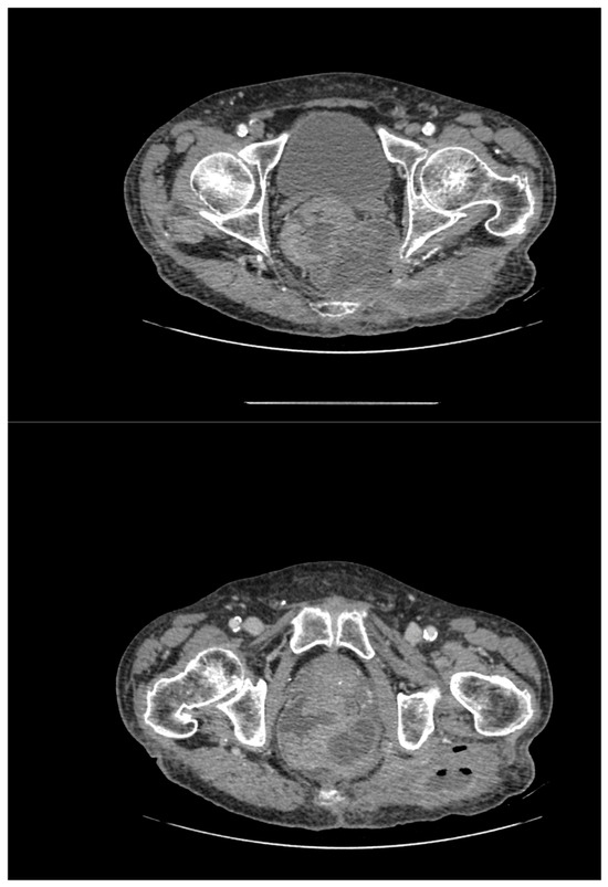
CT performed without and with intravenous contrast confirmed a large multiloculated expansile lesion in the rectal region, measuring approximately 90 × 80 × 108 mm. The lesion showed heterogeneous density due to the coexistence of fluid components and peripheral contrast enhancement. Posterior extension into the left gluteal musculature was observed, associated with a fistulous tract and intralesional gas bubbles. These findings were consistent with rectal carcinoma-associated abscess formation rather than an isolated perirectal infectious collection (Figure 7).
Figure 7.
CT correlation in Case 2. Contrast-enhanced CT demonstrates a large multiloculated rectal-region lesion with fluid components, peripheral enhancement, posterior extension into the left gluteal musculature, a fistulous tract, and intralesional gas bubbles. The coexistence of abscess-like morphology and enhancing pathological tissue supports the diagnosis of rectal carcinoma-associated abscess.
The key imaging red flags in this case were the large expansile mass-like morphology, mixed solid–cystic architecture, enhancement of the solid component, irregular rectal wall thickening, fistulization into the gluteal region, and extension pattern inconsistent with an uncomplicated abscess. MRI was particularly useful for tissue characterization and local mapping of the pelvic and gluteal components, while CT provided a comprehensive assessment of lesion extent, gas-containing abscess-like areas, and associated abdominal findings.
4. Multimodality Imaging Red Flags of “Cancer–Abscess”
The two cases illustrate a recurring diagnostic principle: an abscess-like lesion should not be interpreted only as infection when imaging demonstrates features suggesting viable tumor, atypical extension, or persistence despite the expected clinical course. The “cancer–abscess” concept is therefore most useful as a practical warning pattern rather than as a distinct pathological diagnosis [12,13,14,15].
Several red flags should prompt suspicion of underlying malignancy. First, persistent mass-like architecture is unusual for a simple abscess, particularly when the lesion maintains a solid or infiltrative configuration over time. Second, nodular or irregular enhancement within the wall or periphery of a presumed collection should raise concern for a viable tumor. Third, enhancing solid components within or adjacent to necrotic-colliquative areas are particularly suspicious, as they may reflect tumor necrosis or a neoplasm complicated by secondary infection. Fourth, associated suspicious lymphadenopathy, peritoneal implants, or implant-like nodules should prompt staging-oriented interpretation rather than purely inflammatory assessment. Fifth, fistulization into unusual compartments, such as gluteal musculature, or extension along unexpected anatomical pathways should suggest locally advanced disease. Finally, poor radiological or clinical response to adequate antibiotic or drainage treatment should trigger reassessment, second-level imaging, and tissue diagnosis when appropriate [16].
Ultrasound can be useful as a first-line modality, especially in urgent or postoperative settings, but its role is often limited to detection and triage. Complex echogenic components, nodular tissue adjacent to a collection, and suspicious lymph nodes should prompt contrast-enhanced CT or MRI. CT provides a panoramic evaluation of lesion morphology, enhancement, regional spread, gas, collections, lymph nodes, implants, and distant disease. MRI is particularly valuable in the pelvis, where tissue contrast allows better differentiation among fluid, necrosis, viable tumor, fibrosis, inflammatory changes, and fistulous tracts (Table 1).
Table 1.
Practical imaging comparison between uncomplicated infectious abscesses and malignancy-associated abscess-like lesions. These features should be interpreted as educational red flags rather than validated diagnostic criteria and should always be integrated with clinical, laboratory, endoscopic, surgical, and histopathological correlation when appropriate.
5. Differential Diagnosis of Gastrointestinal and Pelvic Abscess-like Lesions
The differential diagnosis of gastrointestinal and pelvic abscess-like lesions is broad and includes both infectious/inflammatory and neoplastic conditions. Uncomplicated postoperative or inflammatory abscesses, complicated appendicitis, diverticulitis, Crohn-related collections, tubo-ovarian abscesses, infected hematomas, and foreign-body reactions may all show fluid collections, inflammatory fat stranding, gas, and peripheral enhancement [17]. Conversely, several malignancies may mimic abdominopelvic abscesses, including appendiceal neoplasms, colorectal carcinoma, gastrointestinal stromal tumors, lymphoma, ovarian malignancy, gallbladder carcinoma, and recurrent pelvic tumors. Imaging interpretation should therefore focus on features that are discordant with a purely infectious process, such as persistent mass-like architecture, enhancing solid tissue, nodular or irregular wall thickening, suspicious lymph nodes, implant-like nodules, mucinous components, atypical fistulization, or poor response to appropriate treatment. These findings should prompt correlation with the clinical course, inflammatory markers, and tumor markers, when available, as well as endoscopy, biopsy, or surgical evaluation.
6. Practical Diagnostic Approach
A practical diagnostic approach should begin with careful assessment of whether the imaging appearance is fully concordant with an uncomplicated abscess. In patients with fever, pain, inflammatory markers, or recent surgery, infection is often the most likely initial diagnosis. However, radiologists should actively search for discordant features, particularly solid enhancement, persistent mass-like morphology, suspicious nodes, implant-like lesions, atypical extension, or fistulous tracts. When these features are present, the report should explicitly raise the possibility of underlying malignancy and recommend appropriate further evaluation.
In the acute setting, ultrasound may identify a collection and guide initial management. Still, contrast-enhanced CT should be performed when the lesion is large, complex, atypical, postoperative, recurrent, or associated with suspicious nodular tissue. CT should evaluate not only the collection itself but also the adjacent bowel, appendix or appendiceal stump, mesentery, peritoneum, lymph nodes, liver, and potential routes of extension. In pelvic disease, MRI should be considered when CT findings are indeterminate or when local staging, fistula mapping, or tissue characterization may influence management.
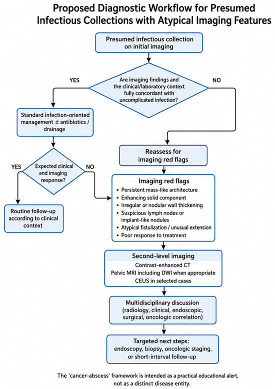
A possible workflow is as follows. If a presumed abscess has typical imaging features and an appropriate clinical context, standard infection-oriented management may be pursued. If atypical features are present at baseline, or if there is an incomplete response to appropriate treatment, second-level imaging should be recommended. If enhancing solid tissue, nodular wall thickening, suspicious lymph nodes, implant-like disease, or atypical fistulization is confirmed, the case should be discussed in a multidisciplinary setting, with consideration of endoscopy, biopsy, surgical assessment, oncologic staging, or targeted follow-up according to the anatomical site and clinical status. This proposed diagnostic pathway is summarized in Figure 8.
Figure 8.
Proposed diagnostic workflow for presumed infectious collections with atypical imaging features. When a presumed abscess shows imaging findings fully concordant with uncomplicated infection and an appropriate clinical and laboratory context, standard infection-oriented management may be pursued. Conversely, the presence of discordant features—such as persistent mass-like architecture, enhancing solid components, irregular or nodular wall thickening, suspicious lymph nodes or implant-like disease, atypical fistulization, or poor response to treatment—should prompt second-level imaging, multidisciplinary discussion, and consideration of endoscopic evaluation, biopsy, oncologic staging, or short-interval follow-up.
7. Discussion
This case report highlights a clinically important imaging pitfall in gastrointestinal and pelvic malignancies: tumors may occasionally present with abscess-like morphology because of necrosis, perforation, fistulization, obstruction, or secondary infection. The proposed “cancer–abscess” concept should therefore be interpreted as a practical imaging-oriented framework and diagnostic alert, rather than a distinct disease entity. Its purpose is to encourage radiologists to recognize imaging features that are discordant with uncomplicated infection and to communicate the need for appropriate clinical, laboratory, endoscopic, surgical, and oncologic correlation [18,19]. Although true abscesses are common and are often appropriately managed with antibiotics, drainage, or surgery, an abscess-like appearance should not preclude careful assessment for an underlying tumor. The two illustrative cases demonstrate how multimodality imaging can reveal suspicious features not expected in uncomplicated infection.
The available literature suggests that malignancies presenting as abscess-like lesions are uncommon but clinically important because they may lead to delayed diagnosis, incomplete treatment, or inappropriate interpretation as uncomplicated infection. In the gastrointestinal tract, this scenario has been described in periappendiceal abscesses associated with appendiceal neoplasms, colorectal cancers presenting with retroperitoneal or iliopsoas abscesses, and rectal carcinomas complicated by perirectal or gluteal abscess formation. Reported outcomes are heterogeneous and depend on tumor site, disease stage, septic complications, feasibility of drainage or surgical treatment, and the timing of oncologic diagnosis. Because most available evidence consists of retrospective cohorts, pictorial reviews, and case-based reports, robust estimates of diagnostic accuracy and prevalence remain limited. Therefore, the present manuscript does not propose validated diagnostic criteria, but rather a practical framework to support earlier suspicion and appropriate escalation of diagnostic work-up.
The first case emphasizes the importance of reassessing presumed postoperative collections when imaging shows solid enhancement, persistent mass-like morphology, and nodal or implant-like disease. Appendiceal-region malignancies are uncommon but may present with clinical and radiological features resembling acute appendicitis, appendiceal inflammatory mass, or periappendiceal abscess. This overlap is particularly relevant after conservative treatment or drainage of complicated appendicitis, because the underlying neoplasm may be detected only during follow-up or interval evaluation. In this context, persistent or atypical pericecal findings should prompt a careful search for enhancing tissue, mucinous components, peritoneal implants, and suspicious lymph nodes [20].
The second case illustrates the pelvic counterpart of the same diagnostic problem. Rectal and colorectal cancers may rarely present with abscess formation, perirectal infection, fistulization, or extension into adjacent compartments. In such cases, symptoms and imaging may initially suggest infection, especially when gas-containing fluid collections, drainage catheters, or inflammatory changes are present. However, irregular rectal wall thickening, enhancing solid tissue, expansile mass-like morphology, and fistulous spread into the gluteal region should raise suspicion for locally advanced malignancy complicated by abscess formation. MRI is particularly helpful in this setting because it provides high soft-tissue contrast and improves characterization of viable tumor, necrosis, fistulous tracts, and involvement of pelvic compartments [21,22,23].
This framework should be applied with caution. Not every atypical abscess-like lesion is malignant, and histopathological confirmation remains necessary whenever imaging and clinical findings raise suspicion for cancer. The value of the “cancer–abscess” concept lies in prompting timely reassessment when the clinical course, treatment response, or imaging morphology is discordant with uncomplicated infection.
Rather, it encourages radiologists to avoid premature closure when a presumed collection demonstrates features inconsistent with simple infection. This is especially relevant in postoperative patients, older adults, patients with recurrent or persistent collections, and cases involving the gastrointestinal tract, where perforated or mucinous tumors can mimic inflammatory disease [24].
In selected challenging cases, additional techniques may further support lesion characterization. Diffusion-weighted MRI may help identify viable solid tumor components, restricted diffusion within cellular malignant tissue, or complex abscess-like areas, although diffusion restriction can be observed in both malignancy and infection and should therefore be interpreted together with morphological and contrast-enhancement findings. Contrast-enhanced ultrasound, when technically feasible and available, may also provide useful information on vascularized solid tissue within or adjacent to a presumed collection, helping distinguish purely fluid components from enhancing mural or nodular tissue. These techniques should be considered complementary rather than standalone tools and should always be interpreted in the clinical context. CT remains the most comprehensive tool for anatomical mapping, detection of enhancement, evaluation of lymph nodes or implants, and assessment of abdominal spread. MRI has particular value in the pelvis and in cases requiring detailed tissue characterization or fistula mapping. The integration of these modalities should guide the radiologist toward a report that not only describes the collection but also communicates the degree of suspicion for underlying malignancy and recommends appropriate next steps [25,26].
This case report has limitations. It is based on two illustrative cases and a discussion of selected literature; therefore, it is not intended to estimate prevalence, diagnostic accuracy, or outcome rates for malignancies presenting as abscess-like lesions. The proposed red flags and workflow are based on case-based interpretation and expert-opinion reasoning and should not be interpreted as evidence-based recommendations, validated diagnostic criteria, or a substitute for clinical judgment, laboratory assessment, histopathological confirmation, or multidisciplinary decision-making. Rather, they are intended to support recognition of discordant imaging features and encourage timely diagnostic escalation in selected cases [27].
8. Future Perspectives
Future research should aim to better define the diagnostic performance of multimodality imaging in distinguishing uncomplicated abscesses from malignancy-associated abscess-like lesions. Diffusion-weighted MRI may help characterize viable tumor components and complex fluid collections, although diffusion restriction is not specific and may be seen in both infection and malignancy. FDG-PET/CT may be useful for whole-body staging and detection of occult disease, but its ability to differentiate cancer from infection is limited by overlapping inflammatory and neoplastic glucose metabolism. Emerging approaches such as radiomics and artificial intelligence may provide quantitative tools for identifying imaging patterns associated with malignancy, treatment non-response, or atypical disease progression. However, these techniques require validation in larger, disease-specific cohorts before they can be incorporated into clinical decision-making. In selected cases, nuclear medicine techniques such as labeled leukocyte scintigraphy or emerging infection-targeted tracers may provide complementary information, although interpretation remains challenging in oncologic patients because infection and malignancy may coexist or produce overlapping uptake patterns.
9. Conclusions
Malignancies may occasionally present as presumed infectious collections, particularly in the gastrointestinal and pelvic regions. The “cancer–abscess” concept is proposed as a descriptive imaging-oriented framework for recognizing abscess-like lesions that may contain or conceal an underlying neoplastic process. This framework should not be interpreted as validated diagnostic criteria or evidence-based recommendations. Persistent mass-like architecture, enhancing solid components, irregular soft-tissue proliferation, necrotic-colliquative areas associated with viable tissue, suspicious lymph nodes or implant-like disease, atypical fistulization, and poor response to treatment should prompt further investigation. Multimodality imaging, especially contrast-enhanced CT and pelvic MRI, can support earlier suspicion, guide tissue diagnosis or staging, and facilitate timely multidisciplinary oncologic management when integrated with clinical, laboratory, endoscopic, surgical, and histopathological correlation.
Take-Home Message
A presumed abscess should be reconsidered when imaging shows solid enhancement, persistent mass-like morphology, suspicious nodal or implant-like disease, or atypical extension. In these settings, radiologists should actively consider the possibility of an underlying malignancy and recommend appropriate second-level imaging, biopsy, endoscopic evaluation, or multidisciplinary assessment.
Author Contributions
Conceptualization, M.M. and G.G.; methodology, M.M., F.M. and G.G.; investigation, M.M., F.M., R.T. and E.M.; resources, R.T. and G.G.; data curation, M.M., F.M. and R.T.; writing—original draft preparation, M.M.; writing—review and editing, M.M., F.M., R.T., E.M. and G.G.; visualization, M.M.; supervision, E.M. and G.G.; project administration, M.M. and G.G. All authors have read and agreed to the published version of the manuscript.
Funding
This research received no external funding.
Institutional Review Board Statement
This study was conducted in accordance with the principles of the Declaration of Helsinki. The study was based on retrospectively collected and fully anonymized clinical and imaging data. No experimental intervention was performed, and no identifiable patient information is presented. Ethical review and approval were waived due to the retrospective and observational nature of the work, in accordance with institutional policy.
Informed Consent Statement
Patient consent for publication was waived because all clinical and imaging material was fully anonymized before inclusion in the manuscript, no direct or indirect identifiers are reported, and the images do not contain names, dates of birth, accession numbers, institutional identification codes, or other identifiable information. The manuscript is presented for scientific purposes only and does not include information that would allow patient re-identification.
Data Availability Statement
No new datasets were generated or analyzed in this study. The imaging material presented in this article is not publicly available due to privacy and ethical restrictions.
Acknowledgments
The authors acknowledge the clinical and radiological teams involved in the diagnostic work-up and multidisciplinary management of the cases presented in this article.
Conflicts of Interest
The authors declare no conflicts of interest.
Abbreviations
The following abbreviations are used in this manuscript:
| CT | Computed Tomography |
| MRI | Magnetic Resonance Imaging |
| MDT | Multidisciplinary Team |
| US | Ultrasound |
References
- Men, S.; Akhan, O.; Köroğlu, M. Percutaneous drainage of abdominal abscess. Eur. J. Radiol. 2002, 43, 204–218. [Google Scholar] [CrossRef] [Scilit] [PubMed]
- De Filippo, M.; Puglisi, S.; D’Amuri, F.; Gentili, F.; Paladini, I.; Carrafiello, G.; Maestroni, U.; Del Rio, P.; Ziglioli, F.; Pagnini, F. CT-guided percutaneous drainage of abdominopelvic collections: A pictorial essay. Radiol. Medica 2021, 126, 1561–1570. [Google Scholar] [CrossRef] [Scilit] [PubMed]
- Fukuya, T.; Hawes, D.R.; Lu, C.C.; Chang, P.J.; Barloon, T.J. CT of abdominal abscess with fistulous communication to the gastrointestinal tract. J. Comput. Assist. Tomogr. 1991, 15, 445–449. [Google Scholar] [CrossRef] [Scilit] [PubMed]
- Pickhardt, P.J.; Levy, A.D.; Rohrmann, C.A., Jr.; Kende, A.I. Primary neoplasms of the appendix: Radiologic spectrum of disease with pathologic correlation. Radiographics 2003, 23, 645–662. [Google Scholar] [CrossRef] [Scilit] [PubMed]
- Pickhardt, P.J.; Levy, A.D.; Rohrmann, C.A., Jr.; Kende, A.I. Primary neoplasms of the appendix manifesting as acute appendicitis: CT findings with pathologic comparison. Radiology 2002, 224, 775–781. [Google Scholar] [CrossRef] [Scilit] [PubMed]
- Leonards, L.M.; Pahwa, A.; Patel, M.K.; Petersen, J.; Nguyen, M.J.; Jude, C.M. Neoplasms of the appendix: Pictorial review with clinical and pathologic correlation. Radiographics 2017, 37, 1059–1083. [Google Scholar] [CrossRef] [Scilit] [PubMed]
- Karande, G.Y.; Chua, W.M.; Yiin, R.S.Z.; Wong, K.M.; Hedgire, S.; Tan, T.J. Spectrum of computed tomography manifestations of appendiceal neoplasms: Acute appendicitis and beyond. Singap. Med. J. 2019, 60, 173–182. [Google Scholar] [CrossRef] [Scilit] [PubMed]
- Fiume, I.; Napolitano, V.; Del Genio, G.; Allaria, A.; Del Genio, A. Cecum cancer underlying appendicular abscess: Case report and review of literature. World J. Emerg. Surg. 2006, 1, 11. [Google Scholar] [CrossRef] [Scilit] [PubMed][Green Version]
- Jongsma, T.E.F.; Puylaert, J.B.C.M. Infection of a malignant appendiceal mucocele masquerading as an appendiceal abscess: Role of preoperative sonography and CT. J. Clin. Ultrasound 2009, 37, 236–238. [Google Scholar] [CrossRef] [Scilit] [PubMed]
- Chandra Mohan, S.; Gummalla, K.M.; H’ng, M.W.C. Malignant tumours mimicking complicated appendicitis and discovered upon follow-up after percutaneous drainage: A case of two patients. Case Rep. Radiol. 2017, 2017, 3253928. [Google Scholar] [CrossRef] [Scilit] [PubMed]
- Mällinen, J.; Rautio, T.; Grönroos, J.; Rantanen, T.; Nordström, P.; Savolainen, H.; Ohtonen, P.; Hurme, S.; Salminen, P. Risk of appendiceal neoplasm in periappendicular abscess in patients treated with interval appendectomy vs follow-up with magnetic resonance imaging: 1-year outcomes of the peri-appendicitis acuta randomized clinical trial. JAMA Surg. 2019, 154, 200–207. [Google Scholar] [CrossRef] [Scilit] [PubMed]
- Son, J.; Park, Y.J.; Lee, S.R.; Kim, H.O.; Jung, K.U. Increased risk of neoplasms in adult patients undergoing interval appendectomy. Ann. Coloproctol. 2020, 36, 366–372. [Google Scholar] [CrossRef] [Scilit] [PubMed]
- Skendelas, J.P.; Alemany, V.S.; Au, V.; Rao, D.; McNelis, J.; Kim, P.K. Appendiceal adenocarcinoma found by surgery for acute appendicitis is associated with older age. BMC Surg. 2021, 21, 228. [Google Scholar] [CrossRef] [Scilit] [PubMed]
- Wazir, S.; Ta, L.; Aguillard, J.; Newell, S.; Wheelis, S.C. Appendiceal adenocarcinoma masquerading as a chronic intra-abdominal abscess. Cureus 2026, 18, e103054. [Google Scholar] [CrossRef] [Scilit] [PubMed]
- Dekker, E.; Tanis, P.J.; Vleugels, J.L.A.; Kasi, P.M.; Wallace, M.B. Colorectal cancer. Lancet 2019, 394, 1467–1480. [Google Scholar] [CrossRef] [Scilit] [PubMed]
- Yang, X.F.; Pan, K. Diagnosis and management of acute complications in patients with colon cancer: Bleeding, obstruction, and perforation. Chin. J. Cancer Res. 2014, 26, 331–340. [Google Scholar] [CrossRef] [Scilit] [PubMed]
- Okita, A.; Kubo, Y.; Tanada, M.; Kurita, A.; Takashima, S. Unusual abscesses associated with colon cancer: Report of three cases. Acta Med. Okayama. 2007, 61, 107–113. [Google Scholar] [CrossRef] [Scilit] [PubMed]
- Moomjian, L.N.; Clayton, R.D.; Carucci, L.R. A spectrum of entities that may mimic abdominopelvic abscesses requiring image-guided drainage. Radiographics 2018, 38, 1264–1281. [Google Scholar] [CrossRef] [Scilit] [PubMed]
- Takakura, Y.; Ikeda, S.; Yoshimitsu, M.; Hinoi, T.; Sumitani, D.; Takeda, H.; Kawaguchi, Y.; Shimomura, M.; Tokunaga, M.; Okajima, M.; et al. Retroperitoneal abscess complicated by necrotizing fasciitis of the thigh in a patient with sigmoid colon cancer. World J. Surg. Oncol. 2009, 7, 74. [Google Scholar] [CrossRef] [Scilit] [PubMed]
- Cacurri, A.; Cannata, G.; Trastulli, S.; Desiderio, J.; Mangia, A.; Adamenko, O.; Pressi, E.; Giovannelli, G.; Noya, G.; Parisi, A. A rare case of perforated descending colon cancer complicated by a fistula and abscess of the left iliopsoas and ipsilateral obturator muscle. Case Rep. Surg. 2014, 2014, 128506. [Google Scholar] [CrossRef] [Scilit] [PubMed]
- Zhou, J.; Wan, S.; Li, C.; Ding, Z.; Qian, Q.; Yu, H.; Li, D. Retroperitoneal abscess as a presentation of colon cancer: The largest case set analysis to date, which was extracted from our unit and the literature. Front. Oncol. 2023, 13, 1198592. [Google Scholar] [CrossRef] [Scilit] [PubMed]
- Saha, B.; Dang, J.; Kahili-Heede, M.; Grobe, J. Rectal adenocarcinoma presenting as a perirectal abscess. Case Rep. Gastroenterol. 2023, 17, 204–211. [Google Scholar] [CrossRef] [Scilit] [PubMed]
- Yano, T.; Nakano, K.; Yoshimitsu, M.; Idani, H.; Okajima, M. Successful resection of rectal cancer and perirectal abscess following systemic chemotherapy and chemoradiotherapy: A case report. Int. J. Surg. Case Rep. 2023, 108, 108403. [Google Scholar] [CrossRef] [Scilit] [PubMed]
- Coyne, B.; Saravanakumar, S.; Murillo-Chavez, F.; Chengappa, N.K.; Ihsan, R. Colorectal carcinoma and gluteal abscesses in the background of a gait disturbance presentation. Case Rep. Oncol. 2024, 17, 1239–1245. [Google Scholar] [CrossRef] [Scilit] [PubMed]
- Gollub, M.J.; Arya, S.; Beets-Tan, R.G.; Deprisco, G.; Gonen, M.; Jhaveri, K.; Kassam, Z.; Kaur, H.; Kim, D.; Knezevic, A.; et al. Use of magnetic resonance imaging in rectal cancer patients: Society of Abdominal Radiology rectal cancer disease-focused panel recommendations. Abdom. Radiol. 2018, 43, 2893–2902. [Google Scholar] [CrossRef] [Scilit] [PubMed]
- Inoue, A.; Sheedy, S.P.; Wells, M.L.; Mileto, A.; Goenka, A.H.; Ehman, E.C.; Yalon, M.; Murthy, N.S.; Mathis, K.L.; Behm, K.T.; et al. Rectal cancer pelvic recurrence: Imaging patterns and key concepts to guide treatment planning. Abdom. Radiol. 2023, 48, 1867–1879. [Google Scholar] [CrossRef] [Scilit] [PubMed]
- Dantas, P.P.; Teixeira, V.B.; Marques, C.F.S.; Nogueira, G.F.; Ortega, C.D. Roles of MRI evaluation of pelvic recurrence in patients with rectal cancer. Insights Imaging 2024, 15, 270. [Google Scholar] [CrossRef] [Scilit] [PubMed]

Disclaimer/Publisher’s Note: The statements, opinions and data contained in all publications are solely those of the individual author(s) and contributor(s) and not of MDPI and/or the editor(s). MDPI and/or the editor(s) disclaim responsibility for any injury to people or property resulting from any ideas, methods, instructions or products referred to in the content. |
© 2026 by the authors. Licensee MDPI, Basel, Switzerland. This article is an open access article distributed under the terms and conditions of the Creative Commons Attribution (CC BY) license.