


Abstract
Ossification of the vascular pedicle following fibula free flap (FFF) reconstruction is an uncommon and typically asymptomatic complication. Symptomatic cases requiring intervention are rare. We report a 29-year-old man with anterior maxillary osteosarcoma who underwent tumor resection followed by reconstruction with an osteocutaneous FFF. Calcification within the surgical site region was noted on imaging at two months after fibular reconstruction. By five months, he developed progressive trismus and pain with mastication. Computed tomography demonstrated a calcified structure extending from the mandible to the reconstructed maxilla along the flap pedicle, raising concern for tumor recurrence. Surgical excision was performed, and histopathology revealed benign woven bone without evidence of malignancy. Postoperatively, trismus improved, and flap viability was preserved. Retained periosteum during FFF harvest maintains osteogenic potential and may result in pedicle ossification. In symptomatic patients, particularly when recurrence is suspected, surgical resection is both diagnostic and therapeutic.
1. Introduction
The fibula free flap (FFF) is widely used for reconstruction of large maxillofacial defects requiring osseous tissue, particularly in the maxilla and mandible. Subperiosteal dissection during flap harvest may increase pedicle length; however, retained periosteum combined with mechanical stress can lead to ossification of the vascular pedicle [1,2,3,4,5,6,7,8,9,10]. This phenomenon may present clinically as a facial mass, trismus, or mastication pain and is typically diagnosed with computed tomography imaging. Symptomatic cases may require surgical excision. We present a case of a 7 cm ossified vascular pedicle following maxillary reconstruction for osteosarcoma, describe its surgical management, and review the current literature.
2. Case Presentation
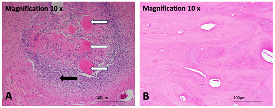
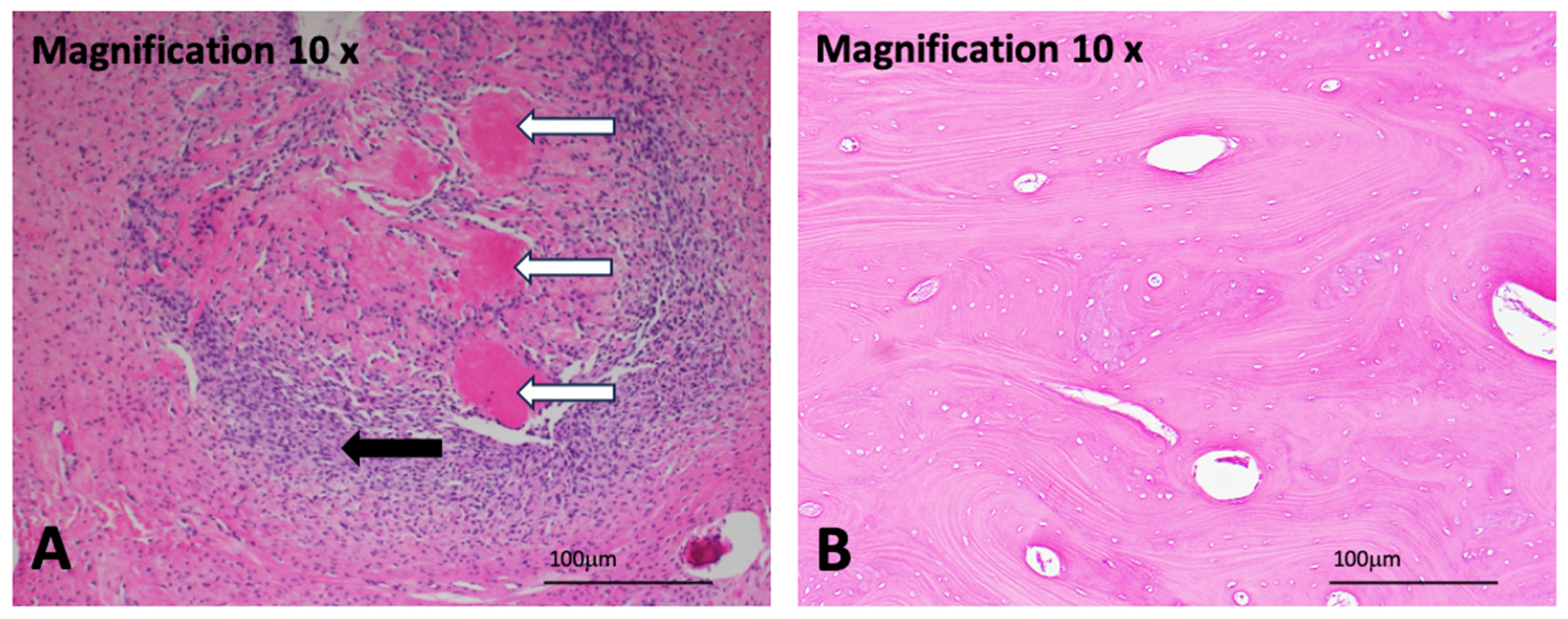
A 29-year-old man presented with a progressively enlarging anterior maxillary mass associated with upper lip deformity and dental displacement. Physical examination revealed a firm, fixed, violaceous lesion involving the anterior maxilla. Computed tomography demonstrated a 4 cm × 3 cm × 3 cm expansile lesion with bony destruction, local invasion, and associated cervical lymphadenopathy. Incisional biopsy confirmed low-grade osteosarcoma (Figure 1A), while nodal biopsy was negative for metastatic disease. The tumor was staged as cT4aN0M0, and the patient began neoadjuvant chemotherapy consisting of cisplatin, doxorubicin, and high-dose methotrexate.
Figure 1.
(A) Pathology slide with hematoxylin and eosin staining from the tumor excision demonstrates well-differentiated, low-grade osteosarcoma with characteristic hypercellularity and ovoid neoplastic cells with eosinophilic cytoplasm (black arrow). There is collagenous amorphous stroma consistent with osteoid formation. The neoplastic bone is fibrillar in appearance (white arrows). Hematoxylin stains nuclei blue-purple, while eosin stains cytoplasm and extracellular matrix pink. (B) Pathology slide of the ossified pedicle demonstrates a benign fragment of dense woven bone.
After three cycles, interval imaging demonstrated tumor reduction, and the patient underwent maxillectomy, palatectomy, and neck dissection with immediate reconstruction using a scapular tip free flap. The scapular flap subsequently failed due to postoperative vascular compromise and was removed, and the defect was temporarily managed with an obturator. Secondary definitive reconstruction (after initial scapular flap failure and completion of neoadjuvant chemotherapy) was performed six months later using a left osteocutaneous fibular free flap. The peroneal artery and venae comitantes were anastomosed to the right facial artery and external jugular vein, respectively. The postoperative course was uncomplicated, and the patient was discharged on postoperative day 10 with a viable flap.
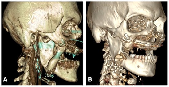
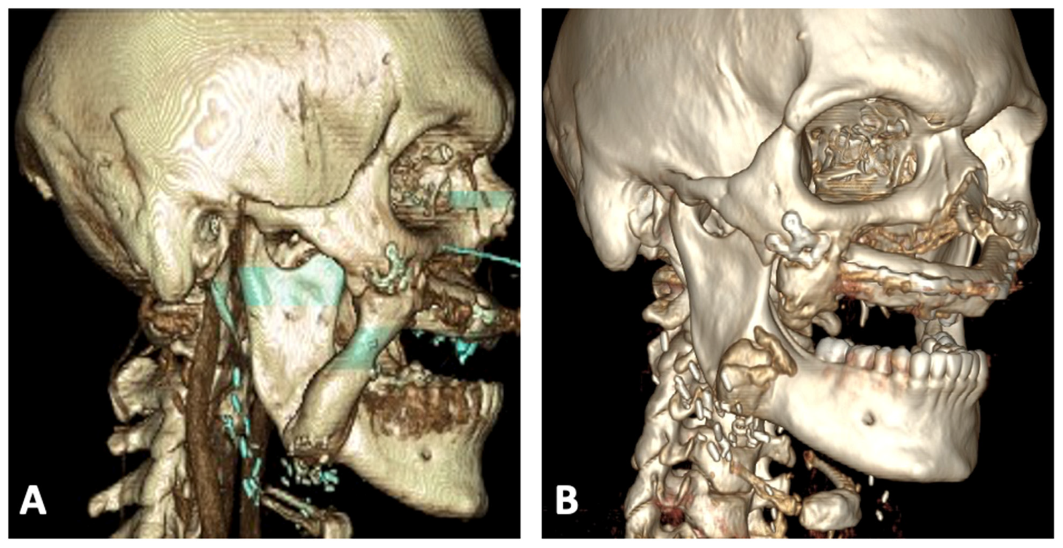
Two months after fibular reconstruction, surveillance CT imaging demonstrated focal calcification near the right masseter muscle. Although initially indeterminate, the finding prompted close follow-up given the patient’s oncologic history. Over subsequent months, he developed progressive trismus, mastication pain, and right-sided facial fullness. Repeat imaging revealed a 7 cm ossified structure extending from the mandibular ramus to the reconstructed superior alveolar ridge along the vascular pedicle (Figure 2A). Given radiographic progression and concern for tumor recurrence, operative exploration was performed.
Figure 2.
CT images with 3D reconstruction demonstrating: (A) preoperative bony ossification of the pedicle and (B) postoperative successful resection of the bony segment.
Through a transoral approach, the ossified pedicle was identified. The lateral aspect of the pedicle was completely calcified, whereas the peroneal vessels remained patent medially. The bony overgrowth adhered to both the reconstructed maxilla and mandibular ramus, forming a rigid connection that restricted jaw motion. The ossified segment was resected using a reciprocating saw, and adjacent bony prominences were contoured with a drill to restore mobility. Postoperatively, the patient experienced marked improvement in trismus. Histopathology confirmed benign woven bone (Figure 1B) without evidence of osteosarcoma recurrence. Follow-up CT confirmed complete removal of the ossified segment (Figure 2B), and the fibular free flap remained viable.
3. Discussion
Vascular pedicle ossification following fibula free flap reconstruction is a recognized but likely underreported phenomenon. Reported incidence varies widely, with higher detection rates in series performing routine postoperative imaging rather than imaging only symptomatic patients (Table 1) [3,4,5,6,11,12,13,14]. This variability suggests that asymptomatic ossification may be more common than clinically appreciated.
Table 1.
The incidence of fibular free flap pedicle ossification reported in the literature varies significantly depending on the imaging protocol.
The pathophysiology likely relates to the osteogenic potential of retained periosteum along the vascular pedicle. Periosteal tissue contains progenitor cells capable of osteoblastic differentiation, particularly when stimulated by bone morphogenetic proteins and local inflammatory mediators [8,9,15,16,17]. Experimental and clinical studies have demonstrated heterotopic bone formation when periosteum remains attached to the pedicle. Conversely, meticulous periosteal stripping during flap harvest has been associated with lower ossification rates, although this technique may increase operative complexity and theoretically risk pedicle injury [12,18,19,20]. Tarsitano et al. reported no ossification when the periosteum was dissected from the pedicle compared with a 17% incidence in controls [6].
Mechanical stress also appears to contribute. Mechanotransduction and micromotion-induced callus formation suggest that repetitive functional forces may stimulate osteogenesis in susceptible periosteal tissue [10]. This may explain the higher frequency of ossification in maxillary reconstruction, where the pedicle traverses the buccal space near active masticatory musculature [21,22,23]. Myon et al. reported on 149 patients who underwent fibula free flap surgery, 10 of whom underwent maxillary reconstruction and 139 underwent mandibular reconstruction. The overall rate of vascular pedicle ossification was 9.4%; however, the rate was much higher in maxillary reconstructions (50%) compared to 6.4% of mandibular reconstructions [3].
Recipient vessel selection and pedicle orientation may further influence ossification. In this case, anastomosis to the facial artery resulted in a pedicle trajectory through the buccal space near the masseter and medial pterygoid muscles, likely subjecting the periosteum to repetitive mechanical stress. Minimizing excess pedicle length, avoiding direct contact between retained periosteum and native bone, and routing the pedicle away from high-motion regions may reduce the risk of clinically significant ossification.
Most cases are asymptomatic and incidentally detected. When symptoms occur, they typically include mass effect, trismus, mastication pain, or, rarely, ankylosis. In oncologic patients, new calcification along the pedicle may raise concern for tumor recurrence, particularly in osteogenic malignancies. Correlation with clinical findings and histopathologic confirmation is therefore essential.
For symptomatic patients, surgical excision is both diagnostic and therapeutic. Prior reports and our experience demonstrate that pedicle resection does not compromise flap viability, and recurrence after excision appears uncommon. Awareness of this complication is especially important in maxillary reconstruction, where functional impairment may be significant.
4. Conclusions
Pedicle ossification following fibular free flap reconstruction is an underrecognized complication with variable incidence. Retained periosteum and mechanical stress contribute to its pathogenesis. Symptomatic cases presenting with trismus, pain, or mass effect should undergo surgical excision. Recognition of this entity is particularly important in maxillary reconstruction to avoid misdiagnosis of tumor recurrence.
Author Contributions
M.R.-S. conceptualization, investigation, data curation, writing—original draft, review and editing; S.K. data, investigation, validation, writing—review and editing; A.A.A. supervision, review and editing; R.M. conceptualization, investigation, data curation, writing—review and editing. All authors have read and agreed to the published version of the manuscript.
Funding
This research received no external funding.
Institutional Review Board Statement
This study was determined to be exempt from institutional review board approval as a single-case report and a retrospective, non-invasive study involving only chart review.
Informed Consent Statement
Informed consent was obtained from all subjects involved in the study.
Data Availability Statement
The original contributions presented in this study are included in the article. Further inquiries can be directed to the corresponding author.
Conflicts of Interest
The authors declare no conflicts of interest.
Abbreviations
The following abbreviations are used in this manuscript:
| FFF | Fibula free flap |
| CT | Computed tomography |
References
- Hidalgo, D.A. Fibula free flap: A new method of mandible reconstruction. Plast. Reconstr. Surg. 1989, 84, 71–79. [Google Scholar] [CrossRef] [Scilit]
- Katz, A.; Gidumal, S.; Mayland, E.; Genden, E. Management of pedicle ossification following free flap reconstruction: A case report. Am. J. Otolaryngol. 2021, 42, 102990. [Google Scholar] [CrossRef] [Scilit] [PubMed]
- Myon, L.; Ferri, J.; Genty, M.; Raoul, G. Consequences of bony free flap’s pedicle calcification after jaw reconstruction. J. Craniofac. Surg. 2012, 23, 872–877. [Google Scholar] [CrossRef] [Scilit]
- Autelitano, L.; Colletti, G.; Bazzacchi, R.; Biglioli, F. Ossification of vascular pedicle in fibular free flaps: A report of four cases. Int. J. Oral Maxillofac. Surg. 2008, 37, 669–671. [Google Scholar] [CrossRef] [Scilit] [PubMed]
- Smith, R.B.; Funk, G.F. Severe trismus secondary to periosteal osteogenesis after fibula free flap maxillary reconstruction. Head Neck 2003, 25, 406–411. [Google Scholar] [CrossRef] [Scilit]
- Tarsitano, A.; Sgarzani, R.; Betti, E.; Oranges, C.M.; Contedini, F.; Cipriani, R.; Marchetti, C. Vascular pedicle ossification of free fibular flap: Is it a rare phenomenon? Is it possible to avoid this risk? Acta Otorhinolaryngol. Ital. 2013, 33, 307–310. [Google Scholar]
- Mays, A.C.; Gillenwater, A.M.; Garvey, P.B. Rare presentation of heterotopic ossification along a fibula free flap pedicle in a high-volume microvascular reconstruction practice. Head Neck 2018, 40, E21–E24. [Google Scholar] [CrossRef] [Scilit]
- Ortak, T.; Özdemir, R.; Uysal, A.; Ulusoy, M.G.; Sungur, N.; Sahin, B.; Koçer, U.; Şensöz, Ö. Osteogenic capacities of periost grafts, periost flaps and prefabricated periosteal flaps: Experimental study. J. Craniofac. Surg. 2005, 16, 594–600. [Google Scholar] [CrossRef] [Scilit]
- Burstein, F.D.; Canalis, R.F. Studies on the osteogenic potential of vascularized periosteum: Behavior of periosteal flaps transferred onto soft tissues. Otolaryngol. Neck Surg. 1985, 93, 731–735. [Google Scholar] [CrossRef] [Scilit] [PubMed]
- Carter, D.R.; Beaupré, G.S.; Giori, N.J.; Helms, J.A. Mechanobiology of skeletal regeneration. Clin. Orthop. Relat. Res. 1998, 355S, S41–S55. [Google Scholar] [CrossRef] [Scilit]
- Karagozoglu, K.H.; Winters, H.A.H.; Forouzanfar, T.; Schulten, E.A.J.M. Periosteal ossification of the vascular pedicle after reconstruction of continuity defects of the mandible and the maxilla with fibular free flaps: A retrospective study. Br. J. Oral Maxillofac. Surg. 2013, 51, 965–967. [Google Scholar] [CrossRef] [Scilit]
- Kim, B.B.; Kaleem, A.; Alzahrani, S.; Yeoh, M.; Zaid, W. Modified fibula free flap harvesting technique for prevention of heterotopic pedicle ossification. Head Neck 2019, 41, E104–E112. [Google Scholar] [CrossRef] [Scilit] [PubMed]
- Wood, C.B.; Rohde, S.L.; Sinard, R.J.; Mannion, K.; Bigcas, J.-L.M. Incidence of pedicle ossification in osseous free flap reconstruction in the head and neck. Oral Oncol. 2020, 103, 104611. [Google Scholar] [CrossRef] [Scilit] [PubMed]
- Baserga, C.; Massarelli, O.; Bolzoni, A.R.; Rossi, D.S.; Beltramini, G.A.; Baj, A.; Giannì, A.B. Fibula free flap pedicle ossification: Experience of two centres and a review of the literature. J. Cranio-Maxillofac. Surg. 2018, 46, 1674–1678. [Google Scholar] [CrossRef] [Scilit]
- Rintala, A.E.; Ranta, R. Periosteal flaps and grafts in primary cleft repair: A follow-up study. Plast. Reconstr. Surg. 1989, 83, 17–24. [Google Scholar] [CrossRef] [Scilit]
- Takato, T.; Harii, K.; Nakatsuka, T.; Ueda, K.; Ootake, T. Vascularized periosteal grafts: An experimental study using two different forms of tibial periosteum in rabbits. Plast. Reconstr. Surg. 1986, 78, 489–497. [Google Scholar] [CrossRef] [Scilit]
- Yu, Y.Y.; Lieu, S.; Lu, C.; Colnot, C. Bone morphogenetic protein 2 stimulates endochondral ossification by regulating periosteal cell fate during bone repair. Bone 2010, 47, 65–73. [Google Scholar] [CrossRef] [Scilit] [PubMed]
- Romana, M.C.; Masquelet, A.C. Vascularized periosteum associated with cancellous bone graft: An experimental study. Plast. Reconstr. Surg. 1990, 85, 587–592. [Google Scholar] [CrossRef] [Scilit]
- Canalis, R.F.; Burstein, F.D. Osteogenesis in vascularized periosteum. Interact. Underlying Bone Arch. Otolaryngol. 1985, 111, 511–516. [Google Scholar] [CrossRef] [Scilit]
- Colletti, G.; Autelitano, L.; Rabbiosi, D.; Biglioli, F.; Chiapasco, M.; Mandalà, M.; Allevi, F. Technical refinements in mandibular reconstruction with free fibula flaps: Outcome-oriented retrospective review of 99 cases. Acta Otorhinolaryngol. Ital. 2014, 34, 342–348. [Google Scholar]
- Abou Ghanem, O.; O’Sick, N.R.; Kalmar, C.L.; Golinko, M. Complete Extensive Heterotopic Ossification of the Entire Free Fibula Flap Vascular Pedicle in Maxillary Reconstruction. J. Craniofac. Surg. 2024, 35, e200–e202. [Google Scholar] [CrossRef] [Scilit] [PubMed]
- Zou, B. Pedicle ossification after fibular flap reconstruction of maxillary defects: A case report and literature review. Clin. Case Rep. 2024, 12, e8810. [Google Scholar] [CrossRef] [Scilit] [PubMed]
- Karkoutli, A.A.; Beiriger, J.; McCrary, H. Heterotopic ossification of the fibula flap pedicle causing trismus in a pediatric maxillary reconstruction. J. Surg. Case Rep. 2026, 2026, rjaf1078. [Google Scholar] [CrossRef] [Scilit] [PubMed] [PubMed Central]

Disclaimer/Publisher’s Note: The statements, opinions and data contained in all publications are solely those of the individual author(s) and contributor(s) and not of MDPI and/or the editor(s). MDPI and/or the editor(s) disclaim responsibility for any injury to people or property resulting from any ideas, methods, instructions or products referred to in the content. |
© 2026 by the authors. Licensee MDPI, Basel, Switzerland. This article is an open access article distributed under the terms and conditions of the Creative Commons Attribution (CC BY) license.