


Abstract
In this work, the impact of UV (ultaviolet) radiation combined with thermal treatments and Ag doping in TiO2 transparent films on crystal structure and light absorption in the visible spectral region is investigated. The nanostructured TiO2:Ag films were obtained via sol–gel spin-coating deposition, using UV radiation between layers. The substrates used were quartz and silicon wafers. X-Ray Diffraction (XRD) and Fourier Transform Infrared (FTIR) spectroscopy revealed that the structural features of TiO2:Ag films were strongly dependent on the annealing temperatures. The presence of Ag nanoparticles and a small silver oxide fraction in the film structure was found. The formation of AgO and Ag nanoparticles is affected by thermal annealing and the UV radiation treatment. The transmittance, absorbance and reflectance spectra were used for determining the optical behavior and localized surface plasmon resonance (LSPR) properties of TiO2:Ag thin films. Optical characterization revealed increased optical absorption in the whole visible range of TiO2 thin films with incorporated Ag NPs. The dependence of the plasmonic absorption bands, the values of the optical band gap on the number of layers (from one to five) and the high-temperature treatments (from 150 to 800 °C) were studied.
1. Introduction
Titanium dioxide (TiO2) is a highly efficient material, distinguished for its high chemical and thermal stability, non-toxicity, low cost, wide band gap, transparency, etc. [1,2]. The three main crystal polymorphs of TiO2 are anatase, rutile and brookite phases [3]. The anatase and rutile structures are of research interest and they offer distinct electronic and optical properties driven by their unique crystal lattices [4].
TiO2 films are obtained through a huge range of deposition techniques, including magnetron sputtering [5], CVD, ALD [6], spray pyrolysis, sol–gel methods [7], pulsed laser deposition [8], etc., usually followed by thermal annealing to promote specific phase modifications [9]. Among these preparation techniques, the sol–gel approach is employed to obtain homogenous coatings with good control of structure, composition, film thickness, uniformity and adhesion to different substrates [10,11]. Other advantages of the sol–gel method include the capacity for homogeneous doping, low-temperature crystallization, and better phase control in complex and mixed oxide systems [11].
TiO2 materials have a broad area of applications, such as photocatalytic degradation of pollutants, photoelectrochemical water splitting, self-cleaning coatings, solar cells, optical waveguides, gas sensors, etc. [2,7,12].
The incorporation of noble metals such as Pt, Pd, Au, and Ag is an effective approach to modify metal oxide properties, to expand their existing applications and to find new ones [13,14]. The doping of TiO2 with plasmonic noble metals lowers the band gap energy and allows the absorption of photons to occur at lower energy towards the visible region in the electromagnetic spectrum, primarily due to the localized surface plasmon effect (LSPR) [15]. LSPR is related to the collective oscillation of free conduction electrons in metallic nanoparticles driven into resonance by the electric field of incident light. This is manifested by light absorption and scattering [16]. This phenomenon occurs at specific wavelengths, which depend on the size, shape, and material of the nanoparticles. It is widely reported that the LSPR bands of metal nanoparticles (NPs) are largely size-dependent [16,17]. It is expected that the absorption peaks shift towards shorter wavelengths (blue shift) with decreasing metal NP sizes (below 10–20 nm), and bigger particles exhibit LSPR bands at longer wavelengths (red shift) [16,18]. Other factors that exert sufficient impact on plasmonic bands include the composition of NPs, size, geometry and shape, dielectric environment and the separation distance of NPs [16,19].
Noble metal dopants in the titanium oxide lattice result in the creation of empty and/or filled localized impurity levels within the TiO2 band gap. For example, in the case of Ag embedment, its 4d orbitals hybridize with the 2p orbitals of oxygen close to the valence band maximum, or Ag 4d orbitals generate discrete impurity states below the conduction band minimum (which are often related to Ti3+ states or oxygen vacancies). This reduces the optical band gap energy [20]. Meanwhile, LSPR enhances visible-light absorption and it creates energetic “hot electrons” transferred into the TiO2 conduction band. This is a resonant optical phenomenon, and in general, LSPR cannot modify the band gap of titania [21]. TiO2:Ag nanostructured films have applications in various fields. These materials are known to enhance photocatalytic activity [22], to be applied in pollution degradation and wastewater treatment [23], to be integrated in solar cells [24] or advanced photodetector applications [25], etc.
The sol–gel deposition of TiO2:Ag films with a silver nitrate precursor is reported by other studies [26,27]. The silver in the sol solution is presented predominantly as solvated Ag+ ions or complexed silver species interacting with the titanium oxide matrix.
UV radiation treatment provokes the low-temperature crystallization, densification, and photochemical activation of sol–gel titanium dioxide nanomaterials [28]. In the case of the sol–gel- method, derived TiO2 materials containing silver ions are exposed to UV radiation, and three major effects appear: UV-induced Ag+ reduction to Ago [29], nucleation and growth of Ag nanoparticles (NPs) [30], and UV-assisted crystallization of the TiO2 matrix [31] The present study finalizes a previous study on the sol–gel deposition of TiO2 films with embedded Ag NPs. Previously, we investigated spin-coated five-layered TiO2:Ag nanocomposite films with one silver concentration. Their structural, optical and vibrational properties had been reported in relation to thermal treatments (varying the annealing temperature (300–600 °C) and the ambient gas (oxygen and nitrogen)) [32]. The next investigation stage was changing the silver concentration (1 and 0.5 wt% AgNO3 concentration in titanium sol solution), and the preliminary results for the structural and optical properties of TiO2:Ag films, obtained from these two sol solutions after annealing at 600 °C, were reported. The results were compared to TiO2:Ag films, obtained from these sols, but the films were UV-treated between layer deposition and finally treated at 600 °C [33].
The systematic investigation of the sol–gel silver-doped TiO2 films was performed to reveal the influence of UV radiation treatment of the prepared mixed sols with different Ag precursor concentrations on the final properties of the fabricated nanostructured films. The samples were thermally treated at temperatures from 300 to 600 °C. The aim was to investigate the impact of the silver doping amount and thermal annealing on the structural, morphological and optical properties of TiO2:Ag films [34].
This work presents a new study and novel results regarding the influence of UV radiation between layers (UV radiation is applied after each layer deposition with no preheating temperature) and different thermal treatments (300–600 °C) on the structural and vibrational properties of the sol–gel-derived TiO2:Ag films. The number of layers (from one to five layers) varies. XRD analysis is extended to reveal the formation of Ag and AgO fractions in relation to the Ag precursor concentration, the additional thermal treatments and the number of layers. Detailed FTIR characterization is presented. The optical properties are monitored deeply by measuring film transmittance, reflectance and absorbance spectra of TiO2:Ag films. The effect of the number of layers and the combination of UV and thermal treatments (300–800 °C) on the plasmonic properties and the values of the optical band gap is determined.
2. Materials and Methods
Sol–gel TiO2 film deposition has been previously described [34]. The synthesized titanium-containing sol solution maintains its film-forming properties for more than two years. This sol stability makes the titanium solution interesting for practical applications.
The silver precursor was AgNO3 (silver nitrate). Doping was performed by using two different concentrations: 1 and 0.5 wt% AgNO3 (silver nitrate, ≤100%, Merck KGaA Frankfurter, Darmstadt, Germany). The silver precursor was added in a certain volume of the titanium sol solution. The mixed solutions were stirred using a magnetic stirrer for 1 h and then were ultrasonically treated at 50 °C for two hours per day for one week. The sols were stored in dark bottles. The mixed sols and the corresponding deposited TiO2:Ag films are labeled Sol A (1 wt% AgNO3) and Sol B (0.5 wt% AgNO3), respectively. The solutions were homogeneous, without precipitation and sediment and stable for three months.
TiO2:Ag thin films were obtained by using the spin-coating method (employing a spin-coater P 6708, PI-KEM Limited, Staffordshire, UK) at 8000 rpm for 30 s. The films were UV-radiated for 15 min between layers. TiO2:Ag films with different numbers of layers (ranging from one to five) were deposited. The substrates were Si wafers (p-type, FZ, resistivity 4.5–7.5 Ω, orientation <100>) and quartz (UV-graded, glass thickness 1 mm ± 0.1). The preheating (150 °C for 30 min) and the annealing treatments (300–800 °C, 60 min) were performed with a constant heating rate of 10 °C/min. A programmable chamber furnace was used (TOKMET-TK, Ltd., Varna, Bulgaria).
Film thickness was determined using an LEF 3 M laser ellipsometer (Siberian Branch of Russian Academy of Sciences, Novosibirsk, Russia), equipped with a HeNe laser at a wavelength of 638.2 nm. It was found that one layer of TiO2:Ag films from Sol A showed values from 55 nm (150 °C) to 48 nm (600 °C); accordingly, the five layer films ranged from 325 nm (150 °C) to 245 nm (600 °C). The sol–gel films deposited from Sol B (0.5 wt% AgNO3) showed film thickness values for one layer of 55–45 nm and 300–230 nm. Film thickness was reduced after thermal treatments. X-Ray Diffraction patterns were recorded by using an XRD diffractometer Bruker D8 (Bruker AXS GmbH, Karlsruhe, Germany) with a Cu anode (λKα = 1.54056 Å), grazing angle of 2o, and step time of 8 s. FTIR spectroscopy was used for analyzing the composition and the vibrational properties of TiO2:Ag films. FTIR spectra were taken witg Shimadzu Spectrophotometer IRPrestige-21 (Shimadzu Corporation, Kyoto, Japan) in the spectral range 350–4000 cm−1 using a bare Si wafer as the background. The transmittance, reflectance and absorbance spectra were carried out by using a UV–VIS–NIR Shimadzu 3600 double-beam spectrophotometer (Shimadzu Corporation, Kyoto, Japan) in the spectral region of 200–1800 nm. The transmittance spectra were measured against air. The reflectance spectra were recorded using the specular reflectance attachment (angle of Incidence 5°) and Al mirror as reference.
3. Results and Discussions
3.1. Structural Study by XRD Measurements
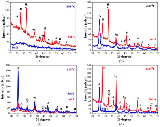
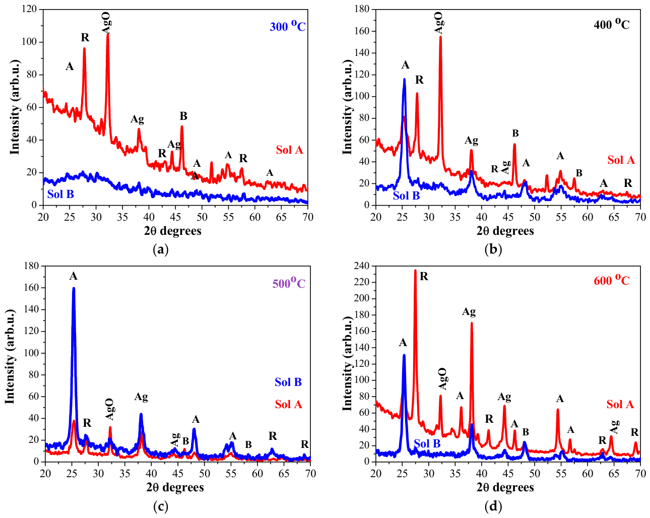
XRD characterization was carried out to reveal the crystalline structure, the crystal phases and the sizes of the crystallites in TiO2:Ag films. Figure 1 presents the diffraction patterns of the five-layered TiO2:Ag films, obtained from Sol A and Sol B with UV radiation after each layer deposition. Their final treatments were at temperatures from 300 to 600 °C. Table 1 presents the estimated crystallite sizes of the crystal phases in the recorded XRD patterns. The crystallite sizes were determined using the Scherrer equation [35].
Figure 1.
XRD patterns of sol–gel TiO2:Ag films, UV-radiated and then annealed at (a) 300, (b) 400, (c) 500 and (d) 600 °C. The films are obtained from Sol A and Sol B and they have five layers. The anatase phase is marked as A, rutile as R, and brookite as B. Ag (metallic silver) and AgO lines are pointed as well.
Table 1.
Crystalline sizes of different crystal phases, estimated from XRD data for sol–gel TiO2:ag films. All samples are have five layers with the exception marked in table. The uncertainties in crystallite size are estimated by assuming a 0.02° uncertainty in the peak positions and in the FWHM.
Figure 1a presents the recorded XRD patterns of 300 °C annealed samples. The XRD pattern of the TiO2:Ag (Sol B) film displays a diffuse scattering band, typical of a highly disordered or amorphous phase (Figure 1a, blue curve). The higher Ag precursor concentration results in a very different diffraction pattern (Figure 1a, red color). XRD analysis reveals that the TiO2:Ag (Sol A) film is polycrystalline, consisting primarily of the rutile phase with a crystallite size of 12.8 nm, along with traces of anatase and brookite fractions indicated by weak, broad reflections. Two clear lines are related to Ag (with cubic crystal structure and crystallite sizes of 18.1 and 27.4 nm, estimated from the planes (111) and (200), respectively). The presence of an AgO phase is detected for the TiO2:Ag (Sol A) film annealed at 300 °C, with a strong XRD line at 2θ = 32.2°.
The increase in the annealing temperature to 400 °C (Figure 1b) induced the crystallization of the TiO2:Ag (Sol B) film. The analysis reveals that the sample crystallized mainly in the anatase phase; however, some contributions from silver oxide and rutile titania are also evident from the broad, low-intensity peaks. The presence of Ag nanoparticles is proved with the line located at 2θ = 38°. On the other hand, the TiO2:Ag (Sol A) film exhibited a mixed crystalline structure, including anatase, rutile, Ag and AgO fractions. The XRD pattern shows a strong line at 32.2°, corresponding to the −111 plane of AgO with a crystallite size of 19.4 nm. The anatase and rutile phases are also well expressed, with the rutile phase showing bigger crystallites measuring 20.4 nm compared to those of the anatase phase (7.0 nm). XRD measurement confirms the formation of the cubic Ag NPs with crystallite sizes of 24.9 and 34.0 nm, determined from the (111) and (200) planes, respectively.
The thermal treatment at 500 °C (Figure 1c) led to interesting structural features. The UV-radiated TiO2:Ag (sol B, blue curve) film shows a predominant presence of the anatase phase (exhibiting strong lines), together with the appearance of well-defined peaks related to the rutile phase (estimated crystallite size of 18.8 nm). Ago and AgO phases are also detected and their corresponding sizes of crystallites are 12.0 and 7.4 nm (see Table 1), respectively. The silver NPs have a crystallite size very close to that observed in the sample annealed at 500 °C. the TiO2:Ag (sol A) film structure consists of four crystal fractions, and the XRD lines of rutile TiO2 are less intense.
Annealing at the highest temperature (600 °C) induced the growth of larger Ag NPs, sharpened the XRD peaks of the rutile phase, and prohibited the appearance of the AgO phase in the UV-irradiated TiO2:Ag film deposited from Sol B. The existence of Ag NPs was confirmed by two well-pronounced XRD peaks. The TiO2:Ag (Sol A) film (see Figure 1d) reveals a greater crystallization degree with well-defined XRD peaks, attributed to the anatase and rutile phases. The AgO fraction is still observed and has a crystallite size of 24.7 nm. Three distinct peaks confirm the presence of face-centered cubic (fcc) silver. Compared to the thermally annealed TiO2:Ag films, UV radiation treatment induces a higher degree of crystallinity and promotes the growth of larger Ag nanoparticles [34].
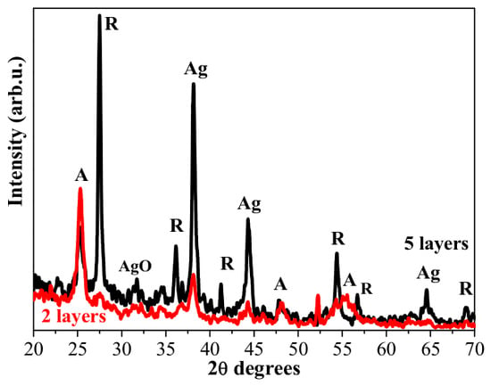
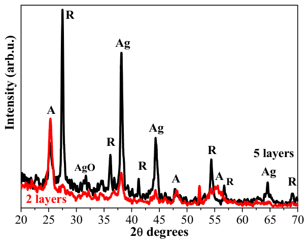
Figure 2 shows the XRD patterns of two-layer and five-layer TiO2:Ag (Sol A) films annealed at 600 °C. Thinner films exhibit reduced intensities for both the rutile XRD lines and the Ago peaks; the AgO phase is not detected. The crystallite sizes of the identified phases are given in Table 1. The anatase phase predominantly appears in thinner films. Consequently, the five-layer film has larger silver NPs, along with a well-defined AgO fraction.
Figure 2.
The XRD patterns of UV-treated TiO2:Ag films, deposited from Sol A with 2 (red curve) and 5 layers (black line). The anatase phase is marked as A and rutile as R. The films were annealed at 600 °C.
The effect of silver precursor concentration was studied by estimating the anatase and rutile fractions in TiO2:Ag films. It must be noted that the other crystal phases were not taken into account. The values were determined by Equations (1) and (2) [36]:
where WA and WR represent the mass fraction of anatase TiO2 and rutile TiO2, respectively. IA and IR represent the integral intensity of diffraction peaks on the crystal surface of anatase TiO2 (101) and rutile TiO2 (110), respectively.
The obtained data are shown in Table 2. The rutile phase is provoked by silver incorporation and UV radiation. Increasing the number of layers in the TiO2:Ag (Sol A) film promotes a higher fraction of the rutile phase. This analysis was performed to clarify the effect of different treatments on the formation of the two main TiO2 phases, anatase and rutile, and their modification with Ag doping, UV radiation, annealing and film thickness.
Table 2.
Crystallographic composition (weight percentage %) of UV-radiated TiO2:Ag films, deposited from Sol A and Sol B as function of annealing temperature from 300 to 600 °C.
Comparing the five-layered samples, the rutile phase fraction shows a strong dependence on the annealing temperature; notably, lower temperatures promote greater rutile phase formation. The two-layered UV-radiated TiO2:Ag (Sol A) film reveals a more pronounced anatase phase. Interestingly, in the case of the films obtained from the sol with a lower silver concentration, the anatase/rutile ratio is strongly influenced by UV radiation. UV radiation provokes an opposite trend for Sol B films as the anatase fraction is increased considerably (see Table 2) compared to films only subjected to thermal treatment.
XRD results show that the presence of Ag and AgO phases affects the anatase/rutile transformation, aligning with previously reported research [37,38]. The XRD study reveals that Ag NP embedment leads to modification of film crystallization and the appearance of new crystal phases.
3.2. FTIR Analysis
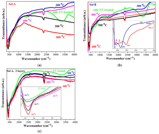
FTIR spectra of five-layered TiO2:Ag films, obtained from Sol A and Sol B with UV irradiation and subsequent annealing at 300–600 °C, are illustrated in Figure 3. The films (Sol A) with only UV treatment show a very broad and strong band at 3330 cm−1 (covering the spectral area of 3000–3590 cm−1). This band is due to the O–H stretching vibrations of surface hydroxyl groups (Ti–OH) and physically adsorbed water molecules [39]. This absorption feature vanished with increasing annealing temperature. Two new weaker lines appear (see Figure 3a) at 3737 and 3610 cm−1. These peaks are assigned to the isolated terminal hydroxyl groups (Ti–OH) bound to single surface titanium cations and to the stretching of non-hydrogen bonding free hydroxyl groups, respectively. They may also be associated with octahedral vacancies, specifically 6Ti3+-OH [39]. A characteristic bending mode corresponding to the hydroxyl groups was observed at 1680 cm−1 [39]. Weak IR lines observed at 1522 cm−1 are attributed to the C=C stretching vibrations of residual organic species. These bands are present in the spectra of UV-radiated TiO2:Ag films, obtained from both Sol A and Sol B.
Figure 3.
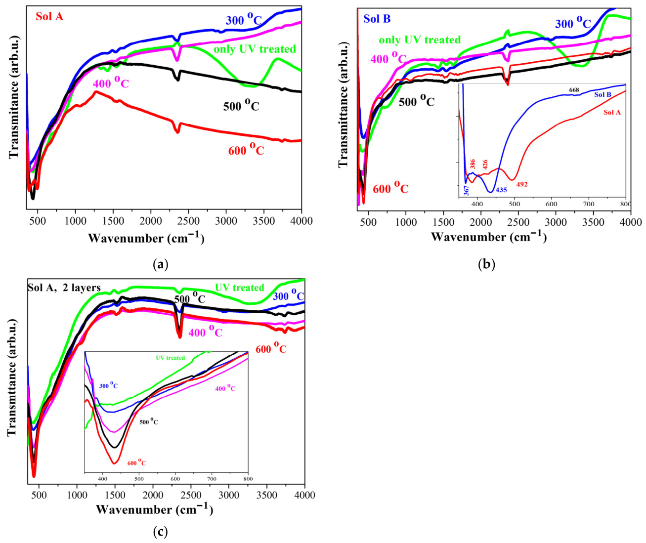
FTIR spectra of TiO2:Ag films: (a) the five-layered films deposited from Sol A; (b) the five-layered films deposited from Sol B. The figure shows a comparison of the main absorption band of the films, deposited from the two sols, and (c) the two-layered films from Sol A in the spectral range 350–800 cm−1. The films are UV-treated and subjected annealing at 300–600 °C.
The broad FTIR bands observed at 750 cm−1 and 436 cm−1 for the TiO2:Ag films, (Sol A) treated at 300 and 400 °C, are related to Ti-O-Ti lattice vibrations and to the characteristic Ti-O stretching mode of anatase titania, respectively. The observed broadening within this spectral range is indicative of structural disorder and amorphness. Annealing at the highest temperature of 600 °C induces splitting of the main band into two distinct lines at 491 and at 383 cm−1. The IR line at 383 cm−1 is assigned to the Ti-O stretching mode in the anatase framework [40,41]. The 491 cm−1 band corresponds to Ti-O bonds vibrations characteristic of the rutile phase [42]. A similar splitting phenomenon is observed at a lower annealing temperature (400 °C) in the FTIR spectra of the UV-treated Sol B TiO2:Ag films, as shown in Figure 3b. The absorption features observed at 370 cm−1 (weak line) and 435 cm−1 (broader and intense band) are attributed to the Ti-O and to Ti-O-Ti stretching vibrations of the anatase phase, respectively [41]. The broad absorption band centered at 435 cm−1 overlaps with the IR band at 443 cm−1, which is assigned to the asymmetric stretching vibration of Ti–O–Ag [43].
The inset in Figure 3b shows the difference between the FTIR spectra of the five-layered TiO2:Ag films obtained from Sol A and Sol B. A pronounced shift in the main absorption bands is seen. For the film deposited from Sol A, the main absorption band is detected at 492 cm−1. XRD analysis revealed that this film possesses a higher fraction of rutile TiO2 (see Table 2). Increasing the Ag concentration in TiO2:Ag films shifts the IR absorption bands toward higher wavenumbers. A similar finding has been reported for Ag NPs incorporated into TiO2 nanocrystals [44]. The splitting of the absorption band may be attributed to the formation of different TiO2 polymorphs and distinct Ag and AgO phases within the film structure. In the literature, the absorption bands observed in the 450–520 cm−1 range are widely attributed to Ag–O bond vibrations [45]. The presence of Ag-O bonds is expected as the AgO phase was confirmed within the structure of TiO2:Ag film via XRD measurements.
Figure 3c presents FTIR spectra of the two-layered TiO2:Ag films obtained from Sol A and treated at different temperatures. As shown in the inset, no splitting of the main bands is observed. The broad absorption bands suggest the possible presence of Ti-Ag-O bonds, even though no silver oxide fraction was detected within the limits of the XRD analysis.
FTIR analysis reveals that vibrational properties are modified by Ag embedment, silver concentration, annealing temperature and UV radiation. Furthermore, the number of layers influences both the absorption features and the presence of the crystalline phases. FTIR conclusions are in good agreement with the XRD findings.
3.3. Optical Properties: Effect of Number of Layers and Annealing Temperature
The optical response and LSPR effect of the TiO2:Ag thin films were investigated using UV-VIS spectrophotometry in both transmittance and absorbance modes. The spectra were recorded in the 200–1800 nm spectral range. The impact of silver concentration and subsequent film treatments (namely UV radiation and thermal treatments) on the optical properties was investigated. Annealing procedures at various temperatures were employed to study the modification of the optical properties of the nanocomposite TiO2:Ag films, with a particular focus on their plasmonic properties.
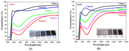
The transmittance spectra of TiO2:Ag films in the 200–1800 nm spectral range are shown in Figure 4. The multilayered films (ranging from one to five layers) were fabricated by sequentially repeating the coating and UV irradiation steps. After each coating, the films were subjected to UV radiation for 15 min. The coating/UV radiation cycle was repeated to obtain multilayered films from one up to five layers. No thermal treatment was applied during the entire deposition process Film transparency decreases significantly as the number of layers increases, resulting in an enhancement in absorption. TiO2:Ag films (Sol A) reveal no distinct absorption bands, demonstrating instead a very broad absorption feature within the 500–650 nm spectral range. The observed absorption feature can be attributed to charge transfer from silver-induced energy levels within the band gap [46]. In the case of TiO2:Ag films (Sol B), the transmittance spectra exhibit distinct variations. As shown in Figure 4b, the single-layer film exhibits a broad absorption band centered at 510 nm. The two-layered film has no distinct absorption bands. The three-layered sample manifests two absorption bands at 365 and at 460 nm. As the number of layers was increased to four, these bands shifted to 395 and 540 nm for the TiO2:Ag film (Sol B, Figure 4b). The thickest five-layered film exhibits a broad absorption band at 540 nm. The absorption bands in the 360–395 nm range are attributed to isolated Ag NPs, whereas the 520 nm plasmon resonance arises from larger nanoparticles or/and quadrupole plasma resonance [47].
Figure 4.
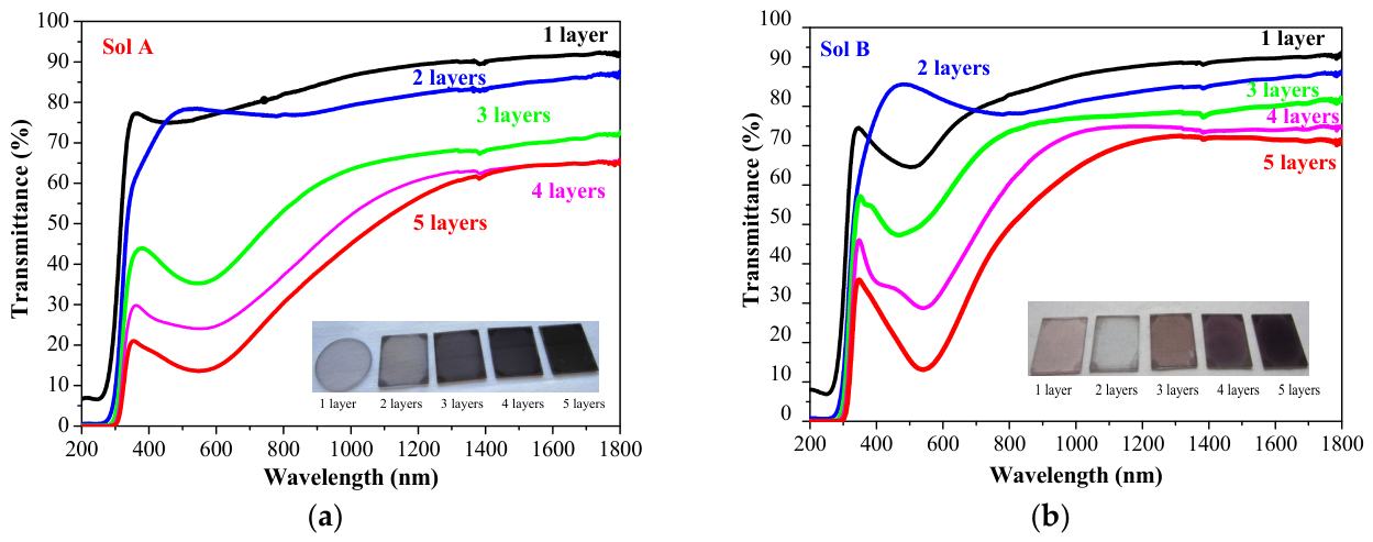
Transmittance spectra of TiO2:Ag films obtained from (a) Sol A and (b) Sol B with 1, 2, 3, 4 and 5 layers and only UV-radiated after each layer deposition. The inset photos present the TiO2:Ag films on quartz substrates.
The absorption features observed in the transmittance spectra are attributed to the localized surface plasmon resonance of the metallic silver NPs, confirming that UV radiation induces the formation of silver nanoparticles within TiO2 films. These findings are consistent with the XRD analysis.
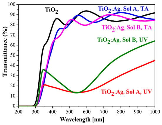
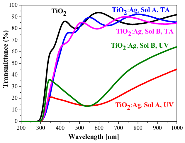
Figure 5 compares the transmittance spectra of five-layered TiO2 and TiO2:Ag films in the spectral range 200–1000 nm. The films, labeled as Sol A, TA and Sol B, TA, were preheated at 150 °C for 30 min. In contrast, the labels Sol A, UV and Sol B, UV correspond to TiO2:Ag films that were exclusively subjected to UV radiation for 15 min. The doped titania film was also preheated at 150 °C.
Figure 5.
Transmittance spectra of TiO2 and TiO2:Ag films obtained from Sol A and Sol B with 5 layers, where TA labels the films which are only preheated at 150 °C UV marks the samples which are UV-radiated after each layer deposition.
The preheated samples without UV treatment show similar spectral transmittance and high transparency, with TiO2:Ag films exhibiting a negligible drop in transmittance. The transmittance spectra are significantly different for the UV-treated samples. In the visible spectral range (400–750 nm), the transmittance drops below 30%. As previously discussed, this is driven by Ag NP formation in the metal oxide matrix. It has been established that film densification and enhanced crystallization are induced by UV treatment [48,49]. UV irradiation simultaneously provides the photon energy required to reduce silver ions Ag+ into silver Ago nanoparticles [49].
It can be concluded from the obtained results that UV radiation alone successfully induces the formation and growth of Ag NPs, requiring no additional thermal treatment. These silver NPs enhance visible-light absorption through local surface plasmon resonance.
The optical properties of UV-radiated multilayered TiO2:Ag films (ranging from one to five layers) were further investigated after annealing at various temperatures ranging from 150 °C to 800 °C.
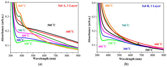
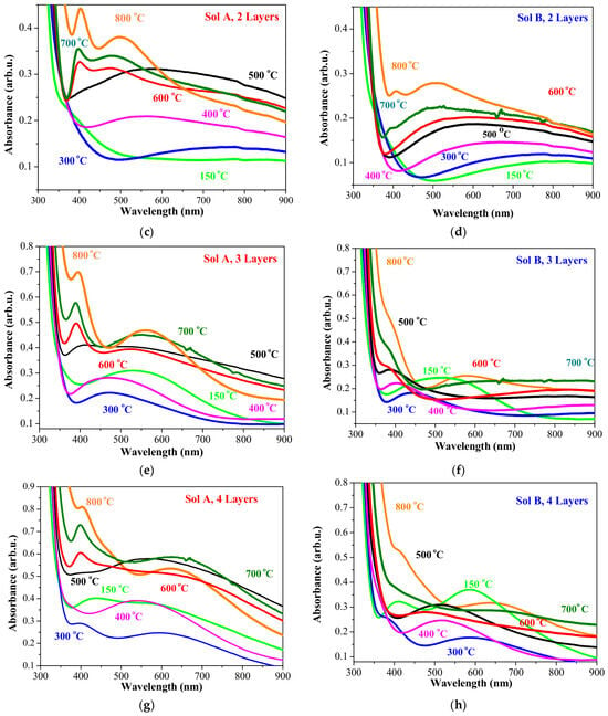
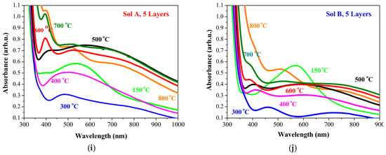
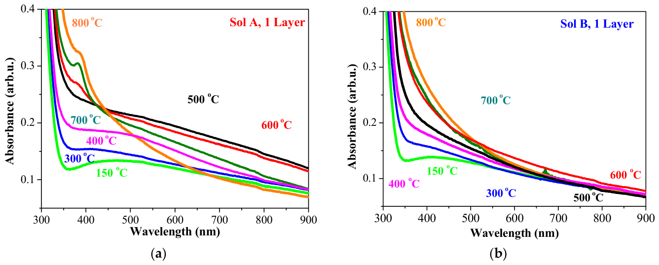
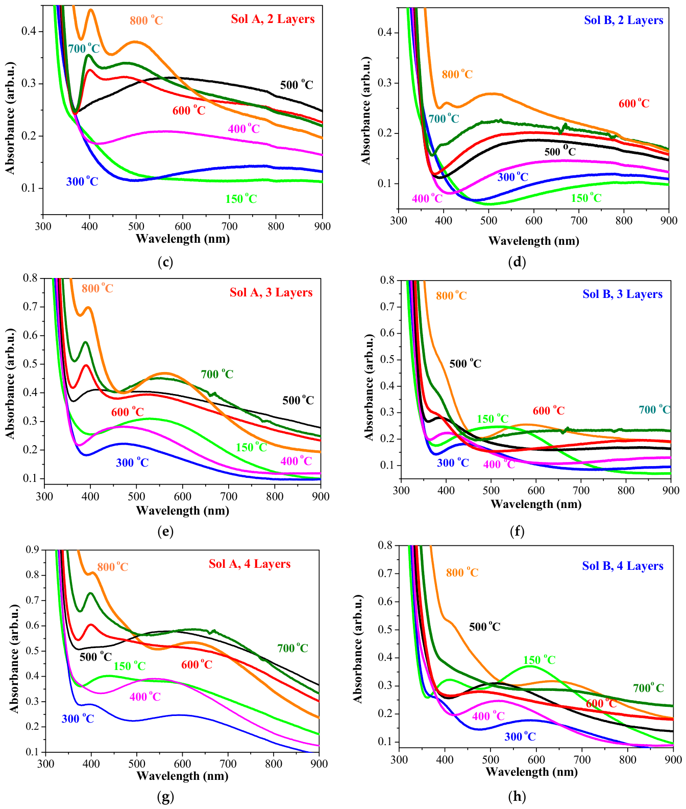
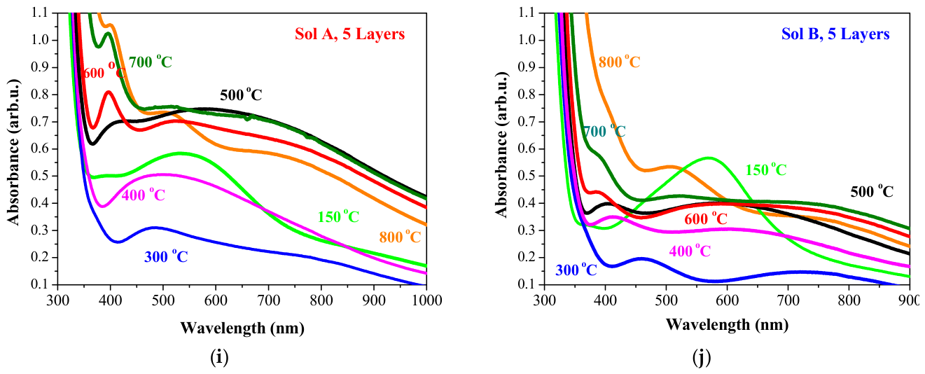
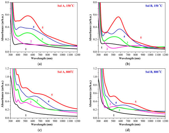
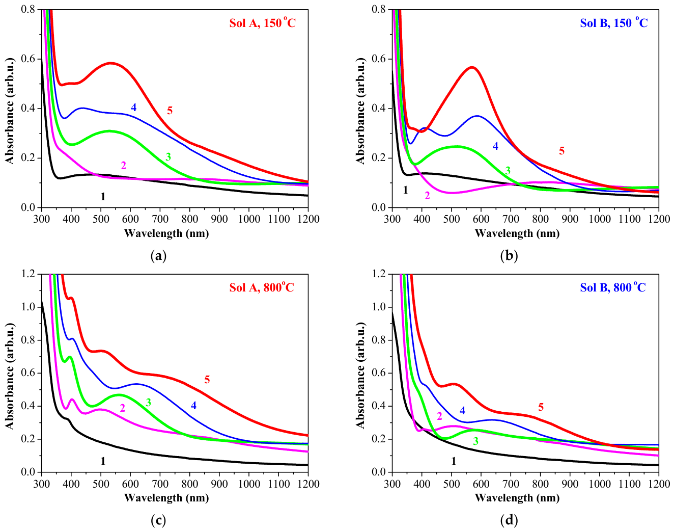
The absorbance spectra of TiO2:Ag films in the 300–900 nm spectral range are given in Figure 6. The films with various numbers of layers are annealed at temperatures from 150 to 800 °C. The impact of the annealing temperature on the optical properties is presented in Figure 7, which compares the multilayered films with various numbers of layers treated at 150 (Figure 7a,b) and 800 °C (Figure 7c,d). It is well known that the positions, shapes and width of LSPR bands vary depending on the concentrations, geometry and dimensions of Ag NPs. Other factors include the refractive index of the metal oxide matrix, crystal structure, the chosen deposition method and annealing procedures [50]. Furthermore, the broadening of the absorption bands for Ag NPs embedded in TiO2 can be provoked by irregular particle shapes and morphologies, growth of larger particles and clustering or agglomeration, which leads to the overlapping of plasmonic bands [50,51]. The plasmonic and optical properties of Ag NPs embedded in TiO2 films depend on annealing temperatures, silver concentrations and film thickness [52]. Notably, high-temperature annealing at 700 and 800 °C induced distinct SPR absorption bands between 385 and 390 nm, even in the thinnest TiO2:Ag films (single layer) deposited from Sol A. This optical feature strongly suggests the formation and growth of isolated Ag nanoparticles. The Sol B films do not exhibit distinct absorption bands; however, a significant increase in absorbance is observed, particularly for the samples annealed at 600 °C and 800 °C (Figure 6b,d,f,h,j). Increasing the number of layers enhances both the plasmonic absorption and the overall absorption features, leading to the emergence of several new, distinct and broad absorption bands. In addition, absorption within the visible spectral range increases with both higher annealing temperatures (Figure 6) and a greater number of layers (Figure 7).
Figure 6.
Absorbance spectra of TiO2:Ag films obtained from Sol A with various numbers of layers, where (a,c,e,g,i) present the spectra of films with one layer to five layers, respectively, and (b,d,f,h,j) show the absorbance spectra of TiO2:Ag films obtained from Sol B with one to five layers. After each layer deposition, the films are UV-radiated for 15 min. The final procedure is annealing at temperatures ranging from 150 to 800 °C.
Figure 7.
Optical absorbance spectra of TiO2:Ag films obtained from Sol A, where (a,c) present the comparison of films with one layer to five layers (labeled in figures as 1, 2, 3, 4, 5) treated at 150 and 800 °C, respectively, and (b,d) show the absorbance spectra of TiO2:Ag films obtained from Sol B (labeled in figures as 1, 2, 3, 4, 5), treated at 150 and 800 °C.
The UV-treated two-layered nanocomposite films deposited from Sol A (Figure 6b) exhibit increased optical absorption due to surface plasmon resonance, showing no distinct bands up to an annealing temperature of 500 °C. Following annealing at 600–800 °C, the absorbance spectra exhibit a distinct peak at 400 nm in parallel with broader absorption features; the latter shift from 480 nm (after heat treatment at 600 °C) to 505 nm (after annealing at 800 °C). In contrast, the two-layered films with lower silver content (Sol B) reveal absorption peaks at 395 nm (700 °C) and 405 nm when annealed at 700 and 800 °C, respectively. The three-layered Sol A samples exhibit a broad band centered at 560 nm after annealing at 150 °C, which is shifted to 460 nm at higher annealing temperatures of 300 and 400 °C. Thermal treatment at 500 °C resulted in a flat absorbance spectrum across the entire visible spectral range. Above 600 °C, intense peaks emerged within the 380–390 nm range. For the three-layered TiO2:Ag films (Sol B), a broad band is initially observed at 520 nm (after annealing at 150 °C). This band blueshifts to 450 nm (for the film, treated at 300 °C) and 400 nm at temperatures of 400 and 500 °C. When the annealing temperature exceeds 600 °C, a distinct shoulder appears at 395 nm. The sample annealed at 800 °C exhibits an additional broad absorption band centered at 570 nm.
The strongest plasmonic absorption in the case of the four and five-layered TiO2:Ag films is manifested by UV-treated films in the absence of subsequent thermal annealing. The TiO2:Ag films deposited from Sol A reveal an absorption band at 390–405 nm, alongside longer-wavelength absorption bands at 510–570 nm and 750–800 nm. Similar band positions are observed for the corresponding four-layered and five-layered TiO2:Ag films obtained from Sol B.
The plasmonic absorption at 395–400 nm is attributed to the formation and growth of isolated spherical Ag NPs [53]. Moreover, the intensity and position of these SPR peaks within the 380–400 nm spectral range are strongly dependent on silver concentrations. The absorbance spectra of the Sol A films manifested well-defined, symmetrical peaks after thermal treatments above 500 °C. In contrast, the TiO2:Ag films obtained from Sol B displayed weaker, asymmetrical absorption bands that that appeared as shoulders within the same spectral region.
The broad character of the observed absorption bands suggests wide dispersion of the silver nanoparticles [54]. An increase in Ag NP size can shift SPR towards longer wavelengths above 500 nm. As the physical dimensions of the Ag NPs expand, the interparticle distance decreases, which intensifies the electromagnetic coupling between adjacent particles and consequently drives the collective coupled surface plasmon oscillation to longer wavelengths [55]. The absorption bands observed between 500 and 1000 nm are attributed to the in-plane or longitudinal surface plasmon resonance (SPR) of anisotropic, non-spherical geometries (such as nanotriangles, nanoprisms, or nanodisks) or large aggregated clusters, rather than isolated, small spherical Ag NPs [56,57]. The flat absorbance spectra and the absence of defined bands can be attributed to the broad LSPR of polydesperse nanoparticles, defect-induced mid-gap states and interband transitions [58]. The plasmonic absorption is significantly enhanced by increasing the annealing temperature and the number of deposited layers. The concentration of silver in the sol solutions plays a crucial role in the manifestation of LSPR peaks. Moreover, UV radiation strongly modulates the optical behavior and plasmonic features; TiO2:Ag nanocomposite films exhibit distinct plasmonic bands exclusively following UV treatment.
The dependence of plasmonic properties on film thickness is also found for TiO2:Ag films prepared by magnetron sputtering [59]. The LSPR peak is reported to be redshifted from a wavelength of 428 nm to 500 nm with TiO2 films of 10–30 nm [59]. The films were prepared via two-step deposition: Ag layer covered by titania films with different thicknesses.
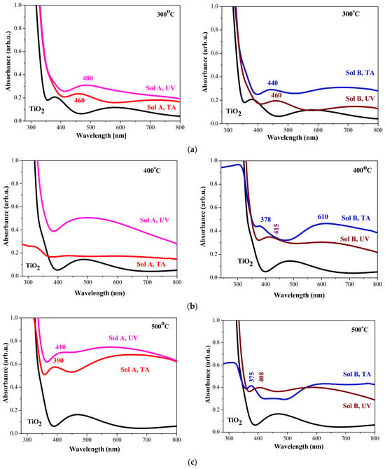
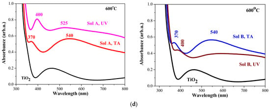
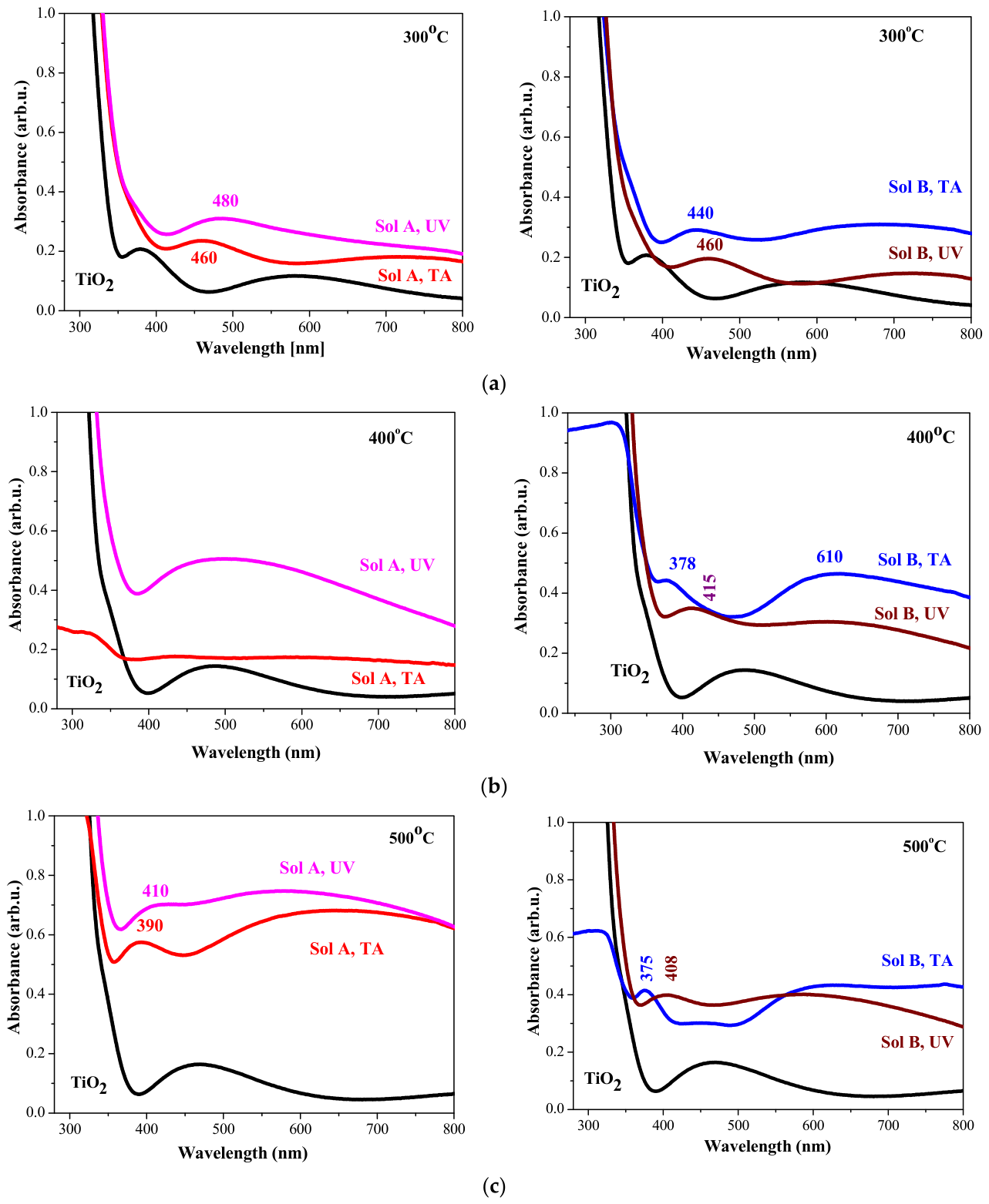
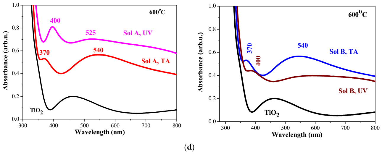
Figure 8 shows the absorbance spectra of TiO2 and TiO2:Ag films recorded in the 280–800 nm spectral range. The films have five layers. The films, labeled as Sol A, TA and Sol B, TA, underwent a preheating procedure at 150 °C for 30 min. Conversely, the Sol A, UV and Sol B, UV labels correspond to TiO2:Ag films that were only exposed to UV radiation for 15 min, without the preheating step. To ensure consistency, all high-temperature annealing processes were executed under strictly identical conditions, maintaining uniform heating rates, annealing temperatures, dwell times, and cooling rates.
Figure 8.
Absorbance spectra of TiO2 and TiO2:Ag films annealed at 300 (a), 400 (b), 500 (c) and 600 °C (d). All films have five layers. TiO2:Ag films are obtained from Sol A and Sol B. The undoped and TA films are thermally treated, and UV samples are UV-radiated after each layer deposition and then annealed at different temperatures.
As shown in Figure 8, UV radiation displays different effects depending on the silver precursor concentration. Regardless of the annealing temperatures, UV-treated TiO2:Ag films obtained from Sol A possess higher absorption in the visible spectral region. Conversely, the nanocomposite UV-radiated films derived from Sol B present lower SPR absorption than those that underwent only thermal treatment. The increased optical absorption in the visible spectral range due to Ag doping is reported for TiO2:Ag films, obtained via plasma-enhanced CVD [60].
Optical measurements indicate that the effect of UV radiation is complex, which aligns with XRD analysis. The XRD data confirm that UV radiation induces distinct modifications in the films deposited from Sol A and Sol B. In particular, TiO2:Ag films from Sol A manifest larger rutile fractions upon UV radiation compared to purely thermally treated films. In contrast, TiO2:Ag films derived from Sol B show an increase in the anatase fraction (see Table 2).
The determination and tuning of the optical band gap in semiconductors are of fundamental and technological importance. For instance, modulating the band gap of TiO2 yields multifunctional, highly efficient materials suitable for a wide range of applications [12,61]. In this study, the optical band gap values were found from the first derivative of transmittance with respect to energy [61,62].
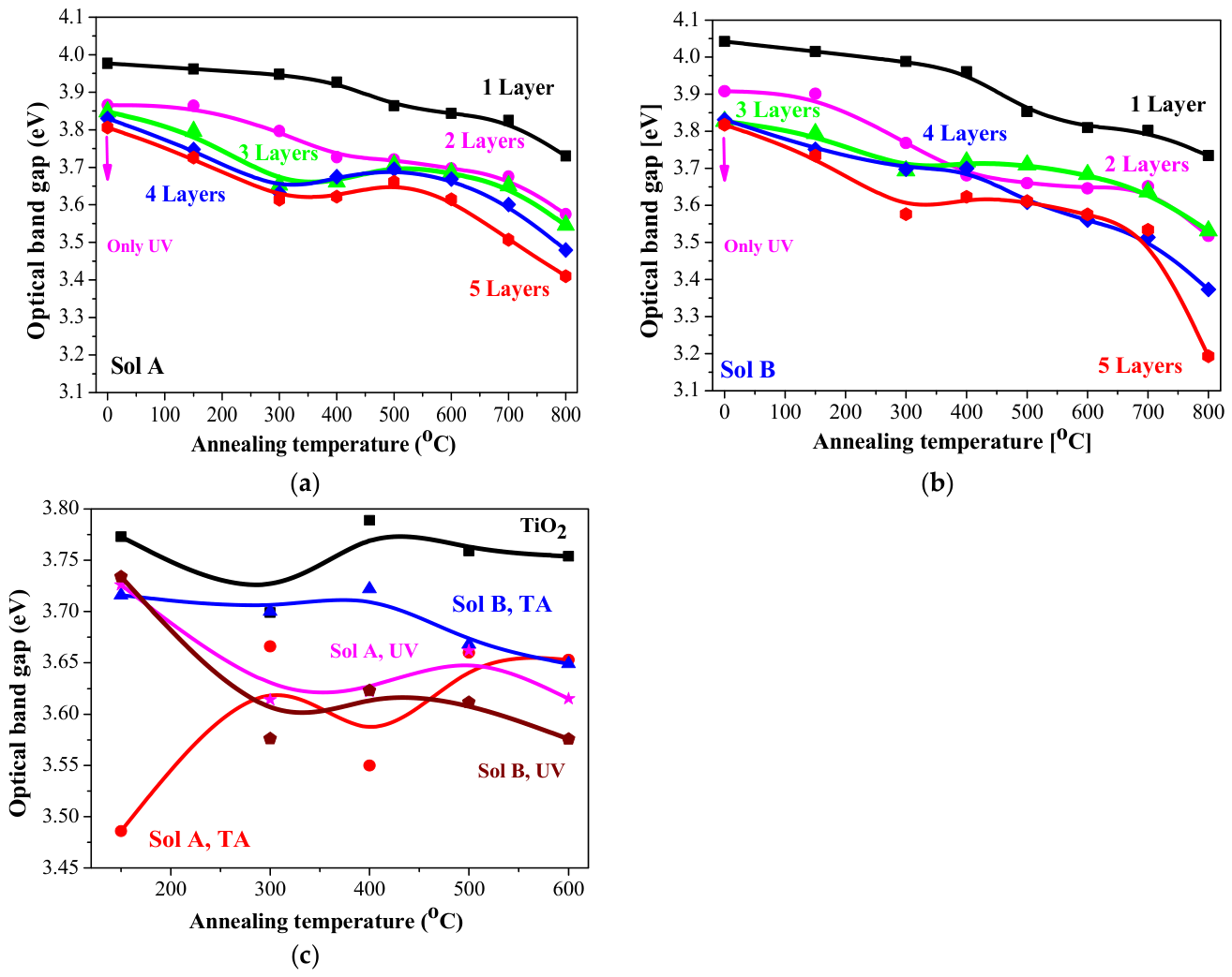
Figure 9a,b show the variations in the optical band gap of UV-treated films, obtained from sol A and B, plotted as a function of both the number of layers and the annealing temperatures within the range of 150–800 °C. The redshift of Eg is observed with increasing the number of layers for nanocomposite TiO2:Ag films. It is reported that the optical band gap of TiO2 films narrows as film thickness increases [61]. The optical band gap decreases as the annealing temperature increases. The reduction in the optical band gap as a function of increasing the number of layers (the film thickness) and the thermal treatments can be attributed to enhanced crystallinity, larger crystallite sizes, and the existence of surface oxygen vacancies [63]. Furthermore, the incorporation of Ag into TiO2 films has been reported to narrow the optical band gap [64].
Figure 9.
Optical band gap (Eg) values of UV-radiated TiO2:Ag films deposited from (a) Sol A and (b) Sol B with different numbers of layers as a function of annealing temperature (150–800 °C). (c) A comparison of Eg values, determined for the five-layered films of undoped TiO2, TiO2:Ag films (only annealed (TA)), and TiO2:Ag films with UV treatment (UV) annealed at temperatures from 150 to 600 °C. The arrow indicates that the films were subjected solely to UV treatment without subsequent thermal annealing.
The Ag dopant can be positioned either substitutionally or interstitially in the oxide lattice. Ag can provoke localized impurity states as oxygen vacancies or Ti3+ states below the conduction band. LSPR fields strongly couple with these sub-band gap defect states, thus causing band gap narrowing [65]. The concentration of silver significantly influences the Eg values of UV-treated TiO2:Ag films. For example, at annealing temperatures exceeding 300 °C, the films obtained from Sol B possess lower Eg values compared to their Sol A counterparts. This trend aligns with the reported data showing that higher Ag NP amounts induce more pronounced narrowing of the optical band gap [66].
In our study, this trend is absent when UV radiation is applied between layers. The observed increase in the energy band gap with higher Ag concentration may be attributed to surface plasmon resonance, which involves the interaction of an electromagnetic field with the conduction electrons of Ag nanoparticles dispersed within TiO2 [67]. Consequently, TiO2:Ag films obtained from Sol A manifest enhanced absorption in the visible spectral range as a direct result of this SPR effect.
The impact of different technological treatments, including only thermal treatments and preliminary interlayer UV radiation, on the optical band gap of five-layered films is presented in Figure 9c. Ag NP incorporation resulted in a significant reduction in the optical band gap. While the Eg values of undoped TiO2 films varied only slightly (3.69–3.79 eV) with increasing annealing temperature, the thermally annealed (TA) nanocomposite films manifested notable band gap narrowing as the Ag concentration increased. Specifically, the Eg values of TiO2:Ag films obtained from Sol B (TA) are higher than those of the corresponding Sol A (TA) films. Furthermore, UV radiation yielded the lowest Eg values for Sol B TiO2:Ag films after annealing at 500 and 600 °C. This reduction in the optical band gap may be due the structural disorders [67] within the samples. Magnetron-sputtered TiO2:Ag films [59] revealed optical band gap reductions from 3.88 to 3.66 eV with increasing thickness of the oxide film. The determined optical band gaps of TiO2:Ag films are in the range of the reported data [60,61,62,63,64,65,66]. The optical properties of the SPR bands and the optical band gap of silver-doped titania nanocomposites are strongly dependent on the deposition method and form (thin films, powder, fibers), as can be seen in Table 3. Table 3 presents some literature data for the optical characteristics of TiO2:Ag materials obtained via various approaches. It can be deduced that most deposition techniques require two-step preparation with or without additional thermal treatments. The nanocomposite films, synthesized via chemical routes, feature Ag NPs embedded within their structural matrix and bulk volume. This leads to the broad distribution of NP sizes and shapes and to broader, asymmetric SPR peaks. The presence of oxidized silver species can also be observed. Moreover, physical vapor deposition induces the formation of isolated, non-agglomerated Ag clusters. The SPR features are exhibited as intense and distinct localized bands. Oxidation (Ag2O or AgO) is almost absent during synthesis under vacuum conditions. The optical band gap generally narrows with Ag incorporation, depending on the silver concentrations.
Table 3.
Literature data for the SPR bands and the optical band gap values of TiO2:Ag nanomaterials deposited by means of various methods.
Our deposition method offers the preparation of uniform and homogeneous TiO2:Ag films. The optical characterization of multilayered TiO2:Ag thin films subjected to interlayer ultraviolet (UV) irradiation reveals highly sophisticated modulation of their plasmonic properties. Varying the number of layers and thermal annealing temperature leads to manifestation of different SPR effects and optical behaviors.
4. Conclusions
The proposed sol–gel spin-coating method has proven to be highly advantageous and relevant for depositing TiO2:Ag nanocomposite thin films. The influence of silver concentration (1 and 0.5 wt% AgNO3 added in Ti sol solution), UV radiation (between the layers), thermal treatment temperature (ranging from 150 to 800 °C) and number of layers (from 1 to 5 layers) on the structural and the optical properties was investigated.
XRD analysis reveals the crystalline structural evolution. The presence of the anatase and rutile fractions was observed. The successful formation of silver nanoparticles was confirmed. The appearance of the AgO phase was strictly governed by the silver precursor concentration. UV radiation induced rutile phase formation in TiO2:Ag films obtained from Sol A. Its phase fraction exceeded that observed in the purely thermally treated TiO2:Ag films. FTIR spectroscopy analysis demonstrates that silver incorporation in TiO2 and UV treatment alter the profiles and intensities of the absorption bands. Variations in the optical absorption spectra indicate the formation of isolated Ag NPs, irregular non-spherical particles or the activation of higher-order resonance modes. These features exhibited a strong dependence on the number of layers and subsequent UV or thermal treatments The incorporation of a silver dopant drives a reduction in the optical band gap of sol–gel TiO2 films. Notably, the minimum Eg values were determined for nanocomposite films obtained from Sol B, corresponding to the lower dopant concentration. The relationship between the technological parameters (UV radiation, annealing) and the resulting changes in the optical band gap was investigated and discussed herein. The obtained results demonstrate that the effect of UV treatment is highly sensitive to the concentration of the silver precursor in the sol solutions.
Author Contributions
Conceptualization, T.I. and A.H.; methodology, T.I.; validation, T.I. and A.H.; formal analysis, T.I.; investigation, T.I. and A.H.; resources, A.H.; data curation, T.I. and A.H.; writing—original draft preparation, T.I.; writing—review and editing, T.I. and A.H.; visualization, A.H.; supervision, T.I. All authors have read and agreed to the published version of the manuscript.
Funding
This research was funded by the Bulgarian National Science Fund (BNSF), grant number KP-06-N78/7, under the project Development and Investigation of Metal Oxide Heterostructures for Transparent Solar Cells.
Data Availability Statement
The original contributions presented in this study are included in the article. Further inquiries can be directed to the corresponding author.
Conflicts of Interest
The authors declare no conflicts of interest.
References
- Potocnik, J.; Popovic, M.; Abazovic, N.; Carevic, M.; Novakovic, M. Zigzag-structured TiO2 thin films: Thickness-controlled nanostructure and related optical and photocatalytic behavior. Mater. Charact. 2026, 238, 116620. [Google Scholar] [CrossRef] [Scilit]
- Alothoum, M.A.S. A Review of the Synthesis, Structural, and Optical Properties of TiO2 Nanoparticles: Current State of the Art and Potential Applications. Crystals 2025, 15, 944. [Google Scholar] [CrossRef] [Scilit]
- Haggerty, J.E.S.; Schelhas, L.T.; Kitchaev, D.A.; Mangum, J.S.; Garten, L.M.; Sun, W.; Stone, K.H.; Perkins, J.D.; Toney, M.F.; Ceder, G.; et al. High-fraction brookite films from amorphous precursors. Sci. Rep. 2017, 7, 15232. [Google Scholar] [CrossRef] [Scilit]
- Hussain, A.; Rauf, A.; Ahmed, E.; Khan, M.S.; Mian, S.A.; Jang, J. Modulating Optoelectronic and Elastic Properties of Anatase TiO2 for Photoelectrochemical Water Splitting. Molecules 2023, 28, 3252. [Google Scholar] [CrossRef] [Scilit]
- Silva, D.; Monteiro, C.S.; Silva, S.O.; Frazão, O.; Pinto, J.V.; Raposo, M.; Ribeiro, P.A.; Sério, S. Sputtering Deposition of TiO2 Thin Film Coatings for Fiber Optic Sensors. Photonics 2022, 9, 342. [Google Scholar] [CrossRef] [Scilit]
- Kang, B.C.; Lee, S.B.; Boo, J.H. Growth of TiO2 thin films on Si(100) substrates using single molecular precursors by metal organic chemical vapor deposition. Surf. Coat. Technol. 2000, 131, 88–92. [Google Scholar] [CrossRef] [Scilit]
- Pant, B.; Park, M.; Park, S.-J. Recent Advances in TiO2 Films Prepared by Sol-Gel Methods for Photocatalytic Degradation of Organic Pollutants and Antibacterial Activities. Coatings 2019, 9, 613. [Google Scholar] [CrossRef] [Scilit]
- Albu, D.F.; Lungu, J.; Popescu-Pelin, G.; Mihăilescu, C.N.; Socol, G.; Georgescu, A.; Socol, M.; Bănică, A.; Ciupina, V.; Mihailescu, I.N. Thin Film Fabrication by Pulsed Laser Deposition from TiO2 Targets in O2, N2, He, or Ar for Dye-Sensitized Solar Cells. Coatings 2022, 12, 293. [Google Scholar] [CrossRef] [Scilit]
- Ambartsumov, M.G.; Tarala, V.A.; Medyanik, E.V.; Kovalenko, V.V.; Chapura, O.M. Post-annealing microstructural evolution of titanium dioxide thin films grown by thermal and plasma-enhanced atomic layer deposition. Appl. Surf. Sci. 2025, 714, 164419. [Google Scholar] [CrossRef] [Scilit]
- Anchieta e Silva, F.; de Almeida, T.A.; Secchi, A.R.; Pinto, J.C.; Rodrigues, T.S. Enhancing Optical Properties and Cost-Effectiveness of Sol–Gel TiO2 Nanomaterials Through Experimental Design. Processes 2025, 13, 1988. [Google Scholar] [CrossRef] [Scilit]
- Nassar, K.I.; Teixeira, S.S.; Graça, M.P.F. Sol–Gel-Synthesized Metal Oxide Nanostructures: Advancements and Prospects for Spintronic Applications—A Comprehensive Review. Gels 2025, 11, 657, Correction in Gels 2026, 12, 216. [Google Scholar] [CrossRef] [Scilit]
- Liao, L.; Wang, M.; Li, Z.; Wang, X.; Zhou, W. Recent Advances in Black TiO2 Nanomaterials for Solar Energy Conversion. Nanomaterials 2023, 13, 468. [Google Scholar] [CrossRef] [Scilit]
- Nawaz, M.N.; Zhang, Z.; Yuan, W.; Khan, S.B. Photocatalytic enhancement of TiO2 through silver, gold, and platinum doping. Energy Nexus 2025, 19, 100495. [Google Scholar] [CrossRef] [Scilit]
- Dou, H.; Wang, J.; Zhao, Y.; Liu, J.; Li, Y. Preparation of Ag-Decorated TiO2 Composite Materials and Study on Photocatalytic Performance. Nanomaterials 2025, 15, 1383. [Google Scholar] [CrossRef] [Scilit]
- Ding, Q.; Li, R.; Chen, M.; Sun, M. Ag nanoparticles-TiO2 film hybrid for plasmon-exciton co-driven surface catalytic reactions. Appl. Mater. Today 2017, 9, 251–258. [Google Scholar] [CrossRef] [Scilit]
- Yoon, H.; Kim, S.; Kim, H.-Y. Modulating localized surface plasmon resonance through metal composition and nanoparticle Shape: Implications for solar cell applications. Mater. Sci. Semicond. Process. 2025, 197, 109677. [Google Scholar] [CrossRef] [Scilit]
- Mcoyi, M.P.; Mpofu, K.T.; Sekhwama, M.; Mthunzi-Kufa, P. Developments in Localized Surface Plasmon Resonance. Plasmonics 2025, 20, 5481–5520. [Google Scholar] [CrossRef] [Scilit]
- Zhao, X.; Zhu, X.; Chen, D.; Wei, Y.; Yao, L.; Li, W.; Li, G.; Sun, Z.; Zhao, Y.; Fan, X.; et al. Systematic investigation of the LSPR characteristics in plasmonic nanoarrays. Microsyst. Nanoeng. 2026, 12, 137. [Google Scholar] [CrossRef] [Scilit]
- Petryayeva, E.; Krull, U.J. Localized surface plasmon resonance: Nanostructures, bioassays and biosensing—A review. Anal. Chim. Acta 2011, 706, 8–24. [Google Scholar] [CrossRef] [Scilit]
- Khedulkar, A.P.; Bolloju, S.; Pradhan, L.K.; Pandit, B.; Nakate, U.T.; Mahato, S.S.; Mahata, S. Synergistic effects of Fe and Ag doping on the structural and optical properties of a TiO2 thin film: A dual function platform for hydrogen generation and dye degradation. Dalton Trans. 2024, 53, 18592–18606. [Google Scholar] [CrossRef] [Scilit]
- Liao, C.; Li, Y.; Tjong, S.C. Visible-Light Active Titanium Dioxide Nanomaterials with Bactericidal Properties. Nanomaterials 2020, 10, 124. [Google Scholar] [CrossRef] [Scilit]
- Chakhtouna, H.; Benzeid, H.; Zari, N.; Qaiss, A.E.K.; Bouhfid, R. Recent progress on Ag/TiO2 photocatalysts: Photocatalytic and bactericidal behaviors. Environ. Sci. Pollut. Res. Int. 2021, 28, 44638–44666. [Google Scholar] [CrossRef] [Scilit]
- Kanakaraju, D.; Kutiang, F.D.; Lim, Y.C.; Goh, P.S. Recent progress of Ag/TiO2 photocatalyst for wastewater treatment: Doping, co-doping, and green materials functionalization. Appl. Mater. Today 2022, 27, 101500. [Google Scholar] [CrossRef] [Scilit]
- Ramadhani, D.A.K.; Sholeha, N.; Khusna, N.N.; Diantoro, M.; Afandi, A.N.; Osman, Z.; Pujiarti, H. Ag-doped TiO2 as photoanode for high performance dye sensitized solar cells. Mater. Sci. Energy Technol. 2024, 7, 274–281. [Google Scholar] [CrossRef] [Scilit]
- Singh, L.P.; Laishram, R.; Bikesh, S.; Chanam, B.; Lenka, T.R.; Rahman, T. Highly Sensitive Ag Nanoparticle Deposited TiO2 Thin Film-Based Broadband Photodetector. IEEE Trans. Electron Devices 2026, 73, 3660–3668. [Google Scholar] [CrossRef] [Scilit]
- Chang, C.; Rad, S.; Gan, L.; Li, Z.; Dai, J.; Shahab, A. Review of the sol–gel method in preparing nano TiO2 for advanced oxidation process. Nanotechnol. Rev. 2023, 12, 20230150. [Google Scholar] [CrossRef] [Scilit]
- Man, S.L.; Hong, S.S.; Mohseni, M. Synthesis of photocatalytic nanosized TiO2-Ag particles with sol-gel method using reduction agent. J. Mol. Catal. A Chem. 2004, 242, 135–140. [Google Scholar] [CrossRef] [Scilit]
- Bretos, I.; Jiménez, R.; Ricote, J.; Calzada, M.L. Low-temperature crystallization of solution-derived metal oxide thin films assisted by chemical processes. Chem. Soc. Rev. 2018, 47, 291–308. [Google Scholar] [CrossRef] [Scilit]
- Chen, W.; Wu, Z.; Li, J.; Peng, R.; Su, Y.; Qin, D.; Zhang, Z.; Gu, J. UV light leads to the formation of reduced graphene oxide and silver nanoparticles in waterborne coating. Mater. Lett. 2024, 357, 135727. [Google Scholar] [CrossRef] [Scilit]
- Lima, M.J.A.; Reis, B.F. Photogeneration of silver nanoparticles induced by UV radiation and their use as a sensor for the determination of chloride in fuel ethanol using a flow-batch system. Talanta 2019, 201, 373–378. [Google Scholar] [CrossRef] [Scilit]
- Yuan, B.; Luo, Z.; Luan, W.; Cao, L.; Zhu, R. Effect of UV irradiation on synthesis of Ag/amorphous TiO2 composites with photocatalytic hydrogen production. J. Mater. Res. 2023, 38, 3628–3637. [Google Scholar] [CrossRef] [Scilit]
- Ivanova, T.; Harizanova, A.; Koutzarova, T.; Vertruyen, B. Optical and structural characterization of TiO2 films doped with silver nanoparticles obtained by sol-gel method. Opt. Mater. 2013, 36, 207–213. [Google Scholar] [CrossRef] [Scilit]
- Ivanova, T.; Harizanova, A.; Koutzarova, T.; Vertruyen, B. Characterization of nanostructured TiO2: Ag films: Structural and optical properties. J. Phys. Conf. Ser. 2016, 764, 012019. [Google Scholar] [CrossRef] [Scilit]
- Ivanova, T.; Harizanova, A.; Koutzarova, T.; Closset, R. Crystallization and Optical Behaviour of Nanocomposite Sol-Gel TiO2:Ag Films. Molecules 2024, 29, 5156. [Google Scholar] [CrossRef] [Scilit]
- Irie, H.; Watanabe, Y.; Hashimoto, K. Carbon-doped Anatase TiO2 Powders as a Visible-light Sensitive Photocatalyst. Chem. Lett. 2003, 32, 772–773. [Google Scholar] [CrossRef] [Scilit]
- He, J.; Du, Y.-e.; Bai, Y.; An, J.; Cai, X.; Chen, Y.; Wang, P.; Yang, X.; Feng, Q. Facile Formation of Anatase/Rutile TiO2 Nanocomposites with Enhanced Photocatalytic Activity. Molecules 2019, 24, 2996. [Google Scholar] [CrossRef] [Scilit]
- Li, J.; Jin, Z.; Zhang, Y.; Liu, D.; Ma, A.; Sun, Y.; Li, X.; Cai, Q.; Gui, J. Ag-induced anatase-rutile TiO2-x heterojunction facilitating the photogenerated carrier separation in visible-light irradiation. J. Alloys Compd. 2022, 909, 164815. [Google Scholar] [CrossRef] [Scilit]
- Singh, A.; Kumar, S. Effect of Ag doping on phase-change and photocatalytic performance of rutile–anatase mixed-phase titanium dioxide (TiO2) nanoparticles. Appl. Phys. A 2021, 127, 839. [Google Scholar] [CrossRef] [Scilit]
- Kumar, P.M.; Badrinarayanan, S.; Sastry, M. Nanocrystalline TiO2 studied by optical, FTIR and X-ray photoelectron spectroscopy: Correlation to presence of surface states. Thin Solid Films 2000, 358, 122–130. [Google Scholar] [CrossRef] [Scilit]
- Connor, P.A.; Dobson, K.D.; McQuillan, A.J. Infrared Spectroscopy of the TiO2/Aqueous Solution Interface. Langmuir 1999, 15, 2402–2408. [Google Scholar] [CrossRef] [Scilit]
- Manikandan, K.; Ahamed, A.J.; Brahmanandhan, G.M. Synthesis, Structural and Optical Characterization of TiO2 Nanoparticles and its Assessment to Cytotoxicity Activity. J. Environ. Nanotechnol. 2017, 6, 94–102. [Google Scholar] [CrossRef] [Scilit]
- Dwivedi, S.; Biswas, S. Effects of Annealing on Pulsed Laser Deposited TiO2 Thin Films. Appl. Mech. Mater. 2014, 446–447, 306–311. [Google Scholar] [CrossRef] [Scilit]
- Suciu, R.-C.; Zagrai, M.; Popa, A.; Toloman, D.; Berghian-Grosan, C.; Tudoran, C.; Stefan, M. The Influence of Ag+/Ti4+ Ratio on Structural, Optical and Photocatalytic Properties of MWCNT–TiO2–Ag Nanocomposites. Inorganics 2023, 11, 249. [Google Scholar] [CrossRef] [Scilit]
- Ahmed, F.; Kanoun, M.B.; Awada, C.; Jonin, C.; Brevet, P.-F. An Experimental and Theoretical Study on the Effect of Silver Nanoparticles Concentration on the Structural, Morphological, Optical, and Electronic Properties of TiO2 Nanocrystals. Crystals 2021, 11, 1488. [Google Scholar] [CrossRef] [Scilit]
- Dhand, V.; Soumya, L.; Bharadwaj, S.; Chakra, S.; Bhatt, D.; Sreedhar, B. Green synthesis of silver nanoparticles using Coffea arabica seed extract and its antibacterial activity. Mater. Sci. Eng. C Mater. Biol. Appl. 2016, 58, 36–43. [Google Scholar] [CrossRef] [Scilit]
- Lozano-Rosas, R.; Ruíz-Osorio, J.J.; Ramos-García, R.; Silva-González, R.; Spezzia-Mazzocco, T.; Robles-Águila, M.J. Photoexcitation of Ag-doped TiO2 nanoparticles with visible light for antimicrobial photodynamic therapy against Candida albicans. J. Nanopart. Res. 2025, 27, 236. [Google Scholar] [CrossRef] [Scilit]
- Wu, C.; Zhou, X.; Wei, J. Localized Surface Plasmon Resonance of Silver Nanotriangles Synthesized by a Versatile Solution Reaction. Nanoscale Res. Lett. 2015, 10, 354. [Google Scholar] [CrossRef] [Scilit]
- Nakajima, A.; Hayashi, N.; Taniguchi, Y.; Kameshima, Y.; Okada, K. Effect of vacuum ultraviolet light illumination on the crystallization of sol-gel-derived titanium dioxide precursor films. Surf. Coat. Technol. 2005, 192, 112–116. [Google Scholar] [CrossRef]
- Alvares, M.R.N.; Queiroz, V.M.; Jardim, P.M.; Thire, R.M.S.M. High-throughput fabrication of antimicrobial PHBV fiber mats via UV-assisted in situ silver nanoparticle synthesis. Colloids Surf. A Physicochem. Eng. Asp. 2026, 736, 139668. [Google Scholar] [CrossRef] [Scilit]
- Hang, Y.; Wang, A.; Wu, N. Plasmonic silver and gold nanoparticles: Shape- and structure-modulated plasmonic functionality for point-of-caring sensing, bio-imaging and medical therapy. Chem. Soc. Rev. 2024, 53, 2932–2971. [Google Scholar] [CrossRef] [Scilit]
- Bakr, O.; Amendola, V.; Aikens, C.; Wenseleers, W.; Li, R.; Dal Negro, L.; Schatz, G.; Stellacci, F. Silver Nanoparticles with Broad Multiband Linear Optical Absorption. Angew. Chem. 2009, 121, 6035–6040. [Google Scholar] [CrossRef] [Scilit]
- Kumar, M.; Parashar, K.K.; Tandi, S.K.; Kumar, T.; Agarwal, D.C.; Pathak, A. Fabrication of Ag:TiO2 Nanocomposite Thin Films by Sol-Gel Followed by Electron Beam Physical Vapour Deposition Technique. J. Spectrosc. 2013, 2013, 491716. [Google Scholar] [CrossRef] [Scilit]
- Ismail, R.K.; Mubarak, T.H.; Al-Haddad, R.M.S. Surface Plasmon Resonance of Silver Nanoparticles: Synthesis, Characterization, and Applications. J. Biochem. Technol. 2019, 10, 62–64. [Google Scholar] [CrossRef] [Scilit]
- Tasnim, T.; Haque, K.M.A.; Adil, M.; Tanishq, A.H.; Sarker, S.M.K.H.; Mohotadi, A.A. Characterization of green synthesized silver nanoparticles (AgNPs) and application in nanocomposites reinforced with natural fibers. Results Mater. 2026, 31, 101001. [Google Scholar] [CrossRef] [Scilit]
- Mondal, S.; Basak, D. Plasmon assisted high ultraviolet to visible broad band photosensitivity in lateral Ag NPs-TiO2 nanocomposite film. Surf. Interfaces 2022, 31, 102090. [Google Scholar] [CrossRef] [Scilit]
- Benalcázar, J.; Lasso, E.D.; Ibarra-Barreno, C.M.; Pareja, J.A.A.; Vispo, N.S.; Chacón-Torres, J.C.; Briceño, S. Photochemical Optimization of a Silver Nanoprism/Graphene Oxide Nanocomposite’s Antibacterial Properties. ACS Omega 2022, 7, 46745–46755. [Google Scholar] [CrossRef] [Scilit]
- Ameer, F.S.; Varahagiri, S.; Benza, D.W.; Willett, D.R.; Wen, Y.; Wang, F.; Chumanov, G.; Anker, J.N. Tuning Localized Surface Plasmon Resonance Wavelengths of Silver Nanoparticles by Mechanical Deformation. J. Phys. Chem. C Nanomater. Interfaces 2016, 120, 20886–20895. [Google Scholar] [CrossRef] [Scilit]
- Prakash, J.; Kumar, P.; Harris, R.A.; Swart, C.; Neethling, J.H.; van Vuuren, A.J.; Swart, H.C. Synthesis, characterization and multifunctional properties of plasmonic Ag–TiO2 nanocomposites. Nanotechnology 2016, 27, 355707. [Google Scholar] [CrossRef] [Scilit]
- Lo, T.-H.; Shih, P.-Y.; Wu, C.-H. The Response of UV/Blue Light and Ozone Sensing Using Ag-TiO2 Planar Nanocomposite Thin Film. Sensors 2019, 19, 5061. [Google Scholar] [CrossRef] [Scilit]
- Lang, J.; Takahashi, K.; Kubo, M.; Shimada, M. Ag-Doped TiO2 Composite Films Prepared Using Aerosol-Assisted, Plasma-Enhanced Chemical Vapor Deposition. Catalysts 2022, 12, 365. [Google Scholar] [CrossRef] [Scilit]
- Guillén, C. Band Gap Energy and Lattice Distortion in Anatase TiO2 Thin Films Prepared by Reactive Sputtering with Different Thicknesses. Materials 2025, 18, 2346. [Google Scholar] [CrossRef] [Scilit]
- Ruvalcaba-Manzo, S.G.; Ramírez-Bon, R.; Ochoa-Landín, R.; Castillo, S.J. A Comprehensive Study of the Optical, Structural, and Morphological Properties of Chemically Deposited ZnO Thin Films. Inorganics 2025, 13, 331. [Google Scholar] [CrossRef] [Scilit]
- Ennaceri, H.; Boujnah, M.; Taleb, A.; Khaldoun, A.; Saez-Araoz, R.; Ennaoui, A.; El Kenz, A.; Benyoussef, A. Thickness effect on the optical properties of TiO2-anatase thin films prepared by ultrasonic spray pyrolysis: Experimental and ab initio study. Int. J. Hydrogen Energy 2017, 42, 19467–19480. [Google Scholar] [CrossRef] [Scilit]
- Sharma, H.; Singhal, R.; Siva Kumar, V.V.; Asokan, K. Structural, optical and electronic properties of Ag–TiO2 nanocomposite thin film. Appl. Phys. A 2016, 122, 1010. [Google Scholar] [CrossRef] [Scilit]
- Benmeddah, N.; Temmar, F. Electronic structure modulation in Fe- and Ag-doped rutile TiO2: A GGA+U approach for optoelectronic and solar applications. Next Mater. 2026, 10, 101568. [Google Scholar] [CrossRef] [Scilit]
- Amin, S.M.A.; Kowser, A. Influence of Ag doping on structural, morphological, and optical characteristics of sol-gel spin-coated TiO2 thin films. Heliyon 2024, 10, e37558. [Google Scholar] [CrossRef] [Scilit]
- Sharif, A.M.; Ashrafuzzaman, M.; Kalam, A.; Al-Sehemi, A.G.; Yadav, P.; Tripathi, B.; Dubey, M.; Du, G. Green Synthesis of Pristine and Ag-Doped TiO2 and Investigation of Their Performance as Photoanodes in Dye-Sensitized Solar Cells. Materials 2023, 16, 5731. [Google Scholar] [CrossRef] [Scilit]
- Abualnaja, K.M.; ElAassar, M.R.; Ghareeb, R.Y.; Ibrahim, A.A.; Abdelsalam, N.R. Development of photo-induced Ag0/TiO2 nanocomposite coating for photocatalysis, self-cleaning and antimicrobial polyester fabric. J. Mater. Res. Technol. 2021, 15, 1513–1523. [Google Scholar] [CrossRef] [Scilit]
- Sun, L.; Que, Z.; Ruan, T.; Yuan, Z.; Gong, W.; Mei, S.; Chen, Z.; Liu, Y. The Synthesis of Ag/TiO2 via the g Method and Its Application in the Photocatalytic Degradation of Methyl Orange in Na2SO4 Solution. Appl. Sci. 2024, 14, 4014. [Google Scholar] [CrossRef] [Scilit]
- Zuo, J. Deposition of Ag nanostructures on TiO2 thin films by RF magnetron sputtering. Appl. Surf. Sci. 2010, 256, 7096–7101. [Google Scholar] [CrossRef] [Scilit]
- Sun, L.; Hu, X.; Yuan, Z.; Liu, Y.; Mei, S.; Meng, F.; Que, Z.; Xiong, Y.; Zhang, K.; Chen, Z. Photocatalytic Properties of Ag-Modifying N-TiO2 Films Prepared via Magnetron Sputtering. Catalysts 2025, 15, 164. [Google Scholar] [CrossRef] [Scilit]
- Kavaliunas, V.; Čeplikas, P.; Sriubas, M.; Laukaitis, G. The Sensitization of TiO2 Thin Film by Ag Nanoparticles for the Improvement of Photocatalytic Efficiency. Appl. Sci. 2022, 12, 5725. [Google Scholar] [CrossRef] [Scilit]
- Zhang, J.; Cha, J.K.; Fu, G.; Cho, E.J.; Kim, H.S.; Kim, S.H. Aerosol processing of Ag/TiO2 composite nanoparticles for enhanced photocatalytic water treatment under UV and visible light irradiation. Ceram. Int. 2022, 48, 9434–9441. [Google Scholar] [CrossRef] [Scilit]
- Tuntithavornwat, S.; Onkan, A.; Nonejuie, P.; Samernate, T.; Sirinonthanawech, N.; Kannan, A.M.; Saisawang, C.; Hunsom, M. Synthesis of Ag/TiO2 photocatalysts via direct photodeposition of silver from industrial electroplating wastewater. Sci. Rep. 2026, 16, 22669. [Google Scholar] [CrossRef] [Scilit]
- Vu, N.N.; Tran, B.V.; Ladhari, S.; Saidi, A.; Assadi, A.A.; Nguyen-Tri, P. Phase transformation-induced intimate Ag deposition in Ag-TiO2 hybrids for enhanced photocatalytic antibacterial performance. Chem. Eng. J. 2025, 524, 169720. [Google Scholar] [CrossRef] [Scilit]
- Cai, P.-F.; Li, J.; Wu, X.-B.; Li, Z.-Y.; Shen, J.; Nie, J.-J.; Cui, Z.-D.; Chen, D.-F.; Liang, Y.-Q.; Zhu, S.-L.; et al. ALD-induced TiO2/Ag nanofilm for rapid surface photodynamic ion sterilization. Rare Met. 2022, 41, 4138–4148. [Google Scholar] [CrossRef] [Scilit]

Disclaimer/Publisher’s Note: The statements, opinions and data contained in all publications are solely those of the individual author(s) and contributor(s) and not of MDPI and/or the editor(s). MDPI and/or the editor(s) disclaim responsibility for any injury to people or property resulting from any ideas, methods, instructions or products referred to in the content. |
© 2026 by the authors. Licensee MDPI, Basel, Switzerland. This article is an open access article distributed under the terms and conditions of the Creative Commons Attribution (CC BY) license.