


Abstract
Acute and chronic wounds remain a major clinical burden, motivating the design of multilayer biomedical composite systems capable of combining structural support, antimicrobial protection, and controlled release of therapeutic agents within a single device. These architectures integrate natural and synthetic biomaterials, hydrogels, electrospun membranes, bioactive nanoparticles, and 3D-printed scaffolds to reproduce the multifunctionality of the native extracellular matrix. This review examines how layer-by-layer design and biomaterial selection govern mechanical strength, as well as bioactivity, and how these parameters can be tuned to the distinct phases of wound repair. Particular emphasis is placed on strategies for incorporating growth factors, antimicrobial agents, metal/metal-oxide nanoparticles to enhance re-epithelialization, angiogenesis, and infection control; emerging gene-based delivery strategies are also discussed. The main technologies and biomaterial combinations reported to date are compared, alongside a critical overview of their in vitro and in vivo performance. Reproducibility, scalability, and regulatory standardization remain the main barriers to clinical translation. We conclude by outlining priority research directions to advance multilayer composites from bench-scale prototypes toward approved wound-care products.
1. Introduction
The skin is the largest organ in the human body, with an average surface area of 1.5–2.0 m2 in adults and weighing approximately 16% of the total body weight [1]. This organ has a high capacity to adapt to environmental conditions [2] and performs multiple functions: it protects internal organs, acts as an immune barrier and against ultraviolet radiation, regulates body temperature, and participates in the elimination of waste products through sweat, in addition to intervening in physiological functions and sensory interactions [3].
The skin is composed of three main layers: the epidermis, dermis, and hypodermis. The epidermis, the outermost layer, acts as a physical and biological barrier against pathogens and irritants. It is composed of 80% keratinocytes and, although it lacks blood vessels, receives oxygen and nutrients from the dermal vessels. It has five sublayers: basal, spinous, granular, lucid, and corneum. The latter is home to the skin microbiome, composed of species such as Corynebacterium, Dermabacter, Propiobacterium, and Brevibacterium, which perform defense and healing functions. The dermis provides strength, flexibility, and structural support thanks to its connective tissue, blood and lymphatic vessels, nerve endings, sebaceous and sweat glands, and hair follicles, which together maintain the skin’s function and homeostasis. Its main components are type I and III collagen (COL) (fibrous) and type IV, V, and VII COL (non-fibrous). The extracellular matrix (ECM), composed of glycosaminoglycans (GAGs), proteoglycans, glycoproteins, hyaluronic acid (HA), and adhesion proteins, together with elastic fibers and COL networks, provides strength and elasticity to the tissue and is collectively known as the matrisome. The dermis is divided into papillary and reticular layers. It contains resident cells, such as fibroblasts and mast cells, as well as migratory cells, such as leukocytes that extravasate during inflammatory processes. Finally, the hypodermis, also known as subcutaneous tissue, is composed of fibroblasts, adipocytes, blood and lymphatic vessels, elastic fibers, and elements of the matrisome. Its main function is structural and protective: it contributes to thermoregulation, absorbs impacts, regulates local immunity, and secretes adipokines and trophic factors. The amount of fat in this layer varies between individuals [1,4,5]. The skin is highly susceptible to the development of lesions, including ulcers and wounds. The latter represent alterations in skin integrity caused by burns, surgery, accidents, cuts, lacerations, fractures, or infections, which affect homeostasis and increase fluid loss and susceptibility to infections, contributing to high morbidity and mortality. Wounds are classified as acute or chronic according to their pathophysiology. In healthy individuals, acute wounds generally heal within 3–4 weeks, depending on their depth, location, size, and the patient’s immune status [6,7,8].
Wound healing is an essential and complex biological process with four interconnected phases: hemostasis, inflammation, proliferation, and remodeling. During hemostasis, platelets are activated, and fibrin clot formation occurs. Subsequently, the inflammatory phase protects against infection and removes damaged tissue. The inflammatory phase elicits local signs such as pain, heat, redness, and swelling, driven by increased blood flow and immune cell activation. As inflammation subsides, the wound prepares for the proliferative phase. The proliferative phase, which lasts 3–21 days, is characterized by active reconstruction of damaged tissue, restoration of vascular channels, formation of granulation tissue, and restoration of the epithelial layer at the damaged surface. Finally, the remodeling phase, which can last up to a year, involves continuous refinement and strengthening of the tissue, with the maturation of blood vessels and progressive strengthening of the structure, culminating in a structure that increasingly resembles the surrounding healthy tissue [2,3,8].
Wound management affects millions of people worldwide, with medical, social, and economic impacts. Inadequate healing can lead to persistent infections, chronic wounds, amputations, and even death [6,7,8].
Conventional treatments include ointments, gels, topical creams, gauze and absorbent dressings, dressings impregnated with antimicrobial agents, films, foam dressings, hydrocolloids, and oral or systemic antibiotics, which primarily provide protection, moisture retention, and basic antibacterial activity. However, advances in biomaterials, biotechnology, and regenerative medicine have enabled the development of more sophisticated therapies, including bioactive dressings, gene therapies, biomimetic scaffolds, and multilayer systems that integrate biomolecules, cells, and nanomaterials. It is important to distinguish between dressings and scaffolds: dressings are passive materials whose purpose is to maintain a favorable environment and prevent infection, while scaffolds are bioactive structures that promote adhesion, cell proliferation, and tissue regeneration by mimicking the ECM [4,6,9]. The integration of multiple layers with distinct functions constitutes an innovative strategy to enhance healing by enabling controlled, synergistic release of bioactive agents.
Although reviews on multilayer dressings have been published, these tend to focus on specific materials, particular manufacturing techniques, or very specific applications [10,11,12], which limits a comparative and integrative view of the field. Consequently, a systematic analysis that articulates the different methodologies, material combinations, and experimental models used in the development of multilayer wound healing systems is still lacking.
In this context, this review article analyzes recent advances in multilayer systems for the controlled release of healing agents, highlighting their design, preclinical applications, and the challenges and prospects for their implementation in regenerative medicine. Unlike previous reviews, which typically address multilayer dressings from an isolated perspective—centered on a single fabrication technique [10], material family [11], or clinical application [12]—this review offers an integrative, cross-cutting comparison spanning conventional, self-assembly-based, and advanced biofabrication strategies together with the full spectrum of constituent biomaterials. By systematically linking manufacturing method, material selection, and preclinical outcome, this work derives explicit design principles and an efficacy–safety–manufacturability trade-off framework intended to guide the rational, application-specific design of next-generation multilayer wound-healing devices.
2. Design and Manufacture of Multilayer Systems
The design of multilayer systems for wound healing is based on the ability to combine biomaterials and biomolecules in hierarchical structures that mimic the architecture of the skin and ECM, facilitating the controlled release of bioactive agents. In recent years, various manufacturing techniques have been developed, each with unique limitations and characteristics. This section describes the main methodologies used to obtain multilayer systems, classified into conventional methods, self-assembly-based strategies, and advanced biofabrication techniques (Figure 1), as well as hybrid systems that integrate elements from different approaches.
Figure 1.
Main manufacturing strategies for multilayer wound healing systems. The methods are grouped into the following categories: conventional, (a) dip-coating, (b) freeze-drying, (c) casting, (d) template casting; self-assembly-based, (e) hydrogels, (f) LbL; and advanced biofabrication, (g) electrospinning, (h) SBS, (i) cell culture.
2.1. Rationale: Multilayer vs. Single-Layer Design
A single-layer dressing or scaffold must simultaneously fulfill several, often conflicting, functional requirements—mechanical protection, moisture and exudate management, antimicrobial activity, and bioactive delivery—using one homogeneous material. This constraint imposes an intrinsic ceiling on performance, since optimizing a monolithic formulation for one function (e.g., mechanical strength) frequently compromises another (e.g., permeability or bioactive loading capacity). Multilayer architectures overcome this limitation by distributing distinct functions across discrete layers, each independently tunable in composition, porosity, and mechanical behavior, thereby allowing a closer approximation of the compartmentalized structure and function of native skin.
From the standpoint of controlled release, single-layer systems typically release their bioactive cargo through simple diffusion from a homogeneous matrix, which favors an early burst release followed by rapid depletion of the active agent—a phenomenon that compromises sustained therapeutic efficacy and is discussed further in Section 7. Multilayer designs allow bioactive agents to be compartmentalized according to their required release profile: a fast-releasing outer layer can address the initial risk of infection, while an inner layer with slower diffusion or degradation kinetics sustains delivery of growth factors or other regenerative agents throughout the later phases of healing. This sequential, multistage release behavior, illustrated by several systems reviewed in Section 5, is structurally unattainable in a single homogeneous layer.
Regarding mechanical properties, a single material must simultaneously provide tensile strength, flexibility, and adhesion to a moist, dynamic wound bed, a combination that is difficult to optimize without trade-offs. Layer-specific assignment of mechanical roles—for instance, a stiffer outer layer for structural support and puncture resistance paired with a softer, more elastic inner layer for conformability to the wound surface—reduces stress concentration at the tissue interface and improves adaptability over joints and other high-movement areas, an approach exemplified by the hybrid systems described in Section 4.
In terms of cellular responses, a homogeneous single-layer environment rarely satisfies the distinct requirements of keratinocytes at the wound surface and fibroblasts or endothelial cells in deeper regions, which differ in their optimal porosity, stiffness, and biochemical cues. Multilayer systems can establish gradients in these properties from layer to layer, supporting spatially differentiated cellular behavior that more closely recapitulates the stratified organization of the epidermis and dermis, as opposed to the uniform microenvironment offered by a single-layer construct.
Finally, with respect to tissue regeneration, single-layer systems generally excel at one dominant function—either as a passive barrier or as a bioactive-releasing matrix—but rarely both. Multilayer systems integrate barrier protection, controlled bioactive delivery, and structural support within a single device, which is consistent with the superior regenerative, anti-inflammatory, antimicrobial, and hemostatic outcomes reported for multilayer dressings relative to conventional single-layer and commercial reference dressings in the in vitro and in vivo studies summarized in Section 6.1 and Section 6.2. Taken together, these four dimensions justify the multilayer design strategy as a means of overcoming the functional trade-offs inherent to single-layer systems, a rationale that also underlies the material-selection trade-off framework discussed in Section 6.4.
2.2. Conventional Methods
Conventional methods for manufacturing multilayer systems have been widely used due to their simplicity, low cost, and ability to generate basic coatings or scaffolds. These strategies include dip-coating, freeze-drying, casting, and template casting, which enable the formation of porous structures or coatings in successive layers. Although their control over architecture and the release of bioactive agents is limited compared to more advanced approaches, they remain the basis for numerous developments in dressings and biomaterials for wound healing.
2.2.1. Dip-Coating
Dip-coating is a simple technique widely used in the manufacture of multilayer coatings, in which a substrate is immersed in a polymer or composite solution and then dried to form a thin, uniform film. This method enables the incorporation of specific characteristics, such as hydrophobicity or antibacterial capacity, depending on the materials used. One example is the use of stearic acid (St) layers in multilayer systems, in which immersion in a St-acetone solution, followed by drying, yields a hydrophobic film that acts as a barrier to moisture loss and microbial penetration [13]. Its advantages include scalability and low cost, although it is limited by the mechanical fragility of the resulting layers, which can be mitigated by combining it with other methodologies within multilayer systems.
2.2.2. Freeze-Drying
Freeze-drying has established itself as an effective strategy for generating interconnected porous structures that are highly suitable for cell migration and nutrient exchange. One of its most widely used variants is iterative layer freeze-drying, which enables the production of multilayer scaffolds with improved structural stability and mechanical properties after cross-linking [14]. For example, multilayer sponges have been developed from mixtures of egg white protein, quaternized chitin derivatives, and montmorillonite (MMT), yielding a spongy, highly porous structure [14]. Likewise, freeze-drying has been used to manufacture multilayer systems based on κ-carrageenan and proteins, with successive layers molded and subsequently freeze-dried under controlled conditions. This approach enabled the generation of gradients in pore size and mechanical strength, mimicking the anisotropic structure of skin and achieving a porosity of 85–95%, which is optimal for tissue regeneration [15]. Using this strategy, bilayer scaffolds were fabricated by one-step lyophilization to mimic the structure of skin. In this simulation, the dermal layer (N1), with greater porosity, promotes fibroblast proliferation, whereas the denser epidermal layer (N2) facilitates keratinocyte adhesion and stratification [16].
2.2.3. Casting
Casting is a technique that involves sequentially pouring and drying polymer solutions, thereby enabling the fabrication of multilayer membranes with a controlled, homogeneous layer arrangement. Its simplicity and reproducibility have facilitated its application in the development of wound-healing systems. One example is the manufacture of membranes composed of silk fibroin (SF), chitosan (CHI), and sodium alginate (SA), in which each layer was sequentially deposited and dried, with drugs such as diclofenac sodium (DS) incorporated into the intermediate layer. The system’s stability was improved by physical cross-linking via UV irradiation, conferring uniformity and resistance to degradation in aqueous media while maintaining the ability to release bioactive agents in a controlled manner [17].
2.2.4. Template Casting
The template-casting method enables the precise fabrication of multilayer structures using preformed molds, thereby facilitating hierarchical and functional designs. A relevant example is the manufacture of the GP-eMN system, inspired by natural structures, in which the inner layer was fabricated using methacrylated gelatin (GelMA) and silver nanowires (AgNWs) to confer antimicrobial and conductive properties. In contrast, the outer layer was made with Polyvinyl alcohol (PVA) loaded with metformin to achieve controlled drug release. The result was a multilayer system with a hierarchical “tree” architecture that improved skin adhesion and tissue penetration, demonstrating strong potential for advanced wound-healing applications [18].
2.3. Self-Assembly-Based Methods
Self-assembly-based approaches, such as hydrogel stacking and the Layer-by-Layer (LbL) technique, enable more precise control over layer arrangement and the incorporation of bioactive molecules. At the nanoscale, these techniques enable the sequential deposition of polymers, proteins, or nanoparticles, thereby modulating the mechanical, chemical, and biological properties of each layer. Thanks to their versatility, they are a key tool for designing “smart” systems that respond to physiological stimuli during the healing process.
2.3.1. Multilayer Hydrogels
Hydrogels are three-dimensional polymer networks that retain large amounts of water, exhibit high biocompatibility, and can release bioactive molecules. In recent years, multilayer versions that integrate multiple functions within the same structure have been developed, including antimicrobial activity, sequential drug release, and regenerative stimulation.
One example is the MRRH@UB hydrogel, designed with three hierarchical layers: an inner layer of ε-poly-L-lysine (PLL) grafted with glucose and cross-linked with (N-(4-boronobenzyl)-N′-(4-boronophenyl)-N,N,N′,N′-tetramethylpropane 1,3-diamine) (TPA), an agent sensitive to reactive oxygen species (ROS)-, an intermediate network of PLL-glucose, HA, TPA, and urolithin B (UB), and finally an outer layer of PLL cross-linked with opamina (AAm) and the photoinitiator phenyl (2,4,6-trimethylbenzoyl) lithium phosphinate (LAP). This arrangement allowed the system to respond to oxidative stimuli and release bioactive agents in a controlled manner [19].
Other designs have focused on sequential release and photothermal response. For example, a system composed of a bottom layer of HA loaded with mupirocin (MUP) and a hybrid top layer of gelatin (GEL) with -N-[tris(hydroxymethyl)methyl] acrylamide (THMA), ibuprofen (IBU), and MXene allowed for rapid release of MUP for antibacterial action and subsequent sustained release of IBU. In addition, the inclusion of MXene conferred photothermal properties, enabling modulation of drug release under NIR irradiation [20].
The combination of stimulus-responsive hydrogels with regenerative biomolecules has also shown promising results. In one study, the inner layer consisted of GEL loaded with mesoporous polydopamine nanoparticles (MPDA) and lysozyme (LZM), rapidly released under NIR irradiation to eradicate bacterial biofilms. The outer layer, composed of photocrosslinked GelMA and epidermal growth factor (EGF), provided mechanical support and sustained growth factor release, thereby promoting tissue regeneration [21].
2.3.2. LbL Self-Assembly
LbL assembly is among the most widely used self-assembly techniques for fabricating multilayer systems. It is based on the alternating deposition of oppositely charged polyelectrolytes, allowing nanometric control over the thickness, composition, and arrangement of the layers. This technique is notable for its modularity and its ability to incorporate bioactive molecules at specific positions within the matrix, making it a powerful tool for healing applications.
Several studies have demonstrated the method’s versatility [22]. They developed multilayer films by alternating PLL and HA, incorporating the molecule Bis[2-(4-hydroxyphenyl)benzimidazole] (BHPB) as a pH modulator. These films reached thicknesses of up to 7.2 µm and released the agent for more than 14 days, which is relevant for chronic wounds.
Other designs have explored incorporating antimicrobial or antioxidant agents. Petkovska et al. [23] applied the LbL method to cotton substrates, depositing layers of chitosan-anthocyanin (CHI-AC) and pectin-anthocyanin, achieving dressings with pH-dependent color-changing properties and high anthocyanin content [24]. Used MMT together with stimulus-sensitive polymers and chlorhexidine, generating coatings with antibiotic release specifically activated by infectious microenvironments.
The modularity of LbL has also been used to incorporate growth factors. Hautmann et al. reported CHI and alginate (ALG) films crosslinked with genipin that modulate the sustained release of fibroblast growth factor 2 (FGF2). Similarly, another group demonstrated that FGF2 can be inserted into a specific bilayer and that its assembly can be monitored in real time using a quartz crystal microbalance with dissipation monitoring (QCM-D) and Fourier transform infrared spectroscopy coupled with attenuated total reflectance (ATR-FTIR) [25].
The technique also enables advanced modifications; for example, Zheng et al. developed a hemostatic dressing comprising sequential layers of carboxymethylcellulose, GEL, and ALG, yielding a system with high fluid absorption and procoagulant properties [26]. On the other hand, C. Liu implemented a spray-assisted LbL approach, in which in situ-generated porogens enabled the production of macroporous multilayer hydrogels, thereby improving cell infiltration and hemostasis [27]. LbL self-assembly is emerging as a highly precise and versatile platform, with applications ranging from smart antibacterial dressings to controlled release systems for regenerative factors.
3. Advanced Biofabrication Techniques
Advanced biofabrication techniques, such as electrospinning, solution blow spinning (SBS), and cell integration via multilayer culture, enable the generation of scaffolds that are more similar to the ECM and can be customized for clinical applications. These methodologies enable more detailed control over the morphology, porosity, and functionalization of the systems, facilitating the incorporation of growth factors, genes, and nanoparticles into defined layers. Their application has driven the development of bioactive dressings and complex scaffolds that promote faster, more efficient healing.
3.1. Electrospinning
Electrospinning has become one of the most widely used techniques for manufacturing multilayer systems for wound healing, owing to its ability to generate nanofibers with a high surface-to-volume ratio and porosity, characteristics that mimic the ECM and promote cell adhesion and proliferation. In addition, it allows modulation of parameters such as voltage, flow, and working distance to adjust fiber diameter, interlayer integration, and the incorporation of bioactive agents, making it a versatile method for developing hierarchical dressings.
Regarding functionalization and bioactive agents, electrospinning has enabled the controlled incorporation of antibiotics such as rifampicin, ciprofloxacin, vancomycin, and tetracycline [28,29,30], as well as natural extracts and essential oils [31,32,33]. Metal nanoparticles and inorganic oxides, such as silver nanoparticles (AgNPs), zinc oxide (ZnO), or TiO2 [34,35,36], and bioactive compounds such as astaxanthin (ATX), tannic acid (TA), or quercetin [36,37,38]. These additives have given the systems antibacterial, antioxidant, hemostatic, and proangiogenic properties, in addition to ensuring sequential and sustained release of the incorporated agents [39,40].
In terms of performance, multilayer electrospun dressings have demonstrated a remarkable ability to maintain an optimal moist environment [28], modulate water vapor permeability (160–170 g/m2·h), control the dual or sequential release of therapeutic molecules [38,41], and provide mechanical properties that balance flexibility and structural integrity [42,43]. In particular, hybrid approaches such as electrospinning combined with electrospray [44] or co-electrospinning [45] have enhanced the homogeneous integration of hydrophilic and hydrophobic components and the creation of hierarchical surfaces with specific micro- and nanoscale structures.
Finally, emerging variants such as needle-free electrospinning, which enables homogeneous, scalable production at an industrial scale, and side-by-side electrospinning, which enables the formation of Janus fibers with dual properties [46], demonstrate the potential of this technique for mass production and the customization of advanced dressings. Evidence indicates that electrospinning is currently the most robust and versatile technique for fabricating multilayer systems that integrate mechanical, antibacterial, and controlled-release functions into a single scaffold.
3.2. SBS
SBS is an alternative to electrospinning for the manufacture of multilayer systems, offering greater scalability and control over fiber morphology. This technique allows sequential layers to be deposited by spraying polymer solutions onto a rotating collector, generating scaffolds with fiber diameter and porosity gradients that mimic the structure of skin [47].
For example, in a multilayer system, the bottom layer was formulated with a solution rich in polyethylene oxide (PEO) (10/90 CHI/PEO), followed by a middle layer (30/70) and a top layer (90/10), achieving fibers with diameters of 300–1200 nm and porosity of 80–50%. The scaffolds were then neutralized with K2CO3, which removed the PEO and stabilized the CHI, thereby improving biocompatibility and preventing solubility in aqueous media [47]. The SBS technique offers key advantages over traditional electrospinning, including efficient fiber production without high voltage, precise control of fiber diameter, and pore interconnection, which promote cell infiltration and nutrient transport. These multilayer systems have high potential for healing applications, as they combine structural gradients with optimized mechanical and bioactive properties.
3.3. Multilayer Cell Cultures
Multilayer cell cultures enable the generation of cell tissue sheets that replicate the organization and function of the ECM, providing a complementary approach to polymer-based biofabrication. By seeding fibroblasts at high density and using specialized culture media (CTS AIM V and HFDM-1), it is possible to induce endogenous ECM production and form cell sheets that can be manipulated for transplantation [48].
These multilayer sheets secrete growth factors in a sustained manner, promote angiogenesis, and generate dense connective tissue, which is essential for the repair of complex wounds, such as bronchial stumps. The technique allows modulation of the number of layers, cell density, and medium composition to adjust tissue bioactivity and mechanics, offering a bioactive system that integrates structural support with molecular signaling to promote tissue regeneration.
3.4. 3D Bioprinting by Extrusion
3D extrusion bioprinting has established itself as one of the most versatile methodologies for manufacturing multilayer systems, thanks to its ability to deposit bioinks precisely and control internal architecture. This technique allows specific functions to be organized into distinct layers, such as in sandwich-type dressings with sequential release of bioactive compounds [49] or in full-thickness skin substitutes that integrate epidermal, dermal, and vascular components through sequential printing [50].
Its ability to position different cell types has been key to generating prevascularized constructs and multilayer dermal models with defined compartments [51,52]. It has also been used to create therapeutic gradients by successively printing layers with different drug concentrations [53].
Advances in portable devices, such as the Biopen and Biowork Pen, have expanded their applicability, enabling direct printing on irregular surfaces and generating stable multilayer structures via continuous-mixing mechanisms [54,55]. At the same time, advanced variants such as direct ink writing and coaxial-chaotic bioprinting have demonstrated the potential to achieve stratified internal architectures and high-fidelity multilayer structures [56,57].
Overall, 3D extrusion stands out for its precision, adaptability, and capacity to integrate cells and bioactives into complex multilayer architectures, positioning it as a key tool for developing advanced dressings and skin substitutes.
The main characteristics, advantages, disadvantages, and applicable fields of different multilayer fabrication methods are summarized in Table 1.
Table 1.
Comparative characteristics, advantages, disadvantages, and applicable fields of multilayer fabrication methods.
4. Hybrid Systems
Hybrid systems represent an emerging trend in the engineering of multilayer dressings and scaffolds for wound healing. These combine different manufacturing techniques—such as electrospinning, 3D printing, freeze-drying, LbL self-assembly, coatings, or electropulverization—to leverage the advantages of each methodology and overcome their individual limitations. This integration enables the design of multifunctional, highly customizable platforms that not only serve as protective barriers but also actively participate in tissue regeneration through the sequential, localized release of bioactive agents [13,58,59].
A representative example is the three-layer functional dressing, where a base of aloe vera (Alo) nanofibers reinforced with multilayer carbon nanotubes (MWCNTs) provides structural support, a freeze-dried middle layer of polyacrylamide (PAAm) and Alo loaded with insulin-like growth factor-1 (IGF1) regulates the release of growth factors, and a top layer of St as a hydrophobic barrier against moisture loss and pathogens [13]. Similarly, hybrid systems combining electrospinning, hydrogels, and extrusion have yielded structures with functional asymmetry, including internal hydrophilic surfaces that promote cell adhesion and external hydrophobic surfaces that regulate exudate absorption, as well as sustained release of bioactive compounds [58].
Other approaches use 3D printing to provide interconnected macroscopic porosity, whereas coaxial electrospinning enables the formation of core–shell nanofibers loaded with antibiotics or growth factors, thereby generating hierarchical systems that more accurately mimic the organization of skin [60,61]. Likewise, the use of bilayer hydrogels modified with TA or nanomaterials [59,62] has been shown to confer pore asymmetry, self-healing capacity, and antioxidant properties, all of which are essential for a healing-conducive microenvironment.
The versatility of hybrid systems is also evident in combinations such as electrospinning and electrospray, in which nanofibers provide structural support and sprayed layers enable the homogeneous distribution of antibacterial nanoparticles or growth factors [63,64,65]. In other cases, the LbL technique is used in conjunction with electrospinning or chemical coatings to generate gradients of bioactivity and enhanced mechanical strength [66,67].
Beyond polymeric systems, hybrids have incorporated bioactive glasses, metal oxides, and cellular sheets, expanding their functions to include angiogenesis, immune modulation, controlled ion release, and even direct integration with living tissues [68,69,70,71]. These combinations generate synergistic properties, including dual antimicrobial activity, adhesiveness inspired by biological systems, directional water transport, and real-time wound monitoring.
Another study developed a drug-loaded superhydrophilic/superhydrophobic laminate material that integrates three functional layers fabricated using complementary strategies. Its architecture includes: (1) a superhydrophobic inner layer, generated by modifying gauze with tetrabutyl titanate and n-Octyltriethoxysilane (OTES), which promotes exudate drainage and reduces tissue adhesion through its directional microporosity; (2) a hydrophilic core of PVA-CHI sponge, lyophilized and loaded with drugs, which absorbs fluids and releases hemostatic and antibacterial agents; and (3) an outer superhydrophobic layer that acts as a barrier against external contaminants. This combined design provides the system with advanced fluid management, localized therapy, and minimal tissue damage during removal [72]. In addition to the techniques already mentioned, it is possible to combine forming and joining methods in a sequential process. An example of this is the strategy used by Usawattanakul et al., who fabricated using a hybrid approach combining vacuum filtration and freeze-drying [73].
Hybrid systems embody a multifunctional paradigm in the biofabrication of dressings: by combining layers with complementary roles (barrier, support, bioactivity, controlled release, adhesiveness, monitoring), they can more faithfully mimic the complex hierarchy of the skin and simultaneously address the critical challenges of both acute and chronic wound healing.
As shown in Table 2, integrating multiple biofabrication techniques enables overcoming the limitations of each method. For example, combining electrospinning with electrospray facilitates the homogeneous incorporation of antibacterial nanoparticles, while coupling 3D printing with coaxial electrospinning generates hierarchical scaffolds that replicate both the macroscopic and nanometric porosity of the ECM Likewise, hybrid systems comprising hydrogels and inorganic materials (ZnO, Metal–organic frameworks-MOFs, bioactive glasses) confer additional bioactive properties, and the integration of cell sheets enables a closer approximation to cell therapy. A representative three-layer hybrid architecture for multilayer wound dressings is schematically illustrated in Figure 2. These strategies demonstrate that hybrid systems are among the most promising approaches for designing advanced dressings and scaffolds for wound healing.
Table 2.
Comparative table of hybrid systems for wound healing.
Figure 2.
Schematic representation of a generic three-layer hybrid architecture for multilayer wound dressings, based on the asymmetric three-layer design described by authors such as Tavakoli et al. [13]. The outer layer provides a hydrophobic barrier against moisture loss and the entry of pathogens; the central, hydrophilic layer allows for the incorporation and controlled release of water-soluble bioactive agents, including antibiotics, anti-inflammatory drugs, antimicrobial nanoparticles, growth factors, therapeutic genes, and hemostatic agents or enzymes (Section 5); and the base layer provides structural support and contact with the wound bed. The representative manufacturing techniques for each layer (coating/electrospray, freeze-drying/molding, and electrospinning, respectively) are listed next to the corresponding layer.
5. Constituent Biomaterials in Multilayer Composites
5.1. Natural Polymers
Natural polymers are the most widely used basis in the design of multilayer systems for wound healing due to their biocompatibility, biodegradability, similarity to the ECM, and ability to modulate key biological processes. This category includes polysaccharides, proteins, and polypeptides, many of which can be chemically modified to improve their mechanical or bioactive properties.
5.1.1. Polysaccharides
SA, derived from seaweed, is one of the most widely studied. Its hydrophilic and anionic nature allows it to form hydrogels via ionic crosslinking with Ca2+, facilitating the absorption of exudates, maintaining a moist environment, and promoting hemostasis [17,26,33,57,58,77,78]. Modifications such as oxidized alginate (O-Alg) introduce aldehyde groups that improve cross-linking with CHI, increase mechanical stability, and control the release of bioactives [67], while alginate modified with photosensitive groups (Alg-NB) allows additional cross-linking, reinforcing cell adhesion [70].
CHI, derived from chitin, exhibits antibacterial, antifungal, hemostatic, and granulation tissue-stimulating properties, owing to its cationic amino groups that interact with bacterial membranes and plasma proteins [17,23,25,36,44,55,72,79]. In addition, it allows for the sustained release of growth factors, and its chemical modification makes it highly versatile: carboxymethyl chitosan (CMCHI) improves hydrophilicity [67]. Quaternized chitosan (QCHI) increases solubility and antibacterial activity [60], trimethylated chitosan (CTL) promotes tissue regeneration [28], and derivatives such as maleic acid-modified chitosan (mCH) or lactose-modified CHI promote cross-linking and cell adhesion [66]. Similarly, low-molecular-weight chitosan oligosaccharides (COSs) have higher solubility and coagulation capacity [65].
Other relevant polysaccharides include HA, present in the ECM, which contributes to hydration, cell migration, and angiogenesis, as well as its anti-inflammatory effects [22,59,80]. Its chemical modification, such as thiolated HA (tHA), enables cross-linking with modified CHI [66], whereas dopamine-doped HA (HAD) enhances cell adhesion and tissue regeneration [81]. Cellulose nanofibers (CNF) are biocompatible and biodegradable [16,55,73,82]. The combination of xanthan gum (XG), a biocompatible, biodegradable, and non-toxic polymer, with cellulose nanocrystals (CNC), which act as mechanical reinforcement, improves rheology, structural stability, and mechanical properties [56]. Fucoidan, structurally similar to heparin, exerts proangiogenic, anti-inflammatory, and cell-proliferative effects [25]. Methacrylated heparin (HepMA) has the unique ability to retain and release growth factors in a sustained manner [51]. Similarly, pectin exhibits anti-inflammatory and antimicrobial properties [23,60], whereas carrageenans (κ-CG, kC), sulfated polysaccharides derived from algae, form hydrogels with structural and hemostatic properties and are helpful in multilayer matrices [15,62]. Other examples include γ-polyglutamic acid (γ-PGA), which can modulate inflammation [80], and chondroitin sulfate (CS), which promotes fibroblast migration and matrix synthesis [40].
5.1.2. Natural Proteins
COL is the principal protein component of the ECM and is widely incorporated into multilayer systems owing to its ability to promote cell adhesion, migration, and tissue regeneration. Variants such as type I COL, marine COL, and recombinant COL have been used to reduce immunogenicity and improve availability [30,37,83]. Along the same lines, the ADM (acellular dermal matrix), a complex biological scaffold derived from decellularized porcine skin, has been used because it retains the main components of the native ECM, such as COL, GAGs, and fibronectin, which gives it high biocompatibility and promotes cell migration and proliferation [50]. Derived from COL, GEL offers hydrophilic, biodegradable, and bioadhesive properties and contains RGD sequences—short peptide [7] motifs meaning Arginine–Glycine–Aspartate—that promote cell binding [46,64,84]. Its many variants include type A GEL, GelMA—which is photoreactive and forms stable hydrogels—and GEL combined with growth factors or cells, all of which have applications in accelerated healing [18,21,43,50,57,72].
Another widely used protein is SF, extracted from Bombyx mori, which has high mechanical strength and biocompatibility. It has been shown to promote fibroblast migration, stimulate COL synthesis, and induce the production of growth factors such as TGF-β and VEGF, thereby promoting angiogenesis and accelerating epithelialization [58,85,86]. Keratin, rich in RGD motifs, promotes cell proliferation and epithelial regeneration [60].
5.1.3. Natural Polypeptides
Among polypeptides, PLL is notable for its broad-spectrum antibacterial activity and ability to promote coagulation, making it a functional reinforcement in surface layers [68].
5.2. Synthetic Polymers
The use of synthetic polymers in the design of multilayer systems for wound healing has become increasingly important owing to their structural versatility, processability, and bioactive agent functionalization. These materials allow critical properties such as biodegradability, mechanical strength, permeability, and the ability to release therapeutic molecules in a controlled manner to be modulated. In addition, their wide availability and the possibility of obtaining specific formulations favor their integration into techniques such as electrospinning, casting, 3D printing, and hydrogel formation [13,29].
Among biodegradable and biocompatible polymers, aliphatic polyesters such as polylactic acid (PLA), poly(L-lactic acid) (PLLA), polylactic-co-glycolic acid (PLGA), and poly(ε-caprolactone) (PCL) are the most widely used. PLA and PLLA exhibit high mechanical strength and enable controlled drug release, although their hydrophobicity limits their interaction with cells [39,66,86]. PLGA, on the other hand, is biodegradable and can modulate release profiles, thereby reducing initial dose peaks [29]. Among all of them, PCL has been the most studied and used polymer due to its biodegradability, flexibility, and excellent mechanical strength, making it an ideal material for scaffolds and outer layers. Its slow degradation favors sustained release of bioactive agents, and its hydrophobic nature helps limit external infections [31,33,34,42,45,46,61,65,71,76,83,84,87].
Hydrophilic polymers have proven essential for promoting a moist environment conducive to healing, as they enhance exudate absorption and biocompatibility. PVA is one of the most widely used polymers, thanks to its ability to absorb fluids, mechanical strength, and compatibility with sensitive biomolecules. In addition, it can be combined with COL or GEL to optimize the physical and biological properties of multilayer systems [16,18,29,30,32,35,36,40,42,44,59,72,77]. Another widely used material is PAAm, whose swelling capacity and flexibility make it suitable for use as an adhesive matrix, providing mechanical properties and promoting infection prevention [13,87]. Polyvinylpyrrolidone (PVP), meanwhile, has been shown to facilitate the homogeneous dispersion of bioactive compounds and promote rapid drug release, thanks to its hydrophilic and hygroscopic nature [39,41,65,87]. In addition, PEO is used as an adjuvant in electrospinning and fiber formation, as it improves processability, is water-soluble, and enables the controlled release of therapeutic molecules [28,34,47]. Other materials in this category include polyethylene glycol (PEG) and its derivatives, such as polyethylene glycol diacrylate (PEGDA), as well as Pluronic F127, which are helpful for modulating flexibility, improving water retention, and forming thermoresponsive hydrogels [21,88,89].
Functional and hydrophobic polymers are mainly used to provide structural stability, moisture resistance, antimicrobial properties, and specific release mechanisms. Polyacrylonitrile (PAN) and its amino-functionalized derivatives have been shown to improve hydrophilicity and cell interactions, expanding their biomedical applications [64,90]. Polystyrene (PS), cellulose acetate (CA), ethylcellulose (EC), and Eudragit L-100 have been used to confer mechanical strength, control moisture, and achieve pH-dependent releases [34,38,43]. Polyurethane (PU), by contrast, is among the most versatile polymers in this group, providing elasticity, mechanical strength, and oxygen permeability, thereby promoting the formation of a moist microenvironment in the wound. In addition, it can be formulated in biodegradable versions or combined with natural polymers to improve its biocompatibility [37,43,60,63,75,89]. Another important polymer is polyvinylidene fluoride (PVDF), widely valued for its high mechanical strength and structural stability, making it a suitable choice for use as a protective outer layer [44,85]. More specifically, quaternized silicone (QS14), derived from polymethylhydrosiloxane, has shown antibacterial and anti-scar properties, being an example of a modified functional polymer [42]. Other materials, such as THMA, poly(styrene-b-butadiene-b-styrene) copolymers, and Soluplus, improve mechanical stability, provide elasticity, and increase drug encapsulation, expanding the range of possible properties in multilayer systems [20,76,85].
Finally, multistimulus and advanced polymers represent an emerging area in biomaterials engineering. One example is the multi-stimulus polymer (PPPB), a copolymer with boronic and disulfide bonds that responds to pH, glucose, and redox conditions, enabling it to release therapeutic agents in a controlled manner at infected sites [24]. Similarly, systems based on N-isopropylacrylamide (NIPAM) and Aam have been shown to confer thermoresponsiveness and mechanical strength, thereby enabling the development of smart patches that dynamically adapt to the wound microenvironment [62,81].
Synthetic polymers provide a broad platform for designing multilayer patches, enabling the integration of mechanical, biological, and controlled-release properties. Their structural diversity, combined with ease of processing, positions them as fundamental pillars in the development of next-generation biomedical devices.
5.3. Bioactive Agents
Bioactive agents incorporated into multilayer systems for wound healing include growth factors, amino acids, cells, enzymes, hormones, and therapeutic genes, all of which participate in cell proliferation, angiogenesis, tissue remodeling, and the regulation of the inflammatory response.
Among growth factors, insulin-like growth factor type1 (IGF-1) promotes proliferation, cell migration, and epithelialization and accelerates angiogenesis [13]. Complementarily, other secreted factors, such as VEGF, HGF, TGF-β1, CXCL1, and MCP-1, are involved in key processes including angiogenesis, tissue regeneration, controlled fibrosis, and cell recruitment [48]. EGF, in both recombinant (rhEGF) and native forms, stimulates cell proliferation and migration, thereby promoting epithelialization and wound closure [21,84]. Similarly, growth factor-rich plasma (PRGF) and platelet lysate are concentrated sources of mediators, including PDGF, VEGF, EGF, bFGF, and cytokines, that drive fibroblast proliferation, ECM synthesis, and angiogenesis [43,63]. FGF-2 stimulates the proliferation and migration of fibroblasts and keratinocytes, and its cross-linking with genipin has been shown to modulate release, reducing initial discharge and maintaining sustained therapeutic concentrations [25,78].
Among amino acids, arginine (Arg) stands out for its antimicrobial and regenerative properties, participating in COL synthesis, modulating inflammation, and promoting angiogenesis through the production of nitric oxide and ornithine [36,49,77,88]. On the other hand, multi-L-arginyl-poly-L-aspartate (iMAPA), derived from recombinant cyanophycin, incorporates Arg and lysine residues into the side chains, thereby enhancing cell proliferation and facilitating tissue integration. In addition, its electrostatic interaction with polymers such as HA and γ-PGA enables controlled drug release, thereby promoting fibroblast migration [80]. Complementarily, combinations of polyglutamic acid (PGA) with PLL exhibit accelerated degradation, suitable for the early release of epithelializing or vasodilator agents [91].
In cell and tissue culture, human skin fibroblasts (HSFs), human epidermal keratinocytes (HaCaT), and human umbilical vein endothelial cells (HUVECs) have been used to promote skin and vascular regeneration [50,70]. Similarly, autologous fibroblasts derived from oral mucosa have demonstrated a high capacity for proliferation and for secreting growth factors such as VEGF, HGF, and TGF-β1, in addition to producing a COL-rich ECM that promotes cell adhesion and tissue graft integration [48].
In the enzymatic sphere, thrombin accelerates clot formation. It is essential in the treatment of hemorrhagic wounds [29]. At the same time, LZM hydrolyzes peptidoglycan in the bacterial cell wall and enhances its antimicrobial activity when combined with strategies that promote penetration into biofilms [21].
Hormones have also been explored as bioactive agents. Insulin, for example, has been shown to accelerate healing by promoting keratinocyte proliferation, regulating inflammation, and stimulating angiogenesis [39,66].
Finally, therapeutic genes represent an emerging field of interest. Plasmids encoding VEGF encapsulated in nanofibers have been shown to release the factor in a sustained manner, promoting angiogenesis and stimulating the differentiation of adipose tissue-derived stem cells (ADSCs) into endothelial, keratinocyte, and fibroblast phenotypes, thus optimizing the healing process [69].
5.4. Nanomaterials/Inorganic Additives
The incorporation of nanomaterials into multilayer systems for wound healing has had a significant impact, owing to their antimicrobial, antioxidant, photothermal, and regenerative properties, which enhance the biocompatibility and functionality of scaffolds. These nanocomponents can serve as controlled drug delivery vehicles, enhance cell adhesion, confer electrical conductivity, and promote tissue regeneration, making them key elements in the design of advanced biomaterials.
In this regard, the use of surface-functionalized aligned PLGA nanofiber monolayer membranes, in which curcumin (CUR) is encapsulated to exploit its anti-inflammatory and antioxidant effects, has been evaluated, with the surface modified by covalently grafting heparin to attract endogenous growth factors [92]. Among structural reinforcements, MWCNTs enhance mechanical properties and cell adhesion and exhibit antibacterial activity [13].
AgNPs are among the most widely studied nanomaterials, owing to a broad-spectrum antibacterial mechanism based on the release of Ag+ ions, which disrupt bacterial membranes and generate free radicals. When incorporated into polymeric matrices such as PCL, their antibacterial efficacy and cytocompatibility are concentration-dependent, as summarized in Table 3 [34,65,76]. Complementarily, AgNWs provide electrical conductivity and an antibacterial effect against common pathogens such as E. coli and S. aureus [18]. Kaolin nanoparticles are used in bioprinting because they improve mechanical properties, promote hemostasis, and stimulate COL secretion and mineralization [55].
Other nanomaterials of growing interest include MXenes, such as Ti3C2 nanosheets, which exhibit photothermal properties that enable controlled drug release and enhance antibacterial activity through direct interaction with bacteria [20,36]. Similarly, MPDA exhibit high photothermal capacity—with temperature increases of approximately 23 °C under NIR irradiation—and enable efficient loading of biomolecules, such as LZM, up to 18% [21].
Metal oxide nanoparticles have been widely applied in this field. Zinc oxide nanoparticles (ZnO-NPs) are characterized by their broad-spectrum antimicrobial activity and safety for human tissues. Their mechanism includes the rapid release of Zn2+ and the generation of ROS under UV/visible radiation [33,35,36,46]. Innovatively, fluorinated variants (F-ZnO NPs) confer superhydrophobic and antifouling properties to surfaces [89]. On the other hand, titanium dioxide (TiO2) nanoparticles, both in their anatase form and in nanotube form (TiO2-NTs), provide prolonged antibacterial activity, thermal stability, and photocatalytic activity without inducing bacterial resistance, and also improve surface roughness and accelerate healing [35,64,90].
Inorganic biomaterials also include bioactive glass nanoparticles (BGNs), composed of SiO2-CaO, which release bioactive ions that promote tissue regeneration and stimulate angiogenesis [68]. Similarly, CaO2-hydroxyapatite (HAp) hybrid nanoparticles can release oxygen and hydrogen peroxide, which simultaneously promote angiogenesis and exert antibacterial effects [87]. HAp nanoparticles alone mechanically reinforce polymers such as PCL and improve their bioactivity [91].
Calcium phosphosilicate (PSC) bioactive glass was used to enhance healing through three key mechanisms: promoting angiogenesis by releasing ions that stimulate vasculogenic gene expression (e.g., CD31 and VEGFA), modulating the immune response toward an anti-inflammatory profile (M2 phenotype), and stimulating type I COL synthesis. In addition, it enhanced the hydrogel’s bioactivity by increasing its porosity and optimizing its viscosity for 3D bioprinting [52].
Carbon and clay-based nanomaterials have also been explored. Carbon nanoparticles (CH-NPs) exhibit toxin and bacterial adsorption properties, generate heat under irradiation, and allow for controlled perspiration [37]. On the other hand, MMT, a laminar clay with a high cation-exchange capacity, promotes the adsorption of bioactive compounds and enables sustained drug release [24].
Polydopamine (PDA), in the form of a coating or nanoparticles, has been used to increase hydrophilicity, improve adhesion between polymer layers, confer photothermal properties, and reduce bacterial adhesion on scaffold surfaces [66,75,87].
At the same time, various functional ions have been applied as bioactive cofactors. Calcium (Ca2+) activates the endogenous coagulation cascade and participates in ALG cross-linking, accelerating hemostasis [26]. Copper (Cu2+) enhances the antibacterial effect and photothermal stability of hydrogels, while gallium (Ga3+) acts as an antimicrobial by mimicking iron and inhibiting critical bacterial enzymes [61]. Zinc (Zn2+) not only exhibits antimicrobial activity but also stimulates cell proliferation and angiogenesis, thereby directly promoting healing [75].
5.5. Monomers, Crosslinkers, and Functional Additives
Multilayer wound-healing systems incorporate not only nanomaterials and functional ions but also monomers, crosslinkers, surfactants, and plasticizers that modulate the mechanical, biological, and release properties of scaffolds. These compounds optimize the structural stability, tissue adhesion, biocompatibility, and antimicrobial activity of the materials.
Among crosslinkers, genipin stands out as a natural agent that is less cytotoxic than traditional EDC/NHS systems and has been reported to improve the mechanical stability and biocompatibility of hydrogels loaded with growth factors or biomolecules. In comparison, EDC/NHS provides efficient crosslinking but is associated with greater inflammation in vivo, limiting its application in specific biomedical systems [78].
As for functional monomers, MTAI (N-[2-(methacryloyloxy) ethyl]-N,N,N-trimethylammonium iodide) is a cationic monomer that provides broad-spectrum antimicrobial activity and promotes tissue adhesion, while Aam contributes to hydrogel network formation and improves adhesion through hydrogen bonding [89]. MEDA, a zwitterionic monomer, confers antibacterial and antifouling properties, preventing bacterial colonization on scaffold surfaces [81]. Additionally, oxygen plasma treatment of matrices such as SBS introduces polar groups on the surface, increasing hydrophilicity without significantly compromising the material’s elasticity [76].
Surfactants and plasticizers are incorporated to improve compatibility between layers of different chemical natures and to adjust the physical properties of multilayer systems. For example, poloxamer 407 (POX), an amphiphilic surfactant, facilitates interaction between the hydrophobic PCL layers and the hydrophilic COS-PVP layers, thereby optimizing the mechanical integrity and stability of the scaffold [65]. Glycerol (Gly) acts as a plasticizer, increasing membrane flexibility and reducing the fragility of the polymer layers [17]. St is used to impart hydrophobicity to surfaces and to provide antibacterial protection, helping limit microbial colonization [13].
Kaolin improves the mechanical properties and structural stability of the hydrogel, enabling the 3D printing of tall, complex structures. It acts as a bioactive agent, promoting cell proliferation and offering hemostatic properties. In addition, it improves the flow properties of the bioink, facilitating printing [54].
Finally, additives such as dyes and markers enable the visualization and tracking of specific components within multilayer systems. Fluorescein (FITC) is used to mark PVA matrices. In contrast, Dil, a lipophilic dye, is used to track the distribution of PCL within scaffolds, facilitating studies of tissue integration and drug release.
5.6. Therapeutic Agents, Antibiotics, and Drugs
In multilayer wound-healing systems, therapeutic agents include natural and synthetic compounds that modulate inflammation, stimulate angiogenesis, and promote tissue regeneration. UB is a natural metabolite with antioxidant and anti-inflammatory properties that promotes angiogenesis and reduces markers of cellular aging, such as p21 and SA-β-Gal. Gelsevirine (Gs) acts as an inhibitor of the STING pathway, reducing inflammation and oxidative stress [58]. Retinoic acid (RA), a metabolite of vitamin A, stimulates fibroblast proliferation and COL synthesis, showing efficacy at low concentrations (0.05%) without inducing cytotoxicity [86]. Other natural antioxidants, such as CUR and ATX, reduce oxidative stress, promote cell proliferation, and favor angiogenesis [36,44,71]. BHPB combines antioxidant and antibacterial activity, neutralizing reactive oxygen species and activating the Keap1/Nrf2/HO-1 pathway, which is essential for cellular antioxidant defense [22].
Among autologous biological agents, platelet-rich fibrin (PRF) and platelet-rich fibrin with leukocytes (A-PRF) are sources of growth factors, including VEGF and PDGF, that stimulate angiogenesis and tissue regeneration [88,93]. ECM derived from bovine aorta provides COL, laminin, and growth factors that reproduce the native tissue microenvironment [40]. Lactobacillus plantarum exhibits probiotic activity with antibacterial and anti-inflammatory effects, reducing the proliferation of Pseudomonas aeruginosa and Staphylococcus aureus [63]. Likewise, whey protein isolates (WPI) provide essential amino acids for cell proliferation and function as cross-linking agents for κ-carrageenan, increasing the mechanical strength of the top layer [15]. Dopamine, meanwhile, enhances adhesion in multilayer systems and exerts hemostatic effects [81].
Antibiotics incorporated into scaffolds play a critical role in preventing bacterial infections. Mup is active against Gram-positive and Gram-negative bacteria and shows synergy with AgNO3 nanoparticles [20,60]. Gentamicin has potent activity against a broad spectrum of bacteria, making it effective against both Gram-positive and, more pronouncedly, Gram-negative bacteria. This dual coverage is vital for protecting open wounds exposed to polymicrobial infections, which can compromise tissue regeneration [49]. Other broad-spectrum antibiotics, such as rifampicin, ciprofloxacin, chloramphenicol, and tetracycline hydrochloride, are used to prevent infections in chronic wounds, including infections caused by resistant pathogens [28,44,46,67,72]. Silver sulfadiazine (AgSD) provides antibacterial protection in infected wounds, whereas quercetin exhibits anti-inflammatory and antibacterial properties and is effective against S. aureus and P. aeruginosa [38,67]. Other critical antibiotics include amoxicillin, vancomycin, and chlorhexidine acetate (CHA), which prevent infections and, in the case of CHA, promote electrostatic binding to matrices such as MMT and PPPB [29,30,32].
Drugs, anti-inflammatory compounds, analgesics, and modulators of the wound microenvironment are incorporated. IBU and DS act as anti-inflammatory and analgesic agents, promoting controlled release from the matrices [17,20,45]. Nitroglycerin (NTG) functions as a vasodilator and anti-inflammatory, improving blood perfusion [39], while deferoxamine (Def) stimulates angiogenesis by stabilizing HIF-1α [85]. Tranexamic acid is a hemostatic drug that inhibits fibrinolysis [72]. Phenytoin (PH) has been repurposed as a modulator of growth factors and neovascularization [37]. Metformin, in addition to its hypoglycemic function, regulates inflammation and improves healing in diabetic wounds [18,87]. The integration of therapeutic agents, antibiotics, and drugs enables the design of multilayer systems with combined anti-inflammatory, antioxidant, antimicrobial, and regenerative properties, thereby optimizing tissue repair and minimizing the risk of infection in complex wounds.
5.7. Natural Products
Natural products are a valuable source of bioactive compounds with antibacterial, anti-inflammatory, antioxidant, and healing effects, and are widely used in multilayer systems for wound repair. Alo is rich in polysaccharides such as acemannan, which promote cell proliferation and have anti-inflammatory, antioxidant, and antibacterial activity [13,30]. Alo derivatives, such as aloin, have also shown anti-inflammatory and wound-healing effects. Dracaena cinnabari has hemostatic and anti-inflammatory properties, helping to stop bleeding and reduce local inflammation [14].
Astragaloside IV, a triterpenoid saponin derived from Astragalus membranaceus, exhibits antibacterial activity against S. aureus and E. coli, together with antioxidant (free-radical scavenging) capacity, as summarized in Table 3 [94]. Other plant extracts, such as Malva sylvestris, enriched in anthocyanins, anthocyanidins, and phenolic compounds exhibit antioxidant and anti-inflammatory effects; their encapsulation in nanofibers allows sustained release, thereby improving therapeutic efficacy [83]. Storax balm combines antibacterial, antioxidant, and wound-healing properties [62].
Grape anthocyanins (AG) are notable for their antioxidant and antimicrobial activities, and their color change with pH (pink in acidic conditions and violet blue in alkaline conditions) makes them useful as visual indicators of wound status [23]. Tea polyphenols (TP) serve as reducing agents for the in situ synthesis of AgNPs during incorporation into PCL, thereby promoting antibacterial activity [76]. Essential oils, such as Tanacetum polycephalum, which contains β-thujone (84.1%) and borneol (2.9%), have antibacterial and anti-inflammatory properties, contributing to wound cleansing and accelerating wound closure [32]. Oils such as calendula also have anti-inflammatory and antibacterial effects [30].
Hamamelis virginiana (HV) extract is rich in tannins, mainly hamamelitanin, and polyphenols, and exhibits antioxidant and anti-inflammatory activity, and inhibits harmful enzymes such as myeloperoxidase and collagenase [45]. Commercial formulations such as 1-PWD (1 Primary Wound Dressing®), which combine neem oil (Azadirachta indica) and Hypericum perforatum flower extract, have anti-inflammatory and antimicrobial properties and promote cell proliferation [31].
Andrographolide is a naturally occurring bioactive compound extracted from the plant Andrographis paniculata, known for its antibacterial and anti-inflammatory properties. These characteristics make it an ideal therapeutic agent for the treatment of chronic wounds, where it helps fight infections and control prolonged inflammatory responses. Its gradual incorporation allows for sustained release, maintaining its effectiveness throughout the healing process [53].
Plant polyphenols, particularly TA, have multiple functions in wound healing systems: they act as antioxidants, eliminating free radicals; they exert antibacterial activity against E. coli and S. aureus; they have adhesive capacity through phenolic groups; and they facilitate the formation of covalent bonds with polymer matrices, improving the stability and bioactivity of the layers [37,59,68,77]. In this context, crude orchid extract, a natural source rich in phenolic compounds, has been used in the same system at varying concentrations to confer dual properties. At a concentration of 1 mg/mL, the extract exhibits anti-inflammatory properties. In contrast, at 51.2 mg/mL, it exhibits potent antimicrobial activity against bacteria such as S. aureus and E. coli, thereby protecting the wound from external infections [73].
Additionally, polymerized pine resin was used for its dual function as an adhesive and to confer antimicrobial properties to the material [72]. These properties make them particularly useful in the early stages of healing, where adhesion, protection against infection, and a reduction in oxidative stress are critical for tissue regeneration.
As shown, materials with distinct properties are selected for multilayer systems, taking into account their functional synergy, biocompatibility, and capacity to modulate the release of drugs and bioactive compounds. ALG combined with Zn2+ forms a gel that gradually releases ions, showing efficacy against Gram-positive and Gram-negative bacteria and fungi, while PDA helps prevent infections without causing cytotoxicity [75]. Likewise, TA acts quickly in hemostasis, whereas PH and COL promote medium-term tissue repair, and the PU@CH matrix facilitates efficient exudate management [37].
In various studies, combinations of materials have been shown to exhibit synergistic properties that control inflammation, reduce pain, and promote tissue regeneration while also enabling the formation of stable, functional structures for the controlled release of therapeutic agents [39,45]. More than 80 multilayer systems have been reported in recent years, with different combinations of polymers and bioactives; Table 3 summarizes some of the most representative.
Table 3.
Multilayer systems for wound healing.
6. Preclinical Evaluation and Clinical Perspectives
6.1. In Vitro Studies
In vitro studies have consistently demonstrated the biocompatibility and therapeutic potential of multilayer systems for wound regeneration. In terms of biocompatibility, most trials conducted with murine fibroblasts (L929, NIH-3T3), human fibroblasts (HFFs, NHDF, HDF), keratinocytes (HaCaT, HEKa), endothelial cells (HUVECs, HSVECs), and mesenchymal stem cells (MSCs) reported high rates of cell viability (>80–95%) and sustained proliferation, as assessed by (3-[4,5-dimethylthiazol-2-yl]-2,5 diphenyl tetrazolium bromide) (MTT), CCK-8, and Live/Dead staining [13,14,15,17,18,21,22,23,24,26,27,28,29,30,31,33,34,35,37,38,39,40,41,43,44,45,46,47,48,58,59,61,62,63,64,65,66,67,68,70,71,77,78,80,81,83,84,85,86,87,88,89,90,91,93].
At the same time, multiple studies have demonstrated strong antibacterial and antifungal activity against pathogens commonly associated with wound infections, including Staphylococcus aureus, Escherichia coli, Pseudomonas aeruginosa, Klebsiella pneumoniae, and Candida albicans. The results showed bacterial growth inhibition of over 90–99% in most models, with efficacy also in the eradication of resistant biofilms and multidrug-resistant strains (MRSA), reinforcing the antimicrobial potential of these biomaterials [13,17,18,20,21,23,24,28,29,30,32,33,34,35,37,38,41,43,45,46,59,61,63,64,65,67,71,75,77,78,80,81,84,86,87,88,89,90,93].
In addition to these core properties, the biomaterials evaluated exhibited complementary functions critical for healing: stimulation of cell migration and COL synthesis; induction of angiogenesis (tube formation in HUVECs); polarization of macrophages toward an anti-inflammatory M2 phenotype; a reduction in intracellular ROS; antioxidant activity; and hemostatic effects [19,26,27,36,40,43,45,47,58,63,65,67,68,70,78,85,86].
The evidence obtained in vitro has allowed us to establish the key properties of multilayer patches, including biocompatibility with fibroblasts and keratinocytes, induction of angiogenesis in endothelial cells, modulation of the immune response in macrophages, and antimicrobial activity against bacteria and biofilms. These results provide fundamental support for advancing toward preclinical studies in animal models and, eventually, in clinical settings. Table 4 summarizes the main cell models used and the most relevant results.
Table 4.
In vitro studies of multilayer systems for wound healing.
6.2. In Vivo Studies
The efficacy of multilayer dressings and other bioactive systems has been widely validated in various animal models of skin wounds and burns. In Wistar rats, for example, the Trilayer0.5 dressing with 0.5% MWCNTs achieved 94.56% closure in 10 days, with less inflammation and greater COL and skin appendage formation than controls, even compared with commercial dressings such as Comfeel Plus® [13,14]. Similar results were observed with bilayer and trilayer scaffolds, which achieved up to 98% closure in 14 days compared to 17% in controls treated with sterile gauze, accompanied by well-organized epidermal regeneration and increased VEGF expression [60,77,85].
In diabetic wound models in rats, various biomaterials promoted accelerated closure (90–100% in 12–14 days), along with increased COL deposition; angiogenesis, as evidenced by CD31 and α-SMA; and reduced oxidative stress, as assessed by DHE and SOD1 [18,19,22,36,66,87]. Other studies in diabetic rats have shown that multilayer dressings containing Arg or bioactive films promote advanced COL reorganization and reduce inflammation [15,84].
Burn models in mice and rats also confirmed notable improvements. In C57BL/6 mice, a reduction in wound area of up to 85.5% was reported in 21 days, along with a transition from M1 to M2 macrophages and blood vessel formation [58]. In Sprague–Dawley rats, PDA and ALG-Zn2+ dressings reduced inflammation and promoted CD31 and α-SMA expression, resulting in 27.5% faster healing than in controls [75]. Additionally, trilaminate systems loaded with tannins or antioxidant molecules decreased inflammatory markers (IL-1β, TNF-α, MMP-9) and increased hydroxyproline and VEGF [37,45].
Antimicrobial activity and efficacy in infected models have also been documented. In wounds colonized by S. aureus and E. coli, PLA-based dressings, Janus membranes, or multilayer systems achieved bacterial reductions greater than 95% and accelerated healing [20,44,61]. Hydrogels combined with NIR irradiation achieved up to 98.08% bacterial reduction and more than 90% closure in 12 days [21], while MTAI2/Aam1-modified dressings achieved almost complete healing in MRSA-infected wounds in 14 days [89]. Likewise, nanocomposites such as 3D-AgMOF-CUR or STPU demonstrated superiority over commercial dressings, including Urgotul SSD and Tegaderm™ [62,71].
In alternative models, such as rabbits, tests with PQCQS dressings and other systems reported 85% regeneration within 10 days, with lower TNF-α expression, greater VEGF-mediated angiogenesis, and significant scar reduction [42,59]. In Kunming and BALB/c mice, materials such as Janus membranes or multilayer systems achieved more than 95% closure within 14–16 days, with the regeneration of skin appendages and the formation of blood vessels [24,41,69].
The findings were consistent at the histological and immunohistochemical levels. H&E and Masson staining confirmed accelerated epithelialization, organized COL deposition, and granulation tissue formation, whereas analyses of CD31, VEGF, TGF-β, and α-SMA demonstrated advanced angiogenesis and tissue remodeling [32,93,94]. In addition, several studies have documented reductions in proinflammatory cytokines such as TNF-α, IL-1β, and IL-6, along with macrophage polarization toward the M2 phenotype and a microenvironment favorable to repair [29,40,68].
Finally, some studies extended to models of liver trauma and hemostasis, where biomaterials with bioactive properties reduced bleeding time from minutes to seconds and blood loss by more than 60%, without causing significant inflammation [26,27,68].
The results obtained in animal models corroborate the in vitro findings and provide solid preclinical evidence of the efficacy of multilayer systems in healing. Among the most notable effects are accelerated wound closure, reduced inflammation, increased angiogenesis, regeneration of skin appendages, and elimination of bacterial and fungal infections. Table 5 summarizes the main contributions in this field.
Table 5.
In vivo studies of multilayer systems for wound healing.
The results from in vitro and in vivo studies consistently demonstrate that the multilayer systems developed possess regenerative, anti-inflammatory, antimicrobial, and hemostatic properties that exceed those of conventional controls and, in many cases, commercial reference dressings. This preclinical evidence provides a solid basis for validating their efficacy across diverse pathological contexts, including models of infected, diabetic, and burn wounds. Although the available literature confirms their therapeutic potential, most research remains in the experimental phase, with no controlled clinical trials in humans conducted to date. In this regard, the accumulation of positive results reinforces the viability of clinical translation of these biomaterials, which requires advancing to validation stages in regulated settings, evaluating safety parameters, assessing production scalability, and standardizing regulatory requirements. This sequence of development and validation is summarized schematically in Figure 3, which illustrates the flow from the design of the multilayer system to its potential clinical translation. Thus, multilayer systems are emerging as promising candidates for future clinical applications in the treatment of acute, chronic, and infected wounds.
Figure 3.
Design, preclinical evaluation, and clinical prospects of multilayer systems for skin regeneration.
6.3. Safety and Biocompatibility
The safety and biocompatibility of multilayer systems developed for wound healing have been extensively evaluated through in vitro and in vivo testing using various cell lines and animal models. Cell assays with fibroblasts (L929, NIH-3T3, HFFs, MRC-5) and endothelial cells (HUVECs) demonstrated high viability (>85–100%) and proliferation, as well as the absence of cytotoxicity when using components such as PLA, PCL, PVP, HA, GEL, CHI, ALG, and COL [14,27,46,94]. In addition, co-cultures of keratinocytes, fibroblasts, and endothelial cells in multilayer membranes showed increased migration and proliferation without cytotoxic effects, confirming the compatibility of the systems with complex cellular interactions [95].
Hemocompatibility has also been evaluated, with hemolysis rates below 5% and no clinically relevant anticoagulant effects, demonstrating a safe profile for topical applications [42,59,75]. In vivo trials in rat and rabbit models confirmed the absence of adverse reactions, significant inflammation, or systemic toxicity, and demonstrated efficient tissue integration, safe angiogenesis, and promotion of healing [45,84,88].
The materials used, including FDA-approved polymers (PCL, PLA, PLLA), non-immunogenic natural components (CHI, ALG, GEL, SF), and encapsulated antibacterial agents (ZnO, Ag+, rifampicin), have been shown to maintain cell viability and a controlled degradation profile, minimizing risks of toxicity and accumulation of by-products [28,34]. The controlled release of bioactive agents and the elimination of solvent or surfactant residues help optimize safety and ensure that systems are compatible with dermal and topical applications without compromising cell or tissue integrity [44,47].
The in vitro and in vivo studies reviewed confirm that the developed systems exhibit robust safety and biocompatibility profiles. The combination of biocompatible polymers, natural components, and therapeutic agents has been shown to maintain cell viability, minimize hemolysis, and avoid adverse inflammatory responses in animal models. These characteristics not only ensure safe performance in topical applications but also support their potential for future clinical applications, offering reliable platforms for tissue regeneration and infection prevention without compromising cellular or tissue integrity.
6.4. Comparative Analysis of Material Classes
While Section 6.1, Section 6.2 and Section 6.3 established that multilayer systems as a whole achieve high in vitro biocompatibility (>80–95% viability), broad-spectrum antibacterial efficacy (90–99% inhibition in most models), and favorable safety profiles (hemolysis < 5%), these aggregate figures conceal meaningful differences between the material classes from which such systems are built. Contrasting the classes reviewed in Section 5 helps clarify which combinations are best suited to specific clinical needs.
Among antimicrobial strategies, metallic and metal-oxide nanoparticles (AgNPs, ZnO-NPs) offer the fastest and most potent bacterial inhibition, but their therapeutic window is narrow: efficacy and cytotoxicity both scale with concentration, and formulations exceeding approximately 1–2% w/w frequently cross into cytotoxic territory (Section 5.4). Conventional antibiotics (rifampicin, gentamicin, mupirocin; Section 5.6) achieve comparable or superior potency at lower doses against a defined pathogen spectrum, but their prolonged local release raises concerns about resistance selection that nanoparticle-based mechanisms largely avoid. Natural antimicrobial compounds (tannic acid, essential oils, plant extracts; Section 5.7) sit at the opposite end of the safety–efficacy spectrum: they are consistently reported as non-cytotoxic even at the concentrations required for activity, but their inhibitory effect is generally more variable and dose-dependent than that of metallic nanoparticles or antibiotics, which limits their use as a stand-alone antimicrobial strategy in heavily infected wounds.
For the regenerative and angiogenic function of multilayer systems, growth factors (EGF, VEGF, FGF-2; Section 5.3) provide the highest biological specificity and the most direct evidence of accelerated closure and angiogenesis in vivo, but this comes at the cost of stability: their bioactivity is vulnerable to protease degradation and requires protective encapsulation strategies (Section 7), which adds manufacturing complexity. Structural biopolymers (collagen, gelatin, hyaluronic acid; Section 5.1) achieve a comparatively modest but more robust regenerative effect, largely mediated by ECM-mimicking cell adhesion rather than direct signaling, and are inherently more stable and lower-cost, which explains their near-universal use as scaffold or carrier layers across the systems summarized in Table 3 rather than as the primary bioactive component.
Taken together, no single material class simultaneously maximizes efficacy, safety, and manufacturability. This trade-off is precisely what justifies the multilayer design strategy reviewed in this article: pairing a fast-acting, higher-risk antimicrobial component (nanoparticles or antibiotics) in an outer or sacrificial layer with a safer, regeneration-promoting biological component (growth factors or natural polymers) in an inner layer allows each material class to be deployed where its risk–benefit profile is most favorable, rather than requiring any single material to perform every function.
7. Limitations and Challenges
Despite encouraging results in preclinical studies, multilayer systems for wound treatment still face significant technological, biological, and scalability limitations that must be addressed before clinical application. In manufacturing, electrospinning techniques combined with 3D printing or freeze-drying face challenges in optimizing parameters such as polymer concentration, voltage, and flow rate, as even slight deviations can lead to aggregation, morphological defects, or inhomogeneous layer distribution [13]. Likewise, integrating multiple materials with different properties increases process complexity and can lead to uneven degradation rates across layers, thereby affecting structural stability and the controlled release of bioactives [58,59].
In terms of biological and safety properties, various systems have reported rapid initial release of active compounds, a phenomenon known as burst release, which compromises therapeutic efficacy and can induce cytotoxic effects, as observed at high concentrations of silver, zinc oxide, or plant extracts containing potentially carcinogenic components (see Section 5.4 and Section 5.7) [14,34,35,75]. This is compounded by the variability in the bioactivity of specific growth factors (Section 5.3), whose stability can be compromised by proteases present in clinical environments [66]. Furthermore, limitations in adhesion, mechanical strength, or exudate absorption have been described and are directly dependent on the chemical nature and thickness of the layers used (Section 5.1 and Section 5.2) [77,86].
On the other hand, challenges of scalability and clinical translation remain. Many of the processes described, such as LbL assembly, coaxial electrospinning, and co-electrospinning, require multiple immersion or washing steps and highly specialized parameters, which hinder mass production and increase the cost of the final product [45,62,80]. Furthermore, the long-term stability of the systems, their performance in complex physiological environments (variable pH, enzymes, LZM and abundant exudate), and their interactions with deep tissues remain insufficiently studied [33,40,42]. A critical limitation is that a significant proportion of research lacks validation in complex animal models and, above all, in human clinical trials, which prevents the true efficacy, safety, and impact of these systems in medical practice from being evaluated [28,85,94].
These limitations highlight the need to optimize materials engineering, ensure the safety of active compounds, and establish standardized manufacturing and evaluation protocols. Overcoming these challenges will be essential for these systems to move from the laboratory to clinical practice.
8. Future Directions in Multifunctional Biomedical Composites
The development of multilayer wound healing systems offers numerous opportunities to optimize and expand clinical applications; however, from a translational perspective, it is crucial to prioritize strategies with the greatest immediate impact on treatment efficacy and safety. One critical area is optimizing the controlled release of bioactive substances, including growth factors such as EGF, VEGF, and FGF-2, as well as antimicrobial agents (ZnO, AgNPs, peptides). In this regard, protection against enzymatic degradation and precise adjustment of release kinetics to the different phases of healing are key bottlenecks, for which encapsulation in proteins such as albumin or the use of nanoparticles represents a priority strategy [36,43,58,59]. Combining multiple bioactive compounds could enhance regeneration and prevent infections, especially in chronic or diabetic wounds. However, this approach requires a rigorous evaluation of interactions and doses to avoid adverse effects [21,45,75,87].
At a structural and mechanical level, the optimization of multilayer architecture should focus on parameters directly related to clinical performance, such as layer homogeneity, exudate management capacity, elasticity in joint areas, and adhesion to moist tissues [27,59,76]. Advanced technologies such as 3D printing and electrospraying offer clear advantages for dressing customization; however, their adoption should be evaluated based on their compatibility with scalable, reproducible processes and to adapt to complex wound geometries and improve the distribution of the ECM and bioactives [40,69,81].
Scalability and reproducibility of production are essential for clinical translation. Therefore, future research should prioritize the standardization of techniques such as electrospinning, dip coating, fusion electrowriting (MEW), and LbL assembly, along with cost reduction and the selection of materials with favorable regulatory backgrounds [40,69,81]. The integration of intelligent functionalities, such as pH or temperature sensors, is a promising avenue, although its clinical implementation will depend on demonstrating a clear therapeutic benefit in the face of increased system complexity [20,45].
Regarding biological validation, progress in the field will depend on the adoption of more representative preclinical models, such as porcine models and models of chronic, diabetic, or infected wounds, as well as the standardized evaluation of parameters of healing, angiogenesis, and tissue integration [39,48,85]. Subsequently, clinical studies in humans should be designed comparatively and in accordance with current regulations, in order to generate solid evidence to support its application in diabetic ulcers, burns, and chronic wounds [62,88,93].
Finally, future innovative lines of research include combination with cell therapies or autologous bioactives (PRF, A-PRF, stem cells), the functionalization of nanomaterials to modulate drug release, and the adaptation of the system for applications in advanced tissue engineering, including bone regeneration, cartilage repair, or complex skin grafts [15,29,46,70]. While these strategies open up new therapeutic possibilities, their development should be approached progressively once the main challenges of efficacy, safety, and manufacturing have been overcome.
9. Conclusions
This review article synthesizes the latest advances in multilayer systems for the controlled release of wound-healing agents, highlighting their ability to integrate mechanical, bioactive, and antimicrobial functions into a single device. Taken together, the analyzed preclinical evidence demonstrates that multilayer dressings and scaffolds can consistently improve wound healing processes in vitro and in vivo, surpassing the performance of conventional commercial dressings in many cases, particularly in complex wound models.
Several key points can be drawn from the comparative literature review. First, the multilayer design allows for functional compartmentalization that promotes the sequential and controlled release of bioactive agents, which is especially relevant for chronic or infected wounds. Second, the strategic combination of natural and synthetic polymers contributes to simultaneously optimizing biocompatibility, mechanical strength, and structural stability. Third, the incorporation of antimicrobial nanoparticles, growth factors, or other therapeutic agents enhances wound-healing efficacy. Finally, the reviewed studies demonstrate that the performance of these systems depends critically on the experimental model used, underscoring the need for more standardized evaluation approaches.
Beyond summarizing individual studies, this review distills these observations into general design principles for multilayer wound-healing devices: layer function should be assigned according to a hierarchy of roles—barrier, bioactive/release, and support—mirroring native skin architecture; material selection should be guided by a deliberate efficacy–safety–manufacturability trade-off, since no single material class optimizes all three simultaneously (Section 6.4); and hybrid fabrication strategies combining complementary techniques are best positioned to overcome the intrinsic limitations of any single method. These principles constitute this review’s distinguishing contribution relative to prior, narrower reviews focused on isolated materials or techniques.
Despite these advances, the clinical translation of multilayer systems continues to face well-defined challenges. Among the most relevant are optimizing the sustained release of bioactive compounds in biologically complex environments, protecting against enzymatic degradation, ensuring the reproducibility of manufacturing processes at a larger scale, and generating robust evidence of safety and efficacy in clinically relevant models. In this regard, future perspectives should prioritize the development of manufacturable platforms using scalable processes, validation in more complex chronic wound models, and the adoption of standardized criteria that allow for the objective comparison of different multilayer designs.
Taken together, the reviewed multilayer systems constitute versatile and functional platforms for the treatment of complex wounds, with high potential for the development of next-generation advanced dressings. This article provides an integrative framework that can serve as a guide for the rational design of new healing patches, facilitating the informed selection of materials, manufacturing techniques, and preclinical evaluation strategies geared towards more efficient clinical implementation.
Author Contributions
J.M.A.-Z.: Write original draft, funding acquisition. J.M.C.-B.: Conceptualization, supervision. A.S.-M.: Writing—review and editing. B.L.-S.: Supervision, Reviewing it critically for important intellectual content. L.J.V.-G.: Validation, visualization. J.M.-R. and A.C.V.-B.: Investigation. All authors have read and agreed to the published version of the manuscript.
Funding
This research was funded by the Secretariat of Sciences, Humanities, Technology and Innovation, grant number [CVU-1012107].
Data Availability Statement
Data will be made available on request.
Acknowledgments
The authors are grateful to the Secretariat of Science, Humanities, Technology and Innovation (SECIHTI), Government of Mexico, for providing a research fellowship to Jocelyn M. Alcalá-Zacarías under CVU-1012107.
Conflicts of Interest
The authors declare no conflicts of interest.
Abbreviations
The following abbreviations are used in this manuscript:
| 1-PWD | 1 Primary Wound Dressing |
| AAm | Acrylamide |
| ADA-GEL | Oxidized starch–gelatin |
| ADM | Acellular dermal matrix |
| ADSCs | Adipose tissue-derived stem cells |
| AG | Grape anthocyanins |
| AgNPs | Silver nanoparticles |
| AgNWs | Silver nanowires |
| AgSD | Silver sulfadiazine |
| ALG | Alginate |
| Alg-NB | Alginate modified with photosensitive groups |
| Alo | Aloe vera |
| A-PRF | Platelet-rich fibrin and leukocytes |
| Arg | Arginine |
| ATR-FTIR | Fourier transform infrared spectroscopy coupled with attenuated total reflectance |
| ATX | Astaxanthin |
| BGN | Bioactive glass nanoparticles |
| BHPB | Bis[2-(4-hydroxyphenyl)benzimidazole] |
| CA | Cellulose acetate |
| Cat | Catechol |
| CHA | Chlorhexidine acetate |
| CHI | Chitosan |
| CHI-AG | Chitosan–anthocyanin |
| CH-NPs | Carbon nanoparticles |
| CMCHI | Carboxymethyl chitosan |
| CNC | Cellulose nanocrystals |
| CNF | Cellulose nanofibers |
| COL | Collagen |
| COS | Chitosan oligosaccharides |
| CS | Chondroitin sulfate |
| CTL | Trimethylated chitosan |
| CUR | Curcumin |
| Def | Deferoxamine |
| DS | Diclofenac sodium |
| EC | Ethylcellulose |
| ECM | Extracellular matrix |
| EGF | Epidermal growth factor |
| FGF2 | Fibroblast growth factor 2 |
| FITC | Fluorescein |
| F-ZnO NPs | Zinc oxide nanoparticles fluorinated |
| Ga | Gallium |
| GAGs | Glycosaminoglycans |
| GEL | Gelatin |
| GelMA | Methacrylated gelatin |
| GH | Methacrylated gelatin and hyaluronic acid |
| Gly | Glycerol |
| Gs | Gelsevirine |
| HA | Hyaluronic acid |
| HaCaT | Human epidermal keratinocyte |
| HAp | Hydroxyapatite |
| HDF | Human Dermal Fibroblasts |
| HEKa | Normal Human Adult Primary Epidermal Keratinocytes |
| HepMA | Methacrylated heparin |
| HFFs | Human Foreskin Fibroblast cells |
| HSFs | Human Skin Fibroblasts |
| HSVECs | Human Saphenous Vein Endothelial Cells |
| HUVECs | Human Umbilical Vein Endothelial Cells |
| HV | Hamamelis virginiana |
| IBU | Ibuprofen |
| IGF-1 | Insulin-like growth factor type 1 |
| iMAPA | Multi-L-arginyl-poly-L-aspartate |
| L929 | Fibroblast derived from the subcutaneous connective tissue of a male mouse C3H/An |
| LAP | Phenyl (2,4,6-trimethylbenzoyl) lithium phosphinate |
| LbL | Layer-by-Layer |
| LZM | Lysozyme |
| mCH | Maleic acid-modified chitosan |
| MEW | Melt electrowriting |
| MMT | Montmorillonite |
| MOFs | Metal–organic frameworks |
| MPDA | Mesoporous polydopamine nanoparticles |
| MPDA-LZM | Mesoporous polydopamine nanoparticles and lysozyme |
| MRC-5 | Human pulmonary fibroblasts |
| MRSA | Multidrug-resistant strains |
| MSCs | Mesenchymal stem cells |
| MTT | (3-[4,5-dimethylthiazol-2-yl]-2,5 diphenyl tetrazolium bromide) |
| MUP | Mupirocin |
| MWCNTs | Multilayer carbon nanotubes |
| NHDF | Normal Human Dermal Fibroblast |
| NIH-3T3 | Fibroblasts derived from mouse embryo tissue NIH Swiss |
| NIPAM | N-isopropylacrylamide |
| NTG | Nitroglycerin |
| O-Alg | Oxidized alginate |
| OTES | n-Octyltriethoxysilane |
| PAAm | Polyacrylamide |
| PAA-Na | Sodium polyacrylate |
| P-AG | Grape pectin–anthocyanin |
| PAN | Polyacrylonitrile |
| PCL | Poly(ε-caprolactone) |
| PDA | Polydopamine |
| PEG | Polyethylene glycol |
| PEGDA | Polyethylene glycol diacrylate |
| PEO | Polyethylene oxide |
| PGA | Polyglutamic acid |
| PH | Phenytoin |
| PLA | Polylactic acid |
| PLGA | Polylactic-co-glycolic acid |
| PLL | ε-poly-L-lysine |
| PLLA | Poly(L-lactic acid) |
| POX | Poloxamer 407 |
| PPPB | Multi-stimulus polymer |
| PRF | Platelet-rich fibrin |
| PRGF | Growth factor-rich plasma |
| PS | Polystyrene |
| PSC | Bioactive calcium phosphosilicate glass |
| PU | Polyurethane |
| PVA | Polyvinyl alcohol |
| PVDF | Polyvinylidene fluoride |
| PVP | Polyvinylpyrrolidone |
| QCHI | Quaternized chitosan |
| QCH-D | Quartz crystal microbalance with dissipation monitoring |
| QS14 | Quaternized silicone |
| RA | Retinoic acid |
| RAW 264.7 | Macrophage cell line derived from a tumor in a male mouse induced with the Abelson murine leukemia virus |
| rhEGF | Recombinant human Epidermal Growth Factor |
| ROS | Reactive oxygen species |
| SA | Sodium alginate |
| SBS | Solution blow spinning |
| SEM | Scanning electron microscope |
| SF | Silk fibroin |
| St | Stearic acid |
| TA | Tannic acid |
| THMA | (N-[tris(hydroxymethyl)methyl]acrylamide) |
| TiO2 | Titanium dioxide |
| TPs | Tea polyphenols |
| TPA | (N-(4-boronobenzyl)-N′-(4-boronophenyl)-N,N,N′,N′-tetramethylpropane-1,3-diamine) |
| UB | Urolithin |
| VEGF | Vascular endothelial growth factor |
| WPIs | Whey protein isolates |
| XG | Xanthan gum |
| ZnO | Zinc oxide |
| ZnO-NPs | Zinc oxide nanoparticles |
| γ-PGA | γ-polyglutamic acid |
References
- Dal Prà, I.; Chiarini, A.; De Santis, D.; Nocini, R.; Chang, S.; Armato, U. The Quest for the Perfect Healing of Human Skin Wounds: Promising Models. Regenesis Repair Rehabil. 2025, 1, 66–79. [Google Scholar] [CrossRef] [Scilit]
- Yadu, N.; Singh, M.; Singh, D.; Keshavkant, S. Mechanistic Insights of Diabetic Wound: Healing Process, Associated Pathways and MicroRNA-Based Delivery Systems. Int. J. Pharm. 2025, 670, 125117. [Google Scholar] [CrossRef] [Scilit] [PubMed]
- Najimudeen, R.A.; Amaldoss, C.; Raghu, A.; Daniel, A.A.; Shanmugam, D.K.; Pandurangan, P.; Ramasamy, S. Integrative Approaches to Network Pharmacology Studies in Wound Healing and Bone Fracture Recovery: A Comprehensive Review. Biophys. Chem. 2025, 324, 107458. [Google Scholar] [CrossRef] [Scilit] [PubMed]
- Lila, A.S.A.; Mostafa, M.; Katamesh, A.A.; Ibrahim, M.; Hassoun, S.M.; El Sayed, M.M.; Subaiea, G.M.; Abdallah, M.H.; Qelliny, M.R. Vesicular Drug Delivery Systems: A Breakthrough in Wound Healing Therapies. Colloids Surf. B Biointerfaces 2025, 256, 115046. [Google Scholar] [CrossRef] [Scilit] [PubMed]
- Mckenna, M.; Allman, M.; Hargest, R. Surgical Anatomy of the Skin. Surgery 2024, 42, 781–787. [Google Scholar] [CrossRef] [Scilit]
- Bogadi, S.; Uddin, M.E.; Rahman, M.H.; Satyanarayana Reddy Karri, V.V.; Begum, R.; Udeabor, S.E. Wound Healing in the Modern Era: Emerging Research, Biomedical Advances, and Transformative Clinical Approaches. J. Drug Deliv. Sci. Technol. 2025, 110, 107058. [Google Scholar] [CrossRef] [Scilit]
- Chen, Z.; Meng, L.; Sethi, G.; Wang, J.; Li, B. Peptide-Driven Approaches in Advanced Wound Healing Materials. Drug Discov. Today 2025, 30, 104440. [Google Scholar] [CrossRef] [Scilit] [PubMed]
- Zhang, T.; Qin, J.; Zhu, B.; Wang, Y.; Liu, Q.; Fang, J.; Zhang, S.; Deng, S.; Chen, J.; Zhang, Y.; et al. Topical VEGF-A MRNA Therapy Promotes Angiogenesis and Accelerates Wound Healing. Cell Investig. 2025, 1, 100043. [Google Scholar] [CrossRef] [Scilit]
- Talukdar, S.; Medhi, J.; Barbhuiya, P.A.; Chakrabarti, S. Complementary Approaches and Advanced Technologies in Ameliorating Rate of Wound Healing. Int. J. Pharm. 2025, 682, 125910. [Google Scholar] [CrossRef] [Scilit] [PubMed]
- Yang, J.; Xu, L. Electrospun Nanofiber Membranes with Various Structures for Wound Dressing. Materials 2023, 16, 6021. [Google Scholar] [CrossRef] [Scilit] [PubMed]
- Bhardwaj, D.; Kumar, S.; Agrawal, G. Multifunctional Bilayer Dressings: A Next Generation Biomaterial for Enhanced Wound Healing. Biomater. Sci. 2025, 13, 4639–4662. [Google Scholar] [CrossRef] [Scilit] [PubMed]
- He, J.; Chen, J.; Liu, T.; Qin, F.; Wei, W. Research Progress of Multifunctional Hydrogels in Promoting Wound Healing of Diabetes. Int. J. Nanomed. 2025, 20, 7549–7578. [Google Scholar] [CrossRef] [Scilit] [PubMed]
- Tavakoli, M.; Mirhaj, M.; Varshosaz, J.; Salehi, S.; Mohanna, S.M.; Salehi, S.; Haghighi, V.; Kazemi, N.; Mehrjoo, M.; Shahriari-Khalaji, M. Asymmetric Tri-Layer Sponge-Nanofiber Wound Dressing Containing Insulin-like Growth Factor-1 and Multi-Walled Carbon Nanotubes for Acceleration of Full-Thickness Wound Healing. Biomater. Adv. 2023, 151, 213468. [Google Scholar] [CrossRef] [Scilit] [PubMed]
- Vakilian, S.; Jamshidi-adegani, F.; Al Yahmadi, A.; Al-Broumi, M.; Ur Rehman, N.; Anwar, M.U.; Alam, K.; Al-Wahaibi, N.; Shalaby, A.; Alyaqoobi, S.; et al. A Competitive Nature-Derived Multilayered Scaffold Based on Chitosan and Alginate, for Full-Thickness Wound Healing. Carbohydr. Polym. 2021, 262, 117921. [Google Scholar] [CrossRef] [Scilit] [PubMed]
- Vigani, B.; Ianev, D.; Adami, M.; Valentino, C.; Ruggeri, M.; Boselli, C.; Icaro Cornaglia, A.; Sandri, G.; Rossi, S. Porous Functionally Graded Scaffold Prepared by a Single-Step Freeze-Drying Process. A Bioinspired Approach for Wound Care. Int. J. Pharm. 2024, 656, 124119. [Google Scholar] [CrossRef] [Scilit] [PubMed]
- Ghafari, R.; Jonoobi, M.; Naijian, F.; Ashori, A.; Mekonnen, T.H.; Taheri, A.R. Fabrication and Characterization of Bilayer Scaffolds—Nanocellulosic Cryogels—For Skin Tissue Engineering by Co-Culturing of Fibroblasts and Keratinocytes. Int. J. Biol. Macromol. 2022, 223, 100–107. [Google Scholar] [CrossRef] [Scilit] [PubMed]
- Pacheco, M.S.; Kano, G.E.; Paulo, L.d.A.; Lopes, P.S.; de Moraes, M.A. Silk Fibroin/Chitosan/Alginate Multilayer Membranes as a System for Controlled Drug Release in Wound Healing. Int. J. Biol. Macromol. 2020, 152, 803–811. [Google Scholar] [CrossRef] [Scilit] [PubMed]
- Liu, Y.; Luo, X.; Li, L.; Chen, L.; Qiao, Z.; Si, C.; Haiyan, J.; Liu, X. Wearable, Battery-Free, and Wireless Microneedle-Based Bioelectronics for Robustly-Integrated Chronic Wound Management and Therapeutic Diagnosis. Nano Energy 2025, 138, 110909. [Google Scholar] [CrossRef] [Scilit]
- Li, J.; Shen, Y.; Wang, X.; Wu, T.; Huang, Q.; Shen, M.; Xu, S.; Li, Y. A Multilayer Hydrogel Incorporating Urolithin B Promotes Diabetic Wound Healing via ROS Scavenging and Angiogenesis. Chem. Eng. J. 2024, 496, 153661. [Google Scholar] [CrossRef] [Scilit]
- Li, C.; Wang, J.T.; Liu, K.; Ding, H.; Li, Q.F.; Liang, G.; Jin, L.; He, D. Antibacterial and Anti-Inflammatory Synergistic Effects of Double-Layer Hydrogel Promoting Bacterial Wound Healing. Chem. Eng. J. 2024, 493, 152513. [Google Scholar] [CrossRef] [Scilit]
- Wang, Y.; Lv, Q.; Chen, Y.; Xu, L.; Feng, M.; Xiong, Z.; Li, J.; Ren, J.; Liu, J.; Liu, B. Bilayer Hydrogel Dressing with Lysozyme-Enhanced Photothermal Therapy for Biofilm Eradication and Accelerated Chronic Wound Repair. Acta Pharm. Sin. B 2023, 13, 284–297. [Google Scholar] [CrossRef] [Scilit] [PubMed]
- Zheng, G.; Zhang, D.; Tang, Q.; Ma, H.W.; Dong, X.Y.; Chen, Y.L.; Ni, W.F.; Wang, B.L.; Xu, H.Z.; Shen, L.Y. Charge-Switchable, Anti-Oxidative Molecule Tuned Polyelectrolyte Multilayered Films: Amplified Polyelectrolyte Diffusivity and Accelerated Diabetes Wound Healing. Chem. Eng. J. 2021, 416, 129521. [Google Scholar] [CrossRef] [Scilit]
- Petkovska, J.; Geskovski, N.; Marković, D.; Dimova, V.; Mirakovski, D.; Radetić, M.; Jordanov, I. Chitosan-Pectin Multilayer Coating with Anthocyanin Grape Dye as PH Indicating Wound Dressing: Synthesis and Characterization. Carbohydr. Polym. Technol. Appl. 2024, 7, 100438. [Google Scholar] [CrossRef] [Scilit]
- Li, W.; Hua, G.; Cai, J.; Zhou, Y.; Zhou, X.; Wang, M.; Wang, X.; Fu, B.; Ren, L. Multi-Stimulus Responsive Multilayer Coating for Treatment of Device-Associated Infections. J. Funct. Biomater. 2022, 13, 24. [Google Scholar] [CrossRef] [Scilit] [PubMed]
- Benbow, N.L.; Karpiniec, S.; Krasowska, M.; Beattie, D.A. Incorporation of FGF-2 into Pharmaceutical Grade Fucoidan/Chitosan Polyelectrolyte Multilayers. Mar. Drugs 2020, 18, 531. [Google Scholar] [CrossRef] [Scilit] [PubMed]
- Zheng, W.; Chen, C.; Zhang, X.; Wen, X.; Xiao, Y.; Li, L.; Xu, Q.; Fu, F.; Diao, H.; Liu, X. Layer-by-Layer Coating of Carboxymethyl Chitosan-Gelatin-Alginate on Cotton Gauze for Hemostasis and Wound Healing. Surf. Coat. Technol. 2021, 406, 126644. [Google Scholar] [CrossRef] [Scilit]
- Liu, C.; Li, J.; Shi, Z.; Wang, C.; Li, J.; Wang, X.; Huang, F. Sprayable Macroporous Alginate/Chitosan Stacked Hydrogels for Enhancing Wound Healing. Int. J. Biol. Macromol. 2025, 319, 145678. [Google Scholar] [CrossRef] [Scilit] [PubMed]
- Gruppuso, M.; Turco, G.; Marsich, E.; Porrelli, D. Antibacterial and Bioactive Multilayer Electrospun Wound Dressings Based on Hyaluronic Acid and Lactose-Modified Chitosan. Biomater. Adv. 2023, 154, 213613. [Google Scholar] [CrossRef] [Scilit] [PubMed]
- Pandey, G.; Pandey, P.; Arya, D.K.; Kanaujiya, S.; Deepak Kapoor, D.; Gupta, R.K.; Ranjan, S.; Chidambaram, K.; Manickam, B.; Rajinikanth, P.S. Multilayered Nanofibrous Scaffold of Polyvinyl Alcohol/Gelatin/Poly (Lactic-Co-Glycolic Acid) Enriched with Hemostatic/Antibacterial Agents for Rapid Acute Hemostatic Wound Healing. Int. J. Pharm. 2023, 638, 122918. [Google Scholar] [CrossRef] [Scilit] [PubMed]
- Tehrani, F.K.; Sheikhi, M.; Rafiemanzelat, F.; Esmaeili, F.; Ghodsi, S.; Koohmareh, G.A.; Ghalavand, B. Protein and Polysaccharide-Based Asymmetric Mat with Tuned Bilayer Configuration for Enhanced Wound Healing Efficiency. Carbohydr. Polym. 2022, 292, 119666. [Google Scholar] [CrossRef] [Scilit] [PubMed]
- Fiaschini, N.; Carnevali, F.; Van der Esch, S.A.; Vitali, R.; Mancuso, M.; Sulli, M.; Diretto, G.; Negroni, A.; Rinaldi, A. Innovative Multilayer Electrospun Patches for the Slow Release of Natural Oily Extracts as Dressings to Boost Wound Healing. Pharmaceutics 2024, 16, 159. [Google Scholar] [CrossRef] [Scilit] [PubMed]
- Soleiman-Dehkordi, E.; Reisi-Vanani, V.; Hosseini, S.; Lorigooini, Z.; Zvareh, V.A.; Farzan, M.; Khorasgani, E.M.; Lozano, K.; Abolhassanzadeh, Z. Multilayer PVA/Gelatin Nanofibrous Scaffolds Incorporated with Tanacetum polycephalum Essential Oil and Amoxicillin for Skin Tissue Engineering Application. Int. J. Biol. Macromol. 2024, 262, 129931. [Google Scholar] [CrossRef] [Scilit] [PubMed]
- Dodero, A.; Alloisio, M.; Castellano, M.; Vicini, S. Multilayer Alginate-Polycaprolactone Electrospun Membranes as Skin Wound Patches with Drug Delivery Abilities. ACS Appl. Mater. Interfaces 2020, 12, 31162–31171. [Google Scholar] [CrossRef] [Scilit] [PubMed]
- Sarviya, N.; Mahanta, U.; Dart, A.; Giri, J.; Deshpande, A.S.; Khandelwal, M.; Bhave, M.; Kingshott, P. Biocompatible and Antimicrobial Multilayer Fibrous Polymeric Wound Dressing with Optimally Embedded Silver Nanoparticles. Appl. Surf. Sci. 2023, 612, 155799. [Google Scholar] [CrossRef] [Scilit]
- Zhang, J.; Li, J.; Zeng, X.F.; Wang, J.X.; Le, Y. Double-Layer Hybrid Nanofiber Membranes by Electrospinning with Sustained Antibacterial Activity for Wound Healing. J. Ind. Eng. Chem. 2023, 127, 416–425. [Google Scholar] [CrossRef] [Scilit]
- Zhan, Y.; Sun, H.; Zhang, Z.; Chen, X.; Xu, Z.; He, Y.; Tao, S.; Fan, L.; Tan, J. Chitosan and Polyvinyl Alcohol-Based Bilayer Electrospun Nanofibrous Membrane Incorporated with Astaxanthin Promotes Diabetic Wound Healing by Addressing Multiple Factors. Int. J. Biol. Macromol. 2025, 311, 143921. [Google Scholar] [CrossRef] [Scilit] [PubMed]
- Aboelkhair, E.G.; Abdel Khalek, M.A.; Abdel Gaber, S.A.; El-Kemary, M.A. Transpirational Charcoal Nanoparticles in a Trilayered Nanofibrous Dressing Containing Marine Collagen and Loaded with Phenytoin and Tannic Acid for Burn Healing. J. Drug Deliv. Sci. Technol. 2023, 89, 105091. [Google Scholar] [CrossRef] [Scilit]
- Ao, F.; Luo, X.; Shen, W.; Ge, X.; Li, P.; Zheng, Y.; Wu, S.; Mao, Y.; Luo, Y. Multifunctional Electrospun Membranes with Hydrophilic and Hydrophobic Gradients Property for Wound Dressing. Colloids Surf. B Biointerfaces 2023, 225, 113276. [Google Scholar] [CrossRef] [Scilit] [PubMed]
- Raei, H.; Jahanshahi, M.; Morad, H. Three-Layer Sandwich-like Drug-Loaded Nanofibers of Insulin, Titanium Oxide Nanotubes and Nitroglycerin as a Promising Wound Healing Candidate. Mater. Chem. Phys. 2022, 292, 126767. [Google Scholar] [CrossRef] [Scilit]
- Sangsefidi, F.; Tamimi, M.; Baaji, K.; Rajabi, S.; Ghadimi, T.; Zandi, M.; Pezeshki-Modaress, M. Multilayered Electrospun Chondroitin Sulfate Nanofiber Impregnated with Aortic Extracellular Matrix Hydrogel for Dermal Regeneration: In Vitro and in Vivo Study. Int. J. Biol. Macromol. 2025, 320, 145898. [Google Scholar] [CrossRef] [Scilit] [PubMed]
- Li, W.; Wang, J.; Cheng, Z.; Yang, G.; Zhao, C.; Gao, F.; Zhang, Z.; Qian, Y. Sandwich Structure Aloin-PVP/Aloin-PVP-PLA/PLA as a Wound Dressing to Accelerate Wound Healing. RSC Adv. 2022, 12, 27300–27308. [Google Scholar] [CrossRef] [Scilit] [PubMed]
- Gao, Z.; Su, C.; Wang, C.; Zhang, Y.; Wang, C.; Yan, H.; Hou, G. Antibacterial and Hemostatic Bilayered Electrospun Nanofibrous Wound Dressings Based on Quaternized Silicone and Quaternized Chitosan for Wound Healing. Eur. Polym. J. 2021, 159, 110733. [Google Scholar] [CrossRef] [Scilit]
- Shams, F.; Moravvej, H.; Hosseinzadeh, S.; Kazemi, B.; Rajabibazl, M.; Rahimpour, A. Evaluation of in Vitro Fibroblast Migration by Electrospun Triple-Layered PU-CA/Gelatin.PRGF/PU-CA Scaffold Using an AAVS1 Targeted EGFP Reporter Cell Line. BioImpacts 2022, 12, 219–231. [Google Scholar] [CrossRef] [Scilit] [PubMed]
- Zhang, W.; Guan, X.; Qiu, X.; Gao, T.; Yu, W.; Zhang, M.; Song, L.; Liu, D.; Dong, J.; Jiang, Z.; et al. Bioactive Composite Janus Nanofibrous Membranes Loading Ciprofloxacin and Astaxanthin for Enhanced Healing of Full-Thickness Skin Defect Wounds. Appl. Surf. Sci. 2023, 610, 155290. [Google Scholar] [CrossRef] [Scilit]
- Mousavi-Ejarestaghi, N.; Shaabani, A.; Milani, S.; Motedayen, M.; Maghsoudian, S.; Fatahi, Y.; Razavi, M.S.; Khanavi, M.; Mousavi-Ejarestaghi, N.; Ramezani, M.; et al. Smart Multilayer Chitosan-Based Nanofibers for Burn Wounds: PH-Responsive Dual Release of Diclofenac Sodium & Hamamelis virginiana Extract: From Design to in Vitro & in Vivo Evaluation. Int. J. Biol. Macromol. 2025, 320, 145826. [Google Scholar] [CrossRef] [Scilit] [PubMed]
- Xu, H.; Zhang, F.; Wang, M.; Lv, H.; Yu, D.G.; Liu, X.; Shen, H. Electrospun Hierarchical Structural Films for Effective Wound Healing. Biomater. Adv. 2022, 136, 212795. [Google Scholar] [CrossRef] [Scilit] [PubMed]
- Tien, N.D.; Geng, T.; Coelho, D.; Reseland, J.E.; Lyngstadaas, S.P.; Blaker, J.J.; Haugen, H.J. Multilayer Gradient Chitosan Fiber Scaffolds for Skin Tissue Regeneration with Enhanced Mechanical Strength and Cellular Infiltration. React. Funct. Polym. 2025, 214, 106276. [Google Scholar] [CrossRef] [Scilit]
- Yoshimine, S.; Ueno, K.; Murakami, J.; Saito, T.; Suzuki, R.; Asai, Y.; Ikeda, E.; Tanaka, T.; Hamano, K. Autologous Multilayered Fibroblast Sheets Can Reinforce Bronchial Stump in a Rat Model. Semin. Thorac. Cardiovasc. Surg. 2022, 34, 349–358. [Google Scholar] [CrossRef] [Scilit] [PubMed]
- Khoshnood, N.; Frampton, J.P.; Zamanian, A. 3D Bioprinted Alginate/Chitosan-Based Sandwich-Structured Wound Dressing with Dual-Drug Release for Enhanced Skin Regeneration. Int. J. Biol. Macromol. 2025, 332, 148428. [Google Scholar] [CrossRef] [Scilit] [PubMed]
- Jin, R.; Cui, Y.; Chen, H.; Zhang, Z.; Weng, T.; Xia, S.; Yu, M.; Zhang, W.; Shao, J.; Yang, M.; et al. Three-Dimensional Bioprinting of a Full-Thickness Functional Skin Model Using Acellular Dermal Matrix and Gelatin Methacrylamide Bioink. Acta Biomater. 2021, 131, 248–261. [Google Scholar] [CrossRef] [Scilit] [PubMed]
- Li, M.; Liu, Z.; Shen, Z.; Han, L.; Wang, J.; Sang, S. A Heparin-Functionalized Bioink with Sustained Delivery of Vascular Endothelial Growth Factor for 3D Bioprinting of Prevascularized Dermal Constructs. Int. J. Biol. Macromol. 2024, 262, 130075. [Google Scholar] [CrossRef] [Scilit] [PubMed]
- Liu, Y.; Liu, X.; Guo, H.; Wang, X.; Li, A.; Qiu, D.; Gu, Q. 3D Bioprinting Bioglass to Construct Vascularized Full-Thickness Skin Substitutes for Wound Healing. Mater. Today Bio 2024, 24, 100899. [Google Scholar] [CrossRef] [Scilit] [PubMed]
- Shen, D.; Hao, J.; Liu, S.; Rahman, M.T.; Wang, S.; Jhatial, S.H. 3D-Printed Multilayer GelMA-PVA Dual-Network Hydrogels with Gradient Andrographolide Loading for Antimicrobial Wound Healing: Fabrication, Characterization, and in Vitro Evaluation. Mater. Today Commun. 2025, 48, 113638. [Google Scholar] [CrossRef] [Scilit]
- Bhattacharyya, A.; Ham, H.W.; Sonh, J.A.; Gunbayar, M.; Jeffy, R.; Nagarajan, R.; Khatun, M.R.; Noh, I. 3D Bioprinting of Complex Tissue Scaffolds with in Situ Homogeneously Mixed Alginate-Chitosan-Kaolin Bioink Using Advanced Portable Biopen. Carbohydr. Polym. 2023, 317, 121046. [Google Scholar] [CrossRef] [Scilit] [PubMed]
- Bhattacharyya, A.; Heo, J.; Priyajanani, J.; Kim, S.H.; Khatun, M.R.; Nagarajan, R.; Noh, I. Simultaneous Processing of Both Handheld Biomixing and Biowriting of Kombucha Cultured Pre-Crosslinked Nanocellulose Bioink for Regeneration of Irregular and Multi-Layered Tissue Defects. Int. J. Biol. Macromol. 2024, 282, 136966. [Google Scholar] [CrossRef] [Scilit] [PubMed]
- Baniasadi, H.; Kimiaei, E.; Polez, R.T.; Ajdary, R.; Rojas, O.J.; Österberg, M.; Seppälä, J. High-Resolution 3D Printing of Xanthan Gum/Nanocellulose Bio-Inks. Int. J. Biol. Macromol. 2022, 209, 2020–2031. [Google Scholar] [CrossRef] [Scilit] [PubMed]
- Song, S.; Agarwal, T.; Guo, S.; Hann, S.Y.; Lee, J.D.; Zhang, L.G. Precise Cell Stratification in Alginate/Gelatin Methacrylate Based 3D Construct Using Coaxial Chaotic Bioprinting. Int. J. Biol. Macromol. 2025, 330, 148102. [Google Scholar] [CrossRef] [Scilit] [PubMed]
- Sun, X.; Zhang, Y.; Cui, J.; Zhang, C.; Xing, C.; Bian, H.; Lv, J.; Chen, D.; Xiao, L.; Su, J.; et al. Advanced Multilayer Composite Dressing with Co-Delivery of Gelsevirine and Silk Fibroin for Burn Wound Healing. Compos. B Eng. 2023, 253, 110549. [Google Scholar] [CrossRef] [Scilit]
- Li, Y.; Fu, R.; Zhu, C.; Fan, D. An Antibacterial Bilayer Hydrogel Modified by Tannic Acid with Oxidation Resistance and Adhesiveness to Accelerate Wound Repair. Colloids Surf. B Biointerfaces 2021, 205, 111869. [Google Scholar] [CrossRef] [Scilit] [PubMed]
- Mirhaj, M.; Varshosaz, J.; Labbaf, S.; Emadi, R.; Marcus Seifalian, A.; Sharifianjazi, F. An Antibacterial Multi-Layered Scaffold Fabricated by 3D Printing and Electrospinning Methodologies for Skin Tissue Regeneration. Int. J. Pharm. 2023, 645, 123357. [Google Scholar] [CrossRef] [Scilit] [PubMed]
- Shahriari-Khalaji, M.; Sattar, M.; Cao, R.; Zhu, M. Angiogenesis, Hemocompatibility and Bactericidal Effect of Bioactive Natural Polymer-based Bilayer Adhesive Skin Substitute for Infected Burned Wound Healing. Bioact. Mater. 2023, 29, 177–195. [Google Scholar] [CrossRef] [Scilit] [PubMed]
- Nakipoglu, M.; Özkabadayı, Y.; Karahan, S.; Tezcaner, A. Bilayer Wound Dressing Composed of Asymmetric Polycaprolactone Membrane and Chitosan-Carrageenan Hydrogel Incorporating Storax Balsam. Int. J. Biol. Macromol. 2024, 254, 128020. [Google Scholar] [CrossRef] [Scilit] [PubMed]
- Shahghasempour, L.; Hosseinzadeh, S.; Haddadi, A.; Kabiri, M. Evaluation of Lactobacillus plantarum and PRGF as a New Bioactive Multi-Layered Scaffold PU/PRGF/Gelatin/PU for Wound Healing. Tissue Cell 2023, 82, 102091. [Google Scholar] [CrossRef] [Scilit] [PubMed]
- Khan, R.; Haider, S.; Khan, M.U.A.; Haider, A.; Razak, S.I.A.; Hasan, A.; Khan, R.; Wahit, M.U. Fabrication of Amine-Functionalized and Multi-Layered PAN-(TiO2)-Gelatin Nanofibrous Wound Dressing: In-Vitro Evaluation. Int. J. Biol. Macromol. 2023, 253, 127169. [Google Scholar] [CrossRef] [Scilit] [PubMed]
- Tang, T.N.; Nguyen, T.H.A.; Tran, C.M.; Doan, V.K.; Nguyen, N.T.T.; Vu, B.T.; Dang, N.N.T.; Duong, T.T.; Pham, V.H.; Tran, L.D.; et al. Fabrication of Silver Nanoparticle-Containing Electrospun Polycaprolactone Membrane Coated with Chitosan Oligosaccharides for Skin Wound Care. J. Sci. Adv. Mater. Devices 2023, 8, 100582. [Google Scholar] [CrossRef] [Scilit]
- Gao, X.; Huang, R.; Jiao, Y.; Groth, T.; Yang, W.; Tu, C.; Li, H.; Gong, F.; Chu, J.; Zhao, M. Enhanced Wound Healing in Diabetic Mice by Hyaluronan/Chitosan Multilayer-Coated PLLA Nanofibrous Mats with Sustained Release of Insulin. Appl. Surf. Sci. 2022, 576, 151825. [Google Scholar] [CrossRef] [Scilit]
- Tao, X.; Wang, Z.; Ren, B.; Li, J.; Zhou, T.; Tan, H.; Niu, X. High-Flexible Chitosan-Based Composite Membrane with Multi-Layer Biopolymer Coatings for Anti-Bacterial Drug Delivery and Wound Healing. Int. J. Biol. Macromol. 2024, 279, 134829. [Google Scholar] [CrossRef] [Scilit] [PubMed]
- Wang, Y.; Luo, M.; Li, T.; Xie, C.; Li, S.; Lei, B. Multi-Layer-Structured Bioactive Glass Nanopowder for Multistage-Stimulated Hemostasis and Wound Repair. Bioact. Mater. 2023, 25, 319–332. [Google Scholar] [CrossRef] [Scilit] [PubMed]
- Zhu, Y.; Liao, Y.; Zhang, Y.; Shekh, M.I.; Zhang, J.; You, Z.; Du, B.; Lian, C.; He, Q. Novel Nanofibrous Membrane-Supporting Stem Cell Sheets for Plasmid Delivery and Cell Activation to Accelerate Wound Healing. Bioeng. Transl. Med. 2022, 7, e10244. [Google Scholar] [CrossRef] [Scilit] [PubMed]
- Wang, H.; Sun, D.; Lin, W.; Fang, C.; Cheng, K.; Pan, Z.; Wang, D.; Song, Z.; Long, X. One-Step Fabrication of Cell Sheet-Laden Hydrogel for Accelerated Wound Healing. Bioact. Mater. 2023, 28, 420–431. [Google Scholar] [CrossRef] [Scilit] [PubMed]
- Chen, J.; Huang, Z.; Zhang, H.; Zhang, Z.; Wang, D.; Xia, D.; Yang, C.; Dong, M. Three-Dimensional Layered Nanofiber Sponge with in Situ Grown Silver-Metal Organic Framework for Enhancing Wound Healing. Chem. Eng. J. 2022, 443, 136234. [Google Scholar] [CrossRef] [Scilit]
- Yang, J.; Long, C.; Liu, K.; Lu, X.; Zhao, J.; Hong, J.; Zhang, R.; Xia, S.; Qing, Y.; Yu, M.; et al. Design of Chitosan-Based Drug-Loaded Laminated Materials with Superhydrophilic/Superhydrophobic Properties for Simultaneous Effective Hemostasis and Antiadhesion. Int. J. Biol. Macromol. 2024, 273, 133075. [Google Scholar] [CrossRef] [Scilit] [PubMed]
- Usawattanakul, N.; Chaisirijaroenpun, N.; Sukyai, P.; Sukatta, U.; Watthanasakphuban, N.; Nimchua, T.; Pakawanit, P.; Kamonsutthipaijit, N.; Torgbo, S. Green Extraction and Isolation of Cellulose Nanofibrils from Orchid (Dendrobium Sonia Earsakul) Stem for Wound Dressing Application. OpenNano 2025, 22, 100229. [Google Scholar] [CrossRef] [Scilit]
- Zhu, H.; Dai, W.; Wang, L.; Yao, C.; Wang, C.; Gu, B.; Li, D.; He, J. Electroactive Oxidized Alginate/Gelatin/MXene (Ti3C2Tx) Composite Hydrogel with Improved Biocompatibility and Self-Healing Property. Polymers 2022, 14, 3908. [Google Scholar] [CrossRef] [Scilit] [PubMed]
- Zhou, S.; Zhang, M.; Xue, L.; Li, J.; Ma, X.; Zheng, Z.; Wang, T.; Wen, X.; Yang, Y.; Yan, S.; et al. Dopamine-Alginate-Zinc Ion Dressings Employing Synergistic Active and Passive Antimicrobial Strategies for Enhanced Burn Wound Infection Management and Accelerated Healing. Carbohydr. Polym. 2025, 359, 123571. [Google Scholar] [CrossRef] [Scilit] [PubMed]
- Yang, L.; Lou, Y.; Zhang, G.; Sun, Y.; Yang, Y.; Wu, J.; Ye, Y.; Chu, X.; Du, L.; Jiang, Z.; et al. Hybrid Manufacturing of Highly Stretchable Functionalized Membrane for Joint Wound Treatment. Colloids Surf. A Physicochem. Eng. Asp. 2024, 680, 132655. [Google Scholar] [CrossRef] [Scilit]
- Eskandarinia, A.; Gharakhloo, M.; Kermani, P.K.; Navid, S.; Salami, M.A.; Khodabakhshi, D.; Samadi, A. Antibacterial Self-Healing Bilayer Dressing for Epidermal Sensors and Accelerate Wound Repair. Carbohydr. Polym. 2023, 319, 121171. [Google Scholar] [CrossRef] [Scilit] [PubMed]
- Hautmann, A.; Kedilaya, D.; Stojanović, S.; Radenković, M.; Marx, C.K.; Najman, S.; Pietzsch, M.; Mano, J.F.; Groth, T. Free-Standing Multilayer Films as Growth Factor Reservoirs for Future Wound Dressing Applications. Biomater. Adv. 2022, 142, 213166. [Google Scholar] [CrossRef] [Scilit] [PubMed]
- Ghobadi, F.; Kalantarzadeh, R.; Ashrafnia Menarbazari, A.; Salehi, G.; Fatahi, Y.; Simorgh, S.; Orive, G.; Dolatshahi-Pirouz, A.; Gholipourmalekabadi, M. Innovating Chitosan-Based Bioinks for Dermal Wound Healing: Current Progress and Future Prospects. Int. J. Biol. Macromol. 2025, 298, 140013. [Google Scholar] [CrossRef] [Scilit] [PubMed]
- Uddin, Z.; Fang, T.Y.; Siao, J.Y.; Tseng, W.C. Wound Healing Attributes of Polyelectrolyte Multilayers Prepared with Multi-l-Arginyl-Poly-l-Aspartate Pairing with Hyaluronic Acid and γ-Polyglutamic Acid. Macromol. Biosci. 2020, 20, e2000132. [Google Scholar] [CrossRef] [Scilit] [PubMed]
- Yuyu, E.; Li, W.; Li, P.; Lei, F.; Yao, X.; Yuan, S.; Li, J.; Zhang, F.; Jiang, J.; Wang, K. Janus Asymmetric Galactomannan-Based Hydrogel with Programmable Deformation for Accelerating Wound Healing. Int. J. Biol. Macromol. 2025, 309, 142625. [Google Scholar] [CrossRef] [Scilit] [PubMed]
- Das, S.; Ghosh, B.; Sarkar, K. Nanocellulose as Sustainable Biomaterials for Drug Delivery. Sens. Int. 2022, 3, 100135. [Google Scholar] [CrossRef] [Scilit]
- Hajati Ziabari, A.; Ebrahimi, S.; Jafari, K.; Doodmani, S.M.; Natouri, O.; Nobakht, A.; Mouseli, S. Bilayer Nanofibers Loaded with Malva sylvestris Extract for Enhanced Wound Healing Applications. J. Drug Deliv. Sci. Technol. 2024, 93, 105373. [Google Scholar] [CrossRef] [Scilit]
- Song, Y.; Hu, Q.; Liu, S.; Wang, Y.; Zhang, H.; Chen, J.; Yao, G. Electrospinning/3D Printing Drug-Loaded Antibacterial Polycaprolactone Nanofiber/Sodium Alginate-Gelatin Hydrogel Bilayer Scaffold for Skin Wound Repair. Int. J. Biol. Macromol. 2024, 275, 129705. [Google Scholar] [CrossRef] [Scilit] [PubMed]
- Ismaeel, F.E.; Kareem, H.R.; Abbas, M.F.; Al-Musawi, M.H.; Kamil, M.M.; Shahriari-Khalaji, M.; Sharifianjazi, F.; Tavamaishvili, K.; Tavakoli, M.; Azamian, F.; et al. Fabrication and Characterization of an Asymmetric Bilayer Wound Dressing for Healing Acceleration of Full Thickness Wounds. Int. J. Pharm. 2025, 676, 125638. [Google Scholar] [CrossRef] [Scilit] [PubMed]
- Zhao, J.; Wang, Y.; Lu, F.; Hu, E.; Xie, R.; Lan, G.; Yu, K. Development of an Aligned Nanofiber Composite Silk Fibroin Multilayer Dressings: Accelerating Wound Healing and Reducing Scar Formation. Int. J. Biol. Macromol. 2025, 310, 143178. [Google Scholar] [CrossRef] [Scilit] [PubMed]
- Liu, T.; Sun, Y.; Jiang, G.; Zhang, W.; Wang, R.; Nie, L.; Shavandi, A.; Yunusov, K.E.; Aharodnikau, U.E.; Solomevich, S.O. Porcupine-Inspired Microneedles Coupled with an Adhesive Back Patching as Dressing for Accelerating Diabetic Wound Healing. Acta Biomater. 2023, 160, 32–44. [Google Scholar] [CrossRef] [Scilit] [PubMed]
- Mirhaj, M.; Tavakoli, M.; Varshosaz, J.; Labbaf, S.; Salehi, S.; Talebi, A.; Kazemi, N.; Haghighi, V.; Alizadeh, M. Preparation of a Biomimetic Bi-Layer Chitosan Wound Dressing Composed of A-PRF/Sponge Layer and L-Arginine/Nanofiber. Carbohydr. Polym. 2022, 292, 119648. [Google Scholar] [CrossRef] [Scilit] [PubMed]
- Xu, H.; Zhang, Y.; Ma, J.; Miao, H.; Chen, S.; Gao, S.; Rong, H.; Deng, L.; Zhang, J.; Dong, A.; et al. Preparation and Characterization of a Polyurethane-Based Sponge Wound Dressing with a Superhydrophobic Layer and an Antimicrobial Adherent Hydrogel Layer. Acta Biomater. 2024, 181, 235–248. [Google Scholar] [CrossRef] [Scilit] [PubMed]
- Khan, R.; Haider, S.; Wahit, M.U.; Rahman, S.U.; Hameed, S.; Haider, A.; Aqif, M.; Bukhari, I.A.; Razak, S.I.A. Preparation of Amine-Functionalized Polyacrylonitrile-TiO2-Chitosan Multilayer Nanofibers as a Potential Wound Dressing: Characterization and Investigation of in Vitro Cell Viability, Proliferation and Antibacterial Study. Int. J. Biol. Macromol. 2025, 305, 141006. [Google Scholar] [CrossRef] [Scilit] [PubMed]
- Uskoković, V.; Velie, P.N.; Wu, V.M. Toward Chronopharmaceutical Drug Delivery Patches and Biomaterial Coatings for the Facilitation of Wound Healing. J. Colloid Interface Sci. 2024, 659, 355–363. [Google Scholar] [CrossRef] [Scilit] [PubMed]
- Liao, H.T.; Lai, Y.T.; Kuo, C.Y.; Chen, J.P. A Bioactive Multi-Functional Heparin-Grafted Aligned Poly(Lactide-Co-Glycolide)/Curcumin Nanofiber Membrane to Accelerate Diabetic Wound Healing. Mater. Sci. Eng. C 2021, 120, 111689. [Google Scholar] [CrossRef] [Scilit] [PubMed]
- Alizadeh, M.; Salehi, S.; Tavakoli, M.; Mirhaj, M.; Varshosaz, J.; Kazemi, N.; Salehi, S.; Mehrjoo, M.; Abadi, S.A.M. PDGF and VEGF-Releasing Bi-Layer Wound Dressing Made of Sodium Tripolyphosphate Crosslinked Gelatin-Sponge Layer and a Carrageenan Nanofiber Layer. Int. J. Biol. Macromol. 2023, 233, 123491. [Google Scholar] [CrossRef] [Scilit] [PubMed]
- Wang, H.; Luo, Y.; Wang, L.; Liu, Z.; Kang, Z.; Che, X. A Separable Double-Layer Self-Pumping Dressing Containing Astragaloside for Promoting Wound Healing. Int. J. Biol. Macromol. 2024, 281, 136342. [Google Scholar] [CrossRef] [Scilit] [PubMed]
- Blanquer, A.; Kostakova, E.K.; Filova, E.; Lisnenko, M.; Broz, A.; Mullerova, J.; Novotny, V.; Havlickova, K.; Jakubkova, S.; Hauzerova, S.; et al. A Novel Bifunctional Multilayered Nanofibrous Membrane Combining Polycaprolactone and Poly (Vinyl Alcohol) Enriched with Platelet Lysate for Skin Wound Healing. Nanoscale 2024, 16, 1924–1941. [Google Scholar] [CrossRef] [Scilit] [PubMed]
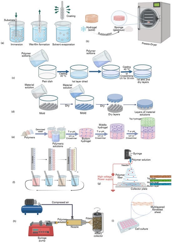
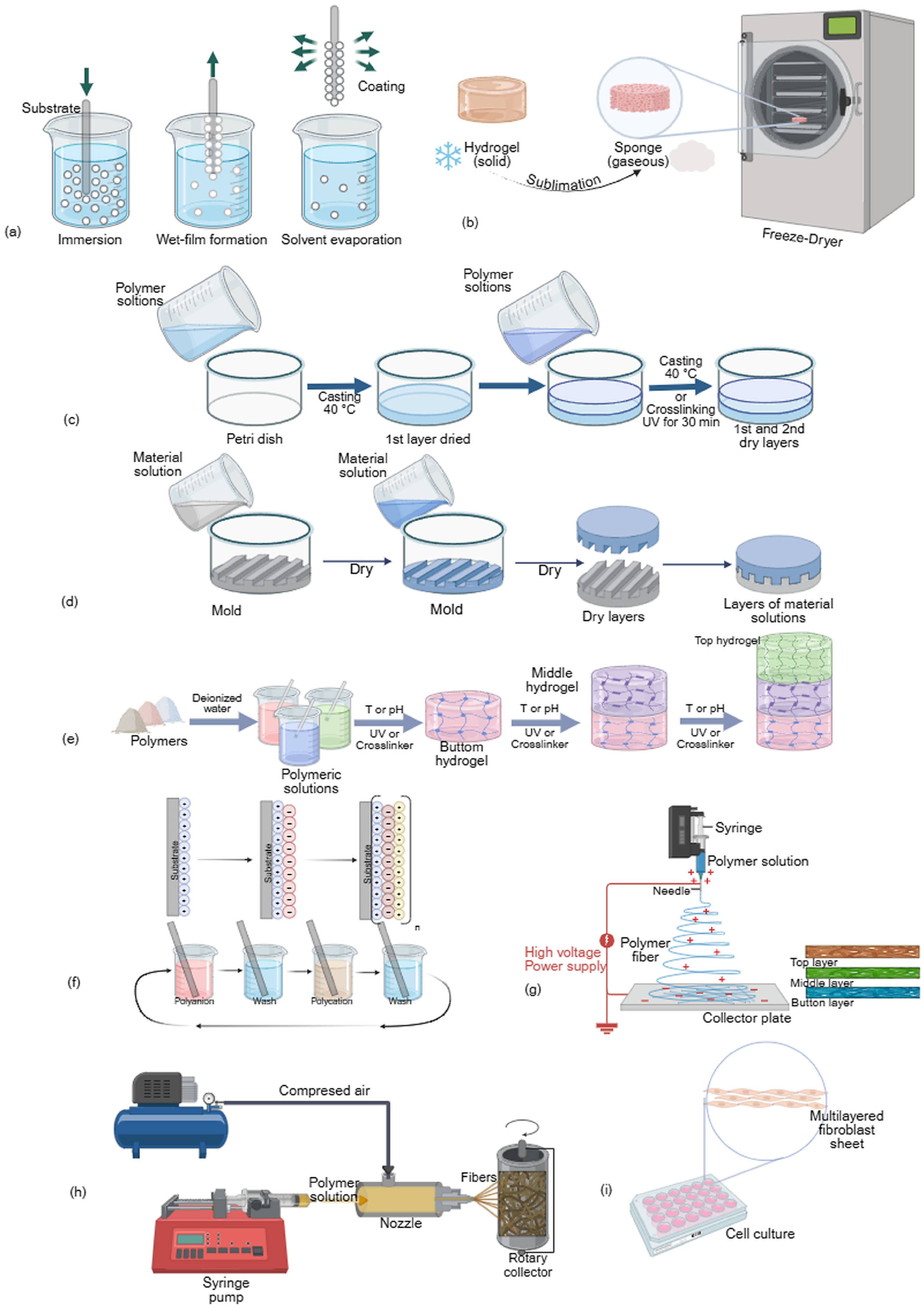
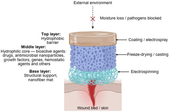
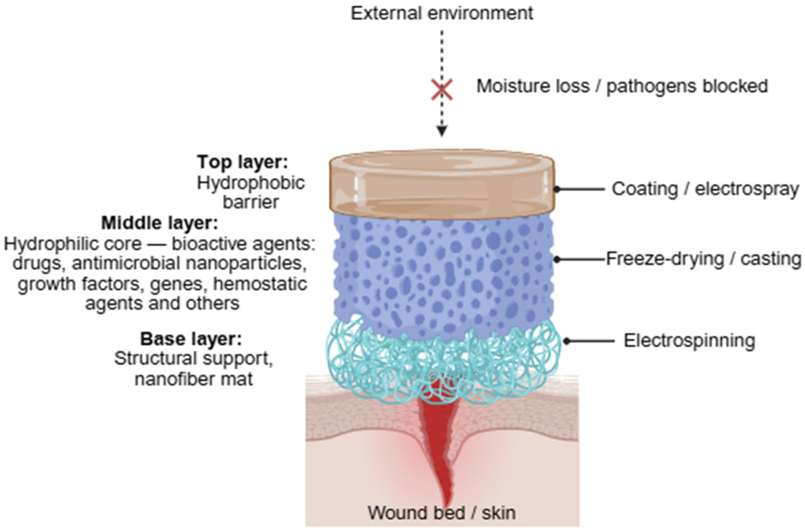
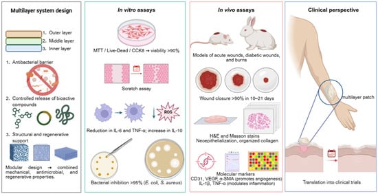
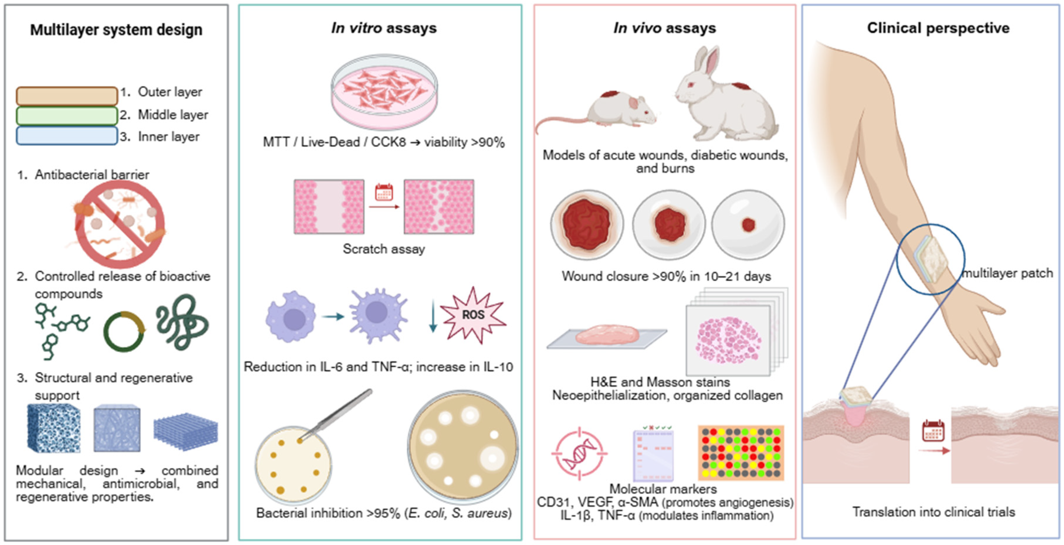

Disclaimer/Publisher’s Note: The statements, opinions and data contained in all publications are solely those of the individual author(s) and contributor(s) and not of MDPI and/or the editor(s). MDPI and/or the editor(s) disclaim responsibility for any injury to people or property resulting from any ideas, methods, instructions or products referred to in the content. |
© 2026 by the authors. Licensee MDPI, Basel, Switzerland. This article is an open access article distributed under the terms and conditions of the Creative Commons Attribution (CC BY) license.