


Abstract
Biodegradable polymers based on poly(lactic acid) (PLA) and polyhydroxybutyrate (PHB) are widely investigated for biomedical engineering applications, particularly for temporary implants and tissue scaffolds fabricated by additive manufacturing. However, their degradation behavior is influenced not only by material composition, but also by manufacturing-related parameters, geometric design, and environmental conditions. This study investigates the in vitro degradation behavior of PLA/PHB structures fabricated using fused filament fabrication (FFF). Degradation was evaluated under two model environmental conditions over a 45 day period. Changes in specimen mass and the evolution of degradation medium pH were monitored as a function of exposure time, specimen geometry, and infill density. The results revealed a progressive degradation process, with pH values decreasing to approximately 2.7–4.1 in physiological saline solution and increasing to 8.9–9.7 in urea solution, depending on specimen geometry and infill density. After 45 days of exposure, the relative mass loss reached approximately 25–32% for type A specimens and 29–41% for type B specimens. The results revealed distinct differences between degradation environments and specimen geometries, while differences related to infill density partially overlapped within the investigated range. The findings indicate that the degradation behavior of additively manufactured PLA/PHB structures cannot be interpreted solely based on material composition, but should be considered in the context of manufacturing strategy and structural design. These results provide useful insights for the design of biodegradable polymer components with more predictable degradation behavior.
1. Introduction
Temporary biomedical implants have traditionally been designed to provide the required mechanical and functional support for a defined period of time, after which they are surgically removed from the body [1]. However, this approach is associated with an additional invasive intervention and an increased risk of complications for the patient. Consequently, current research increasingly focuses on the development of biodegradable implants and tissue scaffolds that are capable of gradually degrading directly in the biological environment after fulfilling their function, thereby eliminating the need for surgical removal and supporting natural tissue regeneration [2,3].
Biodegradation of polymeric materials, however, does not represent only a desirable feature, but also a critical factor influencing the functional performance of such systems throughout their service life. The rate and mechanism of degradation directly affect the preservation of geometric stability and the overall integrity of the implant. Inadequately controlled degradation may lead to premature loss of functionality or undesirable changes in the local chemical environment surrounding the material, potentially impairing its interaction with biological tissues [4,5].
For this reason, systematic investigation of the degradation behavior of biodegradable polymers under controlled and reproducible in vitro conditions is essential. Although such model environments cannot fully replicate the complexity of in vivo conditions, they enable fundamental evaluation of degradation mechanisms, comparison of material behavior in different degradation media, and identification of trends that are important for the further development of biodegradable systems [6,7].
Among biodegradable polymers applied in biomedical engineering, poly(lactic acid) (PLA) and polyhydroxybutyrate (PHB) are frequently investigated due to their biocompatibility and resorbable character [8,9]. PLA is characterized by good processability and mechanical performance, while PHB exhibits higher crystallinity and slower degradation kinetics. The combination of PLA and PHB enables tailoring of the material response by balancing their individual properties, which is particularly relevant for applications requiring controlled degradation behavior [10]. In addition to its biocompatibility and processability, PLA is also attractive for biomedical applications due to its surface roughness tunability, which can be tailored through processing parameters and has been shown to influence cell–material interactions and biological response [11].
Additive manufacturing of biodegradable polymers offers new opportunities in the design of implants and tissue scaffolds; however, it also places increased demands on understanding the relationships between manufacturing technology, material structure, and degradation behavior [12,13]. The degradation of additively manufactured structures may be influenced not only by material composition, but also by geometric design, internal architecture, and porosity, which determine the interaction of the material with the degradation environment.
When processed by fused filament fabrication (FFF), biodegradable polymers are deposited layer-by-layer, resulting in structures with defined geometry and internal architecture. Parameters such as specimen shape, infill density, and internal porosity influence surface area, diffusion pathways, and interaction with the degradation environment. As a result, the degradation behavior of additively manufactured polymer structures may differ from that of bulk materials and should be evaluated with respect to both material composition and structural design [14,15].
The aim of this study is to investigate the in vitro degradation behavior of PLA/PHB specimens fabricated by additive manufacturing under model degradation conditions. The work focuses on the evaluation of changes in specimen mass and the evolution of degradation medium pH during the degradation process as a function of specimen geometry and infill density. The obtained results contribute to a better understanding of the degradation behavior of additively manufactured PLA/PHB systems and provide a basis for optimizing the design of biodegradable structures from both manufacturing and application perspectives.
2. Materials and Methods
2.1. Materials
The investigated material consisted of a blend of poly(lactic acid) (PLA) and polyhydroxybutyrate (PHB) with the addition of a plasticizer at a concentration of 25 wt.%. The PLA and PHB polymers were supplied in pellet form from an academic research source and were further processed within the scope of the present study.
The filament was prepared at the Faculty of Mechanical Engineering, Technical University of Košice, Department of Biomedical Engineering and Measurement. Filament fabrication was carried out using a Filament Maker Composer 450 (3devo, Utrecht, The Netherlands). Prior to filament extrusion, the device was thoroughly cleaned using high-density polyethylene (HDPE) to prevent cross-contamination from previously processed materials. The PLA/PHB pellets were dried at 60 °C for approximately 3 h to reduce residual moisture before processing. Filament production was based on a melt extrusion process. The pellet mixture was gradually melted and homogenized while passing through four heating zones of the extruder, which were set to 153 °C (zone 1), 180 °C (zone 2), 180 °C (zone 3), and 146 °C (zone 4). The extrusion screw speed was set to 4.0 rpm. The extruded filament was actively cooled using a fan operating at 100% speed, and the winding speed was controlled automatically by the system.
The resulting filament had a nominal diameter of 1.75 mm and was wound onto a plastic spool. The prepared filament was subsequently used for fused filament fabrication of the test specimens.
2.2. Specimen Design and Fabrication
Two types of cylindrical specimens with different geometries were designed using SolidWorks 2022 software (Dassault Systèmes, Paris, France).
Specimen type A had a diameter of 6 mm and a height of 2 mm, while specimen type B had a diameter of 5 mm and a height of 10 mm. These geometries were selected to investigate the influence of specimen shape on degradation behavior.
The specimens were fabricated using fused filament fabrication (FFF) on a TRILAB DeltiQ 3D printer (Trilab, Hradec Králové, Czech Republic). The same printing parameters were applied for all specimen types. The extruder temperature was set to 190 °C, while the build platform temperature was maintained at 70 °C. The printing speed was 1200 mm·min−1. No top or bottom solid layers were applied, and two outer perimeters were used to define the specimen contour. Active cooling was applied during the printing process, with the cooling fan operating at 100% capacity.
All specimens were printed with a raster angle of 0°, a constant layer height of 0.4 mm, and identical printing speed and cooling conditions for all sample groups.
A total of 270 specimens were fabricated and included in the degradation study. Specimens of type A consisted of 150 samples, with 50 specimens for each infill density (100%, 75%, and 50%). Specimens of type B consisted of 120 samples, with 40 specimens for each infill density (100%, 75%, and 50%). All specimens were subjected to the same degradation protocol and testing conditions.
2.3. In Vitro Degradation Experiment
The degradation experiment was performed under controlled in vitro conditions using two different degradation media: physiological saline solution and urea solution. The urea degradation medium was prepared as an aqueous solution with a concentration of 20 g/100 mL. After fabrication, the specimens were immersed in the degradation media and placed in a CelCulture® CO2 Incubator CCL-170B-8 (Esco Micro Pte. Ltd., Singapore).
To ensure homogeneous conditions and uniform exposure of the specimens to the degradation media, an orbital shaker (PSU-10i, BioSan, Riga, Latvia) was used during the experiment. Continuous gentle agitation was applied to simulate fluid movement and to promote uniform distribution of the degradation medium around the specimens.
The degradation experiment was carried out over a total period of 45 days. At predefined time points (15, 30, and 45 days), specimens were removed from the degradation media for analysis. After each measurement, the specimens were transferred into fresh degradation medium in order to minimize the influence of accumulated degradation products.
2.4. pH Measurement
Changes in the chemical environment during degradation were monitored by measuring the pH of the degradation media. pH measurements were performed using a pH meter (FiveEasy F20 standard, Mettler-Toledo, Greifensee, Switzerland). Measurements were conducted at regular time intervals during the degradation experiment to capture the evolution of pH in both degradation media.
2.5. Mass Loss Evaluation
The mass of the specimens was measured using an analytical balance MA 50/1.X2.A (Radwag, Radom, Poland). Prior to weighing, the specimens were rinsed, dried, and weighed to determine their dry mass.
The relative mass loss was expressed as (%) and calculated according to the following equation:
where is the initial mass of the specimen before the start of degradation (day 0) and is the dry mass of the specimen at time .
The use of dry mass values minimized the influence of temporary absorption of the degradation medium. Reported values represent average values for each specimen group defined by specimen type, infill density, and degradation environment.
2.6. Data Evaluation
The degradation behavior of the specimens was evaluated by analyzing changes in pH of the degradation media and relative mass loss as a function of exposure time, specimen geometry, and infill density. The obtained results were used to assess the influence of manufacturing-related parameters and structural design on the degradation response of additively manufactured PLA/PHB structures.
3. Results
All reported values represent mean values calculated from multiple specimens within each group. Due to the retrospective nature of data processing, individual raw data required for statistical dispersion analysis were not available; therefore, standard deviations and error bars are not reported.
3.1. Evolution of pH in Degradation Media
The evolution of pH was monitored in both degradation media throughout the 45 day in vitro experiment. Distinct trends in pH development were observed depending on the type of degradation medium, specimen geometry, and infill density.
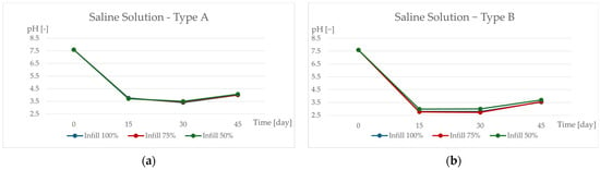
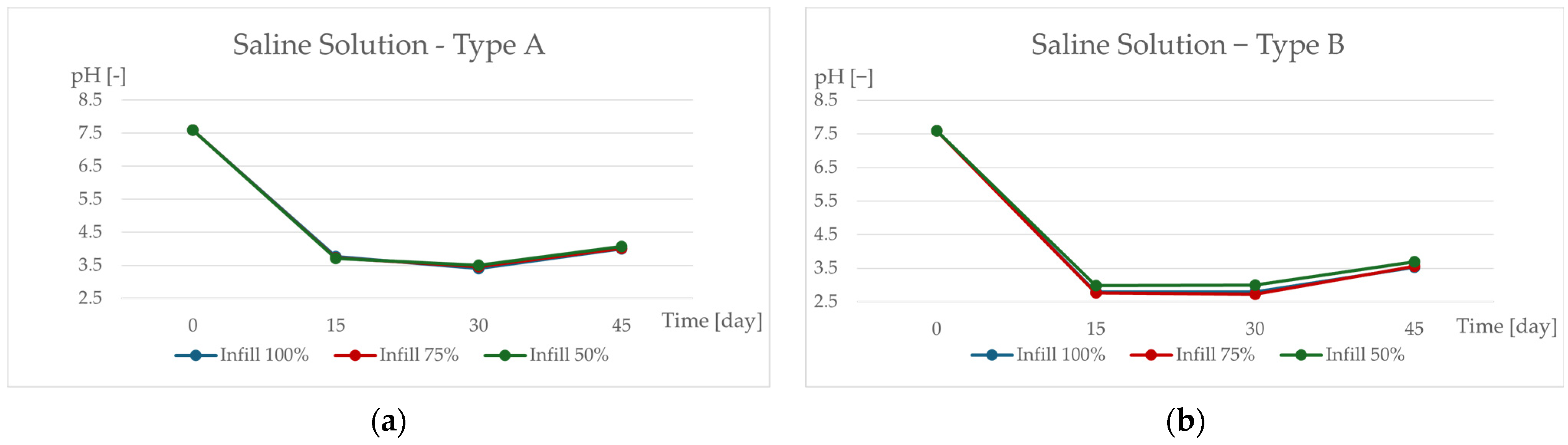
In the physiological saline solution, a pronounced decrease in pH was observed for all specimen groups during the first 15 days of exposure (Figure 1, Table 1).
Figure 1.
Evolution of pH during the in vitro degradation of PLA/PHB specimens in physiological saline solution over a 45 day period: (a) specimen type A and (b) specimen type B, fabricated with infill densities of 100%, 75%, and 50%.
Table 1.
Measured pH values of the physiological saline solution during in vitro degradation of PLA/PHB specimens at exposure times of 0, 15, 30, and 45 days for specimen types A and B with infill densities of 100%, 75%, and 50%.
Initial pH values close to neutrality rapidly decreased to acidic values, indicating the release of acidic degradation products into the surrounding medium. This rapid pH drop suggests an intensive initial degradation phase, most likely associated with hydrolytic cleavage of ester bonds within the PLA/PHB matrix. Between 15 and 30 days, the pH values remained low with only minor variations, followed by a slight increase toward the end of the experiment, which may be related to partial stabilization of the degradation process.
Differences between specimen types A and B were evident throughout the experiment, whereas the curves corresponding to different infill densities partially overlapped within each specimen type. This observation suggests that specimen geometry had a more pronounced influence on pH evolution than infill density within the investigated range.
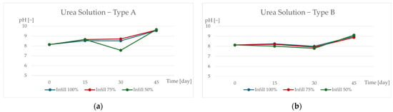
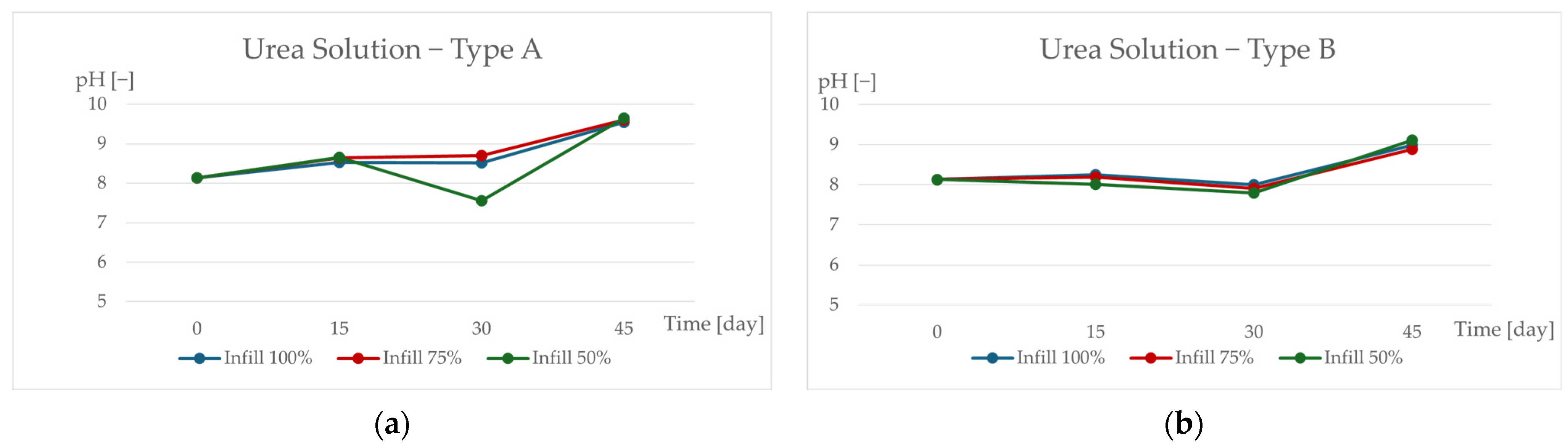
In contrast, degradation in the urea solution exhibited a markedly different pH evolution (Figure 2, Table 2).
Figure 2.
pH variation during the 45 day in vitro degradation of PLA/PHB specimens in urea solution. Results are shown for (a) specimen type A and (b) specimen type B with infill densities of 100%, 75%, and 50%.
Table 2.
pH values of the urea solution recorded during the 45 day in vitro degradation of PLA/PHB specimens at 0, 15, 30, and 45 days. Data correspond to specimen types A and B with infill densities of 100%, 75%, and 50%.
The pH values remained within the alkaline range throughout the entire exposure period and gradually increased with time. This behavior indicates a different degradation environment compared to physiological saline, in which alkaline conditions likely mitigated the accumulation of acidic degradation products. The observed trends were consistent for both specimen types and all infill densities, with only minor differences between individual specimen groups.
3.2. Relative Mass Loss in Physiological Saline Solution
The relative mass loss of PLA/PHB specimens exposed to physiological saline solution is presented in Figure 3a,b and summarized in Table 3. All specimen groups exhibited a substantial increase in relative mass loss after the first 15 days of degradation, corresponding to the initial phase of material degradation. Subsequently, the rate of mass loss became less pronounced, and the values tended to stabilize toward the end of the experiment, indicating a deceleration of the degradation process over time (Figure 3, Table 3).
Figure 3.
Relative mass loss of PLA/PHB specimens exposed to physiological saline solution during 45 days of in vitro degradation: (a) specimen type A and (b) specimen type B, for infill densities of 100%, 75%, and 50%.
Table 3.
Relative mass loss (%) of PLA/PHB specimens exposed to physiological saline solution at exposure times of 15, 30, and 45 days for specimen types A and B with infill densities of 100%, 75%, and 50%.
After 45 days of exposure, specimen type A reached relative mass loss values in the range of approximately 25–28%, depending on infill density, whereas specimen type B exhibited higher mass loss values, approximately 29–32%. These differences suggest that specimen geometry influenced the degradation kinetics, potentially through differences in surface area-to-volume ratio and accessibility of the degradation medium.
At day 15, specimen type A exhibited relative mass loss values between 24.25% and 29.72%, while specimen type B showed lower values ranging from 13.58% to 19.87%, indicating a slower initial degradation of type B specimens. At day 30, the relative mass loss ranged from 21.05% to 25.71% for specimen type A and from 21.64% to 21.76% for specimen type B, suggesting convergence of degradation behavior at intermediate exposure times. After 45 days, specimen type B exceeded type A in relative mass loss, reaching values of 29.63–32.31%, while type A specimens remained in the range of 25.36–27.59%.
Within each specimen type, the curves corresponding to different infill densities partially overlapped, indicating that infill density had a secondary effect compared to specimen geometry under the investigated conditions.
3.3. Relative Mass Loss in Urea Solution
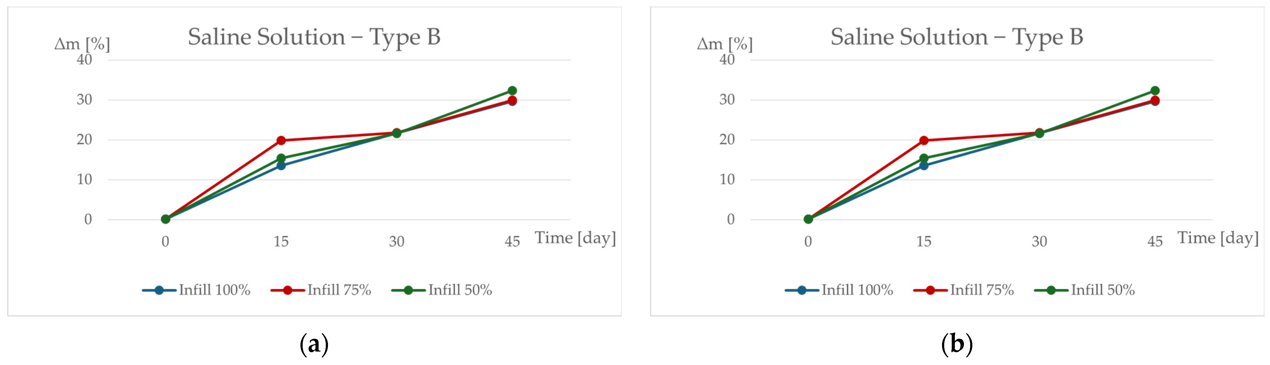
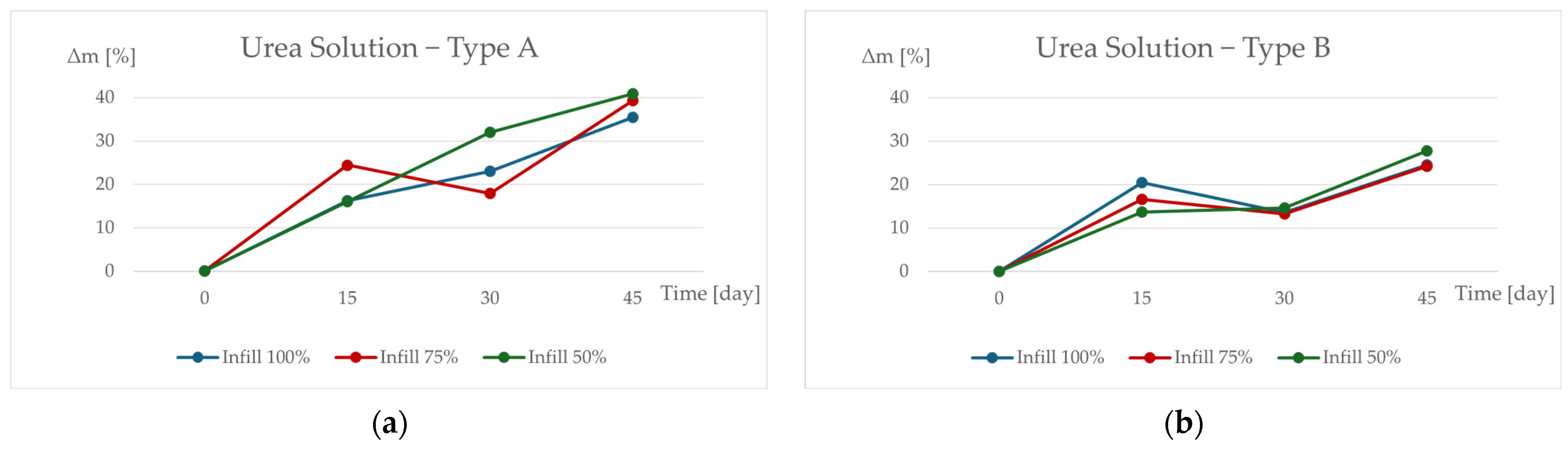
The relative mass loss of PLA/PHB specimens exposed to the urea solution is shown in Figure 4a,b and summarized in Table 4. In contrast to physiological saline solution, the evolution of relative mass loss in the urea environment exhibited different degradation trends between specimen types A and B (Figure 4, Table 4).
Figure 4.
Changes in relative mass (Δm) of PLA/PHB specimens after 45 days of in vitro exposure to physiological saline solution. Data are presented for (a) specimen type A and (b) specimen type B with infill densities of 100%, 75%, and 50%.
Table 4.
Percentage relative mass change (Δm, %) of PLA/PHB specimens measured after 15, 30, and 45 days of in vitro exposure to physiological saline solution. Results are given for specimen types A and B with infill densities of 100%, 75%, and 50%.
Specimen type A showed a progressive increase in relative mass loss with exposure time, reaching values between 35.44% and 40.89% after 45 days, depending on infill density. This continuous increase indicates sustained degradation activity in the urea-based environment. At earlier time points, the relative mass loss values varied between specimen groups, ranging from 16.01% to 24.43% at day 15 and from 17.95% to 32.02% at day 30.
Specimen type B exhibited consistently lower relative mass loss values compared to specimen type A throughout the experiment. After 45 days of exposure, relative mass loss values for specimen type B ranged from 24.17% to 27.78%. At day 15, the values ranged from 13.64% to 20.39%, while at day 30 they remained between 13.23% and 14.63%, indicating a slower degradation progression relative to specimen type A.
As observed in the saline solution, differences related to infill density partially overlapped within each specimen type, suggesting that specimen geometry and degradation environment exerted a stronger influence on mass loss behavior than infill density alone.
4. Discussion
The results of this study indicate that the in vitro degradation behavior of additively manufactured PLA/PHB structures is governed by the combined influence of degradation environment and specimen geometry, while the effect of infill density within the investigated range was less pronounced. These findings confirm that degradation of biodegradable polymer systems cannot be interpreted solely as a material-dependent process, but must be considered in the broader context of manufacturing strategy and structural design, which is consistent with previous studies on additively manufactured biodegradable polymers [13,14].
A pronounced difference in pH evolution was observed between the two degradation environments. In physiological saline solution, a rapid decrease in pH occurred during the first 15 days of exposure for all specimen groups, which can be attributed to hydrolytic cleavage of ester bonds in PLA- and PHB-based materials and the subsequent release of acidic degradation products [8,9]. The stabilization and slight increase in pH at later stages may be associated with changes in degradation kinetics as well as the periodic replacement of the degradation medium, which limited long-term accumulation of acidic species. In contrast, degradation in the urea solution resulted in alkaline pH values throughout the entire exposure period, with a gradual increase over time. This behavior highlights the strong influence of the chemical nature of the degradation medium on the local environment surrounding the degrading polymer and underlines the importance of carefully selecting degradation conditions when comparing in vitro studies [5].
Relative mass loss provided complementary information to pH evolution by reflecting the extent of material removal during degradation. In physiological saline solution, the initial pH decrease was accompanied by a progressive increase in mass loss for both specimen types, with specimen type B exhibiting higher mass loss values at later exposure times. In the urea solution, mass loss increased for both specimen types despite the alkaline pH conditions, with specimen type A showing substantially higher values than specimen type B. These observations demonstrate that degradation behavior cannot be inferred from pH trends alone and that mass loss is influenced by additional factors such as specimen geometry, surface-area-to-volume ratio, and diffusion pathways within the internal structure of additively manufactured specimens [4,7].
Consistent differences between specimen types A and B observed in both degradation environments further emphasize the importance of geometric design in additively manufactured biodegradable structures. Variations in specimen dimensions affect the accessibility of the degradation medium to the internal regions of the structure and the transport of degradation products, which can lead to different degradation dynamics even for identical material compositions [12,16]. In contrast, the influence of infill density on both pH evolution and mass loss was less distinct, as indicated by partial overlap of the corresponding curves within each specimen type. This suggests that, within the investigated infill density range, specimen geometry and degradation environment exert a stronger influence on degradation behavior than moderate variations in infill density [15].
The present study was conducted under controlled in vitro conditions, which enable systematic comparison of degradation trends but do not fully replicate the complexity of in vivo environments. Furthermore, the evaluation of degradation behavior was based on average mass loss values without statistical dispersion analysis. Future research should therefore focus on expanding the range of structural parameters, incorporating mechanical characterization during degradation, and exploring additional degradation environments to further clarify the interplay between material composition, manufacturing strategy, and degradation behavior [3,6]. Despite these limitations, the results provide meaningful insights into the degradation response of additively manufactured PLA/PHB structures and support the need for a holistic approach to the design of biodegradable polymer systems with predictable degradation behavior.
5. Conclusions
This study investigated the in vitro degradation behavior of PLA/PHB structures fabricated by fused filament fabrication under two model degradation environments. The results demonstrate that degradation behavior is strongly influenced by the chemical nature of the surrounding medium and by specimen geometry, while variations in infill density within the investigated range had a less pronounced effect.
A clear distinction was observed between degradation in physiological saline solution and urea-based medium. In saline solution, rapid acidification of the environment during the early stages of degradation was accompanied by progressive mass loss, indicating intensive hydrolytic degradation of the polymer matrix. In contrast, degradation in the urea solution proceeded under alkaline conditions, with different mass loss trends depending on specimen geometry. These findings highlight that pH evolution and mass loss do not necessarily follow the same trends and should be evaluated in parallel to obtain a comprehensive understanding of degradation processes.
Consistent differences between specimen types A and B confirmed that geometric design plays a critical role in governing degradation dynamics of additively manufactured biodegradable structures. Changes in specimen dimensions affect surface-area-to-volume ratio, diffusion pathways, and accessibility of the degradation medium, leading to distinct degradation responses even for identical material compositions and processing parameters. This underscores the importance of considering structural design as an integral factor in the development of biodegradable components fabricated by additive manufacturing.
Although the present study was conducted under controlled in vitro conditions and focused on mass loss and pH evolution, the results provide valuable insights into the degradation behavior of FFF-fabricated PLA/PHB systems. Future work should aim to extend the analysis by incorporating mechanical characterization during degradation, statistical evaluation of data variability, and microstructural analysis to further elucidate degradation mechanisms. Overall, this study supports a holistic approach to the design of biodegradable polymer structures, in which material selection, manufacturing strategy, and geometric design are jointly optimized to achieve predictable degradation behavior.
Author Contributions
Conceptualization, A.F.B.; methodology, A.F.B.; investigation, A.F.B.; formal analysis, A.F.B.; data curation, A.F.B.; writing—original draft preparation, A.F.B.; writing—review and editing, A.F.B., M.T., V.R., J.Č. and D.B.; resources, M.T.; supervision, M.T.; project administration, V.R.; funding acquisition, V.R.; validation, A.F.B., M.T. and D.B.; visualization, A.F.B. All authors have read and agreed to the published version of the manuscript.
Funding
This work was supported by the Scientific Grant Agency of the Ministry of Education, Science, Research and Sport of the Slovak Republic and the Slovak Academy of Sciences (VEGA) under Grant No. VEGA 1/0387/22 and VEGA 1/0403/25 and also by the Cultural and Educational Grant Agency of the Ministry of Education, Science, Research and Sport of the Slovak Republic (KEGA), Grant No. 054TUKE-4/2025 and KEGA 002TUKE-4/2026.
Data Availability Statement
The data supporting the findings of this study are available from the corresponding author upon reasonable request due to their size and experimental nature.
Acknowledgments
The authors acknowledge the technical and administrative support provided by the department during the experimental work. The authors also acknowledge the use of generative artificial intelligence tools during the preparation of this manuscript. During the preparation of this manuscript, the authors used ChatGPT (OpenAI, San Francisco, CA, USA), based on the GPT-4 architecture for language editing and text refinement. The authors have reviewed and edited the generated content and take full responsibility for the content of this publication.
Conflicts of Interest
The authors declare no conflicts of interest. The funders had no role in the design of the study; in the collection, analyses, or interpretation of data; in the writing of the manuscript; or in the decision to publish the results.
Abbreviations
The following abbreviations are used in this manuscript:
| PLA | Poly(lactic acid) |
| PHB | Polyhydroxybutyrate |
| FFF | Fused Filament Fabrication |
| AM | Additive Manufacturing |
| Δm | Relative mass loss |
| CO2 | Carbon dioxide |
| pH | Potential of hydrogen |
References
- Middleton, J.C.; Tipton, A.J. Synthetic biodegradable polymers as orthopedic devices. Biomaterials 2000, 21, 2335–2346. [Google Scholar] [CrossRef] [Scilit] [PubMed]
- Rezwan, K.; Chen, Q.Z.; Blaker, J.J.; Boccaccini, A.R. Biodegradable and bioactive porous polymer/inorganic composite scaffolds for bone tissue engineering. Biomaterials 2006, 27, 3413–3431. [Google Scholar] [CrossRef] [Scilit] [PubMed]
- Dhandayuthapani, B.; Yoshida, Y.; Maekawa, T.; Kumar, D.S. Polymeric scaffolds in tissue engineering application: A review. Int. J. Polym. Sci. 2011, 2011, 290602. [Google Scholar] [CrossRef] [Scilit]
- Araújo, M.A.; Cunha, A.M.; Mota, M. Enzymatic degradation of starch-based thermoplastic compounds used in protheses. Biomaterials 2004, 25, 2687–2693. [Google Scholar] [CrossRef] [Scilit] [PubMed]
- Tas, A.C. The use of physiological solutions or media in calcium phosphate synthesis and processing. Acta Biomater. 2014, 10, 1771–1792. [Google Scholar] [CrossRef] [Scilit] [PubMed]
- Marin, E.; Briceño, M.I.; Caballero-George, C. Critical evaluation of biodegradable polymers used in nanodrugs. Int. J. Nanomed. 2013, 8, 3071–3091. [Google Scholar] [CrossRef] [Scilit] [PubMed]
- Urinov, E.; Hanstein, S.; Weidenkaff, A. Enzymatic Degradation of Fiber-Reinforced PLA Composite Material. Macromol 2022, 2, 522–530. [Google Scholar] [CrossRef] [Scilit]
- Taib, N.-A.A.B.; Rahman, R.; Huda, D.; Kuok, K.K.; Hamdan, S.; Bin Bakri, M.K.; Bin Julaihi, M.R.M.; Khan, A. A review on poly(lactic acid) (PLA) as a biodegradable polymer. Polym. Bull. 2023, 80, 1179–1213. [Google Scholar] [CrossRef] [Scilit]
- McAdam, B.; Fournet, M.B.; McDonald, P.; Mojicevic, M. Production of polyhydroxybutyrate (PHB) and factors impacting its chemical and mechanical characteristics. Polymers 2020, 12, 2908. [Google Scholar] [CrossRef] [Scilit] [PubMed]
- Hussain, M.; Khan, S.M.; Shafiq, M.; Abbas, N. A review on PLA-based biodegradable materials for biomedical applications. Giant 2024, 18, 100261. [Google Scholar] [CrossRef] [Scilit]
- Vesco, S.; Salvi, D. Fuzzy skin in fused filament fabrication: Enhancing morphology, wettability, and friction through a full-factorial experimental plan. Prog. Addit. Manuf. 2025, 10, 11233–11257. [Google Scholar] [CrossRef] [Scilit]
- Bajaj, P.; Schweller, R.M.; Khademhosseini, A.; West, J.L.; Bashir, R. 3D bioprinting strategies for tissue engineering and regenerative medicine. Annu. Rev. Biomed. Eng. 2014, 16, 247–276. [Google Scholar] [CrossRef] [Scilit] [PubMed]
- Wickramasinghe, S.; Do, T.; Tran, P. FDM-based 3D printing of polymer and associated composites: A review on mechanical properties defects and treatments. Polymers 2020, 12, 1529. [Google Scholar] [CrossRef] [Scilit] [PubMed]
- Popescu, D.; Zapciu, A.; Amza, C.; Baciu, F.; Marinescu, R. FDM process parameters influence over the mechanical properties of polymer specimens. Polym. Test. 2018, 69, 157–166. [Google Scholar] [CrossRef] [Scilit]
- Balogová, A.F.; Trebuňová, M.; Ižaríková, G.; Kaščák, Ľ.; Mitrík, L.; Klímová, J.; Feranc, J.; Modrák, M.; Hudák, R.; Živčák, J. In vitro degradation of specimens produced from PLA/PHB by additive manufacturing in simulated conditions. Polymers 2021, 13, 1542. [Google Scholar] [CrossRef] [Scilit] [PubMed]
- Ranakoti, L.; Gangil, B.; Mishra, S.K.; Singh, T.; Sharma, S.; Ilyas, R.; El-Khatib, S. Critical review of polylactic acid: Properties, structure, processing, biocomposites, and nanocomposites. Materials 2022, 15, 4312. [Google Scholar] [CrossRef] [Scilit] [PubMed]

Disclaimer/Publisher’s Note: The statements, opinions and data contained in all publications are solely those of the individual author(s) and contributor(s) and not of MDPI and/or the editor(s). MDPI and/or the editor(s) disclaim responsibility for any injury to people or property resulting from any ideas, methods, instructions or products referred to in the content. |
© 2026 by the authors. Licensee MDPI, Basel, Switzerland. This article is an open access article distributed under the terms and conditions of the Creative Commons Attribution (CC BY) license.