Next Issue
Volume 2, Symmetry 2017
Previous Issue
Volume 1, IMMAGINI? Conference 2017
 
 
proceedings-logo

Journal Browser

Journal Browser

Proceedings, 2017, NPCPT 2017

The 2nd International Conference on Natural Products for Cancer Prevention and Therapy

Kayseri, Turkey | 08-11 November 2017

Issue Editors:
Mukerrem Betul Yerer, Erciyes University, Turkey
Anupam Bishayee, Larkin University, USA

Number of Papers: 106
  • Issues are regarded as officially published after their release is announced to the table of contents alert mailing list.
  • You may sign up for e-mail alerts to receive table of contents of newly released issues.
  • PDF is the official format for papers published in both, html and pdf forms. To view the papers in pdf format, click on the "PDF Full-text" link, and use the free Adobe Reader to open them.
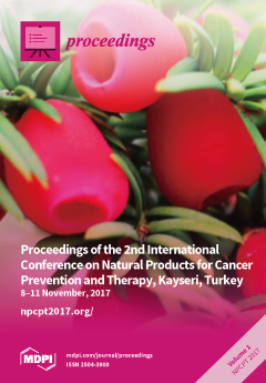
Cover Story (view full-size image): Scientific experts from eight countries gathered to share their views and experience on the latest research on natural products for cancer prevention and therapy. The traditionally used herbal [...] Read more.
Order results
Result details
Select all
Export citation of selected articles as:

Research

113 KiB  
Abstract
Novel Anticancer Capacities of Saffron
by Amr Amin
Proceedings 2017, 1(10), 834; https://doi.org/10.3390/proceedings1100834 - 15 Nov 2017
Cited by 1 | Viewed by 1626
Abstract
Thanks to poor diagnosis and very limited treatment options, hepatocellular carcinoma (HCC) continues to lead the list of cancer-leading causes of death worldwide. Therefore, alternative therapy is crucial to control HCC. Our group has investigated the anticancer effects of the saffron’s main active [...] Read more.
Thanks to poor diagnosis and very limited treatment options, hepatocellular carcinoma (HCC) continues to lead the list of cancer-leading causes of death worldwide. Therefore, alternative therapy is crucial to control HCC. Our group has investigated the anticancer effects of the saffron’s main active ingredient “safranal” against HCC using in vitro, in silico, and network analyses. In addition to the unique and differential cell cycle arrest, safranal showed pro-apoptotic effect through activation of both intrinsic and extrinsic initiator caspases implicating ER stress-mediated apoptosis. Gene set enrichment analysis provided consistent findings where unfolded protein response (UPR) was among the top terms in up-regulated genes in response to safranal treatment. Thus, proteins involved in ER stress and autophagy were regulated through safranal treatment to induce UPR and autophagy-mediated cell death in HepG2 cells. Full article
132 KiB  
Abstract
Cardiac Glycosides as Novel Modulators of Cancer Cell Survival
by Marc Diederich
Proceedings 2017, 1(10), 972; https://doi.org/10.3390/proceedings1100972 - 17 Nov 2017
Cited by 1 | Viewed by 1641
Abstract
Cardiac glycosides (CGs) were initially described as a cure for cardiovascular diseases. More recently a correlation between CG intake and anticancer activity was suggested, further validated by in vivo and in vitro preclinical research. Our recent findings demonstrate that CGs efficiently act on [...] Read more.
Cardiac glycosides (CGs) were initially described as a cure for cardiovascular diseases. More recently a correlation between CG intake and anticancer activity was suggested, further validated by in vivo and in vitro preclinical research. Our recent findings demonstrate that CGs efficiently act on cancer cell proliferation and survival [1]. UNBS1450 is a hemi-synthetic cardenolide that exerts cytostatic and cytotoxic effects in leukemia and solid tumors at low micromolar concentrations. In leukemia, downregulation of Mcl-1 was observed after 4 hours of treatment in the absence of any effect at the transcriptional level. By generalizing our observations with cardenolides and bufadienolides of different plant and animal origins, we showed that Mcl-1can be considered as a universal target for CGs in cancer. Moreover, we demonstrated that Mcl-1 degradation is proteasome-dependent preceding apoptotic cell death. Furthermore, UNBS140 also triggers autophagy and mitophagy in solid tumors like neuroblastoma. So far, UNBS1450 was shown to trigger both complete and incomplete mitophagy in neuroblastoma leading to necroptosis and apoptosis, respectively [2], depending on the subtype of neuroblastoma. In AML, a combination of UNBS1450 and BH3 mimetics like ABT199 or 263 affects leukemia subtypes relying on Mcl-1 expression [3]. Altogether CGs are interesting pharmacological agents that efficiently induce cell death in cancer cell models at nanomolar concentrations encouraging us to further explore the therapeutic potential for CGs alone or in combination with other targeted treatments. Full article
138 KiB  
Abstract
Pristimerin is a Promising Natural Product against Breast Cancer in vitro and in vivo through Apoptosis and the Blockage of Autophagic Flux
by Buse Cevatemre, Konstantinos Dimas, Bruno Botta and Engin Ulukaya
Proceedings 2017, 1(10), 973; https://doi.org/10.3390/proceedings1100973 - 15 Nov 2017
Cited by 1 | Viewed by 1598
Abstract
The exploration of natural products is of importance because the management of cancer is still not satisfactory. Pristimerin is a naturally occurring triterpenoid that has been shown to suppress the proliferation of various cancer cell lines at relatively lower concentrations, of which the [...] Read more.
The exploration of natural products is of importance because the management of cancer is still not satisfactory. Pristimerin is a naturally occurring triterpenoid that has been shown to suppress the proliferation of various cancer cell lines at relatively lower concentrations, of which the IC50 values are around 0.5–4 μM. Its cytotoxic potential on particularly cancer stem cells (CSCs) should be much more important due to the CSCs’ recent role in recurrence of cancer. Although pristimerin exerted growth inhibitory activity in various cancer cells, the mechanisms is still debatable. Its antigrowth effect seems to involve the blockage of autophagic process as well. In our studies, pristimerin showed an anti-growth effect on cancer cells and cancer stem cells with IC50 values ranging at 0.38–1.75 µM. Apoptosis was induced in MCF-7 and MCF-7s (CSCs) cells. It also resulted in an incompleted autophagy as evidenced by the increase of autophagy-related proteins (P62 and LC3-II) as well as an unfolded protein response (UPR). Moreover, it inhibited the growth of xenografts in NOD/SCID mice. Taken together, pristimerin deserves further attention in terms of its use for the treatment of breast cancer. In this talk, quite an unusal cytotoxic potential of pristimerin will be discussed. Full article
151 KiB  
Abstract
Cynaropicrin: A Promising Natural Agent with Antitumor and Antiviral Activities
by Mahmoud F. Elsebai, Jukka Hakkola, Mohamed Mehiri and Juana Diez
Proceedings 2017, 1(10), 974; https://doi.org/10.3390/proceedings1100974 - 17 Nov 2017
Cited by 1 | Viewed by 2171
Abstract
Most natural agents do not induce a high level of toxicity and target multiple signaling pathways involved in cell growth, invasion, apoptosis, angiogenesis and metastasis. Human infection with HCV is currently recognized as the leading cause of hepatocellular carcinoma (HCC) which demands liver [...] Read more.
Most natural agents do not induce a high level of toxicity and target multiple signaling pathways involved in cell growth, invasion, apoptosis, angiogenesis and metastasis. Human infection with HCV is currently recognized as the leading cause of hepatocellular carcinoma (HCC) which demands liver transplantation which was estimated to result in ∼10,000 deaths in the US only in the year 2011. Cynaropicrin is a potential agent for treatment and prevention of HCC by indirect way through inhibition of HCV and in a direct way evidenced by the many antitumor activities in literature. Additionally, cynaropicrin has anti-inflammatory, TNF-α inhibitory, and antioxidant activities and hence it can also prevent the progression of HCC. The compounds cynaropicrin and grosheimol, obtained from the wild Egyptian artichoke (Cynara cardunculus L. var. sylvestris), showed EC50 at 1.03 μM, and 1.27 μM, by using a luciferase-carrying reporter virus. Time-of-addition experiments revealed that cynaropicrin and grosheimol inhibited HCV virus at a time-point during entry. Finally, the results showed that these compounds inhibited HCV particles from genotypes 1a, 1b, 2b, 3a, 4a, 5a, 6a and 7a indicating that these compounds inhibit HCV cell entry independently of viral genotype or subtype. In conclusion, cynaropicrin is a very promising drug as antitumor agent. Full article
153 KiB  
Abstract
Anticancer Potential of Flavones
by Randolph R. J. Arroo, Didem Şöhretoğlu, Demetrios A. Spandidos and Vasilis P. Androutsopoulos
Proceedings 2017, 1(10), 975; https://doi.org/10.3390/proceedings1100975 - 17 Nov 2017
Cited by 2 | Viewed by 1721
Abstract
Many papers have been written on the anticancer properties of dietary flavonoids, and a range of potential mechanisms of action of flavonoids. However, most dietary flavonoids - notably polyphenolic flavonoids—have very poor ADME properties, and the levels necessary to stop growth of tumour [...] Read more.
Many papers have been written on the anticancer properties of dietary flavonoids, and a range of potential mechanisms of action of flavonoids. However, most dietary flavonoids - notably polyphenolic flavonoids—have very poor ADME properties, and the levels necessary to stop growth of tumour cells cannot be sustained in a human body trough dietary intake alone. At present no flavonoid based drugs are clinically used in cancer therapy. Thus, whereas epidemiological and pre-clinical data seem to indicate a high potential for flavonoids, from the point of view of the pharmaceutical industry and drug developers, they are considered poor candidates. The flavones—which constitute a subgroup of the flavonoids—show some structural analogy with oestrogen and are known to interact with human oestrogen receptors, either as agonist or as antagonist. They are classed as phytoestrogens, and may play a role in cancer prevention through a mechanism of action possibly similar to that of the clinically used medication tamoxifen. Flavones are abundantly present in common fruits and vegetables, many of which have been associated with cancer prevention. Their phytoestrogen activity makes that they can assert their biological action at concentrations that are realistically achievable in the human systemic circulation. Full article
Show Figures

Figure 1

134 KiB  
Abstract
Resveratrol in Cancer Prevention and Treatment: Focusing on Molecular Targets and Mechanism of Action
by Adriana Borriello
Proceedings 2017, 1(10), 976; https://doi.org/10.3390/proceedings1100976 - 22 Nov 2017
Cited by 6 | Viewed by 2073
Abstract
Resveratrol (3,5,4′-trihydroxystilbene) is a phytoalexin occurring in grapes, red wines, peanuts, berries and other vegetables that has been reported to provide protection against several human diseases, including metabolic diseases and cancer. More than one thousand preclinical investigations, performed in vitro and in animal [...] Read more.
Resveratrol (3,5,4′-trihydroxystilbene) is a phytoalexin occurring in grapes, red wines, peanuts, berries and other vegetables that has been reported to provide protection against several human diseases, including metabolic diseases and cancer. More than one thousand preclinical investigations, performed in vitro and in animal models, indicate that resveratrol efficaciously affects the development and progression of various tumors such as breast, lung, prostate and colon carcinomas, leukemias, skin tumors and melanomas. On this basis, resveratrol and its derivatives are under investigation in humans as cancer chemopreventive and/or chemotherapeutic agents. Resveratrol mechanism of action has been widely studied in cancer cells and in experimental models of senescence, inflammation, obesity and metabolic diseases. Its molecular targets act at different levels: (1) specific molecular pathways (like NF-B, MAPK/JNK/p38 Kinase, PKC, PI3K-AKT and several others); (2) epigenetic control of gene transcription through sirtuin activation; (3) cell division cycle and differentiation; (4) apoptosis and autophagy; (5) cellular redox homeostasis. In addition, the phytoalexin might modify microbiome composition and mimic hormone activity. Particularly, we recently reported that the molecule is able to substitute insulin in adipogenesis induction of mesenchymal stromal cells derived from human bone marrow. The relevance of these mechanisms and their translation in clinical therapy will be discussed. Full article
126 KiB  
Abstract
Ins and Outs of Flavonoids in Cancer Prevention vs. Cancer Therapy: A Lesson from Quercetin in Leukemia
by Gian Luigi Russo Russo, Maria Usso, Carmela Spagnuolo and Stefania Moccia
Proceedings 2017, 1(10), 977; https://doi.org/10.3390/proceedings1100977 - 13 Nov 2017
Cited by 3 | Viewed by 2152
Abstract
We recently critically reviewed clinical and pre-clinical studies on the concept that polyphenols, being antioxidant compounds, can fight cancer. We suggest that a clear distinction must be done between the use of polyphenols, such as flavonoids, in cancer treatment versus cancer prevention, starting [...] Read more.
We recently critically reviewed clinical and pre-clinical studies on the concept that polyphenols, being antioxidant compounds, can fight cancer. We suggest that a clear distinction must be done between the use of polyphenols, such as flavonoids, in cancer treatment versus cancer prevention, starting from adequate and specifically selected cellular models. As an example, we will present data on the potential application of quercetin against chronic lymphocytic leukemia (CLL). Previous studies from our group indicated that quercetin synergistically sensitizes to apoptosis several leukemic cell lines and B-cells isolated from CLL patients, when associated with other pro-apoptotic agents. Using HG3 cells, derived from primary B-cells immortalized with Epstein-Barr virus, we demonstrated that the association between ABT-737, a BH3-mimetics, and quercetin synergistically induces apoptosis through the inhibition of PI3K/Akt signaling pathway. More importantly, we identified the protein kinase CK2 as a new, direct and primary target of quercetin, since CK2 activity was inhibited by the flavonoid within one minute from the treatment. Considering the rapid uptake of quercetin and its low toxicity against normal peripheral blood cells, we recommend the design of clinical studies aimed to demonstrate the potential use of the molecule in the adjuvant chemotherapy against CLL. Full article
138 KiB  
Abstract
Relationship between Structure of Phenolics and Anticancer Activity
by Müberra Koşar
Proceedings 2017, 1(10), 978; https://doi.org/10.3390/proceedings1100978 - 13 Nov 2017
Cited by 2 | Viewed by 1484
Abstract
Cancer is one of the main reasons of death in both men and women, claiming over 6 million people each year worldwide. Chemoprevention in combination with anticancer treatment is therefore important to reduce morbidity and mortality. Phenolic acids, flavonoids and tannins are very [...] Read more.
Cancer is one of the main reasons of death in both men and women, claiming over 6 million people each year worldwide. Chemoprevention in combination with anticancer treatment is therefore important to reduce morbidity and mortality. Phenolic acids, flavonoids and tannins are very famous compounds found in the foods known as antioxidant and anticancer groups. Antioxidant mechanisms are also related with anticancer activity. Therefore antioxidant phenolics show the anticancer activity in different degree. Many phenolic compounds have been investigated for their potential use as cancer chemopreventive agents. Phenolic compounds consist of one or more hydroxyl substitution on the aromatic ring system. Cinnamic acid esters, such as caffeic acid phenethyl and benzyl esters, display selective antiproliferative activity against some types of cancer cells. Flavonoids consist of a large group of polyphenolic compounds having a benzo-γ-pyrone structure and are ubiquitously present in plants. They are synthesized by phenylpropanoid pathway. Flavonoids and polyphenolic tannins affect the different cancer cells and these activities and mechanisms shown in vitro cell culture assays. Both antioxidant and anticancer activities of phenolics are related with their hydroxyl substitutions on the aromatic rings and also double bounds in the structures. Full article
138 KiB  
Abstract
Growth Factors Responsible from the Cancer Progress: Role of Natural Products
by Mükerrem Betül Yerer
Proceedings 2017, 1(10), 979; https://doi.org/10.3390/proceedings1100979 - 13 Nov 2017
Cited by 1 | Viewed by 1365
Abstract
Growth factors are one of the main factors responsible from the uncontrolled cell progress in cancer. Up to date many scientists have focused on these factors either as the marker or as the targets in several cancer types. Mainly the drugs are designed [...] Read more.
Growth factors are one of the main factors responsible from the uncontrolled cell progress in cancer. Up to date many scientists have focused on these factors either as the marker or as the targets in several cancer types. Mainly the drugs are designed to target these factors are monoclonal antibodies. Nerve growth factor (NGF), epidermal growth factor (EGF), hepatocyte growth factors (HGF), fibroblast growth factors (FGF), vascular endothelial growth factors (VEGF), platelet derived growth factor (PDGF), transforming growth factor (TGF-β) are some of these factors not only increasing the ability of cell proliferation but also playing crucial roles in triggering the invasion and metastasis of the cells. The herbs that are traditionally used for anticancer treatment and target multiple interdependent processes. Given the multiple effects of these agents, their future use for cancer therapy probably lies in synergistic combinations. For instance, Artemisia annua, Viscum album, Curcuma longa, Camellia sinensis, Vitis vinifera, Angelica sinensis, Taxus brevifolia, are some of the herbs that has shown to affect VEGF and those which have additional effects on the molecules related to cancer progress and further can target other growth factors. During active cancer therapy, they should generally be evaluated in combination with chemotherapy and radiation. In this role, they act as modifiers of biologic response, potentially enhancing the efficacy of or reducing the resistance to the conventional therapies. Full article
137 KiB  
Abstract
Can Curcumin be Employed to Promote the Integration of Oncology and Natural Products?
by Mutlu Demiray and Fatemeh Bahadori
Proceedings 2017, 1(10), 980; https://doi.org/10.3390/proceedings1100980 - 13 Nov 2017
Cited by 2 | Viewed by 1898
Abstract
Nuclear Factor-Kappa B (NF-kB) is a key inflammatory transcription factor expressed frequently in tumors which, has an important role in proliferation, apoptosis, inflammatory and immune response. Several recent preclinical and clinical studies have shown that significant NF-kB expression is associated with chemoradiation resistance [...] Read more.
Nuclear Factor-Kappa B (NF-kB) is a key inflammatory transcription factor expressed frequently in tumors which, has an important role in proliferation, apoptosis, inflammatory and immune response. Several recent preclinical and clinical studies have shown that significant NF-kB expression is associated with chemoradiation resistance and poor outcome in several human cancers. It has been shown that curcumin inhibits NF-kB activation pathway at a step before inhibitor kappa B alfa (IKBα) phosphorylation. Preclinical in vivo and in vitro trials that combine curcumin with tyrosine kinase inhibitors (TKIs) have shown that this combination shows better results than each treatment alone. Retrospectively evaluated seventeen NF-kB overexpressed cancer patients were treated with i.v. and oral bioavailable formulation of curcumin and appropriate TKIs. TKIs decision was made according to genetic profiling (FoundationOne®). Toxicities and response was evaluated according to RECIST and common toxicities criteria. All patients are stage IV and heavily pretreated. Our longest experience comes from adenoid cystic carcinoma and he has treated for 72 months by oral curcumin and 8 months by i.v curcumin. Disease control rate was 89.3% (15/17) and no any grade III-IV toxicities was observed related to curcumin. Curcumin is multitargeted molecule with pleotrophic nature. Curcumin inhibits NF-κB and related proteins which promotes effectiveness of TKIs. And might be considered as long as we have to use chemotherapy, radiotherapy and TKI. Full article
127 KiB  
Abstract
Therapeutic Potential of Black Pepper Compound for BRaf Resistant Melanoma
by Neel M. Fofaria, Sharavan Ramachandran and Sanjay K. Srivastava
Proceedings 2017, 1(10), 981; https://doi.org/10.3390/proceedings1100981 - 17 Nov 2017
Cited by 3 | Viewed by 1997
Abstract
Malignant melanoma is significant problem for Caucasian population in the western countries. Mutations in BRAF gene in 60% of patients is responsible for developing resistance to BRAF inhibitors. Our results delineated the mechanism of resistance and identified a suitable drug combination to overcome [...] Read more.
Malignant melanoma is significant problem for Caucasian population in the western countries. Mutations in BRAF gene in 60% of patients is responsible for developing resistance to BRAF inhibitors. Our results delineated the mechanism of resistance and identified a suitable drug combination to overcome the resistance. Treatment of BRAF mutant melanoma cells with vemurafenib or dabrafenib (BRAF inhibitors) alone or in combination with trametinib (MEK1/2 inhibitor) resulted in induced expression of Mcl-1. Melanoma cells resistant to BRAF inhibitors exhibited substantial expression of Mcl-1 as compared to sensitive cell lines. Silencing of Mcl-1 using siRNA completely sensitized resistant cells to growth suppression and induction of apoptosis by BRAF inhibitors. Piperlongumine, an active component of black pepper substantially suppressed the growth of vemurafinib resistant melanoma cells by inducing apoptosis and inhibiting Mcl-1 expression. Vemurafenib resistant A375 xenografts showed substantial tumor growth inhibition in mice when treated with a combination of vemurafenib and Mcl-1 inhibitor or siRNA. Oral administration of piperlongumine also suppressed the growth vemurafinib resistant tumor xenografts in athymic nude mice. Immunohistochemistry and western blot analyses confirmed enhanced expression of Mcl-1 and activation of ERK1/2 in vemurafenib-resistant tumors whereas level of Mcl-1 or p-ERK1/2 was reduced in the tumors of mice treated with either of the combination or piperlongumine. Biopsied tumors from the patients treated with or resistant to BRAF inhibitors revealed overexpression of Mcl-1. These results suggest that the combination of BRAF inhibitors with Mcl-1 inhibitor such as piperlongumine may have therapeutic advantage to melanoma patients with acquired resistance to BRAF inhibitors alone or in combination with MEK1/2 inhibitors. Full article
115 KiB  
Abstract
Natural Products for Cancer Prevention and Therapy: Progress, Pitfalls and Promise
by Anupam Bishayee
Proceedings 2017, 1(10), 982; https://doi.org/10.3390/proceedings1100982 - 15 Nov 2017
Cited by 3 | Viewed by 2106
Abstract
Natural products represent a valuable resource for discovery and development of anticancer drugs. About 80% of drugs approved by the United States Food and Drug Administration during the last three decades for cancer therapy are either natural products per se or are based [...] Read more.
Natural products represent a valuable resource for discovery and development of anticancer drugs. About 80% of drugs approved by the United States Food and Drug Administration during the last three decades for cancer therapy are either natural products per se or are based on, or mimicked natural products. Various extracts, fractions, mixtures and pure compounds from dietary sources, medicinal plants and marine organisms have been found to exhibit potent antiproliferative and cytotoxic activities against numerous cancer cell lines. These agents have also been tested for cancer preventive and therapeutic efficacies using preclinical animal cancer models that resemble human disease. Selected natural products have also entered into clinical studies. The cancer preventive and anticancer attributes of various natural products can be explained by multiple cellular and molecular mechanisms, including scavenging of free radicals, detoxification of carcinogens, DNA repair, alteration of cell cycles, programmed cell death (apoptosis), autophagy, immune surveillance, anti-inflammatory, anti-angiogenic, anti-invasive and antimetastatic activities as well as their ability to modulate a plethora of dysregulated oncogenic signaling pathways. This presentation aims to highlight studies on cancer preventive and therapeutic attributes of various naturally occurring agents and underlying mechanisms of action, with special emphasis on results reported from our laboratory. Current limitations, challenges, and future directions of research for successful cancer drug development based on natural products will also be discussed. Full article
148 KiB  
Abstract
Effect of Pomegranate Extract and Tangeretin on Specific Pathways in the Rat Breast Cancer Model Induced with DMBA
by H. Fatih Gul, Necip Ilhan, Nevin Ilhan and I. Hanifi Ozercan
Proceedings 2017, 1(10), 983; https://doi.org/10.3390/proceedings1100983 - 13 Nov 2017
Cited by 1 | Viewed by 1646
Abstract
It was aimed to investigate the possible chemopreventive effects of Pomegranate extract (PE) and Tangeretin (TAN), in the rat breast cancer model induced with 7,12-Dimethylbenz[a]anthracene (DMBA), on the selected angiogenesis parameters and NF-κB pathway. 56 Sprague Dawley female rats separated into 8 groups. [...] Read more.
It was aimed to investigate the possible chemopreventive effects of Pomegranate extract (PE) and Tangeretin (TAN), in the rat breast cancer model induced with 7,12-Dimethylbenz[a]anthracene (DMBA), on the selected angiogenesis parameters and NF-κB pathway. 56 Sprague Dawley female rats separated into 8 groups. Group I was control, Group II, III and IV were treated with PE, TAN and PE+TAN respectively. Group V was administered with DMBA, Group VI, VII and VIII also received PE, TAN and PE+TAN. The presence of the breast tumour tissue was demonstrated with histopathological examinations. Plasma VEGF, MMP-9 and NF-κB levels were determined using the ELISA method. As a result of the histopathological evaluations, it was determined that most (90%) of the tumors created were invasive ductal carcinoma. When compared to the controls, increased plasma VEGF, MMP-9 and NF- κB levels were determined in the DMBA group (p < 0.001; p < 0.05; p < 0.001). While quite significant decreases were observed in plasma VEGF and NF-κB levels in Group VI, Group VII and Group VIII compared to the DMBA group (p < 0.001; p < 0.001), decreases in MMP-9 levels were statistically insignificant. It was suggested that PE and TAN have chemopreventive effects on the development of breast cancer by effectively inhibiting both angiogenesis pathways and NF-κB pathway due to many natural bioactive agents they contain. Full article
133 KiB  
Abstract
Synergistic Cytotoxic Effects of Resveratrol in Combination with Ceramide Metabolizing Enzymes in Ph + Acute Lymphoblastic Leukemia
by Osman Oğuz and Aysun Adan
Proceedings 2017, 1(10), 984; https://doi.org/10.3390/proceedings1100984 - 16 Nov 2017
Cited by 1 | Viewed by 1567
Abstract
Bioactive sphingolipids are a lipid family including ceramide, sphingosine-1-phosphate (S1P) and glucosyl ceramide (GC). Ceramide produced through de novo synthesis pathway (Serine Palmitoyl Transferase (SPT) is a key enzyme subjected to regulation) plays significant roles in the induction of apoptosis. On the other [...] Read more.
Bioactive sphingolipids are a lipid family including ceramide, sphingosine-1-phosphate (S1P) and glucosyl ceramide (GC). Ceramide produced through de novo synthesis pathway (Serine Palmitoyl Transferase (SPT) is a key enzyme subjected to regulation) plays significant roles in the induction of apoptosis. On the other hand, the conversion of ceramide into S1P and GC by sphingosine kinase 1 (SK-1) and glucosyl ceramide synthase (GCS) induce the proliferation of cancer cells. It is aimed to investigate therapeutic potential of resveratrol on Ph + ALL cells and to identify potential mechanisms behind resveratrol-mediated cytotoxicity in association with targeting of ceramide metabolism. The antiproliferative effects of resveratrol, SPT inhibitor (myriocin), SK-1 inhibitor (SKI II), GCS inhibitor (PDMP), resveratrol: SPT inhibitor, resveratrol: SK-1 inhibitor and resveratrol:GCS inhibitor combinations on SD-1 cells are investigated by cell proliferation assay. The combination indexes are calculated using Calcusyn program. There were synergistic cytotoxic effects of resveratrol with co-administration of myriocin, SK-1 inhibitor and GCS inhibitor. This preliminary data showed for the first time that resveratrol might inhibit the growth of Ph + ALL cells through targeting ceramide metabolism. Molecular studies are still undergoing to reveal the mechanisms behind this synergistic effects. Full article
349 KiB  
Abstract
Characterization of Cycloartane-Type Sapogenol Derivatives for Prostate Cancer Chemoprevention
by Bilge Debelec-Butuner, Mert Burak Ozturk, Ozgur Tag, Ismail Hakki Akgun, Erdal Bedir and Kemal Sami Korkmaz
Proceedings 2017, 1(10), 985; https://doi.org/10.3390/proceedings1100985 - 15 Nov 2017
Cited by 1 | Viewed by 1888
Abstract
Inhibition of inflammation-induced carcinogenesis is an efficient therapeutic strategy for cancer chemoprevention as use of anti-inflammatories was reported to decrease the cancer risk. In this study, we aimed to investigate the inhibition potential of semi-synthetic derivations of cycloartane-type sapogenol molecules on inflammation-related tumorigenic [...] Read more.
Inhibition of inflammation-induced carcinogenesis is an efficient therapeutic strategy for cancer chemoprevention as use of anti-inflammatories was reported to decrease the cancer risk. In this study, we aimed to investigate the inhibition potential of semi-synthetic derivations of cycloartane-type sapogenol molecules on inflammation-related tumorigenic mechanisms in LNCaP prostate cancer cells. Inflammatory microenvironment was stimulated by TNFα/inflammatory conditioned media (CM). WST1/Xcelligence (proliferation), luciferase reporter (NFkB activity), immunoblotting, DCFH (ROS) and Griess (NO release) methods were used. It has been found that TNFα-induced NFkB activation was suppressed by both astragenol and cycloastragenol derivatives through inhibition of IkB phosphorylation. Further, the loss of Androgen Receptor, NKX3.1 and p53 due to inflammatory microenvironment was partially restored. In addition, tumorigenic cellular events such as increased NO release and intracellular ROS levels were both suppressed by the molecules. Inhibition of B-catenin pathway at anti-inflammatory concentrations was determined through decreased levels of pAkt(S473), total B-catenin and B-catenin(S552) induced by both lipopolysaccharide and CM treatments in inflammatory microenvironment. Finally, saponin molecules were found to suppress the proliferation and migration of prostate cancer cells at apoptotic concentrations. Therefore, it is suggested that anti-inflammatory activity of these sapogenol derivatives through NFkB inhibition make them promising agents for chemoprevention of inflammation-related prostate carcinogenesis. Full article
130 KiB  
Abstract
Epibrassinolide Treatment Caused Autophagy or Apoptosis Decision in a Time-Dependent Manner through ER Stress in Colon Cancer Cells
by Pınar Obakan-Yerlikaya, Kaan Adacan, Elif Damla Arısan, Ajda Çoker-Gürkan and Narçın Palavan-Ünsal
Proceedings 2017, 1(10), 986; https://doi.org/10.3390/proceedings1100986 - 16 Nov 2017
Cited by 1 | Viewed by 1666
Abstract
Epibrassinolide (EBR) is a natural plant polyhydroxysteroid with structural similarity to steroid hormones. Lately we showed by SILAC assay that EBR treatment induces apoptosis by significantly altering the expressions of proteins having role in unfolded protein response (UPR) and endoplasmic reticulum (ER) stress. [...] Read more.
Epibrassinolide (EBR) is a natural plant polyhydroxysteroid with structural similarity to steroid hormones. Lately we showed by SILAC assay that EBR treatment induces apoptosis by significantly altering the expressions of proteins having role in unfolded protein response (UPR) and endoplasmic reticulum (ER) stress. ER stress level has been proposed as a critical step in cancer chemotherapy. Moderate ER stress was shown as an inducer of pro-survival machinery via autophagy, which is an important catabolic process that delivers cytoplasmic material to the lysosome for degradation. However chronic ER stress lead cells to apoptosis. EBR treatment induced ER stress in a time-dependent manner within 48 h in SW480 and DLD1 colon cancer cells. Downregulation of p62, LC3 and increase in ATGs expressions indicated that autophagy is induced in these cell lines after 12 h EBR exposure. The mammalian target of rapamycin (mTOR), a coordinator between nutritional stress and cellular growth machinery, is associated with ER stress. Our results indicated that short time EBR exposure induced mTOR expression accompanied by Ser2448 dephosphorylation. After 48 h EBR treatment, with prolonged ER stress, both cell lines undergo apoptosis. Therefore we conclude that time-dependent EBR treatment caused autophagy or apoptosis decision through ER stress in colon cancer cells. Full article
131 KiB  
Abstract
Determination of Silymarin Molecule Activity in Colon Cancer by AgNOR Technique
by Merve Alpay, Recep Eröz, Gorkem Kısmalı and Efe Kurtdede
Proceedings 2017, 1(10), 987; https://doi.org/10.3390/proceedings1100987 - 16 Nov 2017
Cited by 1 | Viewed by 1710
Abstract
Silymarin is from the seeds of S.marinaceous plant which is liver protective agent used in various diseases like cirrhosis, toxic chemicals, hepatitis. Previous researches have reported effects anti-inflammatory, antiapoptotic, anti-carcinogenic, antiviral, anti-fibrotic and antiangiogenic although Silymarin mechanism is still not fully understood. We [...] Read more.
Silymarin is from the seeds of S.marinaceous plant which is liver protective agent used in various diseases like cirrhosis, toxic chemicals, hepatitis. Previous researches have reported effects anti-inflammatory, antiapoptotic, anti-carcinogenic, antiviral, anti-fibrotic and antiangiogenic although Silymarin mechanism is still not fully understood. We aimed to determine this flavonoid role on different cancer type. In study, Silymarin was investigated on colon adenocarcinoma. Cytotoxic effects of Silymarin on Caco-2 cells were determined by MTT. Time-dependent and varied concentrations were evaluated. Optimal activity was found after 100 µM. Specifically, we applied AgNOR technique with 100 µM by 48 h. AgNOR proteins are indicator of cellular proliferation. AgNOR staining was performed by Perle-Lindnerin protocol following cell fixation. AgNOR number and total AgNOR area/nuclear area (TAA/NA) values were calculated. The flavonoid showed cytotoxic and apoptotic effects though. There wasn’t statistically significant difference total AgNOR numbers of experimental group compare to control. Although viewed decrease among TAA/NA values of increased Silymarin administration. This indicate certain amount of Silymarin has protective effect against human colon cancer depending concentration. Further studies at molecular level are required to support our findings and AgNOR can be used as indirect marker to determine treatment strategy and prognosis of cancer by nucleus condition in single cell. Full article
165 KiB  
Abstract
The Cytotoxic Effect of Lysimachia savranii on the Neuroblastoma Cells
by Gonca Dönmez, Fatma Esin Kırık, Işıl Aydemir, İsmail Sarı, Oktay Özkan, Ahmet Savran and Mehmet İbrahim Tuğlu
Proceedings 2017, 1(10), 988; https://doi.org/10.3390/proceedings1100988 - 16 Nov 2017
Cited by 1 | Viewed by 1547
Abstract
Neuroblastoma cells have been used for antitumoral mechanisms such as stopping the cell cycle and inducing apoptosis. The genus of Lysimachia is studied to determine its anti-proliferative effect. The aim of this study is to show the cytotoxic effect of Lysimachia savranii on [...] Read more.
Neuroblastoma cells have been used for antitumoral mechanisms such as stopping the cell cycle and inducing apoptosis. The genus of Lysimachia is studied to determine its anti-proliferative effect. The aim of this study is to show the cytotoxic effect of Lysimachia savranii on neuroblastoma cells in vitro conditions. Neuroblastoma cells and mesenchymal stem cells were cultured and treated with the extract of Lysimachia savranii and IC50 dose was evaluated. The effects on cell viability, oxidative stress, neurite inhibition and apoptosis of Lysimachia savranii at dose of IC50 were studied. In neuroblastoma cells, the rates of eNOS and iNOS stainings used for analysis of oxidative stress were greater than mesenchymal stem cells, and the difference was statistically significant (p < 0.005). TUNEL assay to determine cell apoptosis in neuroblastoma cells was greater than mesenchymal stem cells and also the difference was statistically significant (p < 0.005). The effects of Lysimachia savranii was showed out in vitro on neuroblastoma cells and this plants can be analysis for its cytotoxic effect in vivo models. Full article
152 KiB  
Abstract
Autocrine Growth Hormone-Triggered Curcumin Resistance Abolished by NF-κB Signaling Pathway Dependent on Inflammatory Cytokines and Active Polyamine Catabolic Machinery in MCF-7, MDA-MB-453 and MDA-MB-231 Breast Cancer Cells
by Ajda Çoker Gürkan, Merve Çelik, Merve Uğur, Elif Damla Arisan, Pinar Obakan Yerlikaya and Narçın Palavan Ünsal
Proceedings 2017, 1(10), 989; https://doi.org/10.3390/proceedings1100989 - 16 Nov 2017
Cited by 1 | Viewed by 1727
Abstract
Autocrine Growth Hormone (GH) induces cell growth, proliferation metastasis in breast cancer. Curcumin is a promising therapeutic agent in cancer through affecting different molecular targets. Our aim was to demonstrate the molecular machinery of curcumin-mediated apoptosis in autocrine GH + MCF-7, MDA-MB-453 and [...] Read more.
Autocrine Growth Hormone (GH) induces cell growth, proliferation metastasis in breast cancer. Curcumin is a promising therapeutic agent in cancer through affecting different molecular targets. Our aim was to demonstrate the molecular machinery of curcumin-mediated apoptosis in autocrine GH + MCF-7, MDA-MB-453 and MDA-MB-231 breast cancer cells (BCCs). Stable GH expressing BCCs were generated by GH gene insert PC3.1 plasmid transfection and Neomycin selection. Although GH + cells are resistant to curcumin treatment, dose-dependent drug exposure decreased cell viability, inhibited colony formation, invasion-metastasis via suppressing GH expression in each BCCs. Anti-hormonal concentration of curcumin (20 µM for MCF-7, MDA-MB-453 and 25 µM for MDA-MB-231) inhibited NF-κB p65 (Ser 536) phosphorylation and decreased DNA binding activity of NF-κB p65 in autocrine GH expressing BCCs. In addition, autocrine GH-mediated IL-1α, IL-6, IL-1β pro-inflammatory cytokine expressions downregulated by curcumin treatment. Moreover, curcumin overcome autocrine GH triggered drug resistant and induced caspase-mediated apoptotic cell death through activating Polyamine (PA) catabolic pathway enzymes which led to generation of toxic by-products such as H2O2 in MCF-7, MDA-MB-453 and MDA-MB-231 GH + BCCs. In conclusion, curcumin could overcome GH-mediated resistant phenotype via modulating NF-κB-mediated inflammatory cytokine expression and PA catabolic machinery activation in breast cancer cells. Full article
166 KiB  
Abstract
The Effect of Lysimachia savranii on the Migration of the Breast Cancer Cells
by Işıl Aydemir, İsmail Sari, Gonca Dönmez, Fatma Esin Kırık, Oktay Özkan, Ahmet Savran and Mehmet İbrahim Tuğlu
Proceedings 2017, 1(10), 990; https://doi.org/10.3390/proceedings1100990 - 16 Nov 2017
Cited by 1 | Viewed by 1837
Abstract
The treatment of breast cancer includes chemotherapy, radiotherapy and chemotherapy. Recently, alternative agents have been searched for breast cancer, especially medicinal plants because their anti-cancer effects. It has been reported that the genus of Lysimachia has anti-proliferative effect on cancer cells. In our [...] Read more.
The treatment of breast cancer includes chemotherapy, radiotherapy and chemotherapy. Recently, alternative agents have been searched for breast cancer, especially medicinal plants because their anti-cancer effects. It has been reported that the genus of Lysimachia has anti-proliferative effect on cancer cells. In our study we aimed to evaluate in vitro anti-cancer activity of Lysimachia savranii on migration of breast cancer cells. Breast cancer cell lines MCF-7 and MDA-MB-231 and also adipose tissue derived mesenchymal stem cells as normal cells. IC50 doses of Lysimachia savranii extract and Doxorubicin for each cells were calculated via MTT assay. In vitro wound model with (+) plus shape was created using pipette tips for migration analysis and IC50 doses were exposed. The oxidative stress, vascularization and apoptosis were analyzed by immunocytochemical staining of eNOS, iNOS, VEGF and TUNEL. Data was evaluated by Graphpad software using one-way analysis of variance. Lysimachia savranii and Doxorubicin have anti-proliferative effect on breast cancer cells. Breast cancer cell migration was inhibited by Lysimachia savranii. There was an increase of eNOS and iNOS stainings, and apoptotic cells whereas there was a decrease of VEGF staining. Our findings indicate that Lysimachia savranii extract is a potential herbal product for breast cancer therapy. Full article
166 KiB  
Abstract
Investigation of Cytotoxic Effect of Origanum minutiflorum on Cancer Cells
by Oktay Özkan, Işıl Aydemir, İsmail Sarı, Gonca Dönmez, Fatma Esin Kırık, Ahmet Savran and Mehmet İbrahim Tuğlu
Proceedings 2017, 1(10), 991; https://doi.org/10.3390/proceedings1100991 - 15 Nov 2017
Cited by 1 | Viewed by 1877
Abstract
The number of breast cancer cases have increased gradually in women. During treatment of the breast cancer, chemotherapy needs adjuvant agents. Medicinal plants have an anticancerogenic effect in such cases. Our aim is to search the cytotoxic effect of Origanum minutiflorum O. Schwarz [...] Read more.
The number of breast cancer cases have increased gradually in women. During treatment of the breast cancer, chemotherapy needs adjuvant agents. Medicinal plants have an anticancerogenic effect in such cases. Our aim is to search the cytotoxic effect of Origanum minutiflorum O. Schwarz & P.H. Davis on breast cancer cells in vitro conditions. For this purpose MDA-MB-231 and MCF-7 breast cancer cell lines and adipose tissue derived mesenchymal stem cells were used. Oregano oil from Origanum minutiflorum was exposed to the all cells and IC50 dose was calculated by MTT assay. The immunocytochemical staining of eNOS, p53, PCNA and TUNEL at IC50 dose for each cells. The results were evaluated using one-way-ANOVA by Graphpad software. The proliferation of breast cancer cells were inhibited by oregano oil. Oregano oil did not show any cytotoxic effect in mesenchymal stem cells. It was determined that cytotoxic effect of oregano oil on cancer cells was occurred through increase of eNOS and p53 staining for survival and the TUNEL labeling for apoptosis, and decreased of PCNA staining for proliferation. According to our data, oregano oil has cytotoxic effect on breast cancer cell lines, and it needs to be analysis to detect active components for other biological activities. Full article
154 KiB  
Abstract
Celastrol Modulates Lipid Synthesis via PI3K/Akt/mTOR Signaling Axis to finalize Cell Death Response in Prostate Cancer Cells
by Elif Damla Arisan, Özge Rencüzoğulları, Ajda Çoker-Gürkan, Pinar Obakan-Yerlikaya and Narçın Palavan-Ünsal
Proceedings 2017, 1(10), 992; https://doi.org/10.3390/proceedings1100992 - 15 Nov 2017
Cited by 1 | Viewed by 1676
Abstract
FASN is key enzyme during lipid biogenesis is associated with prostate cancer. In this study, we aim to investigate the potential role of celastrol, root extracts of Tripterygium wilfordii on modulation of lipid biosynthesis-associated PI3K/Akt signaling. To determine the effect of celastrol on [...] Read more.
FASN is key enzyme during lipid biogenesis is associated with prostate cancer. In this study, we aim to investigate the potential role of celastrol, root extracts of Tripterygium wilfordii on modulation of lipid biosynthesis-associated PI3K/Akt signaling. To determine the effect of celastrol on cell viability, prostate cancer cells were exposed with celastrol in dose dependent manner. AR (+) LNCaP and AR (−) DU145 and PC3 cell viability were inhibited by celastrol with IC50 in the range of 0.05–1 µM. To address the role of celastrol on cell death mechanism, celastrol-treated prostate cancer cells were evaluated with immunoblotting and flow cytometric analysis. Celastrol significantly upregulated PARP and caspase 9 cleavage also increased sub-G1 population. Celastrol also inhibited cell migration and invasion. These effects were associated with decreased PI3K/Akt signaling axis and downregulation of epithelial mesenchymal transition in prostate cancer cells. Likewise, lipid biosynthesis was downregulated with celastrol, however inhibition of PI3K/Akt signaling axis via LY294002 further decrease the cell migration and proliferation rate in prostate cancer cells. Our data suggest that, celastrol suppressed cell proliferation via inhibition of lipid biosynthesis through downregulation of PI3K/Akt signal axis. Targeting lipid metabolism-related enzymes in prostate cancer may offer new avenues for therapeutic approaches. Full article
151 KiB  
Abstract
Investigation of the Effect of Paclitaxel and Pycnogenol on Mitochondrial Dynamics in Breast Cancer Therapy
by Suna Sayğılı, Hülya Birinci, Büşra Şen, Mustafa Öztatlıcı, Sevinç İnan and Kemal Özbilgin
Proceedings 2017, 1(10), 993; https://doi.org/10.3390/proceedings1100993 - 16 Nov 2017
Cited by 1 | Viewed by 1711
Abstract
The aim of this study was to investigate the effects of microtubule organization inhibitor Paclitaxel and natural standardized flavonoid extract from the bark of French maritime pine (pycnogenol) on mitochondrial dynamics in breast cancer cell lines which have non-metastatic (67NR) and high metastatic [...] Read more.
The aim of this study was to investigate the effects of microtubule organization inhibitor Paclitaxel and natural standardized flavonoid extract from the bark of French maritime pine (pycnogenol) on mitochondrial dynamics in breast cancer cell lines which have non-metastatic (67NR) and high metastatic (4T1) potential. 67NR and 4T1 breast cancer lines were cultured in DMEM-F12 medium and passaged every 2–3 days. Experimental groups is control group, paclitaxel group and pycnogenol group. We used 0.5 µM for Paclitaxel and 20 µg for pycnogenol and cells incubated 24 h. Mfn-1 antibody for mitochondrial fusion, Drp-1 antibody for mitochondrial fission, Pink1 antibody for mitophagy were evaluated using indirect immunohistochemistry technique. The distributions of immunohistochemical intensities of primary antibodies were graded semi-quantitatively. Scores of staining intensities were graded as mild, moderate, strong and very strong statistics were comparatively evaluated by using H-score. It was found that the usage of paclitaxel and pycnogenol were helpful in terms of cancer therapy. Immunohistochemical studies showed that Mfn-1, Drp-1 and Pink1 expressions significantly changed experimental groups for mitochondrial dynamics. Therefore, our study revealed that mitochondrial dynamics may be a potential target to improve the antineoplastic activity of paclitaxel and pycnogenol in breast cancer in the future. Full article
162 KiB  
Abstract
Effects of Curcumin on Lipid Peroxidation and Antioxidant Enzymes in Kidney, Liver, Brain and Testis of Mice Bearing Ehrlich Solid Tumor
by Mustafa Nisari, Seher Yılmaz, Tolga Ertekin, Dilek Ceylan, Neriman İnanç, Özge Al and Harun Ülger
Proceedings 2017, 1(10), 994; https://doi.org/10.3390/proceedings1100994 - 10 Nov 2017
Cited by 4 | Viewed by 2909
Abstract
Cancer is the second most common cause of death in the world. Several natural products have been studied for anticancer activity and for prevention or repair of oxidative injury. Curcumin is one of the natural products of high medicinal interest. This study was [...] Read more.
Cancer is the second most common cause of death in the world. Several natural products have been studied for anticancer activity and for prevention or repair of oxidative injury. Curcumin is one of the natural products of high medicinal interest. This study was performed to investigate effects of curcumin on lipid peroxidation and antioxidant enzymes in tissues of mice bearing Ehrlich solid tumor. Forty mice were distributed to four groups as healthy control and treatments that received 1 × 10⁶ Ehrlich ascites tumor (EAT) cells and EAT cells plus 25 mg/kg/day or 50 mg/kg/day curcumin subcutaneously. The liver, kidney, brain and testis tissues were collected for the malondialdehyde, superoxide dismutase and catalase analyses. Tumor development increased MDA levels in liver (p = 0.001), kidney (p < 0.001) and testis (p < 0.01) and curcumin reduced liver Mathew decreased liver and kidney SOD activities were increased by both levels of curcumin (p = 0.001) but 50 mg/kg/day curcumin increased brain SOD activity (p < 0.001). The kidney CAT activity was increased by 50 mg/kg/day curcumin (p < 0.001). This study showed that curcumin suppresses tumor progression, and alleviates the lipid peroxidation and improves antioxidant status in the tissues of solid tumor-bearing mice. Full article
148 KiB  
Abstract
Curcumin Enhances the Efficacy of 5-FU in Colo205 Cell Lines
by Ebru Öztürk, Ayşe Kübra Karaboğa Arslan, Basseem Radwan and Mükerrem Betül Yerer
Proceedings 2017, 1(10), 995; https://doi.org/10.3390/proceedings1100995 - 16 Nov 2017
Cited by 1 | Viewed by 2467
Abstract
5-FU is the main antimetabolite drug used in treating colon cancer. However, treatment success is only 10–15% due to drug resistance. To avoid resistance and improve treatment efficacy without increasing general toxicity, activity of combining curcumin with 5-FU in Colo205 cells was investigated [...] Read more.
5-FU is the main antimetabolite drug used in treating colon cancer. However, treatment success is only 10–15% due to drug resistance. To avoid resistance and improve treatment efficacy without increasing general toxicity, activity of combining curcumin with 5-FU in Colo205 cells was investigated for the first time with a real-time cell analyzer system. The cytotoxicity of 5-FU on Colo205 cells alone and in combination with curcumin were evaluated using the xCELLigence system. API-1 was used as positive control. Colo205 cells (25,000 cells/well) treated with 5-FU (1; 4; 8; 16; 32; 64 μM), API-1 (12.5; 25; 50 μM) and curcumin (25; 50 μM) 24 h after cell seeding. Cell viability was monitored for 48 h post-treatment and IC50 values were calculated using xCELLigence software. Concentrations used in the combination were determined as, 64 μM 5-FU, 50 μM curcumin and 25 μM API-1. Due to cytotoxic effect profile similarity between curcumin and protein kinase inhibitor API-1; when used in combination with 5-FU, curcumin was observed to increase the efficacy of 5-FU and accelerate the cytotoxic effect by removing the cytostatic period seen in the first 6 h. The study results show that the combination of 5-FU and curcumin in Colo205 can reduce the dose by increasing the cytotoxic activity of 5-FU and reducing the resistance to the anticancer drug. Full article
130 KiB  
Abstract
Effect of a New Sapogenol Derivative (AG-07) on Cell Death via Necrosis
by Yalcin Erzurumlu, Ozgur Tag, Sinem Yılmaz, Petek Ballar and Erdal Bedir
Proceedings 2017, 1(10), 996; https://doi.org/10.3390/proceedings1100996 - 14 Nov 2017
Cited by 1 | Viewed by 1531
Abstract
Saponins, plant secondary metabolites with high molecular weight, carry sugar groups on the triterpenic or steroidal skeleton. They exhibit biological activities such as anti-HIV, immunoadjuvant, analgesic and anticancer. Saponins have been reported to induce apoptosis, autophagy, stress responses and also inhibit cell cycle [...] Read more.
Saponins, plant secondary metabolites with high molecular weight, carry sugar groups on the triterpenic or steroidal skeleton. They exhibit biological activities such as anti-HIV, immunoadjuvant, analgesic and anticancer. Saponins have been reported to induce apoptosis, autophagy, stress responses and also inhibit cell cycle and invasion-migration in cancer cells. As saponins have partially weaker anti-cancer properties, they are often used as starting compounds for semi-synthesis of biologically more active molecules. With the same aim, we have focused on cycloartane-type saponins from Astragalus species to prepare potent compounds with cytotoxic activity. Cycloastragenol, a major sapogenol encountered only in Astragalus genus, was used to synthesize astragenol (AG) and then AG-07, a novel compound. Herein, we report the anticancer activities of AG and AG-07 against HCC1937 and HeLa cells by WST-1 assay. Additionally, levels of proteins associated with cell death are examined by immunoblotting. Cell death was also evaluated using flow cytometry and AO/EB staining. In comparison to AG, our results show that AG-07 is more cytotoxic. Furthermore, it induces formation of necrosis-associated protein fragments and necrosis-mediated cell death. In conclusion, AG-07, has a lethal effect on cancer cells through non-apoptotic cell death mechanism. Full article
141 KiB  
Abstract
Cytotoxic and Antiinflammatory Activity Guided Studies on Plantago holosteum Scop.
by Yasin Genc, U. Sebnem Harput and Iclal Saracoglu
Proceedings 2017, 1(10), 997; https://doi.org/10.3390/proceedings1100997 - 14 Nov 2017
Cited by 2 | Viewed by 1469
Abstract
The genus Plantago (Plantaginaceae) is represented by 21 species in Turkey. Plantago species are used as folk medicine, especially for wound healing, antiinflammatory and anticancer effects. Iridoid glucosides, phenylethanoid and flavonoid glycosides, caffeic acid derivatives, polysaccharides and lipids are main components of Plantago [...] Read more.
The genus Plantago (Plantaginaceae) is represented by 21 species in Turkey. Plantago species are used as folk medicine, especially for wound healing, antiinflammatory and anticancer effects. Iridoid glucosides, phenylethanoid and flavonoid glycosides, caffeic acid derivatives, polysaccharides and lipids are main components of Plantago species. In this study; aerial parts of Plantago holosteum Scop. were investigated by bioactivity guided fractionation via cytotoxic and antiinflammatory effects. For this purpose; aqueous extract of plant, fractions and compounds isolated from active fractions were tested for their cytotoxic effects against HEp-2 cancer, L929 and RAW 264.7 noncancerous cell lines using MTT assay while anti-inflammatory effects were monitorized by the level of nitric oxide (NO) on LPS stimulated RAW264.7 macrophages. Furthermore; DPPH, NO, SO and ABTS radical scavenging capacities were also investigated to support this study. According to our results; aqueous extract and fractions showed selective cytotoxic effect on HEp-2 cancer cells and reduced the NO level with their increasing concentrations. Moreover, acteoside and isoacteoside isolated from active fractions, showed selective cytotoxic activity against HEp-2 cells and decreased the level of NO not compatible with increasing concentrations; thus cytotoxic and anti-inflammatory activities of aqueous extract of P. holosteum can be attributed to these compounds. Full article
142 KiB  
Abstract
Continuously Monitoring the Cytotoxicity of API-1, α-Chaconine and α-Solanine on Human Lung Carcinoma A549
by Ebru Öztürk, Ayşe Kübra Karaboğa Arslan and Mükerrem Betül Yerer
Proceedings 2017, 1(10), 998; https://doi.org/10.3390/proceedings1100998 - 13 Nov 2017
Cited by 2 | Viewed by 2036
Abstract
Lung adenocarcinoma is one of the most commonly occurring cancer types, leading to cancer-related deaths worldwide. α-Chaconine and α-solanine are the glycoalkaloids found in Potato (Solanum tuberosum L.). In this study, we aim to investigate continuously monitoring the cytotoxicity of API-1, α-chaconine, α-solanine [...] Read more.
Lung adenocarcinoma is one of the most commonly occurring cancer types, leading to cancer-related deaths worldwide. α-Chaconine and α-solanine are the glycoalkaloids found in Potato (Solanum tuberosum L.). In this study, we aim to investigate continuously monitoring the cytotoxicity of API-1, α-chaconine, α-solanine on human lung carcinoma (A549) and human bronchial epithelial (Beas-2b) cell lines. To investigate the cytotoxic effects of these molecules real time cell analyzer xCELLigence system was used. A549 (12.500 cells/well) and Beas-2b (10.000 cells/well) cells were seeded in E-plate then approximately 24 h post-seeding when the cells were in the log growth phase, the cells were treated with these molecules. Cell viability was observed during 48 h after treatment and IC50 values have been calculated. According to results, for A549 cells IC50α-solanine were 12.3 μM and 11.79 μM, and for Beas-2b cells IC50α-solanine were 13.6 μM and 13.3 μM at 24th and 48th h respectively. α-Solanine and API-1 (25 μM) have cytotoxic effects both cell lines at all concentrations. α-Chaconine has no cytotoxic and anti-proliferative effect. These molecules and data provide new information for anticancer studies. In further studies, we are planning to improve our research with identifying the action mechanisms of these compounds on A549 and Beas-2b. Full article
141 KiB  
Abstract
The Effects of α-Chaconine on ER-α Positive Endometrium Cancer Cells
by Ayşe Kübra Karaboğa Arslan and Mükerrem Betül Yerer
Proceedings 2017, 1(10), 999; https://doi.org/10.3390/proceedings1100999 - 15 Nov 2017
Cited by 1 | Viewed by 1576
Abstract
Endometrial cancer is one of the most common cancer types among women in the world. In our study, it was aimed to investigate the potential anticancer effect of α-chaconine, which is one of the major glycoalkaloids found in Solanum tuberosum (potato), on estrogen [...] Read more.
Endometrial cancer is one of the most common cancer types among women in the world. In our study, it was aimed to investigate the potential anticancer effect of α-chaconine, which is one of the major glycoalkaloids found in Solanum tuberosum (potato), on estrogen receptor (ER) positive endometrial cancer cell line RL95-2. The effect of α-chaconine on RL95-2 cell viability was determined by the method of sulforhodamine B. Effect of α-chaconine on cell growth curve was assessed with the real-time cell analyzer system (xcelligence). The ERα inhibitor methyl-piperidino-pyrazole (MPP) dihydrochloride was used as a positive control to evaluate the association of α-chaconine with ERα. Expressions of ERα and p-ERα protein level were investigated by western blot. ERα mRNA expressions were performed by the real-time PCR method. The IC50 values ​​ of MPP dihydrochloride and α-chaconine were calculated as 20.01 μM and 4.72 μM, respectively. At MPP dihydrochloride 20 μM (p < 0.001), α-chaconine; 2.5 (p < 0.001); 5 (p < 0.001) and 10 μM (p < 0.001) concentrations, p-ERα/ERα ratio was decreased in the same significance compared to control. α-Chaconine decreased the level of ERα mRNA expression, after 24 h, but this decrease was not significant. This study showed for the first time the effect of α-chaconine on cell proliferation, ERα activity and expression in RL95-2 cells. Full article
141 KiB  
Abstract
Investigation of Apoptotic Effect of Sinapic Acid in Hep3B and HepG2 Human Hepatocellular Carcinoma Cells
by Canan Eroğlu, Ercan Kurar, Ebru Avcı and Hasibe Vural
Proceedings 2017, 1(10), 1000; https://doi.org/10.3390/proceedings1101000 - 13 Nov 2017
Cited by 1 | Viewed by 1680
Abstract
Hepatocellular carcinoma is one of the most common malignancies in worldwide. Sinapic acid (SA) is a phytochemical found in various fruits and vegetables. The aim of the study was to investigate the effect of SA on apoptosis in Hep3B and HepG2 hepatocellular carcinoma [...] Read more.
Hepatocellular carcinoma is one of the most common malignancies in worldwide. Sinapic acid (SA) is a phytochemical found in various fruits and vegetables. The aim of the study was to investigate the effect of SA on apoptosis in Hep3B and HepG2 hepatocellular carcinoma cells. XTT assay was used to determine the cytotoxic effect of SA. Total RNA isolation was conducted using TRIzol Reagent. Relative mRNA expression levels of important genes in apoptosis including CASP3, CASP7, CASP8, CASP9, BAX, BCL2, FAS and CYCS were quantified using qPCR in control and dose groups. The IC50 dose of SA in Hep3B and HepG2 cells were found to be 1000 µM and 1150 µM for 72 h, respectively. According to qPCR results, significant increases in the expressions of CASP3 and FAS were determined as 23.37 and 27.47 folds in treated Hep3B cells. On the other hand, SA in HepG2 cells caused a significant increase in the expressions of CASP3, CASP8, CASP9, BAX and FAS as 1.53, 1.77, 1.21, 1.47 and 1.39 folds respectively, compared with the control group. It is considered that SA may cause apoptosis in Hep3B and HepG2 cells. Further studies with SA should be conducted to can be therapeutic agent on hepatocellular carcinoma. Full article
155 KiB  
Abstract
Cytotoxic and Antioxidant Activity of Four Cousinia Species of Stenocephalae Bunge. Section
by Leyla Paşayeva, Osman Üstün, Eren Demirpolat, Gökçe Şeker Karatoprak, Osman Tugay and Müberra Koşar
Proceedings 2017, 1(10), 1001; https://doi.org/10.3390/proceedings1101001 - 15 Nov 2017
Cited by 1 | Viewed by 2002
Abstract
Therapeutic efficacy and low toxicity makes natural compounds important for anticancer studies. Cousinia, is one of the most diverse genera of Asteraceae family. There are 6 sections of this genus in Turkey. One of them is Stenocephalae Bunge. Section including the species [...] Read more.
Therapeutic efficacy and low toxicity makes natural compounds important for anticancer studies. Cousinia, is one of the most diverse genera of Asteraceae family. There are 6 sections of this genus in Turkey. One of them is Stenocephalae Bunge. Section including the species C. davisiana, C. foliosa, C. ramosissima and C. stenocephala. In this study, the antioxidant and cytotoxic effects of species were evaluated. Activity tests were carried out on methanol extracts of these plants. As a result, for antioxidant activity, C. davisiana and for cytotoxic activity C. stenocephala were selected as the guide plants due to their potent activity and sub-extracts were prepared, more active sub-extracts were also identified by activity tests. Bioactivity guided fractionations of extracts were evaluated on A549, Beas-2b and Colo205 cell lines and were carried out on dichloromethane sub-extract (IC50 = 130 μg/mL on CoLo 205 cell line) of C. stenocephala and n-butanol sub-extract of C. davisiana. LC-MS/MS and NMR results revealed that the n-butanol sub-extract was found to be rich of with flavonoids and phenolics, dichloromethane sub-extract was found to be rich of with steroidal and triterpenic compounds. Full article
151 KiB  
Abstract
Apoptotic Effect of Ginnalin A on MDA-MB-231 and MCF7 Human Breast Cancer Cell Lines
by Ebru Avcı, Canan Eroğlu, Pınar Özden, Hasibe Vural and Ercan Kurar
Proceedings 2017, 1(10), 1002; https://doi.org/10.3390/proceedings1101002 - 13 Nov 2017
Cited by 3 | Viewed by 2140
Abstract
Breast cancer is the most common type of cancer in women. The aim of this study was to investigate the effects of Ginnalin A (GA), an important phenolic compounds of maple syrup, on apoptosis in MDA-MB-231 and MCF-7 human breast cancer cells. The [...] Read more.
Breast cancer is the most common type of cancer in women. The aim of this study was to investigate the effects of Ginnalin A (GA), an important phenolic compounds of maple syrup, on apoptosis in MDA-MB-231 and MCF-7 human breast cancer cells. The effect of GA on cell viability was determined by using XTT method. Expressions of genes are important in apoptosis including CASP3, CASP7, CASP8, CASP9, BCL2, BAX, CYCS, FAS and P53 were evaluated by qPCR. IC50 dose of GA was found as 160 μM in MDA-MB-231 and 300 μM in MCF7 cells, for 72 h. According to the qPCR results, a significant increase in the expression of CASP3, CASP8, CASP9, CYCS, FAS and P53 genes was observed as 3.88, 12.11, 4.76, 8.17, 4.27 and 3.31 folds, respectively in MDA-MB-231. In MCF-7 cells, the expression of CASP9 and P53 genes significantly increased to 8.24 and 3.39 folds, respectively, while the expression of BCL2 gene significantly decreased to 1.85 fold, compared with the control group. In conclusion, it is thought that GA demonstrates apoptotic effect by regulating expression of important genes in apoptosis on breast cancer cells. However, further functional analyses are required to clarify its effect on breast cancer. Full article
139 KiB  
Abstract
Cytotoxic Effects of Coumarin Compounds Imperatorin and Osthole, Alone and in Combination with 5-Fluorouracil in Colon Carcinoma Cells
by Ayşe Eken, Ayşe Kübra Karaboğa Arslan, Ebru Öztürk, Aysun Ökçesiz and Mükerrem Betül Yerer
Proceedings 2017, 1(10), 1003; https://doi.org/10.3390/proceedings1101003 - 13 Nov 2017
Cited by 1 | Viewed by 1938
Abstract
Colon cancer is one of the causes of cancer-related mortality. So, more efficient therapy strategies are needed. There is an increasing interest in natural products due to their potential cytotoxic activity in various cancer cell lines. Osthole and imperatorin are major active coumarins [...] Read more.
Colon cancer is one of the causes of cancer-related mortality. So, more efficient therapy strategies are needed. There is an increasing interest in natural products due to their potential cytotoxic activity in various cancer cell lines. Osthole and imperatorin are major active coumarins found in a variety of plants. The aim of the present study was to assess the cytotoxic effects of osthole and imperatorin administered separately and in combination with 5-fluorouracil (5-FU) in human colon carcinoma cells and to identify the action of mechanism. Therefore, Colo205 cells were treated with imperatorin (200 μM), osthole (400 μM), and 5-FU (64 μM) 24 h after cell seeding. Real-time cell analysis by xCELLigence System was used to continuously monitor the cell proliferation and viability on Colo205 cells. Furthermore, the effects of the compound on p38 MAPK acitivity and Akt mRNA levels were evaluated. As a result, osthole showed considerable anti-proliferative activity in Colo205 cells and increased the efficacy of 5-FU by accelerating the cytotoxic effect and decreasing the Akt mRNA levels and inhibiting the p38 MAPK activity. Additionally, it was observed that osthole was more effective than imperatorin inhibiting cell proliferation. The findings indicate that osthole may be a promising anti-cancer agent in the treatment of colon cancer. Full article
136 KiB  
Abstract
Screening of Some Apiaceae and Asteraceae Plants for Their Cytotoxic Potential
by Perihan Gürbüz, Kübra Uzun, Ebru Öztürk and Mükerrem Betül Yerer
Proceedings 2017, 1(10), 1004; https://doi.org/10.3390/proceedings1101004 - 13 Nov 2017
Cited by 1 | Viewed by 1912
Abstract
Terpenoids, especially sesquiterpene lactones (SLS) and furanocoumarins are diverse secondary metabolite groups which show a promising efficiency on different cancer cell lines. Therefore the screening of terpenoid and/or coumarin rich palnts from the plant kingdom remains still importance. Breast cancer and lung adenocarcinoma [...] Read more.
Terpenoids, especially sesquiterpene lactones (SLS) and furanocoumarins are diverse secondary metabolite groups which show a promising efficiency on different cancer cell lines. Therefore the screening of terpenoid and/or coumarin rich palnts from the plant kingdom remains still importance. Breast cancer and lung adenocarcinoma are the most common cancer types leading to cancer-related deaths in the world. To investigate the cytotoxic effects of some Asteraceae plants [Jurinea spec., Ptilostemon spec., Circium spec., Centaurea spec., Tanacetum spec., Tragopogon spec., Pulicaria spec., Cyanus spec.] and Apiaceae [Heracleum spec., Pastinaca spec.] plant extracts, real time cell analyzer xCELLigence system was used. A549 (12.500 cells/well) and MCF-7 (5000 cells/well) cells were seeded in E-plate then approximately 24 h post-seeding when the cells were in the log growth phase, the cells were treated with the samples. Cell viability was observed during 48 h after treatment and IC50 values have been calculated. According to results, Jurinea macrocephala, Tanacetum partheneum and Pulicaria dysenterica MeOH extracts showed significant cytotoxic effects on both two cell lines. The IC50 values of those extracts on MCF-7 cell line were calculated as 17 μg/mL, 21.47 μg/mL and 27.05 μg/mL respectively after the end of the 24 h. To understand the secondary metabolite profiles of the extracts; crude 1H NMR experiments were performed. For each active extracts the major compounds and/or group of compounds were discussed. Full article
151 KiB  
Abstract
Cyclodextrine Based Nanogels and Phase Solubility Studies of Flurbiprofen as a Chemopreventive Agent
by Ayşe Nur Oktay, Sibel İlbasmiş Tamer and Nevin Çelebi
Proceedings 2017, 1(10), 1005; https://doi.org/10.3390/proceedings1101005 - 15 Nov 2017
Cited by 1 | Viewed by 1421
Abstract
Abstract: Flurbiprofen (FB) is one of the nonsteroidal anti-inflammatory drugs (NSAIDs) which has low water solubility (1). In recent studies it is shown that NSAIDs tend to demonstrate chemopreventive activity and chemoprotective agents frequently show low water solubility (2). In this study [...] Read more.
Abstract: Flurbiprofen (FB) is one of the nonsteroidal anti-inflammatory drugs (NSAIDs) which has low water solubility (1). In recent studies it is shown that NSAIDs tend to demonstrate chemopreventive activity and chemoprotective agents frequently show low water solubility (2). In this study FB was selected as a model drug. Cyclodextrins (CDs) which are natural products and have special structure (hydrophobic inner phase and hydrophilic outer phase), have been widely used to enhance solubility and stability of drug substances (3). The objective of this study was to prepare CD based nanogels for increasing solubility and stability of FB. Nanogels were prepared with two different ratio (0.02 and 20%) and type of CD (βCD and HPβCD) using emulsification-solvent evaporation technique. Particle size (PS), polydispersity index (PDI), zeta potential (ZP) were investigated and phase solubility studies were performed to optimize CD based nanogels. According to results of phase solubility studies; it was achieved to enhance water solubility of FB with HPβCD 56.7 fold. PS, PDI and ZP were found as 154.5 ± 3.1 nm, 0.25 ± 0.01, −60 ± 2 mV for 0.02% HPβCD and 270.7 ± 58.2nm, 0.52 ± 0.06, −28.9 ± 2.3 mV for 20% HPβCD respectively. It can be concluded that HPβCD based nanogels can be promising carriers for FB as a chemopreventive agent. Full article
293 KiB  
Abstract
Effect of a Synthesized Compound against Cancerous Cell Line and Synthesis of Copper Ion Incorporated 1-(3,4-Diaminophenyl) Ethanone-Based Hybrid Nanoflowers
by Burcu Somtürk Yılmaz, Senem Akkoç and Nalan Özdemir
Proceedings 2017, 1(10), 1006; https://doi.org/10.3390/proceedings1101006 - 15 Nov 2017
Cited by 1 | Viewed by 1273
Abstract
Cancer is a very common disease between men and women in worldwide. Although many studies have been conducted to find effective anticancer drugs by researchers, this disease is not completely exterminated. [...] Full article
Show Figures

Figure 1

129 KiB  
Abstract
Development of Effective Anticancer Drug Candidates against Breast and Colon Cancers
by Senem Akkoç, İlhan Özer İlhan and Veysel Kayser
Proceedings 2017, 1(10), 1007; https://doi.org/10.3390/proceedings1101007 - 15 Nov 2017
Cited by 1 | Viewed by 1472
Abstract
Breast and colon cancers are a global health problem and they are responsible for causing a large number of deaths. Both of them rank among the most dangerous types of cancer. DLD-1, MDA-MB-231 and HEK 293T cells were cultured in DMEM medium supplemented [...] Read more.
Breast and colon cancers are a global health problem and they are responsible for causing a large number of deaths. Both of them rank among the most dangerous types of cancer. DLD-1, MDA-MB-231 and HEK 293T cells were cultured in DMEM medium supplemented with 10% FBS. The cells were seeded into sterile 96-well plates at a density of 1 × 10³ cells/well. Cancerous cells were exposed to synthesized drug candidates at nine different concentrations for 72 h. The normal human cells were exposed to drug candidates in the 0.001–100 µM range for 24 h. All the synthesized compounds showed evidence of enhanced cytotoxic activity against tested cell lines. In general, compounds containing aromatic groups showed more cytotoxic activity compared to compounds containing alkyl groups. Furthermore, a few compounds showed roughly more in vitro cytotoxic activity than the well-known chemotherapeutic agent cisplatin. A lot of new compounds as NHC precursors were synthesized and extensively characterized. The anticancer properties of compounds were tested. Our results demonstrate that a few compounds exhibited promising results against cancerous cell lines that can be warranted for further investigation as a potential anticancer agent. Full article
144 KiB  
Abstract
Synthesis of Copper Ion Incorporated Aminoguanidine Derivatives-Based Hybrid Nanoflowers
by Sevtap Çağlar Yavuz, Burcu Somtürk Yılmaz, Nalan Özdemir and Emin Sarıpınar
Proceedings 2017, 1(10), 1008; https://doi.org/10.3390/proceedings1101008 - 15 Nov 2017
Cited by 1 | Viewed by 1659
Abstract
Aminoguanidine (AG) was presented more than 100 years ago. During the last 30 years important effects of AG have been discovered which have made this molecule attract a lot of interest. AG has anticancer effect on gastric cancer, and it has positive synergistic [...] Read more.
Aminoguanidine (AG) was presented more than 100 years ago. During the last 30 years important effects of AG have been discovered which have made this molecule attract a lot of interest. AG has anticancer effect on gastric cancer, and it has positive synergistic effect with hemotherapeutic drugs. It may play important inhibitory roles in angiogenesis of gastric cancer. The anticancer effect of iNOS inhibitors may include inducing cell apoptosis, suppressing cell proliferation and reducing angiogenesis. Furthermore; we conducted a study on the formation, catalytic activity and stability of hybrid nanoflowers (hNFs) containing AG and copper ions. The synthesis of hNFs was accomplished using a modified method. The activities of hNFs were determined by colorimetric and spectroscopic methods using guaiacol as a chromogenic substrate. The hNFs were characterized using SEM, EDX, FT-IR analysis, UV-Vis spectrometry and XRD. Full article
134 KiB  
Abstract
Evaluation of Anti-Proliferative and Cytotoxic Properties of Chlorogenic Acid against Breast Cancer Cell Lines by Real Time Monitoring
by Onur Bender and Arzu Atalay
Proceedings 2017, 1(10), 1009; https://doi.org/10.3390/proceedings1101009 - 15 Nov 2017
Cited by 1 | Viewed by 2137
Abstract
Chlorogenic acid (CGA) is a major polyphenol in primary human diet, which has a wide range of biological activities including anti-oxidant, anti-inflammatory and anti-cancer effects. Several studies showed CGA’s chemopreventive roles on different types of cancers including breast cancer which is the most [...] Read more.
Chlorogenic acid (CGA) is a major polyphenol in primary human diet, which has a wide range of biological activities including anti-oxidant, anti-inflammatory and anti-cancer effects. Several studies showed CGA’s chemopreventive roles on different types of cancers including breast cancer which is the most common cancer among women worldwide. However, there is no comprehensive study investigating the effects of pure CGA on the phenotype of breast cancer cell lines. In this study, we determined the anti-proliferative and cytotoxic effects of CGA on several breast cancer cell lines (MCF-7, SKBR-3, MDA-MB-231, MDA-MB-468 and BT-20) with well-defined molecular classification and characteristics, in a time and dose dependent manner by using iCELLigence real-time and label-free cell analysis technology. Cells were plated on iCELLigence system-specific E-plate L8 and treated with CGA for 72 h at concentrations ranging from 250 μM to 8 mM. Data were analyzed with RTCA data analysis software 1.0 and IC50 values of 952 ± 32.5 μM for MCF-7, 940 ± 21.2 μM for SKBR-3, 590.5 ± 10.6 μM for MDA-MB-231, 882.5 ± 12.0 μM for MDA-MB-468 and 1095 ± 121.6 μM for BT-20 cell lines were calculated at 72-h time point. Our results demonstrated that CGA displayed no cytotoxic activity and exhibited variable but close anti-proliferative effects on breast cancer cells. Full article
1039 KiB  
Abstract
Investigation of Apoptotic Effects of Usnic Acid on Hepatocellular Carcinoma
by Beste Yurdacan, Ünal Egeli, Gamze Güney Eskiler, Işıl Ezgi Eryılmaz, Gülşah Çeçener and Berrin Tunca
Proceedings 2017, 1(10), 1010; https://doi.org/10.3390/proceedings1101010 - 15 Nov 2017
Cited by 1 | Viewed by 1486
Abstract
Hepatocellular carcinoma (HCC) ranks the sixth among common cancers in the world due to limited treatment option. In recent years, fungicides and lichen metabolites have gained importance in development of new generation chemotherapeutics. For this purpose, we aimed to investigate the cytotoxic and [...] Read more.
Hepatocellular carcinoma (HCC) ranks the sixth among common cancers in the world due to limited treatment option. In recent years, fungicides and lichen metabolites have gained importance in development of new generation chemotherapeutics. For this purpose, we aimed to investigate the cytotoxic and apoptotic effects of a lichen metabolite, usnic acid (UA), on HCC cells in the present study. After treatment with different concentrations (6.25–100 µM) of UA for 48 h, a significant decrease in the number of viable cells was determined in HCC cells. The HepG2 and SNU-449 cell viability decreased to 21.8% and 15.6% at the maximum concentration, respectively (p < 0.05). However cell viability ratio was 104.3% in HUVEC cells. Additionally, UA induced particularly late apoptosis in HCC cells in a dose-dependent manner. The percentage of apoptotic cells was significantly increased to 86.67% and 81.36% at 100 µM UA in HepG2 and SNU-449 cells, respectively. To confirm the apoptosis-inducing effect of UA, cells were stained with AO/EB and the morphological characteristic of apoptotic cell death were observed. In conclusion, UA displayed cytotoxic and apoptotic activity against human HCC cells without damaging control cells. Thus, we think that UA may be a suitable candidate as therapeutic agent for treatment of HCC. Full article
Show Figures

Figure 1

176 KiB  
Abstract
In Vitro Cytotoxic Effect Evaluation of Dioscorea communis (L.) Caddick & Wilkin Rhizome and Stem Extracts on Hepatocellular Carcinoma Cells
by Ünal Egeli, Beste Yurdacan, Huzeyfe Huriyet, Gamze Güney Eskiler, Işıl Ezgi Eryılmaz, Tolga Cavas, Gülşah Çeçener, Hulusi Malyer and Berrin Tunca
Proceedings 2017, 1(10), 1011; https://doi.org/10.3390/proceedings1101011 - 15 Nov 2017
Cited by 1 | Viewed by 1784
Abstract
Plant kingdom still has an important potential for cancer treatment as natural source of numerous active small molecules with various pharmacological properties. The aim of this study was to firstly investigate in vitro cytotoxic activities rhizome and stem extracts of Dioscorea communis (L.) [...] Read more.
Plant kingdom still has an important potential for cancer treatment as natural source of numerous active small molecules with various pharmacological properties. The aim of this study was to firstly investigate in vitro cytotoxic activities rhizome and stem extracts of Dioscorea communis (L.) Caddick & Wilkin (named “Sarmaşık” locally) collected in Turkey. The cytotoxic and genotoxic effects at different concentrations (50–500 µg/mL) of each extract was determined using WST-1 proliferation and Comet assay against hepatocellular carcinoma (HepG2 and SNU-449) cell lines compared with HUVEC cells. The morphological changes of these cells treated with D. communis extracts was screened by acridine orange/ethidium bromide staining. We found that the petroleum ether rhizome extracts showed higher cytotoxic and genotoxic activity than stem extracts to all cell lines in a dose and time dependent manner (p < 0.05). These extracts induced apoptosis and DNA damage at 500 µg/mL after 72 h exposure. However, these extracts exhibited significant cytotoxic, genotoxic and apoptotic damage against HUVEC cells. As a result, our data suggest that the active chemical constituent(s) in different part of plants and their mechanisms of inducing apoptosis and DNA damage should be determined in further investigations by in vitro studies compared with control cells. Full article
155 KiB  
Abstract
The Effect of Herbal Medicine on Neuroblastoma Cell Line in Culture
by Büşra Şen, Pelin Toros, Pinar K. Sönmez, Mahmud Özkut, Şamil Öztürk, Fatih Çöllü, Sevinç Inan and Ibrahim Tuğlu
Proceedings 2017, 1(10), 1012; https://doi.org/10.3390/proceedings1101012 - 15 Nov 2017
Cited by 1 | Viewed by 3234
Abstract
Nowadays, interest in studies of traditional medicine in the world has gradually increased due to its potential to complement modern medicine, there are many phoytotherapautic plants that are used for the herbal medicine. Neuroblastoma (NB), is an embryonal tumor of the sympathetic nervous [...] Read more.
Nowadays, interest in studies of traditional medicine in the world has gradually increased due to its potential to complement modern medicine, there are many phoytotherapautic plants that are used for the herbal medicine. Neuroblastoma (NB), is an embryonal tumor of the sympathetic nervous system, accounts for 15% of pediatric cancer deaths. In this study, we investigated to effect of several plants such as Viscum album, Inula viscosa, Hypericum perforatum, Lysimachia nummularia, Pinus pinaster and Rubus caeisus, Oleocanthal, is popular for the antioxidant, anticancer, antimicrobial features on the mouse neuroblastoma cell line Na2B. The effects of plants on the Na2B cell lines and mesenchymal stem cell. Viability of the cells were investigated via MTT assay for IC50 level. eNOS and VEGF immunohistochemical staining were done to show the oxidative stress and angiogenesis respectively. Also TUNNEL assay were done for the apoptosis. The best results were taken from the Inulo viscosa and Rubus caeisus. These plants showed the greater eNOS expression and the lower VEGF expression and TUNNEL staining. Moreover these plants were not toxic for the mesenchymal stem cell. Our results showed that the effect of especially Inulo viscosa and Rubus caeisus have potential effect for cancer treatment. Full article
118 KiB  
Abstract
The Foods Containing miR-193b May Inhibit the Growth of Breast Cancer Cells
by Dilek Asci Celik and Nilgun Gurbuz
Proceedings 2017, 1(10), 1013; https://doi.org/10.3390/proceedings1101013 - 16 Nov 2017
Cited by 1 | Viewed by 1397
Abstract
The plant and animal derived foods contain fats, carbohydrates, proteins and also nucleic acids, such as miRNA. miRNAs are well known the major players in cancer progression as tumor suppressors or oncogenes. Among many miRNAs, miR-193b was investigated the role on cell growth [...] Read more.
The plant and animal derived foods contain fats, carbohydrates, proteins and also nucleic acids, such as miRNA. miRNAs are well known the major players in cancer progression as tumor suppressors or oncogenes. Among many miRNAs, miR-193b was investigated the role on cell growth in both of triple-negative and estrogen receptor positive breast cancer cells in this study. For this purpose, cell proliferation was assayed using MTS in MDA-MB-231 and MCF-7 cells treated with 25, 50, 75, 100 nM miR-193b mimic or control miRNA for 72 h. Additionally, colony formation was evaluated by crystal violet dye. Our findings revealed that miR-193b mimic caused to inhibit cell viability as a dose dependent manner in both type of breast cancer cells compare to control condition. Whereas 25 nM dose of miR-193b was not enough to kill, 50 nm led to die 44% and 50% of MCF-7 and MDA-MB-231 cells, respectively. miR-193b mimic concentration above 50 nM, especially 100 nM, resulted in some cytotoxicity after 72 h exposure. These MTS data were also supported by colony formation assay. We strongly believed that the foods containing miR-193b might support to prevent the cell growth of breast cancer cells, even more aggressive type as triple negative. Full article
118 KiB  
Abstract
Is the Dietary miR-193b a Novel Cell Cycle Arresting Source for Breast Carcinoma?
by Nilgun Gurbuz and Dilek Asci Celik
Proceedings 2017, 1(10), 1014; https://doi.org/10.3390/proceedings1101014 - 16 Nov 2017
Cited by 1 | Viewed by 1460
Abstract
There are many reasons that make the foods carcinogenic because of containing inorganic and organic molecules, such as nucleic acids, miRNA. Given the tumor suppressor or oncogenic effects of miRNAs, the dietary carcinogenic effect depends on the type of miRNA. The dietary miRNAs [...] Read more.
There are many reasons that make the foods carcinogenic because of containing inorganic and organic molecules, such as nucleic acids, miRNA. Given the tumor suppressor or oncogenic effects of miRNAs, the dietary carcinogenic effect depends on the type of miRNA. The dietary miRNAs having tumor suppressor properties should be consumed to provide cancer protection and prevention. In this study, we aimed to show the regulatory role of miR-193b on key mediators related to cell cycle pathway in breast cancer cells. We evaluated both mRNA and protein expressions of Cyclin D1 and CDK4 in MCF-7, an estrogen receptor (ER) positive breast cancer cell, and MDA-MB-231, a triple-negative breast cancer cell. Based on our data, miR-193 caused to inhibit the cell cycle at G1 checkpoint, since the expressions of Cyclin D1 and CDK4 were markedly down-regulated at both the mRNA and protein levels in MCF-7 and MDA-MB-231 breast cancer cells after treatment with a 50 nM concentration for 72 h. These data suggest that miR-193b is a tumor suppressor in both ER positive and triple-negative breast cancer cells because it leads the cell cycle arrest at G1 checkpoint. The miR-193b rich foods might help to protect and prevent us from breast cancer. Full article
122 KiB  
Abstract
The Effects of Wortmannin and EGCG and Combined Treatments on MDA-MB-231 Breast Cancer Cell Lines via Inactivation of PI3K Signaling Pathway
by Elgin Turkoz Uluer, Melike Ozgul, Tuna Onal, Kemal Ozbilgin and Sevinc Inan
Proceedings 2017, 1(10), 1015; https://doi.org/10.3390/proceedings1101015 - 15 Nov 2017
Cited by 1 | Viewed by 1631
Abstract
Epigallocatechin gallate (EGCG), a major polyphenol in green tea, has been studied as an agent against carcinogenesis and Wortmannin is a microbial steroid and it inhibits phosphatidyl inositol 3-kinase (PI3K) pathway. The aim of this study was to investigate the effects of PI3K [...] Read more.
Epigallocatechin gallate (EGCG), a major polyphenol in green tea, has been studied as an agent against carcinogenesis and Wortmannin is a microbial steroid and it inhibits phosphatidyl inositol 3-kinase (PI3K) pathway. The aim of this study was to investigate the effects of PI3K inhibitor Wortmannin, EGCG and combined treatments on PI3K pathway on human breast cancer cell line MDA-MB-231 using indirect immunohistochemistry method. MDA-MB-231 breast cancer cells were cultured in RPMI-1640 medium containing 10% FBS, 1% l-glutamine and 1% penicillin/streptomycin. Anti-PI3K, anti-AKT, anti-ERK, anti-NFkB, anti-c-jun and anti-EZH2 primary antibodies were used for indirect immunohistochemistry after 24 h administrations of Wortmannin (2.5 µM), EGCG (100 µM) and combination of them. The mean values of the staining intensities (mild, moderate, strong and very strong) and percentage of positively stained cells were calculated using H-Score. The results of this study showed that the combined treatment of Wortmannin and EGCG is more effective on the decreasing of immunoreactivities of PI3K pathway molecules than single administrations. The combined use of these drugs is thought to be advantageous in enhancing the development and efficacy of existing cancer treatments. Full article
127 KiB  
Abstract
The Effects of Paclitaxel and Metformin and Combined Treatments on TLR Signaling Pathway on MDA-MB-231 Breast Cancer Cell Lines
by Melike Ozgul, Elgin Turkoz Uluer, Tuna Onal, Damla Akogullari, Kemal Ozbilgin and Sevinc Inan
Proceedings 2017, 1(10), 1016; https://doi.org/10.3390/proceedings1101016 - 15 Nov 2017
Cited by 1 | Viewed by 1808
Abstract
The aim of this study was to determine the effects of Paclitaxel, Metformin and combined treatments on human breast cancer cell line MDA-MB-231 via TLR signaling pathway using immunocytochemical technique. MDA-MB-231 breast cancer cells were cultured in RPMI-1640 medium containing 10% FBS, 1% [...] Read more.
The aim of this study was to determine the effects of Paclitaxel, Metformin and combined treatments on human breast cancer cell line MDA-MB-231 via TLR signaling pathway using immunocytochemical technique. MDA-MB-231 breast cancer cells were cultured in RPMI-1640 medium containing 10% FBS, 1% L-glutamine and 1% penicillin/streptomycin. Anti-TLR2, anti-TLR4, anti-MyD88, anti-NFkB, anti-IL-6 and anti-ERK primary antibodies were used for indirect immunohistochemistry after 24 h administrations of Paclitaxel, Metformin and combination of them. The mean values of the staining intensities (mild, moderate, strong and very strong) and percentage of positively stained cells were calculated using H-Score. The results showed that the immunoreactivities of TLR-2, TLR-4, MyD88, NFkB and ERK is increased after the drug treatments while the immunoreactivity of IL-6 has not changed between control and treated groups. To conclude that paclitaxel, metformin and combined therapies on breast cancer cells caused the activation of the TLR-MyD88-ERK signaling pathway which mediates tumor growth and progression, metastasis and drug resistance. Full article
143 KiB  
Abstract
Inhibition of Telomerase Activity by Cucurbitacin I in Colon Cancer Cell Line, LS174T
by Emir Tosun and Ahmet Baysar
Proceedings 2017, 1(10), 1017; https://doi.org/10.3390/proceedings1101017 - 13 Nov 2017
Cited by 1 | Viewed by 1713
Abstract
Telomerase activity is dependent on the expression level of human telomerase reverse transcriptase (hTERT) in cancer cells. hTERT messenger ribonucleic acid (mRNA) is expressed in more than 85% of cancer cells, but not in normal human somatic cells. Therefore, the proliferation of human [...] Read more.
Telomerase activity is dependent on the expression level of human telomerase reverse transcriptase (hTERT) in cancer cells. hTERT messenger ribonucleic acid (mRNA) is expressed in more than 85% of cancer cells, but not in normal human somatic cells. Therefore, the proliferation of human cancer cells can be arrested by telomerase inhibition. We aimed to investigate the effect of cucurbitacin I on the inhibition of hTERT mRNA expression in LS174T colorectal carcinoma cells. After treating the LS174T cells with various concentrations (2.5–125 ng/mL) of cucurbitacin I for 24 h, total RNA was extracted and cDNA was synthesized. Real-time PCR was used for quantitative assessment of hTERT mRNA gene expression versus control group. The level of hTERT mRNA was calculated via normalized to β-actin mRNA level which uniformly expressed housekeeping gene, within each sample. The results of our investigation suggested that cucurbitacin I significantly inhibited telomerase activity and showed a considerable decrease in mRNA expression of hTERT in the treated cells in comparison with the control cells in a concentration-dependent manner. Preliminary study shows that cucurbitacin I is effective for the inhibition of telomerase activity in colorectal cancer. Full article
133 KiB  
Abstract
Effect of Cucurbitacin I on Proliferation and Migration in Colorectal Cancer Cell Line, LS174T
by Emir Tosun and Ahmet Baysar
Proceedings 2017, 1(10), 1018; https://doi.org/10.3390/proceedings1101018 - 15 Nov 2017
Cited by 1 | Viewed by 1597
Abstract
Cucurbitacin I is a natural component extracted from plants of the Cucurbitaceae family and has shown anti-cancer activity against various types of cancer. The aim of the present study was to investigate its antiproliferative and antimigratory effects on LS174T colon cancer cell line. [...] Read more.
Cucurbitacin I is a natural component extracted from plants of the Cucurbitaceae family and has shown anti-cancer activity against various types of cancer. The aim of the present study was to investigate its antiproliferative and antimigratory effects on LS174T colon cancer cell line. The inhibitory effect on proliferation was assessed by a 3-(4, 5-dimethylthiazol-2-yl)-2, 5-diphenyl tetrazolium bromide (MTT) assay following exposure to cucurbitacin I at various concentrations (varying between 2.5 and 125 ng/mL) for 24–72 h. The antimigratory effect of cucurbitacin I was investigated by a wound healing assay for 12–48 h. Cucurbitacin I inhibited cell proliferation in a dose- and time-dependent manner. At 125 ng/mL concentration of cucurbitacin I, cancer cells decreased by 16%, 35% and 63% at the end of 24, 48 and 72 h, respectively. The IC50 value following 72 h of incubation was 23 ng/mL. Cucurbitacin I at concentration of 23 ng/mL for 48 h inhibited migration into the scratched area by 48% as compared to the control group. A significant concentration-dependent effect was also observed for the rest of the cucurbitacin I concentration levels in the range for 24 h and 36 h. Preliminary studies indicate that cucurbitacin I is a promising treatment agent for colorectal cancer. Full article
144 KiB  
Abstract
In Vitro Anticancer and Cytotoxic Activities of Some Plant Extracts on HeLa and Vero Cell Lines
by Fulya Tugba Artun, Ali Karagoz, Gul Ozcan, Gulay Melikoglu, Sezin Anil, Sukran Kultur and Nurhayat Sutlupinar
Proceedings 2017, 1(10), 1019; https://doi.org/10.3390/proceedings1101019 - 15 Nov 2017
Cited by 15 | Viewed by 3576
Abstract
The aim of our study was to evaluate the effect of in vitro anticancer and cytotoxic activity of the methanolic extracts of 14 medicinal plants, 8 of which are endemic species in Anatolia, against the human HeLa cervical cancer cell line and to [...] Read more.
The aim of our study was to evaluate the effect of in vitro anticancer and cytotoxic activity of the methanolic extracts of 14 medicinal plants, 8 of which are endemic species in Anatolia, against the human HeLa cervical cancer cell line and to compare to the normal African green monkey kidney epithelial cell line (Vero) using the MTT colorimetric assay. Values for cytotoxicity measured by MTT assay were expressed as the concentration that causes 50% decrease in cell viability (IC50, μg/mL). The degree of selectivity of the compounds can be expressed by its selectivity index (SI) value. High SI value (>2) of a compound gives the selective toxicity against cancer cells (SI = IC50 normal cell/IC50 cancer cell). Dose-dependent studies revealed IC50 of 293 mg/mL and >1000 mg/mL for Cotinus coggygria Scop., IC50 of 265 μg/mL and >1000 mg/mL for Rosa damascena Miller, IC50 of 2 μg/mL and 454 mg/mL for Colchicum sanguicolle K.M. Perss, IC50 of 427 μg/mL and >1000 μg/mL for Centaurea antiochia Boiss. var. praealta (Boiss & Bal) Wagenitz on the HeLa cells and the Vero cells, respectively. Four plants showed significant SI values which were 227 for Colchicum sanguicolle K.M. Perss (endemic species), >3.8 for Rosa damascena Miller, >3.4 for Cotinus coggygria Scop. and >2.3 for Centaurea antiochia Boiss. var. praealta (Boiss & Bal) Wagenitz (endemic species). According to our study, 4 methanolic extracts of 14 tested plants exhibit greater activity on the HeLa cell line and little activity on the Vero cell line, meaning that these plants can be evaluated for potential promising anticancer activity. Full article
164 KiB  
Abstract
Anticancer Effects of Oleocanthal and Pinus Pinaster on Breast Cancer Cell in Culture
by Mahmud Özkut, Gülşah Albayrak, Pınar Kılıçaslan Sönmez, Büşra Şen, Pelin Toros, Şamil Öztürk, Fatih Çöllü, Sevinç İnan and Mehmet İbrahim Tuğlu
Proceedings 2017, 1(10), 1020; https://doi.org/10.3390/proceedings1101020 - 17 Nov 2017
Cited by 2 | Viewed by 1890
Abstract
Phytotherapy has been used for many years due to anticancer and anti-proliferative effects. In this study our purpose was to show the anti-proliferative and apoptotic effects of oleocanthal and pinus pinaster on MCF-7, MDA-MB-231, 67NR and 4T1 breast cancer cell lines. Biological effects [...] Read more.
Phytotherapy has been used for many years due to anticancer and anti-proliferative effects. In this study our purpose was to show the anti-proliferative and apoptotic effects of oleocanthal and pinus pinaster on MCF-7, MDA-MB-231, 67NR and 4T1 breast cancer cell lines. Biological effects of these plants were researched via morphology, MTT assay for cytotoxicity (IC50), immunocytochemical procedure for oxidative stress (eNOS), angiogenesis (VEGF) and TUNEL method for apoptosis. Statistical analysis was performed with the H-score. Oleocanthal and Pinus pinaster extracts showed significant dose and time-dependent inhibition of growth of breast cancer cells. A significant increase in iNOS staining was observed while a decrease in VEGF staining was observed after extract application at IC50 dose. The results showed a significant increase in apoptosis in extract-treated breast cancer cell lines. We showed that the toxic effect of Oleocanthal and Pinus pinaster extracts created by oxidative stress mechanisms. The increase of oxidative stress in breast cancer cells caused the inhibition of cell proliferation and apoptosis. Our results indicated that these plants suggest potential agents in breast cancer treatment. Future studies will focus on the identification of the molecules responsible for anti-cancer activity of these substances in order to improve the quality of life of the patients. Full article
168 KiB  
Abstract
Antiproliferative and Apoptotic Effects of the Medicinal Plants on Breast Cancer Cell Lines
by Pınar Kılıçaslan Sönmez, Gülşah Albayrak, Mahmud Özkut, Büşra Şen, Pelin Toros, Şamil Öztürk, Fatih Çöllü, Sevinç İnan and Mehmet İbrahim Tuğlu
Proceedings 2017, 1(10), 1021; https://doi.org/10.3390/proceedings1101021 - 10 Nov 2017
Cited by 1 | Viewed by 1911
Abstract
Inula viscosa, Rubus caesius and Viscum album known as medicinal plants which have therapeutic properties in treatment of various diseases and particularly cancer. In this study, we aimed to show their anti-proliferative and apoptotic effects on MCF-7, MDA-MB-231, 67NR and 4T1 breast [...] Read more.
Inula viscosa, Rubus caesius and Viscum album known as medicinal plants which have therapeutic properties in treatment of various diseases and particularly cancer. In this study, we aimed to show their anti-proliferative and apoptotic effects on MCF-7, MDA-MB-231, 67NR and 4T1 breast cancer cell lines. The cell lines were cultured to determine the cell growth using the MTT assay. Cells were cultured to evaluate for eNOS, VEGF and apoptosis by immunocytochemical staining and TUNEL assay. Statistical analysis was performed with the H-score. Inula viscosa, Rubus caesius and Viscum album extracts were found to significantly inhibit the growth of breast cancer cell lines in dose and time-dependent manner. eNOS staining was significantly increased and VEGF was decreased after extract application at IC50 dose. Similarly, results indicated that the treated breast cancer cell lines exhibited a marked increase in apoptosis. We showed that the toxic effect of Inula viscosa, Rubus caesius and Viscum album extracts created by oxidative stress and apoptotic mechanisms. Our findings suggested that due to their anticancer potential, these plants may be used as an alternative treatment for the management of cancer. Therefore future studies will focus on the identification of the molecules responsible for the anticancer activity. Full article
135 KiB  
Abstract
The Role of Trophoblastic Stem Cells Conditioned Media on JAR Cell Culture
by Hilal Kabadayı, Remziye Kendirci and H. Seda Vatansever
Proceedings 2017, 1(10), 1022; https://doi.org/10.3390/proceedings1101022 - 10 Nov 2017
Cited by 1 | Viewed by 1354
Abstract
Trophoblasts are placenta-specific epithelial cells which play an important role in embryo implantation. Trophoblast stem cells (TSCs) are precursor cells of placenta and can be derived before or after implantation. During TSCs culture, several molecules and small vesicles such as exosome are secreted. [...] Read more.
Trophoblasts are placenta-specific epithelial cells which play an important role in embryo implantation. Trophoblast stem cells (TSCs) are precursor cells of placenta and can be derived before or after implantation. During TSCs culture, several molecules and small vesicles such as exosome are secreted. Exosomes, play an important role in intercellular communication, signal transduction and cell property regulation via carrying genetic material. The aim of the study was to investigate the role of TSCs released factors on human choriocarcinoma cell line (JAR). TSCs were obtained from E14 placenta and cultured for a week. Then conditioned media were collected. JARs were cultured in JAR culture media with/without TSCs conditioned media for 48h then anti-MMP2, anti-Jagged1, anti-IL10 immunoreactivities were analyzed. The culture media were also collected for evaluation of Exosome and miRNA using miRCURY™ Kit. After TSCs conditioned media incubation; JAR cells spheroids size was different and signaling molecules expression for invasion and adhesion decreased. The amount of miRNA was changed after incubation. In conclusion, TSCs conditioned media was effect JAR cells properties via expressing of signaling molecules and exosomal-fragments. Therefore, TSCs products, which might be used to reduce different cancer cells invasion and adhesion properties, could be further analyzed. Full article
138 KiB  
Abstract
The Effect of Pycnogenol and Paclitaxel on DNA Damage in Human Breast Cancer Cell Line
by Hülya Birinci, Büşra Şen, Suna Sayğılı, Ercüment Ölmez, Elgin Türköz Uluer and Kemal Özbilgin
Proceedings 2017, 1(10), 1023; https://doi.org/10.3390/proceedings1101023 - 15 Nov 2017
Cited by 3 | Viewed by 3106
Abstract
Breast cancer is one of the most common cancer type that affect the woman in worldwide. Paclitaxel is natural product and anticancer drug especially is used for treatment of ovarian, breast and lung cancer. Paclitaxel kills the cancer cells via DNA damage mechanism. [...] Read more.
Breast cancer is one of the most common cancer type that affect the woman in worldwide. Paclitaxel is natural product and anticancer drug especially is used for treatment of ovarian, breast and lung cancer. Paclitaxel kills the cancer cells via DNA damage mechanism. Pycnogenol is a natural product that is extracted from the French maritime pine. It has anti-inflamatory, antioxidant, anti-cancer properties. Hovewer its anticancer activity is not known. In this experiment we aimed to study the effect of pycnogenol on breast cancer cell line compared to paclitaxel. We planned 3 experimental groups. One of them is control; the others are pycnogenol and paclitaxel group. The MDA-MB cells that found in pycnogenol group were exposed to 20 µg/mL pycnogenol, the cells in paclitaxel group were exposed to 0.5 µM paclitaxel for 24 h. At the end of the experiment to show the DNA damage, ATM, BRCA1, USP4, 53BC1 immunostainings were done. Control group showed the lowest expression for DNA damage marker, paclitaxel group was the highest expression for all DNA damage markers. The pycnogenol group showed higher expression compared to control group but lower expression from the paclitaxel group. As a result the pycnogenol may be a target product for anticancer treatment via DNA damage mechanism. Full article
172 KiB  
Abstract
Investigation of the Effects of Paclitaxel and Pycnogenol on Inflammatory Response (PTX3, BDNF, IGF2R) in Human Breast Cancer Cell Line
by Hülya Birinci, Büşra Şen, Suna Sayğılı, Ercüment Ölmez, Sevinç İnan and Kemal Özbilgin
Proceedings 2017, 1(10), 1024; https://doi.org/10.3390/proceedings1101024 - 14 Nov 2017
Cited by 1 | Viewed by 1610
Abstract
Breast cancer is the most frequently diagnosed cancer among women internationally and the second cause of cancer-related death. Paclitaxel is antitumorigenic agent used for several types tumors, especially in breast cancer treatment. Herbal-derived medicines have introduced as sources of novel drugs due to [...] Read more.
Breast cancer is the most frequently diagnosed cancer among women internationally and the second cause of cancer-related death. Paclitaxel is antitumorigenic agent used for several types tumors, especially in breast cancer treatment. Herbal-derived medicines have introduced as sources of novel drugs due to minimum systemic side effects. Pycnogenol is an extract of the French maritime pine bark and is a naturally occurring compound that is well-tolerated with a high oral bioavailability. It has been highly studied for the treatment of many diseases including cancer. In this study, we aimed to determine the effects of pycnogenol and paclitaxel in MCF-7 breast cancer cells. We designed 3 experimental groups including control, pcynogenol and paclitaxel groups. Paclitaxel and pycnogenol were used in dosage of 0.5 µM and 20 µg/mL, respectively. After 24 h, immunostainings of PTX3, BDNF, IGF2R, the inflammatory proteins that play roles in tumorigenesis, were done. According to results, paclitaxel group was showed higher expression for all antibodies compared with control and pcynogenol groups. The pcynogenol group showed also higher expression than control group but not like paclitaxel group. As a conclusion, we suggest that pcynogenol may be as an antitumorigenic agent as paclitaxel or may be used with combined drug therapy. Full article
128 KiB  
Abstract
Is There Any Protective Effect of Pomegranate and Tangeretin On The DMBA-Induced Rat Breast Cancer Model?
by H. Fatih Gul, Nevin Ilhan, Solmaz Susam, Oguzhan Tatar and Necip Ilhan
Proceedings 2017, 1(10), 1025; https://doi.org/10.3390/proceedings1101025 - 10 Nov 2017
Cited by 1 | Viewed by 1834
Abstract
The present study investigated the potential chemoprevention effects of Tangeretin (TAN) and Pomegranate (PE), both alone and in combination, on the oxidant-antioxidant status in 7,12-dimethylbenz [a] anthracene (DMBA)-evoked rat mammary carcinogenesis. Fifty-six female Sprague Dawley rats were divided into eight groups: Group I [...] Read more.
The present study investigated the potential chemoprevention effects of Tangeretin (TAN) and Pomegranate (PE), both alone and in combination, on the oxidant-antioxidant status in 7,12-dimethylbenz [a] anthracene (DMBA)-evoked rat mammary carcinogenesis. Fifty-six female Sprague Dawley rats were divided into eight groups: Group I was control, Group II, III and IV were treated with pomegranate, tangeretin and pomegranate+tangeretin, respectively. Group V was DMBA-induced breast cancer-bearing rats, Group VI, VII and VIII also received simultaneously pomegranate, tangeretin and pomegranate+tangeretin, respectively, and also received a single dose of DMBA. The levels of malondialdehyde (MDA), as well as antioxidant enzyme activities of catalase (CAT), glutathione peroxidase (GPx) and superoxide dismutase (SOD) were measured in the plasma samples of rats. The plasma MDA level was found statistically significant higher in DMBA group (p = 0.001) and decreased significantly in all chemoprevention groups (p < 0.005). Plasma CAT, GPx, SOD activities were found decreased in Group V respect to control group. However; it was observed increases in whole prophylactic groups in respect to Group V. The most increases were found in Group VIII (for all enzyme activities). It was suggested that combination of Tangeretin and Pomegranate might have protective effect against DMBA-induced oxidative stress in breast cancer-bearing animals. Full article
166 KiB  
Abstract
The Neurotoxic Effects of Origanum minutiflorum
by İsmail Sari, Gonca Dönmez, Fatma Esin Kırık, Işıl Aydemir, Oktay Özkan, Ahmet Savran, Kamil Vural and Mehmet İbrahim Tuğlu
Proceedings 2017, 1(10), 1026; https://doi.org/10.3390/proceedings1101026 - 10 Nov 2017
Cited by 1 | Viewed by 1736
Abstract
The natural products are promising products for anti-proliferative effects in cancer. In this study our aim was to analyze the neurotoxic effect of Origanum minutiflorum O. Schwarz & PH. Davis on neuroblastoma cells in in vitro conditions. The neuroblastoma cells were treated with [...] Read more.
The natural products are promising products for anti-proliferative effects in cancer. In this study our aim was to analyze the neurotoxic effect of Origanum minutiflorum O. Schwarz & PH. Davis on neuroblastoma cells in in vitro conditions. The neuroblastoma cells were treated with Oregano oil from Origanum minutiflorum and doxorubicin, and cytotoxic activities of these agents were evaluated by MTT assay for IC50 doses. The immunocytochemistry staining of eNOS, iNOS and TUNEL assay were performed. The results were analyzed using Graphpad software for one-way-ANOVA. The immunoreactivities of eNOS and iNOS were increased in neuroblastoma cells treated with oregano oil and doxorubicin. Both of Oregano oil and doxorubicin showed inhibitory activity against neuroblastoma cells via oxidative stress. Also the number of apoptotic cells was greater than the control group. The oregano oil enhanced the cytotoxic activity of doxorubicin. The compound of oregano oil should be considered with the other neuroblastoma cells for neurotoxicity. Full article
391 KiB  
Abstract
The Cytotoxic and Apoptotic Effects of Usnic Acid on Prostate Cancer versus Normal Cells
by Işıl Ezgi Eryılmaz, Gamze Güney Eskiler, Beste Yurdacan, Ünal Egeli, Gülşah Çeçener and Berrin Tunca
Proceedings 2017, 1(10), 1027; https://doi.org/10.3390/proceedings1101027 - 15 Nov 2017
Cited by 1 | Viewed by 1616
Abstract
Prostate cancer (PC) is the most common epithelium-derived solid tissue cancer in males in developed countries. Lichen seconder metabolites have an important potential as new generation chemotherapeutics. Therefore, we evaluated the cytotoxic and apoptotic effects of a lichen seconder metabolite, usnic acid (UA), [...] Read more.
Prostate cancer (PC) is the most common epithelium-derived solid tissue cancer in males in developed countries. Lichen seconder metabolites have an important potential as new generation chemotherapeutics. Therefore, we evaluated the cytotoxic and apoptotic effects of a lichen seconder metabolite, usnic acid (UA), on androgen receptor-positive PC cells (LNCaP) and also prostate normal cell line (RWPE-1) using WST-1 proliferation assay, acridine orange/ethidium bromide (AO/EB) staining, and annexin V analysis. The viability of LNCaP cells was significantly reduced (p < 0.05) after exposure to increasing concentrations of UA (6.25–150 µM). The LNCaP cell viability decreased to 34.14%, whereas RWPE-1 viability ratio was 109.79% at 150 µM UA for 48 h. UA induced especially early apoptosis in LNCaP cells and total apoptotic cell ratio was 69.01% at 150 µM for 48 h. Apoptotic morphology was also detected with AO/EB in LNCaP cells but not in RWPE-1 cells. Consequently, UA displayed more cytotoxic and apoptotic effects on PC cells than prostate normal cells. Thus, we suggest that UA might be one of the important lichen metabolites in further investigations for prostate cancer therapy. Full article
Show Figures

Figure 1

149 KiB  
Abstract
Antiproliferative Effect of Methanolic Extract of Linum arboreum on A549 Cells
by Ozgur Vatan, Ozer Yılmaz, Huzeyfe Huriyet, Tolga Cavas and Nilufer Cinkılıç
Proceedings 2017, 1(10), 1028; https://doi.org/10.3390/proceedings1101028 - 14 Nov 2017
Cited by 1 | Viewed by 1353
Abstract
Many anticancer drugs in clinical use are natural product derived from natural sources. Today, investigations of anti cancer effect of different natural products are increase. The aimed of the current study was to investigate possible antiproliferative effects of methanolic extract of Linum arboreum [...] Read more.
Many anticancer drugs in clinical use are natural product derived from natural sources. Today, investigations of anti cancer effect of different natural products are increase. The aimed of the current study was to investigate possible antiproliferative effects of methanolic extract of Linum arboreum leaves (MELAL) and fruits (MELAF). Linumarboreum is a plant species that member of Linacea efamily. We evaluated the antiproliferative effect of MELAL and MELAF on human lung carcinomacells A549 by XTT assay. Cells exposed different concentration of MELAL and MELAF (0.1–2000 µg/mL) for 24 h. IC50 value was determined 0.771 ± 0.043 µg/mL of MELAL and IC50 value was determined 1.456 ± 0.995 µg/mL of MELAF. MELAL has shown higher antiproliferative activity compare with MELAF. Alsoboth of MELAL and MELAF have higher antiproliferative activity with low IC50 values in compared cisplatin. Especially MELAL has shown strong anticancer activity with low IC50 value. Nevertheless, anti cancer activity of MELAL should be supported by further studies. Full article
142 KiB  
Abstract
Investigation of In Vitro Cytotoxic Effects of Montivipera xanthina on Healthy and Cancer Human Lung Cell Lines
by Huzeyfe Huriyet, Tolga Cavas, Ozgur Vatan and Nilufer Cinkilic
Proceedings 2017, 1(10), 1029; https://doi.org/10.3390/proceedings1101029 - 14 Nov 2017
Cited by 1 | Viewed by 1366
Abstract
Biotoxins have been used for therapeutic purposes in many cultures since ancient times. It is known that from simple to more complex organisms the animal kingdom contain poisons. Works in the field of anti-cancer seems to concentrate especially on bees, spiders, scorpions and [...] Read more.
Biotoxins have been used for therapeutic purposes in many cultures since ancient times. It is known that from simple to more complex organisms the animal kingdom contain poisons. Works in the field of anti-cancer seems to concentrate especially on bees, spiders, scorpions and snake venoms. In the present study, it was aimed to investigate the in vitro cytotoxic effects of Montivipera xanthina on normal and cancer human lung cell lines. For this purpose the cytotoxic effects of venoms collected from both species were investigated using XTT and Clonogenic assay on A549 human lung cancer and Beas-2B human healthy bronchial epithelial cell lines. In cytotoxicity tests it has been determined that venoms of M. xanthina concentration dependently decreased the viability of both A549 and Beas-2B cells. As a result of the XTT test, the IC50 values of the M. xanthina venom on A549 and Beas-2B were calculated as 1553 μg/mL and 2156 μg/mL, respectively. In the clonogenic test, these values were determined as 2112 μg/mL and 2457 μg/mL for the A549 and Beas-2B cells, respectively. Full article
159 KiB  
Abstract
Development and Characterization of Paclitaxel-loaded PLGA Nanoparticles and Evaluation of Cytotoxicity on MCF-7 Cell Line by MTT Assay
by Merve Çelik Tekeli, Çiğdem Yücel, Sedat Ünal, Gökçe Şeker Karatoprak, Yeşim Aktaş and Erem Bilensoy
Proceedings 2017, 1(10), 1030; https://doi.org/10.3390/proceedings1101030 - 10 Nov 2017
Cited by 3 | Viewed by 2274
Abstract
Paclitaxel (PTX), cytotoxic agent extracted from Taxus brevifolia has significant antineoplastic activity against breast cancer. Poly d,l-lactide-co-glycolide (PLGA) has been widely used for the preparation of nanoparticles carrying cytotoxic drug agents. The aim of this study was to evaluate the effect of PTX [...] Read more.
Paclitaxel (PTX), cytotoxic agent extracted from Taxus brevifolia has significant antineoplastic activity against breast cancer. Poly d,l-lactide-co-glycolide (PLGA) has been widely used for the preparation of nanoparticles carrying cytotoxic drug agents. The aim of this study was to evaluate the effect of PTX solution and paclitaxel-loaded PLGA nanoparticles (PTX-PLGA-NPs) on MCF-7 cell line. PTX was encapsulated within PLGA nanoparticles by nanoprecipitation method as described by Fessi et al. (1989). Particle size, polydispersity index, zeta potential and encapsulation efficiency of nanoparticles were investigated. Cytotoxic effects of the various concentrations of PTX and PTX-PLGA-NPs on MCF-7 cell line were determined by MTT assay. The cells were treated with various PTX and PTX-PLGA-NPs concentrations (1–10 µg/mL) for 24 h. Particle size, polydispersity index, zeta potential and encapsulation efficiency were found as 267 ± 14 nm, 0.29, −28 ± 3 mV and 76% respectively. According to MTT results, the cytotoxic effect of PTX-PLGA-NPs on MCF-7 cells significantly increased in comparison to PTX solution at all concentrations. This can be attributed to adsorption of PLGA nanoparticles on to the MCF-7 cells to serve a drug reservoir around the cell membrane. It can be concluded that PTX-PLGA-NPs can be promising carriers for anticancer drug delivery. Full article
155 KiB  
Abstract
Effects of Fulvic Acid on Different Cancer Cell Lines
by S. Kerem Aydin, Seher Dalgic, Merve Karaman, O. Faruk Kirlangic and Hatice Yildirim
Proceedings 2017, 1(10), 1031; https://doi.org/10.3390/proceedings1101031 - 14 Nov 2017
Cited by 4 | Viewed by 10264
Abstract
Humic substances are commonly found in decaying organic matter including plants, animal residues, sewage and soil. Although fulvic-acid accounts for ∼90% of all substances and its biological significance recognized for many years, there is still minimal scientific understanding the claims of its biological [...] Read more.
Humic substances are commonly found in decaying organic matter including plants, animal residues, sewage and soil. Although fulvic-acid accounts for ∼90% of all substances and its biological significance recognized for many years, there is still minimal scientific understanding the claims of its biological properties. However, these formulations contain numerous toxic elements that make their use clinically impossible. Recently, there has been development of a pure form of fulvic-acid, carbohydrate derived fulvic-acid (CHD-FA) that has been shown to be safe to use clinically and absent from environmental contaminants known to be harmful to the host. The aim of this study is to evaluate the effects of fulvic-acid on different cancer cells. Hep3B, HT29 and PC3 cells were treated different concentrations fulvic-acid for 48 and 72 h and cell proliferation was performed MTT test. Changes in the mRNA levels of apoptotic genes were also analyzed in PC3 cells. Fulvic-acid inhibited the proliferation of all the cell lines used in this study determined by MTT analysis. Specifically, Hep3B cells were found to be most sensitive for 48-hour application with 1.58–2.43 μg/μL IC50 value. Additionally, fulvic-acid significantly upregulated the apoptotic genes at mRNA levels compared to non-treated control group. Full article
185 KiB  
Abstract
Antioxidant, Antibacterial and Antiproliferative Activities of Turkish Rhubarb (Rheum palmatum L.) Leaf Extracts
by Mehmet Berköz, Metin Yıldırım, Oruc Allahverdiyev, Mirosław Krośniak, Renata Francik, Nazım Bozan and Serap Yalın
Proceedings 2017, 1(10), 1032; https://doi.org/10.3390/proceedings1101032 - 14 Nov 2017
Cited by 2 | Viewed by 2110
Abstract
Natural resources right from the beginning of the human civilization has paved the way to human being to combat different challenges. The big challenge was to safe the human being from diseases and shortage of food. Plants helped the man in both areas [...] Read more.
Natural resources right from the beginning of the human civilization has paved the way to human being to combat different challenges. The big challenge was to safe the human being from diseases and shortage of food. Plants helped the man in both areas very efficiently. No doubt when plants are used as food actually we are also taking lot of compounds of medicinal values in an excellent combination which naturally reduce the risk of diseases. Extraction and purification of several medicinally important compounds also gave the way to develop pharmaceutical industry in addition to its own therapeutic effects against different lethal diseases. Rheum palmatum L. has been widely used in traditional medicine for the treatment of various diseases in Asian countries. Antioxidant and biological studies showed very important results. A good coherence was found among extraction yield (9.48 to 16.09%), total phenolics (2.47 to 7.13 mg GAE/100 g), total flavonoids (0.34 to 0.85 mg CE/100 g) and antioxidant potential (≈64%). Antibacterial assays of peel and puree extracts advocated good potential to stop the growth and division of pathogenic bacteria. Further biological activity study was carried out using the human larynx HEp-2 cancer cells. The growth inhibitory effect on cancer cell line using MTT assay showed ethanol extracts of Rheum palmatum L. leaf both remained efficient to inhibit growth (≈38%) and cell division of cancer cells. Our results showed that extracts of Rheum palmatum L. leaf may be utilize to prepare functional food against pathogenic born diseases and most active compounds may also be extracted, concentrated and converted into tablets or suspension form for therapeutic purposes. Full article
163 KiB  
Abstract
The Effect of Herbal Medicine on Colon Cancer Cells in Culture
by Pelin Toros, Büşra Şen, Pınar K. Sönmez, Mahmud Özkut, Şamil Öztürk, Fatih Çöllü, Sevinç İnan and İbrahim Tuğlu
Proceedings 2017, 1(10), 1033; https://doi.org/10.3390/proceedings1101033 - 14 Nov 2017
Cited by 2 | Viewed by 2165
Abstract
Medicinal herbs are being increasingly recognized as useful complementary treatments for cancer. Colon cancer is the third most common cancer in men and the second in women worldwide. Colon cancer diseases is very heterogeneous in terms of grade, genetics, ploidy, and oncogene/tumor suppress [...] Read more.
Medicinal herbs are being increasingly recognized as useful complementary treatments for cancer. Colon cancer is the third most common cancer in men and the second in women worldwide. Colon cancer diseases is very heterogeneous in terms of grade, genetics, ploidy, and oncogene/tumor suppress or gene expression and its biological, hormonal, and molecular characteristics are extremely complex. In this study our aim was to identify the effect of different medical plants Viscum album, Inula viscosa, Hypericum perforatum, Lysimachia nummularia, Oleocanthal, Pinus pinaster and Rubus caeisus on colon cancer cell line. Colo320 cancer cells and human adipose tissue derived mesenchymal stem cell were analysed for four medical plants in culture. Firstly, the cytotoxicity rate (IC50) determined by MTT. After that, immunocytochemical staining were done eNOS, VEGF and TUNEL method for apoptosis. The stainings were evaluated by H-score. As a result, Inula viscosa and Rubus caeisus have a higher inhibitory effect on cell proliferation and apoptosis in both cancers than the other medicinal plants. Colo320 cancer cells expressed strong eNOS by these plants and also both plants were not toxic for adipose tissue derived mesenchymal stem cells. Full article
164 KiB  
Abstract
The Effect of Herbal Medicine on Prostate Cancer Cells in Culture
by Pelin Toros, Büşra Şen, Pınar K. Sönmez, Mahmud Özkut, Şamil Öztürk, Fatih Çöllü, Sevinç İnan and İbrahim Tuğlu
Proceedings 2017, 1(10), 1034; https://doi.org/10.3390/proceedings1101034 - 17 Nov 2017
Cited by 1 | Viewed by 2562
Abstract
Herbal medicine has been used historically for anticancer effect and potent antiproliferative effects on different cancer cell lines. Prostate cancer has become the most common malignancy and the second highest cause of cancer death Western world. The antioxidant, antidepressant and antimicrobial effect of [...] Read more.
Herbal medicine has been used historically for anticancer effect and potent antiproliferative effects on different cancer cell lines. Prostate cancer has become the most common malignancy and the second highest cause of cancer death Western world. The antioxidant, antidepressant and antimicrobial effect of Viscum album, Inula viscosa, Hypericum perforatum, Lysimachia nummularia, Oleocanthal, Pinus pinaster and Rubus caeisus on cancer cell lines. In this study, we investigated whether these seven plants show any anticancer properties on prostat cancer cell line. Du145 cancer cells and human adipose tissue derived mesenchymal stem cell were assessed for seven medical plants in culture with different doses. Firstly, the cytotoxicity rate (IC50) determined by MTT and then immunocytochemical procedure were done for oxidative stress (eNOS), angiogenesis (VEGF) and TUNEL method for apoptosis. The stainings were evaluated by H-score. According to the results, Inula viscosa and Rubus caeisus have a higher improvement effect than the other medicinal plants. And these plants were not toxic for adipose tissue derived mesenchymal stem cells. These plants caused the inhibition of cell proliferation and apoptosis in prostat cancer cells. Our results indicated that these plants may be potential agents in prostat cancer treatment. Full article
138 KiB  
Abstract
Determination of Antioxidant Capacity, Phenolic Acid Composition and Antiproliferative Effect Associated with Phenylalanine Ammonia Lyase (PAL) Activity in Some Plants Naturally Growing under Salt Stress
by Seda Şirin and Belma Aslım
Proceedings 2017, 1(10), 1035; https://doi.org/10.3390/proceedings1101035 - 10 Nov 2017
Cited by 1 | Viewed by 1634
Abstract
Secondary metabolites are involved in plant response to salt stress and provide a significant contribution to the antioxidant and anticancer activity of plant tissues. PAL is a key gateway enzyme in the secondary metabolic pathway. The objective of this study is to determine [...] Read more.
Secondary metabolites are involved in plant response to salt stress and provide a significant contribution to the antioxidant and anticancer activity of plant tissues. PAL is a key gateway enzyme in the secondary metabolic pathway. The objective of this study is to determine the antioxidant capacity, phenolic acid composition and antiproliferative effect associated with PAL activity in some plants naturally growing under salt stress. Firstly, PAL activity and antioxidant capacity of Cyathobasis fruticulosa (CF), Salsola nitraria and Salvia halophila were evaluated. In addition, phenolic acids in the extracts were screened by LC-MS/MS. Antiproliferative effects of the extracts on HT-29 cells were determined by MTT assay. Among all three plants, CF stood out in terms of all the results. CF had the highest PAL specific activity and antioxidant capacity. The richest plant extract in terms of phenolic acids were found to be CF. CF exhibited a markea antiproliferative activity against HT-29 cells compared to other extracts. In conclusion, high PAL activities of plants that naturally growing under salt stress could contribute to antioxidant and anticancer properties. Therefore, compounds obtained through plants exhibiting high levels of PAL activities could be used in the development of new pharmaceuticals as an antioxidant and anticancer agent. Full article
162 KiB  
Abstract
Development and Characterization of Paclitaxel-Loaded PLGA Nanoparticles and Cytotoxicity Assessment by MTT Assay on A549 Cell Line
by Sedat Ünal, Çiğdem Yücel, Merve Çelik Tekeli, Gökçe Şeker Karatoprak, Yeşim Aktaş and Erem Bilensoy
Proceedings 2017, 1(10), 1036; https://doi.org/10.3390/proceedings1101036 - 10 Nov 2017
Cited by 2 | Viewed by 2492
Abstract
Abstract: Paclitaxel (PTX) is a cytotoxic agent derived from Taxus brevifolia used for lung cancer. Poly d,l-lactide- co-glycolide (PLGA) is an FDA-approved biodegradable polymer used for preparation of nanoparticles carrying cytotoxic drug agents. The purpose of this study was to evaluate the [...] Read more.
Abstract: Paclitaxel (PTX) is a cytotoxic agent derived from Taxus brevifolia used for lung cancer. Poly d,l-lactide- co-glycolide (PLGA) is an FDA-approved biodegradable polymer used for preparation of nanoparticles carrying cytotoxic drug agents. The purpose of this study was to evaluate the effect of PTX solution and paclitaxel-loaded PLGA nanoparticles (PTX-PLGA-NPs) on A549 cells. PTX-PLGA-NPs were prepared using nanoprecipation technique as described by Fessi et al. Particle size, polydispersity index, zeta potential and encapsulation efficiency of nanoparticles were measured. Cytotoxic effects of the various concentrations of PTX and PTX-PLGA-NPs on A549 cell line were determined by MTT assay. The cells were treated with various PTX and PTX-PLGA-NPs concentrations (1–10 µg/mL) for 24 h. Particle size, polydispersity index, zeta potential values and encapsulation efficiency were found as 267 ± 14 nm, 0.29, −28 ± 3 mV and 76% respectively. According to MTT results, the cytotoxicity effect of PTX-PLGA-NPs on A549 cells significantly increased in comparison to PTX solution at all concentrations. The enhancement of cytotoxicity can be explained by the sustained release of the PTX from the PLGA nanoparticles and the increase in PTX amount inside the A549 cells. It can be concluded that PTX-PLGA-NPs can be promising systems for anticancer therapy. Full article
171 KiB  
Abstract
Evaluation of In Vitro Anti-Proliferative Activity of St. John’s Wort (Hypericum perforatum Linn.) Plant Extract on Cervix Adenocarcinoma
by Rana Kavurmacı and Serap Yalcin
Proceedings 2017, 1(10), 1037; https://doi.org/10.3390/proceedings1101037 - 10 Nov 2017
Cited by 1 | Viewed by 1674
Abstract
St. John’s wort (Hypericum perforatum L.) is a medicinal plant used for many diseases. Hypericum perforatum has strong historical background about the therapeutic potential. In the present study, we have investigated the cytotoxic activity of the Hypericum perforatum against cultured cervix adenocarcinoma [...] Read more.
St. John’s wort (Hypericum perforatum L.) is a medicinal plant used for many diseases. Hypericum perforatum has strong historical background about the therapeutic potential. In the present study, we have investigated the cytotoxic activity of the Hypericum perforatum against cultured cervix adenocarcinoma (HeLa) cell line. Antiproliferative effects of Hypericum perforatum on HeLa cells were evaluated by means of the XTT Cell Proliferation Kit (Biological Industries, Israel) according to manufacturer's instructions, and then the IC50 value was calculated. IC50 value of Hypericum perforatum was found as 120 μg/mL. Therefore, this plant may be a potential natural antitumor drug for cervix adenocarcinoma. Full article
171 KiB  
Abstract
The Cytotoxic Effect of Annona muricata Leaf Extract on Triple Negative Breast Cancer Cell Line
by Rana Kavurmacı and Serap Yalcin
Proceedings 2017, 1(10), 1038; https://doi.org/10.3390/proceedings1101038 - 10 Nov 2017
Cited by 1 | Viewed by 2241
Abstract
Annona muricata, also known as soursop, has been reported to show cytotoxic activities against cancer cell lines. In this study, we evaluated the antiproliferative effect of Annona muricata leaf extract on the triple negative breast cancer cell line (MDA-MB-231). The antiproliferative effects [...] Read more.
Annona muricata, also known as soursop, has been reported to show cytotoxic activities against cancer cell lines. In this study, we evaluated the antiproliferative effect of Annona muricata leaf extract on the triple negative breast cancer cell line (MDA-MB-231). The antiproliferative effects of leaf extracts on MDA-MB-231cell line were evaluated by means of cell proliferation assay with XTT Reagent (Biological Industries, Israel). The assay was a colorimetric test based on the reduction tetrazolium salt, XTT to colored formazan products by mitochondria of live cells. In brief, cells were seeded into 96-well microtiter plates (Greiner) at a concentration of 5.0 × 10⁴ cells/well and incubated for 72 h in medium containing horizontal dilutions of leaf extract. In each plate, the assay was performed with a column of blank medium control and a cell control column. Then, XTT reagent was added and the soluble product was measured at 500 nm with Biotek Elisa Reader, 96-well plate reader. IC50 value was calculated 320 µg/mL on MDA-MB-231 cells. In conclusion, these results may suggest that Annona muricata is a promising candidate for metastatic breast cancer treatment. Full article
145 KiB  
Abstract
Cytotoxic Activity of Achillea coarctata Poir. Extract
by Sevil Albayrak and Nurcan Silahtarlıoğlu
Proceedings 2017, 1(10), 1039; https://doi.org/10.3390/proceedings1101039 - 17 Nov 2017
Cited by 2 | Viewed by 1312
Abstract
Achillea L. (Asteraceae) is a traditional medicinal herb which contains different phenol and flavonoid compounds that are responsible for Achillea pharmacological effects. Turkey is one of the most important centers of diversity for the genus Achillea in the world. The aim of this [...] Read more.
Achillea L. (Asteraceae) is a traditional medicinal herb which contains different phenol and flavonoid compounds that are responsible for Achillea pharmacological effects. Turkey is one of the most important centers of diversity for the genus Achillea in the world. The aim of this research was the investigation of the cytotoxic activity of A. coarctata Poir. extract on a human breast cancer cell line (MCF-7). The cytotoxic activity of the extract on the MCF-7 cell line and mouse embryo fibroblast cells (NIH/3T3) were evaluated by 3-(4,5-dimethyl thiazol-2-yl)-2,5-diphynyl tetrazolium bromide (MTT) assay. The extract has cytotoxic activity on MCF-7 cell line with IC50 values 37.39 and 33.98 µg/mL after 24 and 48 h treatments, respectively. The results showed that the extract inhibited significantly MCF-7 cell. Further investigation is required to assess the cytotoxic potential of the extract in cancer therapy, and to isolation and purification of bioactive compounds. Full article
140 KiB  
Abstract
Cytotoxic Activity of Endemic Astragalus argaeus Boiss. from Turkey
by Sevil Albayrak and Onur Kaya
Proceedings 2017, 1(10), 1040; https://doi.org/10.3390/proceedings1101040 - 10 Nov 2017
Cited by 1 | Viewed by 1461
Abstract
Astragalus L. is the largest genus in the family Leguminosae with over 3000 species and is represented in Turkey by 445 species, of which 224 are endemic. Several Astragalus species have many biological properties such as hepatoprotective, immunostimulant, antiviral, anti-aging, antitumor, antioxidant, antimicrobial, [...] Read more.
Astragalus L. is the largest genus in the family Leguminosae with over 3000 species and is represented in Turkey by 445 species, of which 224 are endemic. Several Astragalus species have many biological properties such as hepatoprotective, immunostimulant, antiviral, anti-aging, antitumor, antioxidant, antimicrobial, antimalarial, antileshmanial, antiperspirant, anti-inflammatory, diuretic and tonic. The aim of this research was the investigation of the cytotoxic activities of extracts obtained from underground and aerial parts of Astragalus argaeus Boiss. on a human breast cancer cell line (MCF-7). The cytotoxic effects of extracts on MCF-7 (human breast cancer cell lines) and fibroblast cells during 24 and 48 h were determined by 3-(4,5-dimethyl thiazol-2-yl)-2,5-diphynyl tetrazolium bromide (MTT) assay. The extracts showed weak cytotoxic activity on MCF-7 during 24 h. To the best of our knowledge, this study is the first report on cytotoxic activity of A. argaeus. Full article
140 KiB  
Abstract
Lactic Acid Bacteria Mediated Apoptosis Induction: Natural Way of Colon Cancer Cells’ Inhibition
by Şebnem Kurhan and İbrahim Çakir
Proceedings 2017, 1(10), 1041; https://doi.org/10.3390/proceedings1101041 - 10 Nov 2017
Cited by 2 | Viewed by 1481
Abstract
Lactic acid bacteria, one of the members of human microbiome and gut microbiota, are known for their anti-carcinogenic properties. Apoptosis is a programmed cell death that can be named as “cell suicide”. This physiological process has critical role in cancer treatments due to [...] Read more.
Lactic acid bacteria, one of the members of human microbiome and gut microbiota, are known for their anti-carcinogenic properties. Apoptosis is a programmed cell death that can be named as “cell suicide”. This physiological process has critical role in cancer treatments due to tumor formation is the result of the loss of apoptosis ability of the normal cells. Lactic acid bacteria mediated cancer treatment is related to the apoptosis induction in the cancer cells. In this study, we aimed to show the apoptotic effect of lactic acid bacteria named as Lactobacillus plantarum (responsible for plant based fermentations and probiotic bacteria) and Lactobacillus rhamnosus NRRL B-442 (probiotic bacteria and use in some specific dairy fermentations) in human colon adenocarcinoma cell line HT-29. Apoptosis was determined two different methods: Acridine Orange-Ethidium Bromide Microscopic and Annexin V-Propidium Iodide Flow Cytometric assays. In flow cytometric assay necrosis was not detected. Results showed that the lactic acid bacteria used in the study leaded to apoptosis in HT-29 which is a colon adenocarcinoma cell line. Full article
135 KiB  
Abstract
Synthesized a New Organic Compound’s Cytotoxic Activity Quantum Mechanics Calculations and Docking Studies
by Senem Akkoç, Burçin Türkmenoğlu and Sevtap Çağlar Yavuz
Proceedings 2017, 1(10), 1042; https://doi.org/10.3390/proceedings1101042 - 10 Nov 2017
Cited by 1 | Viewed by 1556
Abstract
There are many drugs currently on the market which are organic compounds and natural products. Various coordination compounds have been extensively studied in the chemotherapy treatment of cancer. For finding an effective anticancer drug, a new organic compound synthesized and characterized. The cytotoxic [...] Read more.
There are many drugs currently on the market which are organic compounds and natural products. Various coordination compounds have been extensively studied in the chemotherapy treatment of cancer. For finding an effective anticancer drug, a new organic compound synthesized and characterized. The cytotoxic effect of the synthesized compound against MDA-MB-231 and DLD-1 cell lines using in vitro 3-(4,5-dimethylthiazol-2-yl)-2,5-diphenyltetrazolium bromide assays measured in terms of their IC50 values. According to obtained results, new compound have anticancer activity toward both breast and colon cancer cell lines with low IC50 values and it will be more developed for in vivo studies in the near future. The theoretical calculations of this active molecule also were performed in this study. This molecule has examined using the Spartan 10 package program with the DFT method (B3LYP). By determining the most appropriate conformer of the molecule, data about stable structure was obtained. Furthermore, molecular docking studies were performed to elucidate the attachment patterns and properties of ligand. For the molecular docking study, the Glide module method of the Schrodinger Suite was utilized. From the best docking exposures, it was determined which amino acid residues of this ligand interacted with active residues. Full article
167 KiB  
Abstract
Anticancer Activity of Centaurea babylonica L.
by Elif Dündar, Gülşen Akalın Çiftçi and Ayhan Altıntaş
Proceedings 2017, 1(10), 1043; https://doi.org/10.3390/proceedings1101043 - 17 Nov 2017
Cited by 1 | Viewed by 1530
Abstract
Today, new agents are required to protect from cancer due to the side effects and some deficiencies in the medical drugs used to treat cancer. Because of this reason, active substances used in drugs which is herbal origin of the treatment of tumors [...] Read more.
Today, new agents are required to protect from cancer due to the side effects and some deficiencies in the medical drugs used to treat cancer. Because of this reason, active substances used in drugs which is herbal origin of the treatment of tumors as an alternative agent studies continues to increase. It has been determined that some species belonging to the genus Centaurea have cytotoxic effect. Centaurea babylonica plant has not been performed on cytotoxicity before. Plant was collected from Çamlıyayla district of Mersin province. Methanol extract was prepared from the plant and cytotoxicity tests of the extracts were performed. In vitro cytotoxic effects of extraneous material obtained from the supernatant were determined by MTT assay using C6 (Glioma), A549 (human lung adenocarcinoma) and MCF-7 (human breast cancer) cell lines. Cytotoxic effect was found high in methanol extract. In the DNA synthesis inhibition test of extract, the extract was found to show a high inhibition from the cisplatin used as a control. The 3T3 test, in which the methanol extract showed no toxic effect on healthy fibroblast cells, was supported. Full article
165 KiB  
Abstract
Cytotoxic Effects of Functional Foods Momordica charantia L. and Lycium barbarum L. Extracts on Prostate Cancer Cells
by Guzide Satir Basaran, Hatice Bekci, Ayse Baldemir, Selen İlgün and Ahmet Cumaoğlu
Proceedings 2017, 1(10), 1044; https://doi.org/10.3390/proceedings1101044 - 17 Nov 2017
Cited by 2 | Viewed by 1819
Abstract
Prostate cancer is a growing health problem around the world. Although there are different therapeutic methods, none of them possessed complete efficacy up to now. Therefore, discovery of novel anti-cancer drugs is important. Bitter melon (Momordicacharantia L.) and Goji berry (Lycium [...] Read more.
Prostate cancer is a growing health problem around the world. Although there are different therapeutic methods, none of them possessed complete efficacy up to now. Therefore, discovery of novel anti-cancer drugs is important. Bitter melon (Momordicacharantia L.) and Goji berry (Lycium barbarum L.) as functional foods may prevent prostate cancer. In this study, we investigated the effects of ripe and unripe fruit (peel and pulp), seed and aril extracts of bitter melon and also black and red fruit extracts of L. Barbarum on cancer cell viability. In vitro cytotoxicity activities of the extracts (0–150 µg/mL) against androgen dependent (LnCAP) and independent (PC-3) prostate cancer cell lines were screened by MTT assay. Our results reveal that, all tested extracts exhibited cytotoxic activity towards the investigated tumour cells and bitter melon seed extract was the most effective extracts having an IC50 value of 14.7 µg/mL for LnCAP cells. The overall findings demonstrate that specially seed extract of bitter melon, could be a potential source of a new anti-cancer compound and serve as a possible drug against prostate cancer. Full article
135 KiB  
Abstract
Cytotoxic Effects of Kynurenic Acid and Quinaldic Acid in Hepatocellular Carcinoma (HepG2) Cell Line
by Pınar Atalay Dündar and N. Nalan İmamoğlu
Proceedings 2017, 1(10), 1045; https://doi.org/10.3390/proceedings1101045 - 10 Nov 2017
Cited by 1 | Viewed by 1544
Abstract
Kynurenic acid (KYNA), tryptophan metabolite synthesized in the kynurenine pathway, is present in human body fluids as well as in variety of foods, herbal medicines and honeybee derived foodstuffs. KYNA is an endogenous antagonist of all ionotropic glutamate receptors. It has been found [...] Read more.
Kynurenic acid (KYNA), tryptophan metabolite synthesized in the kynurenine pathway, is present in human body fluids as well as in variety of foods, herbal medicines and honeybee derived foodstuffs. KYNA is an endogenous antagonist of all ionotropic glutamate receptors. It has been found that glutamate antagonists inhibit proliferation of different human tumor cells. Quinaldic acid (QUINA), a derivative of KYNA, is also a tryptophan metabolite and shows high similarity to KYNA in physiological and pathological processes. It has been suggested that both of these endogenous substances have antiproliferative activities in colon cancer cell lines. In this study, the cytotoxic activities of KYNA and QUINA in human hepatocellular carcinoma (HepG2) cell line were investigated. HepG2 cells were exposed to different concentrations (1 µM–10 mM) of KYNA and QUINA for 24, 36 and 48 h. The cell viability was determined by MTT assay. Our results showed that KYNA and QUINA decreased the viability of HepG2cells on a dose- and time-dependent manner, and have cytotoxic effects in milimolar concentrations. To the best of our knowledge, this is the first report on cytotoxic effects of KYNA and QUINA in HepG2 cell lines. Further studies needed to explore the potential roles of KYNA and QUINA as a chemotherapeutic or chemopreventive agent in hepatocellular carcinoma. Full article
145 KiB  
Abstract
The Effects of Benzoxasol Derivate Compounds in Breast Cancer Cells
by Funda Kosova, Özlem Temiz-Arpacı, Ercüment Ölmez and İbrahim Tuğlu
Proceedings 2017, 1(10), 1046; https://doi.org/10.3390/proceedings1101046 - 17 Nov 2017
Cited by 2 | Viewed by 1508
Abstract
Breast cancer today is the most frequent cancer among women, and the second most common cause of cancer deaths among women. The aim of this study was to synthesize a new benzoxazole derivative, scan it for anti-cancer potential by MTT test using different [...] Read more.
Breast cancer today is the most frequent cancer among women, and the second most common cause of cancer deaths among women. The aim of this study was to synthesize a new benzoxazole derivative, scan it for anti-cancer potential by MTT test using different breast cancer cell lines, and examine its effects on NF-κB and apoptosis related proteins by the western blot method. A newly synthesized benzoxazole derived compound was applied to cancer cell lines and its cytotoxicity was measured quantitatively by MTT test. Later, the level of its effects on NF-κB and apoptosis related proteins were examined. The structure of the compound synthesized in our study (5-amino-2-(p-bromobenzyl) benzoxazoleand 5-[4-chlorobutanamido]-2-(p-methylphenyl) benzoxazole were proved by elemental analysis. In the assay of the proteins by western, when heterocyclic compounds were added to the MDA and MCF-7 cell line, there was no difference from the control group in Apaf-1 and BCL-2 levels, but a reduction was observed in caspase and Nfkβ levels compared with the control group. It is seen that this newly synthesized heterocyclic compound increases apoptosis by reducing the activation of Nfkβ, and in this way has shown an effect of inhibiting tumor growth in cancer treatment. Full article
173 KiB  
Abstract
Potential Cytotoxic Activity of Psephellus pyrrhoblepharus Extracts
by Pelin Taştan, Güliz Armagan, Taner Dağcı and Bijen Kıvçak
Proceedings 2017, 1(10), 1047; https://doi.org/10.3390/proceedings1101047 - 17 Nov 2017
Cited by 3 | Viewed by 1563
Abstract
Many species of Psephellus and Centaurea genuses have pharmacological activities including antiinflamatory, antipyretic, cytotoxic etc. Psephellus pyrrhoblepharus (Boiss.) Wagenitz (Centaurea pyrrhoblephara), an endemic plant, was collected from Elazığ, Turkey. Liver cancer is affecting millions of people all over the world. Recent days [...] Read more.
Many species of Psephellus and Centaurea genuses have pharmacological activities including antiinflamatory, antipyretic, cytotoxic etc. Psephellus pyrrhoblepharus (Boiss.) Wagenitz (Centaurea pyrrhoblephara), an endemic plant, was collected from Elazığ, Turkey. Liver cancer is affecting millions of people all over the world. Recent days interest in use of plant-derived compounds for therapeutic purposes in cancer is increasing. So any approach to treat liver cancer is extremely valuable. We aimed to evaluate the cytotoxic potential of extracts (methanol:water 1:1, chloroform and n-hexane) of aerial parts of P. pyrrhoblepharus using human liver cancer cell (HepG2) by utilizing the 3-(4,5-dimethylthiazol-2-yl)-2,5-diphenyltetrazoliumbromide (MTT) assay in different concentrations (50,100,200 µg/mL) and time points (6, 12, 24 h). Regarding the cytotoxicity, cell viability was decreased following extract exposures at different time points. However, the highest cytotoxic activity was observed in 100 µg/mL concentrations of chloroform extract at 24 h. Chloroform extract of plant showed the highest cytotoxic activity in 200 µg/mL concentrations when compared to methanol:water and n-hexane extracts at 12 h. It can be suggested that used extracts of P. pyrrhoblepharus exert cytotoxic effect in HepG2 carcinoma cells in a dose- and time-dependent manner. Generally, results provide information regarding the threshold concentrations of P. pyrrhoblepharus extracts that might be used in different applications without toxicity hazards. Full article
150 KiB  
Abstract
Screening of Onosma Species for Cytotoxic Activity
by Özge Güzel, Seda Duman, Sinem Yılmaz, Ademi Fahri Pirhan and Erdal Bedir
Proceedings 2017, 1(10), 1048; https://doi.org/10.3390/proceedings1101048 - 17 Nov 2017
Cited by 1 | Viewed by 1782
Abstract
Onosma L. genus (Boraginaceae) comprises of 230 species that is represented by 102 species in the flora of Turkey with almost 50% endemism ratio (48 species), implying Anatolia as the gene center of Onosma species. In this study, we evaluated the petroleum ether, [...] Read more.
Onosma L. genus (Boraginaceae) comprises of 230 species that is represented by 102 species in the flora of Turkey with almost 50% endemism ratio (48 species), implying Anatolia as the gene center of Onosma species. In this study, we evaluated the petroleum ether, dichlorometane and methanol root extracts of 5 Onosma species (O. mite, O. mollis, O. taurica var. taurica, O. frutescens, and an unidentified Onosma member) collected from Southwestern Turkey for their cytotoxic activities. The extracts were screened versus seven human cancer cell lines, namely DU145, Capan-1, HCC-1937, MCF-7, HeLa, HEPG2, A-459 and a normal cell line, MRC-5, using MTT assay. As a result, the petroleum ether and dichlorometane extracts of Onosma species, rich in naphthoquinones, were more active than the methanol extracts in a dose range of 8 to 32 μg/mL. The petroleum ether and dichlorometane extracts of O. taurica and the unidentified species along with the petroleum ether extract of O. mollis showed strong cytotoxicity versus all the cell lines with IC50 values lower than 8 μg/mL (the lowest test dose). Thus, O. taurica var. taurica and O. mollis were taken into cytotoxic activity-guided fractionation studies to isolate the bioactive compounds. Full article
161 KiB  
Abstract
Apoptotic Effects of Mount Bulgar Viper (Montivipera bulgardaghica) PLA2 and SVMPs Venom Peptide fractions on HeLa and A549 Cancer Cells
by Yalcin Erzurumlu, Daniel Petras, Bayram Goçmen, Benjamin-Florian Hempel, Paul Heiss, Mehmet Zülfü Yildiz, Roderich D. Süssmuth and Ayse Nalbantsoy
Proceedings 2017, 1(10), 1049; https://doi.org/10.3390/proceedings1101049 - 14 Nov 2017
Cited by 3 | Viewed by 1835
Abstract
Snake venoms are extremely important for alternative medicine applications and discovery of new bioactive molecules. Especially in the recent years, there are promising studies in the literature on the development of new therapies for targeting the cancer with the use of snake venom [...] Read more.
Snake venoms are extremely important for alternative medicine applications and discovery of new bioactive molecules. Especially in the recent years, there are promising studies in the literature on the development of new therapies for targeting the cancer with the use of snake venom peptides. In the present study, PLA2 and SVMPs venom peptide fractions of M. bulgardaghica were investigated for their apoptotic effects on human cervix adenocarcinoma cell line, HeLa and human lung adenocarcinoma cell line, A549. The cytotoxicity of crude venom was assessed by MTT assay with IC50 values of 4.34 ± 0.19 and 2.60 ± 0.18 µg/mL against HeLa and A549 cells, respectively. Following treatment, the cells with fractions for 48 h, the percentage of apoptotic cells was quantified by Annexin V/PI double staining assay and analyzed with flow cytometry. Our results show that, M. bulgardaghica venom fractions weren’t significantly triggered apoptotic effect on cells but affected the cell cycle in PI staining experiments. As a result, snake venom peptides might have potential use in the treatment of cancer with new drug targeting techniques. Full article
131 KiB  
Abstract
Turkish Propolis Extract Increases Apoptosis via Induction of Mitochondrial Membrane Potential Loss in MCF-7 Cells
by Sema Misir, Selim Demir, Ibrahim Turan and Yuksel Aliyazicioglu
Proceedings 2017, 1(10), 1050; https://doi.org/10.3390/proceedings1101050 - 14 Nov 2017
Cited by 1 | Viewed by 1912
Abstract
Breast cancer is the second most common cancer type among women worldwide. Chemotherapy is the most commonly used strategy against in treatment of breast cancer, but chemotherapeutics also have toxic effects on healthy cells. Natural products have low toxicity, so numbers of chemotherapeutic [...] Read more.
Breast cancer is the second most common cancer type among women worldwide. Chemotherapy is the most commonly used strategy against in treatment of breast cancer, but chemotherapeutics also have toxic effects on healthy cells. Natural products have low toxicity, so numbers of chemotherapeutic agents extracted from natural products for treatment of various types of cancers have been increasing for yearly. Propolis is a bee product and contains various polyphenolic compounds. It has been shown that polyphenols affect various signal pathways, such as inhibit cell proliferation, and activate apoptosis, mitochondrial dysfunction. In this study, we aimed to evaluate the effect of Turkish propolis ethanolic extract (EEP) on mitochondrial membrane potential (MMP) alterations, the levels of p53-Ser46 and p53-Ser15 protein in human breast cancer (MCF-7) cells. MMP alterations, protein levels of p53-Ser46 and p53-Ser15 were determined using spectrofluorometric analysis and western blot method, respectively. Results: EEP significantly reduced MMP in dose dependent manner in MCF-7 cells. Moreover, EEP caused increase p53-Ser46 and p53-Ser15 protein levels at 72 h in these cells. Mitochondrial dysfunction is as an important constituent of apoptosis process. EEP may have apoptotic effects in MCF-7 cells through loss of MMP depend on increased phosphorylation of p53. Full article
167 KiB  
Abstract
The Effect of Gilaburu (Viburnum opulus) Juice on Ehrlich Ascites Tumor (EAT) Cell Culture
by Özge Al, Harun Ülger, Tolga Eetekin, Mehtap Nisari, Hatice Susar, Dilek Ceylan and Gökçe Şeker Karatoprak
Proceedings 2017, 1(10), 1051; https://doi.org/10.3390/proceedings1101051 - 15 Nov 2017
Cited by 5 | Viewed by 1761
Abstract
Cancer is a significant public health problem in both developed and developing countries. Phytotherapy studies have gained importance due to the side effect of standard cancer therapy protocols. Viburnum opulus, belongs to the Caprifoliaceae family, is used in the treatment of various [...] Read more.
Cancer is a significant public health problem in both developed and developing countries. Phytotherapy studies have gained importance due to the side effect of standard cancer therapy protocols. Viburnum opulus, belongs to the Caprifoliaceae family, is used in the treatment of various diseases. Anticarcinogenic effects of Viburnum opulus have also been reported. The aim of this study is to compare the cytotoxic effect of Gilaburu juice extract given at various doses against EAT cell in culture. The juice of the fermented Gilaburu fruit were centrifuged filtered through a 50 kDa filter and the fraction below 50 kDa was lyophilized. The EAT cells in the ascitic fluid from the previously prepared stock animal were counted and plated on 96 well plates. The first group of five was determined as the control group and the other groups were given 50–100–200 μg/mL Gilaburu extract. At the end of 3, 24 and 48 h cultured periods, the cells were counted and the number of viable and dead cells was noted. The number of dead and alive cells in control and experimental groups did not change after 3 hours period but there were 50% dead cells in experimental groups after 24 h culture period. Full article
158 KiB  
Abstract
Synthesis and Characterizations of Folate-Conjugated PLGA-PEG Nanoparticles Loaded with Dual Agents
by Yüksel Öğünç, Müzeyyen Demirel and Zerrin Seller
Proceedings 2017, 1(10), 1052; https://doi.org/10.3390/proceedings1101052 - 14 Nov 2017
Cited by 1 | Viewed by 1688
Abstract
The purpose of this study is that the formulation and characterization of folate-conjugated poly(lactide-co-glycolide)-block-poly(ethyleneglycol)(PLGA-PEG-Folate) nanoparticles loaded with the dual agents; vincristine sulfate(VS) and ε-viniferine(EV). Folate-conjugated nanoparticles are useful vehicles to facilitate active targeting of varies chemotherapeutic agents to cancer cells. PLGA-PEG-folate was synthesized [...] Read more.
The purpose of this study is that the formulation and characterization of folate-conjugated poly(lactide-co-glycolide)-block-poly(ethyleneglycol)(PLGA-PEG-Folate) nanoparticles loaded with the dual agents; vincristine sulfate(VS) and ε-viniferine(EV). Folate-conjugated nanoparticles are useful vehicles to facilitate active targeting of varies chemotherapeutic agents to cancer cells. PLGA-PEG-folate was synthesized four stages; folic acid activation, PLGA activation, synthesis of folate-PEG-NH2 and synthesis of PLGA-PEG-folate conjugate copolymer. Characterization of the conjugates was performed using FTIR and H‒NMR. Drug loaded nanoparticles was prepared by the nanoprecipitation method. After the formulation of nanoparticles, particle size, polydispersity index and zeta potential were measured using both non-lyophilise and lyophilise formulations. The results were found to be 82.76 ± 22.42/342.2 ± 20.7 nm, 0.311 ± 0.015/0.394 ± 0.04 and −8.33 ± 3.68/8.05 ± 1.69 mV respectively. The drug-loading percentages were found to be 8.87 ± 0.68 and 2.60 ± 0.20 (mean ± SE) for EV and VS, respectively. The free EV was found about 12.00% and EV release from nanoparticles was determined 31.83% at 6 h. In the other hand, VS release did not occur from nanoparticles while free VS reached 100% in same duration. These results suggest that VS and EV loaded PLGA-PEG-folate nanoparticles could be potential delivery system for targeting the drugs to cancer cells. Therefore, in vitro studies with these nanoparticles are already carrying out in our laboratory using varies cancer cells. Full article
140 KiB  
Abstract
Selective Cytotoxic Activity of Scutellaria Species
by Zeynep Dogan and Iclal Saracoglu
Proceedings 2017, 1(10), 1053; https://doi.org/10.3390/proceedings1101053 - 14 Nov 2017
Cited by 2 | Viewed by 1527
Abstract
The genus Scutellaria (Labiatae) is represented by 25 species in Turkish flora. Some of Scutellaria species are used as tonic, wound healing, hemostatic, antioxidant and antitumor in the world. This is a comparative study to evaluate cytotoxic efficacy of three endemic Scutellaria species; [...] Read more.
The genus Scutellaria (Labiatae) is represented by 25 species in Turkish flora. Some of Scutellaria species are used as tonic, wound healing, hemostatic, antioxidant and antitumor in the world. This is a comparative study to evaluate cytotoxic efficacy of three endemic Scutellaria species; S. salviifolia Bentham (SS), S. glaphyrostachys Rech.f. (SG), S. rubicunda Hornem. subsp. brevibracteata J.R.Edm. (SB) against three different cell lines (HEp-2: human cervix epithelial carcinoma, HeLa: human cervix epithelial cancer cell lines and L929: mouse fibroblast non-cancerous cell line) by MTT method. For the activity tests, methanol extracts of the aerial parts were used. IC50 values were found in a range of 378.0–494.7, 381.7–423.7 μg/ mL against HeLa and HEp-2 cell lines, respectively. All extracts showed lower cytotoxicity on L929 cell line than cancer cells with IC50 value (753.0–1524.6 μg/mL). Due to the results, SB was the most effective extract to both cancer cell lines. SS and SG were more sensitive on HEp-2 than HeLa cell line. Our findings indicated that Scutellaria extracts have selective cytotoxic activity on both tested cancer and non-cancerous cell lines. This selectivity is important for discovery of new anticancer agents. Full article
165 KiB  
Abstract
The Antiproliferative Effect of Alpha Tocopherol in F98 Cell Culture
by Remzi Soner Cengiz, Ayşenur Gök, Efe Kurtdede, Görkem Kısmalı and Tevhide Sel
Proceedings 2017, 1(10), 1054; https://doi.org/10.3390/proceedings1101054 - 15 Nov 2017
Cited by 1 | Viewed by 1527
Abstract
Alpha tocopherol is the most common and biologically active form of Vitamin E. The aim of this study was to evaulate the possible antiproliferative effect of alpha tocopherol on F-98 Glioblastoma cells. F-98 glioblastoma cell line was seeded at a density of 50.000/mL [...] Read more.
Alpha tocopherol is the most common and biologically active form of Vitamin E. The aim of this study was to evaulate the possible antiproliferative effect of alpha tocopherol on F-98 Glioblastoma cells. F-98 glioblastoma cell line was seeded at a density of 50.000/mL per well in 96 well plates in 100 µL medium DMEM. Cells treated with Alpha tocopherol (200,100, 50, 25, 12.5, 6.2, 3.3 µM) for 24 h, 48 h and 72 h incubation. Measurement of Alpha tocopherol treated and control groups’ cell proliferation performed with MTT assay and Wound Healing assay was employed to show migration capacity. MTT Assay data shown are there was significant change in cell viability in 24 h, 48 h and 72 h. However significant decrease was observed at 50, 100 and 200 µM. In the present study, antiproliferative effect of alpha tocopherol was observed via wound healing assay. Our results here show that Alpha Tocopherol maybe a possible avenue for brain cancer treatment. Full article
143 KiB  
Abstract
Analysis of the Cytotoxic Effects of Eryngium billardieri Delar. Extracts on MCF7 Cell Line
by Leyla Paşayeva, Esra Köngül, Rojen Geylan, Gökçe Şeker Karatoprak and Osman Tugay
Proceedings 2017, 1(10), 1055; https://doi.org/10.3390/proceedings1101055 - 14 Nov 2017
Cited by 3 | Viewed by 2325
Abstract
The Apiaceae (previously known as the Umbelliferae) is one of the largest plant families in the world. This family comprises approximately 450 genera and 3700 species worldwide. The members of this family are well known as vegetables, culinary and medicinal plants. Members of [...] Read more.
The Apiaceae (previously known as the Umbelliferae) is one of the largest plant families in the world. This family comprises approximately 450 genera and 3700 species worldwide. The members of this family are well known as vegetables, culinary and medicinal plants. Members of Apiaceae possess various compounds with many biological activities. Some of the main properties are ability to induce apoptosis, antibacterial, hepatoprotective, vaso-relaxant, cyclooxygenase inhibitory and antitumor activities. In this study, 70% methanol extract prepared from the aerial parts of Eryngium billardieri, which belongs to Apiaceae family, and ethyl acetate, n-butanol fractions obtained from this extract were investigated by MTT method for cytotoxic effect on MCF7 (human breast cancer cell line) cell line. In result, the methanol extract was found more cytotoxic than fractions with 99.50 µg/mL IC50 value on MCF7 cell line. Full article
155 KiB  
Abstract
Cytotoxic Effects of Alchemilla mollis (Buser) Rothm. Extracts on MCF 7 Cell Line
by Selen İlgün, Gökçe Şeker Karatoprak and Müberra Koşar
Proceedings 2017, 1(10), 1056; https://doi.org/10.3390/proceedings1101056 - 14 Nov 2017
Cited by 1 | Viewed by 1427
Abstract
Alchemilla is a genus of herbaceous perennial plants in the Rosaceae, with the common name Lady’s Mantle. Aerial part of the plant which is used medicinally, traditionally used for skin irritations, wounds, female conditions such as menorrhagia, menopause and painful periods. Alchemilla mollis [...] Read more.
Alchemilla is a genus of herbaceous perennial plants in the Rosaceae, with the common name Lady’s Mantle. Aerial part of the plant which is used medicinally, traditionally used for skin irritations, wounds, female conditions such as menorrhagia, menopause and painful periods. Alchemilla mollis (Buser) Rothm. which grown in Black Sea Region of Turkey were investigated for its cytotoxic activities on MCF 7 cell line with SRB method A. mollis herb material was extracted with, methanol using a Soxhlet apparatus for 8 h. and separately A. mollis herb material was extracted with water using reflux for 3 h. Deodorized water extract of A. mollis was prepared using Clevenger apparatus. Results clearly demonstrated that the water extract of A. mollis has the highest cytotoxic effect among the other extracts. Water extract was significantly cytotoxic between the 62.5–1000 µg/mL concentrations (p < 0.05). The IC50 value of the water, deodorized water and methanol extracts are 59.34 ± 3.41 µg/mL, 87.37 ± 25.15 µg/mL, 68.18 ± 6.12 µg/mL, respectively. The cytotoxic activity on MCF7 cell line of the A. mollis was first studied by us and the results are promising. Further studies are needed to confirm in vivo anti-tumorgenicity and subsequent chemical characterization of the active molecule(s). Full article
154 KiB  
Abstract
Comparative Evaluation of the Cytotoxic Effects of Stem and Flower Extracts of Rhaponticoides iconiensis (Hub.-Mor.) M.V.Agab. & Greuter
by Eren Demirpolat, Leyla Paşayeva and Osman Tugay
Proceedings 2017, 1(10), 1057; https://doi.org/10.3390/proceedings1101057 - 14 Nov 2017
Cited by 1 | Viewed by 1755
Abstract
Asteraceae is an exceedingly large and widespread family of flowering plants. The family has more than 1100 genus and 2500 species worldwide. Centaurea, is one of the largest and most diverse genera of this family. Rhaponticoides iconiensis is one of the species [...] Read more.
Asteraceae is an exceedingly large and widespread family of flowering plants. The family has more than 1100 genus and 2500 species worldwide. Centaurea, is one of the largest and most diverse genera of this family. Rhaponticoides iconiensis is one of the species from this genera and endemic for Konya (Turkey). It was reported that various species from Centaurea genus has showed considerable cytotoxic activity against various cancer cell lines. To the best of our knowledge, there are not any phytochemical and cytotoxic activity studies on this species. Therefore, in this study the cytotoxic effects of methanol extracts of steam and flowers of R. iconiensis were evaluated against A-549, HEPG2, MCF-7, COLO-205 and BEAS-2b cell lines by MTT and SRB methods. Both methods supported each other. Extracts were cytotoxic on all cell lines except A549. Consequently both of the methanol extracts partioned to n-hexane, dichloromethane, ethyl acetate and n-buthanol sub-extracts. Then cytotoxic effect of these sub-extracts also evaluated. In result, n-hexane, dichloromethane and ethyl acetate sub-extracts obtained from methanol extracts of steam and flower of plant is more effective against cancer cell lines than methanol extracts itself. The phytochemical and activity studies on this species are on going. Full article
135 KiB  
Abstract
Goji Berry Fruit Extract Suppresses Cell Proliferation of Breast Cancer Cells by Inhibiting EGFR/ERK Signalling
by Hatice Bekci, Guzide Satır Basaran, Ayse Baldemir and Ahmet Cumaoglu
Proceedings 2017, 1(10), 1058; https://doi.org/10.3390/proceedings1101058 - 14 Nov 2017
Cited by 1 | Viewed by 1766
Abstract
Herbal medicines have been used in cancer treatment, with many exhibiting favorable side effect and toxicity profiles compared with conventional chemotherapeutic agents. The aim of this study was to verify whether Lycium barbarum fruit (red and black fruit) extracts inhibit proliferation of MDA-MB-231cells [...] Read more.
Herbal medicines have been used in cancer treatment, with many exhibiting favorable side effect and toxicity profiles compared with conventional chemotherapeutic agents. The aim of this study was to verify whether Lycium barbarum fruit (red and black fruit) extracts inhibit proliferation of MDA-MB-231cells through EGFR/ERK pathway. Cytotoxicity with MTT reduction assay and phosphorylation of EGFR and ERK were analyzed by western blot. IC50 values were 87.0 and 79.4 µg/mL for goji berry black fruit extract (GBBFE) and goji berry red fruit extract (GBRFE) expectively. Pretreatment with both extract inhibited phosphorylation of EGFR/ERK in EGF-treated cells. Goji berry fruit extracts cause cellular death of MDA-MB-231 breast cancer cells by inhibiting EGFR/ERK signaling and this study suggests that Goji berry fruit extracts could be beneficial for treating breast cancer. Full article
142 KiB  
Abstract
Biologically Transformed Propolis Exhibits Cytotoxic Effect on A375 Malignant Melanoma Cells In Vitro
by Hikmet Memmedov, Burak Durmaz, Latife Merve Oktay, Nur Selvi, Hatice Kalkan Yıldırım and Eser Yıldırım Sözmen
Proceedings 2017, 1(10), 1059; https://doi.org/10.3390/proceedings1101059 - 14 Nov 2017
Cited by 2 | Viewed by 1838
Abstract
Propolis has been used for its health benefits, due to high phenolic content. Recently it has been shown that the extraction methods which yielded phenolic molecules, affected the anti-oxidant and anticancer effect of propolis. In our previous study we showed that biotransformation of [...] Read more.
Propolis has been used for its health benefits, due to high phenolic content. Recently it has been shown that the extraction methods which yielded phenolic molecules, affected the anti-oxidant and anticancer effect of propolis. In our previous study we showed that biotransformation of propolis via Lactobacillus plantarum might increase antioxidative effect. In this study, we investigated the cytotoxic effect of this propolis sample on A375 melanoma cells. The propolis samples were extracted in water. The phenolic molecules were determined with LC MS/MS. Cytotoxicity was evaluated by means of the WST. Water-extracted propolis samples were incubated with L. plantarum (1.5%) in 37 °C for 24 h. A375 cells were treated by propolis with doses of from 25 to 1000 µg/mL, for periods of 24 h, 48 h and 72 h. Cytotoxicity MTT tests were performed. The significantly high phenolic compounds mainly; Quercetine (514 ng/mL), rutin (623 ng/mL), ellagic acid (331 ng/mL), epicatechin (125 ng/mL) were found in propolis samples IC50 values were 412.5 µg/mL (24 h) and 314 µg/mL (48 h) and 353 µg/mL (72 h). In conclusion, our data showed that the cytotoxic effect of biologically transformed propolis which have high content of rutin, quercetin, ellagic acid, epicatechin. Biotransformation might be a useful strategy to increase bioavailability of phenolic molecules in propolis. Full article
140 KiB  
Abstract
Rheum ribes Extract Increase the Expression Level of miR-200 Family in Human Colorectal Cancer Cells
by Ilknur Cinar, Sumeyra Cetinkaya and H. Gul Dursun
Proceedings 2017, 1(10), 1060; https://doi.org/10.3390/proceedings1101060 - 15 Nov 2017
Cited by 1 | Viewed by 1674
Abstract
Natural products obtained from plants have been used in the treatment of various diseases such as cancer since ancient times. Herbal medicine are the mixtures of therapeutic or preventive components, and so might have more activity than single purified chemicals alone. Rheum ribes [...] Read more.
Natural products obtained from plants have been used in the treatment of various diseases such as cancer since ancient times. Herbal medicine are the mixtures of therapeutic or preventive components, and so might have more activity than single purified chemicals alone. Rheum ribes species that one of the herbs commonly used in pharmacological researches. R. ribes has been determined that Rheum ribes plant species contain strong active compounds. The samples of the stem and root of Rheum ribes were dried and powdered, and then extracted with n-hexane, ethyl acetate and methanol solvents respectively. The cytotoxic effects of Rheum ribes extracts on HCT-116 and HT-29 cells were assessed using the XTT test. In order to determine the IC50 dose, plant extracts were applied to the cells at time (24 h, 48 h, 72 h) and dose-dependent at 50–1000 μM. To examine the effects on miR-200 family of the methanol extract, expression profile of the target miRNAs (miR-200a, miR-200b, miR-200c, miR-141) are determined by qPCR. According to the results, while the expression level of miR-200a and miR-141 increased in both cell lines, miR-200c increased only in dose group HCT116 cells compared with control cells. It is thought that R. Ribes extract may be significant agent for treatment of colorectal cancer. Full article
134 KiB  
Abstract
Potential Effects of Liquidambar orientalis Mill. Against HT-29 and HCT-116 Cell Lines
by Sumeyra Cetinkaya, Ilknur Cinar and H. Gul Dursun
Proceedings 2017, 1(10), 1061; https://doi.org/10.3390/proceedings1101061 - 10 Nov 2017
Cited by 2 | Viewed by 1526
Abstract
Medicinal plants are natural sources which contain a wide range of substances. They are often used to treat various diseases due to their antimicrobial, antimutagenic and antioxidant effects. Also they are best source of many drugs. Liquidambar orientalis Mill. (L. orientalis), a [...] Read more.
Medicinal plants are natural sources which contain a wide range of substances. They are often used to treat various diseases due to their antimicrobial, antimutagenic and antioxidant effects. Also they are best source of many drugs. Liquidambar orientalis Mill. (L. orientalis), a medical plant is an endemic tree species in the Mediterranean region. L. orientalis also called as sweet gum, is used for such as some skin, respiratory and gastric ulser disease. We prepared 3 extracts each of fruid, leaf and storax by use different polarity solutions. For all extracts are studied cytotoxicity assay by XTT methods in colorectal cancer cell line (HT-29, HCT-116). The most cytotoxic extract is chosen to quantitative real-time PCR (qRT-PCR) analysis. To gene expression, total RNA isolated from both cell lines and NF-kB Signaling Pathway PCR Array (88 gene and 8 housekeeping gene) was studied. Leaf methanol extracts estimated IC50 values of respectively 28.30 uM, 40.42 uM in HT-29, HCT-116 cell lines after treatment with different doses of extract for 48h. According to our results some significant inflammation and immune response genes expression seem to be change (such as IL1B, TNFRSF10A). L. orientalis extract may strong potent cytotoxic and anti-inflammatuar agent for colorectal cancer. Full article
153 KiB  
Abstract
The Effect of Tocopherol-α on the Cell Viability in Caco-2 Cell Line
by Ayşenur Gök, Efe Kurtdede, Remzi Soner Cengiz, Görkem Kısmalı and Tevhide Sel
Proceedings 2017, 1(10), 1062; https://doi.org/10.3390/proceedings1101062 - 16 Nov 2017
Cited by 1 | Viewed by 1174
Abstract
Tocopherols, the major forms of vitamin E, are a family of fat-soluble phenolic compounds. Each tocopherol contains a chromanol ring system and a phytyl chain containing 16 carbons. Tocopherols, as effective antioxidants, have been proposed to protect against carcinogenesis. Colon carsinoma cell line [...] Read more.
Tocopherols, the major forms of vitamin E, are a family of fat-soluble phenolic compounds. Each tocopherol contains a chromanol ring system and a phytyl chain containing 16 carbons. Tocopherols, as effective antioxidants, have been proposed to protect against carcinogenesis. Colon carsinoma cell line were treated with tocopherol-α (0, 3.12, 6.25, 12.5, 25, 50, 100, 200 uM). Cell viability and migration were examined. Cell viability was determined MTT assay and cell migration was determined by wound healing assay. Caco-2 cells were treated with for 24 h, 48 h and 72 h incubation. There is a significant decrease was observed at 50, 100 and 200 µM for 48 h and 72 h incubation on cell viability in the MTT assay. Wound healing method observed decrease on migration at 12, 5, 25 and 50 µM in 24 h. These results suggest that tocopherol-α have promising antiproliferative effect on cell viability for research on cancer. Full article
175 KiB  
Abstract
In Vitro Antioxidant and Anticancer Activities of Some Local Plants from Bolu Province of Turkey
by Kadriye Nur Kasapoğlu, Abdurrahim Kocyigit, Vildan Betül Yenigun, Ezgi Balkan, Evren Demircan, Funda Karbancıoğlu-Güler and Beraat Özçelik
Proceedings 2017, 1(10), 1063; https://doi.org/10.3390/proceedings1101063 - 14 Nov 2017
Cited by 2 | Viewed by 2195
Abstract
Polyphenolic compounds of plant origin are of growing interest in cancer prevention and treatment especially because of their antioxidant potential contributing anticarcinogenic and antimutagenic effects. The diversity of Turkeys’ flora is remarkable for the development of new nutraceuticals or pharmaceuticals. In the present [...] Read more.
Polyphenolic compounds of plant origin are of growing interest in cancer prevention and treatment especially because of their antioxidant potential contributing anticarcinogenic and antimutagenic effects. The diversity of Turkeys’ flora is remarkable for the development of new nutraceuticals or pharmaceuticals. In the present study, five local underutilized plant species: Kaldırık (Trachystemon orientalis), Sirken (Chenopodium album), Yağlı Mancar (Rumex spp.1), Efelek (Rumex spp.2), Ebegümeci (Malva spp.) that are located in the Bolu Region of Turkey were investigated spectrophotometrically for total phenolic content and antioxidant capacity. Methanolic plant extracts were assessed for in vitro antioxidant activity by DPPH and CUPRAC assays. The cytotoxicity of plant extracts (100 to 1000 μg/mL) on MCT-7 cell lines were determined by MTT assay. The strongest antioxidant activities were shown for Rumex spp.1 and Rumex spp.2 with total phenolic contents of 26.71 and 16.17 mg Gallic acid equivalent/g dry weight (dw), respectively. Rumex spp.1 exhibited the highest total antioxidant capacity with 110.51 mg Trolox equivalent/g. Phenolic compound analysis done by Ultra-Fast Liquid Chromatography (UFLC) showed that both of the plants contain rutin, gallic acid, chlorogenic acid at most. The strongest cytotoxic activity was shown for Rumex spp.1 at highest concentration (1000 μg/mL) of these extracts with 56% cell viability. Full article
148 KiB  
Abstract
Survey of the Apoptotic Effect of Ginnalin A on Hep3b Human Hepatocellular Carcinoma Cell Line
by Pınar Özden, Ebru Avcı and Hasibe Vural
Proceedings 2017, 1(10), 1064; https://doi.org/10.3390/proceedings1101064 - 15 Nov 2017
Cited by 3 | Viewed by 2169
Abstract
Hepatocellular carcinoma is the third most common cause of cancer-related deaths worldwide. Ginnalin A (GA) is one of the most important phenolic compounds of maple syrup and its anticancer effect has been shown that in several cancer cell lines. In [...] Read more.
Hepatocellular carcinoma is the third most common cause of cancer-related deaths worldwide. Ginnalin A (GA) is one of the most important phenolic compounds of maple syrup and its anticancer effect has been shown that in several cancer cell lines. In this study, objective was to investigate the apoptotic effect of GA in Hep3B human hepatocellular carcinoma cell line. Cell viability was determined by using XTT method after the treatment with GA. Total RNA was isolated with TRIzol Reagent in control and dose group. Expressions of important genes in apoptosis including CASP3, CASP8, CASP9, CYCS, FAS and P53 were evaluated by qPCR. IC50 dose of GA was found as 155 μM for 72h in Hep3B cells. According to the qPCR results, a significant increase in the expression of CASP3, CASP8, CASP9, CYCS and P53 genes was observed as 12.09, 10.14, 3.37, 16.15 and 4.15 folds, respectively. In conclusion, it is thought that GA demonstrates the apoptotic effect on Hep3B human hepatocellular carcinoma cell line. GA can be evaluated as an effective anticancer agent in hepatocellular carcinoma after further molecular and functional analysis. Full article
137 KiB  
Abstract
Ameliorative Effects of Carvacrol on Cyclophosphamide-Induced Testis Damage and Oxidative Stress
by Mustafa Cengiz, Ozgun Teksoy, Varol Sahinturk, Yasemin Tekin, Sibel Gunes and Adnan Ayhanci
Proceedings 2017, 1(10), 1066; https://doi.org/10.3390/proceedings1101066 - 15 Nov 2017
Cited by 1 | Viewed by 1666
Abstract
Cyclophosphamide (CP) is an effective drug widely used for treating clinical cancer and non-malignant diseases. However, using CP causes cytotoxicity in one or more tissues. The present study aims to investigate possible protective effects of Carvacrol (Cr), which is an essential component of [...] Read more.
Cyclophosphamide (CP) is an effective drug widely used for treating clinical cancer and non-malignant diseases. However, using CP causes cytotoxicity in one or more tissues. The present study aims to investigate possible protective effects of Carvacrol (Cr), which is an essential component of thyme and also known for its antioxidant properties, upon testis toxicity. For this study, 35 male rats were categorized into 5 groups, each of them including 7 members. Blood samples and testis tissues were obtained from each animal for the evaluation of biochemical markers and histopathological examination. Assessment of serum and testis sampled revealed that Cr has a significant role in protecting against CP-caused testis damage. Our data also show that Cr is a very effective antioxidant with cell-protecting properties. For this reason, we conclude that Cr could help reduce the side-effects of anticancer drugs as far as chemotherapy protocols are concerned. Full article
136 KiB  
Abstract
Synthesis of Anthocyanin-Rich Red cabbage Nanoflowers and Their Antimicrobial and Cytotoxic Properties
by Suheyl Furkan Konca, Didar Tasdemir, Gulizar Aydogdu, Erkan Yilmaz, Asuman Bozkir and Ismail Ocsoy
Proceedings 2017, 1(10), 1067; https://doi.org/10.3390/proceedings1101067 - 15 Nov 2017
Cited by 1 | Viewed by 1789
Abstract
In this report we developed, for the first time, a novel plant extract–metal ions nanoflower (NFs) and investigate their cytotoxic properties. The major anthocyanins (Anth) of Red cabbage, acted as organic component and metal ions, copper (II) (Cu2+) and silver ions [...] Read more.
In this report we developed, for the first time, a novel plant extract–metal ions nanoflower (NFs) and investigate their cytotoxic properties. The major anthocyanins (Anth) of Red cabbage, acted as organic component and metal ions, copper (II) (Cu2+) and silver ions (Ag+) acted as inorganic components. Although Anth-Cu2+ nanoflowers are quite compact, uniform and porous morphology, Anth-Ag+ nanoflower gives splayed shape. Scanning Electron Microscopy (SEM), Fourier Transform Infrared spectrometry (FT-IR) and Energy-Dispersive X-ray (EDX) spectroscopy and Powder X-ray Diffraction (PXRD) were used to examine the structrues of the NFs. The FT-IR results show Cu–O and Cu–N bonds in NF, can be indication of the NFs formation. While antimicrobial activities of the NFs were tested against Staphylococcus aureus (ATCC 25923), Escherichia coli (ATCC 25922) and Candida albicans (ATCC 90028), cytotoxicity studies were performed with MCF7 cell line. The potential mechanisms of antimicrobial and cytotoxic effects of the NFs can be the negatively/positively charged molecules in the plant extracts, which may enhance reaction between the NFs and the cells and the action of the NFs as Fenton like agent for production of Cu+ and Ag+ ions and various radicals, which cause membrane damage and cell death through oxidative stress. Full article
157 KiB  
Abstract
Cytotoxic Potentials of Some Asteraceae Plants from Turkey on HeLa Cell Line
by Kübra Uzun, Pınar İkiz, Ruziye Daşkın, Perihan Gürbüz and Funda Nuray Yalçın
Proceedings 2017, 1(10), 1068; https://doi.org/10.3390/proceedings1101068 - 15 Nov 2017
Cited by 1 | Viewed by 1774
Abstract
Screening of plants plays a considerable role in the discovery of new biologically active compounds. In the past few decades, numerous useful antineoplastic drugs were discovered in higher plants by following up ethnomedicinal uses or as a result of antitumor screening. Many plants [...] Read more.
Screening of plants plays a considerable role in the discovery of new biologically active compounds. In the past few decades, numerous useful antineoplastic drugs were discovered in higher plants by following up ethnomedicinal uses or as a result of antitumor screening. Many plants in the Asteraceae family are used in folk medicine for a variety indications all over the world, but only limited data are available concerning the anticancer effects. Asteraceae is the richest family not an only number of the species but also rate of endemism in the flora of Turkey. In the current study, the methanol extracts of Asteraceae species from Turkey [Achillea setacea, Achillea sieheana, Achillea cucullata, Inula fragilis, Inula oculus-christi, Centaurea foliosa, Centaurea ertugriliana, Calendula suffruticosa, Erigeron acer] were tested against HeLa cell line in different concentrations (200 µg/mL, 100 µg/mL, 50 µg/mL, 25 µg/mL, 12.5 µg/mL, 6.25 µg/mL). Cell viability assessed with colorimetric MTT assay after 48 h treatment. According to the results; all tested extracts decreased cell viability in a dose dependent manner. The methanol extract of Centaurea ertugriliana is the most active extract and it has a significant cytotoxic activity with 32.35 µg/mL IC50 value on HeLa cell line. Full article
122 KiB  
Abstract
The Role of Lidocaine in the Dunning Model Rat Prostate Cancer Cells: Cell Kinetics and Motility
by Esma Purut and Seyhan Altun
Proceedings 2017, 1(10), 1069; https://doi.org/10.3390/proceedings1101069 - 15 Nov 2017
Cited by 1 | Viewed by 2835
Abstract
Deaths resulting from prostate cancer are due to metastases rather than primary tumor. Therefore, understanding metastatic mechanisms play a significant role. It has been determined that metastatic progression can be avoided by the voltage-gated sodium channels (VGSCs) in cell membranes. We aimed to [...] Read more.
Deaths resulting from prostate cancer are due to metastases rather than primary tumor. Therefore, understanding metastatic mechanisms play a significant role. It has been determined that metastatic progression can be avoided by the voltage-gated sodium channels (VGSCs) in cell membranes. We aimed to evaluate the effects of Lidocaine, a VGSC blocker, on the cell proliferation of rat prostate cancer cells (Mat-LyLu and AT-2) and the lateral motility, which indicates metastases. The effect of lidocaine at different concentrations (0.1–10 mM) on the proliferation of cells was determined by the MTT method, and the effects on DNA synthesis was determined by the autoradiography. Motility of cells was realized with the wound heal method, and tetrodotoxin was used as the positive control. We have obtained, lidocaine decreased the proliferations of high metastatic Mat-LyLu cells and low metastatic AT-2 cells from 0.5 mM and 1 mM, respectively. Labeling index results are also parallel with proliferation results. While lidocaine decreased the motility of Mat-LyLu cells expressing Nav.1.7 VGSC (p < 0.05), it did not cause a change in the motility of AT-2 cells. It has been demonstrated for the first time that lidocaine has the potential to be used as an ion channel blocker in rat prostate cancer metastases. Full article
140 KiB  
Abstract
Pelargonium endlicherianum Fenzl. Root Extract Suppresses Cell Proliferation of Prostate Cancer Cells
by Selda Eren, Hatice Bekci, Guzide Satır Basaran, Gokce Seker Karatoprak and Ahmet Cumaoglu
Proceedings 2017, 1(10), 1070; https://doi.org/10.3390/proceedings1101070 - 15 Nov 2017
Cited by 1 | Viewed by 1892
Abstract
Cancer cells are refractory to most forms of chemotherapy. Conventional and alternative drugs, such as herbal extracts, have been developed to target cancer cells. This study aims to test the cytotoxic effects of Pelargonium endlicherianum root extracts on prostate cancer cells. In vitro [...] Read more.
Cancer cells are refractory to most forms of chemotherapy. Conventional and alternative drugs, such as herbal extracts, have been developed to target cancer cells. This study aims to test the cytotoxic effects of Pelargonium endlicherianum root extracts on prostate cancer cells. In vitro cytotoxic activities of the methanolic and ethanolic root extracts (0–150 µg/mL) were screened against androgen dependent independent (PC-3) prostate cancer cell lines by MTT assay. Ethanolic root extract induced a significant decrease in cell viability characterized by 100 < IC50 < 150 µg/mL. The results of this study support the efficacy of P. endlicherianum as an anticancer agent for prostate cancer and a potential adjuvant treatment to current chemotherapeutic agents used in the treatment of prostate tumors. Further studies of the effects of individual flavonoids alone and in combination with each other and with currently used therapies are needed. Full article
136 KiB  
Abstract
Assessment of Antioxidant and Cytotoxic Activity of Known Antioxidants Compared to Neopterin
by Gözde Girgin, Suna Sabuncuoğlu, Ayşe Zeynep Ünal and Terken Baydar
Proceedings 2017, 1(10), 1071; https://doi.org/10.3390/proceedings1101071 - 15 Nov 2017
Cited by 2 | Viewed by 1478
Abstract
Free radicals and other reactive oxygen species (ROS) are constantly formed in the human body. Free-radical mechanisms have been implicated in the pathology of several human diseases. Free radicals are generated physiologically during immune activation. Neopterin, an early marker of immune activation is [...] Read more.
Free radicals and other reactive oxygen species (ROS) are constantly formed in the human body. Free-radical mechanisms have been implicated in the pathology of several human diseases. Free radicals are generated physiologically during immune activation. Neopterin, an early marker of immune activation is released from monocyte-macrophages with the induction of interferon-gamma. Its biological levels are known to be increased in different types of pathologies including cancer. Neopterin is also known to be related with oxidative stress. In this study, the antioxidant effect of neopterin and silymarin, melatonin and homocystein was evaluated by NO, SO, and DPPH radicals scavenging assays. Possible cytotoxic effects of the compounds were evaluated in 3 different cell lines by MTT and LDH assays. According to the results all the compounds have the radical scavenging activities. Therefore, none of them have a significant cytotoxic potential. However, only neopterin has a significant proliferative effect at very high concentrations. The results should be confirmed and reevaluated by in vivo and in vitro experiments. Full article
132 KiB  
Abstract
Comparison of Radical Scavenging and Cytotoxic Activities of Well-Known Non-Enzymatic Antioxidants
by Suna Sabuncuoğlu, Ayşe Zeynep Ünal and Gözde Girgin
Proceedings 2017, 1(10), 1072; https://doi.org/10.3390/proceedings1101072 - 16 Nov 2017
Cited by 1 | Viewed by 1335
Abstract
Oxidative stress is related to many pathologies and diseases including diabetes, atherosclerosis and cancer. Increase or decrease of reactive oxygen species can affect disease progression and treatment. Hence, both the development and treatment of diseases involve reactive oxygen species and antioxidants. Especially in [...] Read more.
Oxidative stress is related to many pathologies and diseases including diabetes, atherosclerosis and cancer. Increase or decrease of reactive oxygen species can affect disease progression and treatment. Hence, both the development and treatment of diseases involve reactive oxygen species and antioxidants. Especially in the last decade, antioxidants became popular in the prevention of diseases and aging. Antioxidants exert their effects either by chain breaking or prevention of free radicals. In this study, the radical scavenging capacities and cytotoxic potentials of 5 non-enzymatic antioxidants were evaluated by in vitro analysis. The scavenging activities of ascorbic acid, glutathione, l-cysteine, N-acetyl-cysteine and uric acid against NO, SO, DPPH radicals were analyzed. MTT and LDH assays were performed in order to evaluate the cytotoxic activities of the compounds in HeLa, HepG2 and Hep2 cell lines. All the compounds showed comparable antioxidant activities. On the other hand, cytotoxic effect of the compound was not significant except at high concentrations. The results was approved ones again all the compounds have strong radical scavenging activity and all the antioxidants may use for treatment of inflammatory diseases. Full article
138 KiB  
Abstract
A Study on the Synthesis and Anticancer Activities of Novel 6-Methoxy Flavonyl Piperazine Derivatives
by Meltem Ceylan-Ünlüsoy and Oya Bozdağ-Dündar
Proceedings 2017, 1(10), 1073; https://doi.org/10.3390/proceedings1101073 - 17 Nov 2017
Cited by 1 | Viewed by 1456
Abstract
Cancer is an extensive and complicated family of malignancies that can affect virtually every part of the body. It is reported that over 1.2 million new cases are diagnosed every year. In most of the patients, meaningful clinical results cannot be achieved by [...] Read more.
Cancer is an extensive and complicated family of malignancies that can affect virtually every part of the body. It is reported that over 1.2 million new cases are diagnosed every year. In most of the patients, meaningful clinical results cannot be achieved by the currently available cancer therapies and we still need efficient probes as drugs to cure. Natural products have long been a source of antitumor drugs and new natural products are reasonable to be expected as promising leads for antitumor drug discovery. Among natural products, flavonoids are a group of polyphenolic compounds present in plants and they have attracted considerable attention by virtue of their diverse and interesting biological activities such as anti-inflammatory, neuroprotection and antitumor activity. In this study, we describe the synthesis of a new series of N-substituted 6-methoxyflavonyl piperazine derivatives (Formula) with the aim of testing their cytotoxic and anticancer activities. The structural evaluation of the synthesized compounds was based on the 1H NMR, Mass and elementary analysis data. The synthesized compounds are under investigation for their anticancer activities. Full article
179 KiB  
Abstract
Effect of Paclitaxel Loaded Chitosan Nanoparticles and Quantum Dots on Breast Cancer
by Gülen Melike Demir, Mert Ilhan, Esra Küpeli Akkol, Hakkı Taştan, Aynur Işık and İsmail Tuncer Değim
Proceedings 2017, 1(10), 1074; https://doi.org/10.3390/proceedings1101074 - 13 Nov 2017
Cited by 3 | Viewed by 2037
Abstract
The aim of the study was to investigate the effect of paclitaxel loaded nanocarrier systems (chitosan nanoparticles or quantum dots) on breast cancer. Paclitaxel loaded chitosan nanoparticles and quantum dots were prepared with the particle size of 153.6 nm and 302.8 nm respectively. [...] Read more.
The aim of the study was to investigate the effect of paclitaxel loaded nanocarrier systems (chitosan nanoparticles or quantum dots) on breast cancer. Paclitaxel loaded chitosan nanoparticles and quantum dots were prepared with the particle size of 153.6 nm and 302.8 nm respectively. The activities nanocarrier systems were determined using 7–12-dimethylbenzaantracene (DMBA)-induced breast cancer model in six-week-old female, nonpregnant, Wistar albino rats. Paclitaxel loaded nanocarrier systems were administered intraperitoneally to tumor bearing rats and their tumor volumes were measured. At the end of the experiment rats were sacrificed and their tissue sections were analyzed. Both nanocarrier systems (chitosan nanoparticles or quantum dots) successfully reduced the tumors size. This study provides some information about the preparation techniques of paclitaxel loaded nanocarriers and some possible alternatives or strategies for the treatment of breast cancer using these nanocarriers. Full article
153 KiB  
Abstract
Turkish Medicinal Plants Used in Cancer Treatment and Evaluation of Plant Usage in the Oncology Clinic of the İstanbul University Faculty of Medicine
by Büşra Teke and Emine Akalın Uruşak
Proceedings 2017, 1(10), 1075; https://doi.org/10.3390/proceedings1101075 - 16 Nov 2017
Cited by 1 | Viewed by 1877
Abstract
Plants are used as complementary and protective agents in the treatment of simple diseases as well as the severe diseases such as cancer. The plants in this poster are naturally grown in Turkey. Priority to plant selection has been given to past from [...] Read more.
Plants are used as complementary and protective agents in the treatment of simple diseases as well as the severe diseases such as cancer. The plants in this poster are naturally grown in Turkey. Priority to plant selection has been given to past from in vivo tests and clinical tests. The plants are divided into two classes first. The first section contains 3 plants that are effective in protecting against cancer. In the second section, the plants used in cancer treatment are described and these plants are divided into 6 parts. These parts are; Direct Anticancer Effect Plants (18 Plants), Plants That Increase Chemotherapy Effect (10 Plants), Plants That Reduce Drug Resistance (2 Plants), Plants That Reduce the Side Effect of Chemotherapy (4 Plants), Plants That Reduce the Side Effect of Radiotherapy (3 Plants) and Plants That Strengthen the Immun System (1 Plant). The information in the literature has been added in each section. Some of the plants which have been proved end-effective effects by in vivo and in vitro tests are presented to the market as drugs. One hundred (51 male and 49 female) cancer patients were interviewed one by one. The rate of herbal treatment use of the patients was found to be 26%. Patients use the herbal treatment mainly to improve general health, to treat cancer, to reduce the side effects of drugs and to boost the immune system. One hundred (51 male and 49 female) cancer patients were interviewed one by one. While 76.92% of the respondents stated that they benefit from herbal remedies, 23.07% of them said that they did not benefit or suffer any damage. No serious side effects were reported among the participants. Full article
140 KiB  
Abstract
Is Acteosid Effects on Colon Cancer Stem Cells via Inflamation and/or Apoptosis?
by Fatma Firat, Canan Türkoğlu, Feyzan Özdal Kurt and H. Seda Vatansever
Proceedings 2017, 1(10), 1076; https://doi.org/10.3390/proceedings1101076 - 13 Nov 2017
Cited by 1 | Viewed by 2076
Abstract
Colorectal cancer, as one of the main health problems worldwide. Cancer stem cells (CSCs) are called tumor-initiating cells involved in tumour heterogeneity and dormancy. CSCs may cause drug resistance, metastasis and relapse of primary and metastatic cancers. Dysregulation of survive and their cross-talk [...] Read more.
Colorectal cancer, as one of the main health problems worldwide. Cancer stem cells (CSCs) are called tumor-initiating cells involved in tumour heterogeneity and dormancy. CSCs may cause drug resistance, metastasis and relapse of primary and metastatic cancers. Dysregulation of survive and their cross-talk with other cell pathways may alternative way for effective treatment. We aimed whether acteosid, affects stemness and inflamatuvary prosess in primary (HCT-116) and metastatic (Colo-741) colon CSCs. CSCs were obtained from both type of colon cancer cell lines using MINIMACS system using anti-CD133. CD133+ and CD133- cells from Colo-741 and HCT-116 were cultured without or with Acteosid for 48 h. Distribution of Caspase-3, Bcl, Bax, IL-1β, TNF-α, IL-6, IL8 and IL-10 analyzed using indirect immunohistoperoxidase technique. Acteosid increased the intensity of Bax/Bcl ratio on CD133+, CD133- Colo-741 cells, there wasn’t differences of Caspase-3 immunoreactivity. Oct-4 was also decreased in CD133+ and CD133- Colo-741 cells. Caspase 3 was slightly increased CD133+ cells after incubation with Acteoside. In conclusion, it has been observed that the viability and stemness of the HCT-116 was increased after Acteoside, these properties were not changed in CD133+ Colo-741 cells. Therefore inhibition of ROS may play a role during CSCs stemness and viability. Full article
142 KiB  
Abstract
Analysis of the Cytotoxic Effects of Achillea millefolium L. Extracts on MCF7 Cell Line
by Esra Köngül, Ömer Taş, Leyla Paşayeva and Gökçe Şeker Karatoprak
Proceedings 2017, 1(10), 1077; https://doi.org/10.3390/proceedings1101077 - 13 Nov 2017
Cited by 2 | Viewed by 1888
Abstract
Achillea millefolium L. an herbaceous perennial plant from Asteraceae family which has antitumor, anti-inflammatory and antioxidant activity. Breast cancer is the most common cancer in woman. Therefore, in this study we aimed to investigate the effects of the different extracts of A. millefolium [...] Read more.
Achillea millefolium L. an herbaceous perennial plant from Asteraceae family which has antitumor, anti-inflammatory and antioxidant activity. Breast cancer is the most common cancer in woman. Therefore, in this study we aimed to investigate the effects of the different extracts of A. millefolium on breast cancer with in vitro cytotoxicity assay. Within the scope of this study, the compositions of extracts prepared with water, 30% ethanol, 60% ethanol, 90% ethanol and absolute ethanol obtained from the above-ground parts of A. millefolium, was investigated by Folin-Ciocalteu method for total phenolics, AlCl3 colorimetric method for total flavonoid content. Cytotoxic effects on MCF7 (human breast cancer cell line) cell line were studied with MTT assay. It has been elucidated that 60% ethanol extract is rich in total phenolics content and 90% ethanol extract is rich in total flavonoid content. Water extract was found to be most cytotoxic extract with 30.67 ± 2.27 µg/mL IC50 value on MCF7 cell line. Full article
Back to TopTop