


Abstract
Schistosoma japonicum aminopeptidase N (SjAPN) is a conserved M1 family metalloprotease, but its role in schistosome biology remains unclear. Bioinformatics analysis, quantitative PCR, RNA interference, confocal microscopy, and scanning electron microscopy were used to characterize SjAPN expression and function. SjAPN contained typical metalloprotease domains and showed high sequence similarity to homologs from other trematodes. Its transcription increased during schistosome development, was higher in female worms than in males, and was mainly detected in the anterior region of both sexes. RNA interference effectively reduced SjAPN transcript levels in adult worms. Functional analysis showed that SjAPN silencing was associated with reduced egg output during in vitro culture, altered vitellocyte staining, and tegumental surface changes in female worms. In the in vivo experiment, worm burden was significantly lower in the SjAPN RNAi group than in the NC group, but not in the PBS group. These findings indicate that SjAPN transcript levels are associated with changes in the development and reproductive biology of S. japonicum, particularly in female worms, and may be a potential target for further studies on schistosome control. Further studies are required to determine whether these phenotypes are mediated by reduced SjAPN protein abundance or enzymatic activity.
1. Introduction
Schistosomiasis is a major parasitic disease caused by schistosome infection. It can cause severe damage to the host’s liver, intestine, or urogenital system [1], and is characterized mainly by hepatosplenomegaly, egg-induced granuloma formation, and subsequent tissue fibrosis [2]. As one of the neglected tropical diseases, schistosomiasis is endemic in 78 countries [3] and regions in tropical and subtropical areas. Worldwide, about 600 million people are at risk of infection each year, and over 12,000 deaths are annually attributed to schistosomiasis-related chronic complications and advanced disease sequelae [4].
Although large-scale preventive chemotherapy (PC) and integrated control strategies have reduced the burden of schistosomiasis and other neglected tropical diseases in recent years, transmission has not yet been eliminated. The disease remains endemic in some areas, with most cases occurring in Africa, while focal transmission is still present in parts of Asia and Latin America [5,6]. China is approaching the elimination of schistosomiasis japonica. However, the persistence and re-emergence of Oncomelania hupensis habitats pose a risk of transmission resurgence, underscoring the need for sustained surveillance and control [7]. Due to limited funding, the repeated proliferation of intermediate host snails, and the difficulty of controlling wildlife reservoirs, elimination remains challenging even in regions where control has been effective [8,9]. Vaccine development for schistosomiasis has been ongoing for more than 60 years [10], but no commercial vaccine is currently available. Praziquantel remains the drug of choice for schistosomiasis control, and mass drug administration has long been one of the most effective interventions [11]. However, the long-term widespread use of praziquantel has raised concerns about potential drug resistance [12,13]. Therefore, the development of vaccines that can reduce infection, worm burden, and pathological damage is of great importance for the long-term control and eventual elimination of schistosomiasis.
Aminopeptidase N (APN), also known as CD13, is a Zn2+-dependent type II transmembrane exopeptidase widely expressed on the surface of mammalian cells [14]. As a member of the M1 metalloprotease family, APN mainly catalyzes the hydrolysis of N-terminal neutral amino acids, such as alanine and leucine. In mammals, it plays important roles in several physiological and pathological processes, including peptide digestion in the small intestine, regulation of the renin-angiotensin system, cell migration, angiogenesis, and tumor invasion and metastasis [15,16]. Recent studies also suggest that APN may be involved in parasite nutrient uptake, growth, and development, making it a potential target for antiparasitic intervention [17,18,19]. In Haemonchus contortus, the APN protein, also known as the H11 antigen, has already been used as a major component of a commercial vaccine [20].
Accordingly, APN was selected as the target of this study. To further investigate the biological function of APN in Schistosoma japonicum, we performed bioinformatic analyses and used RNA interference (RNAi) to suppress its expression and to evaluate its effects on the growth, development, and reproduction of S. japonicum.
2. Materials and Methods
2.1. Animals, Parasites and Infection
Male BALB/c mice (6–7 weeks old, 18 ± 2 g) were obtained from Shanghai Jiesijie Laboratory Animal Co., Ltd. (Shanghai, China) and maintained under specific pathogen-free (SPF) conditions. All animals were housed in SPF-grade facilities at the Shanghai Veterinary Research Institute, Chinese Academy of Agricultural Sciences (Shanghai, China). Cercariae of the Chinese strain of S. japonicum were provided by the Key Laboratory of Animal Schistosomiasis of the Ministry of Agriculture and Rural Affairs by light-induced shedding from infected Oncomelania hupensis snails. For infection, cercariae were applied percutaneously to the shaved abdominal skin of the animals. Animals were euthanized at predetermined time points according to the experimental design, and adult worms were recovered via hepatic portal perfusion as previously described [21].
2.2. Sequence Analysis of SjAPN
Based on the amino acid sequence of SjAPN (Genbank no. TNN11936.1), the cDNA sequence of this protein was found in the S. japonicum genome sequence. Putative conserved domains were analyzed using NCBI [22]. The physicochemical properties (molecular weight, isoelectric point, transmembrane region, signal peptide) of SjAPN were analyzed using the ProtParam (SIB Swiss Institute of Bioinformatics, Lausanne, Switzerland; https://web.expasy.org/protparam/, accessed on 20 January 2025) [23], TMHMM-2.0 (DTU Health Tech, Technical University of Denmark, Kongens Lyngby, Denmark) [24], and SignalP-6.0 (DTU Health Tech, Technical University of Denmark, Kongens Lyngby, Denmark) [25] tools. The supposed N-linked glycosylation sites were analyzed by the NetNGlyc-1.0 (DTU Health Tech, Technical University of Denmark, Kongens Lyngby, Denmark) [26] tool. Homologous sequencing of multiple species, including Schistosoma mansoni (XP_018648942.1, XP_018648944.1), Schistosoma mekongi (KAK4473952.1), Schistosoma haematobium (CAH8475287.1), Schistosoma intercalatum (CAH8469872.1), Trichobilharzia szidati (CAH8834823.1) and Haemonchus contortus (AAV68383.1), were performed using the ApE software (v3.1.10.1, by M. Wayne Davis) [27].
2.3. Phylogenetic Analysis of SjAPN
The sequence of SjAPN and its homologous sequence of multiple species were acquired from the database of NCBI. Fasciola hepatica (THD26434.1), Dicrocoelium dendriticum (CAH8440214.1), Opisthorchis felineus (TGZ75429.1), Spirometra sp. Aus1 (KAL7063805.1); Paragonimus westermani (KAF8561569.1); Taenia asiatica (VDK22283.1); Echinococcus granulosus (CDS17860.1); Hymenolepis microstoma (CDS32250.1); Toxocara canis (KHN82915.1); Caenorhabditis elegans (NP_001041160.1); Homo sapiens (NP_001141.2); Mus musculus (NP_032512.2) and Oryctolagus cuniculus (NP_001075795.1) were used to construct the phylogenetic tree by MEGA11 software (Version 11.0.13) [28] with the Neighbor-joining method.
2.4. Molecular Modelling of SjAPN
The 3-dimensional (3D) structure for SjAPN was modelled by the homology modelling method using SWISS-MODEL (SIB Swiss Institute of Bioinformatics, Basel, Switzerland) [29]. The protein modelling was performed based on the reference sequence of the crystal structure of S. japonicum aminopeptidase as a template (Uniprot accession number: A0A4Z2D655). The validation of the 3D model was assessed using Ramachandran plots [30], QMEANDisCo global [31], and GMQE estimate [32].
2.5. Expression Pattern of APN in S. mansoni
The expression data of Sm aminopeptidase PILS (GenBank no: XP_018648942.1, also referred to as Smp_247070, obtained from WormBase ParaSite [33]) in a publicly RNAseq dataset [34] and a single-cell RNAseq (scRNAseq) dataset [35] of S. mansoni were taken as a reference for the expression of SjAPN. A heatmap representing the Smp_247070 gene expression throughout life stages (egg, miracidium, sporocyst, cercaria, schistosomulum, 21-day juvenile, 28-day juvenile, 35-day adult, 38-day adult, adult older than 42 days) of mated male (MM) and mated female (MF) was constructed.
2.6. Quantitative Real-Time PCR (qPCR)
To preliminarily detect the expression patterns of SjAPN, mice were exposed to cercariae, and worms were collected and separated manually. Total RNA of the whole worm or different parts of worms (anterior, ovary and vitellaria for female, and anterior, testes, posterior for male) was then extracted with TRIzol reagent (TIANGEN, Beijing, China) according to the manufacturer’s instructions [36]. Reverse transcription was performed using a PrimeScript™ RT Master Mix (Takara, Kusatsu, Japan), and the resulting cDNA was used as a template for qPCR with Hieff® qPCR SYBR Green Master Mix (Yeasen, Shanghai, China). The relative mRNA expression levels of genes were quantified, with 26S proteasome non-ATPase regulatory subunit 4 (PSMD4) serving as an endogenous control [37]. The LightCycler 96 system (Roche, Shanghai, China) was used for qPCR analysis with the following cycling conditions: preincubation, 95 °C for 60 s; 2-step amplification, 95 °C for 5 s and 60 °C for 30 s, for 40 cycles; melting, 95 °C for 10 s, 65 °C for 60 s, and 97 °C for 1 s. All samples were assessed in triplicate. Subsequently, the relative transcript levels of SjAPN were analyzed using the 2−ΔΔCt method [38], with PSMD4 as the endogenous reference gene. For each sample, ΔCt was calculated as Ct(SjAPN) − Ct(PSMD4), and ΔΔCt was calculated by subtracting the mean ΔCt of the corresponding calibrator group from the ΔCt of each sample. For the developmental-stage analysis, male worms collected at 18 dpi were used as the common calibrator for both male and female samples. For the anatomical-region analyses, the anterior region of female worms and the anterior region of male worms served as the respective calibrators. Relative expression was expressed as fold change with respect to the corresponding calibrator. The primers used in the qPCR analysis are listed in Table 1. The specificity of the SjAPN and PSMD4 qPCR products was assessed by amplification curves and melting-curve analysis.
Table 1.
Primers used for qPCR.
2.7. RNai Assay
For biological characteristic analysis of SjAPN, RNAi was performed using the method as described previously with modification [39]. Mice were exposed to approximately 200 cercariae. Worms were recovered at 28 days post-infection (dpi), washed five times with Dulbecco’s Modified Eagle Medium (DMEM, Corning, Corning, NY, USA), and cultured in six-well plates at five worm pairs per well (five males and five females) in 1.5 mL of DMEM supplemented with 10% Fetal Bovine Serum (FBS, UElandy, Suzhou, China) and 1% penicillin-streptomycin (Sinopharm, Beijing, China). For each treatment, 1 OD of siRNA (Three specific siRNA [S1, S2, and S3] pairs and a non-specific [negative control, NC] siRNA pair), corresponding to approximately 33 μg or 2.5 nmol according to the manufacturer’s specifications, was dissolved in 125 μL of diethyl pyrocarbonate (DEPC)-treated water. The entire siRNA solution was added to each well at 0 h, resulting in a total culture volume of 1.625 mL and a nominal siRNA concentration of approximately 1.54 μM. At 24 h, the medium was not replaced, and a second identical dose of siRNA (1 OD in 125 μL of DEPC-treated water) was added directly to each well. Thus, each well received a cumulative siRNA dose of 5.0 nmol (approximately 66 μg), with a final culture volume of 1.75 mL and a nominal cumulative concentration of approximately 2.86 μM. For the PBS (Phosphate-Buffered Saline) control, 125 μL of PBS (Corning, USA) was added at both 0 and 24 h. After 48 h, worms were collected, and the efficiency of transcript knockdown by these siRNA pairs was evaluated by qPCR.
After the interference efficiency was compared, SjAPN S1 siRNA was selected for subsequent assays to assess the effect of SjAPN on the development and reproduction of S. japonicum. Subsequently, a separate in vitro functional experiment was performed to assess its effects on egg production. In this experiment, 1OD S1 siRNA was added at both 0 h and 48 h, and the worms were maintained in culture for a total of 96 h. Eggs released into the culture medium during the 96 h culture period were collected and counted. The in vivo interference assay was conducted in parallel. Mice were exposed to 40 ± 2 cercariae, and SjAPN S1, NC siRNA (dissolved in 125 µL DEPC-treated water) or 125 µL PBS were given via the tail vein every 4 days starting at 4 dpi. Each siRNA-treated mouse received 1 OD of siRNA per injection, equivalent to approximately 2.5 nmol or 33 µg according to the manufacturer’s specifications. Over 10 injections, the cumulative dose was approximately 25 nmol (330 µg) of siRNA per mouse. Mice were sacrificed at 42 dpi, and worms were collected and counted. The repeated-dose regimen was adapted from a previously reported long-term in vitro RNAi approach [39] for use in the in vivo experiment. Part of the worms were used to evaluate transcript knockdown efficiency by qPCR, and the rest were prepared for morphological observation. In addition, livers were collected for the calculation of liver egg count and the liver egg-hatching rate according to previous studies [39].
The siRNA pairs used in this study were synthesized by Shanghai GenePharma Co., Ltd. (Shanghai, China), and the detailed sequences of these siRNA pairs are shown in Supplementary Table S1.
2.8. Fast Blue BB Staining
Fast Blue BB can strongly label the polyphenol-rich vitelline droplets of mature vitellocytes [40]. It can be visualized by confocal microscopy using excitation with a 561 nm laser. Briefly, female worms were separated from males manually and fixed for 4 h in 4% formaldehyde (Acmec, Shanghai, China) in PBS with 0.3% Triton X-100 (Solarbio, Beijing, China), referred to as PBSTx. Parasites were washed in PBSTx for 10 min, stained in freshly made, filtered 1% Fast Blue BB (Solarbio, Beijing, China) in PBSTx for 5 min, and rinsed in PBSTx 3 times. Worms were incubated with 1 μg/mL DAPI (Sigma-Aldrich, Burlington, MA, USA) in PBSTx for 2 h, cleared in 80% glycerol (BBI, Shanghai, China) in PBSTx, diluted gradually with PBSTx, and finally mounted on slides. Colorimetric detection of Fast Blue BB was imaged using a Zeiss LSM880 (Zeiss, Oberkochen, Germany) equipped with a 561 nm laser.
2.9. Scanning Electron Microscopy (SEM)
42 dpi male and female worms from the long-term in vivo interference assay were washed with PBS three times (15 min/time), fixed in 1% osmium tetroxide for 2 h, and then dehydrated in a graded ethanol series of 30%, 50%, 70%, 80%, 90%, 95%, and 100% for 20 min each. After desiccation in a CO2 critical point drying apparatus, the dehydrated worms were then coated with a gold film in a vacuum-coating device and then examined under a SU8010 scanning electron microscope (HITACHI, Tokyo, Japan). Three female and three male worms from each experimental group were examined by SEM.
2.10. Statistical Analysis
Data were analyzed using SPSSAU (version 26.0) [41] and are presented as mean ± standard deviation (SD), unless otherwise indicated. The number of replicates in each group and the experimental unit used for statistical analysis are specified in the corresponding figure legends. Unless otherwise stated, n refers to the number of culture wells for in vitro experiments and the number of individually treated mice for in vivo experiments. Comparisons between two independent groups were performed using an unpaired two-tailed Student’s t-test. Comparisons among more than two groups were performed using analysis of variance (ANOVA) followed by Tukey’s multiple-comparisons test. A p-value of <0.05 was considered statistically significant in statistical analysis.
3. Results
3.1. Bioinformatics Analysis of SjAPN
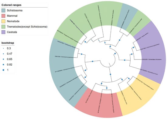
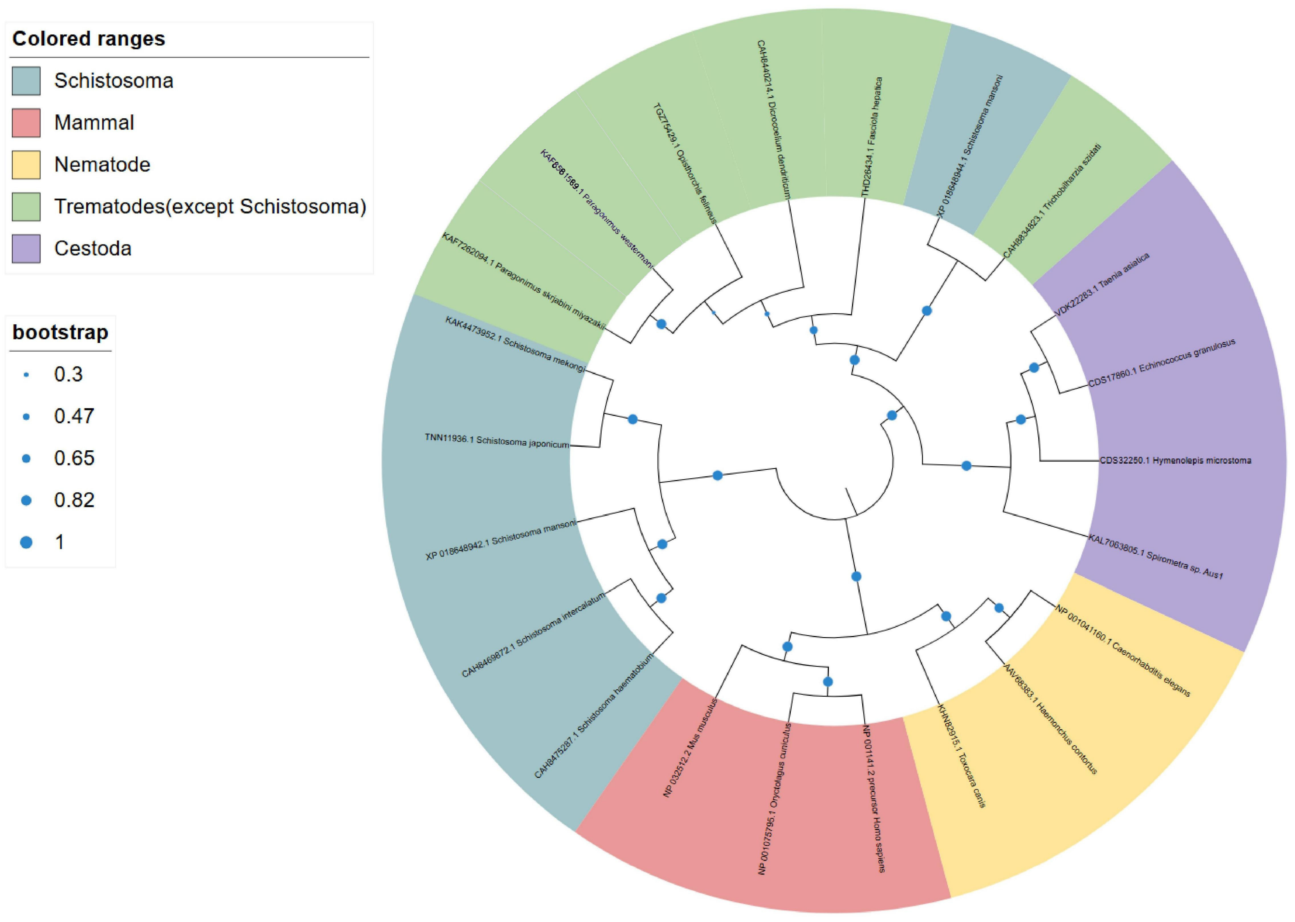
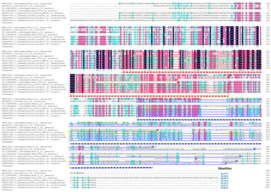
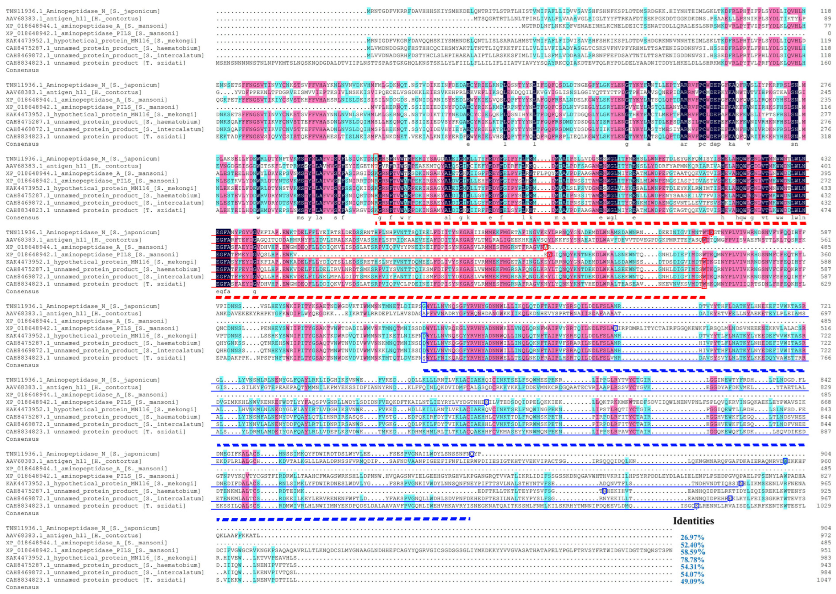
SjAPN is homologous to the H11 antigen of H. contortus, which is one of the active ingredients in a commercialized vaccine in Australia, the UK, and South Africa, called Barbervax [20]. To investigate its evolutionary relationship with homologous proteins from other species, a phylogenetic tree was constructed using SjAPN and related sequences retrieved from the NCBI database (Figure 1). The phylogenetic analysis showed that proteins from the Schistosoma genus formed a distinct clade, indicating a close evolutionary relationship among Schistosoma species. Within this clade, SjAPN clustered most closely with a hypothetical protein from S. mekongi, followed by aminopeptidase PILS from S. mansoni, which was used as the reference protein for the scRNA-seq dataset. Multiple sequence alignment further revealed that SjAPN shared 78.78% sequence identity with the homologous protein from S. haematobium, 58.59% and 54.07% identity with two homologous proteins from S. mansoni, 53.73% with T. szidati, 52.40% with S. intercalatum, 49.09% with S. mekongi, and 26.97% with H. contortus (Figure 2), indicating that SjAPN is highly conserved among trematodes but more divergent from nematode homologs.
Figure 1.
Phylogenetic analysis of SjAPN and its homologous proteins. The phylogenetic tree was constructed using the amino acid sequences of SjAPN and its homologous proteins from Schistosoma, mammals, nematodes, other trematodes, and cestodes. The tree is presented in a circular layout. The colored sectors indicate the taxonomic groups of the homologous proteins: Schistosoma, mammals, nematodes, trematodes excluding Schistosoma, and cestodes. The species names and corresponding protein accession numbers are shown at the branch tips. Bootstrap support values are represented by blue circles at the internal nodes, with larger circles indicating higher bootstrap support. The bootstrap values are shown in the scale bar on the left (0.30–1.00).
Figure 2.
Multiple sequence alignment of SjAPN with homologous proteins. Sequence identities of SjAPN with homologs from S. haematobium, S. mansoni, T. szidati, S. intercalatum, S. mekongi, and H. contortus were 78.78%, 58.59%/ 54.07%, 53.73%, 52.40%, 49.09%, and 26.97%, respectively. The Peptidase_M1 and ERAP1_C domains are indicated by red and blue lines, respectively. The red and blue boxes enclose the amino acid residues corresponding to the N- and C-terminal boundaries of the respective domains. The bold red and blue dashed lines highlight the ranges of the corresponding domains in SjAPN. The domain ranges (amino acid positions; Peptidase_M1/ERAP1_C, respectively) are 320–560/640–902 in SjAPN, 289–534/617–954 in H. contortus, 321–561/641–948 in S. mekongi, 320–560/641–896 in S. haematobium, 320–560/641–947 in S. intercalatum, and 362–602/685–1011 in T. szidati; in S. mansoni, Peptidase_M1 spans residues 279–484 in XP_018648944.1 and 160–295 in XP_018648942.1, while ERAP1_C spans residues 464–589 in XP_018648942.1.
The full-length SjAPN gene is 4319 bp, with an open reading frame of 2715 bp, encoding a protein of 904 amino acids, with a predicted molecular weight of 106.15 kDa and an isoelectric point of 5.48. Conserved domain analysis revealed that SjAPN possesses two characteristic domains of the M1 aminopeptidase family, including the M1_APN-Q_like domain (aa 107–562) and the ERAP1_C domain (aa 640–902) (Supplementary Figure S1A). In addition, domain analysis indicated the presence of active sites and Zn-binding sites, which are typical features of the gluzincin metalloprotease superfamily.
Sequence feature prediction further showed that SjAPN lacks a signal peptide but contains a single transmembrane region at residues 41–63, suggesting that the N-terminal region (1–40) is intracellular, whereas the C-terminal region (64–904) is extracellular (Supplementary Figure S1B). Moreover, multiple potential N-glycosylation sites were predicted in the protein sequence. Hydrophobicity analysis indicated that SjAPN is predominantly hydrophilic.
The 3D structure of SjAPN was predicted using the SWISS-MODEL homology modeling pipeline based on a homologous aminopeptidase template (Supplementary Figure S1C). The predicted model exhibited a QMEANDisCo global score of 0.75 ± 0.05 and a GMQE score of 0.84, indicating a reliable structural model. Ramachandran plot analysis showed that 92.35% of residues were located in the most favored regions, 2.77% in additionally allowed regions, and 2.05% in disallowed regions, further supporting the high quality of the predicted structure.
To explore the potential biological role of SjAPN, the expression profile of its S. mansoni homolog (Smp_247070) was analyzed using publicly available RNA-seq and scRNA-seq datasets (Supplementary Figure S1D). The results showed that SmAPN expression was low in early developmental stages but increased markedly in juvenile and adult worms after 21 dpi, with the highest expression observed in adult female worms at 38 dpi, while expression remained relatively low in males.
Overall, these bioinformatics analyses indicate that SjAPN is a conserved M1 family aminopeptidase with characteristic metalloprotease domains and a reliable predicted structure, and its female-biased expression pattern in late developmental stages suggests a potential role in female maturation and reproduction.
3.2. Expression Patterns of SjAPN
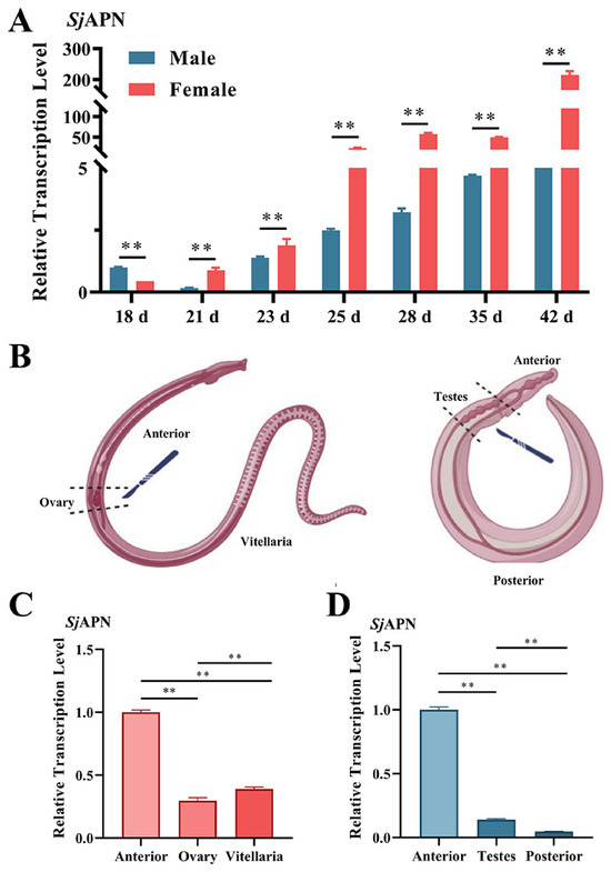
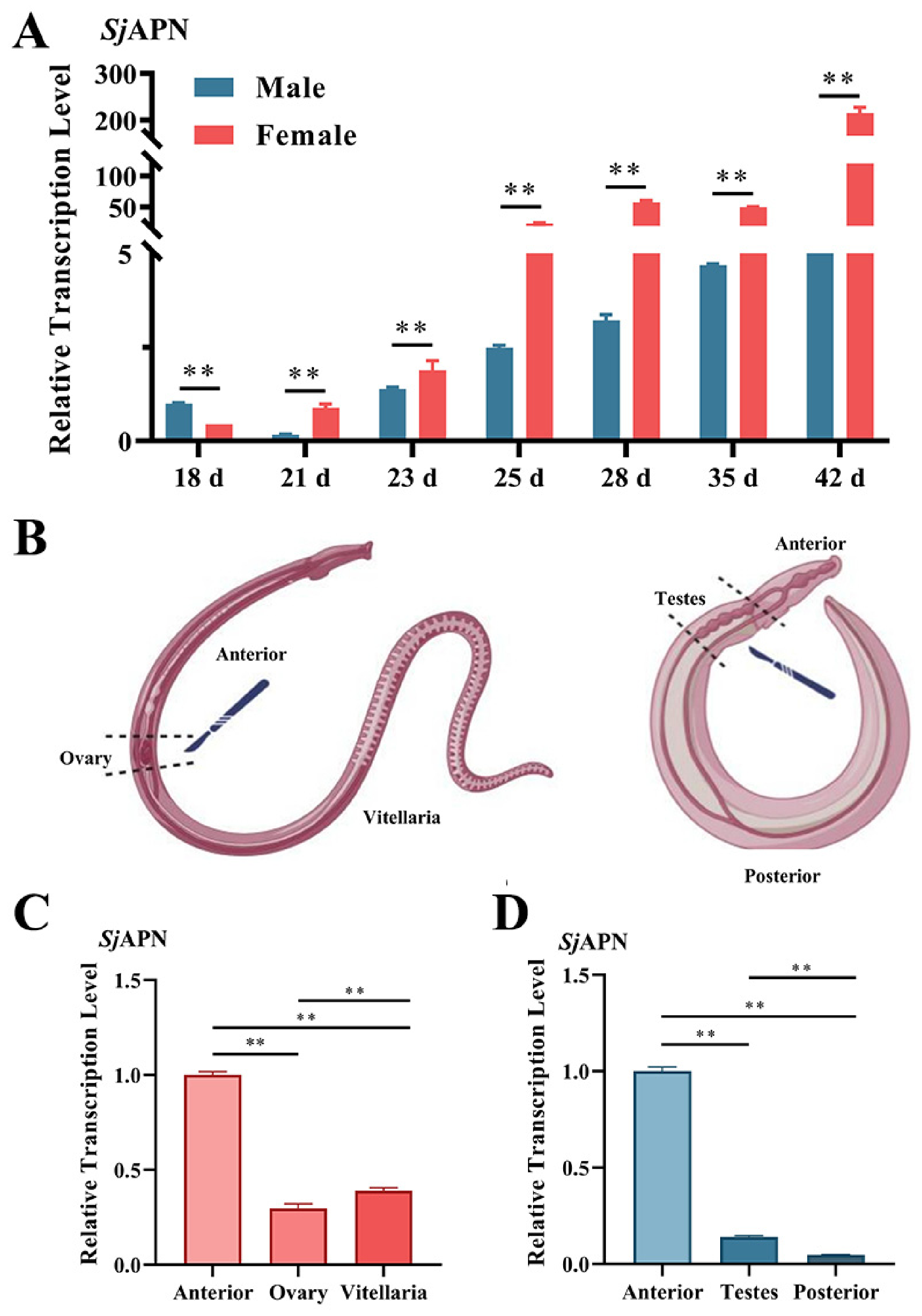
To investigate the transcriptional profile of SjAPN during schistosome development, the relative mRNA expression levels were analyzed in male and female S. japonicum worms at different developmental stages (18–42 dpi) by qPCR. The results showed that SjAPN expression gradually increased as the worms developed, with significantly higher transcription levels observed in female worms compared with males at most developmental stages, particularly from 25 dpi to 42 dpi (Figure 3A).
Figure 3.
Expression patterns of SjAPN. (A) Relative transcription levels of SjAPN in male and female worms at different developmental stages (18–42 dpi) determined by qPCR. Male worms at 18 dpi were used as the common calibrator for both sexes. (B) Schematic diagram showing the anatomical regions dissected for tissue-specific expression analysis. (C) Relative transcription levels of SjAPN in different regions of female worms (anterior, ovary, and vitellaria). The anterior region of female worms was used as the calibrator. (D) Relative transcription levels of SjAPN in different regions of male worms (anterior, testes, and posterior). The anterior region of male worms was used as the calibrator. Values represent fold changes relative to the corresponding calibrator. Statistical differences were assessed using one-way ANOVA followed by Tukey’s multiple-comparison test. Data are presented as mean ± SD; error bars represent SD. Statistical significance is indicated as ** p < 0.01.
To further determine the tissue distribution of SjAPN, worms were dissected into different anatomical regions according to the schematic diagram shown in Figure 3B. In female worms, SjAPN was predominantly expressed in the anterior region, while lower expression levels were detected in the ovary and vitellaria (Figure 3C). Similarly, in male worms, SjAPN transcription was mainly enriched in the anterior region, whereas much lower expression levels were observed in the testes and posterior region (Figure 3D).
Overall, these results indicate that SjAPN exhibits stage-dependent expression and is predominantly expressed in the anterior region of both male and female worms, with notably higher expression in females, suggesting that SjAPN may play an important role in schistosome development, particularly in female reproductive biology.
3.3. SjAPN Transcript Knockdown Is Associated with the Development of Female S. japonicum
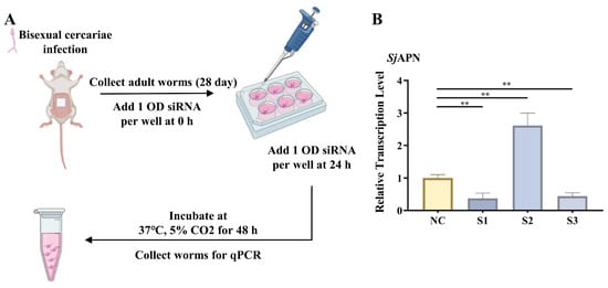
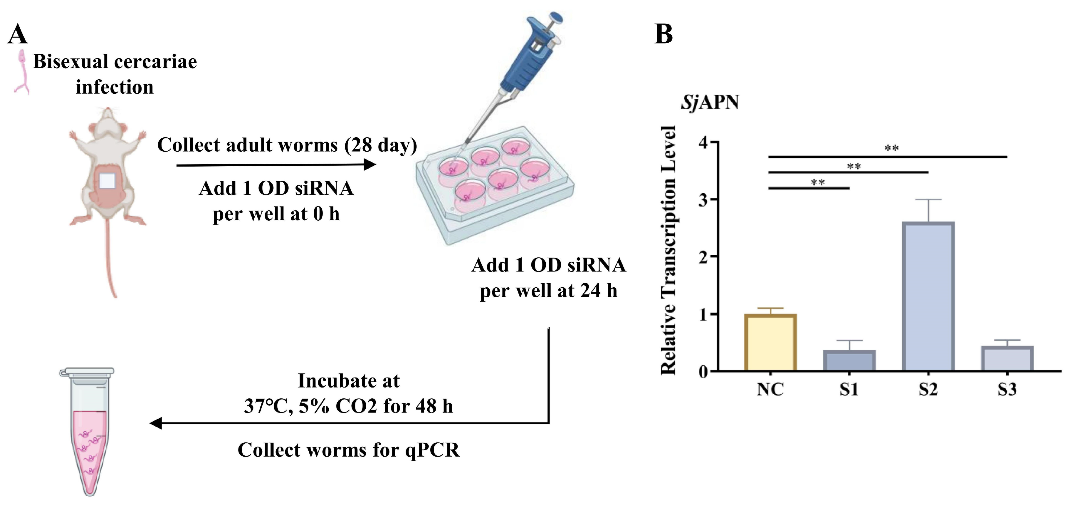
RNAi was performed to investigate the biological function of SjAPN. Three specific siRNAs targeting SjAPN (S1, S2, and S3) were first tested in vitro to determine the most effective sequence. Adult worms were recovered from infected mice at 28 dpi and cultured in six-well plates. SjAPN siRNAs were added at 0 h and 24 h, and worms were incubated for 48 h at 37 °C with 5% CO2 before RNA extraction for qPCR analysis (Figure 4A). The results showed that S1 and S3 significantly reduced SjAPN transcript levels by 62.12% and 33.83%, respectively, compared with the non-targeting NC siRNA group. Among them, S1 exhibited the strongest silencing effect and was therefore selected for subsequent experiments (Figure 4B). In contrast, SjAPN transcript levels were significantly higher in the S2 group than in the NC group.
Figure 4.
Screening of effective siRNAs targeting SjAPN in vitro. (A) Experimental workflow of RNA interference in cultured adult worms. Adult worms were cultured at five worm pairs per well. Each well received 2.5 nmol (approximately 33 μg) of siRNA at 0 and 24 h, corresponding to a cumulative dose of 5.0 nmol (approximately 66 μg) per well. (B) Relative transcription levels of SjAPN after treatment with three different siRNAs (S1, S2, and S3) compared with the NC. Data are presented as mean ± SD from three independent biological replicates (n = 3 wells per group). Statistical differences were assessed using one-way ANOVA followed by Tukey’s multiple-comparison test, with each SjAPN-targeting siRNA group compared with the NC group. Data are presented as mean ± SD; error bars represent SD. Statistical significance is indicated as ** p < 0.01.
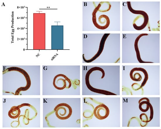
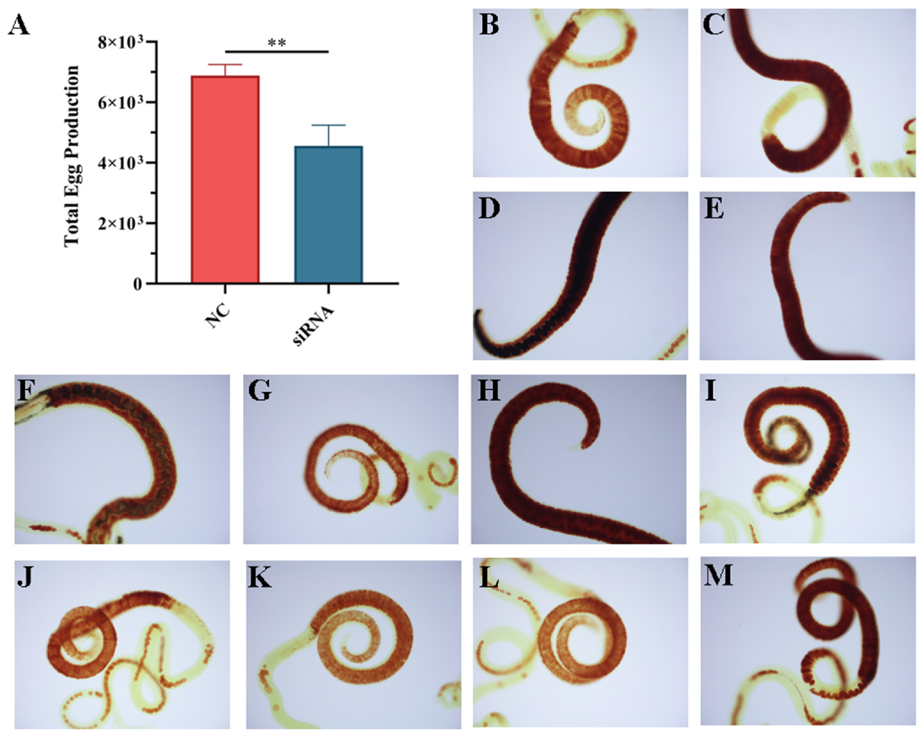
To further investigate the effect associated with SjAPN transcript knockdown on female fecundity, eggs in the culture medium were counted, and vitellocytes were visualized using Fast Blue BB staining. Compared with the NC group, the total egg production in the SjAPN RNAi group was reduced by 33.77% during the 96 h culture (Figure 5A). Moreover, microscopic examination of Fast Blue BB-stained female worms showed apparent vitelline gland abnormalities, defined by markedly reduced staining and discontinuity of the glandular structures, in 71.43% (5/7) of worms in the RNAi group (Figure 5G–M), compared with 20% (1/5) in the NC group (Figure 5B–F).
Figure 5.
Effects of in vitro transcript knockdown of SjAPN on females. (A) Eggs produced by five females per well during 96 h of in vitro culture following SjAPN transcript knockdown (n = 3 wells per group). (B–F) Representative light-microscopic images of females from the NC group after in vitro interference and Fast Blue BB staining. 5 female worms were examined, collected from three independently treated culture wells. (G–M) Representative light-microscopic images of worms after SjAPN siRNA treatment and Fast Blue BB staining. 7 female worms were examined, collected from three independently treated culture wells. Vitelline gland abnormalities were assessed per female worm based on markedly reduced Fast Blue BB staining and discontinuity of the glandular structures. Statistical differences were assessed using an unpaired two-tailed t-test. Data are presented as mean ± SD; error bars represent SD. Statistical significance is indicated as ** p < 0.01.
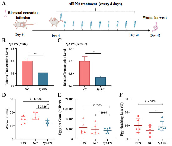
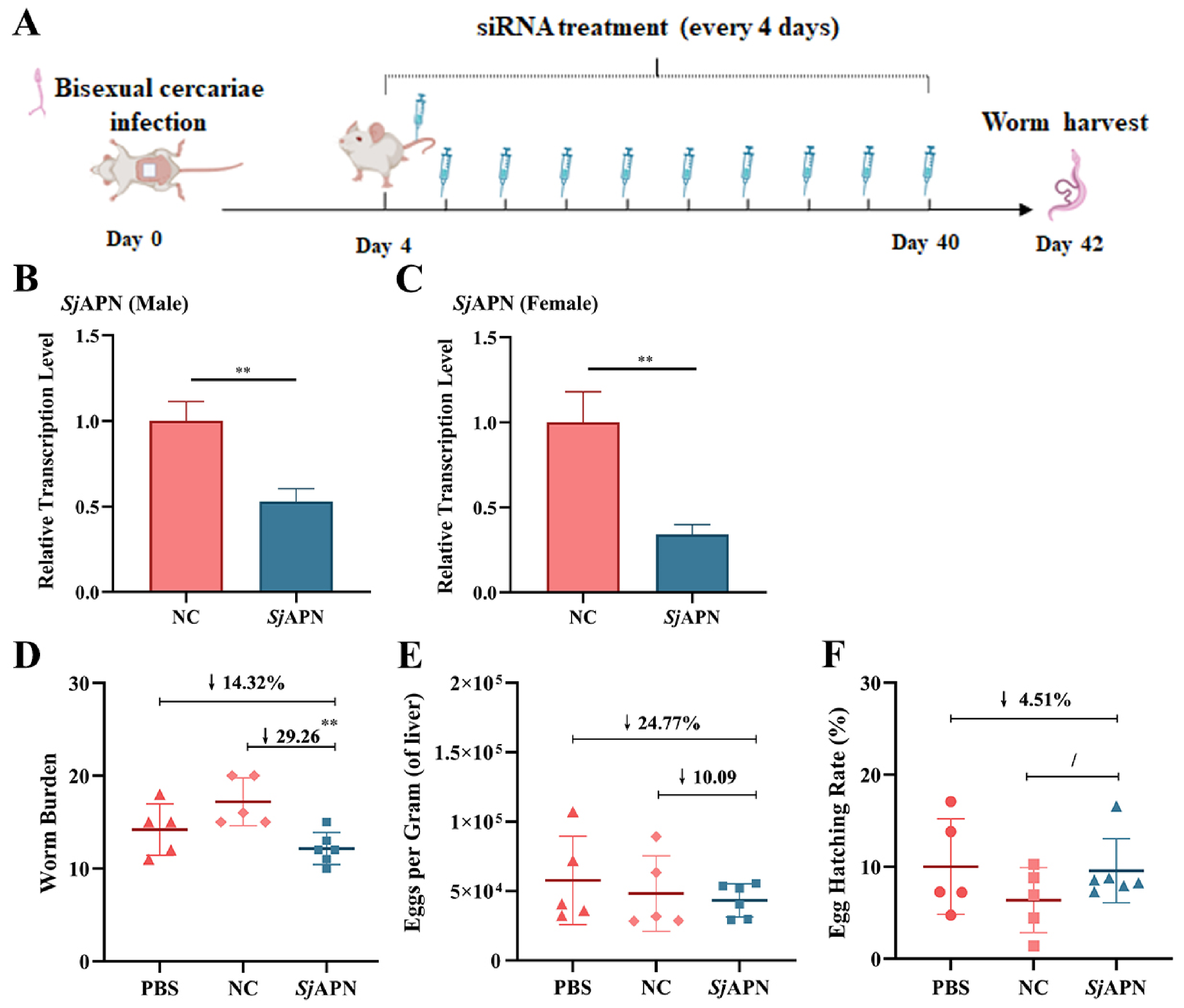
To further evaluate the role of SjAPN in vivo, mice infected with bisexual cercariae were treated with SjAPN S1 siRNA every four days from 4 dpi to 42 dpi, and worms were harvested at 42 dpi (Figure 6A). qPCR analysis showed that SjAPN transcription levels were significantly reduced in both male and female worms in the SjAPN RNAi group compared with the NC group (Figure 6B,C), confirming effective SjAPN transcript knockdown in vivo.
Figure 6.
Effects of SjAPN transcript knockdown in vivo. (A) Experimental design for in vivo RNA interference. (B,C) Relative transcription levels of SjAPN in male (B) and female (C) worms after in vivo RNAi treatment. (D) Worm burden in mice from PBS (n = 5 mice), NC (n = 5 mice), and SjAPN RNAi (n = 6 mice) groups. (E) Number of eggs per gram of liver in infected mice. (F) Egg hatching rate. Statistical differences were assessed using an unpaired two-tailed t-test for panels (B,C), and one-way ANOVA followed by Tukey’s multiple-comparison test for panels (D–F). Data are presented as mean ± SD; error bars represent SD. Statistical significance is indicated as ** p < 0.01. Downward arrows indicate the percentage decrease in the SjAPN RNAi group relative to the PBS or NC group. The “/” symbol between groups indicates a non-significant difference between the indicated groups.
The effects associated with SjAPN transcript knockdown on schistosome development and reproduction were further assessed. Compared with the control groups, worm burden in the SjAPN RNAi group decreased by 29.26% compared with the NC group and by 14.32% compared with the PBS group (Figure 6D). The difference was statistically significant compared with the NC group, but not compared with the PBS group. In addition, egg numbers per gram of liver were reduced by 24.77% and 10.09% compared with the PBS and NC groups, respectively, but these differences were not statistically significant (Figure 6E). Egg-hatching rates also did not differ significantly among the groups (Figure 6F). Thus, the in vivo data do not establish a significant reduction in liver egg burden or egg-hatching rate, and the worm-burden result should be interpreted cautiously.
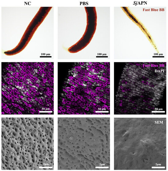
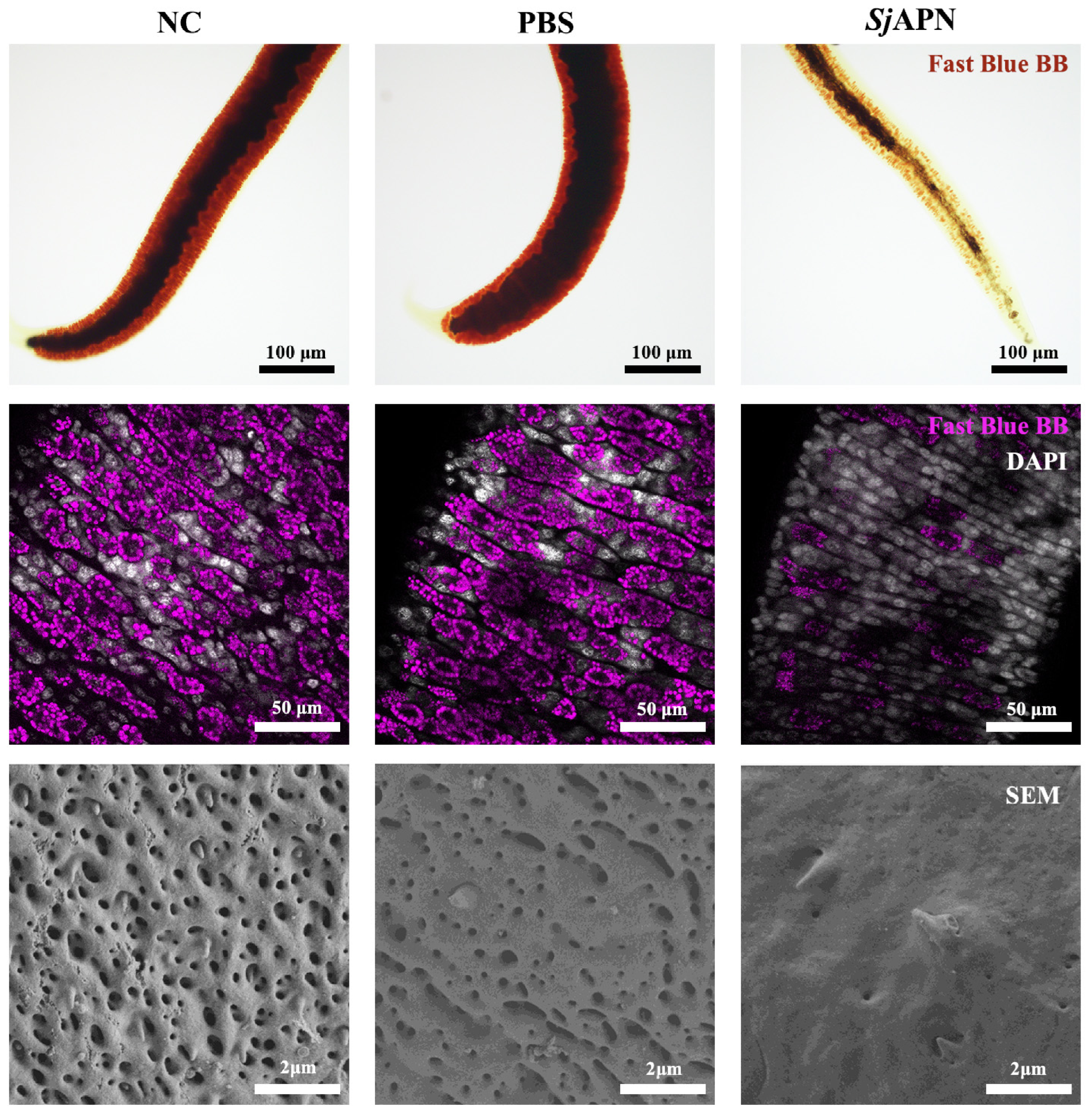
To further investigate the effect of SjAPN knockdown on female reproductive tissues, Fast Blue BB staining was used to visualize vitellocytes. Fast Blue BB staining followed by confocal microscopy revealed that the fluorescence intensity of mature vitellocytes in female worms from the SjAPN RNAi group was markedly reduced compared with the PBS and NC groups, suggesting impaired vitellocyte development (Figure 7).
Figure 7.
Morphological changes in female worms associated with SjAPN transcript knockdown. Representative images showing morphological alterations in female worms after SjAPN RNAi treatment. Upper panels: Bright-field images of worms stained with Fast Blue BB to visualize vitelline cells. Middle panels: Confocal microscopy images showing vitellocyte distribution (Fast Blue BB staining) with nuclei stained by DAPI. Lower panels: SEM images showing tegumental surface structures. Scale bars: 100 μm (upper panels), 50 μm (middle panels), and 2 μm (SEM). Five female worms per group were examined by fluorescence microscopy. Three female worms per group were then selected for SEM based on an unobstructed view of the vitelline glands and a relatively extended body posture. SEM images are representative of three female worms examined per group; three male worms per group were also examined but are not shown because no obvious morphological changes were observed in the male worms.
SEM was also used to examine tegumental morphology. In the control groups, the worm tegument displayed a continuous and well-organized surface structure with abundant spines and pits. In contrast, female worms in the SjAPN RNAi group exhibited a smoother tegument surface with fewer spines and reduced pitted structures, particularly in the anterior region (Figure 7).
Overall, these results demonstrate that SjAPN plays an important role in the development and reproductive biology of S. japonicum, particularly in female worms, and its silencing leads to reduced worm burden, decreased egg production, and impaired vitellocyte maturation.
4. Discussion
Previous studies have shown that APN in several parasites is closely linked to development, and nutrient acquisition, suggesting that it may be a promising antiparasitic target. For example, inhibition of PfA-M1 in Plasmodium falciparum disrupts hemoglobin utilization and parasite development, leading to reduced proliferation, developmental arrest, and a lower parasite burden [17]. EtAPN1 in Eimeria tenella is involved in oocyst maturation and sporulation, and its suppression has marked effects on schizogony, development, and progeny sporozoite formation [18]. TgAPN2, which is homologous to PfA-M1, is likely involved in metabolism and development in Toxoplasma gondii [19]. In H. contortus, APN, also known as the H11 antigen, has already been used as a component of a commercial vaccine [20]. Given that schistosomes and H. contortus are both blood-feeding parasites, SjAPN may also be involved in intestinal digestion or other host–parasite interaction processes and therefore has potential as a vaccine candidate or drug target.
Bioinformatic analysis in this study indicated that SjAPN contains the typical structural features of the M1 aminopeptidase family, supporting its predicted or putative peptide-hydrolyzing function. Consistent with this, analysis of publicly available transcriptomic and single-cell transcriptomic data for the homologous protein Aminopeptidase PILS in S. mansoni showed relatively high expression in female worms, especially in late-stage females. This is broadly consistent with our qPCR results in S. japonicum, which showed that SjAPN was expressed at higher levels in females than in males at both 28 dpi and 42 dpi and was also relatively enriched in the anterior region of both sexes. Since the anterior region of schistosomes contains feeding structures such as the oral sucker and pharynx [42] and is closely associated with anterior gut tissue, the higher expression of SjAPN in this region suggests a possible role in nutrient uptake, foregut digestion, or anterior surface function. The stronger expression in females also indicates that SjAPN may be associated not only with basic metabolism but also with female development and reproduction.
To further clarify its biological role, we used RNAi to suppress SjAPN expression. In vitro screening identified S1 siRNA as the most effective construct, and it was therefore used in the subsequent in vivo experiment. However, S2 siRNA unexpectedly increased SjAPN expression; the reason for this increase remains unclear, but it may reflect an off-target effect or a compensatory response. Further experiments are needed to examine these possibilities. SjAPN transcript knockdown was associated with reduced egg output, suggesting a potential role for SjAPN in schistosome reproduction. However, because SjAPN protein abundance and enzymatic activity were not measured, these data do not establish a direct protein-level or enzymatic mechanism. In vivo, worm burden was significantly lower in the knockdown group than in the NC group, although the difference relative to the PBS group was not significant. Liver egg burden also tended to be lower, whereas egg-hatching rates were comparable among groups. Together, these findings support further investigation of SjAPN’s role in schistosome development and reproduction, while the in vivo effects require confirmation. The limited effect on egg reduction and hatching may be due to incomplete silencing in vivo, partial functional compensation by related molecules, or variation caused by sample size and individual differences. Further validation with improved silencing efficiency or additional functional assays will be needed. In particular, an independent repeat is needed to determine whether the observed reduction in worm burden is reproducible and biologically meaningful.
Morphological observations provided additional support for these findings. Fast Blue BB staining showed weakened yolk droplet signals in mature vitelline cells of female worms in the SjAPN interference group, suggesting that SjAPN may be involved in vitelline gland development or vitelline cell maturation. These qualitative observations, although based on a limited number of worms, suggest possible alterations in vitellocyte maturation that warrant further investigation. SEM further showed that the anterior tegument of female worms in the interference group was smoother, with fewer spines and reduced pit-like structures, whereas no obvious changes were observed in males. This phenotype is consistent with the higher expression of SjAPN in female worms and in the anterior region and suggests that SjAPN may contribute to the maintenance of the anterior surface and possibly to female reproductive-associated functions. Nevertheless, although qPCR confirmed a reduction in SjAPN transcript levels, changes in SjAPN protein abundance, protein turnover, and enzymatic activity were not evaluated in the present study. Therefore, the effects on egg production, vitellocyte morphology, and tegumental structure are interpreted as phenotypic outcomes associated with SjAPN transcript knockdown, rather than as direct evidence of reduced SjAPN protein function. In the long-term interference experiment, the reduction in liver egg burden was not statistically significant. These findings suggest that the gene may primarily be involved in the morphological development or cellular differentiation of the vitelline gland in S. japonicum, rather than exerting a direct and essential role in egg formation or embryonic development. Although the observed alterations in vitelline cells after siRNA-mediated transcript knockdown, the lack of significant changes in egg production and hatching rate indicates that the residual function of the gland may still have been sufficient to support normal reproductive output. In addition, the schistosome may possess certain compensatory mechanisms that help maintain relatively stable egg production and developmental competence despite defects in vitelline gland development. It is also possible that the limited number of experimental animals reduced the statistical power of the analysis, thereby preventing these differences from reaching significance.
From a vaccine perspective, gut- and surface-associated molecules of blood-feeding parasites often include so-called hidden antigens. Unlike exposed antigens, these molecules are not readily accessible to the host immune system during natural infection and therefore are under weaker immune selection pressure [43]. At the same time, they may become exposed during feeding and nutrient uptake, making them attractive vaccine targets or drug targets. Hidden antigens have already been shown to induce protective immunity in blood-feeding parasites such as ticks [44], H. contortus, and hookworms [45]. The H11 antigen of H. contortus has been extensively studied and confirmed as a hidden antigen localized to the parasite digestive tract. As a homolog of H11, if SjAPN is a membrane-associated or surface-related protein involved in intestinal digestion or blood feeding, its potential hidden-antigen properties are worth further investigation.
Overall, our data suggest that SjAPN is biologically significant in S. japonicum. Its preferential expression in female worms and anterior regions, together with the phenotypic changes observed after RNAi-mediated transcript knockdown, including effects on worm recovery, female surface structure, and vitelline development, indicate that SjAPN may participate in nutrient utilization, development, and reproduction. These findings highlight the value of further investigating its potential as a vaccine candidate or drug target for schistosomiasis control.
Supplementary Materials
The following supporting information can be downloaded at https://www.mdpi.com/article/10.3390/tropicalmed11110299/s1, Figure S1: Bioinformatics analysis of SjAPN; Table S1: siRNAs used for RNA interference experiments.
Author Contributions
Conceptualization, D.Z.; methodology, D.Z., Y.S., J.X., R.Z. and L.G.; software, D.Z.; validation, D.Z.; formal analysis, H.L., K.L. and L.G.; investigation, Z.F. and Y.J.; resources, Y.J.; data curation, D.Z.; writing—original draft preparation, D.Z.; writing—review and editing, H.Z. and Y.J.; visualization, D.Z.; supervision, H.Z.; project administration, Y.J.; funding acquisition, H.Z. and Y.J. All authors have read and agreed to the published version of the manuscript.
Funding
This work was supported by the Central Public-interest Scientific Institution Basal Research Fund (no.2026JB16) and the Agricultural Science and Technology Innovation Program (ASTIP) (no. CAAS-ASTIP-2026-SHVRI). The funders had no role in study design, data collection and analysis, decision to publish, or preparation of the manuscript.
Institutional Review Board Statement
All animal procedures were conducted in accordance with the regulations of the Committee for the Care and Use of Laboratory Animals at the Shanghai Veterinary Research Institute, Shanghai, China (permit no. SYXK-20160010). The experimental protocol was reviewed and approved by the Ethics and Animal Welfare Committee of the Shanghai Veterinary Research Institute, Shanghai, China (approval no. SV-20240712-Y01) on 12 July 2024.
Informed Consent Statement
Not applicable.
Data Availability Statement
The datasets presented in this study can be found in online repositories. The names of the repository/repositories and accession number(s) can be found in the article. Further inquiries can be directed to the corresponding author.
Acknowledgments
We sincerely thank our colleagues in the Schistosomiasis Department of the Shanghai Veterinary Research Institute for their kind assistance in challenge infection, sacrifice, and worm perfusion.
Conflicts of Interest
The authors declare no conflicts of interest.
Abbreviations
The following abbreviations are used in this manuscript:
| APN | Aminopeptidase N |
| RNAi | RNA interference |
| SPF | Specific pathogen-free |
| 3D | 3-dimensional |
| MM | Mated male |
| MF | Mated female |
| DMEM | Dulbecco’s Modified Eagle Medium |
| FBS | Fetal Bovine Serum |
| DEPC | Diethyl pyrocarbonate |
| PBS | Phosphate-Buffered Saline |
| NC | Negative control |
| dpi | day post infection |
References
- Barsoum, R.S.; Esmat, G.; El-Baz, T. Human schistosomiasis: Clinical perspective: Review. J. Adv. Res. 2013, 4, 433–444. [Google Scholar] [CrossRef] [Scilit]
- Song, L.J.; Yin, X.R.; Mu, S.S.; Li, J.H.; Gao, H.; Zhang, Y.; Dong, P.P.; Mei, C.J.; Hua, Z.C. The Differential and Dynamic Progression of Hepatic Inflammation and Immune Responses During Liver Fibrosis Induced by Schistosoma japonicum or Carbon Tetrachloride in Mice. Front. Immunol. 2020, 11, 570524. [Google Scholar] [CrossRef] [Scilit]
- He, L.; Zhao, C.; Lu, Y.; Wei, H.; Deng, Z.; Zhang, Y.; Yin, T. Pancreatic Schistosomiasis, China, 2020–2024. Emerg. Infect. Dis. 2025, 31, 2308–2311. [Google Scholar] [CrossRef] [Scilit]
- Osakunor, D.N.M.; Alonso, S.; Jumbe, S.; Lamberton, P.H.L. Unravelling the true biopsychosocial impact of schistosomiasis. Trends Parasitol. 2026, 42, 408–425. [Google Scholar] [CrossRef] [Scilit]
- Dong, B.; Hou, Z.; Lu, K. Schistosomiasis trends and control efforts: A global perspective from 1990 to 2050. Eur. J. Clin. Microbiol. Infect. Dis. 2025, 44, 1219–1230. [Google Scholar] [CrossRef] [Scilit]
- Li, Q.; Li, Y.L.; Guo, S.Y.; Li, S.Z.; Wang, Q.; Lin, W.N.; Zhang, L.J.; Li, S.Z.; Zhou, X.N.; Xu, J. Global trends of schistosomiasis burden from 1990 to 2021 across 204 countries and territories: Findings from GBD 2021 study. Acta Trop. 2025, 261, 107504. [Google Scholar] [CrossRef] [Scilit]
- He, J.; Zhang, L.; Guo, S.; Li, S.; Li, Y.; Yang, F.; Cao, C.; Xu, J.; Li, S. Progress of schistosomiasis control in the People’s Republic of China in 2025. Chin. J. Schistosomiasis Control. 2026, 38, 223–231. [Google Scholar] [CrossRef] [Scilit]
- Yang, X.; Zhang, Y.; Sun, Q.X.; Zhou, J.X.; Zhou, X.N. SWOT analysis on snail control measures applied in the national schistosomiasis control programme in the People’s Republic of China. Infect. Dis. Poverty 2019, 8, 13. [Google Scholar] [CrossRef] [Scilit]
- Sanogo, B.; Echoru, I.; Jones, C.; Butala, C.; Lin, D.; Maiga, H.; Samano-Sanchez, H.; Sacko, M.; Kassegne, K.; Welburn, S.C. A reservoir at the gates: Nonhuman mammalian hosts for human schistosomiasis in western Africa and the critical challenge for elimination. Infect. Dis. Poverty 2026, 15, 6. [Google Scholar] [CrossRef] [Scilit]
- Hsu, S.Y.; Hsu, H.F. New approach to immunization against Schistosoma japonicum. Science 1961, 133, 766. [Google Scholar] [CrossRef] [Scilit]
- Nono, J.K.; Bitye Zambo, B.M.; Kameni Poumeni, M.P.; Soh Bayeck, E.B.; Meyo Kamguia, L.M.; Noubissi Fotseu, M.M.; Owona Ayissi, V.B.; Spangenberg, T.; Demarta-Gatsi, C. Mass drug administration of praziquantel lowers the susceptibility of school-aged children to Schistosoma mansoni in endemic areas. Front. Immunol. 2026, 17, 1746003. [Google Scholar] [CrossRef] [Scilit]
- Melman, S.D.; Steinauer, M.L.; Cunningham, C.; Kubatko, L.S.; Mwangi, I.N.; Wynn, N.B.; Mutuku, M.W.; Karanja, D.M.S.; Colley, D.G.; Black, C.L.; et al. Reduced Susceptibility to Praziquantel among Naturally Occurring Kenyan Isolates of Schistosoma mansoni. PLoS Negl. Trop. Dis. 2009, 3, e504. [Google Scholar] [CrossRef] [Scilit]
- Wang, W.; Wang, L.; Liang, Y.S. Susceptibility or resistance of praziquantel in human schistosomiasis: A review. Parasitol. Res. 2012, 111, 1871–1877. [Google Scholar] [CrossRef] [Scilit]
- Mina-Osorio, P. The moonlighting enzyme CD13: Old and new functions to target. Trends Mol. Med. 2008, 14, 361–371. [Google Scholar] [CrossRef] [Scilit]
- Piedfer, M.; Dauzonne, D.; Tang, R.; N’Guyen, J.; Billard, C.; Bauvois, B. Aminopeptidase-N/CD13 is a potential proapoptotic target in human myeloid tumor cells. FASEB J. 2011, 25, 2831–2842. [Google Scholar] [CrossRef] [Scilit]
- Enyedi, K.N.; Tóth, S.; Szakács, G.; Mező, G. NGR-peptide-drug conjugates with dual targeting properties. PLoS ONE 2017, 12, e0178632. [Google Scholar] [CrossRef] [Scilit]
- Bounaadja, L.; Schmitt, M.; Albrecht, S.; Mouray, E.; Tarnus, C.; Florent, I. Selective inhibition of PfA-M1, over PfA-M17, by an amino-benzosuberone derivative blocks malaria parasites development in vitro and in vivo. Malar. J. 2017, 16, 382. [Google Scholar] [CrossRef] [Scilit]
- Gras, S.; Byzia, A.; Gilbert, F.B.; McGowan, S.; Drag, M.; Silvestre, A.; Niepceron, A.; Lecaille, F.; Lalmanach, G.; Brossier, F. Aminopeptidase N1 (EtAPN1), an M1 metalloprotease of the apicomplexan parasite Eimeria tenella, participates in parasite development. Eukaryot. Cell 2014, 13, 884–895. [Google Scholar] [CrossRef] [Scilit]
- Li, Q.; Jia, H.; Cao, S.; Zhang, Z.; Zheng, J.; Zhang, Y. Biochemical characterization of aminopeptidase N2 from Toxoplasma gondii. J. Vet. Med. Sci. 2017, 79, 1404–1411. [Google Scholar] [CrossRef] [Scilit][Green Version]
- Kebeta, M.M.; Hine, B.C.; Walkden-Brown, S.W.; Kahn, L.P.; Doyle, E.K. Evaluation of Barbervax(R) vaccination for lambing Merino ewes. Vet. Parasitol. 2020, 283, 109187. [Google Scholar] [CrossRef] [Scilit]
- Zhong, H.; Dong, B.; Zhu, D.; Fu, Z.; Liu, J.; Jin, Y. Sja-let-7 suppresses the development of liver fibrosis via Schistosoma japonicum extracellular vesicles. PLoS Pathog. 2024, 20, e1012153. [Google Scholar] [CrossRef] [Scilit]
- Madej, T.; Lanczycki, C.J.; Zhang, D.; Thiessen, P.A.; Geer, R.C.; Marchler-Bauer, A.; Bryant, S.H. MMDB and VAST+: Tracking structural similarities between macromolecular complexes. Nucleic Acids Res. 2014, 42, D297–D303. [Google Scholar] [CrossRef] [Scilit]
- Walker, J.M. The Proteomics Protocols Handbook; Humana Press: Totowa, NJ, USA, 2005. [Google Scholar] [CrossRef] [Scilit]
- Möller, S.; Croning, M.D.; Apweiler, R. Evaluation of methods for the prediction of membrane spanning regions. Bioinformatics 2001, 17, 646–653. [Google Scholar] [CrossRef] [Scilit]
- Nielsen, H.; Teufel, F.; Brunak, S.; von Heijne, G. SignalP: The Evolution of a Web Server. In Methods in Molecular Biology; Springer: New York, NY, USA, 2024; Volume 2836, pp. 331–367. [Google Scholar] [CrossRef] [Scilit]
- Gupta, R.; Brunak, S. Prediction of glycosylation across the human proteome and the correlation to protein function. Biocomputing 2002, 7, 310–322. [Google Scholar] [CrossRef] [Scilit]
- Davis, M.W.; Jorgensen, E.M. ApE, A Plasmid Editor: A Freely Available DNA Manipulation and Visualization Program. Front. Bioinform. 2022, 2, 818619. [Google Scholar] [CrossRef] [Scilit]
- Tamura, K.; Stecher, G.; Kumar, S. MEGA11: Molecular Evolutionary Genetics Analysis Version 11. Mol. Biol. Evol. 2021, 38, 3022–3027. [Google Scholar] [CrossRef] [Scilit]
- Bienert, S.; Waterhouse, A.; de Beer, T.A.; Tauriello, G.; Studer, G.; Bordoli, L.; Schwede, T. The SWISS-MODEL Repository-new features and functionality. Nucleic Acids Res. 2017, 45, D313–D319. [Google Scholar] [CrossRef] [Scilit]
- Ramachandran, G.N.; Ramakrishnan, C.; Sasisekharan, V. Stereochemistry of polypeptide chain configurations. J. Mol. Biol. 1963, 7, 95–99. [Google Scholar] [CrossRef] [Scilit]
- Studer, G.; Rempfer, C.; Waterhouse, A.M.; Gumienny, R.; Haas, J.; Schwede, T. QMEANDisCo-distance constraints applied on model quality estimation. Bioinformatics 2020, 36, 1765–1771. [Google Scholar] [CrossRef] [Scilit]
- Waterhouse, A.; Bertoni, M.; Bienert, S.; Studer, G.; Tauriello, G.; Gumienny, R.; Heer, F.T.; de Beer, T.A.P.; Rempfer, C.; Bordoli, L.; et al. SWISS-MODEL: Homology modelling of protein structures and complexes. Nucleic Acids Res. 2018, 46, W296–W303. [Google Scholar] [CrossRef] [Scilit]
- Howe, K.L.; Bolt, B.J.; Shafie, M.; Kersey, P.; Berriman, M. WormBase ParaSite—A comprehensive resource for helminth genomics. Mol. Biochem. Parasitol. 2017, 215, 2–10. [Google Scholar] [CrossRef] [Scilit]
- Lu, Z.; Zhang, Y.; Berriman, M. A web portal for gene expression across all life stages of Schistosoma mansoni. BioRxiv 2018, 308213. [Google Scholar] [CrossRef] [Scilit]
- Wendt, G.; Zhao, L.; Chen, R.; Liu, C.; O’Donoghue, A.J.; Caffrey, C.R.; Reese, M.L.; Collins, J.J., 3rd. A single-cell RNA-seq atlas of Schistosoma mansoni identifies a key regulator of blood feeding. Science 2020, 369, 1644–1649. [Google Scholar] [CrossRef] [Scilit]
- Rio, D.C.; Ares, M.; Hannon, G.J.; Nilsen, T.W. Purification of RNA using TRIzol (TRI reagent). Cold Spring Harb. Protoc. 2010, 2010, pdb.prot5439. [Google Scholar] [CrossRef] [Scilit]
- Liu, S.; Cai, P.; Hou, N.; Piao, X.; Wang, H.; Hung, T.; Chen, Q. Genome-wide identification and characterization of a panel of house-keeping genes in Schistosoma japonicum. Mol. Biochem. Parasitol. 2012, 182, 75–82. [Google Scholar] [CrossRef] [Scilit]
- Livak, K.J.; Schmittgen, T.D. Analysis of relative gene expression data using real-time quantitative PCR and the 2(-Delta Delta C(T)) Method. Methods 2001, 25, 402–408. [Google Scholar] [CrossRef] [Scilit]
- Zhong, H.; Hou, L.; Qin, F.; Ren, Y.; Dong, B.; Zhu, D.; Li, H.; Lu, K.; Fu, Z.; Liu, J.; et al. Molecular and functional characterization of Schistosoma japonicum annexin A13. Vet. Res. 2023, 54, 116. [Google Scholar] [CrossRef] [Scilit]
- Wang, J.; Chen, R.; Collins, J.J., 3rd. Systematically improved in vitro culture conditions reveal new insights into the reproductive biology of the human parasite Schistosoma mansoni. PLoS Biol. 2019, 17, e3000254. [Google Scholar] [CrossRef] [Scilit]
- Wang, C.; Hou, X.; Zhang, P.; Liu, C.; Yang, Y.; Zhao, Z.; Jiang, W.; Su, G.; Jiang, X.; Lin, L.; et al. Prevalence of Kinesiophobia and Associated Factors Among Patients After Percutaneous Coronary Intervention: A Multicenter Cross-Sectional Study. Nurs. Open 2026, 13, e70748. [Google Scholar] [CrossRef] [Scilit]
- Li, X.H.; Stark, M.; Vance, G.M.; Cao, J.P.; Wilson, R.A. The anterior esophageal region of Schistosoma japonicum is a secretory organ. Parasites Vectors 2014, 7, 565. [Google Scholar] [CrossRef] [Scilit]
- Antunes, S.; Domingos, A. Tick Vaccines and Concealed versus Exposed Antigens. Pathogens 2023, 12, 374. [Google Scholar] [CrossRef] [Scilit]
- Mendoza-Martínez, N.; Alonso-Díaz, M.; Merino-Charrez, J.O.; Lagunes-Quintanilla, R. Exploring the Combined Effect of Bm86 and Subolesin Polypeptide Vaccines in Cattle Naturally Infested with Rhipicephalus microplus. Vet. Sci. 2026, 13, 301. [Google Scholar] [CrossRef] [Scilit]
- Hotez, P.J.; Bethony, J.M.; Diemert, D.J.; Pearson, M.; Loukas, A. Developing vaccines to combat hookworm infection and intestinal schistosomiasis. Nat. Rev. Microbiol. 2010, 8, 814–826. [Google Scholar] [CrossRef] [Scilit]

Disclaimer/Publisher’s Note: The statements, opinions and data contained in all publications are solely those of the individual author(s) and contributor(s) and not of MDPI and/or the editor(s). MDPI and/or the editor(s) disclaim responsibility for any injury to people or property resulting from any ideas, methods, instructions or products referred to in the content. |
© 2026 by the authors. Licensee MDPI, Basel, Switzerland. This article is an open access article distributed under the terms and conditions of the Creative Commons Attribution (CC BY) license.