


Abstract
Background: Acute myeloid leukemia (AML) is biologically heterogeneous and associated with poor outcomes. Although oxidative stress contributes to AML pathogenesis, the prognostic value of related biomarkers remains uncertain. Objective: This study aimed to evaluate associations between oxidative stress-related biomarkers and prognosis in adults with AML, focusing on overall survival (OS), complete remission (CR), relapse, relapse-free survival (RFS), event-free survival (EFS), and early mortality. Methods: This PRISMA-compliant systematic review was registered in PROSPERO (CRD420261364956). MEDLINE/PubMed, Scopus, Cochrane Library, Science Citation Index, ClinicalTrials.gov, and WHO ICTRP were searched from January 2015 through 31 July 2026. Eligible studies included adults with AML, oxidative stress-related biomarkers measured in biological samples, and extractable prognostic data. Risk of bias was assessed using QUIPS and certainty of evidence using GRADE. Results: Thirteen of 537 records met the inclusion criteria; 203 supplementary reports yielded no additional studies. Investigated biomarkers included ROS-related phenotypes, antioxidant and redox-regulatory genes, glutathione metabolism, iron/inflammation-related markers, and oxidative DNA damage indicators. Adverse outcomes were associated with ROS-related phenotypes, combined ROS/aldehyde dehydrogenase activity, GPX3, GSTP1, ferritin, gamma-glutamyl transpeptidase-to-albumin ratio, SOD1, and glutathione-related metabolic profiles. Conclusions: These biomarkers may have prognostic value, particularly for OS, but heterogeneity limits clinical application. Standardized validation and integration into applicable risk models are required.
1. Introduction
Acute myeloid leukaemia (AML) is a biologically heterogeneous hematologic malignancy, accounting for about 1% of all cancers and characterized by marked variability in genetic alterations and clinical presentation [1,2]. AML remains a major clinical burden, with an estimated 22,720 new cases and 11,500 deaths expected in the United States in 2026, and most cases occurring in adults with a median age at diagnosis of approximately 69 years [2]. Despite advances in molecular classification and treatment, overall survival remains poor, with a reported five-year relative survival rate of approximately 31.9% [3]. This limited survival is largely attributed to biological and clinical heterogeneity, which complicates diagnosis, risk stratification, and treatment selection [4].
Among mechanisms contributing to this heterogeneity, oxidative stress has emerged as an important factor in AML pathophysiology. Reactive oxygen species (ROS) are increased in leukemic cells compared with normal counterparts and participate in proliferation, apoptosis, and therapy resistance [5]. However, existing evidence is heterogeneous in study design, biomarker measurement, and outcome reporting. Therefore, this systematic review evaluates the prognostic value of oxidative stress-related biomarkers in AML, focusing on studies published from 2015 onwards to reflect recent advances in disease classification and biomarker assessment.
1.1. AML Definition, Classification, Prognosis and Risk Factors
Acute myeloid leukemia (AML) is a biologically complex hematopoietic malignancy defined by clonal proliferation of myeloid precursor cells in the bone marrow and peripheral blood [6,7]. Despite intensive chemotherapy and hematopoietic stem cell transplantation (HSCT), relapse remains frequent and long-term outcomes remain unfavorable because of disease heterogeneity, treatment resistance, and therapy-related toxicity [7,8]. The latest World Health Organization (WHO) classification defines AML through an integrated clinical, morphologic, and molecular genetic approach, dividing cases into genetically defined entities and entities classified according to differentiation. In several genetically defined subtypes, the traditional ≥20% blast threshold is no longer uniformly required, while updated categories such as AML with myelodysplasia-related features (AML-MR) highlight the growing role of molecular features in classification [9]. Prognostic stratification is primarily based on cytogenetic and molecular abnormalities according to the 2022 European LeukemiaNet (ELN) recommendations, which classify patients into favorable, intermediate, and adverse risk groups and guide risk-adapted therapy [10].
Prognosis is also influenced by age, immune status, treatment response, and measurable residual disease (MRD) [10,11]. At the biological level, AML arises from hematopoietic stem and progenitor cells and is characterized by expansion of immature myeloid cells with impaired differentiation. Relapses are frequently linked to measurable residual disease and therapy-resistant leukemic clones, including leukemic stem cells (LSCs), which remain quiescent, resist chemotherapy, and re-initiate leukemic proliferation [12,13]. Although current prognostic models have improved risk stratification, variability persists even within the same risk category, indicating the need for additional biomarkers [14]. Established risk factors include advanced age, male sex, prior chemotherapy or radiotherapy, antecedent hematologic disorders, environmental or occupational exposures such as benzene and tobacco smoke, and inherited genetic susceptibility [2,15]. Together, the biological heterogeneity and clinical complexity of AML support the need for more robust prognostic biomarkers to refine risk assessment and personalized therapy.
1.2. Role of Oxidative Stress in AML Pathogenesis
Oxidative stress reflects an imbalance between ROS generation and antioxidant defence mechanisms, leading to accumulation of reactive molecules that can cause cellular damage, inflammation, and disease progression [16]. Antioxidant defences include non-enzymatic components, such as vitamins A, C, and E and glutathione, and enzymatic systems, including superoxide dismutase (SOD), catalase (CAT), glutathione peroxidase (GPx), and glutathione reductase (GR), which maintain redox homeostasis [17]. ROS arise mainly from mitochondrial electron transport chain activity, but enzymatic sources such as cytochrome P450 monooxygenases, xanthine oxidoreductase, uncoupled nitric oxide synthase, cyclooxygenases, and NADPH oxidases also contribute. Exogenous sources, including ionizing radiation and chemical agents, may further increase oxidative stress [18,19]. ROS act as regulators of signaling pathways involved in proliferation and differentiation; however, excessive ROS can disrupt redox homeostasis, promote genomic instability, and alter critical cellular processes [20].
In AML, dysregulated ROS can impair myeloid differentiation through redox-sensitive transcription factors such as NF-κB, AP-1, and HIF-1α [21]. Recurrent mutations in genes such as FLT3, KIT, RAS, and IDH1/2 may enhance ROS production and promote oncogenic signaling, lipid peroxidation, genomic instability, disease progression, and chemotherapy resistance [22]. ROS also exert dose-dependent effects: low-to-moderate levels may support leukemic survival and adaptation, whereas excessive levels can trigger oxidative stress-mediated cell death [23]. Ferroptosis has emerged as a distinct iron-dependent form of regulated cell death driven by lipid peroxidation and disruption of antioxidant systems, including glutathione depletion and GPX4 inactivation [24]. Redox metabolism is also linked to mitochondrial oxidative phosphorylation (OXPHOS), which supports AML cell survival and is therapeutically relevant, as shown by the activity of venetoclax, a BCL-2 inhibitor that disrupts mitochondrial metabolism [25].
These findings highlight the dual role of ROS as both signaling mediators and drivers of leukemic progression, supporting the investigation of oxidative stress-related molecules as prognostic biomarkers.
1.3. Oxidative Stress Biomarkers in AML
In the broader AML literature, several oxidative stress-related biomarkers have been investigated in AML, reflecting molecular damage, antioxidant adaptation, and redox-regulatory pathways. These include lipid peroxidation products such as malondialdehyde (MDA), protein oxidation markers such as advanced oxidation protein products (AOPPs), oxidative DNA damage indicators such as 8-OHdG, and antioxidant or immune-related molecules such as thioredoxin (TRX) and indoleamine 2,3-dioxygenase (IDO). Increased IDO activity has been associated with reduced overall and relapse-free survival, supporting its potential prognostic relevance [26]. Leukemic cells may also upregulate antioxidant systems such as glutathione (GSH), enhancing therapy resistance. Among redox-regulatory enzymes, elevated glutathione peroxidase 1 (GPX1) has been identified as an adverse prognostic biomarker and independent risk factor in AML [27]. Ferroptosis-related markers, including GPX4 and AIFM2, have also been associated with poor prognosis, suggesting resistance to lipid peroxidation and ferroptotic cell death [28]. In addition, NOX2-related gene signatures have shown prognostic value for overall survival [29], while increased ITPR2 expression has been linked to poor prognosis and mitochondrial dysfunction [30]. Genetic susceptibility may further influence oxidative stress regulation. Polymorphisms in detoxification enzymes, including CYP3A4 A290G, CYP2B6 G516T, and NQO1 C609T (NAD(P)H: quinone oxidoreductase 1), have been associated with disease risk or poorer survival, possibly through impaired ROS detoxification [31,32]. Overall, oxidative stress biomarkers in AML include damage markers, redox-regulating enzymes, gene-expression signatures, and genetic alterations, all of which may contribute to disease progression and prognostic stratification.
1.4. Aim and Rationale of the Review
Despite advances in molecular classification and risk stratification, clinical outcomes in acute myeloid leukemia (AML) remain highly variable and are not fully predicted by existing prognostic models. Although oxidative stress-related biomarkers have been increasingly studied in AML, current evidence is heterogeneous and lacks a comprehensive synthesis regarding their prognostic value. Therefore, this systematic review aims to critically evaluate the association between oxidative stress biomarkers and clinical outcomes in AML in order to clarify their potential role in prognostic stratification and support future research directions.
2. Materials and Methods
2.1. Protocol and Registration
This systematic review was conducted in accordance with the Preferred Reporting Items for Systematic Reviews and Meta-Analyses (PRISMA) guidelines and the updated PRISMA 2020 statement. The complete PRISMA checklist is provided as Supplementary Table S1. Prior to the initiation of the review process, a detailed protocol was developed and registered in the International Prospective Register of Systematic Reviews (PROSPERO; registration number CRD420261364956). Any deviations from the original protocol will be clearly described and justified in the present manuscript.
2.2. Research Question and Objectives
The primary objective of the present study is to evaluate the prognostic role of oxidative stress biomarkers in acute myeloid leukemia (AML), with a particular focus on their association with overall survival (OS). Secondary objectives include: (i) assessing the association between oxidative stress biomarkers and additional clinical outcomes, including complete remission (CR), treatment response, relapse, relapse-free survival (RFS), event-free survival (EFS), and early mortality; (ii) systematically summarizing and categorizing the oxidative stress biomarkers investigated in the included studies according to their prognostic value, biological specimen (e.g., blood, serum, plasma, bone marrow), and measurement methods; and (iii) exploring whether the prognostic significance of these biomarkers varies according to disease-related and patient-related factors, such as disease status, patient characteristics, AML subtype, treatment setting, and biomarker category.
Furthermore, this review aims to investigate whether oxidative stress biomarkers may influence disease progression and clinical course in AML patients. The overall objective is to provide a comprehensive synthesis of the available evidence and to clarify the potential contribution of these biomarkers to risk stratification and prognostic assessment in AML.
2.3. Eligibility Criteria
The eligibility criteria for this review were defined a priori based on the PICO (Population, Intervention, Comparator, Outcome) framework (Table 1). The population of interest consisted of adult patients (≥18 years) diagnosed with acute myeloid leukemia (AML), including newly diagnosed and relapsed/refractory cases. The intervention corresponded to the assessment of one or more oxidative stress-related biomarkers measured in patient-derived biological specimens. The comparator was based on differences in biomarker expression, concentration, activity, or status (e.g., high versus low levels, positive versus negative findings) and their relationship with clinical outcomes. The outcomes of interest included overall survival (OS) as the primary outcome and complete remission (CR), treatment response, relapse, relapse-free survival (RFS), event-free survival (EFS), progression-free survival (PFS), early mortality, and treatment-related mortality as secondary outcomes. Studies were considered eligible if they included adult patients (≥18 years) diagnosed with acute myeloid leukemia (AML) according to recognized diagnostic criteria and investigated one or more oxidative stress-related biomarkers measured in human biological samples, such as peripheral blood, serum, plasma, bone marrow, or leukemic cells, in relation to clinically relevant prognostic outcomes.
Table 1.
PICO framework used to define the scope of the systematic review.
A broad range of primary study designs was deemed acceptable, including prospective and retrospective cohort studies, case–control studies with clearly defined temporal associations, cross-sectional studies reporting outcome-related data, clinical trials evaluating biomarker–outcome relationships, and other observational studies providing extractable prognostic information. Only primary research articles published in languages understood by the review team (English, Greek, German, French, and Italian) were included. Studies were excluded if they did not provide AML-specific data, focused exclusively on pediatric populations, or investigated non-AML malignancies. Secondary literature, including systematic reviews, meta-analyses, narrative reviews, editorials, commentaries, case reports, small case series, conference abstracts without sufficient data, and preclinical studies, including animal and in vitro research, were also excluded. Studies assessing antioxidant therapies were not considered eligible unless they reported biomarker-based prognostic associations.
Studies based exclusively on public transcriptomic or bioinformatic datasets, such as TCGA, GEO, GEPIA, Oncomine, or UALCAN, were excluded when no oxidative stress-related biomarker was directly measured in biological samples from AML patients. However, studies using public datasets were considered eligible if they also included independent validation or direct biomarker assessment in patient-derived biological specimens. This criterion was applied to ensure that the review focused on measured oxidative stress biomarkers with potential clinical prognostic relevance, rather than exclusively in silico gene-expression signatures. To ensure a comprehensive and methodologically robust selection process, the reference lists of included studies and relevant reviews were manually screened, and citation tracking was performed to identify additional eligible studies. These criteria were established in accordance with the registered protocol in PROSPERO.
2.4. Search Strategy
A comprehensive and systematic literature search was conducted across multiple electronic databases using their native search interfaces, including MEDLINE (via PubMed-NCBI), Scopus (Elsevier), the Cochrane Library (CENTRAL), and the Science Citation Index, in order to identify relevant studies. Additional sources were explored to enhance coverage, including ClinicalTrials.gov, the WHO International Clinical Trials Registry Platform (ICTRP), and Google Scholar for supplementary citation tracking. The search strategy combined controlled vocabulary (where applicable) with free-text terms, Boolean operators, and truncation techniques to maximize sensitivity. The search included studies published from January 2015 through 31 July 2026, a timeframe selected to ensure consistency with contemporary biomarker assessment approaches and current acute myeloid leukemia classification systems. A final update search was performed on 31 July 2026 across all included electronic databases and supplementary sources immediately prior to manuscript submission in order to identify any additional eligible studies published during the review process. The full search strategies for each database are provided in Supplementary Table S2. The search strategy was developed by the review team and internally reviewed to ensure methodological rigor and alignment with established reporting standards, including PRISMA and PRESS recommendations. Both published and unpublished studies were considered eligible. To further minimize the risk of missing relevant evidence, additional studies were identified through manual screening of reference lists (backward citation tracking), forward citation searching, and consultation of trial registries, and where necessary, experts in the field were contacted to identify additional relevant data sources. Only studies published in languages understood by the review team (English, Greek, German, French, and Italian) were included. No study was excluded solely due to lack of immediate access to the full text; efforts were made to obtain all potentially eligible articles through institutional subscriptions, interlibrary services, or open-access sources.
2.5. Screening and Data Management
All retrieved records were imported into the Rayyan web application (Rayyan Systems Inc., Cambridge, MA, USA) for systematic review screening. Duplicate records were identified and removed prior to the screening process. Study selection was conducted in two sequential stages by two independent reviewers, who remained blinded to each other’s decisions throughout. In the first stage, titles and abstracts were screened against the predefined eligibility criteria. In the second stage, the full texts of potentially eligible studies were assessed for inclusion. Any discrepancies between reviewers were resolved through discussion and consensus, and when necessary, a third reviewer was consulted to reach a final decision. For all studies excluded at the full-text stage, explicit reasons for exclusion were documented. The overall study selection process, from initial identification to final inclusion, is illustrated in the PRISMA flow diagram (Figure 1). To ensure completeness and enhance methodological rigor, the reference lists of relevant reviews and included studies were manually screened to identify additional eligible primary studies that may not have been captured through the initial database search.
Figure 1.
PRISMA 2020 flow diagram of the search and study selection process. The diagram illustrates the identification, screening, eligibility assessment, and inclusion of studies retrieved from databases, registers, and additional sources for this systematic review.
2.6. Data Extraction
Following the selection of eligible studies, data was systematically extracted and organized to address the research objectives of the review. The extracted information was compiled into two structured tables. The first table summarizes the general characteristics of the included studies, including study identification details (author and year), study design, population characteristics, disease status, type of biological sample, investigated biomarkers, analytical methods, assessed outcomes, and the main findings reported by each study. A second, more detailed table was developed to focus specifically on oxidative stress biomarkers and their prognostic relevance in acute myeloid leukemia. For each biomarker, detailed information was collected, including the study source, biomarker name, biomarker category and type, biological sample and fraction, disease status of the population, analytical method or assay used, comparator or grouping strategy, direction of association with outcomes, outcomes assessed, reported effect size or statistical measures, and overall prognostic interpretation.
For protein-coding biomarkers, the functional role of the corresponding gene/protein was additionally annotated using UniProt to support standardized biological interpretation within the biomarker table; this annotation was used only for descriptive purposes and did not influence study eligibility or prognostic classification. Data extraction was performed independently by two reviewers to ensure accuracy and consistency. Any discrepancies were resolved through discussion and consensus. Extracted data were also used to construct summary visualizations, including biomarker-category distributions, clinical evidence heatmaps, descriptive forest plots, evidence-tiering matrices, and exploratory protein-interaction analyses.
2.7. Quality and Certainty Assessment
The methodological quality and risk of bias of the included studies were evaluated using the QUIPS instrument (Table 2), which is specifically designed for prognostic factor research. This tool assesses potential bias across multiple domains, including study participation, study attrition, measurement of prognostic factors, outcome assessment, handling of confounding variables, and statistical analysis and reporting. Each domain was judged as having low, moderate, or high risk of bias based on predefined criteria. The assessment process was carried out independently by two reviewers to ensure consistency and minimize subjective bias. Any disagreements were resolved through discussion and consensus, with involvement of a third reviewer when necessary. Where relevant information was incomplete or unclear, attempts were made to obtain additional details from the study authors. Beyond individual study quality, attention was also given to the possibility of selective reporting or incomplete presentation of results, which could influence the overall interpretation of findings.
Table 2.
Risk of bias assessment with QUIPS (Quality in Prognosis Studies) tool.
The overall certainty of the evidence was assessed using the Grading of Recommendations Assessment, Development and Evaluation (GRADE) approach, adapted for prognostic factor research (Table 3). Certainty was evaluated per outcome rather than per individual biomarker, taking into account the heterogeneity of oxidative stress markers and the limited number of studies available for each biomarker. The assessment considered the following domains: risk of bias, inconsistency of findings, indirectness of evidence, imprecision of effect estimates, and potential publication bias. Based on these criteria, the certainty of evidence for each outcome was classified as high, moderate, low, or very low. Where appropriate, a narrative summary was provided to justify the assigned ratings and to support the interpretation of the findings. The results of the risk-of-bias and certainty assessments were considered when interpreting the strength of prognostic evidence and when assigning biomarkers to evidence tiers for translational prioritization.
Table 3.
Summary of findings and certainty of evidence (GRADE) for the association between oxidative stress biomarkers and clinical outcomes in AML.
2.8. Data Synthesis, Visualization, and Translational Prioritization
Because of substantial heterogeneity in biomarker type, biological sample, assay method, cut-off definition, clinical outcome, and statistical reporting, a pooled meta-analysis was not performed. Instead, findings were synthesized narratively and visually to support interpretation of the direction, strength, and clinical relevance of the reported prognostic associations. Oxidative stress-related biomarkers were first grouped according to their main biological category, including ROS-related phenotypes, antioxidant enzymes or redox-regulatory genes, glutathione metabolism, iron- and inflammation-related markers, and oxidative DNA damage indicators. A biomarker-category bar chart was generated to summarize the distribution of biomarkers across these biological groups. A clinical evidence heatmap was created to summarize the associations between each biomarker and clinically relevant outcomes, including overall survival (OS), disease-free survival (DFS), event-free survival (EFS), complete remission (CR), chemoresistance, and induction death. Associations were coded according to the direction of evidence: adverse or worse prognosis, trend or borderline adverse association, no significant association, or outcome not assessed. For CR, adverse association was interpreted as association with a lower complete remission rate. For biomarkers with extractable hazard ratios and 95% confidence intervals, a descriptive forest plot was generated to provide a quantitative overview of the magnitude and direction of reported prognostic associations. This plot was descriptive only and did not represent a pooled meta-analysis because the included studies differed substantially in biomarker measurement, statistical adjustment, comparator definitions, outcomes, and study populations. Where estimates were highly imprecise, such as very wide confidence intervals, they were described narratively rather than incorporated into the main forest plot.
To support translational interpretation, biomarkers were additionally ranked using a pragmatic evidence-tier framework. This prioritization considered the strength and consistency of prognostic association, availability of effect estimates, evidence from multivariable analyses, assay feasibility, and clinical interpretability. Tier 1 represented the strongest and most clinically feasible candidates for future validation, Tier 2 represented promising biomarkers requiring further validation, Tier 3 represented supportive or exploratory evidence, and Tier 4 represented biomarkers with limited prognostic evidence or markers mainly reflecting oxidative stress or DNA damage burden. An evidence-tiering matrix was generated to visually summarize evidence tier, independent prognostic evidence, and assay feasibility. Protein-coding oxidative stress-related biomarkers were further explored using STRING-based protein–protein interaction and functional enrichment analysis. This analysis was restricted to protein-coding biomarkers or representative proteins, whereas non-protein biomarkers such as ROS, GSH, and 8-OHdG were not directly included in the network. The STRING analysis was used as an exploratory biological synthesis tool to identify functional relationships and enriched biological processes among the protein-coding biomarkers identified in the review.
All visualizations, including the biomarker-category bar chart, clinical evidence heatmap, descriptive forest plot, and evidence-tiering matrix, were generated using R version 4.6.0 (R Foundation for Statistical Computing, Vienna, Austria) in RStudio version 2026.04.0+526 (Posit Software, PBC, Boston, MA, USA), using ggplot2 version 4.0.3 and reshape2 version 1.4.5. All graphical and visual outputs were used to support the narrative synthesis and did not alter the underlying extracted data.
3. Results
3.1. PRISMA Flow Diagram
The study selection process is summarized in the PRISMA flow diagram (Figure 1). A total of 537 records were identified through database and register searches. After the removal of 102 duplicate records, 435 records were screened by title and abstract, of which 387 were excluded. Forty-eight reports were sought for retrieval and subsequently assessed for eligibility. Of these, 35 were excluded following full-text assessment, leaving 13 studies that met the inclusion criteria. The reasons for full-text exclusion were no primary clinical measurement of oxidative stress biomarkers in biological samples (n = 18), no prognostic outcome reported (n = 7), mechanistic or non-clinical design (n = 4), genetic polymorphism studies (n = 2), no AML-specific data available (n = 2), and no oxidative stress biomarker studied (n = 2). Supplementary searching identified 203 additional reports through Google Scholar (n = 200) and citation searching (n = 3). Of these, 196 were excluded during title and abstract screening, while the remaining seven reports were excluded following full-text assessment because they did not include a primary measurement of an oxidative stress biomarker (n = 4) or did not report a prognostic outcome (n = 3). Consequently, no additional studies were included through supplementary searching. The characteristics and findings of the 13 included studies are summarized in Table 4, while Table 5 provides a detailed synthesis of oxidative stress-related biomarkers and their prognostic associations.
3.2. Quality and Certainty Assessment Results
The methodological quality assessment of the 13 included studies [33,34,35,36,37,38,39,40,41,42,43,44,45] is presented in Table 2. Most studies showed low risk of bias for prognostic factor and outcome measurement, indicating that biomarkers and clinical outcomes were generally assessed using appropriate methods. Study participation and attrition were also mostly low risk, although some studies raised moderate concerns because of small sample size, mixed AML populations, or incomplete follow-up. The main limitation was confounding, as several studies did not adequately adjust for age, cytogenetic or molecular risk, disease status, treatment regimen, response, or MRD. Statistical analysis and reporting were also of moderate risk in some studies because biomarkers were often evaluated using univariable or exploratory approaches without consistent multivariable validation. GRADE certainty is summarized in Table 3. For the OS outcome, 11 of the 13 included studies contributed data; Lo Presti et al. (2021) [41] and Akgün Cagliyan et al. (2025) [44] were not included in the OS-specific GRADE assessment because they did not report biomarker-specific OS analyses. Evidence for OS was rated as moderate because most studies showed an adverse direction of association between oxidative stress-related biomarker profiles and poorer OS, despite confounding and heterogeneity. Evidence for CR, EFS, RFS, DFS, and relapse was rated as low because fewer studies were available and reporting of outcomes and effect estimates was inconsistent.
Table 4.
Main results of the 13 included studies.
Table 5.
Summary of oxidative stress-related biomarkers, measurement characteristics, and prognostic associations in AML.
3.3. Main Results of the Included Studies
The characteristics and main findings of all 13 included studies are summarized in Table 4. Of these, 12 studies contributed direct biomarker–outcome evidence and therefore formed the basis of the structured prognostic biomarker synthesis presented in Table 5. The study of Akgün Çağlıyan et al. (2025) [44] was retained in the overall qualitative synthesis because it provided clinically relevant information on oxidative stress and DNA-damage burden in AML; however, it did not report biomarker-specific survival or relapse associations and was therefore not used to support conclusions regarding prognostic biomarker performance.
3.4. Visual Synthesis of Biomarker–Outcome Associations and Protein Interaction Networks
To facilitate interpretation of the heterogeneous evidence, the extracted data were summarized using complementary visual approaches. These figures were intended to provide an overview of biomarker categories, clinical outcome associations, quantitative effect estimates where available, translational prioritization, and potential biological interactions among protein-coding biomarkers. The visual synthesis was descriptive and exploratory and was not intended to replace the narrative synthesis or imply pooled quantitative inference. For terminological consistency, “oxidative stress-related marker” is used as the broad descriptive term for the investigated variables, whereas “prognostic biomarker” is reserved for markers showing an association with predefined clinical outcomes. Markers demonstrating biological or mechanistic relevance without consistent prognostic associations are described accordingly as biological or mechanistic markers.
3.4.1. Distribution of Oxidative Stress-Related Biomarkers by Biological Category
Oxidative stress-related biomarkers were grouped according to their main biological category to provide an overview of the types of biomarkers investigated across the included AML studies. The identified biomarkers were classified into five categories: antioxidant enzymes and redox-regulatory genes, oxidative stress or DNA damage indicators, iron- and inflammation-related markers, ROS-related phenotypes, and glutathione metabolism markers. This categorization showed that antioxidant and redox-regulatory markers represented the largest biomarker group, followed by oxidative stress or DNA damage indicators and iron- or inflammation-related markers. The distribution of biomarker categories is provided in Figure 2.
Figure 2.
Distribution of oxidative stress-related biomarkers by biological category. Bar chart showing the number of oxidative stress-related biomarker groups identified in the included AML studies according to their main biological category. Biomarkers were grouped into antioxidant enzymes/redox-regulatory genes, oxidative stress/DNA damage indicators, iron- and inflammation-related markers, ROS-related phenotypes, and glutathione metabolism. The five categories were denoted depending on the biomarker’s main biological role in oxidative stress biology.
3.4.2. Clinical Evidence Heatmap
To summarize the direction of clinical associations across outcomes, a clinical evidence heatmap was constructed (Figure 3). This figure integrates the reported associations between each oxidative stress-related biomarker and clinically relevant outcomes, including OS, DFS, EFS, CR, chemoresistance, and induction death. The heatmap provides a compact overview of which biomarkers were consistently associated with adverse clinical outcomes and which biomarkers showed no significant association or were not assessed for specific outcomes.
Figure 3.
Clinical evidence heatmap of oxidative stress-related biomarkers in AML. Red indicates adverse prognostic or outcome-related association, orange indicates borderline or trend-level adverse association, grey indicates no significant association, and white indicates outcomes not assessed in the included studies. For complete remission (CR), red indicates association with lower CR rate. Outcomes include overall survival (OS), disease-free survival (DFS), event-free survival (EFS), complete remission (CR), chemoresistance, and induction death.
3.4.3. Forest Plot of Oxidative Stress-Related Biomarkers with Reported Hazard Ratios in AML
A descriptive forest plot (Figure 4) was constructed for oxidative stress-related biomarkers with extractable hazard ratios and 95% confidence intervals. The plot provides a descriptive visualization of the direction and magnitude of the reported prognostic associations. The individual hazard ratios, 95% confidence intervals, sample sizes, analysis type (univariable or multivariable), reference categories, biomarker coding, and available information on statistical adjustment are presented in Supplementary Table S3. Owing to substantial heterogeneity in biomarker type, assay method, cut-off definition, clinical outcome, and study population, no pooled meta-analysis was performed.
Figure 4.
Forest plot of oxidative stress-related biomarkers with reported hazard ratios in AML. Hazard ratios and 95% confidence intervals are shown for biomarkers with extractable effect estimates. Values greater than 1 indicate an adverse association with the corresponding outcome, whereas values below 1 indicate a protective association or inverse coding of the biomarker variable. The plot is descriptive and does not represent a pooled meta-analysis because biomarkers, assays, outcomes, cut-offs, and study populations were heterogeneous. Individual hazard ratios are therefore presented for descriptive visualization only and should not be interpreted as directly quantitatively comparable across studies. TRX was retained in Supplementary Table S3 for completeness but was not displayed in the forest plot because of its very wide confidence interval. Analysis type, reference category, biomarker coding, and adjustment information for each estimate are provided in Supplementary Table S3.
3.4.4. Evidence Tiering and Translational Prioritization of Oxidative Stress-Related Biomarkers in AML
To move beyond a descriptive summary of biomarkers, oxidative stress-related markers were ranked using a pragmatic evidence-tier framework (Figure 5). Prioritization was based on the strength and consistency of the reported prognostic association, availability of effect estimates, evidence from multivariable analyses, assay feasibility, and clinical interpretability. A detailed biomarker-level summary of the assigned evidence tiers, supporting clinical outcomes, independent prognostic evidence, assay feasibility, and translational interpretation is provided in Supplementary Table S4. This framework was intended to prioritize biomarkers for future validation based on their relative prognostic evidence and clinical feasibility, rather than to imply biomarker specificity or immediate clinical applicability. Based on this framework, serum ferritin and GAR were ranked as Tier 1 biomarkers, representing the strongest and most clinically feasible candidates identified in this review. Both biomarkers were assessed using routine serum-based measurements and showed adverse associations with survival-related outcomes. Ferritin was associated with poorer OS, DFS, EFS, lower CR rate, and early death, while GAR was associated with shorter OS and EFS. These findings suggest that circulating markers reflecting iron metabolism, inflammation, nutritional status, and redox imbalance may represent the most immediately testable candidates for future prospective validation.
Figure 5.
Evidence tiering and translational prioritization matrix of oxidative stress-related biomarkers in AML. Biomarkers are summarized according to three translational criteria: evidence tier, independent prognostic evidence, and assay feasibility. Green indicates stronger evidence or higher feasibility, yellow/orange indicates intermediate or uncertain evidence, red indicates lower translational readiness, and grey indicates absence of independent prognostic evidence. Tier assignments reflect relative prognostic evidence and clinical feasibility for future validation and should not be interpreted as indicating biomarker specificity or immediate clinical applicability.
Tier 2 biomarkers included low GPX3 expression, GPX3 promoter methylation, ROS high phenotype, and the combined ROS/ALDH high phenotype. These biomarkers showed clinically relevant adverse associations, particularly with OS, but their translation into clinical practice is limited by the need for independent validation, assay standardization, and consistent cut-off definitions. GPX3-related markers remain of particular interest because promoter hypermethylation showed an adverse prognostic association, whereas findings for reduced GPX3 expression were inconsistent across studies.
Tier 3 biomarkers were considered supportive or exploratory. This group included SOD1, GST-Pi/GSTP1, GSH/GSHhi MITOhi phenotype, ALDH1A1/ALDH2, COPS5/JAB1, TRX high expression, and EV-associated GPX3. These biomarkers were biologically relevant and generally aligned with oxidative stress-related mechanisms, including antioxidant adaptation, redox signaling, mitochondrial metabolism, and chemoresistance. However, their current prognostic value is limited by small study numbers, exploratory analyses, lack of consistent multivariable adjustment, or dependence on specialized assays.
Tier 4 biomarkers included FTH1, MRP-1, TXN low expression, 8-OHdG, TAS/TOS/OSI, and comet assay-derived DNA damage parameters. These markers mainly reflected oxidative stress, iron metabolism, drug resistance, or DNA damage burden, but they did not provide sufficient direct evidence of prognostic value in AML. Therefore, they should be interpreted primarily as biological or mechanistic markers rather than clinically useful prognostic biomarkers at this stage.
Overall, the evidence-tier framework highlights that only a small subset of oxidative stress-related biomarkers currently demonstrates sufficient prognostic consistency and practical feasibility to justify prioritization for future clinical validation. Serum-based biomarkers, particularly ferritin and GAR, appear closest to clinical applicability, whereas cellular redox phenotypes and gene-expression markers require further methodological standardization. Importantly, this prioritization should be considered hypothesis-generating and does not imply immediate clinical implementation.
3.4.5. STRING Protein–Protein Interaction Network
In addition to clinical outcome synthesis, an exploratory protein–protein interaction analysis (Figure 6) was performed to examine potential biological relationships among protein-coding oxidative stress-related biomarkers. The STRING network was restricted to proteins corresponding to biomarkers identified in the included studies. Non-protein biomarkers, such as ROS, GSH, and 8-OHdG, were not directly included in the network but were considered in the broader biological interpretation. The STRING protein–protein interaction analysis was performed using Homo sapiens as the background species, with a minimum required interaction score of 0.400 (medium confidence). The full STRING network was used, incorporating evidence from text mining, experiments, databases, co-expression, neighborhood, gene fusion, and co-occurrence. No additional interactors were added beyond the query proteins.
Figure 6.
STRING-based protein–protein interaction network of protein-coding oxidative stress-related biomarkers in AML. Nodes represent proteins corresponding to oxidative stress-related biomarkers identified in the included studies, while edges represent predicted functional associations based on STRING evidence channels. The network includes antioxidant enzymes, redox-regulatory proteins, iron metabolism-related proteins, and serum biochemical marker components. Non-protein biomarkers such as ROS, GSH, and 8-OHdG were not directly included in the STRING network. The network was generated in STRING using Homo sapiens as the background species and a minimum interaction score of 0.400 (medium confidence), with no additional interactors added beyond the query proteins. Edge colors represent different STRING evidence channels, while the images within nodes indicate available or predicted protein structural information; node colors are used for visual distinction.
3.4.6. STRING Functional Enrichment Analysis of Protein-Coding Oxidative Stress-Related Biomarkers in AML
Functional enrichment analysis (Figure 7) was subsequently used to explore biological processes represented by the protein-coding biomarkers included in the STRING network. This analysis was intended to provide mechanistic context for the clinical findings by identifying enriched pathways related to antioxidant defense, redox homeostasis, oxidative stress response, iron metabolism, and inflammatory signaling. Functional enrichment analysis was performed in STRING using Homo sapiens (NCBI taxon ID: 9606) and the default STRING human proteome as the statistical background. Gene Ontology Biological Process terms were analyzed, and enrichment significance was assessed using false discovery rate (FDR)-adjusted p-values. For visualization, enriched terms were grouped at a similarity threshold of ≥0.8, ranked by enrichment signal, and the six highest-ranking representative terms were displayed.
Figure 7.
STRING functional enrichment analysis of protein-coding oxidative stress-related biomarkers in AML. The plot shows enriched biological process terms associated with the proteins included in the STRING network. Functional enrichment was performed using Homo sapiens (NCBI taxon ID: 9606) and the default STRING human proteome as the statistical background. Bar color represents the false discovery rate (FDR), while circle size indicates the number of proteins contributing to each enriched term. Lower FDR values indicate stronger statistical enrichment. This analysis was restricted to protein-coding biomarkers and representative proteins; non-protein biomarkers such as ROS, GSH, and 8-OHdG were not directly included.
4. Discussion
4.1. Summary of the Main Findings
This systematic review synthesized clinical evidence on oxidative stress-related biomarkers and prognosis in adult AML. Thirteen studies were included in the systematic review. Study characteristics are presented in Table 4, while the direct prognostic biomarker evidence is summarized in Table 5. Overall, the findings suggest that redox-related mechanisms are involved in AML biology and may be associated with adverse clinical features, treatment resistance, or poorer outcomes. These mechanisms include ROS-related signaling [35,36,38], impaired antioxidant defense [33,34,36,37,43,45], glutathione metabolism and metabolic adaptation [38,41], oxidative DNA damage [44,45], and iron- or inflammation-related pathways [39,40]. The clearest adverse prognostic signals involved ROS-related phenotypes, combined ROS/ALDH activity, GPX3 promoter hypermethylation, GST-Pi, serum ferritin, GAR, SOD1, and glutathione-associated metabolic profiles, although the prognostic significance of GPX3 expression itself was inconsistent across studies.
In contrast, TXN, 8-OHdG, TAS, TOS, OSI, and comet assay parameters mainly reflected oxidative stress or DNA damage burden without consistent prognostic associations. No universal oxidative stress biomarker emerged. Instead, the evidence suggests a heterogeneous but biologically coherent pattern in which ROS production, antioxidant enzyme activity, glutathione metabolism, oxidative DNA damage, iron metabolism, and inflammatory redox-related markers may contribute to AML progression, chemoresistance, and adverse outcomes. This heterogeneity likely reflects both AML biology and methodological differences between studies.
Importantly, the reported prognostic associations should be interpreted cautiously because several included studies had moderate-to-high risk of bias related to confounding and did not consistently adjust for established AML prognostic factors, including age, ELN 2022 risk category, treatment regimen, and measurable residual disease (MRD) status. Consequently, hazard ratios derived from univariable or incompletely adjusted analyses may partly reflect differences in these established prognostic factors rather than an independent effect of the investigated oxidative stress biomarker. This limitation reduces the clinical validity and reliability of the reported effect estimates and prevents firm conclusions regarding the independent prognostic value of several biomarkers.
4.2. Interpretation by Biomarker Category
To facilitate a structured interpretation of the findings, the included studies were grouped according to the main biomarker categories and shared redox-related mechanisms investigated. This approach allows a clearer comparison of studies evaluating similar oxidative stress pathways while also highlighting differences in prognostic strength, biological sample type, assay methodology, and clinical outcome assessment. The following sections therefore discuss the evidence according to biomarker category, including ROS-related phenotypes, antioxidant defense markers, glutathione metabolism, iron- and inflammation-related redox markers, and oxidative DNA damage indicators.
4.2.1. ROS and Redox Phenotype
The studies that evaluated ROS and redox-related phenotypes [35,36,38] suggest that ROS-related alterations are clinically relevant in AML, but their interpretation should not be reduced to a simple “high ROS equals poor prognosis” model. Zhou et al. (2017) [35] reported increased ROS in AML-M5 patients, particularly at relapse, and high ROS was associated with poorer OS in Kaplan–Meier analysis, although it was not an independent predictor in multivariable analysis. Increased expression of redox-related mediators, including JAB1 and TRX, was also associated with poorer OS. Venton et al. (2024) [36] similarly found that high ROS levels in bone marrow leukemic blasts were associated with shorter OS, with median OS of 8.2 months in ROShigh AML versus 24.6 months in ROSlow AML. The combined ROShigh/ALDHhigh phenotype identified an especially adverse subgroup, suggesting that ROS may be more informative when interpreted with antioxidant or detoxification capacity. In contrast, Forte et al. (2024) [38] found that aggressive AML cells were characterized not simply by high ROS, but by low ROS with high antioxidant and mitochondrial activity, particularly the GSHhigh/MITOhigh phenotype.
Taken together, these findings indicate that ROS should be interpreted within the broader context of redox adaptation. Increased ROS may reflect oxidative stress burden, relapse biology, and disease aggressiveness, whereas enhanced antioxidant buffering and mitochondrial fitness may allow leukemic cells to maintain lower measurable ROS while remaining clinically aggressive.
4.2.2. Antioxidant Enzymes and Redox-Regulatory Genes
Among antioxidant enzymes and redox-regulatory genes, GPX3 was the most frequently investigated biomarker, being assessed in four included studies through different biological and methodological approaches. These studies evaluated GPX3 expression, GPX3 promoter methylation, and extracellular vesicle-associated GPX3, providing complementary but heterogeneous evidence regarding its relationship with oxidative stress biology and prognosis in AML.
GPX3 expression was examined in two studies with partially divergent findings. Zhou et al. (2015) [42] reported that GPX3 mRNA expression was significantly lower in bone marrow samples from de novo AML patients compared with healthy controls. However, low GPX3 expression was not significantly associated with complete remission rates or overall survival, although it was related to cytogenetic risk groups. Therefore, in this study, GPX3 appeared to reflect AML disease biology and cytogenetic profile rather than acting as an independent prognostic biomarker.
In contrast, Shaaban et al. (2020) [43] found a clearer prognostic association for GPX3 expression in newly diagnosed AML. Low GPX3 expression was associated with shorter OS and DFS, lower complete remission rates, and higher induction death. Moreover, GPX3 remained an independent prognostic factor in multivariate analysis. These findings support the interpretation that impaired GPX3-mediated antioxidant defense may contribute to treatment resistance and adverse clinical outcomes in at least some AML populations.
GPX3 was also evaluated at the epigenetic level. Zhou et al. (2015) [34] showed that GPX3 promoter hypermethylation was associated with shorter OS in non-M3 AML and remained an independent adverse prognostic factor. Interestingly, GPX3 promoter methylation was not clearly correlated with GPX3 mRNA expression, suggesting that the prognostic relevance of GPX3 dysregulation may not be fully captured by expression analysis alone. This finding highlights the importance of considering different regulatory layers, including epigenetic modification, when evaluating oxidative stress-related biomarkers in AML.
Finally, Forte et al. (2024) [38] investigated GPX3 in circulating extracellular vesicles. GPX3-enriched extracellular vesicles were associated with adverse biological behavior and poor clinical outcomes, but GPX3 was mainly presented as part of an extracellular vesicle-mediated redox and metabolic mechanism rather than as a standalone prognostic biomarker. Thus, this study expands the relevance of GPX3 beyond intracellular antioxidant defense, suggesting that GPX3 may also participate in intercellular communication and redox adaptation within the AML microenvironment.
Taken together, the available evidence suggests that GPX3 is biologically relevant in AML, but its prognostic value depends on the level at which it is assessed. GPX3 promoter methylation and low GPX3 expression showed adverse prognostic associations in some studies, whereas GPX3 expression alone was not consistently associated with survival across cohorts. EV-associated GPX3 appears promising as a mechanistic redox-related marker but remains exploratory. Overall, GPX3 may represent an important component of antioxidant dysregulation in AML, but further standardized studies are needed to clarify whether it can serve as a clinically reliable prognostic biomarker.
Beyond GPX3, other antioxidant and redox-regulatory markers also supported the relevance of antioxidant defense pathways in AML prognosis. GST-Pi expression, assessed by Ebeed et al. (2017) [33], was associated with chemoresistance and significantly shorter OS and DFS, suggesting a role for glutathione-dependent detoxification mechanisms in treatment failure. SOD1, evaluated by Yu et al. (2023) [37] within a multi-gene prognostic model, was also associated with poorer OS, although its independent contribution was less clear because the overall risk score, rather than SOD1 alone, was the main prognostic factor.
In contrast, TXN and ALDH1A1/2 provided more nuanced evidence. Kamal et al. (2016) [45] showed that TXN underexpression was associated with increased oxidative DNA damage, reflected by higher serum 8-OHdG levels and DNA damage, as assessed by comet assay. However, neither TXN nor 8-OHdG was significantly associated with OS or EFS, suggesting that these markers reflect redox imbalance and oxidative DNA damage burden rather than validated prognostic value. ALDH1A1/2 activity, evaluated by Venton et al. (2024) [36], showed only a trend toward worse OS when assessed alone, but the combined ROShigh/ALDHhigh phenotype was significantly associated with adverse prognosis, suggesting that ALDH may be most informative as part of a broader redox-adaptive phenotype.
Taken together, these findings indicate that antioxidant defense pathways are closely involved in AML biology, but their prognostic value differs across biomarkers. GPX3 methylation/expression, GST-Pi and SOD1 expression showed adverse prognostic associations, whereas TXN expression and 8-OHdG mainly reflected oxidative DNA damage burden, and ALDH appeared most relevant when combined with ROS. Therefore, antioxidant enzymes and redox-regulatory genes should not be interpreted as a uniform biomarker class; rather, their clinical meaning depends on their specific biological function, sample source, assay method, and whether they are assessed individually or as part of an integrated redox phenotype.
4.2.3. Glutathione Metabolism and Metabolic Adaptation
Glutathione metabolism was evaluated in two included studies, Lo Presti et al. (2021) [41] and Forte et al. (2024) [38], both of which highlighted the potential role of antioxidant metabolic adaptation in AML aggressiveness and treatment resistance.
Lo Presti et al. (2021) [41] reported that increased glutathione levels were associated with chemoresistance and adverse-risk features in de novo AML. However, no survival-specific statistics, such as hazard ratios or Kaplan–Meier analyses, were reported. Therefore, although glutathione appeared to be related to treatment resistance and adverse disease biology, it could not be considered an independent prognostic biomarker in this study.
In a similar context, Forte et al. (2024) [38] showed that a GSHhigh/MITOhigh phenotype was associated with poorer clinical outcomes. This phenotype was characterized by increased glutathione levels and enhanced mitochondrial activity, suggesting that antioxidant pathways may promote leukemic cell survival, metabolic fitness, and resistance to therapy.
Taken together, these findings suggest that AML cells may acquire prognostically adverse behavior not only through increased oxidative stress, but also through enhanced antioxidant buffering and mitochondrial metabolic adaptation. Therefore, glutathione-related biomarkers may be particularly relevant as indicators of redox adaptation and chemoresistant AML biology, although further studies are needed to clarify their independent prognostic value.
4.2.4. Iron Metabolism, Ferritin and Inflammation-Related Redox Markers
Iron metabolism and inflammation-related redox markers were mainly evaluated by Bertoli et al. (2019) [39] and Song et al. (2026) [40]. Both studies suggest that serum-based markers linked to oxidative stress and inflammation may have prognostic relevance in AML.
Bertoli et al. (2019) [39] showed that elevated serum ferritin was independently associated with poorer survival-related outcomes, lower complete remission rates, and increased early mortality. These findings support ferritin as one of the most clinically accessible adverse prognostic biomarkers identified in this review. In the same study, FTH1 was also overexpressed in AML and leukemic stem cells and was enriched in cytarabine-resistant AML cells, suggesting a link between iron-related oxidative stress pathways and chemoresistance.
Similarly, Song et al. (2026) [40] evaluated the gamma-glutamyl transpeptidase (GGT)/albumin ratio (GAR) and reported that higher GAR values were independently associated with shorter OS and EFS. GAR was interpreted as a serum-based marker reflecting oxidative stress and inflammation and therefore may represent another accessible prognostic indicator in AML.
Taken together, ferritin and GAR appear to be among the more clinically applicable biomarkers identified in this review because they are serum-based and associated with survival-related outcomes. However, both markers should be interpreted within the broader context of inflammation, iron metabolism, and established AML prognostic factors. Their prognostic value should therefore be considered alongside established clinical and molecular risk factors.
4.2.5. Oxidative DNA Damage and Genotoxicity Markers
Oxidative DNA damage and genotoxicity markers were mainly evaluated by Kamal et al. (2016) [45] and Akgün Çağlıyan et al. (2025) [44]. Overall, these studies suggest that oxidative DNA damage is present in AML and may reflect disease-related oxidative injury, but its independent prognostic value remains insufficiently demonstrated.
Kamal et al. (2016) [45] reported that TXN underexpression was associated with increased oxidative DNA damage, as reflected by higher serum 8-OHdG levels and increased comet assay tail moment. However, neither TXN nor 8-OHdG was significantly associated with OS or EFS, suggesting that these markers reflect oxidative DNA damage burden rather than validated prognostic value.
Similarly, Akgün Çağlıyan et al. (2025) [44] showed that AML patients had increased oxidative stress and DNA damage, particularly at diagnosis. OSI was higher in newly diagnosed and remission AML patients compared with controls, TAS was lower in remission AML, while serum 8-OHdG did not differ significantly between groups. Importantly, no biomarker-survival or biomarker-relapse analysis was reported.
Taken together, these findings support the biological relevance of oxidative DNA damage and genotoxicity in AML. However, based on the available evidence, markers such as 8-OHdG, TXN, TAS, TOS, OSI, and comet assay parameters should currently be interpreted mainly as indicators of oxidative stress or DNA damage burden, rather than as established prognostic biomarkers.
4.3. Biological Context of the Identified Oxidative Stress-Related Markers
The findings of this review suggest that oxidative stress-related markers in AML should be interpreted within a broader network of redox adaptation, metabolic fitness, and treatment resistance. The ROS-related findings from Zhou et al. (2017) [35] and Venton et al. (2024) [36] are consistent with evidence that ROS contribute to AML pathophysiology through redox signaling, DNA damage, genomic instability, and chemotherapy resistance (Sillar et al., 2019) [22]. However, Forte et al. (2024) [38] showed that low measurable ROS may still characterize aggressive AML when accompanied by enhanced antioxidant buffering and mitochondrial adaptation, consistent with evidence that leukemic stem cells can maintain low oxidative-stress levels through low mitochondrial activity and increased ROS-removing pathways [1]. These apparently divergent observations are consistent with the broader cancer literature, in which oxidative stress exerts context-dependent effects on tumor behavior through DNA damage, genomic instability, metabolic adaptation, cellular senescence, and remodeling of the inflammatory and immune microenvironment. These mechanisms may either constrain tumor growth or facilitate tumor progression, treatment resistance, and recurrence, providing a broader biological rationale for investigating redox-related markers as potential prognostic indicators while emphasizing that biological relevance does not necessarily imply independent prognostic utility [46]. Antioxidant defense markers, including GPX3, GST-Pi, SOD1, TXN, and ALDH1A1/2, align with known redox-regulating systems involved in peroxide detoxification, superoxide handling, thiol-redox balance, aldehyde detoxification, and glutathione-dependent drug resistance. These pathways may contribute to leukemic transformation, AML cell survival, and chemoresistance [47]. Similarly, glutathione metabolism and mitochondrial activity may support resistant AML populations through antioxidant defense and oxidative metabolism [48]. This metabolic dependence is also therapeutically relevant, as mitochondrial OXPHOS represents an important vulnerability of leukemia stem cells (LSCs) in AML and is closely linked to response and resistance to venetoclax-based therapy, including venetoclax combined with azacitidine [49,50]. Ferritin-related findings are also biologically plausible because ferritin and iron metabolism influence oxidative-stress regulation, leukemic proliferation, survival, and chemosensitivity. AML-specific literature suggests that ferritin may function not only as a marker of inflammation or iron overload but also as a regulator of cellular iron metabolism and signaling [51].
In contrast, oxidative DNA damage markers such as 8-OHdG and comet assay parameters showed biological relevance but limited prognostic utility [52]. GAR should also be interpreted cautiously as a composite marker related to inflammation, nutritional status, hepatic function, and glutathione/redox metabolism rather than a specific oxidative-stress biomarker. Together, these findings indicate that oxidative stress may have prognostic relevance mainly when interpreted in relation to antioxidant capacity, mitochondrial adaptation, iron regulation, and therapy resistance rather than through single biomarkers alone.
4.4. Clinical and Translational Implications
Oxidative stress-related biomarkers may contribute to AML prognostic assessment by capturing biological features not fully represented by conventional clinical, cytogenetic, and molecular risk models. These biomarkers reflect redox imbalance, antioxidant adaptation, mitochondrial metabolism, iron regulation, inflammation, oxidative DNA damage, and treatment resistance. However, their current role should be considered complementary and investigational rather than a replacement for established AML prognostic systems. The translational relevance of these biomarkers is also constrained by the methodological limitations of the available evidence. In particular, the frequent lack of adequate multivariable adjustment makes it difficult to determine whether reported biomarker–outcome associations are independent of established prognostic factors such as age, ELN 2022 risk category, treatment regimen, and MRD status. Accordingly, validation in well-characterized cohorts using appropriately adjusted multivariable models will be required before these biomarkers can be incorporated into clinical risk-stratification frameworks.
Among the biomarkers identified, serum ferritin and GAR appear to have the highest immediate translational potential because they are inexpensive, serum-based, routinely available, and associated with survival-related outcomes. Nevertheless, both are nonspecific. Ferritin reflects iron metabolism but is also influenced by inflammation, immune activity, and malignancy, while GAR combines GGT, related to oxidative stress, liver function, and systemic inflammation, with albumin, which reflects inflammation-nutrition status and clinical vulnerability [53,54]. Other biomarkers, including ROS-related phenotypes, ROS/ALDH activity, GPX3 dysregulation, GST-Pi, SOD1, and glutathione-associated metabolic profiles, are biologically informative but require standardization and validation.
Recent advances in AML and precision medicine further support a broader shift from isolated biomarkers toward mechanism-based and integrated translational approaches. Han et al. demonstrated that solamargine suppresses AML cell growth, promotes apoptosis and differentiation, and reduces tumor growth in vivo, with TLR8 activation contributing to its differentiation-inducing effect, illustrating the therapeutic potential of targeting defined biological pathways in AML [55]. In parallel, recent developments in CRISPR/Cas9-engineered tumor-associated immune cells highlight the potential of precision cellular immunotherapy; notably, CRISPR-based modification of CAR-NK cells has been explored to enhance targeting of AML, although challenges related to editing efficiency, off-target effects, and targeted delivery remain [56]. From a methodological perspective, interpretable machine-learning approaches may also offer a strategy for integrating multiple routinely available but individually nonspecific biomarkers. Although demonstrated in a non-oncological setting, Ling et al. showed that combining routine laboratory variables within an interpretable multivariable model enabled accurate and externally validated clinical prediction [57]. Recent AML-specific evidence further supports these complementary translational directions. Metabolic profiling studies emphasize that altered mitochondrial function, oxidative phosphorylation, and metabolic reprogramming represent important vulnerabilities that contribute to AML survival and treatment resistance and may provide opportunities for targeted therapeutic intervention [58]. In parallel, a recent systematic review and meta-analysis of AI-based prognostic models in AML demonstrated moderate predictive performance but substantial between-study heterogeneity and limited prospective validation, underscoring both the potential of integrated prognostic modeling and the need for rigorous external validation before clinical implementation [59]. Such approaches may therefore provide a framework for future AML studies integrating oxidative stress-related biomarkers with established clinical, molecular, and metabolic prognostic factors.
Overall, oxidative stress biomarkers may be most useful as components of integrated prognostic models rather than isolated standalone markers.
5. Conclusions
This systematic review supports the potential prognostic relevance of oxidative stress-related biomarkers in adult acute myeloid leukemia. The available evidence suggests that redox imbalance is closely linked to AML biology and may contribute to adverse clinical outcomes through mechanisms involving ROS production, antioxidant defense, glutathione metabolism, mitochondrial adaptation, iron regulation, inflammation, oxidative DNA damage, and chemoresistance.
Several biomarkers, including ROS-related phenotypes, combined ROS/ALDH activity, GPX3 promoter methylation or altered GPX3 expression, GST-Pi, serum ferritin, GAR, SOD1, and glutathione-associated metabolic profiles, were associated with poorer prognosis or treatment resistance in the included studies. For GPX3 specifically, the evidence was heterogeneous: promoter hypermethylation was associated with adverse prognosis, whereas findings for GPX3 expression were inconsistent across studies. Serum ferritin and GAR appear to have the highest immediate translational potential because they are clinically accessible, serum-based markers and are associated with survival-related outcomes.
Importantly, not all oxidative stress-related biomarkers demonstrated consistent prognostic value. TXN, 8-OHdG, TAS, TOS, OSI, and comet assay parameters mainly reflected oxidative stress or DNA damage burden without clear or reproducible prognostic associations.
Overall, oxidative stress-related biomarkers should currently be regarded as investigational tools rather than ready-to-use clinical prognostic markers. Further prospective studies with larger cohorts, standardized biomarker assays, harmonized cut-off definitions, consistent outcome reporting, and appropriate multivariable adjustment are required to determine whether these biomarkers provide independent and clinically useful prognostic information. If validated, oxidative stress-related biomarkers may contribute to more refined risk stratification and support a more personalized approach to AML management.
6. Limitations
The findings of this review should be interpreted in light of several limitations. Regarding the limitations of the included evidence, only a small number of studies fulfilled the predefined inclusion criteria, and the available evidence was highly heterogeneous in terms of study design, AML population, disease status, biological samples, biomarker assays, cutoff definitions, outcomes, and statistical approaches. Several studies also had limitations related to confounding and statistical reporting, including limited adjustment for established AML prognostic factors and reliance on univariable or exploratory analyses. These issues reduced the certainty of evidence, particularly for secondary outcomes. Limitations of the review process should also be acknowledged. Because of substantial clinical and methodological heterogeneity across the included studies, a meta-analysis was not feasible and the findings were synthesized narratively. In addition, the review included only studies published in languages understood by the review team, namely English, Greek, German, French, and Italian, which may have introduced language bias.
7. Future Research Directions
Future research should focus on prospective validation of the most promising oxidative stress-related biomarkers in large, well-characterized AML cohorts. Priority should be given to biomarkers with both prognostic signal and clinical feasibility, particularly serum ferritin and GAR, as well as biologically informative markers such as GPX3-related alterations, ROS/ALDH phenotypes, glutathione-associated metabolic profiles, and selected antioxidant or redox-regulatory genes. A major priority is the standardization of biomarker measurement. Future studies should use clearly defined biological samples, validated laboratory methods, predefined cut-off values, and harmonized outcome definitions. This is particularly important for cellular redox phenotypes and gene-expression or methylation-based biomarkers, where differences in assay platforms and thresholds may strongly influence prognostic interpretation.
Future studies should also incorporate multivariable models adjusted for established AML prognostic factors, including age, ELN risk category, cytogenetic and molecular abnormalities, disease status, treatment intensity, treatment response, and measurable residual disease. This will be essential to determine whether oxidative stress-related biomarkers provide independent and incremental prognostic value beyond existing risk stratification systems. Finally, future research should evaluate whether combined biomarker panels are more informative than single markers. Integrated models combining serum-based markers, redox phenotypes, antioxidant defense markers, iron/inflammation-related markers, and metabolic adaptation indices may better capture the complexity of AML redox biology and may support more personalized prognostic assessment in the future.
8. Critical View
This review provides, to our knowledge, the first systematic synthesis focused specifically on the prognostic significance of oxidative stress-related biomarkers in adult acute myeloid leukemia (AML). Previous reviews have mainly addressed oxidative stress biology, redox signaling, leukemic stem-cell adaptation, or therapeutic targeting in AML, whereas the present review evaluates clinical prognostic evidence across predefined outcomes, including overall survival, complete remission, relapse, relapse-free survival, event-free survival, and early mortality. By integrating structured biomarker categorization, QUIPS risk-of-bias assessment, GRADE certainty assessment, evidence-tier prioritization, visual synthesis, and exploratory protein-interaction analyses, this review provides a clinically oriented and translational overview of the current evidence.
A major contribution of this review is the distinction between biomarkers with potential prognostic utility and markers that mainly reflect oxidative stress or DNA damage burden without consistent prognostic value. This distinction is important because biological plausibility alone is insufficient for clinical implementation. Prognostic biomarkers must show reproducible associations with patient outcomes and ideally provide information beyond established AML risk-stratification systems.
Although several oxidative stress-related biomarkers showed potential prognostic relevance, their current clinical value remains limited. Serum ferritin and the gamma-glutamyl transpeptidase-to-albumin ratio (GAR) appear to be the most clinically feasible candidates because they are inexpensive, serum-based, and routinely available. However, both are nonspecific. Ferritin is influenced by iron metabolism, inflammation, immune activation, transfusion burden, liver dysfunction, and disease severity, while GAR reflects redox activity, hepatic function, nutritional status, systemic inflammation, and overall patient condition. Therefore, these markers should be interpreted as composite indicators of redox-inflammatory and systemic disease burden rather than specific oxidative stress biomarkers.
More specific markers, including ROS-related phenotypes, combined ROS/ALDH activity, GPX3 dysregulation, GST-Pi, SOD1, and glutathione-associated metabolic profiles, may reflect redox-adaptive and treatment-resistant AML states. However, their translation is restricted by small study numbers, heterogeneous populations, different biological samples and assays, inconsistent cut-offs, limited external validation, and insufficient adjustment for established prognostic factors such as age, ELN risk category, cytogenetic and molecular abnormalities, measurable residual disease, treatment strategy, and treatment response.
Overall, the current evidence does not support the routine use of oxidative stress-related biomarkers as standalone prognostic tools in AML. Their most realistic future role may be within integrated prognostic models combining clinical, cytogenetic, molecular, MRD, inflammatory, metabolic, and redox-related information. Future studies should determine whether these biomarkers improve discrimination, calibration, risk reclassification, and clinical decision-making when added to established prognostic frameworks. Until such evidence is available, oxidative stress biomarkers should be considered promising but investigational tools.
Supplementary Materials
The following supporting information can be downloaded at: https://www.mdpi.com/article/10.3390/sci8090253/s1, Table S1: PRISMA 2020 checklist; Table S2: Complete search strategies for all databases and registers; Table S3: Hazard ratios for survival outcomes extracted from the included studies; Table S4: Evidence tiering and translational prioritization of oxidative stress-related biomarkers in AML.
Author Contributions
Conceptualization: E.P. and E.G. (Elisavet Georgiou); data curation: E.P., M.-L.T.A., E.G. (Efthymios Giraleas), F.-M.M. and E.G. (Elisavet Georgiou); formal analysis: E.P., M.-L.T.A., E.G. (Efthymios Giraleas), F.-M.M. and E.G. (Elisavet Georgiou); funding acquisition: E.G. (Elisavet Georgiou); investigation: E.P., M.-L.T.A., E.G. (Efthymios Giraleas), F.-M.M. and E.G. (Elisavet Georgiou); methodology: E.P., M.-L.T.A., E.G. (Efthymios Giraleas), F.-M.M. and E.G. (Elisavet Georgiou); project administration: E.P. and E.G. (Elisavet Georgiou); resources: E.P. and E.G. (Elisavet Georgiou); software: E.P.; supervision: E.P. and E.G. (Elisavet Georgiou); validation: E.P. and E.G. (Elisavet Georgiou); visualization: E.P.; writing—original draft: E.P., M.-L.T.A., E.G. (Efthymios Giraleas), F.-M.M. and E.G. (Elisavet Georgiou); writing—review and editing: E.P., M.-L.T.A., E.G. (Efthymios Giraleas), F.-M.M. and E.G. (Elisavet Georgiou). All authors have read and agreed to the published version of the manuscript.
Funding
This research received no external funding.
Institutional Review Board Statement
Not applicable.
Informed Consent Statement
Not applicable.
Data Availability Statement
All data extracted and analyzed in this systematic review are presented in the manuscript and Supplementary Materials. Additional data extraction materials are available from the corresponding author upon reasonable request.
Acknowledgments
Generative AI tools, specifically ChatGPT (OpenAI, GPT-5.5 Thinking), were used solely to support the refinement of language, clarity, and readability during manuscript preparation. The authors reviewed, edited, and verified all AI-assisted content and, therefore accept full responsibility for the accuracy and integrity of the final manuscript.
Conflicts of Interest
The authors declare no conflicts of interest.
Abbreviations
The following abbreviations are used in this manuscript:
| AML | Acute Myeloid Leukemia |
| ROS | Reactive Oxygen Species |
| OS | Overall Survival |
| CR | Complete Remission |
| RFS | Relapse-Free Survival |
| EFS | Event-Free Survival GSH: Glutathione |
| DFS | Disease-Free Survival |
| PFS | Progression-Free Survival |
| MRD | Measurable Residual Disease |
| ELN | European LeukemiaNet |
| ALDH | Aldehyde Dehydrogenase |
| GPX3 | Glutathione Peroxidase 3 |
| GST-Pi | Glutathione S-Transferase Pi 1 |
| SOD1 | Superoxide Dismutase 1 |
| TXN | Thioredoxin |
| GAR | Gamma-Glutamyl Transpeptidase-to-Albumin Ratio |
| GGT | Gamma-Glutamyl Transpeptidase |
| TAS | Total Antioxidant Status |
| TOS | Total Oxidant Status |
| OSI | Oxidative Stress Index |
| 8-OHdG | 8-Hydroxy-2′-Deoxyguanosine |
| QUIPS | Quality In Prognosis Studies |
| GRADE | Grading of Recommendations Assessment, Development and Evaluation |
| PRISMA | Preferred Reporting Items for Systematic Reviews and Meta-Analyses |
| PROSPERO | International Prospective Register of Systematic Reviews |
| PPI | Protein–Protein Interaction |
| WHO | World Health Organization |
| ICTRP | International Clinical Trials Registry Platform |
References
- Costa, R.G.A.; Silva, S.L.R.; Dias, I.R.S.B.; Oliveira, M.d.S.; Rodrigues, A.C.B.d.C.; Dias, R.B.; Bezerra, D.P. Emerging drugs targeting cellular redox homeostasis to eliminate acute myeloid leukemia stem cells. Redox Biol. 2023, 62, 102692. [Google Scholar] [CrossRef] [Scilit] [PubMed]
- American Cancer Society. Risk Factors for Acute Myeloid Leukemia (AML). Available online: https://www.cancer.org/cancer/types/acute-myeloid-leukemia/causes-risks-prevention/risk-factors.html (accessed on 2 May 2026).
- Görlich, D.; Lanwer, L.; Sauerland, C.; Krug, U.; Faldum, A. Characterizing overall survival of patients with acute myeloid leukemia: A competing risk analysis of SEER data covering 46 years. Cancers 2025, 17, 3735. [Google Scholar] [CrossRef] [Scilit] [PubMed]
- Chen, Z.; Song, J.; Wang, W.; Bai, J.; Zhang, Y.; Shi, J.; Bai, J.; Zhou, Y. A novel 4-mRNA signature predicts the overall survival in acute myeloid leukemia. Am. J. Hematol. 2021, 96, 1385–1395. [Google Scholar] [CrossRef] [Scilit] [PubMed]
- Dong, C.; Zhang, N.; Zhang, L. The multi-omic prognostic model of oxidative stress-related genes in acute myeloid leukemia. Front. Genet. 2021, 12, 722064. [Google Scholar] [CrossRef] [Scilit] [PubMed]
- Javidan, A.; Azarboo, A.; Jalali, S.; Fallahtafti, P.; Azimi Shahrabi, Y.; Yaghmaie, M.; Fathi, A.T. Secondary mutational and cytogenetic alterations in core binding factor-acute myeloid leukemia (CBF-AML): A systematic review and meta-analysis. Crit. Rev. Oncol. Hematol. 2025, 212, 104770. [Google Scholar] [CrossRef] [Scilit] [PubMed]
- Bakhtiyaridovvombaygi, M.; Yazdanparast, S.; Mikanik, F.; Izadpanah, A.; Parkhideh, S.; Shahbaz Ghasabeh, A.; Roshandel, E.; Hajifathali, A.; Gharehbaghian, A. Cytokine-induced memory-like NK cells: Emerging strategy for AML immunotherapy. Biomed. Pharmacother. 2023, 168, 115718. [Google Scholar] [CrossRef] [Scilit] [PubMed]
- Tang, Y.; Li, J.; Ye, W.; Du, Y.; Zhang, Y.; Ye, Y.; Gong, Y. Overcoming therapeutic challenges in acute myeloid leukemia: Active targeting strategies by nano-drug delivery systems. J. Transl. Med. 2026, 24, 345. [Google Scholar] [CrossRef] [Scilit] [PubMed]
- Khoury, J.D.; Solary, E.; Abla, O.; Akkari, Y.; Alaggio, R.; Apperley, J.F.; Bejar, R.; Berti, E.; Busque, L.; Chan, J.K.C.; et al. The 5th edition of the World Health Organization classification of haematolymphoid tumours: Myeloid and histiocytic/dendritic neoplasms. Leukemia 2022, 36, 1703–1719. [Google Scholar] [CrossRef] [Scilit] [PubMed]
- Döhner, H.; Wei, A.H.; Appelbaum, F.R.; Craddock, C.; DiNardo, C.D.; Dombret, H.; Ebert, B.L.; Fenaux, P.; Godley, L.A.; Hasserjian, R.P.; et al. Diagnosis and management of AML in adults: 2022 recommendations from an international expert panel on behalf of the ELN. Blood 2022, 140, 1345–1377. [Google Scholar] [CrossRef] [Scilit] [PubMed]
- Huang, K.; Xie, L.; Wang, F. A novel defined pyroptosis-related gene signature for the prognosis of acute myeloid leukemia. Genes 2022, 13, 2281. [Google Scholar] [CrossRef] [Scilit] [PubMed]
- Azlan, A.; Zakaria, N.; Rafsanjani, M.R.; Solayappan, M.; Yik, M.Y.; Mohamed Said, M.S.; Mohamed, S.; Yusoff, N.M.; Moses, E.J. Current progress in the elucidation of acute myeloid leukemia (AML) immune landscapes. Crit. Rev. Oncol. Hematol. 2026, 217, 105002. [Google Scholar] [CrossRef] [Scilit] [PubMed]
- Kellaway, S.G.; Potluri, S.; Keane, P.; Blair, H.J.; Ames, L.; Worker, A.; Chin, P.S.; Ptasinska, A.; Derevyanko, P.K.; Adamo, A.; et al. Leukemic stem cells activate lineage inappropriate signalling pathways to promote their growth. Nat. Commun. 2024, 15, 1359. [Google Scholar] [CrossRef] [Scilit] [PubMed]
- Wang, N.; Feng, Z.; Zheng, H.; Chen, X.; Cong, X.; Zhang, L.; Li, L. Diagnostic updates and research advances in AML-MR: Integrating molecular genetics and immune mechanisms. Leuk. Res. 2026, 163, 108200. [Google Scholar] [CrossRef] [Scilit] [PubMed]
- Mayo Clinic Staff. Acute Myelogenous Leukemia. Available online: https://www.mayoclinic.org/diseases-conditions/acute-myeloid-leukemia/symptoms-causes/syc-20369109 (accessed on 2 May 2026).
- Fallahzadeh, A.; Mahmoodi, T.; Kwon, S.; Liu, M.; Nolan, A. Multi-omics of oxidative stress and particulate matter exposure: A systematic review. Respir. Res. 2026, 27, 47. [Google Scholar] [CrossRef] [Scilit] [PubMed]
- Rasool, M.; Farooq, S.; Malik, A.; Shaukat, A.; Manan, A.; Asif, M.; Sani, S.; Qazi, M.H.; Kamal, M.A.; Iqbal, Z.; et al. Assessment of circulating biochemical markers and antioxidative status in acute lymphoblastic leukemia (ALL) and acute myeloid leukemia (AML) patients. Saudi J. Biol. Sci. 2015, 22, 106–111. [Google Scholar] [CrossRef] [Scilit] [PubMed]
- Udensi, U.K.; Tchounwou, P.B. Dual effect of oxidative stress on leukemia cancer induction and treatment. J. Exp. Clin. Cancer Res. 2014, 33, 106. [Google Scholar] [CrossRef] [Scilit] [PubMed]
- Trombetti, S.; Cesaro, E.; Catapano, R.; Sessa, R.; Lo Bianco, A.; Izzo, P.; Grosso, M. Oxidative stress and ROS-mediated signaling in leukemia: Novel promising perspectives to eradicate chemoresistant cells in myeloid leukemia. Int. J. Mol. Sci. 2021, 22, 2470. [Google Scholar] [CrossRef] [Scilit] [PubMed]
- Akter, S.; Madhuvilakku, R.; Kar, A.K.; Nila, I.S.; Liu, P.; Inuzuka, H.; Wei, W.; Hong, Y. Reactive oxygen species (ROS) in cancer: From mechanism to therapeutic implications. Signal Transduct. Target. Ther. 2026, 11, 111. [Google Scholar] [CrossRef] [Scilit] [PubMed]
- Ye, Z.-W.; Zhang, J.; Townsend, D.M.; Tew, K.D. Oxidative stress, redox regulation and diseases of cellular differentiation. Biochim. Biophys. Acta Gen. Subj. 2015, 1850, 1607–1621. [Google Scholar] [CrossRef] [Scilit] [PubMed]
- Sillar, J.R.; Germon, Z.P.; De Iuliis, G.N.; Dun, M.D. The role of reactive oxygen species in acute myeloid leukaemia. Int. J. Mol. Sci. 2019, 20, 6003. [Google Scholar] [CrossRef] [Scilit] [PubMed]
- Fan, C.; Yang, X.; Yan, L.; Shi, Z. Oxidative stress is two-sided in the treatment of acute myeloid leukemia. Cancer Med. 2024, 13, e6806. [Google Scholar] [CrossRef] [Scilit] [PubMed]
- Tavakoli, F.; Araskhan, M.A.; Karimpour, F.; Soleimani Samarkhazan, H.; Hasheminasab, H.; Roshandel, E.; Vazifeh Shiran, N.; Dehghanifard, A.; Mohammadi, M.H. Ferroptosis in AML: Nanoparticles, biomarkers, and immune rewiring for therapeutic breakthroughs. Discov. Oncol. 2025, 16, 1937. [Google Scholar] [CrossRef] [Scilit] [PubMed]
- Khorashad, J.S.; Rizzo, S.; Tonks, A. Reactive oxygen species and its role in pathogenesis and resistance to therapy in acute myeloid leukemia. Cancer Drug Resist. 2024, 7, 5. [Google Scholar] [CrossRef] [Scilit] [PubMed]
- Chamuleau, M.E.D.; van de Loosdrecht, A.A.; Hess, C.J.; Janssen, J.J.W.M.; Zevenbergen, A.; Delwel, R.; Valk, P.J.M.; Löwenberg, B.; Ossenkoppele, G.J. High INDO (indoleamine 2,3-dioxygenase) mRNA level in blasts of acute myeloid leukemic patients predicts poor clinical outcome. Haematologica 2008, 93, 1894–1898. [Google Scholar] [CrossRef]
- Zhang, J.; Peng, Y.; He, Y.; Xiao, Y.; Wang, Q.; Zhao, Y.; Zhang, T.; Wu, C.; Xie, Y.; Zhou, J.; et al. GPX1-associated prognostic signature predicts poor survival in patients with acute myeloid leukemia and involves in immunosuppression. Biochim. Biophys. Acta Mol. Basis Dis. 2022, 1868, 166268. [Google Scholar] [CrossRef] [Scilit] [PubMed]
- Ma, Z.; Ye, W.; Huang, X.; Li, X.; Li, F.; Lin, X.; Hu, C.; Wang, J.; Jin, J.; Zhu, B.; et al. The ferroptosis landscape in acute myeloid leukemia. Aging 2023, 15, 13486–13503. [Google Scholar] [CrossRef] [Scilit] [PubMed]
- Ijurko, C.; González-García, N.; Galindo-Villardón, P.; Hernández-Hernández, Á. A 29-gene signature associated with NOX2 discriminates acute myeloid leukemia prognosis and survival. Am. J. Hematol. 2022, 97, 448–457. [Google Scholar] [CrossRef] [Scilit] [PubMed]
- Huang, X.; Lu, N.; Zhu, S.; Liang, X.; Huang, Y.; Luo, J.; Liu, Z. ITPR2 promotes acute myeloid leukemia progression through calcium-mediated mitochondrial dysfunction. Front. Biosci. (Landmark Ed.) 2026, 31, 47824. [Google Scholar] [CrossRef] [Scilit] [PubMed]
- Botros, S.K.A.; El Gharbawi, N.; Shahin, G.; Al Lithy, H.; El Sherbiny, M. Impact of cytochromes P450 3A4 and 2B6 gene polymorphisms on predisposition and prognosis of acute myeloid leukemia: An Egyptian case-control study. Egypt. J. Med. Hum. Genet. 2021, 22, 38. [Google Scholar] [CrossRef] [Scilit]
- Zachaki, S.; Stavropoulou, C.; Koromila, T.; Manola, K.N.; Kalomoiraki, M.; Daraki, A.; Koumbi, D.; Athanasiadou, A.; Kanavakis, E.; Kollia, P.; et al. High frequency of NAD(P)H:quinone oxidoreductase 1 (NQO1) C609T germline polymorphism in MDS/AML with trisomy 8. Leuk. Res. 2013, 37, 742–746. [Google Scholar] [CrossRef] [Scilit] [PubMed]
- Ebeed, S.A.; Sadek, N.A.; Zaher, E.R.; Mahmoud, M.M.; Nabil, G.; Elbenhawy, S.A. Role of MRP-1 and GST-Pi in MDR and their inhibition by indomethacin in AML. Alex. J. Med. 2017, 53, 251–259. [Google Scholar] [CrossRef] [Scilit]
- Zhou, J.-D.; Yao, D.-M.; Zhang, Y.-Y.; Ma, J.-C.; Wen, X.-M.; Yang, J.; Guo, H.; Chen, Q.; Lin, J.; Qian, J. GPX3 hypermethylation serves as an independent prognostic biomarker in non-M3 acute myeloid leukemia. Am. J. Cancer Res. 2015, 5, 1786–1794. [Google Scholar] [PubMed]
- Zhou, F.; Pan, Y.; Wei, Y.; Zhang, R.; Bai, G.; Shen, Q.; Meng, S.; Le, X.-F.; Andreeff, M.; Claret, F.X. Jab1/Csn5-thioredoxin signaling in relapsed acute monocytic leukemia under oxidative stress. Clin. Cancer Res. 2017, 23, 4450–4461. [Google Scholar] [CrossRef] [Scilit] [PubMed]
- Venton, G.; Colle, J.; Tichadou, A.; Quessada, J.; Baier, C.; Labiad, Y.; Perez, M.; De Lassus, L.; Loosveld, M.; Arnoux, I.; et al. Reactive oxygen species and aldehyde dehydrogenase 1A as prognosis and theragnostic biomarker in acute myeloid leukaemia patients. J. Cell. Mol. Med. 2024, 28, e70011. [Google Scholar] [CrossRef] [Scilit] [PubMed]
- Yu, X.; Chen, C.; Hu, Y.; Li, K.; Zhang, Y.; Chen, Z.; Nie, D.; Gao, R.; Huang, Y.; Zhong, M.; et al. High expression of LOC541471, GDAP1, SOD1, and STK25 is associated with poor overall survival of patients with acute myeloid leukemia. Cancer Med. 2023, 12, 9055–9067. [Google Scholar] [CrossRef] [Scilit] [PubMed]
- Forte, D.; Pellegrino, R.M.; Falvo, P.; Garcia-Gonzalez, P.; Alabed, H.B.R.; Maltoni, F.; Lombardi, D.; Bruno, S.; Barone, M.; Pasini, F.; et al. Parallel single-cell metabolic analysis and extracellular vesicle profiling reveal vulnerabilities with prognostic significance in acute myeloid leukemia. Nat. Commun. 2024, 15, 10878. [Google Scholar] [CrossRef] [Scilit] [PubMed]
- Bertoli, S.; Paubelle, E.; Bérard, E.; Saland, E.; Thomas, X.; Tavitian, S.; Larcher, M.-V.; Vergez, F.; Delabesse, E.; Sarry, A.; et al. Ferritin heavy/light chain (FTH1/FTL) expression, serum ferritin levels, and their functional as well as prognostic roles in acute myeloid leukemia. Eur. J. Haematol. 2019, 102, 131–142. [Google Scholar] [CrossRef] [Scilit] [PubMed]
- Song, M.; Sun, X.; Wang, P.; Liu, N.; Wei, M.; Shi, J. Serum gamma-glutamyl transpeptidase/albumin ratio in patients with acute myeloid leukemia: A new perspective on prognostic assessment. Hematology 2026, 31, 2611526. [Google Scholar] [CrossRef] [Scilit] [PubMed]
- Lo Presti, C.; Fauvelle, F.; Jacob, M.-C.; Mondet, J.; Mossuz, P. The metabolic reprogramming in acute myeloid leukemia patients depends on their genotype and is a prognostic marker. Blood Adv. 2021, 5, 156–166. [Google Scholar] [CrossRef] [Scilit] [PubMed]
- Zhou, J.-D.; Wen, X.-M.; Zhang, Y.-Y.; Yang, L.; Ma, Y.-J.; Ma, J.-C.; Yang, J.; Guo, H.; Yao, D.-M.; Lin, J.; et al. Down-regulation of GPX3 is associated with favorable/intermediate karyotypes in de novo acute myeloid leukemia. Int. J. Clin. Exp. Pathol. 2015, 8, 2384–2391. [Google Scholar] [PubMed]
- Shaaban, Y.; Aref, S.; Taalab, M.; Ayed, M.; Mabed, M. Implications of glutathione peroxidase 3 expression in a cohort of Egyptian patients with acute myeloid leukemia. Asian Pac. J. Cancer Prev. 2020, 21, 3567–3572. [Google Scholar] [CrossRef] [Scilit] [PubMed]
- Akgun Cagliyan, G.; Altıntaş, F.; Ünver Koluman, B.; Tunç Ata, M.; Baser, M.N.; Kılıç Toprak, E.; Çört, A.; Küçükatay, Z.M. Does the level of oxidative stress and genotoxicity increase in patients with acute myeloid leukemia? Pamukkale Med. J. 2025, 18, 749–759. [Google Scholar] [CrossRef] [Scilit]
- Kamal, A.M.; El-Hefny, N.H.; Hegab, H.M.; El-Mesallamy, H.O. Expression of thioredoxin-1 (TXN) and its relation with oxidative DNA damage and treatment outcome in adult AML and ALL: A comparative study. Hematology 2016, 21, 567–575. [Google Scholar] [CrossRef] [Scilit] [PubMed]
- Jin, P.; Feng, X.-D.; Huang, C.-S.; Li, J.; Wang, H.; Wang, X.-M.; Li, L.; Ma, L.-Q. Oxidative stress and cellular senescence: Roles in tumor progression and therapeutic opportunities. MedComm Oncol. 2024, 3, e70007. [Google Scholar] [CrossRef] [Scilit]
- Romo-González, M.; Ijurko, C.; Hernández-Hernández, Á. Reactive oxygen species and metabolism in leukemia: A dangerous liaison. Front. Immunol. 2022, 13, 889875. [Google Scholar] [CrossRef] [Scilit] [PubMed]
- Forte, D.; García-Fernández, M.; Sánchez-Aguilera, A.; Stavropoulou, V.; Fielding, C.; Martín-Pérez, D.; López, J.A.; Costa, A.S.H.; Tronci, L.; Nikitopoulou, E.; et al. Bone marrow mesenchymal stem cells support acute myeloid leukemia bioenergetics and enhance antioxidant defense and escape from chemotherapy. Cell Metab. 2020, 32, 829–843.e9. [Google Scholar] [CrossRef] [Scilit] [PubMed]
- Culp-Hill, R.; Stevens, B.M.; Jones, C.L.; Pei, S.; Dzieciatkowska, M.; Minhajuddin, M.; Jordan, C.T.; D’Alessandro, A. Therapy-resistant acute myeloid leukemia stem cells are resensitized to venetoclax + azacitidine by targeting fatty acid desaturases 1 and 2. Metabolites 2023, 13, 467. [Google Scholar] [CrossRef] [Scilit] [PubMed]
- Sheth, A.I.; Althoff, M.J.; Tolison, H.; Engel, K.; Amaya, M.L.; Krug, A.E.; Young, T.N.; Minhajuddin, M.; Pei, S.; Patel, S.B.; et al. Targeting acute myeloid leukemia stem cells through perturbation of mitochondrial calcium. Cancer Discov. 2024, 14, 1922–1939. [Google Scholar] [CrossRef] [Scilit] [PubMed]
- Reikvam, H.; Rolfsnes, M.G.; Rolsdorph, L.; Sandnes, M.; Selheim, F.; Hernandez-Valladares, M.; Bruserud, Ø. Ferritin in acute myeloid leukemia: Not only a marker of inflammation and iron overload, but also a regulator of cellular iron metabolism, signaling and communication. Int. J. Mol. Sci. 2025, 26, 5744. [Google Scholar] [CrossRef] [Scilit] [PubMed]
- Chao, M.-R.; Evans, M.D.; Hu, C.-W.; Ji, Y.; Møller, P.; Rossner, P.; Cooke, M.S. Biomarkers of nucleic acid oxidation—A summary state-of-the-art. Redox Biol. 2021, 42, 101872. [Google Scholar] [CrossRef] [Scilit] [PubMed]
- Kernan, K.F.; Carcillo, J.A. Hyperferritinemia and inflammation. Int. Immunol. 2017, 29, 401–409. [Google Scholar] [CrossRef] [Scilit] [PubMed]
- Li, H.; Liu, R.; Li, J.; Li, J.; Wu, H.; Wang, G.; Li, Z.; Li, D. Prognostic significance of gamma-glutamyl transpeptidase to albumin ratio in patients with intrahepatic cholangiocarcinoma after hepatectomy. J. Cell. Mol. Med. 2022, 26, 3196–3202. [Google Scholar] [CrossRef] [Scilit] [PubMed]
- Han, Q.; Chen, Y.; Li, Z.; Peng, Y.; Hu, Z.; Deng, Y.; Fu, Y.; Cheng, M.; Tan, S.; Tian, J.; et al. Solamargine induces acute myeloid leukemia differentiation through TLR8 activation. VIEW 2026, 7, 20250161. [Google Scholar] [CrossRef] [Scilit]
- Ma, Y.; Chen, J.; Du, Y.; Cao, Z.; Ji, W.; Niu, Y.; Yang, Z. CRISPR/Cas9-edited tumor-associated immune cells in cancer immunotherapy. VIEW 2025, 6, 20250002. [Google Scholar] [CrossRef] [Scilit]
- Ling, J.; Zhang, S.; Lin, J.; Xu, Z.; Xu, R.; Lv, Q.; Zhang, K.; Liu, S.; Guo, J.; Hua, C.; et al. Interpretable machine learning enables early and accurate detection of drug-induced liver injury: A multicenter study with real-world clinical translation. Interdiscip. Med. 2026, 4, e70091. [Google Scholar] [CrossRef] [Scilit]
- Addanki, S.; Kim, L.; Stevens, A. Understanding and targeting metabolic vulnerabilities in acute myeloid leukemia: An updated comprehensive review. Cancers 2025, 17, 1355. [Google Scholar] [CrossRef] [Scilit] [PubMed]
- Zhang, X.; Xiao, N.; Du, S.; Anipindi, M.; Lee, C.-H.; Yoshino, T.; Anand, A.; Xiao, L.; Kumar, A. Artificial intelligence-based prognostic models in acute myeloid leukemia: Systematic review and meta-analysis. Blood Neoplasia 2026, 3, 100217. [Google Scholar] [CrossRef] [Scilit] [PubMed]
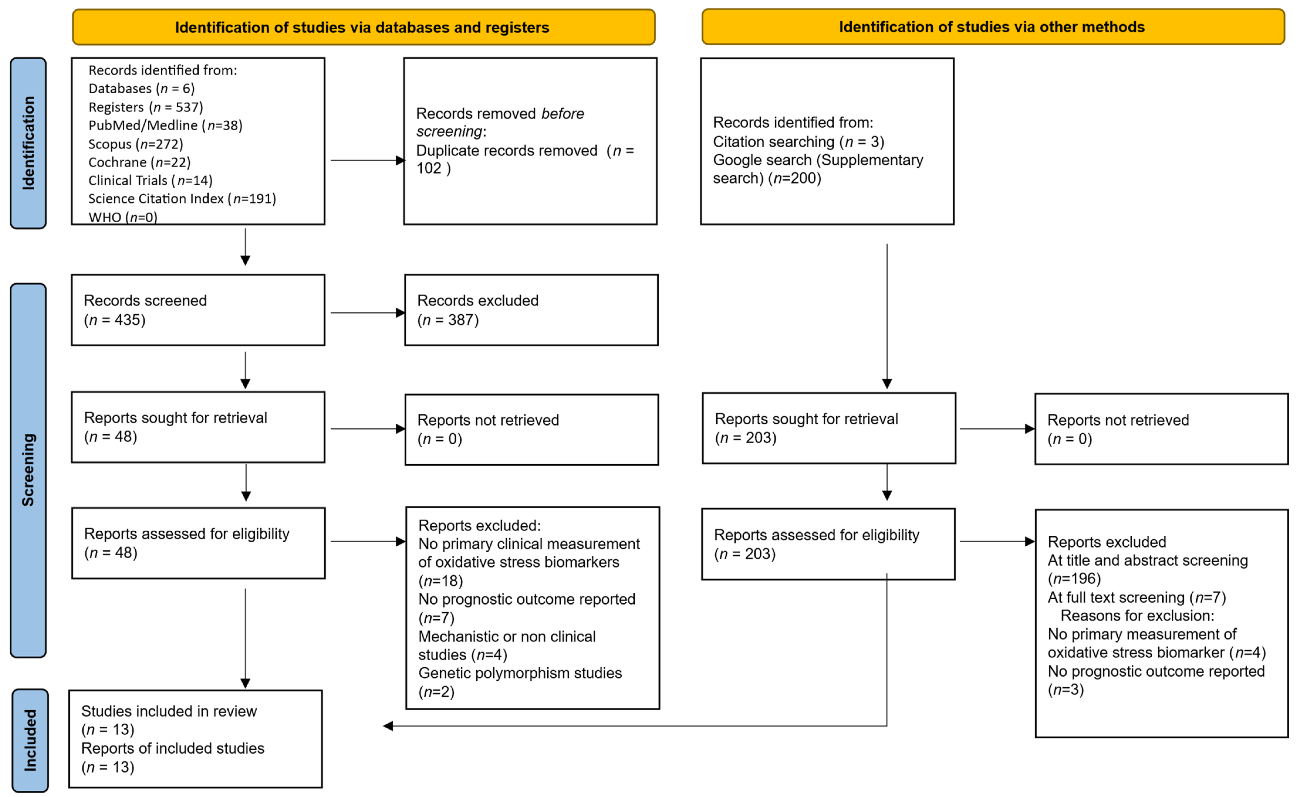
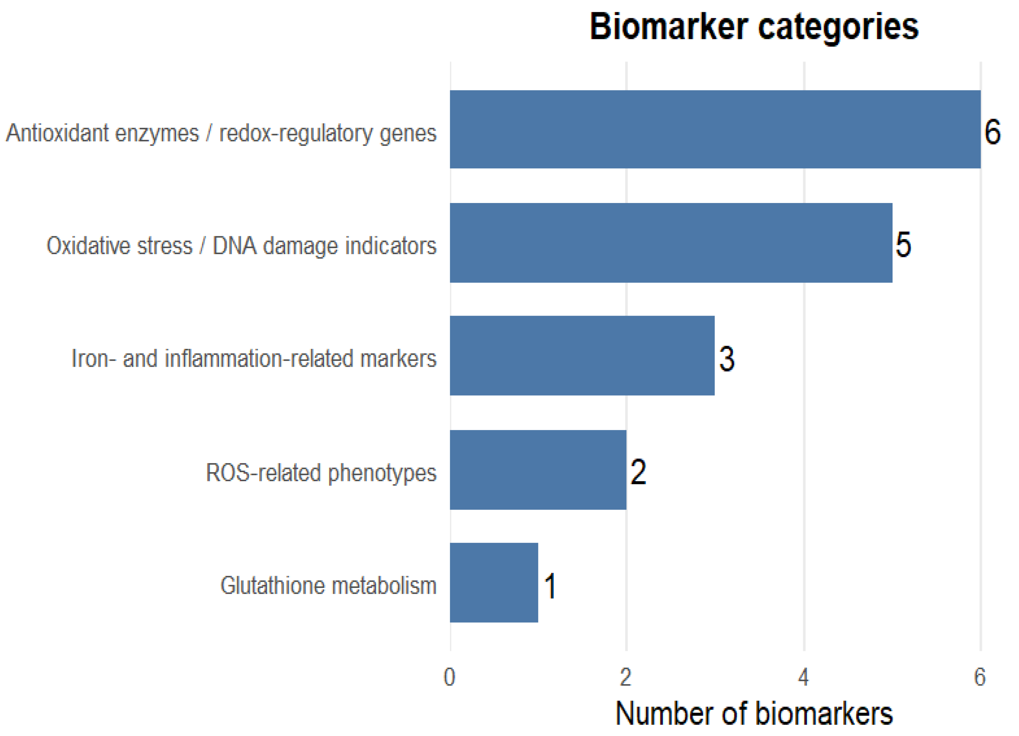
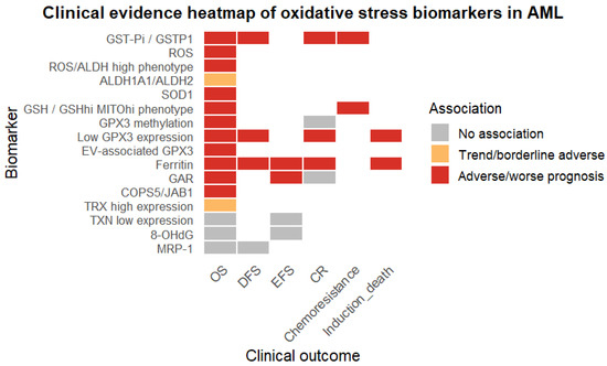
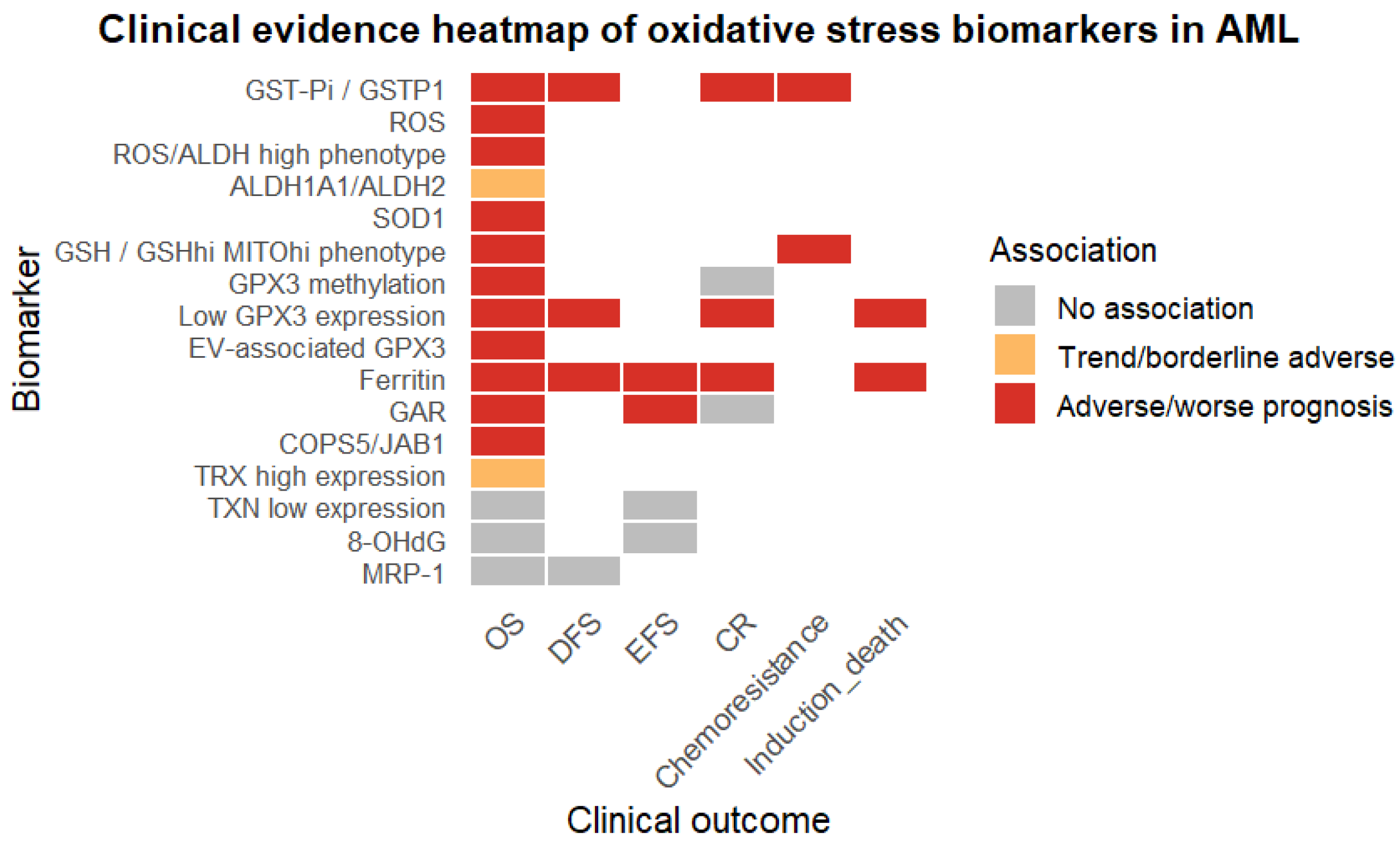
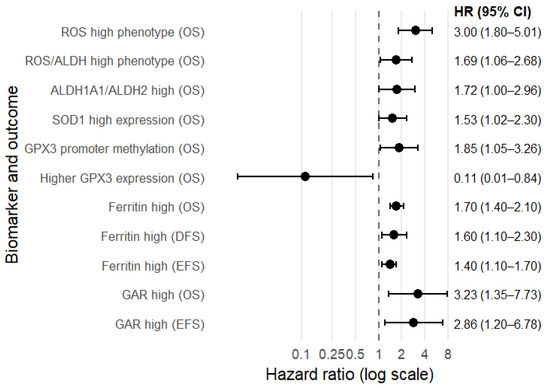
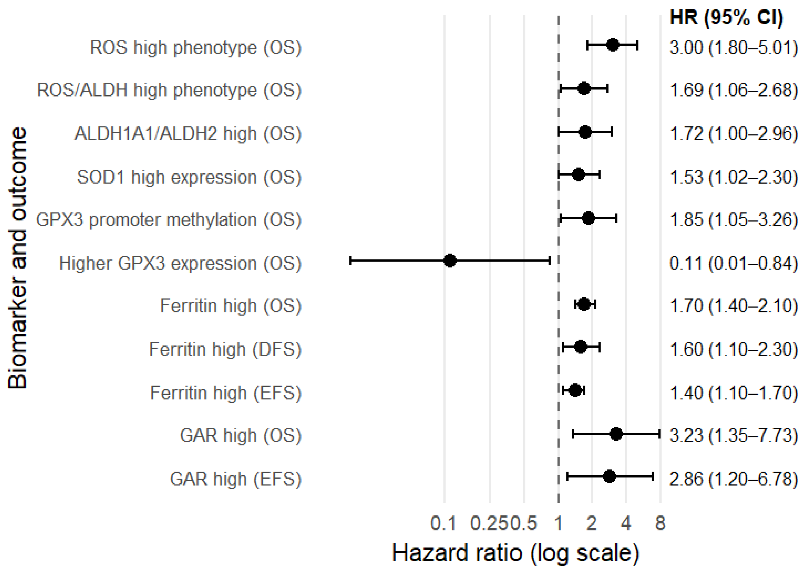
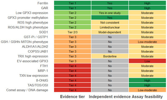
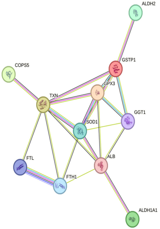
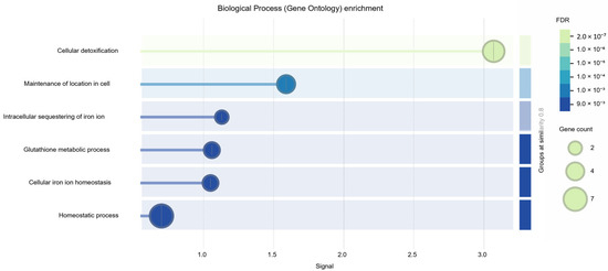
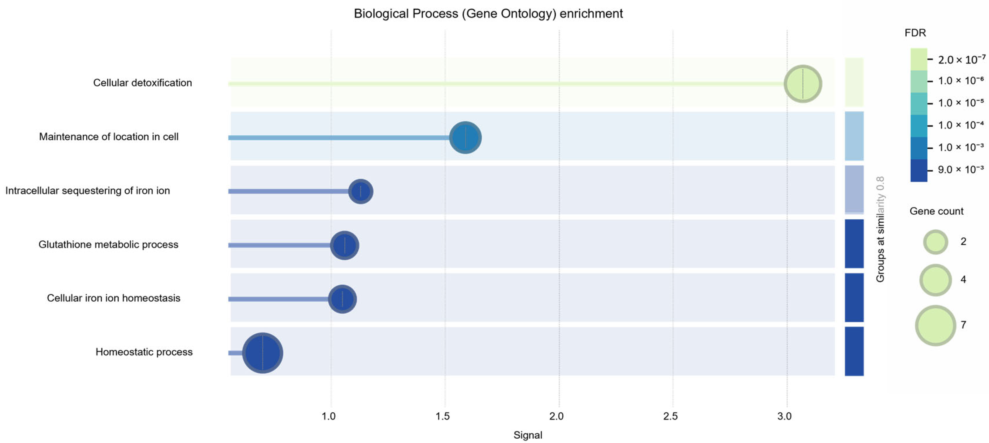

Disclaimer/Publisher’s Note: The statements, opinions and data contained in all publications are solely those of the individual author(s) and contributor(s) and not of MDPI and/or the editor(s). MDPI and/or the editor(s) disclaim responsibility for any injury to people or property resulting from any ideas, methods, instructions or products referred to in the content. |
© 2026 by the authors. Licensee MDPI, Basel, Switzerland. This article is an open access article distributed under the terms and conditions of the Creative Commons Attribution (CC BY) license.