


Abstract
We present a dataset from a wearable sensor that monitors respiration, electrocardiography (one lead), body position/actigraphy, and skin temperature, suitable for sleep studies. We collected the dataset in parallel with standard in-laboratory polysomnography (PSG) and used it to validate ASPEN-derived metrics against clinical assessments. Data were collected from seven volunteers (mean age = 55.7 ± 12.5 years and body mass index (BMI) = 29.9 ± 3.78 kg/m2) for a total of 63 h during sleep studies at the Flinders University Sleep Lab. Our preliminary analysis shows broad agreement between standard metrics used to assess sleep quality measured from the wearable sensor and the resident (Compumedics) polysomnography system, such as respiratory rate, heart rate, and sleep posture. Therefore, we make this dataset of high-resolution ASPEN signals, along with synoptic participant data (age, gender, BMI), available to support the development and benchmarking of algorithms for wearable sleep monitoring.
1. Introduction
Sleep quality strongly influences our health, performance, and overall well-being [1]. Poor sleep quality is associated with many adverse health outcomes, including increased prevalence and severity of cardiovascular diseases and increased mortality, particularly among older adults [1,2]. The complex relationship between cardiovascular diseases (including heart attacks in the early morning) and sleep is yet to be determined. Consequently, extensive global research is underway as the health and economic burdens of these diseases continue to grow [3,4].
A polysomnogram (PSG) study is typically required to determine sleep disturbances by analysing numerous physiological parameters. However, due to the high costs and limited availability of PSG, many people with sleep disorders are left undiagnosed and untreated [1,2,5,6]. Advances in wearable technology and personal electronic devices such as smartphones have made many health and sleep-tracking apps available. Numerous research works have compared portable wearable devices and tracking apps with PSG for sleep quality assessment, as they overcome two primary limitations of PSG: the inability to easily and comfortably use them over multiple nights, and the inability to continue daytime use to correlate daily activity and wearer respiratory/cardiac performance with sleep quality [2,5,6].
In recent years, a growing number of studies have explored consumer- and research-grade wearable devices for sleep monitoring, typically focusing on wrist- or finger-worn systems that estimate sleep stages, heart rate, and breathing rate using indirect measures such as photoplethysmography and accelerometry [7,8]. While these devices have shown promising accuracy for certain metrics when benchmarked against polysomnography (PSG), most publicly available datasets emphasise sleep staging performance and provide limited access to raw, multi-channel biomedical signals suitable for algorithm development and performance validation [9,10]. More recent work has highlighted the importance of direct respiratory measurements and multimodal sensing for improving sleep-stage classification and detecting sleep-related breathing disorders, but the corresponding datasets are often proprietary or only partially accessible [9,11].
For these reasons and to address this gap, over the last three years, we have perfected and tested a wearable device called ASPEN that incorporates two elastomer sensors (enabling respiratory rate and effort assessment), a single-lead ECG, skin temperature, and an accelerometer [12]. Although the elastomer sensors can detect cardiac signals (mechanically) without intimate skin contact, enabling the device to be worn over clothing, the sensor also integrates a single-lead ECG with automated heart rate extraction, recording the heart’s electrical activity [12].
Based on simultaneous sleep-laboratory assessments, we demonstrate that ASPEN-derived respiratory rate, heart rate, and sleep position metrics show broad agreement with PSG-derived metrics in this cohort. The dataset presented in this work provides a prepared collection of ASPEN wearable recordings, with study participants undergoing clinical sleep assessment. The ASPEN system simultaneously captures several of the key physiological and postural parameters critical for advanced wearable sleep analytics. In contrast to many consumer-focused resources, we make available high-resolution raw signals, detailed channel descriptions, and relevant metadata, enabling the development and benchmarking of algorithms for breath-by-breath respiratory rate estimation, heart rate tracking, sleep position classification, and artefact handling. By situating ASPEN within the broader landscape of wearable sleep resources, this dataset provides a flexible platform for exploring multimodal feature extraction and algorithm development with cross-device transferability in sleep-related applications.
Although the present dataset is limited to seven male participants with sleep apnoea, it provides a benchmark resource for work on multi-signal wearable sleep analytics.
2. Materials and Methods
2.1. Participants
Seven men with previously diagnosed sleep apnoea (via overnight polysomnography) volunteered to wear the ASPEN wearable device during a research polysomnography study. Inclusion criteria were: (i) 18–70 yr and (ii) apnoea–hypopnea index (AHI) > 10 events/hr-sleep. Exclusion criteria were: (i) cardiovascular disease, (ii) neurological disorder, and (iii) pregnant or breastfeeding (women only). Synoptic data from the participants with sleep apnoea are presented in Table 1.
Table 1.
Summary of participants.
2.2. Data Acquisition
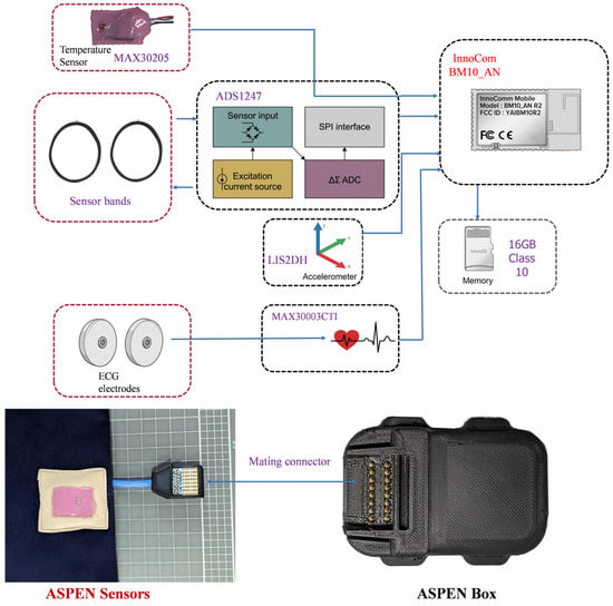
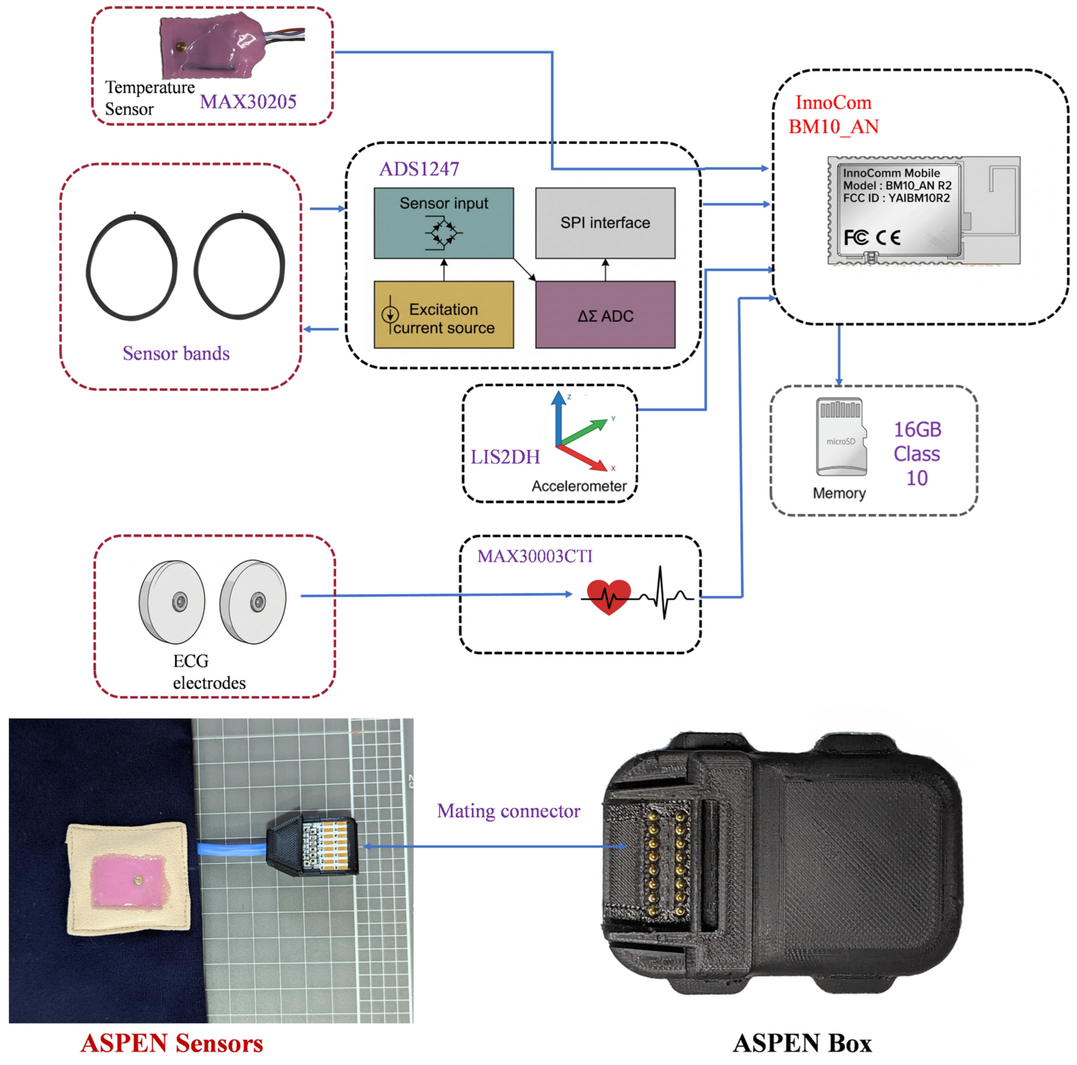
As mentioned, physiological data are acquired via a bespoke ASPEN Garment with embedded sensors mated to control electronics enclosed in a custom 3D-printed enclosure (ASPEN Box). The ASPEN Box electronics (see Figure 1) save data locally (to a removable SD card) encoded in a binary format and simultaneously transmit these data in real time to a smartphone app that allows real-time data visualisation [13]. Bluetooth wireless communications are enabled by an Innocom BM10_AN BLE built around the Texas Instruments CC2640R2F.
Figure 1.
ASPEN’s generalised architecture, adapted from [8,11], showing the device outlay, sensor types and microprocessors.
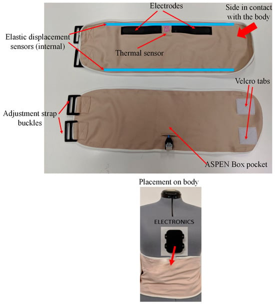
Integrated into the ASPEN Box [14] is an ECG frontend designed around the Analog Devices MAX30003 single-channel biopotential frontend, a tri-axis accelerometer (LIS2DH) to detect the position of the wearer relative to the gravity field, assuming as a reference position the box inserted into a dedicated pocket (see Figure 2), and a 24-bit ADC Texas Instruments ADS1247 to acquire and convert the analogue data from a pair of elastic displacement sensors integrated into the ASPEN Garment [15,16]. Sample rates for each sensor are detailed in Table 2.
Figure 2.
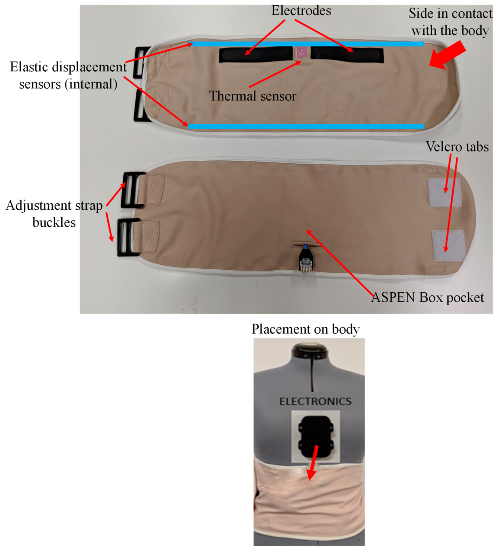
ASPEN sensors and placement on the body.
Table 2.
Data type and sampling rates.
The multi-pin connector branching directly from the ASPEN Garment allows connections for a digital medical-grade temperature sensor (Maxim Integrated MAX30205), the conductive silicone ECG electrodes, and the elastic displacement sensors. To avoid contact between electronics and the subject’s skin, the temperature sensor is encased in skin-safe silicone, allowing contact only for the necessary thermally conductive element. The ASPEN Garment is worn on the abdomen, with the thermometer patch positioned near the celiac (solar) plexus. Velcro straps adjust the wearer’s garment size (Figure 2). For the database uploaded to PhysioNet, we resampled the skin temperature to 100 Hz, although it was initially recorded at 1.619 Hz, as shown in Table 2.
2.3. Dataset Description
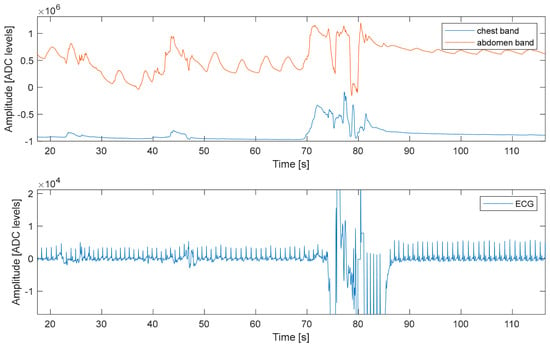
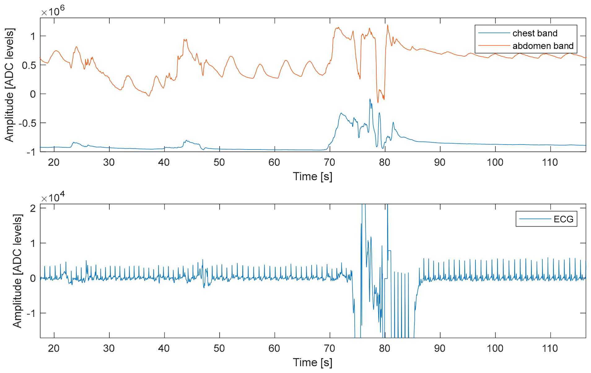
The ASPEN device continuously saves data to the SD card in a binary format during each night study. Converted raw data is first synchronised with the PSG raw data, as the recording duration may vary depending on when recordings started/stopped on APSEN and PSG. Further, blank data sections were removed from the start of the study, when the participant was being prepared, and at the end of the recording, when sensors were removed. No other artefacts have been removed, and the data are one contiguous block with no interruptions. The remaining data include polluted sections, e.g., during toilet use, poor ECG electrode contact, and PSG sensor refitting, e.g., due to a loose mask or ECG/EEG lead/electrode. An example of a retained artefact is depicted in Figure 3; artefact-polluted sections are not marked in the supplied dataset.
Figure 3.
Retained artefact example (data from ASPEN).
For our preliminary analysis only, data have been further cleaned from artefacts using our developed “GroundTruth” MATLAB App Version 1.0 [17]. The data presented to Figshare is provided raw, as recorded. Data from the displacement sensors have been converted to [mV] (ADC quantisation level, 24-bit over a 3.3 V range). Accelerometer data are in ADC quantisation levels, 12-bits over a range of ±2 g. ECG data are in [mV], and temperature sensor data are in [Celsius].
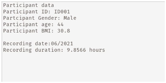
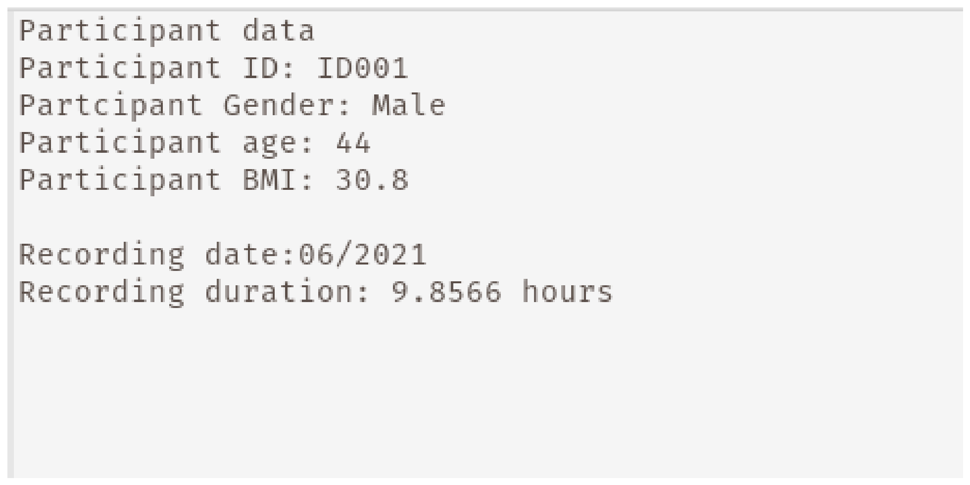
Data have been exported as WFDB files. Because the temperature data have a lower sampling rate and therefore a much shorter data column, the temperature data were resampled to 100 Hz. Data have been presented as *.hea and *.dat files named after the participant IDs. The header files shown in Figure 4 contain synoptic information for each participant (as detailed in Table 1) as .txt files. The recording date is restricted to the month/year and does not specify the exact day.
Figure 4.
Header file sample.
Our preliminary analysis confirmed that heart rate and respiratory rate are in broad agreement with the PSG; however, due to the different sample rates, technology of the sensors, and position and averaging algorithms for the computation of the Compumedics hypnogram, there are differences between the two sleep position sensors (see Technical Validation in the Results Section 3.1).
3. Results
3.1. Technical Validation
3.1.1. Respiration Rate
The data from the elastic displacement sensors provide information about torso displacement induced by respiration and heartbeat. As expected during the data recording, artefacts are generated due to sensor movements/shifts or wearer body movements. To minimise the effect of artefacts on the respiration data analysis, signals are filtered using a low-pass filter with a cut-off frequency of 1 Hz. Respiration peaks are initially detected using the MATLAB built-in window-based threshold method (Peak Detect). Instantaneous breath-by-breath respiratory rates and time (Td) are subsequently obtained by differentiating the position of two consecutive respiratory peaks (Pi and Pj) divided by the sampling rate (FS = 100 Hz), formally:
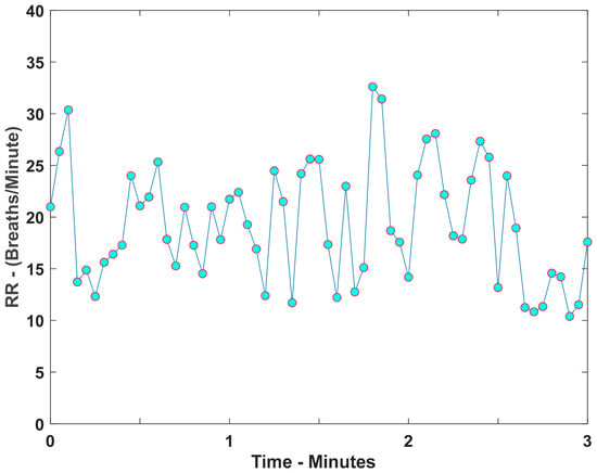
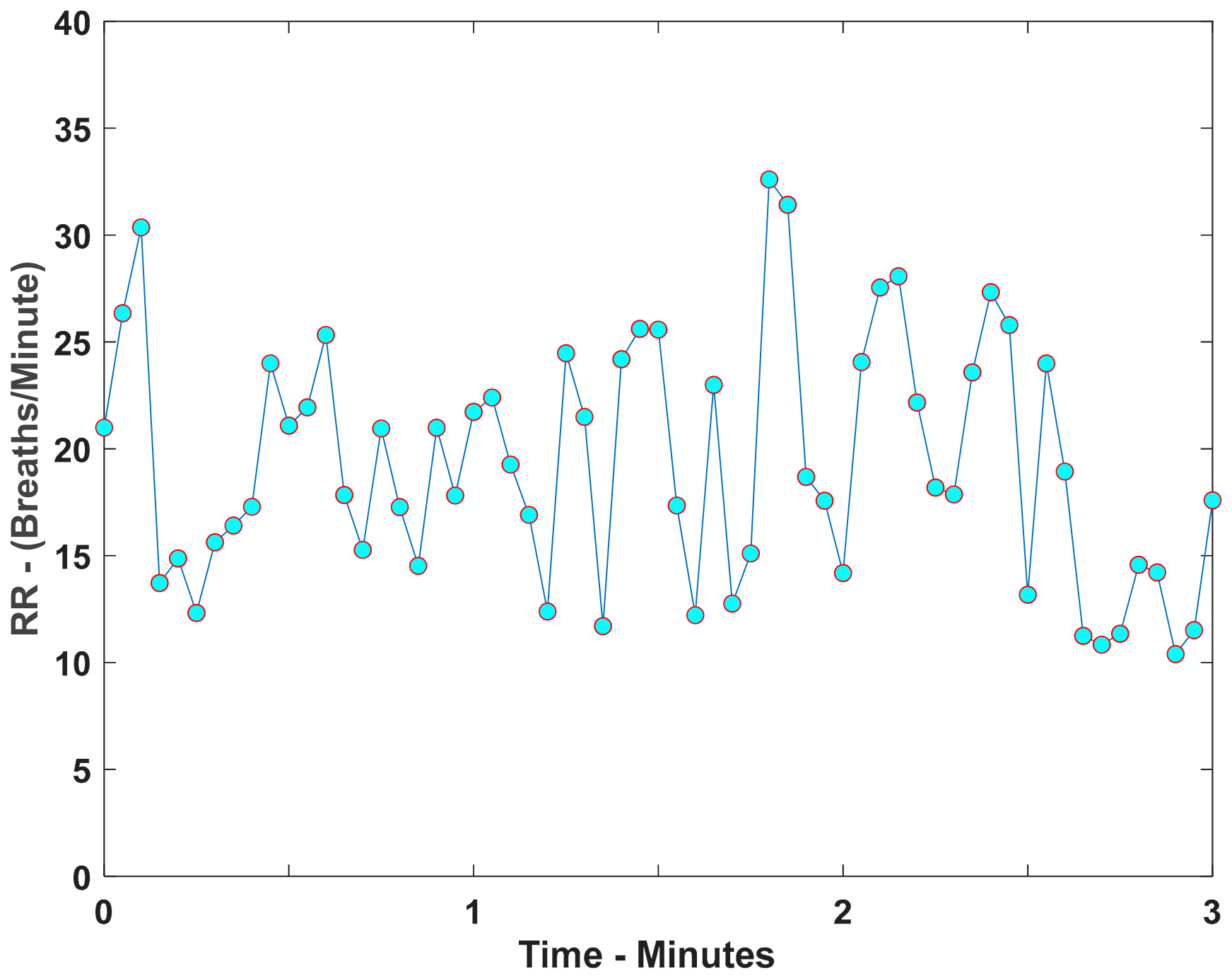
The instantaneous respiration rate (FR) is then calculated by taking the inverse of i.e., FR = 1/. The average, maximum, and minimum respiration rates are then calculated using every respiration event duration, in breaths per minute. An example of the respiration rate computed for one of the participants is shown in Figure 5.
Figure 5.
Sample respiration rate (breaths/minute) from ASPEN band shown with blue dots indicating the respiration rate are joined by lines.
The instantaneous respiration rate shown in Figure 5 illustrates expected breath-by-breath variation of 10–33 breaths per minute across a single overnight recording, with rate fluctuating around the participant’s mean value as sleep stage and posture change. This variation is consistent with normal respiratory rate changes during sleep, where transient increases can relate to arousals or brief position changes. Additionally, sustained lower rates are typical of stable sleep. Comparable breath-by-breath variability has been reported in other wearable respiratory monitoring studies [11], supporting the reliability of the ASPEN-derived respiration signal for capturing physiologically meaningful trends rather than a single static value.
Figure 5 demonstrates the breath-by-breath variation in respiration rate obtained from the elastic displacement signal. The rate is not expected to remain constant over the recording, as breathing patterns can change with sleep stage, body position, respiratory events, and transient movement. The mean respiration rates derived from ASPEN were broadly comparable with those obtained from PSG for most participants (Table 3), demonstrating that the respiratory band supplies a useful representation of the overnight respiratory pattern. Differences in the minimum and maximum values between systems are expected because the sensors use different measurement modalities, sampling rates, and peak-detection processes.
Table 3.
Mean, maximum, and minimum of respiration and heart rate from PSG and ASPEN data.
3.1.2. Heart Rate
In principle, upper-chest elastic displacement sensors can measure the minute displacements impressed by the heart on the chest; hence, heart rate can be extracted from this mechanical movement [13,16]. However, for this study, heart rate is derived from the ASPEN ECG signal computed using the Pan–Tompkins ECG QRS detector algorithm [18], using a period of 20 beats. Heart rate is presented as the mean, maximum, and minimum rates over the entire dataset. The results of respiration rate and heart rate for both PSG and ASPEN bands are tabulated in Table 3.
The mean heart rate values in Table 3 fall within the expected range for adults during sleep, and the observed spread between minimum and maximum rates reflects normal night-time variation associated with sleep-stage transitions and apnoea events, consistent with the participants’ diagnosed sleep apnoea. Where ASPEN- and PSG-derived heart rates diverge, this is most likely attributable to differences in beat-detection sensitivity between the ECG-based Pan–Tompkins algorithm and the PSG system’s own HR derivation, rather than a fundamental limitation of the ASPEN sensor.
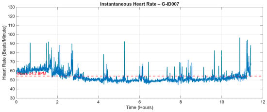
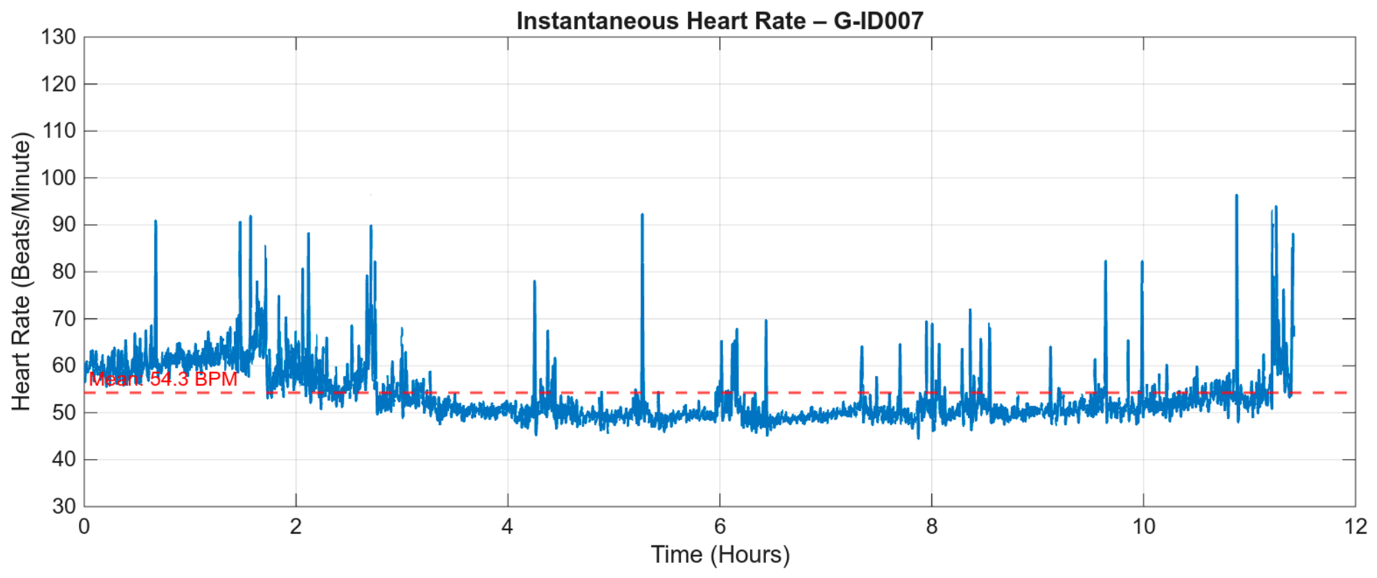
Figure 6 shows the HR trend for subject 7. The recorded HR increases reflect the expected trend during apnoea events [19]. The recorded HR increases are consistent with the expected post-event tachycardic response associated with apnoea and arousal.
Figure 6.
The heart rate trend for subject 7, over more than 11 h of sleep. Peak heart rates may indicate apnoea events.
3.1.3. Sleep Position
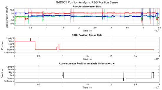
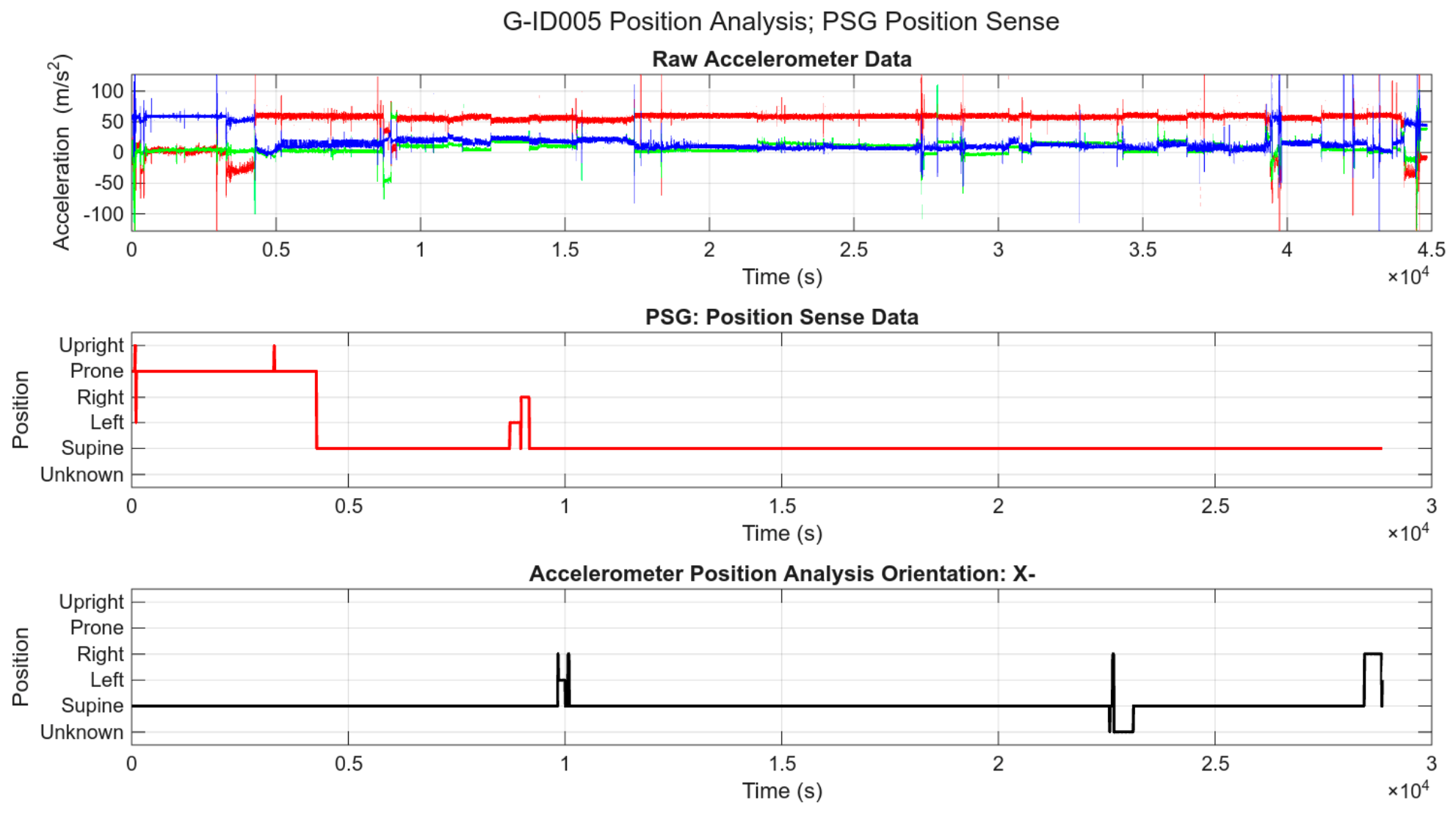
Sleep position was computed using the embedded accelerometer. The accelerometer returns x-, y-, and z-axis data at 100 Hz as signed byte numbers calibrated to ±2 g at full-scale reading. We computed sleep position using an algorithm available on request. Examples of sleep position for one of the participants are shown in Figure 7, and all the individual participants’ sleep positions are reported in Table 4.
Figure 7.
Sleep position for one of the participants—computed using embedded accelerometer data. (Top) Indicates the accelerometer vectors, showing z axis (blue), y axis (red) and x axis (green) acceleration. (Middle) Shows the calculated position vector, and (Bottom) the PSG ground truth subject orientation.
Table 4.
Sleep position of all participants (%) with ASPEN bands in correct orientation.
A comparison of the data in Table 4 shows that the major postural patterns identified by the ASPEN and PSG systems were broadly consistent after selecting the closest-fit orientation. For example, the PSG and ASPEN recordings both identified supine position as the predominant posture for participants ID001, ID004, ID005, and ID007. However, agreement was not uniform across all subjects’ posture categories, particularly where the PSG system recorded an “Unknown” position. These discrepancies are consistent with differences in the position sensor location, reference orientation, sampling rate, and classification method used by the two systems. In some cases, operator error meant that the ASPEN sensor was not inserted into the band in the correct position. To avoid participant discomfort, the ASPEN Box fits quite loosely inside the garment pocket, which may also cause misalignment of the X- and Y-axes. Further misalignment can be caused by wearing the garment slightly asymmetrically (e.g., the box is not centred on the abdomen). Additionally, Z-axis misalignment can be caused by the participant’s anatomy (e.g., a prominent abdominal wall) or operator error (e.g., the box is flipped/tilted before insertion into the pocket).
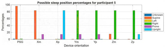
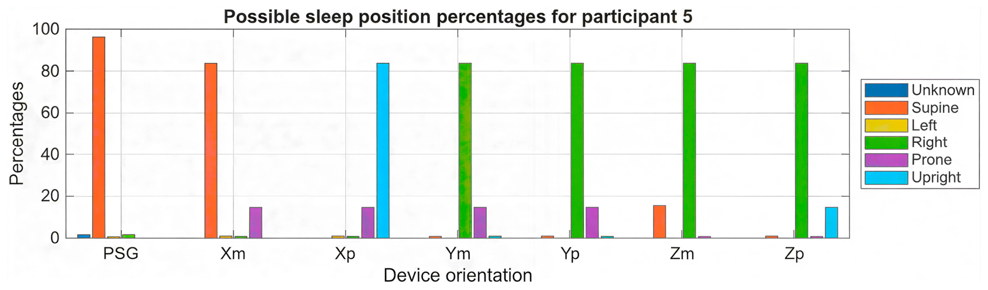
Because of these issues, the position data was calculated for all possible insertion orientations, and the closest-fit orientation was selected, as can be seen in Figure 8, where the positive X orientation provides a close fit to the PSG position data, which shows an “Unknown” position for almost the same amount of time as the ASPEN band records a “right” position. This approach enabled comparison between each possible orientation and the PSG-derived position data, while also identifying cases where residual disagreement may be attributable to device location, body anatomy, or differences in posture definitions.
Figure 8.
The calculated sleep positions for all possible device orientations, with negative X orientation providing the closest match to the PSG data. Additional time in the prone position may be due to the ASPEN device’s position on the upper body.
3.1.4. Temperature Data
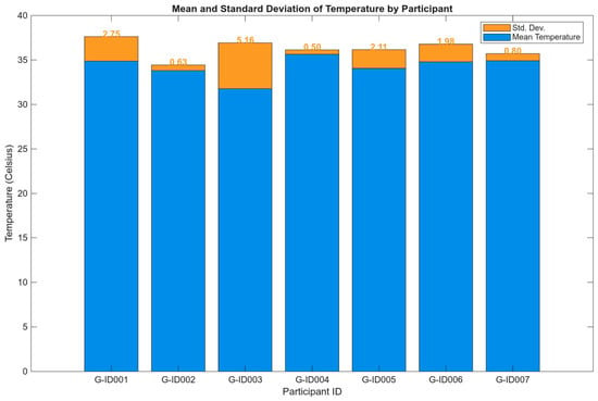
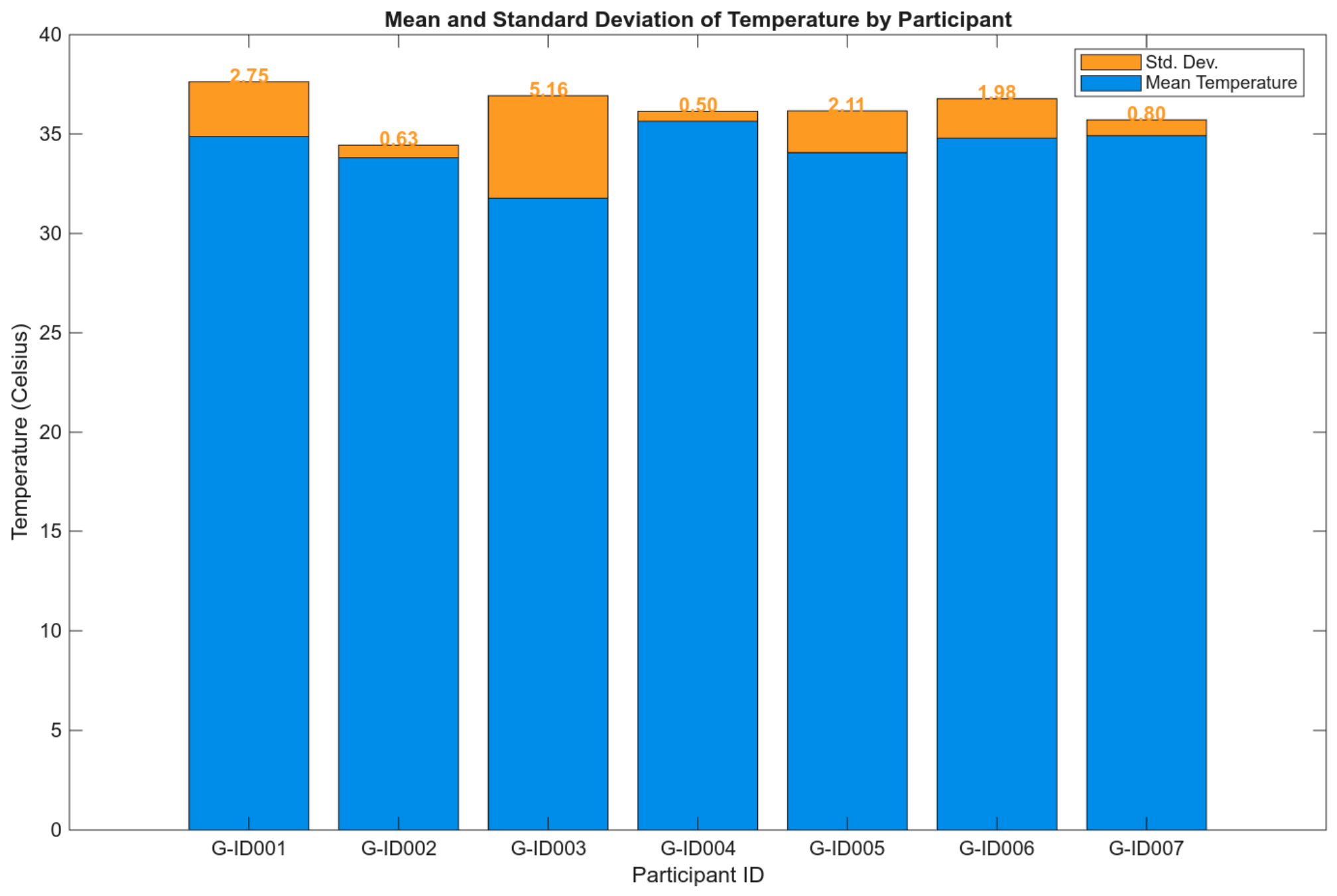
During data collection, skin temperature was continuously recorded for each participant using the ASPEN Garment. The mean and standard deviation of the participant temperature (Celsius) recordings are presented in Figure 9. Mean temperatures were generally clustered between 33.81 and 35.64 °C, except for participant ID003, who showed a lower mean temperature and greater variability. This subject’s lower temperature may reflect intermittent sensor contact or local skin-temperature variation over the overnight recording. These temperature signals are included as raw data in the dataset and may be useful for future work investigating sensor contact or sleep-related body temperature regulation.
Figure 9.
Temperature plot (mean ± SD) for all participants.
4. Discussion
The presented ASPEN dataset contains raw multimodal signals, including respiratory effort, cardiac data, and accelerometry, as well as temperature data recorded by a unique device fitted into a single garment. The device’s data has broad agreement with the recorded PSG data and was recorded in a sleep lab. The database includes discrepancies expected given the use of different sensors and sampling rates, but it may not reflect the low-quality recordings that are typical of in-home sleep analysis. This, however, supports ASPEN as a viable resource for algorithm development, including RR and HR trajectories. Positional data from the accelerometer may have been influenced by the user’s anatomy and in-pocket placement, and while this affects mapping between raw accelerometer data and actual posture, this can be determined from an assessment of all possible postures.
By maintaining artefacts in the database, we reflect real-world usage and data recording, but this does impose the requirement on the database user to develop robust artefact detection. While the ASPEN database has a modest sample size, the recordings are a total of 63 h in duration, providing ample opportunities for quality algorithm development. Making these WFDB files publicly available to the community along with detailed headers is a key contribution, enabling the testing and development of multimodal algorithms.
5. Limitations and Future Directions
While the ASPEN dataset provides a rich set of physiological and positional data, several considerations should be kept in mind when interpreting the results and reusing the provided recordings. The study’s cohort consists of seven male participants with previously diagnosed sleep apnoea, and the sample size is modest compared with recent large-scale evaluations of consumer wearables [9,20]. As a result, the distributions of age, BMI, and apnoea severity are not intended to represent the general population or all types of sleep-disordered breathing. Furthermore, all recordings were conducted in a controlled laboratory environment, which may not capture the variability, motion profiles, and environmental considerations encountered in home-based monitoring described in recent contactless and wearable studies [21].
The accuracy of sleep position metrics is influenced by the placement of the ASPEN device within the garment pocket and by individual anatomy, as discussed in Results Section 3.1.3. We addressed this by systematically evaluating sleep position across all eight possible accelerometer insertion orientations (Figure 8) and selecting the orientation that most closely agreed with PSG-derived position assessments, rather than assuming a fixed-axis mapping. This approach demonstrates that misalignment can be identified and corrected post hoc, but it also means that any deployment of ASPEN outside a supervised research setting would need either a calibration step or an orientation-detection algorithm to achieve equivalent accuracy. Future work could incorporate an automated orientation-detection step, using a short calibration posture at the start of each recording, to remove this dependency on manual best-fit selection.
Additionally, future work could expand the dataset to include larger and more diverse cohorts, use additional sensors such as photoplethysmography, and incorporate repeated measurements across multiple nights, in line with recent trends toward multimodal, longitudinal sleep monitoring [22]. The dataset will also be extended to include home environments and reference information for complex events, such as arousal or waking episodes and limb movements.
Beyond validating ASPEN against PSG in a clinical cohort, the raw, high-resolution bio-signal recordings released with this dataset are intended to support the wider development of multimodal wearable sleep algorithms. This includes sensor fusion approaches that combine respiratory, cardiac, and postural information. As wearable sleep monitoring may move toward home-based, longitudinal use, datasets that expose raw multi-channel signals are needed to support the research community in benchmarking new algorithms under realistic signal conditions.
6. Conclusions
Overall, despite the modest cohort size and single-night, laboratory-based design, the ASPEN dataset provides a multi-channel reference against which clinical polysomnography is compared, and is released with sufficient raw signal detail to support algorithm development beyond the specific analyses presented here. Findings from this cohort should be interpreted as a proof-of-concept validation of the ASPEN platform rather than a characterisation of sleep physiology for the larger population. Any generalisation of developed algorithms or ASPEN-derived metrics to unsupervised or home-based settings should be validated against a larger and more demographically diverse cohort.
These recordings reveal that a wearable garment can successfully capture clinically relevant biomedical data with broad agreement with PSG in a sleep-lab setting. The ASPEN data contributes to publicly available datasets of elastic chest bands, ECG, and accelerometer data, as well as temperature in WFDB format. The dataset can support the development of algorithms for respiration rate estimation, heart rate tracking, posture classification, and artefact management in sleep monitoring, despite the modest size of the dataset.
The ASPEN device recordings offer a practical foundation for advancing wearable polysomnography approaches and help bridge real-world multimodal sleep recording devices and lab-grade polysomnography.
Author Contributions
Conceptualization, G.D.G. and P.P.B.; methodology, G.D.G. and P.P.B.; software, G.R.N. and B.B.; validation, B.B. and U.G.; formal analysis, B.B. and G.R.N.; investigation, B.B. and G.R.N.; resources, U.G.; data curation, B.B. and U.G.; writing—original draft preparation, B.B. and G.R.N.; writing—review and editing, G.D.G., U.G. and D.J.E.; visualisation, B.B. and U.G.; supervision, G.D.G., D.J.E. and P.P.B.; project administration, G.D.G.; funding acquisition, G.D.G. and P.P.B. All authors have read and agreed to the published version of the manuscript.
Funding
This research was funded by the Cooperative Research Centre Project (CRC-P) titled: “Targeted Therapy for Sleep Apnoea: A Novel Personalised Approach”.
Institutional Review Board Statement
This study was approved by the South Eastern Sydney Local Health District Human Research Ethics Committee (Approval Code: 2019/ETH05012, Approval Date: 2 December 2019).
Informed Consent Statement
Written informed consent has been obtained from the patients for the publication of this paper.
Data Availability Statement
The dataset will be available as “Towards a wearable polysomnography approach: A dataset of key physiology and body position metrics validated against in-laboratory polysomnography” on Physionet.
Conflicts of Interest
Co-authors P.P.B. and G.D.G. are minority shareholders of Medical Monitoring Solutions (MMS) Pty. Ltd. MMS holds the rights to commercialize the morphic sensor assessed in the paper. However, MMS had no role in the design of the study; in the collection, analysis, or interpretation of data; in the writing of the manuscript; or in the decision to publish the results.
Usage Notes
As noted, the data files are in PhysioNet WFDB format and are accompanied by a header file that includes descriptions of variables within each dataset. The dataset will be available as “Towards a wearable polysomnography approach: A dataset of key physiology and body position metrics validated against in-laboratory polysomnography” on Figshare, https://doi.org/10.6084/m9.figshare.33399838. We have added a brief description of each dataset in a .TXT file. To facilitate reuse of this dataset, we recommend that users follow a standardised workflow when processing the ASPEN signals. The WFDB headers provide channel descriptions and note sampling frequencies, which can be used to develop analysis pipelines for participants. For respiratory analysis, the elastic displacement channels can be low-pass-filtered to isolate the respiratory band, followed by peak detection to derive breath-by-breath intervals and respiratory rate trajectories, similar to approaches used in recent wearable and contactless monitoring studies [22]. ECG-derived heart rate can be measured using established QRS detection algorithms and assessed to produce summary statistics. When working with accelerometer-derived sleep positions, users should preferentially note the assumed axis direction and verify the position trajectories over time, particularly in segments where the ASPEN device may have shifted or been adjusted.
Code Availability
All codes used for formatting, cleaning, and variable computations were written in MATLAB language and will be made available to users with reasonable requests.
Abbreviations
The following abbreviations are used in this manuscript:
| AHI | Apnoea–hypopnoea index |
| ADC | Analog-to-digital converter |
| ASPEN | Wearable sensor system/device name used in the paper |
| BMI | Body mass index |
| BPM | Beats per minute |
| ECG | Electrocardiography |
| EEG | Electroencephalography |
| Hz | Hertz |
| PSG | Polysomnography |
References
- Kwon, S.; Kim, H.; Yeo, W.H. Recent advances in wearable sensors and portable electronics for sleep monitoring. iScience 2021, 24, 102461. [Google Scholar] [CrossRef] [Scilit] [PubMed]
- Wolkove, N.; Elkholy, O.; Baltzan, M.; Palayew, M. Sleep and aging: 1. Sleep disorders commonly found in older people. Can. Med. Assoc. J. 2007, 176, 1299–1304. [Google Scholar] [CrossRef] [Scilit] [PubMed]
- Malhotra, A.; Loscalzo, J. Sleep and cardiovascular disease: An overview. Prog. Cardiovasc. Dis. 2009, 51, 279–284. [Google Scholar] [CrossRef] [Scilit] [PubMed]
- Yeghiazarians, Y. Obstructive Sleep Apnea and Cardiovascular Disease: A Scientific Statement from the American Heart Association. Circulation 2021, 144, 56–67. [Google Scholar] [CrossRef] [Scilit] [PubMed]
- Dezambotti, M.; Cellini, N.; Goldstone, A.; Colrain, I.M.; Baker, F.C. Wearable Sleep Technology in Clinical and Research Settings. Med. Sci. Sports Exerc. 2019, 51, 1538–1557. [Google Scholar] [CrossRef] [Scilit] [PubMed]
- Lee, J.M.; Byun, W.; Keill, A.; Dinkel, D.; Seo, Y. Comparison of Wearable Trackers’ Ability to Estimate Sleep. Int. J. Environ. Res. Public Health 2018, 15, 1265. [Google Scholar] [CrossRef] [Scilit] [PubMed]
- Du, Y.; Gu, J.; Duan, S.; Trueb, J.; Tzavelis, A.; Shin, H.-S.; Arafa, H.; Li, X.; Huang, Y.; Carr, A.N.; et al. A skin-interfaced wireless wearable device and data analytics approach for sleep-stage and disorder detection. Proc. Natl. Acad. Sci. USA 2025, 122, e2501220122. [Google Scholar] [CrossRef] [Scilit] [PubMed]
- Birrer, V.; Elgendi, M.; Lambercy, O.; Menon, C. Evaluating reliability in wearable devices for sleep staging. npj Digit. Med. 2024, 7, 74. [Google Scholar] [CrossRef] [Scilit] [PubMed]
- Lee, T.; Cho, Y.; Cha, K.S.; Jung, J.; Cho, J.; Kim, H.; Kim, D.; Hong, J.; Lee, D.; Keum, M.; et al. Accuracy of 11 Wearable, Nearable, and Airable Consumer Sleep Trackers: Prospective Multicenter Validation Study. JMIR MHealth UHealth 2023, 11, e50983. [Google Scholar] [CrossRef] [Scilit] [PubMed]
- de Zambotti, M.; Goldstein, C.; Cook, J.; Menghini, L.; Altini, M.; Cheng, P.; Robillard, R. State of the science and recommendations for using wearable technology in sleep and circadian research. Sleep 2024, 47, zsad325. [Google Scholar] [CrossRef] [Scilit] [PubMed]
- Natarajan, A.; Su, H.-W.; Heneghan, C.; Blunt, L.; O’Connor, C.; Niehaus, L. Measurement of respiratory rate using wearable devices and applications to COVID-19 detection. npj Digit. Med. 2021, 4, 136. [Google Scholar] [CrossRef] [Scilit] [PubMed]
- Andreozzi, E.; Gargiulo, G.D.; Esposito, D.; Bifulco, P. A Novel Broadband Forcecardiography Sensor for Simultaneous Monitoring of Respiration, Infrasonic Cardiac Vibrations and Heart Sounds. Front. Physiol. 2021, 12, 725716. [Google Scholar] [CrossRef] [Scilit] [PubMed]
- Jayarathna, T.; Gargiulo, G.D.; Breen, P.P. Continuous Vital Monitoring During Sleep and Light Activity Using Carbon-Black Elastomer Sensors. Sensors 2020, 20, 1583. [Google Scholar] [CrossRef] [Scilit] [PubMed]
- Polley, C. Wearable Bluetooth Triage Healthcare Monitoring System. Sensors 2021, 21, 7586. [Google Scholar] [CrossRef] [Scilit] [PubMed]
- Gargiulo, G.D. A Wearable Contactless Sensor Suitable for Continuous Simultaneous Monitoring of Respiration and Cardiac Activity. J. Sens. 2015, 2015, 151859. [Google Scholar] [CrossRef] [Scilit]
- Gargiulo, G.D.; O’Loughlin, A.; Breen, P.P. Electro-resistive bands for non-invasive cardiac and respiration monitoring, a feasibility study. Physiol. Meas. 2015, 36, N35–N49. [Google Scholar] [CrossRef] [Scilit] [PubMed]
- Naik, G.R.; Gargiulo, G.D.; Serrador, J.M.; Breen, P.P. Groundtruth: A Matlab GUI for Artifact and Feature Identification in Physiological Signals. Front. Physiol. 2019, 10, 850. [Google Scholar] [CrossRef] [Scilit] [PubMed]
- Pan, J.; Tompkins, W.J. A Real-Time QRS Detection Algorithm. IEEE Trans. Biomed. Eng. 1985, 32, 230–236. [Google Scholar] [CrossRef] [Scilit] [PubMed]
- DiCaro, M.V.; Lei, K.; Yee, B.; Tak, T. The Effects of Obstructive Sleep Apnea on the Cardiovascular System: A Comprehensive Review. J. Clin. Med. 2024, 13, 3223. [Google Scholar] [CrossRef] [Scilit] [PubMed]
- Ravindran, K.K.G.; della Monica, C.; Atzori, G.; Lambert, D.; Hassanin, H.; Revell, V.; Dijk, D.-J. Reliable Contactless Monitoring of Heart Rate, Breathing Rate, and Breathing Disturbance During Sleep in Aging: Digital Health Technology Evaluation Study. JMIR MHealth UHealth 2024, 12, e53643. [Google Scholar] [CrossRef] [Scilit] [PubMed]
- Yoon, H.; Choi, S.H. Technologies for sleep monitoring at home: Wearables and nearables. Biomed. Eng. Lett. 2023, 13, 313–327. [Google Scholar] [CrossRef] [Scilit] [PubMed]
- Gabb, V.G.; Blackman, J.; Morrison, H.; Li, H.; Kendrick, A.; Turner, N.; Greenwood, R.; Biswas, B.; Heslegrave, A.; Coulthard, E. Longitudinal Remote Sleep and Cognitive Research in Older Adults With Mild Cognitive Impairment and Dementia: Prospective Feasibility Cohort Study. JMIR Aging 2025, 8, e72824. [Google Scholar] [CrossRef] [Scilit] [PubMed]

Disclaimer/Publisher’s Note: The statements, opinions and data contained in all publications are solely those of the individual author(s) and contributor(s) and not of MDPI and/or the editor(s). MDPI and/or the editor(s) disclaim responsibility for any injury to people or property resulting from any ideas, methods, instructions or products referred to in the content. |
© 2026 by the authors. Licensee MDPI, Basel, Switzerland. This article is an open access article distributed under the terms and conditions of the Creative Commons Attribution (CC BY) license.