


Abstract
Bio-based scaffolds are being investigated in tissue engineering because they can provide temporary support for tissue repair while offering biocompatibility and biodegradability. A scaffold need not rely solely on the properties of its starting material: surface treatments, bioactive compounds, therapeutic agents, nanoscale constituents and delivery systems can provide further functions. The discussion begins with the main categories of bio-based scaffolds and considers how functionalization alters their biological and therapeutic behaviour. Particular attention is paid to the effects of these modifications on cell attachment and growth, differentiation, blood-vessel formation, immune activity, matrix remodelling and the development of functional tissue. The path from laboratory research to clinical use is considered in terms of consistent and scalable manufacture, sterilisation, safety, regulatory assessment, clinical evidence and cost. Developments in computational modelling, artificial intelligence, advanced biomanufacturing, responsive biomaterials and precision regenerative medicine are examined as possible ways forward. Introducing several functions into one scaffold may address different regenerative needs, but the resulting complexity can make production, quality assurance and regulatory assessment harder. Future development must weigh regenerative performance against the need for a product that can be reproduced, used safely and implemented in practice. By linking scaffold selection and functionalization with biological performance and translational considerations, this review provides an integrated perspective on the development of bio-based scaffolds for regenerative medicine.
1. Introduction
Damage to human tissues may result from trauma, congenital abnormalities, degenerative disease, chronic wounds, infection or tumour resection. Depending on the tissue affected, the consequences can extend from impaired organ function and reduced quality of life to wider demands on healthcare services [1,2,3]. Ageing populations and the burden of chronic disease have increased interest in therapies that restore tissue structure as well as biological function [4]. Repair or replacement has traditionally relied on autografts, allografts, xenografts and synthetic implants. Each has limitations: donor tissue may be scarce, harvesting may cause morbidity, and implanted materials may be associated with rejection, disease transmission, poor integration or mechanical incompatibility [5,6]. Tissue engineering offers another approach, bringing together biology, materials science, engineering and medicine to promote functional regeneration [7]. Its three principal elements are cells, bioactive signalling molecules and scaffolds. The scaffold provides a temporary, three-dimensional structure resembling the extracellular matrix (ECM), within which cells can attach, proliferate, migrate, differentiate and form tissue [8]. Research has consequently explored different biomaterials, fabrication techniques and functionalization methods, both to improve regeneration and to make these therapies more suitable for clinical translation [9,10].
The appeal of naturally sourced scaffold materials stems from their potential compatibility with living tissues, their degradability and, in some cases, their resemblance to native ECM [5,11,12]. Unlike unmodified synthetic polymers, certain bio-based materials also contain recognition motifs and other biochemical features that affect cell attachment and matrix remodelling [13,14]. Their renewable origins may offer an additional advantage where environmentally responsible material production is a consideration [15]. Investigated materials include collagen, gelatin, silk fibroin and keratin, as well as the polysaccharides chitosan, alginate, hyaluronic acid, cellulose and pectin. Their reported applications span bone, cartilage, skin, vascular, neural and musculoskeletal tissue engineering [5,16]. Nevertheless, favourable biological characteristics do not necessarily translate into a scaffold with adequate mechanical strength, degradation control or consistency. Problems such as weak mechanical stability, variable material composition, limited antimicrobial activity and insufficient control over cell responses can restrict performance [17,18]. Rather than assuming that similarity to native tissue is sufficient, researchers modify natural scaffolds to provide the functions needed for particular regenerative applications [19,20].
Scaffold design must address more than mechanical support: how the material influences the surrounding healing environment is equally important [21,22]. By modifying a scaffold physically, chemically or biologically, functionalization can influence how it behaves within the healing environment [13,23]. Surface treatments and chemical modifications may change stability, degradation or the way cells interact with the material. Other approaches incorporate growth factors, peptides, antimicrobial compounds, nanoparticles or naturally derived therapeutics. These additions have been studied in relation to blood-vessel formation, inflammatory responses, bacterial control and stem-cell differentiation [24,25,26]. Therapeutic availability may also be managed through release systems designed to control how much agent is present locally and for how long [27,28]. The choice of modification is therefore linked to the particular biological functions required for a given regenerative application [29,30].
Despite the breadth of experimental work on scaffolds, comparatively few designs have reached clinical practice. Thousands of formulations reporting improved biological performance have appeared over the past decade [31,32], yet relatively few have advanced from preclinical testing to regulatory approval and routine use [33,34]. Promising results from biological studies do not necessarily mean that a scaffold is suitable for clinical application. Beyond demonstrating its regenerative potential, the material must be produced with consistent quality, retain its stability, meet the relevant regulatory requirements, and be economically viable to manufacture [35,36,37]. Achieving this consistency can be particularly challenging with naturally derived materials, as differences in biological sources may affect their composition and physicochemical characteristics. Sterilisation, storage conditions, production scale-up, mechanical reliability, regulatory compliance, and manufacturing costs introduce further considerations that must be addressed collectively [38,39]. Early proof-of-concept studies are generally performed under carefully controlled laboratory conditions, which may not fully reflect the demands of clinical use [40,41]. It is therefore important to consider these practical requirements during the initial stages of scaffold development, alongside investigations of biological performance.
Previous reviews have discussed the range of naturally derived materials used in scaffold development, their fabrication methods, and their applications in different tissues [5,42,43]. However, an important question remains as to whether the biological improvements achieved through scaffold functionalisation can be maintained when these materials are developed into products intended for clinical use. Many studies emphasise improvements in biological performance while giving comparatively less attention to the challenges associated with manufacturing, regulatory approval, and commercialisation. Examining these aspects alongside the available biological evidence provides a more complete understanding of the factors that may limit the translation of promising scaffold technologies from laboratory research into clinical practice.
The review first considers the principal categories of bio-based scaffold materials and the reasons for selecting them. It then examines functionalization methods used to alter their biological performance, followed by the scientific and practical obstacles that arise when laboratory scaffolds are developed for clinical use. Manufacturing, regulation and commercial feasibility are considered alongside emerging approaches intended to address these obstacles. Taken together, these topics connect material design with regenerative performance and the requirements of clinical translation. Figure 1 presents this organising framework, tracing the relationship between scaffold platforms, bioactive modifications, biological outcomes, translational challenges and possible routes towards clinical implementation.
Figure 1.
Conceptual framework linking bio-based scaffold design to clinical translation for tissue regeneration. The illustration was generated using ChatGPT (OpenAI, GPT-5.5) based on the scientific concepts and content of this review manuscript, as provided and directed by the author. The author subsequently reviewed and refined the illustration to ensure scientific accuracy and consistency with the manuscript.
Literature Search and Selection Approach
Relevant publications were identified through a broad literature search, including ScienceDirect, and by examining the reference lists of relevant articles to identify additional studies. Publications were selected based on their relevance to the main themes of the review, particularly scaffold composition, functionalization approaches, biological performance, biomedical applications, and translational considerations.
The selected literature includes experimental investigations, clinical studies, and regulatory documentation where relevant. Representative studies were examined in relation to their scaffold composition, functional components, intended applications, experimental approaches, reported outcomes, and limitations. Particular attention was given to distinguishing findings obtained from in vitro and animal studies from those supported by human clinical investigations or regulatory approval. As a narrative review, the literature selection was intended to support a critical discussion of representative developments rather than provide an exhaustive or systematic assessment of the published literature.
2. Bio-Based Scaffold Platforms for Tissue Regeneration
The composition and architecture of a scaffold affect cellular behaviour, particularly when they reproduce features of the ECM. Materials obtained from natural sources may offer biochemical cues together with favourable biocompatibility and biodegradability. These characteristics have prompted studies of bio-based scaffolds for bone, cartilage, skin, vascular, neural and musculoskeletal repair [13,44].
For the purposes of this review, scaffold materials are considered under four overlapping headings: proteins, polysaccharides, decellularized extracellular matrix (dECM) and composites [5,30,45]. These categories are used to organise the discussion and are not necessarily mutually exclusive. Protein-based and polysaccharide-based scaffolds are grouped according to their predominant material composition, while dECM scaffolds are considered separately because they originate from decellularized tissues and retain components of the native ECM. Composite scaffolds are formed by combining two or more materials, such as proteins, polysaccharides, dECM-derived components or synthetic polymers. As a result, some scaffolds may fit into multiple material categories. For the purpose of this review, they are classified according to the material properties or design considerations most relevant to the discussion.
Selecting a suitable scaffold material requires consideration of the target tissue’s needs, as the four material groups differ in their biological properties, processing requirements and mechanical behaviour. Protein-based scaffolds share similarities with the native extracellular matrix (ECM) and contain biological recognition sites that support cell–material interactions. Polysaccharide-based scaffolds, meanwhile, are valued for their biocompatibility, adjustable physicochemical properties and flexibility in processing [30,46]. dECM scaffolds offer another advantage by retaining aspects of the original tissue architecture and signalling molecules, which may help provide an environment that supports tissue regeneration [45,47,48]. Composites take a different approach: two or more biomaterials are combined so that their structural, mechanical and biological contributions may compensate for limitations of individual components [49].
No single bio-based scaffold platform fulfils all the biological, mechanical, physicochemical and translational requirements of tissue regeneration. Natural biomaterials may exhibit limitations, including insufficient mechanical strength, uncontrolled degradation behaviour, batch-to-batch variability, limited antimicrobial activity, or inadequate regulation of tissue-specific cellular responses. Accordingly, the focus of scaffold development has progressively evolved beyond the identification of suitable biomaterials towards the engineering of multifunctional scaffold platforms capable of overcoming these intrinsic limitations through rational material design and targeted bioactive functionalization. These materials provide a basis for scaffold development, although further modifications and evaluation for the intended application may be necessary. The following sections discuss the characteristics and limitations of each material group, drawing on selected examples to highlight their strengths and challenges. This is followed by a discussion of strategies used to enhance scaffold functionality.
2.1. Protein-Based Scaffold Platforms
Protein-derived materials are commonly investigated for scaffold fabrication because their composition and biological characteristics share similarities with those of the native extracellular matrix (ECM). Many of these proteins contribute to the structural organisation of tissues and possess biochemical motifs that participate in cellular processes, including adhesion, migration, proliferation, differentiation and matrix remodelling [5,50]. However, these favourable biological properties do not determine scaffold performance on their own. Variations in protein origin and fabrication conditions can influence the characteristics of the resulting material. Its degradation behaviour and ability to provide cell-recognition sites must also be considered when assessing suitability for tissue regeneration. Among the available protein sources, collagen, gelatin, silk fibroin and keratin have been investigated extensively because their structural, physicochemical and biological properties differ. Reported applications include bone, cartilage, skin, vascular, neural and musculoskeletal tissue engineering [51,52].
Protein scaffolds do more than hold developing tissue in place: they can interact directly with cells during repair. Molecular recognition sequences and other biochemical cues within these materials participate in such interactions [50,53], affecting cell attachment, growth, differentiation and deposition of ECM [54,55]. The rate at which protein-based scaffolds degrade is another important consideration. Gradual resorption can allow newly formed tissue to occupy the space previously taken up by the scaffold, potentially eliminating the need for a separate procedure to remove it [56]. This degradation behaviour, together with the ability of protein-derived materials to interact with cells, contributes to their suitability for various tissue engineering applications.
Although collagen, gelatin, silk fibroin and keratin have all been extensively investigated for scaffold development, each offers distinct properties that influence its suitability for tissue regeneration. Collagen is a major component of native tissues and provides an ECM-like environment that supports cell attachment [57]. Gelatin retains many of collagen’s biological characteristics but offers greater flexibility in processing and chemical modification. Its relatively low immunogenicity has also made it a material of interest for hydrogels and injectable scaffolds [58]. Silk fibroin is valued for its mechanical strength and stability, particularly in applications where structural support is important. Its degradation behaviour can also be adjusted to suit different requirements [59]. Keratin, meanwhile, contains amino acid sequences that may promote cell–material interactions and has been explored for skin, nerve and bone regeneration [6]. These differences are important when selecting a scaffold material. Collagen and gelatin may be preferred where cellular interactions are a priority, whereas silk fibroin offers advantages when greater mechanical support is required. Keratin provides another option where its bioactive characteristics may contribute to tissue repair.
While protein-derived scaffolds offer several biological advantages, they also have limitations that need to be considered during scaffold design. Their mechanical strength may be inadequate for load-bearing applications, and some materials degrade too rapidly to maintain structural support until sufficient new tissue has formed. Batch consistency is also affected by differences among biological sources, which make material composition harder to standardise [60,61]. Manufacturing at scale further requires evidence that sterilisation and prolonged storage do not compromise material consistency [62]. Achieving the necessary strength, degradation profile and production consistency may therefore require reinforcement or further engineering before clinical use.
When a protein scaffold does not meet the demands of its intended use, its properties can be modified. Approaches under investigation include mechanical reinforcement, control of degradation, enhancement of antibacterial activity and the addition of therapeutic molecules that affect cellular behaviour and tissue repair [63,64]. Performance therefore reflects both the choice of protein and the way it is engineered to meet application-specific biological and mechanical requirements. Regenerative goals and the practical demands of translation must inform these decisions, which lead into the functionalization strategies considered below.
2.2. Polysaccharide-Based Scaffold Platforms
Polysaccharides provide several options for designing and processing scaffolds because their chemical structures can be modified in different ways. Depending on the material, hydroxyl, carboxyl and amino groups offer sites for crosslinking or other reactions that can be used to adjust properties such as stiffness and swelling [65]. These naturally occurring carbohydrates are generally hydrophilic and potentially biocompatible, although their degradation behaviour varies; their reactive groups also allow further modification [66]. As a result, polysaccharide scaffolds have been explored for wound healing and for engineering bone, cartilage, vascular, neural and other soft tissues, where architecture and physicochemical behaviour affect regenerative performance [67,68].
A particular advantage of polysaccharides is the range of scaffold forms that can be produced from them. Crosslinking, blending and chemical modification permit adjustments to porosity, mechanical strength, swelling, degradation and therapeutic-agent loading. Researchers have used these methods to prepare hydrogels, injectable systems, porous constructs, electrospun fibres and three-dimensional bioprinted scaffolds [69,70]. In wound care, their capacity to retain water while allowing nutrient and waste transport may also help maintain the moist conditions required for healing [71].
Different polysaccharides offer different combinations of properties, making the choice of material important. Chitosan has been investigated for its biocompatibility, biodegradability, and antimicrobial activity and has been widely investigated for wound healing and skin tissue engineering [72]. Alginate is particularly useful for hydrogel-based scaffolds because it can undergo gelation under relatively mild conditions and has a high capacity for water absorption. It may also encapsulate cells and bioactive molecules, supporting its use in hydrogel fabrication and bioprinting [73]. Hyaluronic acid (HA) is a natural component of the ECM that helps maintain tissue hydration and participates in cellular activities, including migration and proliferation. Cellulose, in contrast, is primarily used to reinforce scaffold structures and improve their mechanical properties [74]. Pectin has also been investigated for wound dressing applications and controlled drug delivery, particularly in hydrogel formulations, owing to its biocompatibility and gel-forming properties [75]. These examples illustrate how polysaccharides differ in their characteristics and why material selection should be guided by the requirements of the intended application.
Compared with protein-based materials, polysaccharides generally have fewer inherent cell-recognition sites, which may restrict cell attachment. To overcome this limitation, some scaffold formulations incorporate adhesion peptides or other bioactive compounds to promote interactions between cells and the material. A further consideration is that some polysaccharide scaffolds are mechanically weak or degrade over a period that does not correspond to the required duration of tissue support [67]. Depending on the application, these limitations may need to be addressed through further modification.
The reactive chemistry of polysaccharides offers ways to address some of these limitations. Functional groups can be used to introduce growth factors, peptides, nanoparticles, antimicrobial substances and naturally derived therapeutic agents. Because polysaccharides are compatible with several fabrication routes, such components can be incorporated into different scaffold forms. This permits biological or therapeutic functions that are weak or absent in the original material to be added for a specific application [76]. Polysaccharides can thus act as a foundation on which structural properties and additional functions are combined. The methods used to achieve these changes are discussed in the following section.
2.3. Decellularized Extracellular Matrix Scaffold Platforms
Removing cellular material from tissue while retaining as much of its original matrix architecture and biochemical composition as possible produces a decellularized extracellular matrix (dECM). How much of the native matrix remains depends on both the tissue source and the decellularization procedure. Unlike scaffolds prepared from conventional individual biomaterials, dECM may preserve a tissue-derived environment that supports adhesion, proliferation, migration, differentiation and ECM remodelling. Applications investigated include the regeneration of skin, cartilage, cardiac, vascular, liver, skeletal muscle and neural tissues [77,78,79].
Decellularized matrices may preserve more than the architecture of their source tissue. Depending on the decellularization process, ECM proteins, glycosaminoglycans, growth factors and signalling molecules can remain and contribute to cellular responses. The combination of these components may support attachment, tissue-specific differentiation, vascularisation and matrix remodelling in ways that differ from some single-component scaffolds. However, the biochemical constituents retained in dECM vary with the tissue of origin, and so may their effects on cell fate. Selecting an appropriate tissue source is consequently part of designing dECM for a defined regenerative purpose [80,81,82].
Retaining features of the native matrix does not, on its own, resolve the obstacles to developing clinically usable dECM. Donor-to-donor variation, tissue source and differences in decellularization or subsequent processing all influence the final material, making consistent product specifications difficult to establish. Incomplete removal of cellular material may provoke an unwanted immune response, whereas excessive processing can disrupt matrix structure and diminish biological activity. Additional challenges emerge when dECM is subjected to sterilisation and preservation processes, particularly when production must also be scaled up to meet regulatory requirements [83,84]. These challenges may differ from those associated with conventional natural-polymer scaffolds and, in some cases, require more extensive evaluation.
Rather than treating dECM as suitable for every application, it is more appropriate to consider it a tissue-derived material platform. Combining it with synthetic polymers, proteins or polysaccharides may preserve native biochemical signals while addressing mechanical performance, reproducibility and manufacturing limitations. Improvements in decellularization, biofabrication and quality control are likewise being studied as routes towards clinical application [85,86,87]. The presence of a complex biological matrix is not, by itself, evidence of clinical suitability. Development must account for tissue-specific activity as well as the ability to manufacture and validate a consistent product.
2.4. Composite Bio-Based Scaffold Platforms
Unlike the preceding categories, which are primarily based on material composition or tissue origin, composite bio-based scaffolds are distinguished by the deliberate combination of two or more distinct materials to achieve properties that may be difficult to obtain using individual materials alone.
A composite scaffold must balance the biological contribution of each material against the engineering demands of the application. Proteins may encourage interactions with cells without providing sufficient strength for every use. Polysaccharides can be processed and modified in many ways, but frequently have fewer inherent signalling functions. Although dECM retains characteristics of its tissue of origin, challenges remain in achieving consistent product quality, scaling up manufacturing and meeting regulatory standards [88,89]. One approach to overcoming the limitations of individual materials is to combine them within a single scaffold. Such combinations may offer complementary properties, but their formulation needs to be guided by the biological, mechanical and physicochemical demands of the tissue being repaired.
The advantages of combining materials can be seen in several composite scaffold formulations. For skin regeneration, collagen and chitosan have been used together to provide a structure that supports cellular interactions while introducing antimicrobial properties associated with chitosan [90]. Gelatin–alginate scaffolds have also been investigated for three-dimensional bioprinting, where their gelation characteristics and capacity to encapsulate cells are particularly useful [91]. In other formulations, silk fibroin is incorporated to improve mechanical stability, whereas cellulose is used to reinforce hydrogels and minimise dimensional changes [92]. Incorporating dECM into protein or polysaccharide matrices may retain tissue-specific signals while making the constructs easier to fabricate and handle [93]. Whether a combination is useful ultimately depends on how well it meets the target tissue’s biological, mechanical and physicochemical needs.
A material combination cannot be assumed to preserve every benefit observed for its separate constituents. Additional constituents complicate fabrication and may interact in ways that alter degradation, mechanical response or biological function. Properties demonstrated by an individual material cannot therefore be assumed to remain unchanged in a composite. Formulations with several components may also be harder to manufacture consistently, sterilise, scale up and assess against regulatory requirements [42]. Composite development calls for deliberate optimisation of composition, processing and biological function, rather than simply increasing the number of ingredients.
Composite designs make use of different materials within the same construct rather than treating the scaffold categories as exclusive alternatives. Such combinations may provide several functions relevant to a particular regenerative application, but their biological benefits must be evaluated alongside mechanical integrity, reproducibility, manufacturing practicality and clinical requirements. Once the material system has been established, additional functions can be introduced through therapeutic or bioactive agents and other engineering approaches.
The target tissue and intended use should guide scaffold selection. Candidate platforms then need to be compared for biological performance, mechanical adequacy and practical prospects for translation. The development of the main scaffold platforms is illustrated in Figure 2; Table 1 sets out examples together with their strengths, constraints and potential engineering directions. Subsequent modification can then be used to influence cellular behaviour, improve therapeutic action or add functions that the original material does not provide. The approaches used for such modifications are examined in the next section.
Figure 2.
Overview of bio-based scaffold platforms. The illustration was generated using ChatGPT (OpenAI, GPT-5.5) based on the scientific concepts and content of this review manuscript, as provided and directed by the author. The author subsequently reviewed and refined the illustration to ensure scientific accuracy and consistency with the manuscript.
Table 1.
Comparative analysis of the major bio-based scaffold platforms and their implications for scaffold engineering and functionalization.
The suitability of bio-based scaffolds for tissue engineering depends not only on their material composition but also on their physicochemical, mechanical, structural, and biological properties. These properties are assessed using different characterisation techniques, depending on the scaffold material and its intended application. Table 2 compares the properties commonly investigated across the four scaffold platforms, together with representative characterisation techniques and selected quantitative findings from published studies.
Table 2.
Comparison of key properties, representative characterisation techniques, and selected quantitative findings for bio-based scaffold platforms.
Table 2 presents selected quantitative findings to illustrate how scaffold properties have been evaluated in published studies. As these findings relate to specific experimental formulations, they should not be taken as performance benchmarks for clinical applications. The importance of individual properties also varies according to the tissue being targeted, making direct comparisons between scaffold platforms difficult. Moreover, quantitative specifications for clinically established scaffold products are not consistently available in the published literature. Scaffold systems that have progressed into clinical use are discussed separately in Section 5.1.
3. Engineering Bioactive Scaffolds Through Functionalization
Even a carefully selected scaffold material may not provide every property needed for tissue regeneration. Because the platforms described above differ in both biological behaviour and engineering performance, additional modification is often required. Functionalization addresses a defined shortcoming of the base material in relation to its intended biological or therapeutic use.
The choice of scaffold modification should be guided by the specific functional requirements of its intended application.Depending on the limitation identified, this may involve changing the surface or incorporating bioactive molecules, therapeutic agents or nanostructured components. Scaffold functionalisation has been investigated as a means of regulating several biological processes, including cell adhesion, proliferation, differentiation, angiogenesis and inflammatory activity. Certain modifications may also provide antimicrobial effects or facilitate the delivery of therapeutic compounds [96,97]. However, the choice of functionalisation strategy must take account of the characteristics of the underlying scaffold material and the biological response required at the implantation site.
The discussion below considers functionalisation strategies in relation to their specific purposes, rather than the materials to which they are applied. It covers surface modification, incorporation of bioactive and therapeutic agents, nanoscale engineering and controlled-release approaches, with attention given to both their potential contributions to tissue regeneration and the practical limitations associated with their implementation.
3.1. Surface Functionalization
The surface of a scaffold is where the material first interacts with cells and surrounding tissues. Altering its surface characteristics can affect these interactions without necessarily changing the scaffold’s bulk composition or mechanical behaviour [98]. Factors such as wettability, electrical charge, surface topography and chemical composition play a role in determining how proteins adsorb onto the material and how they are subsequently arranged. These interactions, in turn, can affect subsequent cell attachment and cellular responses [99].
Surface engineering can involve physical treatments, chemical modifications, or the application and immobilisation of biological coatings. Plasma treatment, ultraviolet irradiation, laser processing and ion implantation are among the physical methods used to adjust surface energy or wettability, thereby influencing protein adsorption and cell attachment [97,100]. Chemical approaches such as grafting, oxidation, silanization and crosslinking introduce functional groups, stabilise the interface or provide attachment sites for bioactive molecules [101]. Alternatively, ECM proteins, Arg-Gly-Asp (RGD) peptides, polysaccharides and other ligands can be immobilised to create sites for cellular recognition and signalling [102,103].
The effects of surface treatment must persist after implantation if they are to remain useful. Immobilised molecules may detach, break down or change conformation under physiological conditions, with a corresponding loss of activity. Some methods also depend on specialist equipment or extra processing steps, creating difficulties for reproducible large-scale production [98]. Biological effectiveness therefore needs to be considered together with durability and manufacturing feasibility.
The main influence of surface modification is at the initial interface between scaffold and tissue. Later stages of regeneration may require biochemical signals within the scaffold rather than at its surface alone. One option is to incorporate biologically active molecules into the material, as considered in the following section.
3.2. Bioactive Functionalization
Surface modification mainly changes the scaffold–tissue interface; bioactive functionalization instead seeks to provide signalling cues during the regenerative process. Such cues may influence cell movement and growth, differentiation, matrix deposition and subsequent remodelling. Their presence alone is insufficient: signalling activity, local concentration and timing must be appropriate for the stage of healing.
Investigated signalling molecules include vascular endothelial growth factor (VEGF), bone morphogenetic proteins (BMPs), transforming growth factor-β (TGF-β) and fibroblast growth factors (FGFs), which are associated with angiogenesis, osteogenesis, chondrogenesis and tissue remodelling [104,105,106]. Cell attachment and signalling may also be supported by Arg-Gly-Asp (RGD) peptides or ECM proteins such as collagen, fibronectin and laminin [107]. Other candidates include phytochemicals, polysaccharide-derived bioactives and constituents of honey, all of which have been explored for possible regenerative effects [108,109].
A molecule capable of eliciting the desired cellular response is not necessarily suitable for incorporation into a scaffold. Fabrication and implantation may compromise its activity, and a short half-life or degradation under physiological conditions can restrict the duration of signalling. Dose and exposure time must also be controlled to avoid ineffective or undesirable responses. Incorporation strategies should consequently maintain molecular activity and appropriate local availability. The practicality of an agent is further affected by production complexity, cost and regulatory demands [110,111], all of which need to be considered alongside its biological purpose.
Bioactive signalling alone may be insufficient to support tissue repair when infection, oxidative stress or persistent inflammation interferes with healing. Incorporating therapeutic agents into scaffolds offers a potential means of addressing these adverse local conditions, as discussed in the following section.
3.3. Therapeutic Functionalization
Providing structural support and a suitable environment for cell growth does not always ensure successful tissue regeneration. Infection, oxidative stress and prolonged inflammation can interfere with healing, even when the scaffold itself exhibits favourable biological properties. Therapeutic functionalisation offers a means of addressing these conditions by incorporating agents that act within the surrounding tissue environment.
Preventing microbial colonisation is particularly important for implanted scaffolds, as bacterial growth can interfere with cellular activity, delay tissue repair and potentially lead to implant failure. Various antimicrobial agents have therefore been investigated for incorporation into scaffold systems, including antibiotics, antimicrobial peptides, metal ions, silver nanoparticles and naturally derived compounds [112,113]. Antimicrobial activity may also be associated with the scaffold material itself. For example, chitosan can inhibit microbial growth under certain conditions, while honey has attracted interest for its potential roles in infection control and wound healing. The antimicrobial effects of honey have been attributed to several factors, including its osmotic properties, acidity, hydrogen peroxide production and the presence of bioactive constituents [114,115].
Beyond antimicrobial protection, therapeutic functionalisation may help regulate oxidative stress and inflammation within the healing tissue. Excessive production of reactive oxygen species and sustained inflammatory responses can disrupt cell proliferation, ECM deposition and the formation of new blood vessels, potentially delaying tissue regeneration. Scaffolds containing flavonoids, polyphenols, medicinal-plant phytochemicals or honey-derived compounds have therefore been investigated for antioxidant and anti-inflammatory effects [116,117]. Materials derived from Aloe vera, Curcuma longa, Centella asiatica, Melastoma malabathricum and Cosmos caudatus have likewise been studied [118,119]. Their usefulness depends on the activity and dose of the incorporated substances and their compatibility with the scaffold.
Adding a therapeutic component creates challenges beyond achieving the intended biological effect. Fabrication and storage can alter agent activity, while the incorporated dose must be sufficient for therapy without causing cytotoxicity. Interactions with the scaffold matrix may change material properties or destabilise the compound. Concerns about antimicrobial resistance also motivate investigation of alternatives to antibiotic-based strategies [120]. Each additional component may further complicate manufacturing and regulatory assessment.
Where several factors contribute to tissue damage, incorporating different therapeutic functions into one scaffold may be useful for impaired tissue repair. Achieving this, however, requires suitable methods for incorporating different agents without compromising scaffold properties or their biological activity. Nanotechnology provides one approach for introducing nanoscale features and functional components into scaffold systems and is discussed in the following section.
3.4. Nanotechnology-Enabled Functionalization
Nanoscale engineering provides another means of altering scaffold structure and function. This is particularly relevant to tissue engineering because many components of the native ECM have nanoscale dimensions. Nanofibrous structures, nanoscale surface features, and nanoparticles can alter surface area, protein adsorption, cell–material interactions, and the mechanical or physicochemical properties of a scaffold [121], [122,123]. Nanomaterials offer another approach to delivering bioactive or therapeutic compounds within scaffolds, introducing additional functionality while leaving the overall scaffold structure largely unchanged.
The value of incorporating a nanomaterial must be assessed against the needs of the tissue being regenerated. Hydroxyapatite nanoparticles have been incorporated into scaffolds to improve osteoconductivity and provide mechanical reinforcement for bone tissue engineering [124,125]. Silver, gold, zinc oxide, and copper oxide nanoparticles have been investigated for antimicrobial activity and other biological effects that depend on factors such as their composition and concentration [126]. Carbon-based materials, including graphene oxide and carbon nanotubes, have been explored for their effects on mechanical and electrical properties in bone, neural, and cardiac tissue engineering [127]. Naturally derived nanomaterials such as nanocellulose, nanochitin, and protein-based nanofibres offer another means of introducing nanoscale features into scaffold systems [128].
Nanomaterial behaviour in biological systems varies with characteristics, including particle size, morphology, surface chemistry, concentration, and degradation behaviour. These factors can affect how nanomaterials interact with cells and tissues and may also influence their toxicity, biodistribution, immune response, and long-term fate. These issues need to be evaluated when considering nanomaterials for regenerative applications. Reproducible manufacturing, quality control, scale-up, and regulatory requirements introduce further challenges for clinical translation [129].
Nanotechnology can introduce structural, biological, or therapeutic functions into scaffold systems. When nanoparticles or other components are used to carry bioactive or therapeutic agents, however, scaffold performance also depends on how these agents become available after implantation. Their release from the scaffold therefore becomes an important design consideration. Methods designed specifically to control this release are considered in the following section.
3.5. Controlled Therapeutic Delivery
The biological effect of an incorporated agent depends on its release behaviour as well as its presence in the scaffold after implantation. Rapid or uncontrolled release can result in early depletion of the agent, concentrations outside the desired therapeutic range, or insufficient availability during later stages of tissue repair. Controlled delivery systems are therefore used to regulate the amount and duration of bioactive or therapeutic agents available within the local tissue environment.
Release profiles can be designed according to the biological processes occurring during tissue regeneration. Healing involves overlapping stages that include inflammation, cell proliferation, ECM deposition, angiogenesis, and tissue remodelling [130]. Because the biological requirements change during these stages, sustained or sequential delivery may be useful for maintaining therapeutic activity or providing different signals over time. Stimuli-responsive systems provide another approach by releasing incorporated agents in response to changes within the local environment.
Researchers have tested several ways of regulating the release of agents incorporated into bio-based scaffolds. Encapsulating molecules in nanoparticles, microspheres, liposomes, hydrogels or polymeric carriers may protect them against early degradation and prolong their availability [131]. Where the timing of delivery matters, affinity-based interactions, degradable carriers and layered structures can be used to alter release profiles or provide different agents during successive stages of healing [132]. Stimulus-responsive systems take another approach, releasing agents in response to local pH, temperature, enzymes or reactive oxygen species, or to external light and magnetic fields [133]. Selection of a delivery strategy should follow the properties of the agent and scaffold and the release pattern required.
Obtaining an appropriate release profile becomes especially difficult when a scaffold contains agents with different physicochemical properties are incorporated into the same scaffold. Release behaviour may also change during fabrication, sterilisation, storage, or scaffold degradation. More complex delivery systems can introduce additional manufacturing and regulatory requirements, while reproducibility, scale-up, and cost remain important considerations for clinical translation [134]. Controlled delivery strategies therefore need to balance the required release characteristics with manufacturing feasibility, reproducibility, and the complexity introduced by additional functional components.
These approaches allow the availability of biological and therapeutic agents to be regulated over time rather than functioning simply as carriers. This adds another level of control to scaffold functionalization and may allow treatment to be better matched to changes occurring during tissue repair.
Different functionalisation approaches allow the properties of bio-based scaffolds to be adjusted according to the requirements of the intended tissue application. Surface treatments primarily modify the interface between the scaffold and surrounding tissues, whereas the incorporation of bioactive or therapeutic agents aims to provide biological signals, support regenerative processes or address conditions that may interfere with healing. Nanotechnology offers additional opportunities to modify scaffold properties and incorporate functional components at the nanoscale, whereas controlled delivery determines how incorporated agents become available over time. Such approaches are not mutually exclusive and may be combined within the same scaffold to achieve the required biological and therapeutic functions. Table 3 compares the main functionalization strategies, their engineering purposes, representative functional agents, and principal biological effects. Ultimately, the value of these modifications depends on the biological responses they produce and whether these responses contribute to effective tissue regeneration. The following section therefore examines the biological performance of functionalized bio-based scaffolds.
Table 3.
Comparative summary of scaffold functionalization strategies and their roles in enhancing the biological performance of bio-based scaffolds.
4. Biological Responses Induced by Functionalized Scaffolds
The strategies described in Section 3 modify scaffold interactions with cells and surrounding tissues. Their effectiveness therefore depends on the biological responses that occur after implantation and whether these responses support tissue repair.
Tissue regeneration involves interconnected events, including protein adsorption, cell adhesion and proliferation, differentiation, angiogenesis, immune regulation, ECM remodelling, and the formation of functional tissue [135]. These processes overlap and influence one another throughout healing. Scaffold properties and incorporated functional components can affect different stages of regeneration, linking scaffold design with the biological responses that follow.
The following subsections examine how these interconnected responses are influenced by scaffold functionalization, beginning with cell–material interactions and progressing towards functional tissue regeneration.
4.1. Cell Adhesion and Proliferation
Cell adhesion can begin soon after scaffold implantation. Proteins from the surrounding biological fluids first adsorb onto its surface, creating an interfacial layer between the material and incoming cells. Differences in the composition and organisation of this protein layer can affect how cells attach and spread and may subsequently influence tissue regeneration [61].
Initial cellular responses arise from an interaction between scaffold surface properties and the mechanisms by which cells recognise that surface. Surface chemistry, wettability, roughness, topography, charge, and adhesive ligands can alter protein adsorption and integrin-mediated attachment [136,137]. To encourage attachment, investigators have used plasma-treated surfaces, immobilised ECM proteins, adhesion peptides and nanoscale surface features. Following cell attachment, signalling through cell receptors influences cytoskeletal organisation and regulates processes such as cell migration, proliferation and survival [138].
Cell attachment is followed by the need to expand the cell population sufficiently for tissue formation. Proliferation may be influenced by growth factors, cytokines, matrix proteins or naturally derived bioactive substances delivered through the scaffold. Other agents incorporated into the scaffold may indirectly promote cell proliferation by limiting microbial activity, oxidative stress or inflammatory responses [104,112]. The resulting viable cell population thus depends on both scaffold–cell interactions and the local tissue environment.
Favourable attachment and proliferation also depend on the extent and nature of surface modification. Extensive modification can change physicochemical properties or compromise scaffold stability, whereas poorly controlled presentation of bioactive ligands may produce different responses among cell types and tissue environments. Even when attachment and proliferation are successful, these responses represent only the beginning of tissue regeneration [61]. The attached cells must subsequently acquire the specialised characteristics needed to form the intended tissue, making differentiation and maturation the next important stage of the regenerative process.
Cell adhesion and proliferation are important early events in scaffold-mediated tissue regeneration by establishing the initial interactions between implanted biomaterials and host cells. However, successful regeneration ultimately depends on the ability of these attached and proliferating cells to differentiate into functional tissue-specific phenotypes capable of producing mature ECM and restoring tissue function. The regulation of cell differentiation and tissue formation is discussed in the following section.
4.2. Cell Differentiation and Tissue Formation
Cell differentiation and tissue formation are important stages of scaffold-mediated regeneration, during which proliferating cells acquire tissue-specific phenotypes and begin synthesising ECM components required for functional tissue repair. While successful cell adhesion and proliferation establish the initial cellular population within the scaffold, regeneration cannot be achieved unless these cells undergo appropriate lineage-specific differentiation according to the biological requirements of the target tissue. Consequently, directing cell fate has become one of the principal objectives of scaffold functionalization, enabling engineered biomaterials to actively influence tissue-specific regenerative processes rather than merely supporting cellular growth.
Biochemical signals, physicochemical conditions and mechanical factors act together to regulate cell differentiation. Surface functionalization establishes favourable cell–material interactions that initiate intracellular signalling, whereas bioactive functionalization supplies growth factors, adhesive peptides, ECM proteins, and other signalling molecules capable of directing lineage-specific differentiation [98]. Therapeutic functionalization may also support cell differentiation by reducing oxidative stress and excessive inflammation within the local tissue environment [112]. Together, these approaches can provide conditions that support the differentiation of stem and progenitor cells towards tissue-specific lineages.
Several functionalization approaches have been examined for their ability to direct cells towards tissue-specific differentiation. In bone tissue engineering, for example, scaffolds incorporating osteogenic growth factors, hydroxyapatite nanoparticles, or ECM-derived components can support osteogenic differentiation and mineralised matrix formation [40,139]. Bioactive molecules and scaffold modifications have also been used to influence lineage-specific responses in cartilage, neural, vascular, and skin tissue engineering [23]. Naturally derived compounds, including phytochemicals, polysaccharide-derived bioactives, and honey-derived constituents, have also been explored for their effects on signalling pathways involved in cell differentiation, ECM synthesis, and tissue maturation [108]. These examples illustrate that differentiation can be influenced through different aspects of scaffold design, depending on the cell type and target tissue.
Consistent tissue-specific differentiation is nevertheless difficult to obtain. Cell behaviour is influenced by scaffold composition, mechanical properties, degradation behaviour, and the availability of biochemical signals. The type and combination of cues required also vary between tissues, making it difficult to develop a scaffold design that is suitable across different regenerative applications [13]. Scaffold functionalization therefore needs to be tailored to the biological and mechanical requirements of the target tissue rather than relying on a common set of regenerative cues.
Although differentiation is an important stage in tissue formation, the survival and further development of newly formed tissue also depend on an adequate supply of oxygen and nutrients and the removal of metabolic waste. Establishing a vascular network is therefore particularly important as tissue develops beyond the immediate diffusion range of existing blood vessels. Strategies used to promote angiogenesis and vascularisation are discussed in the following section.
4.3. Angiogenesis and Vascularisation
A functional vascular network is needed for nutrient supply, waste removal and integration with the host circulation [30]. Angiogenesis and vascularisation are particularly important in thicker or metabolically active engineered tissues, where diffusion alone may be insufficient to maintain cell viability [61]. Scaffold functionalization can therefore be directed towards promoting vascular development and integration.
New blood vessels form through a sequence of endothelial-cell recruitment, migration, proliferation and organisation [140]. Scaffold functionalization can support these processes by providing structural features and biochemical signals that influence endothelial cell behaviour. Growth factors such as VEGF and FGFs, together with angiopoietins and ECM-derived signalling molecules, have been incorporated into scaffolds to promote angiogenic responses [104,105,106]. Nanoscale modifications can influence the environment encountered by endothelial cells, while controlled delivery systems can regulate the availability of angiogenic factors over time [122,131].
Naturally derived compounds have also been studied as possible promoters of angiogenesis. These include phytochemicals, polysaccharide-derived bioactives, honey-derived constituents, and ECM-derived molecules, some of which may also influence inflammation and oxidative stress [94,108]. Angiogenic components may also be combined with antimicrobial, antioxidant, or immunomodulatory functions to address different aspects of the local tissue environment [141]. When several functions are incorporated into the same scaffold, their interactions and combined effects on vascular development and tissue integration need to be considered.
Establishing a vascular network that works effectively remains a major difficulty. Excessive or poorly controlled angiogenic stimulation can result in immature or disorganised vessels, while inadequate vascular development may restrict nutrient transport and compromise tissue survival. Vascular development also needs to occur alongside scaffold degradation, tissue formation, and integration with the surrounding host tissue [61]. Scaffold designs intended to promote angiogenesis therefore need to consider not only the presence of angiogenic signals but also their amount, duration, and relationship with other regenerative processes.
Vascularisation supports the survival and integration of developing tissue, but the response of the host immune system also influences scaffold integration and subsequent tissue remodelling. Rather than focusing solely on avoiding adverse immune reactions, scaffold design can be used to influence immune responses in ways that support tissue repair. These immunomodulatory responses are discussed in the following section.
4.4. Immunomodulation and Inflammation
Scaffold integration, tissue remodelling and later regeneration are all influenced by the host immune response [142]. Inflammation is a normal part of the healing response following implantation, but excessive or prolonged inflammation can interfere with cellular activity and delay tissue repair. Functionalization may help shape immune activity at the implantation site, but should not suppress the inflammation necessary for tissue repair and host defence [143].
Implantation brings immune cells into contact with the scaffold, initiating responses that form part of the repair process. Macrophages have an important role in this process and are also involved in angiogenesis, ECM remodelling, and tissue regeneration. Macrophage responses are often described using the M1 and M2 classifications, which are broadly associated with inflammation and tissue repair, respectively. However, macrophage activation is more complex, with cells exhibiting a range of functional states beyond these two categories [144]. Modifying scaffold properties has been investigated as a means of influencing macrophage behaviour and encouraging the resolution of inflammation while maintaining the immune functions necessary for host defence and tissue remodelling [145]. Bioactive or therapeutic agents may contribute to this process by regulating inflammatory responses within the surrounding tissue. Rather than eliminating inflammation, these approaches seek to limit excessive or prolonged immune activity while preserving the responses that support tissue repair and regeneration. This is particularly relevant because immune activity is closely linked with other regenerative processes, including angiogenesis and ECM remodelling.
Achieving a predictable immune response is difficult because the response varies with the scaffold and its biological surroundings. Too much suppression may interfere with host defence, while persistent inflammation can contribute to fibrosis, foreign body reactions, or implant failure [146]. Such factors make it difficult to develop a single immunomodulatory approach that is suitable for all scaffold materials and regenerative applications.
As healing proceeds, immune activity continues to shape tissue development, notably through interactions with ECM deposition and remodelling. The organisation and turnover of this matrix are important for the development of functional regenerated tissue and are considered in the following section.
4.5. Extracellular Matrix Remodelling and Functional Tissue Regeneration
The formation of functional regenerated tissue depends in part on ECM remodelling. As healing progresses, newly synthesised matrix must be deposited, organised, and matured to support the structure and function of the developing tissue [14]. The outcome of regeneration therefore depends not only on earlier cellular responses but also on whether these processes lead to the formation of an ECM that can support tissue-specific functions.
Throughout tissue repair, extracellular matrix components are deposited, broken down and reorganised as the tissue develops. Cellular activity, biochemical signalling, mechanical stimulation, vascularisation, and immune regulation all contribute to this process [147]. Through surface treatments, incorporated bioactive or therapeutic substances, nanoscale structures and regulated delivery, scaffold engineering can act on several processes involved in matrix remodelling [13]. As the scaffold degrades, newly formed ECM can gradually occupy the space previously supported by the scaffold, making the relationship between scaffold degradation and matrix formation an important consideration in regenerative design.
Depositing a new matrix is only one part of functional tissue regeneration. The developing tissue must also acquire appropriate organisation, mechanical properties, vascular integration, and biological function [148]. Depending on the target tissue, this may involve processes such as organised collagen deposition, mineralised matrix formation, cartilage maturation, neural regeneration, or soft tissue reconstruction. These outcomes depend on interactions among the cellular, vascular, immune, and matrix-remodelling responses described throughout this section rather than on any single process acting independently.
Reproducing the properties of native tissue remains challenging, particularly for large, load-bearing, or highly specialised tissues. Engineered tissues may not fully reproduce the structural complexity, biomechanical properties, or physiological functions of their native counterparts. Scaffold degradation must also occur at a rate that remains compatible with ECM formation, tissue maturation, vascular remodelling, and integration with the host tissue [23]. A mismatch between these processes can compromise the structural or biological development of the regenerated tissue.
Functionalized bio-based scaffolds are therefore intended to provide temporary support while creating conditions that allow new tissue to develop and gradually replace the scaffold. Their regenerative performance depends on how well scaffold degradation, matrix remodelling, vascularisation, immune responses, and tissue maturation proceed together. The biological responses discussed in this section should therefore be viewed as interconnected events rather than independent outcomes. Figure 3 illustrates this relationship, from early cell adhesion and proliferation through differentiation, vascularisation, immune regulation, and ECM remodelling, leading to functional tissue regeneration. Achieving these biological responses under controlled experimental conditions, however, does not necessarily ensure successful clinical translation, and the barriers involved in moving bio-based scaffolds towards clinical application are considered in the following section.
Figure 3.
Interconnected biological responses involved in tissue regeneration using functionalized bio-based scaffolds. The illustration was generated using ChatGPT (OpenAI, GPT-5.5) based on the scientific concepts and content of this review manuscript, as provided and directed by the author. The author subsequently reviewed and refined the illustration to ensure scientific accuracy and consistency with the manuscript.
5. Clinical Translation and Future Perspectives
Advances in biomaterials and functionalization have expanded the range of bio-based scaffolds being studied for tissue regeneration. However, favourable performance under laboratory or preclinical conditions does not necessarily lead to successful clinical application. A candidate scaffold must retain its intended function after fabrication, sterilisation, storage, and handling, not merely during an initial laboratory assay.
A scaffold that performs well biologically must still satisfy several practical requirements before it can be used clinically. Clinical feasibility depends on whether production can be reproduced and scaled, products can be sterilised and quality-controlled, long-term safety can be established, regulatory approval can be obtained and costs remain acceptable [149]. Adding multiple functional components, such as bioactive agents, nanomaterials or therapeutic delivery systems, may further complicate the requirements that a scaffold must satisfy before clinical application.
Moving a scaffold from laboratory research into clinical practice involves more than demonstrating its regenerative potential. Scientific evidence must be supported by reliable manufacturing processes and consideration of the practical requirements for clinical use. The following section discusses the progress made in this area, the challenges that continue to limit clinical translation and possible strategies for addressing them.
5.1. Clinical Translation of Functionalized Bio-Based Scaffolds
Although numerous scaffold systems have been investigated experimentally, only a relatively small proportion have progressed into clinical practice [150]. Demonstrating favourable biological responses is an important part of development, but it does not necessarily indicate that a scaffold is ready for use in patients. Other requirements, including reproducible manufacturing, product safety, quality assurance and regulatory compliance, must also be addressed. These factors become particularly relevant when a scaffold moves from laboratory investigations towards preclinical testing and eventual clinical evaluation.
Scaffolds produced from well-characterised materials with an established history of use have generally made greater progress towards clinical application. One example is the collagen-based Integra Dermal Regeneration Template, which has received regulatory approval for certain applications involving severe burns and scar contracture repair [151]. Another is matrix-induced autologous chondrocyte implantation (MACI), which uses a porcine collagen membrane carrying cultured autologous chondrocytes to treat symptomatic full-thickness cartilage defects of the knee in adults [152]. These products demonstrate that bio-based scaffolds can be successfully introduced into clinical practice, although their approved applications are confined to specific indications. Their development also illustrates the importance of understanding material characteristics, establishing consistent production methods and demonstrating safety within the applicable regulatory framework.
The nature of the evidence available for scaffold performance changes as development progresses. In vitro studies are commonly used to investigate cytocompatibility, cellular behaviour and biological activity under controlled laboratory conditions. Animal studies extend these investigations by allowing researchers to examine tissue responses, scaffold integration and regenerative potential within a living system. Nevertheless, outcomes observed in laboratory or animal models may not accurately predict treatment responses in humans. Clinical investigations provide more direct information about patient safety and therapeutic outcomes, whereas regulatory approval establishes that a particular product has satisfied the applicable requirements for its specified use. It is therefore important to distinguish between experimental promise and demonstrated clinical benefit when evaluating functionalised bio-based scaffolds. Table 4 brings together representative examples at different stages of development, including their reported findings, observation periods and limitations.
Table 4.
Representative bio-based scaffold systems at different stages of preclinical and clinical development.
Comparison of the representative studies in Table 4 highlights differences in the experimental approaches, reported outcomes, and levels of evidence supporting their intended biomedical applications. In vitro investigations of functionalized composite scaffolds provide useful information on cytocompatibility, cellular responses, and functional properties, but do not establish regenerative effectiveness in living tissues. Animal studies provide further information on scaffold integration and tissue repair, although differences between experimental models and human conditions limit the extent to which their findings can be applied clinically. Human investigations of matrix-induced autologous chondrocyte implantation (MACI), meanwhile, report patient outcomes, including improvements in pain and function. Although MACI and the Integra Dermal Regeneration Template have received regulatory approval for specific indications, these approvals cannot be extended to other scaffold formulations or applications. Overall, the studies presented in Table 4 highlight the importance of considering study design, observation period, and the strength of the available evidence when assessing the clinical relevance of scaffold technologies.
As more functional components are incorporated into a scaffold, the requirements for its clinical development may become increasingly complex. Bioactive compounds, nanomaterials, stimuli-responsive delivery systems and hybrid structures introduce additional variables that must be characterised and carefully controlled. Their presence can also affect the feasibility of scaling up production, the suitability of sterilisation procedures and the stability of the finished product during storage. Furthermore, these added features may necessitate more extensive testing and documentation to satisfy applicable regulatory requirements. A further consideration is that differences between experimental models and human physiology can affect how well preclinical findings predict clinical performance [154,155]. Increasing the biological functionality of a scaffold therefore does not necessarily make it easier to translate into clinical use.
Clinical development requires a workable compromise between biological function and the practical conditions of manufacture and use. Rather than maximising the number of incorporated functional components, future scaffold design should prioritise clinically meaningful improvements in regenerative performance while maintaining manufacturing reproducibility, cost-effectiveness, regulatory compliance, and ease of clinical implementation. This shift from technology-driven innovation towards patient-centred engineering is expected to accelerate the successful adoption of advanced scaffold technologies in routine regenerative medicine.
Bringing a scaffold into clinical practice involves more than bringing a single product to market. Nevertheless, several scientific, technological, manufacturing, and regulatory barriers continue to limit the widespread clinical adoption of advanced functionalized scaffold systems. These challenges are discussed in the following section.
5.2. Challenges and Barriers to Clinical Translation
Scientific, manufacturing and regulatory considerations shape the route by which functionalized bio-based scaffolds reach clinical and economic factors. Scaffold performance observed under controlled laboratory conditions may be difficult to reproduce consistently when manufacturing is scaled up or when the material is used across different clinical settings. For this reason, manufacturing controls and clinically relevant performance measures need to be considered together rather than treated as separate stages of development.
Natural materials can make consistent scaffold manufacturing difficult. Variations in the biological source and in extraction, purification, or processing can result in differences between batches. The number of variables increases further when bioactive molecules, therapeutic agents, nanomaterials, or stimuli-responsive components are added, because their composition and biological activity also need to remain controlled during fabrication [23]. Procedures developed at laboratory scale must therefore be adapted carefully for larger-scale production without introducing unacceptable changes in scaffold properties.
The obstacles encountered during manufacturing scale-up depend strongly on the materials and processing methods used. For protein-based scaffolds, differences in biological sources and processing conditions can affect material composition and make consistent fabrication difficult [156,157]. Polysaccharide-based scaffolds can be fabricated using various techniques, including crosslinking, electrospinning, and three-dimensional bioprinting. Nevertheless, processing conditions must be carefully controlled to obtain reproducible scaffold properties [69,70]. dECM-based scaffolds present different challenges because tissue decellularisation must remove cellular components without excessively altering the remaining ECM. Maintaining consistent ECM composition during subsequent processing is therefore particularly important [83,84]. Composite scaffolds introduce further complexity, as combining different materials may require additional fabrication steps and closer control of scaffold composition and structure [42]. These differences should be considered when assessing the feasibility of manufacturing bio-based scaffolds for clinical applications.
For scaffolds containing biologically active substances, the choice of sterilisation method and storage conditions requires particular attention. Some incorporated agents may lose their activity when exposed to conventional sterilisation procedures or during prolonged storage. The scaffold material itself may also be affected, with possible changes to its structure, mechanical behaviour and stability. Storage conditions can further influence how incorporated agents are released from the scaffold [158]. These factors should be investigated during formulation development so that suitable sterilisation and preservation procedures can be established before manufacturing conditions are finalised.
The introduction of multiple materials or functional components may also increase the complexity of regulatory evaluation [159]. Scaffolds incorporating bioactive agents, nanomaterials or therapeutic delivery systems may need additional evidence to demonstrate consistent product quality, safety and clinical effectiveness. Regulatory requirements are not uniform across jurisdictions, creating further considerations for products intended for use in different markets [23]. Identifying the applicable requirements early can help developers determine which studies are necessary and plan the time and resources needed to support regulatory approval.
The regulatory classification of a bio-based scaffold depends on the materials it contains, its intended clinical application and the potential risks associated with its use. Its principal mode of action and the requirements of the relevant jurisdiction determine whether it falls under the regulatory framework for medical devices, medicinal products or combination products. Relevant standards also address aspects of product development, including biological safety, risk management, manufacturing quality, sterilisation, and material characterisation. Selected regulatory frameworks, guidance documents, and standards relevant to the preclinical and clinical development of bio-based scaffolds are summarised in Table 5.
Table 5.
Selected regulatory frameworks, standards, and guidelines relevant to the preclinical and clinical translation of bio-based scaffolds.
Taken together, these frameworks show that favourable results in laboratory tests are only one part of regulatory evaluation. The evidence required to establish biological safety, manufacturing consistency, and product performance depends on the intended clinical application and regulatory classification. Additional considerations may arise when scaffolds incorporate living cells, therapeutic agents or other functional components. Regulatory requirements should accordingly be considered during scaffold development rather than only after experimental performance has been established.
The cost of production and the availability of sufficient clinical evidence remain important factors limiting the wider adoption of scaffold technologies. Multifunctional scaffolds, in particular, may involve expensive materials, specialised fabrication techniques and additional quality control measures, all of which can increase manufacturing expenditure [167,168]. Clinical evidence remains insufficient for many emerging scaffold technologies, particularly regarding their long-term safety and effectiveness. Large-scale randomised controlled trials are still uncommon, and relatively few studies have followed patients over extended periods [169,170]. Clinical investigations involving more participants and longer observation periods would help determine whether the improvements demonstrated in laboratory studies translate into sustained therapeutic benefits for patients.
Table 6 outlines the principal barriers to clinical translation and possible strategies for overcoming them. These challenges are closely related, as changes intended to improve one aspect of scaffold performance may introduce difficulties elsewhere. For example, incorporating additional functional components can increase the complexity of manufacturing, sterilisation, safety testing and regulatory assessment, while also raising production costs. Such implications need to be examined alongside biological performance during scaffold development, rather than being considered only when a promising formulation is ready for clinical evaluation. Emerging technologies may help address some of these limitations and are discussed in the following section.
Table 6.
Challenges and emerging strategies for the clinical translation of functionalized bio-based scaffolds.
5.3. Emerging Trends and Future Perspectives
Researchers are increasingly bringing together advanced biomaterials, computational methods and patient-specific scaffold design. An important direction is the development of scaffolds that can respond to changes in the local biological environment rather than maintaining fixed properties throughout tissue repair. At the same time, greater attention to manufacturing, safety, and clinical feasibility will be needed if increasingly complex scaffold designs are to progress beyond experimental studies.
Digital approaches are attracting increasing interest in scaffold design and optimisation. Computational approaches are receiving increasing attention in scaffold research, particularly for their potential to support material selection and design optimisation. Artificial intelligence (AI), machine learning, computational modelling and digital twins are being investigated to predict biomaterial behaviour, examine scaffold geometries, compare material combinations and develop patient-specific constructs [171,172,173]. These approaches could allow researchers to screen different designs and identify suitable candidates before undertaking extensive laboratory experiments. However, computational predictions are only as reliable as the data and assumptions on which they are based. Their contribution to clinical development will therefore depend on appropriate validation and agreement between predicted behaviour and findings obtained from experimental and clinical investigations.
Fabrication technologies are also evolving, offering greater flexibility in the design of scaffold structures. Three-dimensional (3D) bioprinting allows biomaterials and cells to be positioned within complex architectures according to the requirements of the intended tissue. More recently, four-dimensional (4D) approaches have been investigated to produce constructs that undergo changes in structure or function when exposed to specific stimuli [174,175]. Although these methods offer new possibilities for scaffold fabrication, producing complex structures consistently remains an important consideration. Automation, in-line monitoring and standardised procedures may help improve reproducibility and support larger-scale manufacturing. Their suitability for clinical production, however, still needs to be established through appropriate validation and regulatory assessment.
Another area of interest involves developing scaffolds that can respond to conditions within the surrounding tissue. For example, changes in pH, temperature, enzymatic activity, inflammatory signals or mechanical forces may trigger the release of incorporated therapeutic agents [176,177]. Such systems could allow treatment to be delivered at particular locations or stages of tissue repair. Alongside these developments, researchers are examining the use of renewable raw materials, less resource-intensive fabrication processes and functional compounds obtained from natural sources. These efforts extend scaffold development beyond regenerative performance to include environmental sustainability and accessibility. Nevertheless, the advantages of responsive and sustainable approaches need to be assessed against their effectiveness, safety and feasibility for routine manufacturing.
The development of scaffolds for clinical use also benefits from early involvement of clinicians, manufacturing specialists and regulatory experts alongside materials researchers. Their contributions can help ensure that decisions concerning scaffold composition, fabrication and functionalisation take account of biological testing, clinical evidence requirements and regulatory expectations [178,179]. This is particularly relevant for multifunctional scaffolds, where each additional component may introduce new variables affecting production and quality control. Although greater functionality can improve certain regenerative responses, the benefits should be sufficient to justify the associated manufacturing and validation challenges. Scaffold design should therefore be guided by the requirements of the intended clinical application, with emphasis on achieving the necessary biological performance without introducing unnecessary complexity.
6. Conclusions
The regenerative potential of a scaffold depends on more than the natural material from which it is produced. Its performance can also be influenced by surface modifications, the incorporation of bioactive compounds, nanoscale engineering and controlled-release mechanisms. These approaches may affect cell behaviour, vascularisation, immune responses and ECM remodelling. However, not every modification is necessary for every application. The choice of functionalisation strategy should reflect the particular biological needs of the tissue being repaired and the role that the scaffold is expected to perform.
Although bio-based scaffold research has advanced considerably, the number of technologies that have progressed into routine clinical use remains limited. Favourable laboratory findings are an important starting point, but they do not establish whether a scaffold can be manufactured reliably, remain stable during storage, meet safety and regulatory requirements or provide meaningful benefits to patients at an acceptable cost. These challenges may become more pronounced when additional functional components are introduced. A modification that improves a particular biological response may also complicate fabrication, sterilisation, quality control and regulatory evaluation. The decision to incorporate such features should therefore consider their practical implications alongside their regenerative benefits. Addressing these issues requires collaboration among researchers, clinicians, manufacturers and regulatory authorities, particularly during the early stages of development. Future investigations could also examine the environmental and economic implications of producing bio-based scaffolds, including the sourcing of raw materials, consumption of resources and costs associated with manufacturing. Life cycle assessment (LCA) offers a means of evaluating environmental impacts, while techno-economic analysis (TEA) could be used to examine production costs and manufacturing feasibility. Applying these approaches alongside assessments of scaffold performance and clinical suitability may provide a more comprehensive basis for determining which technologies are practical to develop further.
Current investigations are examining whether computational modelling, artificial intelligence, advanced manufacturing and responsive biomaterials can improve scaffold development. The value of these methods will depend on their ability to deliver scaffolds that can be made consistently, used safely, accessed clinically and shown to support repair. A technically elaborate design is not necessarily a better one unless it satisfies both regenerative needs and the practical conditions of clinical use.
Funding
The APC was funded by the Universiti Brunei Darussalam Research Grant No. UBD/RSCH/1.3/FICBF/2026/009.
Data Availability Statement
The original contributions presented in this study are included in the article. Further inquiries can be directed to the author.
Acknowledgments
During the preparation of this manuscript, the author used Grammarly v1.2.214.1792 for the purpose of correcting grammatical errors and ChatGPT (OpenAI, GPT-5.5) for the generation of the graphical illustrations in Figure 1, Figure 2 and Figure 3. The author has reviewed and edited the output and take full responsibility for the content of this publication.
Conflicts of Interest
The author declares no conflicts of interest.
References
- Sen, C.K. Human Wound and Its Burden: Updated 2025 Compendium of Estimates. Adv. Wound Care 2025, 14, 429–438. [Google Scholar] [CrossRef] [Scilit] [PubMed]
- Bogoev, S. Chronic Wounds as a Socioeconomic Burden. Knowl. Int. J. 2021, 49, 885–888. Available online: http://ojs.ikm.mk/index.php/kij/article/view/4573 (accessed on 12 August 2026).
- Ji, R.; Cao, W.; Fei, Z.; Li, X.; Zhou, H. Outcomes of arthroscopic meniscal repair versus partial meniscectomy on knee function and quality of life in middle-aged and elderly patients: A retrospective control study of 80 cases. Knee 2026, 60, 104416. [Google Scholar] [CrossRef] [Scilit] [PubMed]
- Cai, X.-J.; Cui, Y.-Y.; Ding, C.-Y.; Liu, H.-B.; Yu, M.; Chen, L.-Y.; Ding, C.-Y.; Wu, X.-L.; Zhang, H.; Li, C.; et al. Nanomaterial-based strategies for anti-aging and regeneration in oral and maxillofacial tissues: Mechanisms and applications. Biomaterials 2026, 327, 123747. [Google Scholar] [CrossRef] [Scilit] [PubMed]
- Krishani, M.; Shin, W.Y.; Suhaimi, H.; Sambudi, N.S. Development of Scaffolds from Bio-Based Natural Materials for Tissue Regeneration Applications: A Review. Gels 2023, 9, 100. [Google Scholar] [CrossRef] [Scilit] [PubMed]
- Krishani, M.; Chong, J.N.; Lim, W.R.; Jusoh, N.; Sambudi, N.S.; Suhaimi, H. Synthesis and Characterization of Keratin-Based Scaffold for Potential Tissue Engineering Applications. Fibers 2025, 13, 97. [Google Scholar] [CrossRef] [Scilit]
- Tabatabai, T.S.; Alizadeh, M.; Rezakhani, L.; Kebria, M.M.; Mikaeili, A.; Gharedaghi, S.; Namdar, A.; Salahinejad, E.; D’AMora, U.; Salehi, M.; et al. From lab to life: Advances in tissue engineering and 3D bioprinting for regenerative female reproductive health. Tissue Cell 2026, 103, 103736. [Google Scholar] [CrossRef] [Scilit] [PubMed]
- Thanachotkullapat, P.; Sukyai, P.; Li, Y.-C.E.; Morakul, S.; Torgbo, S. Fabrication, physicochemical characterization and in vitro evaluation of pre-osteoblast cells on bacterial cellulose/hydroxyapatite reinforced with chitosan composite scaffold for bone tissue engineering. Int. J. Biol. Macromol. 2025, 334, 148779. [Google Scholar] [CrossRef] [Scilit] [PubMed]
- Hsu, C.-Y.; Ahmed, A.Y.; Almajidi, Y.Q.; Kubaev, A.; Raval, A.D.; Maharana, L.; Bainsal, N.; Shukla, S.K.; Iqbal, M.S.; Smerat, A. Advanced Biodegradable and Nanoengineered Scaffolds for bone tissue regeneration: Recent progress and future perspective. J. Drug Deliv. Sci. Technol. 2026, 122, 108308. [Google Scholar] [CrossRef] [Scilit]
- Jayabal, R.; Khambhati, D.; Rajendran, N.K.; Choudhury, P.; Nanda, B.; Smerat, A. Waste-derived carbon scaffolds and carbon-hydrogel composites for sustainable regenerative engineering: From circular bioeconomy to clinical translation. Carbon Trends 2026, 23, 100646. [Google Scholar] [CrossRef] [Scilit]
- Wani, S.U.D.; Gautam, S.P.; Qadrie, Z.L.; Gangadharappa, H.V. Silk fibroin as a natural polymeric based bio-material for tissue engineering and drug delivery systems-A review. Int. J. Biol. Macromol. 2020, 163, 2145–2161. [Google Scholar] [CrossRef] [Scilit] [PubMed]
- Krishani, M.; Sambudi, N.S.; Suhaimi, H. Green Catalysts in the Synthesis of Biomaterials for Biomedical Applications BT—Sustainable Green Biomaterials as Drug Delivery Systems; Malviya, R., Sundram, S., Eds.; Springer Nature: Cham, Switzerland, 2025; pp. 217–252. [Google Scholar] [CrossRef] [Scilit]
- Del Gaudio, L.; Lattanzio, S.; Di Pietro, R.; Sancilio, S. Advances in Biomaterials for Tissue Regeneration: From Scaffold Design to CAP-Enabled Interfaces and AI-Driven Optimization. Biomimetics 2026, 11, 330. [Google Scholar] [CrossRef] [Scilit] [PubMed]
- Abolhasani, S.; Ahmadi, Y.; Rostami, Y.; Baravar, E.; Fattahi, D. Biomaterials in tissue repair and regeneration: Key insights from extracellular matrix biology. Front. Med. Technol. 2025, 7, 1565810. [Google Scholar] [CrossRef] [Scilit] [PubMed]
- Mahmun, A.; Barua, E.; Deoghare, A.B.; Bharali, L.; Hussain, I.; Arya, N.; Mohan, A.J. Bio-waste utilization for tissue engineering: Coconut shell-derived graphene oxide reinforced HA/PMMA composite scaffolds with conductive properties. Surf. Interfaces 2026, 98, 110260. [Google Scholar] [CrossRef] [Scilit]
- Asadi, N.; Del Bakhshayesh, A.R.; Davaran, S.; Akbarzadeh, A. Common biocompatible polymeric materials for tissue engineering and regenerative medicine. Mater. Chem. Phys. 2020, 242, 122528. [Google Scholar] [CrossRef] [Scilit]
- Mozammal, H.M.D.; Lee, H. A comprehensive review in the advancements of bioprinting for tissue engineering using polysaccharide biomaterials and a future strategies. Int. J. Biol. Macromol. 2025, 322, 146667. [Google Scholar] [CrossRef] [Scilit] [PubMed]
- Aljabali, A.A.A.; Alwattar, J.K.; Obeid, M.A.; Tambuwala, M.M. Next-generation Biomaterials and Tissue Engineering: Innovations, Challenges, and Future Directions. Curr. Nanosci. 2026, 22, 33–54. [Google Scholar] [CrossRef] [Scilit]
- Guo, X.; Liu, Y.; Huangfu, J.; She, S.; Li, T.; Yang, C.; Wang, L.; Xi, L.; Liang, Z.; Wang, W.; et al. PCL/SSAD bioactive scaffolds loaded aminoguanidine-functionalized MSN for enhanced diabetic bone regeneration. Mater. Des. 2026, 267, 116351. [Google Scholar] [CrossRef] [Scilit]
- Adhikari, J.; Dasgupta, S.; Das, P.; Dey, S.; Rajora, A.D.; Barui, A.; Basak, P.; Bhadra, S.; Gouripriya, D.A.; Ghosh, M.; et al. Bilayer electrospun amine modified regenerated cellulose-collagen/functionalized bioactive glass-ceramic scaffold for potential bone tissue engineering applications. Biomater. Adv. 2026, 180, 214604. [Google Scholar] [CrossRef] [Scilit] [PubMed]
- Zhang, Q.; Mochalin, V.N.; Neitzel, I.; Hazeli, K.; Niu, J.; Kontsos, A.; Zhou, J.G.; Lelkes, P.I.; Gogotsi, Y. Mechanical properties and biomineralization of multifunctional nanodiamond-PLLA composites for bone tissue engineering. Biomaterials 2012, 33, 5067–5075. [Google Scholar] [CrossRef] [Scilit] [PubMed]
- Vargas-Osorio, Z.; García-Acevedo, P.; Piñeiro, Y.; Michálek, M.; Luzardo-Álvarez, A.; Otero-Espinar, F.J.; Boccaccini, A.R.; Rivas, J. Multifunctional hybrid chitosan/κ-carrageenan sponges integrating engineered SBA-15@Fe3O4 composites and nano-hydroxyapatite for bone tissue engineering. Int. J. Biol. Macromol. 2026, 353, 151201. [Google Scholar] [CrossRef] [Scilit] [PubMed]
- Farjaminejad, R.; Farjaminejad, S.; Garcia-Godoy, F.; Marya, A.; Nucci, L.; Jamilian, A. Advances and Challenges in Tissue Engineering: Biomaterials, Cellular Strategies, and Clinical Applications. J. Funct. Biomater. 2026, 17, 184. [Google Scholar] [CrossRef] [Scilit] [PubMed]
- Ana, I.D.; Vrana, N.E.; Morita, A.; Satria, G.A.P.; Hathroubi, S. Antibacterial surface functionalization of biomedical scaffolds: A transformation towards more adaptive, resilient regenerative therapy. Results Surf. Interfaces 2025, 19, 100481. [Google Scholar] [CrossRef] [Scilit]
- Khan, A.R.; Gholap, A.D.; Grewal, N.S.; Jun, Z.; Khalid, M.; Zhang, H.-J. Advances in smart hybrid scaffolds: A strategic approach for regenerative clinical applications. Eng. Regen. 2025, 6, 85–110. [Google Scholar] [CrossRef] [Scilit]
- Dupuy, S.; Salvador, J.; Morille, M.; Noël, D.; Belamie, E. Control and interplay of scaffold–biomolecule interactions applied to cartilage tissue engineering. Biomater. Sci. 2025, 13, 1871–1900. [Google Scholar] [CrossRef] [Scilit] [PubMed]
- Rasekh, M.; Arshad, M.S.; Ahmad, Z. Advances in Drug Delivery Integrated with Regenerative Medicine: Innovations, Challenges, and Future Frontiers. Pharmaceutics 2025, 17, 456. [Google Scholar] [CrossRef] [Scilit] [PubMed]
- Keshari, R.; Muddala, G.; Mishra, A.; Srivastava, R.; Kumari, P. Next-generation tissue regeneration through theranostic nanogels: Rewiring repair pathways for precision and personalised therapy. Int. J. Pharm. 2026, 696, 126813. [Google Scholar] [CrossRef] [Scilit] [PubMed]
- Praveen, H.S.; Naik, P.; Bhavya, B.S.; Sudarshan, H.S.; Goudanavar, P. Personalized bioactive 3D printed scaffolds for bone and cartilage repair: Engineering strategies toward clinical precision regeneration. Next Mater. 2026, 13, 102968. [Google Scholar] [CrossRef] [Scilit]
- Sindhi, K.; Pingili, R.B.; Beldar, V.; Bhattacharya, S.; Rahaman, J.; Mukherjee, D. The role of biomaterials-based scaffolds in advancing skin tissue construct. J. Tissue Viability 2025, 34, 100858. [Google Scholar] [CrossRef] [Scilit] [PubMed]
- El-Sherbiny, I.M.; Yacoub, M.H. Hydrogel scaffolds for tissue engineering: Progress and challenges. Glob. Cardiol. Sci. Pract. 2013, 2013, 316–342. [Google Scholar] [CrossRef] [Scilit] [PubMed]
- Flores-Rojas, G.G.; Gómez-Lázaro, B.; López-Saucedo, F.; Vera-Graziano, R.; Bucio, E.; Mendizabal, E. Electrospun Scaffolds for Tissue Engineering: A Review. Macromol 2023, 3, 524–553. [Google Scholar] [CrossRef] [Scilit]
- Mela, P. Subject- and Leaflet-Specific Remodeling of Polymeric Heart Valves for In Situ Tissue Engineering: Challenges Towards Clinical Translation. JACC Basic Transl. Sci. 2020, 5, 32–34. [Google Scholar] [CrossRef] [Scilit] [PubMed]
- Luan, C.; Ming, D.; Gu, X. Tissue engineering for traumatic spinal cord injury: Research advances and clinical translation. Bioact. Mater. 2026, 66, 922–954. [Google Scholar] [CrossRef] [Scilit] [PubMed]
- Pedroso, A.F.V.; Silva, L.; Barbosa, M.L.S.; Ding, W.; Zhao, B.; Qian, N.; Silva, F.J.G. Additive Manufacturing of Biomaterials: Integrated Translational Ecosystems, Process–Structure–Property Interactions and Emerging Paradigms for Next-Generation Biomedical Engineering. J. Funct. Biomater. 2026, 17, 365. [Google Scholar] [CrossRef] [Scilit] [PubMed]
- Hollister, S.J.; Murphy, W.L. Scaffold translation: Barriers between concept and clinic. Tissue Eng. Part B. Rev. 2011, 17, 459–474. [Google Scholar] [CrossRef] [Scilit] [PubMed]
- Mohite, P.; Puri, A.; Gholap, A.; Chaudhari, Y.; Nagime, P.; Singh, S. Regulatory Aspects, Future Prospects, and Challenges Associated with Biomaterials-Based Additive Manufacturing for Tissue Engineering. Biomater.-Based Addit. Manuf. Tissue Eng. Regen. 2025, 7, 531–551. [Google Scholar] [CrossRef] [Scilit]
- Moore, E.; Cortese, Y.J.; Colbert, D.M. A Review of Sterilization Methods and Their Commercial Impacts on Polysaccharide-Based Biomaterials. Macromol 2025, 5, 45. [Google Scholar] [CrossRef] [Scilit]
- Marsit, N.M.; Saadawi, S.; Alennabi, K. Challenges of growth-based microbiological methods in sterility assurance of pharmaceutical product manufacturing. Discov. Pharm. Sci. 2025, 1, 13. [Google Scholar] [CrossRef] [Scilit]
- Sultana, N. Bone Tissue Engineering: Scaffold Design Principles, Biomaterial Advances, and Strategies for Functional Regeneration and Clinical Translation. Bioengineering 2026, 13, 514. [Google Scholar] [CrossRef] [Scilit] [PubMed]
- Zhao, X.; Li, N.; Zhang, Z.; Hong, J.; Zhang, X.; Hao, Y.; Wang, J.; Xie, Q.; Zhang, Y.; Li, H.; et al. Beyond hype: Unveiling the Real challenges in clinical translation of 3D printed bone scaffolds and the fresh prospects of bioprinted organoids. J. Nanobiotechnol. 2024, 22, 500. [Google Scholar] [CrossRef] [Scilit] [PubMed]
- Sultana, N.; Cole, A.; Strachan, F. Biocomposite Scaffolds for Tissue Engineering: Materials, Fabrication Techniques and Future Directions. Materials 2024, 17, 5577. [Google Scholar] [CrossRef] [Scilit] [PubMed]
- Francis, A.; Augustus, A.R.; Chandramohan, S.; Bhat, S.A.; Priya, V.V.; Rajagopalan, R. A review on biomaterials-based scaffold: An emerging tool for bone tissue engineering. Mater. Today Commun. 2023, 34, 105124. [Google Scholar] [CrossRef] [Scilit]
- Tidu, A.; Ghoubay-Benallaoua, D.; Teulon, C.; Asnacios, S.; Grieve, K.; Portier, F.; Schanne-Klein, M.-C.; Borderie, V.; Mosser, G. Highly concentrated collagen solutions leading to transparent scaffolds of controlled three-dimensional organizations for corneal epithelial cell colonization. Biomater. Sci. 2018, 6, 1492–1502. [Google Scholar] [CrossRef] [Scilit] [PubMed]
- Zhang, X.; Chen, X.; Hong, H.; Hu, R.; Liu, J.; Liu, C. Decellularized extracellular matrix scaffolds: Recent trends and emerging strategies in tissue engineering. Bioact. Mater. 2022, 10, 15–31. [Google Scholar] [CrossRef] [Scilit] [PubMed]
- Alkabli, J. Recent advances in the development of chitosan/hyaluronic acid-based hybrid materials for skin protection, regeneration, and healing: A review. Int. J. Biol. Macromol. 2024, 279, 135357. [Google Scholar] [CrossRef] [Scilit] [PubMed]
- Ramaswamy, A.K.; Vorp, D.A.; Weinbaum, J.S. Functional Vascular Tissue Engineering Inspired by Matricellular Proteins. Front. Cardiovasc. Med. 2019, 6, 74. [Google Scholar] [CrossRef] [Scilit] [PubMed]
- Catoira, M.C.; Fusaro, L.; Di Francesco, D.; Ramella, M.; Boccafoschi, F. Overview of natural hydrogels for regenerative medicine applications. J. Mater. Sci. Mater. Med. 2019, 30, 115. [Google Scholar] [CrossRef] [Scilit] [PubMed]
- Vyas, J.; Raytthatha, N.; Vyas, P.; Prajapati, B.G.; Uttayarat, P.; Singh, S.; Chittasupho, C. Biomaterial-Based Additive Manufactured Composite/Scaffolds for Tissue Engineering and Regenerative Medicine: A Comprehensive Review. Polymers 2025, 17, 1090. [Google Scholar] [CrossRef] [Scilit] [PubMed]
- Purohit, S.D.; Singh, H.; Bhaskar, R.; Yadav, I.; Chou, C.-F.; Gupta, M.K.; Mishra, N.C. Gelatin—Alginate—Cerium oxide nanocomposite scaffold for bone regeneration. Mater. Sci. Eng. C 2020, 116, 111111. [Google Scholar] [CrossRef] [Scilit] [PubMed]
- Grabska-Zielińska, S.; Sionkowska, A.; Carvalho, Â.; Monteiro, F.J. Biomaterials with potential use in bone tissue regeneration-collagen/chitosan/silk fibroin scaffolds cross-linked by EDC/NHS. Materials 2021, 14, 1105. [Google Scholar] [CrossRef] [Scilit] [PubMed]
- Szychlinska, M.A.; Calabrese, G.; Ravalli, S.; Dolcimascolo, A.; Castrogiovanni, P.; Fabbi, C.; Puglisi, C.; Lauretta, G.; Di Rosa, M.; Castorina, A.; et al. Evaluation of a cell-free collagen type i-based scaffold for articular cartilage regeneration in an orthotopic rat model. Materials 2020, 13, 2369. [Google Scholar] [CrossRef] [Scilit] [PubMed]
- Kimura, A.; Yoshida, F.; Ueno, M.; Taguchi, M. Application of radiation crosslinking technique to development of gelatin scaffold for tissue engineering. Radiat. Phys. Chem. 2021, 180, 109287. [Google Scholar] [CrossRef] [Scilit]
- Sarrami, P.; Karbasi, S.; Farahbakhsh, Z.; Bigham, A.; Rafienia, M. Fabrication and characterization of novel polyhydroxybutyrate-keratin/nanohydroxyapatite electrospun fibers for bone tissue engineering applications. Int. J. Biol. Macromol. 2022, 220, 1368–1389. [Google Scholar] [CrossRef] [Scilit] [PubMed]
- Dou, J.; Wang, Y.; Jin, X.; Li, P.; Wang, L.; Yuan, J.; Shen, J. PCL/sulfonated keratin mats for vascular tissue engineering scaffold with potential of catalytic nitric oxide generation. Mater. Sci. Eng. C 2020, 107, 110246. [Google Scholar] [CrossRef] [Scilit] [PubMed]
- Xia, B.; Liu, Y.; Xing, Y.; Shi, Z.; Pan, X. Biodegradable Medical Implants: Reshaping Future Medical Practice. Adv. Sci. 2025, 12, e08014. [Google Scholar] [CrossRef] [Scilit] [PubMed]
- He, Q.; Feng, T.; Xie, Y.; Swamiappan, S.; Zhou, Y.; Zhou, Y.; Zhou, H.; Peng, X. Recent Advances in the Development and Application of Cell-Loaded Collagen Scaffolds. Int. J. Mol. Sci. 2025, 26, 4009. [Google Scholar] [CrossRef] [Scilit] [PubMed]
- Sonwane, S.; Bonde, S.; Bonde, C.; Chandarana, C. Advances in gelatin-based scaffolds for tissue engineering applications: A review. J. Drug Deliv. Sci. Technol. 2025, 107, 106789. [Google Scholar] [CrossRef] [Scilit]
- Ma, L.; Dong, W.; Lai, E.; Wang, J. Silk fibroin-based scaffolds for tissue engineering. Front. Bioeng. Biotechnol. 2024, 12, 1381838. [Google Scholar] [CrossRef] [Scilit] [PubMed]
- Wang, Q.; Wang, X.; Feng, Y. Chitosan Hydrogel as Tissue Engineering Scaffolds for Vascular Regeneration Applications. Gels 2023, 9, 373. [Google Scholar] [CrossRef] [Scilit] [PubMed]
- Jigar, V.; Nensi, R.; Puja, V.; Prajapati, B.; Panraksa, P.; Singh, S.; Chittasupho, C. Bioinspired Polymeric Scaffolds for Improvement of Angiogenesis and Tissue Engineering: A Review. Polymers 2026, 18, 1224. [Google Scholar] [CrossRef] [Scilit] [PubMed]
- Schwingenschlögl-Maisetschläger, V.; Vila, X.M.; Duman, I.; Okuducu, C.; Kuess, P.; Hacker, M.; Kraule, C.; Teuschl-Woller, A.; Pichler, V. Sustainable and standardized fabrication, recycling, and sterilization of salt-bed casted silk fibroin sponges. Int. J. Biol. Macromol. 2025, 312, 144212. [Google Scholar] [CrossRef] [Scilit] [PubMed]
- Qin, J.; Chen, F.; Sun, G. Recent Advances in Bioengineered Scaffolds for Cutaneous Wound Healing. Front. Bioeng. Biotechnol. 2022, 10, 841583. [Google Scholar] [CrossRef] [Scilit] [PubMed]
- Ujjwal, R.R.; Slaughter, G. Advances in Bacterial Cellulose-Based Scaffolds for Tissue Engineering: Review. J. Biomed. Mater. Res. Part A 2025, 113, e37912. [Google Scholar] [CrossRef] [Scilit] [PubMed]
- Zhang, B.; Zhang, M.; Jiang, C.; Yan, W.; Pan, Y.; Meng, F. Engineered polysaccharide scaffolds for cartilage regeneration: Mechanisms, functionalization, and clinical prospects. Colloids Surf. B Biointerfaces 2026, 257, 115134. [Google Scholar] [CrossRef] [Scilit] [PubMed]
- Jin, M.; Shi, J.; Zhu, W.; Yao, H.; Wang, D.-A. Polysaccharide-Based Biomaterials in Tissue Engineering: A Review. Tissue Eng. Part B Rev. 2021, 27, 604–626. [Google Scholar] [CrossRef] [Scilit] [PubMed]
- Tiwari, S.; Patil, R.; Bahadur, P. Polysaccharide Based Scaffolds for Soft Tissue Engineering Applications. Polymers 2018, 11, 1. [Google Scholar] [CrossRef] [Scilit] [PubMed]
- Sharma, R.; Sharma, P.; Malviya, R. Polysaccharide-based Scaffolds for Bone Marrow Regeneration: Recent Work and Commercial Utility (Patent). Curr. Smart Mater. 2019, 4, 29–35. [Google Scholar] [CrossRef] [Scilit]
- Moreno, J.M.C.; Chelu, M.; Popa, M. Biocompatible Stimuli-Sensitive Natural Hydrogels: Recent Advances in Biomedical Applications. Gels 2025, 11, 993. [Google Scholar] [CrossRef] [Scilit] [PubMed]
- Chen, S.; Wang, M. Fabrication, Properties, and Applications of Scaffolds for Bone Tissue Regeneration. Adv. Mater. Technol. 2026, 11, e01877. [Google Scholar] [CrossRef] [Scilit]
- Liu, M.; Jin, J.; Zhong, X.; Liu, L.; Tang, C.; Cai, L. Polysaccharide hydrogels for skin wound healing. Heliyon 2024, 10, e35014. [Google Scholar] [CrossRef] [Scilit] [PubMed]
- Kim, Y.; Zharkinbekov, Z.; Raziyeva, K.; Tabyldiyeva, L.; Berikova, K.; Zhumagul, D.; Temirkhanova, K.; Saparov, A. Chitosan-Based Biomaterials for Tissue Regeneration. Pharmaceutics 2023, 15, 807. [Google Scholar] [CrossRef] [Scilit] [PubMed]
- Murugan, S.S.; Anil, S.; Venkatesan, J.; Seong, G.H. 16—Alginate-based biomaterials for tissue engineering applications. In Woodhead Publishing Series in Biomaterials; Jayakumar, R., Murali, V.P., Eds.; Woodhead Publishing: Cambridge, UK, 2023; pp. 435–447. [Google Scholar] [CrossRef] [Scilit]
- Su, C.-M.; Shie, M.Y.; Huang, W.N.; Chiu, F.J.; Chen, H.K.; Chen, Y.W.; Shen, Y.F. Hyaluronic Acid-Based Biomaterials in Tissue Engineering: From Molecular Properties to Re-Generative Applications. J. Funct. Biomater. 2026, 17, 246. [Google Scholar] [CrossRef] [Scilit] [PubMed]
- David, N. 24—Pectin in tissue engineering. In Woodhead Publishing Series in Biomaterials; Jayakumar, R., Murali, V.P., Eds.; Woodhead Publishing: Cambridge, UK, 2023; pp. 609–626. [Google Scholar] [CrossRef] [Scilit]
- Papadopoulou, S.K.; Poulios, E.; Tsopelas, F.; Tsantili-Kakoulidou, A.; Giaginis, C. Natural Products in Modern Drug Discovery: Advances, Challenges and Emerging Technologies. Sci. Pharm. 2026, 94, 61. [Google Scholar] [CrossRef] [Scilit]
- Golebiowska, A.A.; Intravaia, J.T.; Sathe, V.M.; Kumbar, S.G.; Nukavarapu, S. Decellularized extracellular matrix biomaterials for regenerative therapies: Advances, challenges and clinical prospects. Bioact. Mater. 2024, 32, 98–123. [Google Scholar] [CrossRef] [Scilit] [PubMed]
- Liu, H.; Xing, F.; Yu, P.; Lu, R.; Ma, S.; Shakya, S.; Zhou, X.; Peng, K.; Zhang, D.; Liu, M. Biomimetic fabrication bioprinting strategies based on decellularized extracellular matrix for musculoskeletal tissue regeneration: Current status and future perspectives. Mater. Des. 2024, 243, 113072. [Google Scholar] [CrossRef] [Scilit]
- Sakthiabirami, K.; Park, Y.; Jo, S.K.; Kim, M.H.; Song, J.H. Decellularized Extracellular Matrix (dECM) in Tendon Regeneration: A Comprehensive Review. Adv. Healthc. Mater. 2026, 15, e03676. [Google Scholar] [CrossRef] [Scilit] [PubMed]
- Savitri, C.; Park, H.S.; Kim, Y.; Jang, Y.-J.; Hyun, J.; Lee, D.-H.; Kang, M.; Park, K.M.; Bhang, S.H.; Park, K. Immunomodulatory role of decellularized extracellular matrix in skin wound healing. Mater. Today Bio 2026, 36, 102672. [Google Scholar] [CrossRef] [Scilit] [PubMed]
- Sharma, N.S.; Karan, A.; Tran, H.Q.; John, J.V.; Andrabi, S.M.; Shahriar, S.S.; Xie, J. Decellularized extracellular matrix-decorated 3D nanofiber scaffolds enhance cellular responses and tissue regeneration. Acta Biomater. 2024, 184, 81–97. [Google Scholar] [CrossRef] [Scilit] [PubMed]
- Ramos-Rodriguez, D.H.; Leach, J.K. Decellularized Cell-Secreted Extracellular Matrices as Biomaterials for Tissue Engineering. Small Sci. 2025, 5, 2400335. [Google Scholar] [CrossRef] [Scilit] [PubMed]
- Neishabouri, A.; Khaboushan, A.S.; Daghigh, F.; Kajbafzadeh, A.-M.; Zolbin, M.M. Decellularization in Tissue Engineering and Regenerative Medicine: Evaluation, Modification, and Application Methods. Front. Bioeng. Biotechnol. 2022, 10, 805299. [Google Scholar] [CrossRef] [Scilit] [PubMed]
- Cui, X.; Liu, D.; Lu, X.; Wang, W.; Du, S.; Wang, Z.; Wei, R.; Li, M.; Chao, B.; Wang, Y. Advanced dECM-based bioprinting strategy for tissue regeneration. Mater. Today Adv. 2026, 29, 100733. [Google Scholar] [CrossRef] [Scilit]
- Wang, Z.; Lin, X.; Shi, Y.; Yan, H.; Shang, Y.; Ren, H. Decellularized extracellular matrix: Advanced bioplatforms for functional tissue restoration via innovative decellularization techniques. Biomed. Technol. 2026, 13, 100131. [Google Scholar] [CrossRef] [Scilit]
- Jin, W.; Ning, W.; Wang, R.; Zhao, D.; Lu, L.; Duan, F.; Yang, J.; Zhang, C.; Song, K. Meniscus Tissue Engineering Scaffolds: Biomaterials, Biofabrication, and Translation. Polymers 2026, 18, 1717. [Google Scholar] [CrossRef] [Scilit] [PubMed]
- Jin, C.; Zhang, X.; Jin, Y.; Chien, P.N.; Heo, C.Y. Acellular Extracellular Matrix Scaffolds in Regenerative Medicine: Advances in Decellularization and Clinical Applications. J. Funct. Biomater. 2025, 16, 383. [Google Scholar] [CrossRef] [Scilit] [PubMed]
- Somasekhar, L.; Griffiths, L.G. Current Challenges and Future Promise for Use of Extracellular Matrix Scaffold to Achieve the Whole Organ Tissue Engineering Moonshot. Stem Cells Transl. Med. 2023, 12, 588–602. [Google Scholar] [CrossRef] [Scilit]
- Farjaminejad, S.; Farjaminejad, R.; Hasani, M.; Garcia-Godoy, F.; Abdouss, M.; Marya, A.; Harsoputranto, A.; Jamilian, A. Advances and Challenges in Polymer-Based Scaffolds for Bone Tissue Engineering: A Path Towards Personalized Regenerative Medicine. Polymers 2024, 16, 3303. [Google Scholar] [CrossRef] [Scilit] [PubMed]
- Batista, M.; Pimenta, M.; Fernández, N.; Duarte, A.R.C.; Bronze, M.D.R.; Marto, J.; Gaspar, F.B. Collagen-Chitosan Composites Enhanced with Hydroxytyrosol for Prospective Wound Healing Uses. Pharmaceutics 2025, 17, 618. [Google Scholar] [CrossRef] [Scilit] [PubMed]
- Kang, D.; Liu, Z.; Qian, C.; Huang, J.; Zhou, Y.; Mao, X.; Qu, Q.; Liu, B.; Wang, J.; Hu, Z.; et al. 3D bioprinting of a gelatin-alginate hydrogel for tissue-engineered hair follicle regeneration. Acta Biomater. 2023, 165, 19–30. [Google Scholar] [CrossRef] [Scilit] [PubMed]
- Madappura, A.; Madduri, S. A comprehensive review of silk-fibroin hydrogels for cell and drug delivery applications in tissue engineering and regenerative medicine. Comput. Struct. Biotechnol. J. 2023, 21, 4868–4886. [Google Scholar] [CrossRef] [Scilit]
- Xu, J.; Fang, H.; Zheng, S.; Li, L.; Jiao, Z.; Wang, H.; Nie, Y.; Liu, T.; Song, K. A biological functional hybrid scaffold based on decellularized extracellular matrix/gelatin/chitosan with high biocompatibility and antibacterial activity for skin tissue engineering. Int. J. Biol. Macromol. 2021, 187, 840–849. [Google Scholar] [CrossRef] [Scilit] [PubMed]
- Suhaimi, H.; Murdini, N.; Shamsuddin, N.; Ma’amor, A.; Abas, E. Synthesis and Characterization of Honey-Pectin Hydrogel: Exploring Potential Applications in Wound Healing. Malays. J. Fundam. Appl. Sci. 2025, 21, 1907–1927. [Google Scholar] [CrossRef] [Scilit]
- Crapo, M.; Gilbert, T.W.; Badylak, S.F. An overview of tissue and whole organ decellularization processes. Biomaterials 2011, 32, 3233–3243. [Google Scholar] [CrossRef] [Scilit]
- Aponte-Lopez, N.; Rondón, J.; Gonzalez-Lizardo, A.; Lugo, C. A review on bioactive scaffolds in biomedical engineering: Functionalization with nanoparticles and biomolecules. Cienc. Ing. 2025, 47, 75–84. [Google Scholar]
- Rivera, V.; Ocasio, J.R.; Rondón, J.; Lugo, C. Evaluation of plasma-induced surface modification in PLA-alginate biocomposites and their dielectric response: A review. Cienc. Ing. 2026, 47, 199–212. [Google Scholar]
- Kim, Y.S.; Shin, Y.S. Surface Functionalization of 3D-Printed Bio-Inspired Scaffolds for Biomedical Applications: A Review. Biomimetics 2024, 9, 703. [Google Scholar] [CrossRef] [Scilit] [PubMed]
- Selim, M.; Mousa, H.M.; Khan, M.U.A.; Abdel-Jaber, G.T.; Mubarak, N.M.; Barhoum, A.; Al-Anazi, A.; Abdal-Hay, A. Enhancing 3D scaffold performance for bone tissue engineering: A comprehensive review of modification and functionalization strategies. J. Sci. Adv. Mater. Devices 2024, 9, 100806. [Google Scholar] [CrossRef] [Scilit]
- Pillai, R.R.; Mohan, L. Plasma Surface Modification of Biomedical Implants and Devices: Emphasis on Orthopedic, Dental, and Cardiovascular Applications. Prosthesis 2025, 7, 143. [Google Scholar] [CrossRef] [Scilit]
- Madhushree, M.; Mahesha, G.T.; Venkatachalam, H.; Bhat, K.S. Green Approaches to the Surface Modification of Cellulose: Methods and Mechanisms. J. Compos. Sci. 2026, 10, 99. [Google Scholar] [CrossRef] [Scilit]
- Shineh, G.; Janghour, L.M.; Xia, Y.; Shao, J.; Gulati, K.; Yeo, G.C.; Akhavan, B. Biomolecule-functionalized dental implant surfaces: Towards augmenting soft tissue integration. Bioact. Mater. 2025, 53, 540–590. [Google Scholar] [CrossRef] [Scilit] [PubMed]
- Huettner, N.; Dargaville, T.; Forget, A. Discovering Cell-Adhesion Peptides in Tissue Engineering: Beyond RGD. Trends Biotechnol. 2018, 36, 372–383. [Google Scholar] [CrossRef] [Scilit]
- Li, B.; Wang, H.; Qiu, G.; Su, X.; Wu, Z. Synergistic Effects of Vascular Endothelial Growth Factor on Bone Morphogenetic Proteins Induced Bone Formation In Vivo: Influencing Factors and Future Research Directions. BioMed Res. Int. 2016, 2016, 2869572. [Google Scholar] [CrossRef] [Scilit] [PubMed]
- Han, S.; Yang, H.; Ni, X.; Deng, Y.; Li, Z.; Xing, X.; Du, M. Programmed release of vascular endothelial growth factor and exosome from injectable chitosan nanofibrous microsphere-based PLGA-PEG-PLGA hydrogel for enhanced bone regeneration. Int. J. Biol. Macromol. 2023, 253, 126721. [Google Scholar] [CrossRef] [Scilit] [PubMed]
- Nokhbatolfoghahaei, H.; Moradi, M.; Shojaei, S.; Mohammadi-Yeganeh, S. Growth Factors and Signaling Pathways. In Handbook of Oral and Maxillofacial Surgery and Implantology; Springer: Cham, Switzerland, 2026; pp. 1–25. [Google Scholar] [CrossRef] [Scilit]
- Morwood, A.J.; El-Karim, I.A.; Clarke, S.A.; Lundy, F.T. The Role of Extracellular Matrix (ECM) Adhesion Motifs in Functionalised Hydrogels. Molecules 2023, 28, 4616. [Google Scholar] [CrossRef] [Scilit] [PubMed]
- Peršurić, Ž.; Pavelić, S.K. Bioactives from Bee Products and Accompanying Extracellular Vesicles as Novel Bioactive Components for Wound Healing. Molecules 2021, 26, 3770. [Google Scholar] [CrossRef] [Scilit] [PubMed]
- Aw, E.X.Y.; Mahmud, S.N.; Abas, P.E.; Suhaimi, H. Stingless Bee Honey-Based Hydrogel Incorporating Plant Extracts for Wound Healing Applications. Malays. J. Fundam. Appl. Sci. 2026, 22, 857–875. [Google Scholar]
- Liu, M.; Svirskis, D.; Proft, T.; Loh, J.; Yin, N.; Li, H.; Li, D.; Zhou, Y.; Chen, S.; Song, L.; et al. Progress in peptide and protein therapeutics: Challenges and strategies. Acta Pharm. Sin. B 2025, 15, 6342–6381. [Google Scholar] [CrossRef] [Scilit]
- Szwed-Georgiou, A.; Płociński, P.; Kupikowska-Stobba, B.; Urbaniak, M.M.; Rusek-Wala, P.; Szustakiewicz, K.; Piszko, P.; Krupa, A.; Biernat, M.; Gazińska, M.; et al. Bioactive Materials for Bone Regeneration: Biomolecules and Delivery Systems. ACS Biomater. Sci. Eng. 2023, 9, 5222–5254. [Google Scholar] [CrossRef] [Scilit] [PubMed]
- Calais, G.B.; Garcia, G.D.; Júnior, C.F.d.M.; Soares, J.D.M.; Lona, L.M.F.; Beppu, M.M.; Hernandez-Montelongo, J.; Neto, J.B.M.R. Therapeutic functions of medical implants from various material categories with integrated biomacromolecular systems. Front. Bioeng. Biotechnol. 2024, 12, 1509397. [Google Scholar] [CrossRef] [Scilit] [PubMed]
- Blanco, A.F.; Crecente-Campo, J.; Alonso, M.J. Functionalization of Implantable Systems for Controlled Drug Delivery and Beyond. Regen. Eng. Transl. Med. 2025, 11, 831–848. [Google Scholar] [CrossRef] [Scilit]
- Al-Musawi, S.; Albukhaty, S.; Al-Karagoly, H.; Sulaiman, G.M.; Alwahibi, M.S.; Dewir, Y.H.; Soliman, D.A.; Rizwana, H. Antibacterial Activity of Honey/Chitosan Nanofibers Loaded with Capsaicin and Gold Nanoparticles for Wound Dressing. Molecules 2020, 25, 4770. [Google Scholar] [CrossRef] [Scilit] [PubMed]
- Sarhan, W.A.; Azzazy, H.M.E.; El-Sherbiny, I.M. Honey/Chitosan Nanofiber Wound Dressing Enriched with Allium sativum and Cleome droserifolia: Enhanced Antimicrobial and Wound Healing Activity. ACS Appl. Mater. Interfaces 2016, 8, 6379–6390. [Google Scholar] [CrossRef] [Scilit] [PubMed]
- Liu, S.; Liu, J.; Wang, Y.; Deng, F.; Deng, Z. Oxidative Stress: Signaling Pathways, Biological Functions, and Disease. MedComm 2025, 6, e70268. [Google Scholar] [CrossRef] [Scilit] [PubMed]
- Manful, C.F.; Fordjour, E.; Ikumoinein, E.; Abbey, L.; Thomas, R. Therapeutic Strategies Targeting Oxidative Stress and Inflammation: A Narrative Review. BioChem 2025, 5, 35. [Google Scholar] [CrossRef] [Scilit]
- Sánchez, M.; González-Burgos, E.; Iglesias, I.; Gómez-Serranillos, M. Pharmacological Update Properties of Aloe Vera and its Major Active Constituents. Molecules 2020, 25, 1324. [Google Scholar] [CrossRef] [Scilit] [PubMed]
- Hein, Z.M.; Gopalakrishna, P.K.; Kanuri, A.K.; Thomas, W.; Hussan, F.; Naik, V.R.; Shantakumari, N.; Ramli, M.D.C.; Moklas, M.A.M.; Nassir, C.M.N.C.M.; et al. Centella asiatica: Advances in Extraction Technologies, Phytochemistry, and Therapeutic Applications. Life 2025, 15, 1081. [Google Scholar] [CrossRef] [Scilit] [PubMed]
- Moghaddam, M.; Aghajanzadeh, M.; Imani, R. Engineering therapeutic scaffolds: Integrating drug delivery with tissue regeneration. J. Mater. Chem. B 2025, 13, 10780–10835. [Google Scholar] [CrossRef] [Scilit] [PubMed]
- Parvin, N.; Aslam, M.; Alam, M.N.; Mandal, T.K. Nanotechnology Driven Innovations in Modern Pharmaceutics: Therapeutics, Imaging, and Regeneration. Nanomaterials 2025, 15, 1733. [Google Scholar] [CrossRef] [Scilit] [PubMed]
- Badshah, S.M.; Lin, K.-S.; Liao, H.-J.; Chen, C.-S.; Mdlovu, N.V.; Hussain, A. Nanostructured Chitosan-Based Scaffolds as Dual-Functional Nanoplatforms for Localized Bone Cancer Therapy and Postresection Bone Regeneration: A Review. ACS Appl. Nano Mater. 2026, 9, 8518–8538. [Google Scholar] [CrossRef] [Scilit]
- Le, T.H.; Phan, L.M.T. Nanotechnology-Enhanced Stemcell Therapy for Precision Regeneration Medicine: Mechanistic Advances, Engineering Strategies, and Clinical Translation. Adv. Ther. 2026, 9, e00573. [Google Scholar] [CrossRef] [Scilit]
- Jadbabaee, S.; Far, F.M.; Esmaeili, J.; Kolahdoozan, M. The Role of Ion-Doped Hydroxyapatite in Drug Delivery, Tissue Engineering, Wound Healing, Implants, and Imaging. Chemistry 2025, 7, 137. [Google Scholar] [CrossRef] [Scilit]
- Krishani, M.; Suhaimi, H.; Sambudi, N.S. A review of hydroxyapatite: Sustainable product development in terms of waste valorization. In What To Know About Hydroxyapatite; Nova Science Publishers: New York, NY, USA, 2023; pp. 219–243. [Google Scholar]
- Ravikumar, S.; Velappan, K.; Munusamy, S.; M, V.; S, N.; Shanmugam, R. Emerging nanotechnologies in wound care: The role of metal and polymeric nanocomposites in enhancing healing and combating infections. Int. J. Pharm. 2025, 684, 126143. [Google Scholar] [CrossRef] [Scilit]
- Parveen, A.; Chatterjee, A.; Karak, P. Biomedical Applications of Carbon-Based Nanomaterials: Exploring Recent Advances in Therapeutics, Diagnostics, and Tissue Engineering. Adv. Pharm. Bull. 2025, 15, 232–247. [Google Scholar] [CrossRef] [Scilit] [PubMed]
- Islam, M.A.; Hasan, M.N.; Evan, M.S.H.; Uddin, M.J.; Tulin, W.S.; Islam, M.S.; Khandaker, M.U.; Rahman, I.M.M.; Chowdhury, F.I. Chitin nanofibers: Recent advances in preparation and applications in biomedical and beyond. RSC Adv. 2025, 15, 14655–14690. [Google Scholar] [CrossRef] [Scilit] [PubMed]
- He, Y.; Peng, Q. Advances and Clinical Translation Potentials of Functional Nanomaterials in Tissue Engineering. Bioengineering 2026, 13, 902. [Google Scholar] [CrossRef] [Scilit] [PubMed]
- Maan, M.; Joshi, S.; Saini, A. Deciphering skin architecture, wound pathophysiology and molecular mechanisms of healing: Insights into immune response and therapeutic strategies. Front. Bioeng. Biotechnol. 2026, 14, 1812108. [Google Scholar] [CrossRef] [Scilit] [PubMed]
- Trucillo, P. Biomaterials for Drug Delivery and Human Applications. Mater. 2024, 17, 456. [Google Scholar] [CrossRef] [Scilit] [PubMed]
- Shi, Y.; Guo, S.; Tian, J.; Xie, X.; Shi, J.; Zhang, X.; Wang, C. Biomaterials-mediated sequential drug delivery: Emerging trends for wound healing. Asian J. Pharm. Sci. 2025, 20, 101088. [Google Scholar] [CrossRef] [Scilit] [PubMed]
- Zuo, H.; Jiao, Y.; Chen, J.; Tong, S.; Li, Y.; Zhao, W. Recent Advances in Smart Stimulus-Responsive Hydrogels for Precision Drug Delivery in Tumours. Gels 2026, 12, 98. [Google Scholar] [CrossRef] [Scilit]
- Gil, C.J.; Li, L.; Hwang, B.; Cadena, M.; Theus, A.S.; Finamore, T.A.; Bauser-Heaton, H.; Mahmoudi, M.; Roeder, R.K.; Serpooshan, V. Tissue engineered drug delivery vehicles: Methods to monitor and regulate the release behavior. J. Control. Release 2022, 349, 143–155. [Google Scholar] [CrossRef] [Scilit] [PubMed]
- Salthouse, D.; Novakovic, K.; Hilkens, C.M.U.; Ferreira, A.M. Interplay between biomaterials and the immune system: Challenges and opportunities in regenerative medicine. Acta Biomater. 2023, 155, 1–18. [Google Scholar] [CrossRef] [Scilit] [PubMed]
- Khalili, A.A.; Ahmad, M.R. A Review of Cell Adhesion Studies for Biomedical and Biological Applications. Int. J. Mol. Sci. 2015, 16, 18149–18184. [Google Scholar] [CrossRef] [Scilit] [PubMed]
- Herrera, B.M.; Gomez-Maldonado, D. Exploring the Link Between Surface Physicochemistry of Cellulose-Based Scaffold and Cellular Activity for Sustainable Tissue Engineering. ACS Appl. Bio Mater. 2026, 9, 4316–4332. [Google Scholar] [CrossRef] [Scilit] [PubMed]
- Katoh, K. Mechanostimulation-Induced Cell Adhesion and Interaction with the Extracellular Matrix. Biomolecules 2025, 16, 60. [Google Scholar] [CrossRef] [Scilit] [PubMed]
- Murugiah, K.; Zakaria, M.I.; Suhaimi, H.; Caesarendra, W.; Sambudi, N.S. Synthesis and Characterisation of Hydroxyapatite (HAp) from Asiatic Hard Clam (Meretrix meretrix) and Blood Cockle Clam (Anadara granosa) Using Wet Precipitation Process. In Proceedings of the 2021 IEEE National Biomedical Engineering Conference (NBEC), Kuala Lumpur, Malaysia, 9–10 November 2021; pp. 1–6. [Google Scholar] [CrossRef] [Scilit]
- Stephen, N.M.; Deepika, U.R.; Maradagi, T.; Sugawara, T.; Hirata, T.; Ganesan, P. Insight on the cellular and molecular basis of blood vessel formation: A specific focus on tumor targets and therapy. MedComm—Oncol. 2023, 2, e22. [Google Scholar] [CrossRef] [Scilit]
- Zhao, M.; Yang, H.; Yang, Q.; Zhang, C.; Liu, J.; Wu, Z.; Wang, L.; Zhang, W.; Wang, B.; Liu, W. A Multifunctional PEEK Composite Scaffold with Immunomodulatory, Angiogenic, and Osteogenic Properties for Enhanced Bone Regeneration. Polymers 2025, 17, 1206. [Google Scholar] [CrossRef] [Scilit] [PubMed]
- Peshkova, M.; Kosheleva, N.; Shpichka, A.; Radenska-Lopovok, S.; Telyshev, D.; Lychagin, A.; Li, F.; Timashev, P.; Liang, X.-J. Targeting Inflammation and Regeneration: Scaffolds, Extracellular Vesicles, and Nanotechnologies as Cell-Free Dual-Target Therapeutic Strategies. Int. J. Mol. Sci. 2022, 23, 13796. [Google Scholar] [CrossRef] [Scilit] [PubMed]
- Riaz, M.; Iqbal, M.Z.; Klar, A.S.; Biedermann, T. Immunomodulatory Mechanisms of Chronic Wound Healing: Translational and Clinical Relevance. MedComm 2025, 6, e70378. [Google Scholar] [CrossRef] [Scilit]
- Li, J.; Xie, J.; Wang, Y.; Li, X.; Yang, L.; Zhao, M.; Chen, C. Development of Biomaterials to Modulate the Function of Macrophages in Wound Healing. Bioengineering 2024, 11, 1017. [Google Scholar] [CrossRef] [Scilit] [PubMed]
- Du, Q.; Dickinson, A.; Nakuleswaran, P.; Maghami, S.; Alagoda, S.; Hook, A.L.; Ghaemmaghami, A.M. Targeting Macrophage Polarization for Reinstating Homeostasis following Tissue Damage. Int. J. Mol. Sci. 2024, 25, 7278. [Google Scholar] [CrossRef] [Scilit] [PubMed]
- Moghaddam, A.S.; Bahrami, M.; Sarikhani, E.; Tutar, R.; Ertas, Y.N.; Tamimi, F.; Hedayatnia, A.; Jugie, C.; Savoji, H.; Qureshi, A.T.; et al. Engineering the Immune Response to Biomaterials. Adv. Sci. 2025, 12, e2414724. [Google Scholar] [CrossRef] [Scilit] [PubMed]
- Suhag, D. Mechanisms of ECM Remodeling. In Extracellular Matrix Biomimicking for Theranostics; Springer: Singapore, 2026; pp. 61–86. [Google Scholar] [CrossRef] [Scilit]
- Son, B. Regulation of Tissue Regeneration by Immune Microenvironment-Fibroblast Interactions. Int. J. Mol. Sci. 2025, 26, 11950. [Google Scholar] [CrossRef] [Scilit]
- van Hengel, E.V.A.; van der Laan, L.J.W.; de Jonge, J.; Verstegen, M.M.A. Towards Safety and Regulation Criteria for Clinical Applications of Decellularized Organ-Derived Matrices. Bioengineering 2025, 12, 136. [Google Scholar] [CrossRef] [Scilit]
- Alonzo, M.; Primo, F.A.; Kumar, S.A.; Mudloff, J.A.; Dominguez, E.; Fregoso, G.; Ortiz, N.; Weiss, W.M.; Joddar, B. Bone tissue engineering techniques, advances and scaffolds for treatment of bone defects. Curr. Opin. Biomed. Eng. 2021, 17, 100248. [Google Scholar] [CrossRef] [Scilit] [PubMed]
- U.S. Food & Drug Administration. Premarket Approval P900033/S008: Integra Dermal Regeneration Template. Available online: https://www.accessdata.fda.gov/scripts/cdrh/cfdocs/cfpma/pma.cfm?ID=P900033S008 (accessed on 14 October 2026).
- U.S. Food and Drug Administration. MACI (Autologous Cultured Chondrocytes on Porcine Collagen Membrane): Prescribing Information. 2024. Available online: https://www.fda.gov/media/101914/download (accessed on 14 October 2026).
- Brittberg, M.; Recker, D.; Ilgenfritz, J.; Saris, D.B.F. Matrix-Applied Characterized Autologous Cultured Chondrocytes Versus Microfracture: Five-Year Follow-up of a Prospective Randomized Trial. Am. J. Sports Med. 2018, 46, 1343–1351. [Google Scholar] [CrossRef] [Scilit] [PubMed]
- Chen, F.; Wang, L.; Liu, H.; Wang, Y.; Tang, B.; Bao, N. Multiscale Stimuli-Responsive Nanocomposite Scaffolds for Neural Regeneration and Neurodegenerative Disease Therapy. Mater. Today Bio 2026, 102822. [Google Scholar] [CrossRef] [Scilit]
- Kamaraj, I.; Kamaraj, S.; Shanmugam, G. Emerging strategies and multifunctional applications of nanomaterials in modern nanomedicine. Med. Nov. Technol. Devices 2025, 28, 100400. [Google Scholar] [CrossRef] [Scilit]
- Mirzaei, M.; Okoro, O.V.; Nie, L.; Petri, D.F.; Shavandi, A. Protein-Based 3D Biofabrication of Biomaterials. Bioengineering 2021, 8, 48. [Google Scholar] [CrossRef] [Scilit] [PubMed]
- Wray, L.S.; Hu, X.; Gallego, J.; Georgakoudi, I.; Omenetto, F.G.; Schmidt, D.; Kaplan, D.L. Effect of processing on silk-based biomaterials: Reproducibility and biocompatibility. J. Biomed. Mater. Res. B Appl. Biomater. 2011, 99, 89–101. [Google Scholar] [CrossRef] [Scilit] [PubMed]
- Ozsel, H.; Ceylan, A.; Erdog, O.C.; Dikici, B.A. Effect of Sterilization and Disinfection Methods on the Physicochemical, Mechanical, and Biological Properties of Highly Porous Thermoset Polycaprolactone-Based Scaffolds. ACS Omega 2026, 11, 19880–19893. [Google Scholar] [CrossRef] [Scilit]
- Reis, M.E.; Bettencourt, A.; Ribeiro, H.M. The regulatory challenges of innovative customized combination products. Front. Med. 2022, 9, 821094. [Google Scholar] [CrossRef] [Scilit]
- U.S. Food & Drug Administration. Use of International Standard ISO 10993-1, Biological Evaluation of Medical Devices—Part 1: Evaluation and Testing Within a Risk Management Process. 2023. Available online: https://www.fda.gov/regulatory-information/search-fda-guidance-documents/use-international-standard-iso-10993-1-biological-evaluation-medical-devices-part-1-evaluation-and (accessed on 15 October 2026).
- U.S. Food and Drug Administration. Quality Management System Regulation (QMSR). Available online: https://www.fda.gov/medical-devices/postmarket-requirements-devices/quality-management-system-regulation-qmsr (accessed on 15 October 2026).
- European Parliament and Council of the European Union. Regulation (EC) No. 1394/2007 on Advanced Therapy Medicinal Products. 2007. Available online: https://eur-lex.europa.eu/legal-content/EN/TXT/?uri=CELEX:32007R1394 (accessed on 15 October 2026).
- European Medicines Agency. Guideline on Quality, Non-clinical and Clinical Requirements for Investigational Advanced Therapy Medicinal Products in Clinical Trials. 2025. Available online: https://www.ema.europa.eu/en/scientific-guidelines/guideline-quality-non-clinical-clinical-requirements-investigational-advanced-therapy-medicinal-products-clinical-trials (accessed on 15 October 2026).
- ISO 14971:2019; Medical Devices—Application of Risk Management to Medical Devices. International Organization for Standardization: Geneva, Switzerland, 2019. Available online: https://www.iso.org/standard/72704.html (accessed on 15 October 2026).
- ISO 11135:2014; Sterilization of Health-Care Products—Ethylene Oxide—Requirements for the Development, Validation and Routine Control of a Sterilization Process for Medical Devices. International Organization for Standardization: Geneva, Switzerland, 2014. Available online: https://www.iso.org/standard/56137.html (accessed on 15 October 2026).
- ASTM F2150-19; Standard Guide for Characterization and Testing of Biomaterial Scaffolds Used in Regenerative Medicine and Tissue-Engineered Medical Products. ASTM International: West Conshohocken, PA, USA, 2019. Available online: https://store.astm.org/f2150-19.html (accessed on 15 October 2026).
- Tan, T.-E.; Peh, G.S.L.; Finkelstein, E.A.; Mehta, J.S. A practical model for economic evaluation of tissue-engineered therapies. Wiley InterdisciRev. Syst. Biol. Med. 2015, 7, 91–100. [Google Scholar] [CrossRef] [Scilit]
- Yuan, Y.; Lin, D.; Chen, F.; Liu, C. Clinical translation of biomedical materials and the key factors towards product registration. J. OrthoTransl. 2014, 2, 49–55. [Google Scholar] [CrossRef] [Scilit]
- Cuzick, J. The importance of long-term follow up of participants in clinical trials. Br. J. Cancer 2023, 128, 432–438. [Google Scholar] [CrossRef] [Scilit]
- Fogel, D. Factors associated with clinical trials that fail and opportunities for improving the likelihood of success: A review. ContemClin. Trials Commun. 2018, 11, 156–164. [Google Scholar] [CrossRef] [Scilit] [PubMed]
- Kong, M.; Zeng, Y.; Wu, Z.; Deng, H.; Zhang, B.; Feng, D.; Zhang, Y.; Zhang, W.; Fu, X.; Wang, L. Artificial intelligence for design strategies of tissue engineering materials. Fundam. Res. 2026, 6, 2670–2681. [Google Scholar] [CrossRef] [Scilit] [PubMed]
- Ahmed, N.; Narayanan, L.K.; Shirwaiker, R. A framework for digital twin integration in biofabrication and a scaffold 3D bioplotting case study. Manuf. Lett. 2024, 41, 1182–1191. [Google Scholar] [CrossRef] [Scilit]
- Gharibshahian, M.; Torkashvand, M.; Bavisi, M.; Aldaghi, N.; Alizadeh, A. Recent advances in artificial intelligent strategies for tissue engineering and regenerative medicine. Ski. Res. Technol. 2024, 30, e70016. [Google Scholar] [CrossRef] [Scilit]
- Aftab, M.; Ikram, S.; Ullah, M.; Khan, S.U.; Wahab, A.; Naeem, M. Advancement of 3D Bioprinting Towards 4D Bioprinting for Sustained Drug Delivery and Tissue Engineering from Biopolymers. J. Manuf. Mater. Process. 2025, 9, 285. [Google Scholar] [CrossRef] [Scilit]
- Narayanan, K.B. Advances in 3D Bioprinting for Scaffold-Based and Scaffold-Free Tissue Engineering and Regenerative Medicine. Gels 2026, 12, 691. [Google Scholar] [CrossRef] [Scilit]
- Mohammadzadeh, M.; Farzin, A.; Pazhouhnia, Z.; Hoseinpour, M.; Beheshtizadeh, N. Smart biomaterials for cardiovascular, bone, and skin tissue engineering: Mechanisms, applications, and future prospects. J. Biol. Eng. 2026, 20, 31. [Google Scholar] [CrossRef] [Scilit]
- Febres, F.J.; Godoi, M.T.; de Oliveira, F.A.; Taba, M. Hydrogel-Based Therapies for Periodontal Wound Healing. Appl. Sci. 2026, 16, 7329. [Google Scholar] [CrossRef] [Scilit]
- Korram, P.; Satapathy, T. Engineered living materials for controlled drug delivery: Design strategies, therapeutic applications and quality-by-design considerations for clinical translation. Biomater. Living Med. Pharm. 2026, 1, 100002. [Google Scholar] [CrossRef] [Scilit]
- Wu, W.; Shi, Z.; Pang, Q. Resolving the multiscale design trade-off in bone tissue engineering: From generative design to digital twins. Biofabrication 2026, 18, 22007. [Google Scholar] [CrossRef] [Scilit] [PubMed]
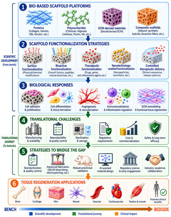
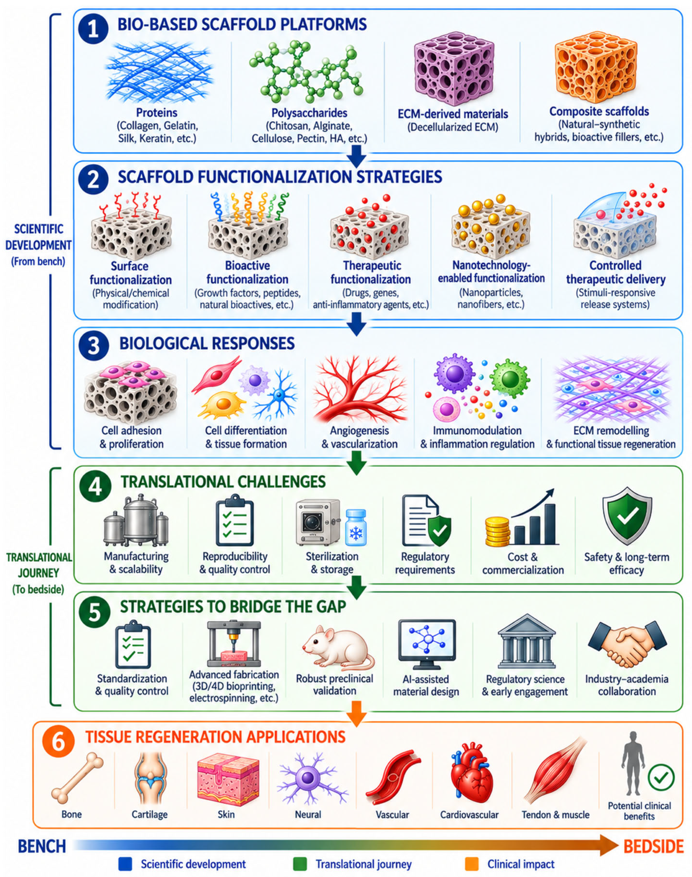
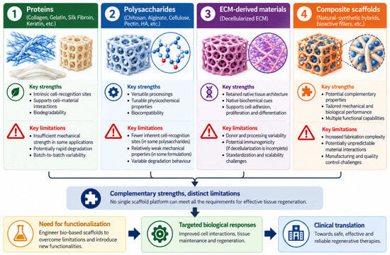
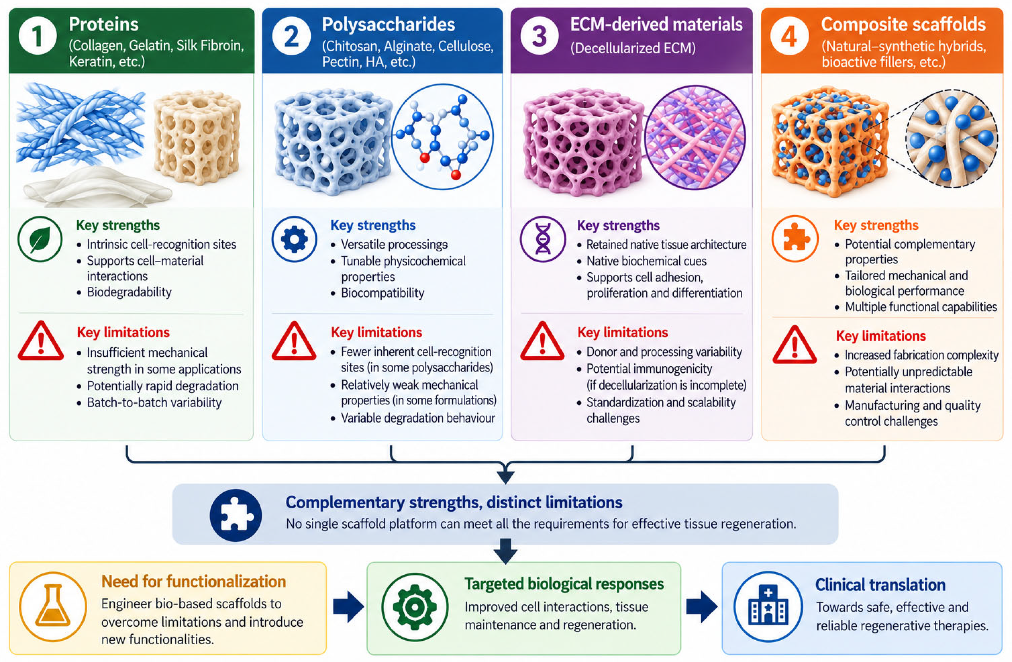
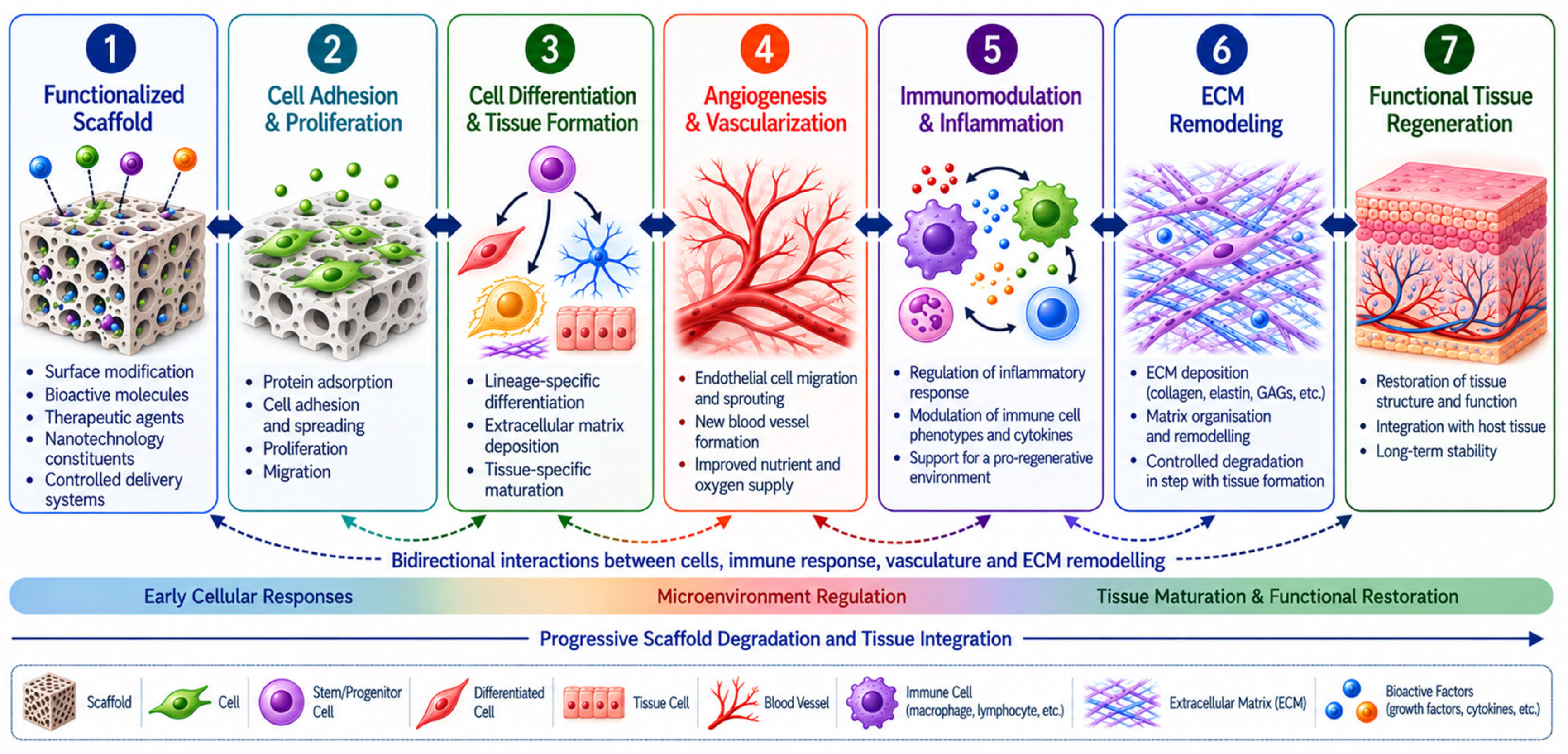

Disclaimer/Publisher’s Note: The statements, opinions and data contained in all publications are solely those of the individual author(s) and contributor(s) and not of MDPI and/or the editor(s). MDPI and/or the editor(s) disclaim responsibility for any injury to people or property resulting from any ideas, methods, instructions or products referred to in the content. |
© 2026 by the author. Licensee MDPI, Basel, Switzerland. This article is an open access article distributed under the terms and conditions of the Creative Commons Attribution (CC BY) license.