


Abstract
One of the promising methods for generating low-temperature plasma to address oncological issues is a spark discharge initiated by a piezotransformer. This is attributed to its relative simplicity and versatility in application, both in the direct treatment of biological objects in vitro and in vivo, as well as indirectly through the production of plasma-activated solutions. The study presents the results of a comprehensive study of the effect of spark discharge initiated by a piezotransformer in argon flow on the metabolic activity and survival rate of cancer HEp-2 cells. For this purpose, adhesive cells cultured in DMEM were subjected to plasma exposure of different duration (from 10 to 120 s). The produced effect was assessed by studying the spark discharge optical emission spectra, as well as by measuring the concentrations of reactive oxygen and nitrogen species in the liquid medium. Cell viability and metabolic rate were determined by MTT test and fluorescence microscopy using propidium iodide (PI) and Hoechst dyes. The metabolic activity of cells is reduced by half after 20 s of treatment, cell viability reduced after 150 s of treatment. The concentration of hydrogen peroxide H2O2 reaches a value of ~50 μM, and the concentration of nitrite ion NO2− reaches a value of ~20 μM.
1. Introduction
Modern technologies of surface treatment, including modification of biological objects, require the development of methods that combine high efficiency, environmental safety and minimal damaging effects. One of the most promising areas is the use of low-temperature plasma (LTP), which allows for controlled changes in surface properties without thermal degradation of the material being processed [1]. The impact of plasma treatment depends on the parameters of the resulting plasma, the type and configuration of the discharge. The most common types of discharge for creating LTP during treatment are barrier discharge, spark discharge, corona discharge and sliding arc discharge. Also, based on these types of discharges, plasma jets with a flow of noble gases and nitrogen are created. In the practical implementation in biomedicine and agriculture, important requirements are compactness, flexibility and tunability of the LTP source, and the possibility of its scaling.
The interaction of plasma and liquid is significant for addressing biological issues due to the unique plasma-chemical reactions near plasma-gas–liquid interfaces that result in the creation of reactive long-lived and short-lived species of oxygen and nitrogen (RONS) in the liquid [2,3]. This biological enhancement of liquid using LTP has been applied in various research fields, including the inactivation of pathogenic microorganisms (sterilization of medical equipment and antimicrobial action in the food industry, agriculture, veterinary medicine, and implantology), as well as in dentistry, dermatology, diabetic foot treatment, and antitumor therapy, among others [4,5,6,7]. The use of LTP in antitumor therapy [8,9,10] is relevant due to the claimed selectivity of the method and a gentler effect compared to known approaches involving radiation therapy, chemotherapy, surgery, or combinations of these methods. Notably, the use of plasma technologies for chemoresistant cancers is especially intriguing and promising; in these instances, it is essential to explore alternative treatment strategies that do not result in resistance to cytostatic effects. The multiplicative application of low-temperature plasma together with known chemical agents is also considered [11,12,13,14]. In this case, it is supposed to reduce the required dose of the drug, increase selectivity, and achieve a total gentler effect on the organism. Two methods of exposure are commonly considered: direct treatment with gas discharges and through plasma-treated solutions (PTS) [15].
Recent years have seen growing interest in biomedical research regarding the use of low-temperature plasma sources that rely on spark discharge initiated by a piezotransformer (PT), commonly referred to as piezoelectric direct discharge (PDD) [16,17,18]. This trend is primarily driven by improved prospects for device miniaturization, as the high voltage is generated at the very end of the working manipulator. The working manipulator can also be configured to change its configuration on the fly to generate several types of plasma: corona discharge, spark discharge, barrier discharge, and plasma jets. An increase in the treatment area is achieved by connecting the working devices to the matrix [19]. Taken together, this allows for the most flexible implementation of this type of source in medical practice: whether for generating plasma-activated solutions or directly treating biological tissue.
Thus, plasma treatment from a PT source significantly improves the tribological (wear resistance) and biocompatible properties of orthopedic implant materials, reducing wear rate by up to 60 times and increasing biocompatibility by 2–3 times compared to untreated surfaces [20]. Plasma treatment increases the surface roughness and hydrophilicity of polymers (e.g., PVDF), improving cell adhesion and cytocompatibility, which is valuable for tissue engineering [21]. PT sources plasma is also capable of inducing apoptosis and autophagy in cancer cells by disrupting glycolysis and key survival pathways, which is promising for oncotherapy, and it effectively inhibits the viability of bacteria (e.g., E. coli) on surfaces and in liquids [22,23].
LTP can be used for highly selective cancer treatment (to target and destroy tumors without damaging normal tissues). Plasma treatment affects a wide range of mechanisms: apoptosis, immunogenic cell death, pyroptosis, ferroptosis, necrosis, and modulation of the tumor immune microenvironment [24]. In addition, plasma treatment can increase the sensitivity of aggressive forms of cancer to chemotherapy by modulating intracellular cell cycle regulation pathways in vitro and in vivo [25]. One of the primary mechanisms of plasma action on cancer cells is the enhancement of the generation of active forms of oxygen and nitrogen [26]. The influence of the treatment method on the composition of active forms and their effect on cancer cells are described. Direct exposure can reduce the viability of YD-10B cells by enhancing the generation of ROS and RNS. Indirect plasma treatment causes RNS-dependent cancer cell death [27].
In a series of studies using the LTP source “CAPKO”, PDD has been shown to exert a dose-dependent cytotoxic effect on cancer cells in vitro. At the same time, it is possible to increase the effectiveness of antitumor drug (doxorubicin) with combined action on HeLa cells [28,29]. Further research has demonstrated that saline treated with PDD can serve as a non-specific cyto- and genotoxic agent, exhibiting prolonged effects [30]. A study [31] reported a comparison of the effects of four PTS prepared with PDD from isotonic solutions and investigated their combined cytotoxic effects with doxorubicin and medroxyprogesterone acetate.
Nevertheless, the results of using PDD-based plasma sources in solving oncologic problems to date are fragmentary and require a broader coverage: both from the side of cell line selection and from the side of treatment methods. Scientific publications using this type of source are not so common due to the much later appearance of commercial devices. In the present article, we examined the in vitro exposure of HEp-2 laryngeal carcinoma epithelial cells to argon plasma jet (initiated by PDD). The study aims to both systematically investigate the properties of argon flow discharge when applied to liquid media and to determine a semi-lethal dose of exposure to cells for transition to in vivo studies. The concentrations of long-lived reactive oxygen and nitrogen species were measured to assess the measure of plasma exposure.
2. Materials and Methods
2.1. Low-Temperature Plasma Source
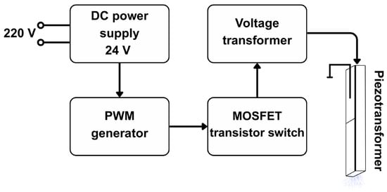
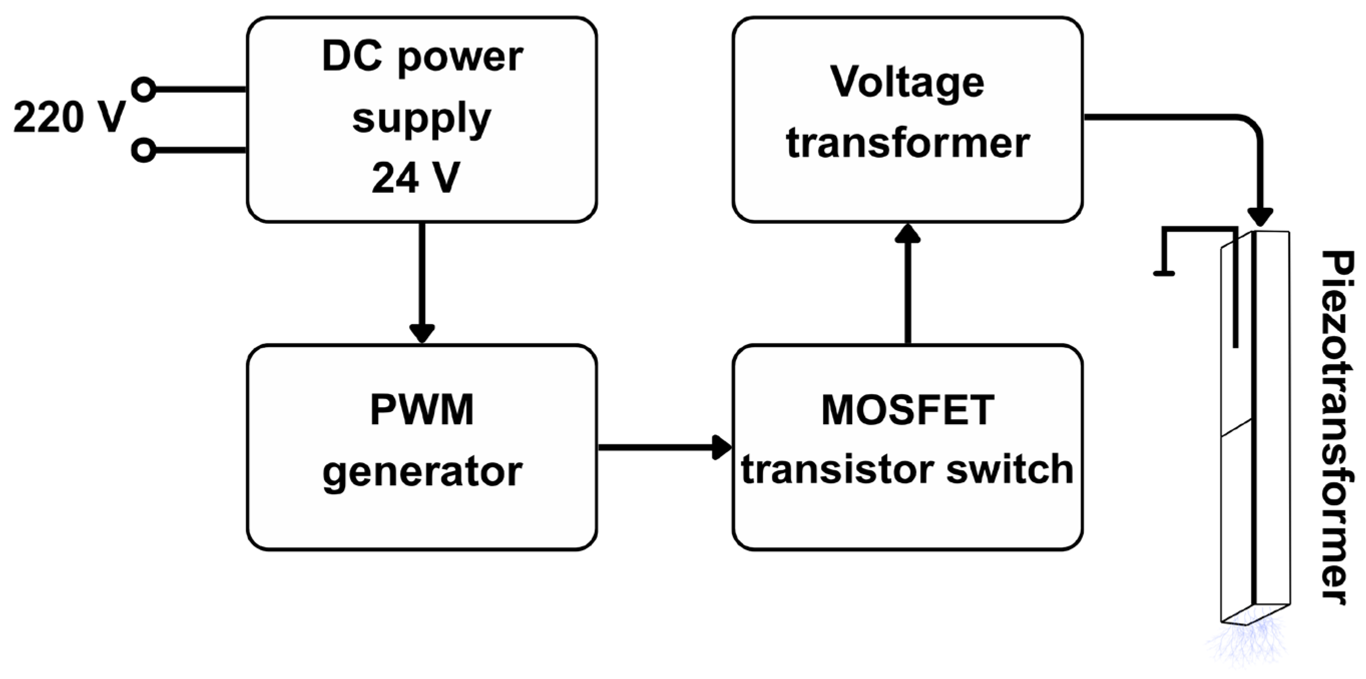
The second generation of the plasma source “CAPKO” was used in this work [23,32]. This source generates nanosecond spark discharges at the end of the piezotransformer [18,33] in each half-period of the supply voltage. The block diagram of the plasma source is shown in Figure 1. It consists of a DC source with a voltage of 24 V, which feeds a PWM generator necessary for feeding a signal to the step-up transformer through a transistor switch. The PWM generator makes it possible to adjust the frequency and amplitude of the voltage of the supplied signal. This source used a piezoelectric transformer from the Elpa Research Institute (Moscow, Russia), operating at half resonance frequency ~22 kHz. The voltage and current oscillograms of a nanosecond spark discharge in air were studied in detail in the article by Artem’ev et al. [33]. The discharge current duration was approximately 20 ns with an average peak value of 0.34 A. The energy released during one discharge was (2.05 ± 3.5) × 10−5 J. Four spark discharges occurred during one period (~45 μs). The voltage across the air gap before breakdown was (9.5 ± 1.1) kV, while after the spark discharge the voltage decreased to (4.55 ± 1.15) kV.
Figure 1.
Block diagram of a piezoelectric transformer source.
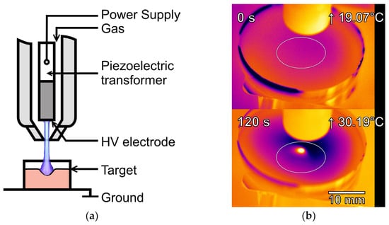
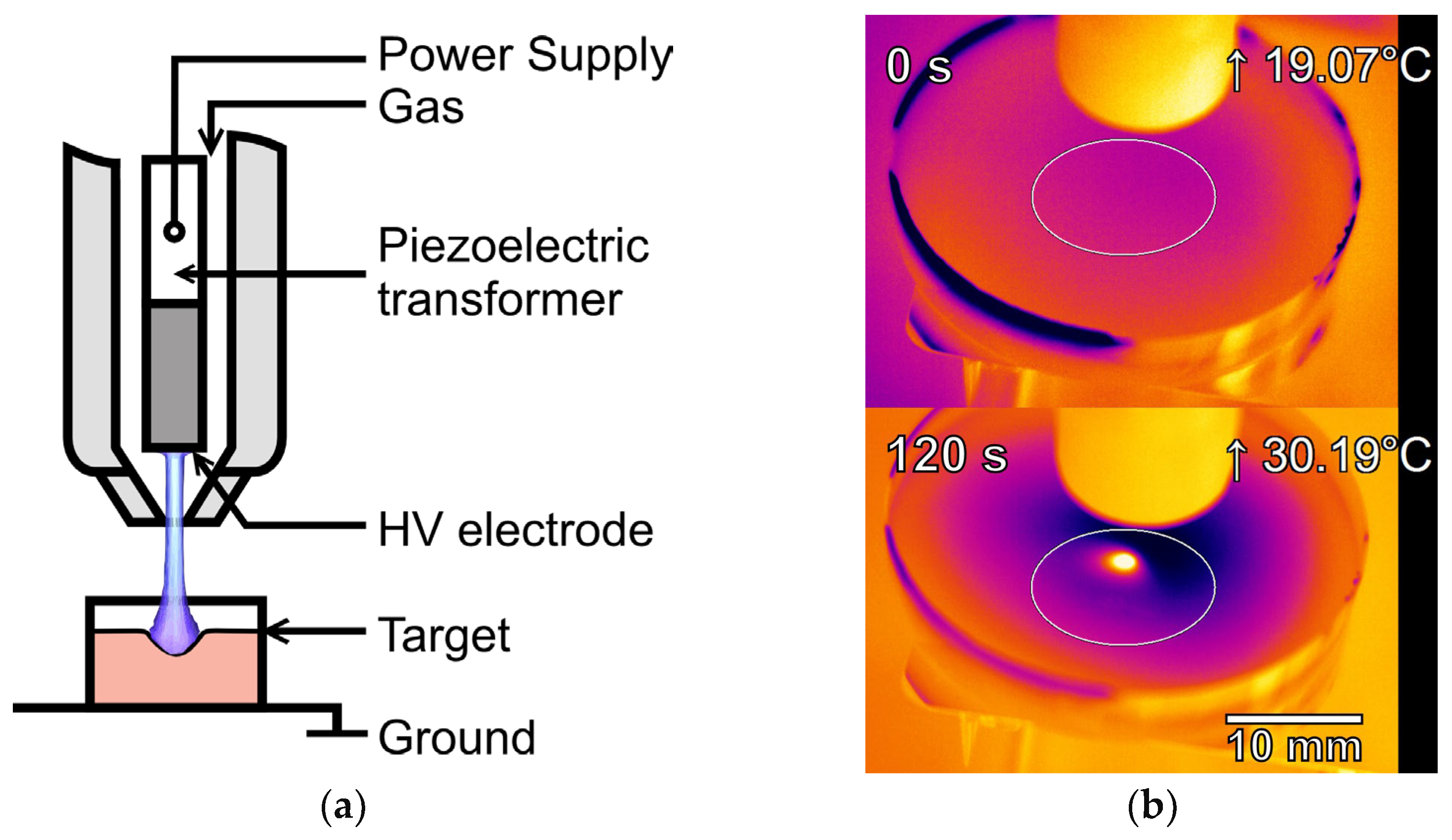
For a spark discharge in argon, the voltage and current waveforms are the same, but the energy released in one discharge should depend not only on the distance to the surface and its material, but also on the gas flow. We set the distance between the high-voltage electrode (the end of the piezoelectric transformer) and the surface to 10 mm, which allowed us to treat liquids placed in the wells of the different culture plates. The argon flow was set to a minimum (in our case, it was 2 slm), but sufficient to ensure stable discharge reproducibility and prevent overheating of the liquid surface (Figure 2b). The heating spot diameter on the surface is ~4 mm. The effective area (i.e., the area of significant RONS generation above the surface) is several times larger and is described by a spot diameter of ~10 mm at 45 s of treatment and ~26 mm at 300 s of treatment [23]. Setting a low gas flow when using a piezoelectric transformer as a discharge generator is also described in publications [16,34]. Nevertheless, studying the energy characteristics of this type of discharge at different gas flows, different distances to the target, and different target materials is an important task, which we will consider in future articles.
Figure 2.
Schematic illustration (a) of the HEp-2 laryngeal carcinoma cells treatment in liquid medium using discharge in an argon flow-through system with piezotransformer and maximum heating temperature (b) of the liquid surface before treatment and at 120 s of treatment. The distance between the high-voltage (HV) electrode and the liquid surface is ~10 mm, the argon flow is ~2 slm. The material of the liquid vessel is polystyrene (6 or 24 well plate); it is placed on a grounded metal lifting table.
2.2. Optical Emission Spectroscopy and Colorimetric Analysis of RONS
Emission spectroscopy was employed to analyze the discharge and its characteristics. Spectra were recorded using spectrometers AvaSpec-DUAL (Avantes, Apeldoorn, The Netherlands, spectral ranges 219–381 and 379–521 nm, average optical resolution 0.12 and 0.1 nm), StarLine AvaSpec-ULS2048CL (Avantes, Apeldoorn, The Netherlands, spectral range 517–748 nm, average optical resolution 0.3 nm), AvaSpec-3648 0802081U1 (Avantes, Apeldoorn, The Netherlands, spectral range 367–909 nm, average optical resolution 1.2 nm) with a treatment time of 1–5 s.
To evaluate the biological activity of the solutions acquired during plasma treatment, measurements of the concentrations of long-lived reactive oxygen and nitrogen species (H2O2, NO2−) were used in this work. The concentrations of H2O2, NO2− were determined by spectrophotometry: for this purpose, the methods were tested for selectivity towards other compounds present in the liquid. Hydrogen peroxide concentrations were measured using peroxide the FOX assay [35], nitrite ions using the Griess assay [36,37]. To measure the concentration of H2O2, NO2−, 600 μL of solutions were treated in a 24-well plate.
The reagent FOX stands for “ferrous oxidation in xylenol orange”—oxidation of iron in xylenol orange. The method is based on selective oxidation by iron hydroperoxides to ions in dilute acid (1). The iron ions formed can be determined using iron-sensitive dyes as an indirect measure of hydrogen peroxide concentration (Fenton reaction).
Fe2+ + ROOH → Fe3++ RO∙ + OH−
Fe3+ + XO → orange complex (560 nm)
Xylenol orange (XO) binds iron ions with high selectivity, forming a colored complex (2).
The Griess assay is based on the ability of nitrite ions to diazotize sulfanilic acid to form a red-violet azo dye diazo compound by reaction with α-naphthylamine; the optical density of the solution was measured at λ = 525 nm.
2.3. Analysis of Cytotoxic Effects
The viability of HEp-2 cells was analyzed using MTT test and fluorescent microscopic method. HEp-2 cells were cultured and subcultered by standard protocols on DMEM/F12 medium, supplemented by 10% fetal bovine serum, 2 mM L-glutamine, 25 units/mL penicillin and 25 μg/mL streptomycin (PanEco, Moscow, Russia). After culturing, cells were detached from the culture vial using 0.05% trypsin 0.53 mM EDTa mixture (PanEco, Moscow, Russia). Cells were seeded in a 24-well plate at a rate of 50,000 cells in 600 μL of DMEM culture medium per well for MTT test, and in a 6-well plate at a rate of 300,000 cells in 3 mL of culture medium for microscopic analysis. After 24 h, medium replacement was performed followed by argon plasma jet treatment and incubation of cells for 48 h. The incubation time was chosen on base on the literature data on HEp-2 cells viability assays in studies of anticancer screening and plasma-dependent effects [38,39]. After incubation, MTT assay (λ = 570 nm, formazan solvent was DMSO) and fluorescence microscopy analysis (PI and Hoechst dyes) were performed. In this work, the protocol for the assay was taken from [40].
2.4. Statistical Processing
The data were processed using SigmaPlot v 10 (Grafiti LLC, Palo Alto, CA, USA) and Origin 2021 software (OriginLab Corporation, Northampton, MA, USA). The data were presented as mean values ± SE or medians and interquartile ranges depending on the distribution of values. Normality was assessed using the Shapiro–Wilk test. The statistical significance of differences was assessed using the t-test and Kruskal–Wallis ANOVA with Dunn’s test. For each variant, 3–5 independent measurements were performed.
3. Results and Discussion
3.1. Characteristics of Emission Spectra and RONS Concentrations
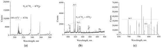
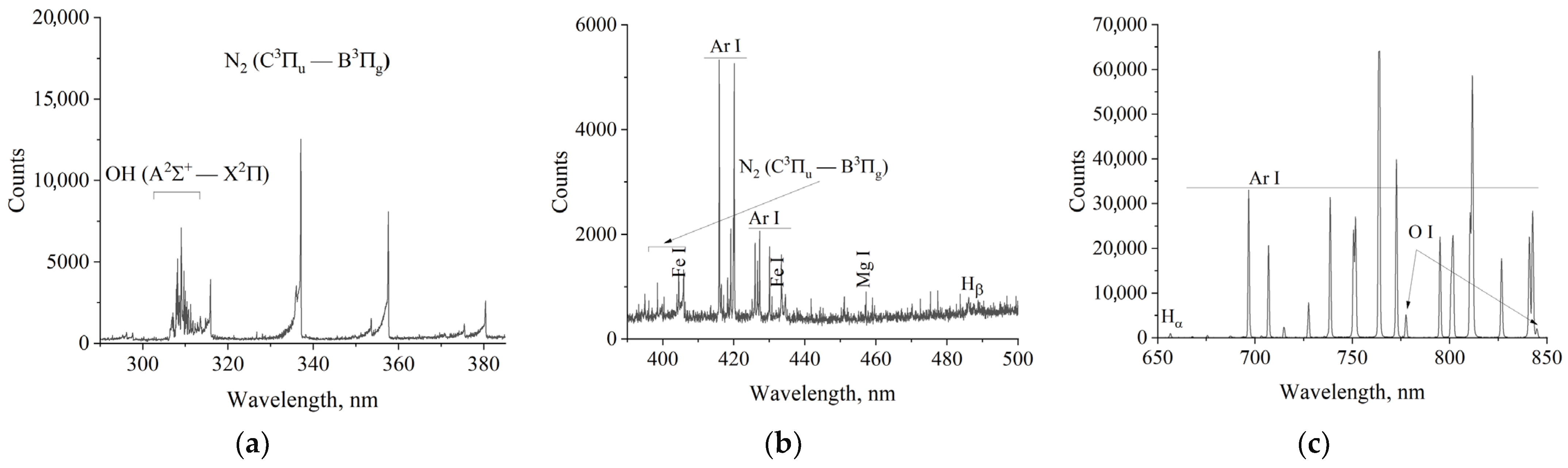
The spectra (Figure 3a–c) show molecular bands N2, OH and lines of atoms O I (777.4 and 844.6 nm), Hα (656.3 nm), Ar I, as well as some lines corresponding to the solution components—Fe I, Mg I. The rotational temperature when the medium is exposed to a discharge in argon flow is 700 ± 100 K in the OH band.
Figure 3.
Optical emission spectra of piezotransformer-based discharge in argon flow during DMEM treatment in the ranges of 290-380 nm (a), 380-500 nm (b) and 650-850 nm (c).
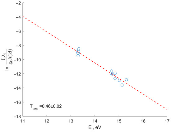
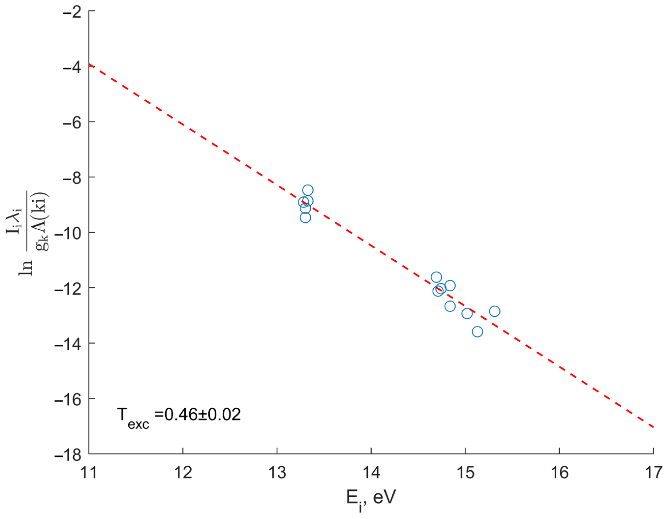
To determine the excitation temperature, argon Ar I lines are used (Table 1). The temperature is determined by a Boltzmann plot ln(Iλ/gA), where I is the intensity of the emission line in the spectrum, λ is the line length, A is the probability of a radiative transition, g is the statistical weight of the upper transition level, and Ei is the energy of the upper level.
Table 1.
Observed argon Ar I lines in the spectral composition in the range 600–740 nm.
The excitation temperature (Figure 4), determined by Ar I lines in the exposure mode of a nanosecond pulsed discharge in argon flow (argon plasma jet) and characterizing the low-energy part of the EEDF, is 0.46 ± 0.02 eV.
Figure 4.
Boltzmann plot.
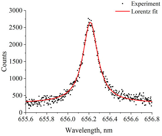
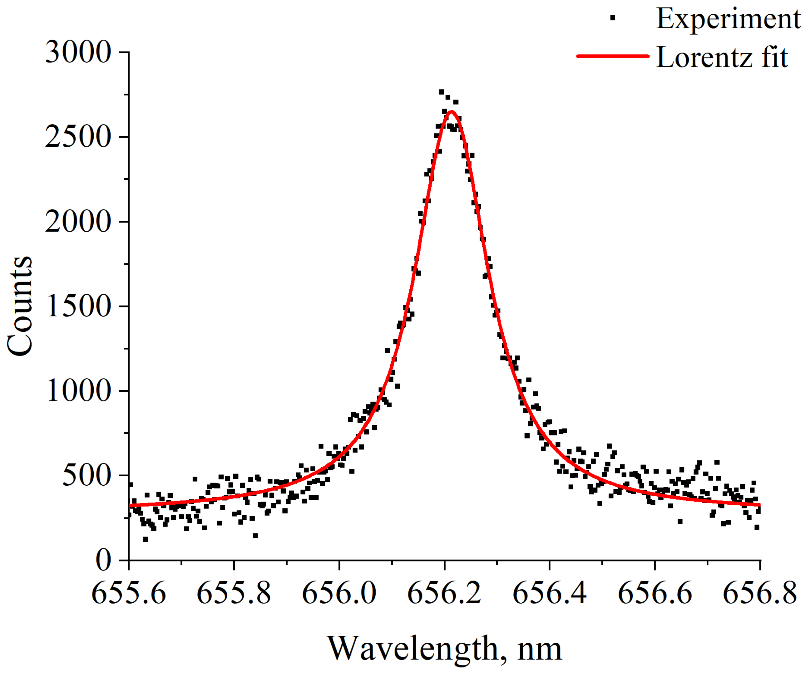
The concentration of low-temperature plasma was determined by the broadening of the Hα line due to the linear Stark effect (Figure 5) [41]. The half-width of the experimental Hα line in the case of exposure to DMEM nutrient medium was 1.70 Å, which, with a half-width of the instrumental function of 0.38 Å, corresponds to concentrations of ne~9.5 × 1015 cm−3 (estimated by half-area, FWHA) [42].
Figure 5.
Experimental spectrum of Hα line and approximation by Lorentz function.
From the point of view of plasma-treated solutions application, the analysis of the concentration of long-lived active forms of oxygen and nitrogen fixed in the liquid is of great importance. The easiest substances to determine are hydrogen peroxide H2O2 and nitrite ions NO2−. Under plasma exposure of DMEM culture medium to a nanosecond discharge in argon flow (argon plasma jet), the concentrations of hydrogen peroxide and nitrite ions were measured using the methods described above. The volume of liquid in each well of a 24-well culture plate was 600 μL and the surface area was 1.9 cm2.
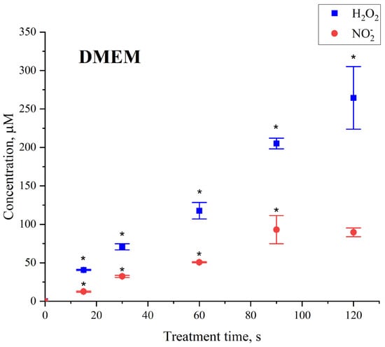
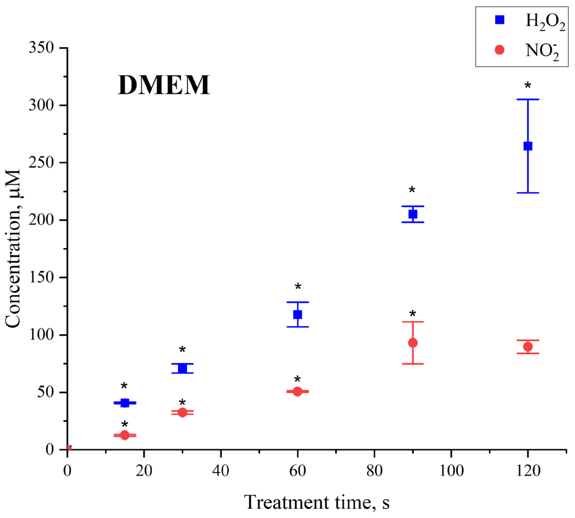
The concentration of hydrogen peroxide increases with treatment time, with a semi-lethal dose corresponding to a concentration on the order of 50 µM. Measurements were limited to the time at which the MTT test resulted in the lowest cancer cell survival (up to 10%). The concentration of nitrite ions also increased with exposure time, with a value of 20 μM (Figure 6).
Figure 6.
Dependence of hydrogen peroxide and nitrite ion concentration on treatment time of DMEM. Data presented as mean ± SE. *—t-test vs. corresponding control (n = 3, three different solution (independent preparations) samples were used in each experimental group).
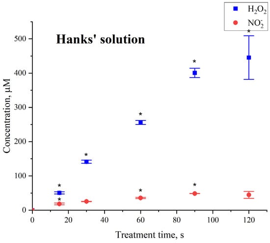
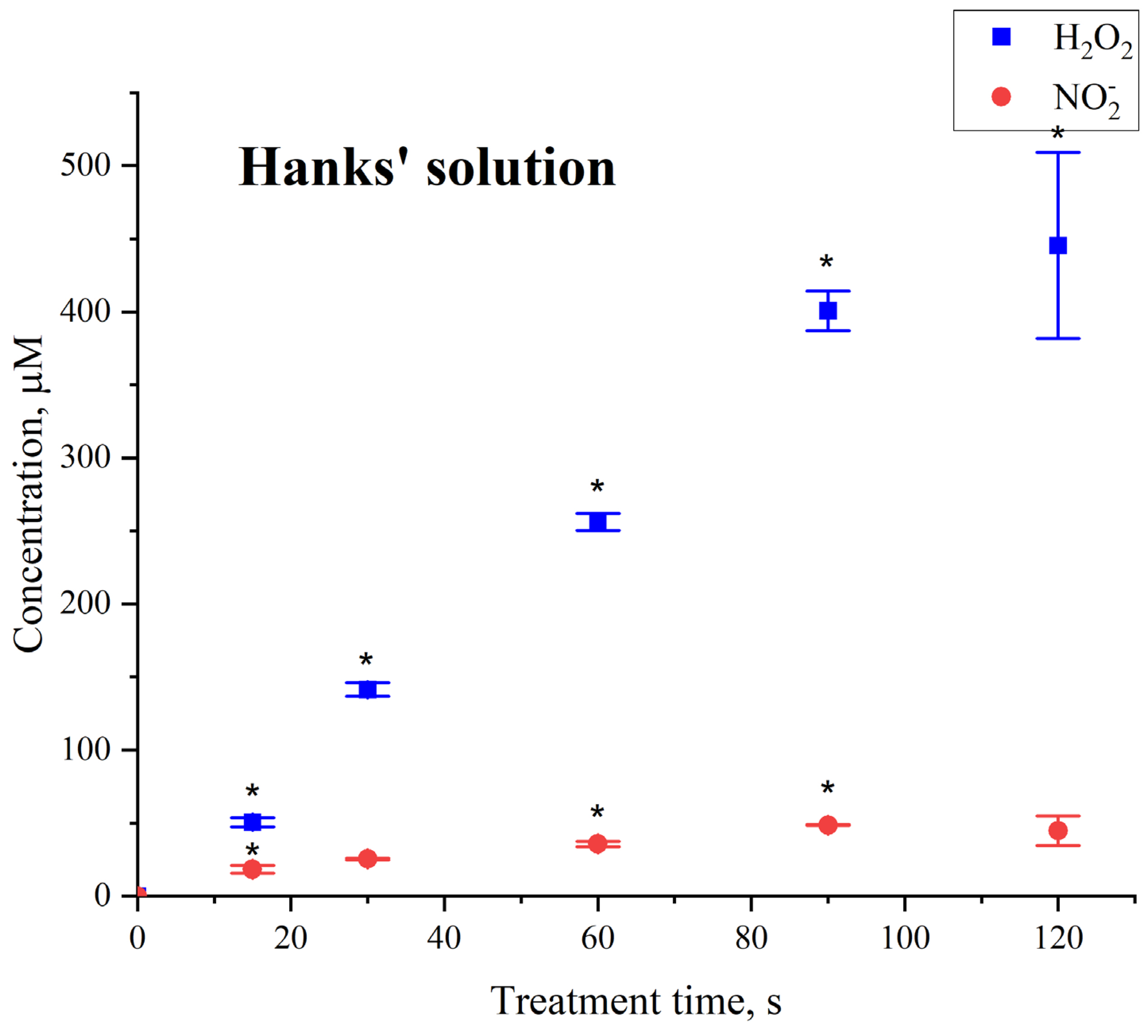
The production of reactive oxygen and nitrogen species was also assessed when exposed to Hanks’ solution (Figure 7). Under these conditions, the concentration of nitrite ions attained lower levels and stabilized after 30 s of exposure, while the concentration of hydrogen peroxide was higher and plateaued at 90 s of exposure.
Figure 7.
Dependence of hydrogen peroxide and nitrite ion concentration on treatment time of Hanks’ solution. Data presented as mean ± SE. *—t-test vs. corresponding control (n = 3, three different solution (independent preparations) samples were used in each experimental group).
Hanks’ solution does not contain nitrogen compounds, nitrates, nitrites, ammonium compounds, and/or amino acids. Therefore, RNS generation in this solution is very low and may be caused by a small amount of dissolved molecular nitrogen. DMEM contains the following nitrogen-containing compounds: ferric nitrate, amino acids (L-arginine, L-Cystine, Glycine, L-Lysine L-Isoleucine, etc., 14 in total), vitamins (Choline Chloride, Folic Acid, Niacinamide, D-Pantothenic Acid, Pyridoxal, Pyridoxine Riboflavin Thiamine). The total mass of nitrogen-containing compounds in the medium is 1.06 g/L. The mass fraction of nitrogen in the compounds varies from 6.39 to 31.17% with an average value of ~15%. Based on the mass fractions of nitrogen for each compound, the total concentration of this element is 0.15 g/L. Consequently, the concentration of atomic nitrogen is 11 mM. For a medium volume of 600 µL subjected to plasma treatment, provided that all nitrogen is completely converted to RNS, generation of up to 6.6 mM NO2− is possible. In the experiment, we observed the production of no more than 100 µM NO2− in the DMEM medium with an exit to a plateau. This indicates that only a portion of the compounds in DMEM can act as a nitrogen atom donor for the generation of RNS. Only 1.5% of all atomic nitrogen in organic compounds is converted to NO2− after 150 s of plasma treatment. We believe that the most likely nitrogen donor for the generation of RNS may be L-arginine as a natural nitrogen “depot” in a body and a natural source of NO and NO2− generation during the regulation of vascular tone [43,44]. The contribution of other compounds is also likely, but their concentrations and/or mass fractions of nitrogen are significantly lower. However, even for L-arginine, the conversion of nitrogen to NO2− was far from the theoretical maximum ~470 µM NO2− (provided that only one nitrogen atom is oxidized). It is worth noting that the conversion of all nitrogen-containing compounds into RNS was not the objective of plasma treatment, as it is expected that, along with the reduction in tumor cell viability, the viability of adjacent living cells must be maintained. We assume that, in our case, the conversion of no more than 1.5% organic nitrogen into RNS (subject to a reduction in cancer cell viability) is adequate. Clarification of the molecular transformations of individual medium components and an assessment of the cytotoxicity of the transformation products will be the goal of future research. According to the literature, plasma treatment of DMEM in an argon environment causes its slight alkalization. This change in pH cannot lead to serious chemical modifications of the components of the medium; however, it can cause cell alkalosis and be one of the mechanisms for reducing cellular viability.
3.2. Cell Viability and Potential Mechanism of Cell Death
The HEp-2 (ATCC CCL-23) human carcinoma culture was selected as the model. Initially, it was believed that this line was derived from squamous cell carcinoma of the larynx, but subsequently, based on isoenzyme analysis, HeLa cell marker chromosomes and DNA fingerprinting, it was established that it was created by contamination with HeLa cells [45,46]. We have previously conducted work on HeLa cells. The work on HEp-2 in this case is a continuation of this cycle of work.
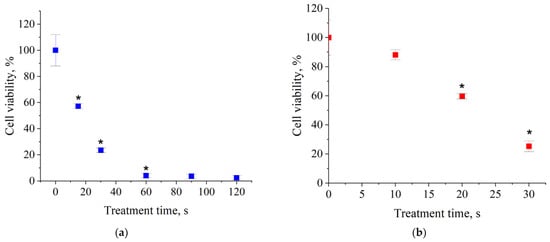
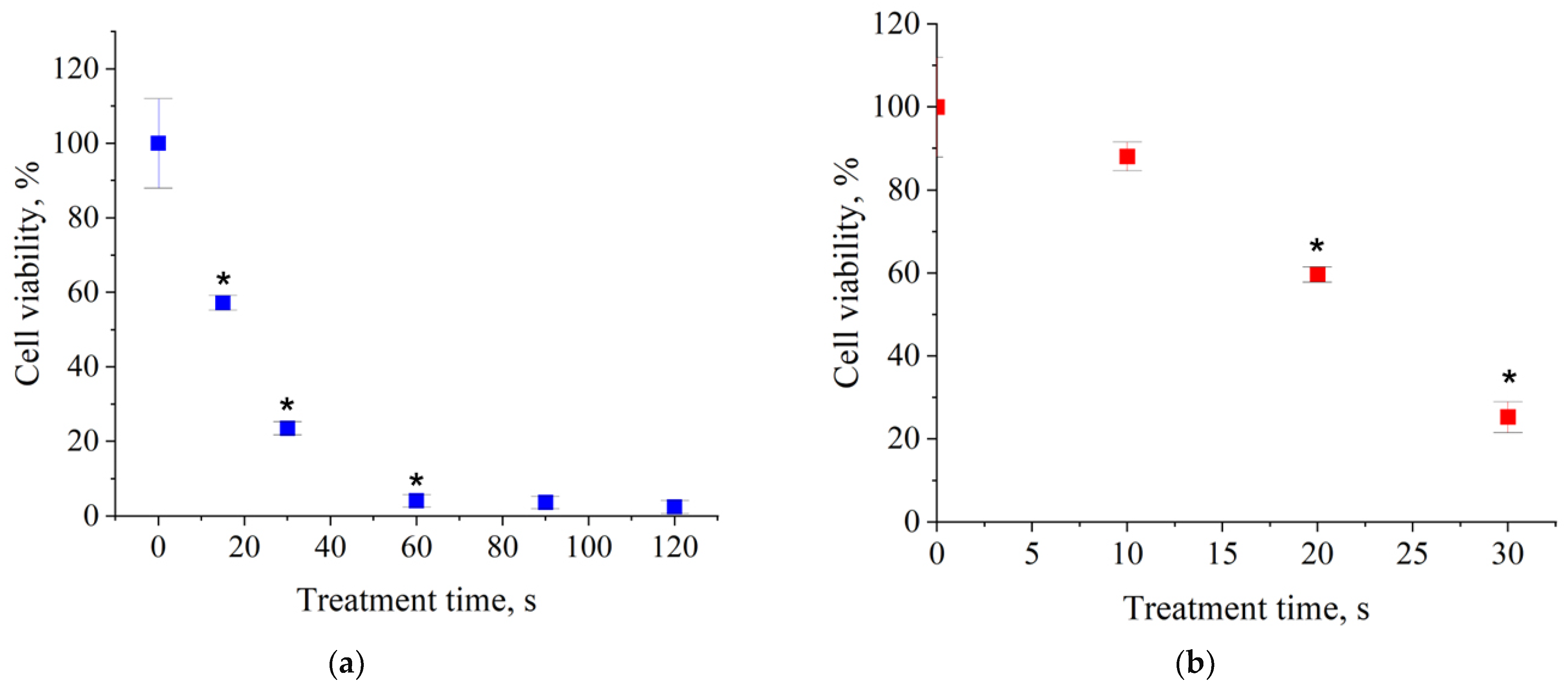
In the first stage, we analyzed the effect of plasma treatment on the metabolic activity of cells in culture using the MTT test. The rate of tetrazolium dye reduction to detectable formazan depends on the activity of mitochondrial NADPH-dependent oxidoreductases [47,48]. At this stage, we observed a decrease in the metabolic activity of cells after plasma treatment for ≥20 s (Figure 8a,b). Metabolic activity measured by MMT often correlates with culture viability; however, a decrease in mitochondrial oxidoreductase activity may indicate not only cell death but also a switch from aerobic metabolism to glycolysis [49]. We suggest that plasma treatment also causes cells to switch to stress-induced glycolysis. The impairment of mitochondrial function requires confirmation in tests involving potential-sensitive mitochondrial probes, calcein, or an assessment of mitochondrial colocalization with lysosomes. Verification and refinement of our hypotheses is the subject of future research.
Figure 8.
Dependence of HEp-2 cells metabolic activity (NADPH-dependent cellular oxidoreductases activity evatuated by MTT test) on treatment time in two scales: up to 120 s of treatment (a) and up to 30 s of treatment (b). Data presented as mean ± SE, *—p < 0.05 vs. corresponding control, Kruskal–Wallis ANOVA with Dunn’s test (n = 3, three biological replicates (independent culture preparations) were performed in each experimental group).
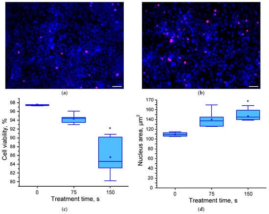
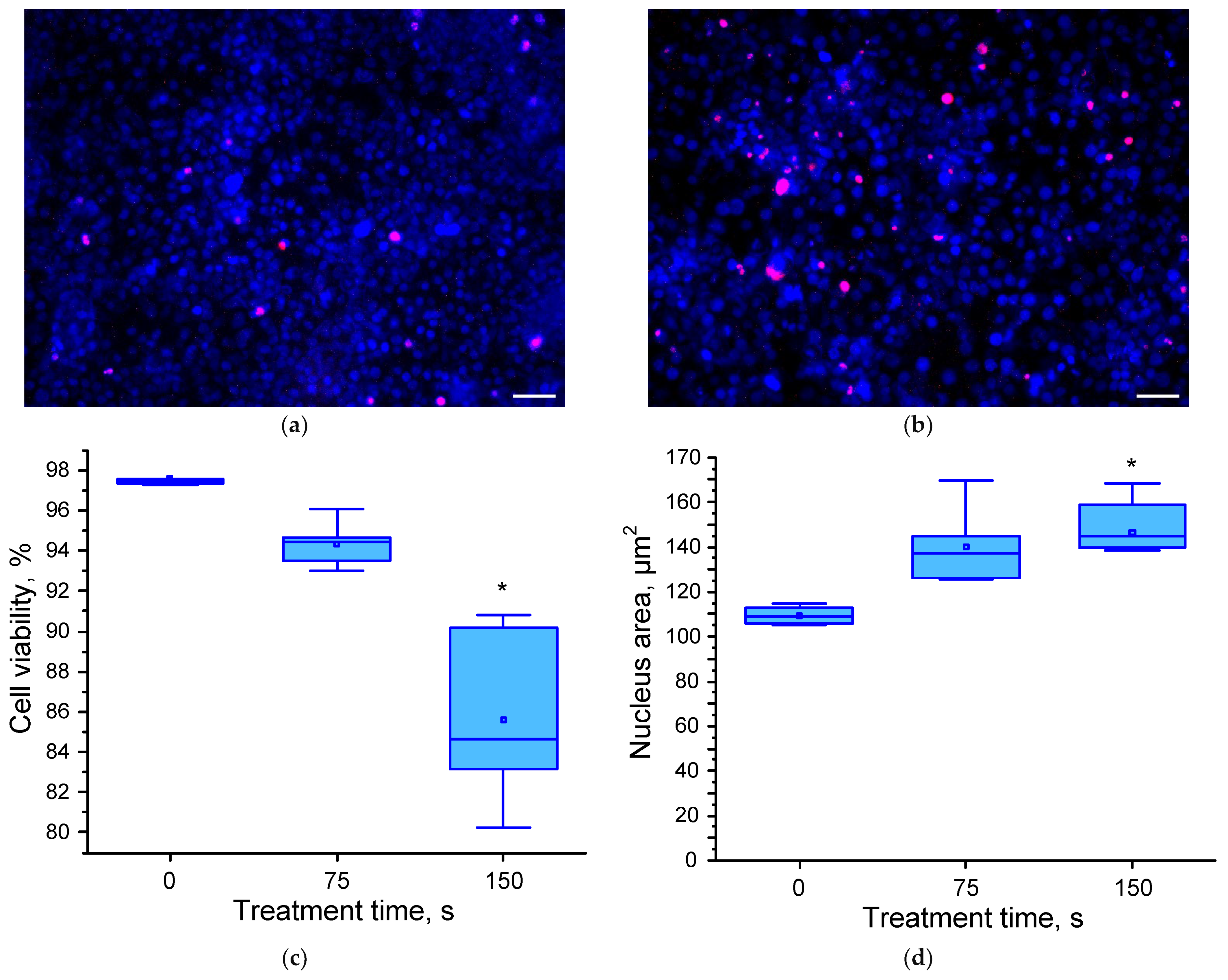
To clarify the results of the MTT test, we performed a series of viability measurements using double staining with Hoechst vital dye and PI (which selectively stains dead cells). For microscopy the volume of liquid being treated was increased to 3 mL (5-fold), and the duration of plasma treatment was proportionally increased to 150 s (5-fold). We can expect that with such small liquid volumes, the concentration of RONS generated in the liquid will remain approximately the same, and consequently, the “dose” of exposure to the cells will remain approximately the same. To assess the potential mechanism of cell death, we evaluated the average nucleus size. Apoptosis induction is accompanied by a decrease in the average nucleus size, while necrosis, on the contrary, is accompanied by an increase in the nucleus size [50,51]. The initial viability of cells in culture without treatment was ~98%. After 150 s of plasma treatment, there was a clear increase in the number of PI-positive cells (dead) compared to the control (Figure 9a,b). The decrease in viability after 150 s of treatment was ~14% compared to the control (Figure 9c). The differences between the microscopy and MTT test results indicate that plasma treatment initially affects the activity of mitochondrial oxidoreductases. It is noteworthy that after 150 s of treatment, an increase in nucleus size is observed, indicating that the cells most likely die by necrosis (Figure 9d).
Figure 9.
Dependence of HEp-2 viability and nucleus area on plasma treatment time. Examples of fluorescence microscopy photos before (a) and after (b) 150 s exposure. Average cell viability (c) and area of nucleus (d). Data presented as median and 10, 25, 75, and 90 percentiles. *—p < 0.05 vs. corresponding control, Kruskal–Wallis ANOVA with Dunn’s test (n = 5, five biological replicates (independent culture preparations) were performed in each experimental group). Blue color—living and early apoptotic cells, red—necrotic and late apoptotic cells. Scale bar is 50 µm.
In present study, we found a decrease in the metabolic activity of cancer cells and a significant reduction in their viability after plasma treatment. At the expected level, a flexible cell may undergo necrosis. In the case of apoptosis, nuclear pyknosis has been described [52]. In our case, nuclear pyknosis was not observed. However, a more detailed assessment of the cell dead pathway during plasma treatment requires Annexin V/PI double staining, caspase activation analysis, and a time-lapse viability study with sequential exposure geometry. These studies are the focus of future research. Furthermore, the effectiveness of plasma treatment on different cell lines will be studied.
In helium plasma micron jet was investigated on murine breast cancer 4T1 cells in vitro and in vivo [53]. According to the literature data, the major cell population dies by late apoptosis during plasma exposure, and when chemotherapy is used, the predominant mode of cell death is necrosis. An in vivo study showed that micron sized plasma jet treatment inhibited tumor growth. Plasma can induce apoptosis in tumor cells through activation of p-53 and Bax/Bcl-2 proteins [53]. Our results seem to coincide with data on plasma-induced necrosis of cancer cells [54,55]. The choice of a specific cell death pathway probably depends on the specific experimental conditions, in particular, the difference in the cell lines used. Optimization of treatment regimens and methods for assessing pro-apoptotic and pro-necrotic activities will be the subject of future research. In the case of plasma-dependent induction of necrosis, specificity of action can be ensured by localizing the effect. The ability to induce cell death through increased NOx and H2O2 generation is described in the literature [56,57]. Our data on increased ROS and RNS generation are consistent with the mechanism described in the literature.
Reactive oxygen and nitrogen species (RONS) can be generated in normal cells during mitochondrial functions and/or work of NO-synthase (NOS) and SOD1. Low concentrations of RONS are successfully eliminated by antioxidant systems of a cell [58,59,60].
High concentration of reactive oxygen and nitrogen species (RONS) can induce cell, including cancer lines, death due apoptosis or necrosis. This is described in detail in the literature on other cancer cells, such as LN229, [61,62]. ROS can induce next sequence of events: mitochondrial membrane depolarization, cytochrome c releasing, caspase-3 activation and finally apoptosis induction [63]. RNS disrupt the cellular redox balance, alter the activity of intracellular signaling proteins, and cause oxidative damage to DNA, proteins, and lipids [64].
The literature describes drug approaches to increasing ROS production as a potential anticancer therapy, using pancreatic adenocarcinoma cell lines, A549 and NCI-H1299 lung cancer cells, and MCF-7 and MDA-MB-468 breast cancer cells as an example [65,66,67,68].
ROS-induced apoptosis, triggered by mitochondrial depolarization followed by activation of PI3K/Akt-dependent signaling pathways, has been demonstrated and well described for HeLa and HEp-2 cells [69,70,71].
Based on measurements of changes in RONS concentrations in Hank’s saline and DMEM culture medium, we suggest the involvement of RONS in increasing HEp-2 cell death in our case.
Since we detected the generation of both RNS (NO2−) and ROS (H2O2) in the culture medium, we hypothesize a combined effect of these two classes of compounds. We propose a combined contribution of the lower angle to the proportional forms of the ratio of generated active substances. In our case, ~2 times more occurs in ROS than in RNS (Figure 6). Therefore, ROS-induced oxidative stress may be a more probable mechanism of the cytotoxic effect. The paper details that ROS and RNS can participate in each other’s generation and/or work together [60]. For more detailed assessments of their interactions and contribution to cell death, we recommend studies with quantitative assessment of both RONS incorporated into cells (fluorescence microscopy using RONS-sensitive probes: DCF, MitoSOX, DAF-2, etc.) and modifications of intrapulmonary signaling pathways (activation of caspase-3, p52, etc.)
In study [72] two types of devices were used as a source of LTP. For helium flow, a single-electrode dielectric barrier discharge (DBD) jet was used, in which the high-voltage electrode consists of a conducting cylindrical shell placed on a dielectric insulator (quartz). For the argon jet, the system also consisted of a single electrode, with the difference that the high-voltage electrode was placed as a needle inside a quartz tube. Both were powered by an AC source with a frequency of 20 kHz and a sinusoidal waveform, and the voltage was 4.5 kV. Helium and argon streams were supplied at a rate of 2 L per minute. The effects on U-87 MG (glioblastoma) cells were investigated both directly and through treatment of culture medium (DMEM) and then addition of plasma-activated medium to the cells. Incubation times ranged from 24 to 72 h. Based on the calculated semi-lethal dose values, the optimal treatment times were 70.15 and 138 s at the 48 h time point (incubation time) after treatment for direct and indirect helium jet exposure, and 202 and 116 s at the 72 h time point after treatment for direct and indirect argon jet exposure. It was also shown by flow cytometry data that the direct argon jet exposure route was the most effective at inducing apoptosis.
When HEp-2 cells were treated with the LTP source used in the present work, 50% of cells transitioned from a state of stress-induced glycolysis. The dose was achieved in 20 s, and cell viability began to decline after 150 s. The activation of glycolysis can be regarded as a consequence of mitophagy induction [73]. Further clarification of the presence of plasma-induced mitophagy is the aim of future work.
3.3. Prospects
The current research was conducted using a laboratory plasma source operating at driving frequencies up to 30 kHz, which has several limitations: low repetition frequency of discharge pulses, imperfect synchronization of voltage and current waveforms, and the absence of adaptive control under varying load conditions. In contrast, modern piezoelectric plasma generators based on high-performance piezoelectric transformers (e.g., piezobrush® PZ3 device) typically operate close to their mechanical resonance at substantially higher frequencies of about 80 kHz, enabling very high voltage transformation ratios (>103) and efficient generation of chemically active species at relatively low input voltages of 5–25 V and powers in the range of only a few watts (often below 10 W) [18]. For example, it has been shown that an atmospheric-pressure plasma jet powered by a piezoelectric direct discharge using a CeraPlas™-type transformer produces stable air and compressed dry air discharges with substantial ozone formation, with the ozone production rate increasing monotonically with input power at a fixed resonance frequency in the tens-of-kilohertz range, reaching ≈70–80 mg/h at 8 W of input power (for example, see piezobrush® PZ3 ozone data). Since these long-lived oxidizing species (O3, H2O2, NO2−, etc.) are efficiently transferred into the liquid phase during plasma-activated water production, a higher pulse repetition rate at the transformer resonance (while keeping the discharge in the so-called cold-plasma regime) directly translates into a higher cumulative dose of reactive oxygen and nitrogen species delivered to the treated liquid per unit time compared to our present 30 kHz prototype.
Furthermore, several experimental studies have demonstrated that the ignition, stability and efficiency of piezoelectric-transformer-based plasma sources are strongly governed by the instantaneous resonance frequency and the effective voltage transformation ratio of the transformer, both of which are modified by the plasma load and by changes in external conditions such as gas composition, discharge gap or the presence of a liquid layer [74,75]. Spatiotemporally resolved measurements of the electric field around a piezoelectric transformer using the E-FISH technique revealed a pronounced modification of the local electric field distribution and an effective down-shift in the resonance frequency when the transformer is loaded by an atmospheric-pressure plasma jet, which clearly indicates the need for active resonance tracking and adaptive control to maintain the desired discharge characteristics under dynamic loading conditions. Finally, earlier works on discharge plasmas generated by several piezoelectric transformers connected in parallel have already demonstrated that multi-transformer configurations can be used to scale up the discharge area and power at atmospheric pressure, provided that the excitation is properly matched to the joint resonance of the system. These observations together provide a strong motivation for the development of a new high-frequency piezoelectric-transformer-based plasma source with adaptive resonance control and the possibility of operating multiple transformers, which is expected to increase the production rate of reactive species in liquids and to ensure stable operation over a wide range of external loads, beyond what is achievable with the present 30 kHz laboratory source.
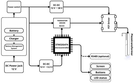
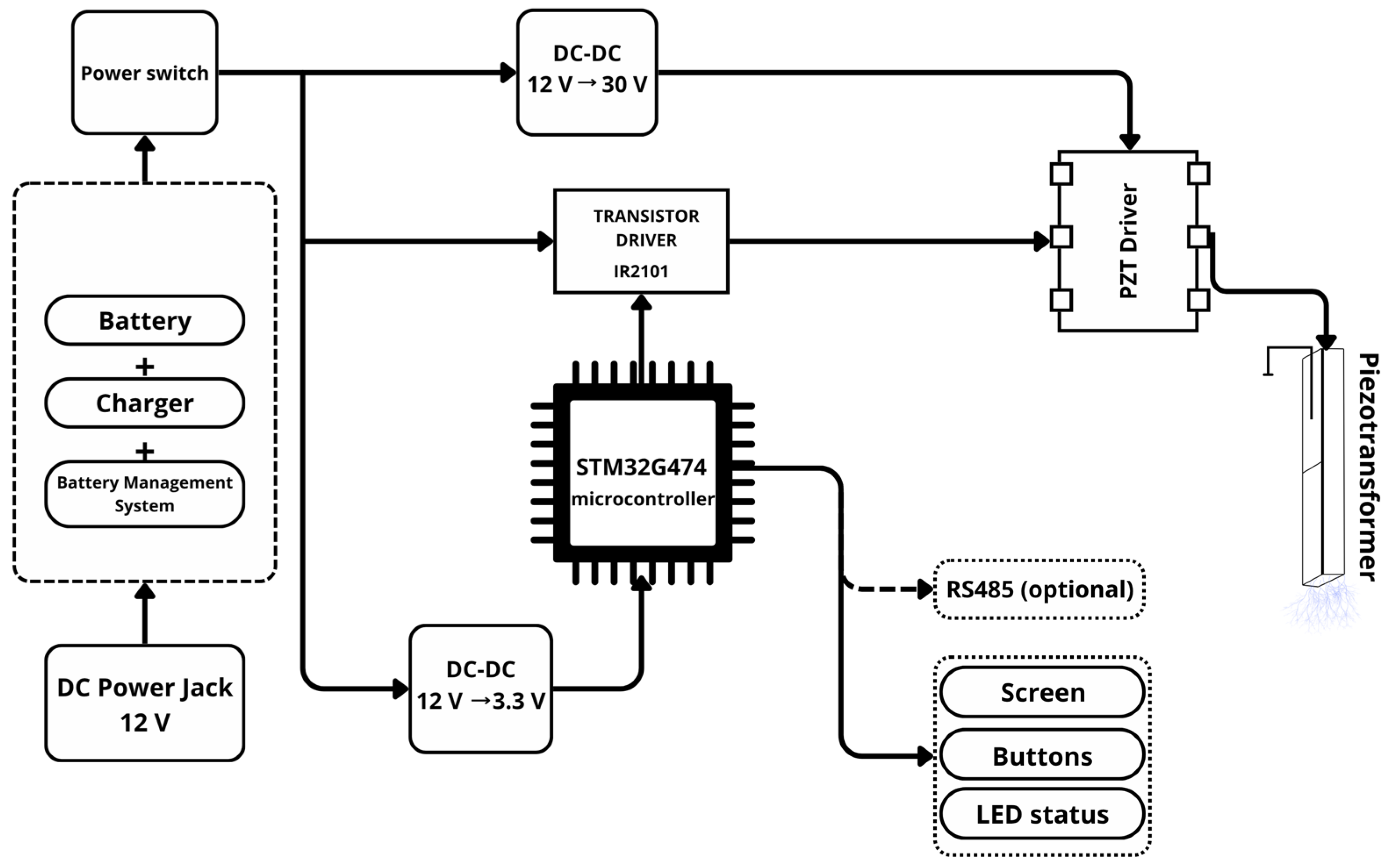
Based on the conducted research and approaches to efficient control of piezoelectric transformers [18,76], a third-generation plasma source was developed. The new source is based on a digitally controlled Class E amplifier topology (the block diagram is shown in Figure 10). The key difference from the previous-generation laboratory setup is the use of a microcontroller control system with voltage and current feedback.
Figure 10.
Block diagram of a third-generation piezoelectric transformer source of low-temperature plasma.
The new device operates using a microcontroller that generates a signal with an adjustable frequency (from a few hertz to 100 kHz). A Class E amplifier in the piezoelectric transformer driver (PZT driver) provides pulsed amplification the signal, efficiently converting the power supply into a high-frequency oscillation during each half-cycle of the input voltage. The piezoelectric transformer converts the electrical signal into a high voltage to initiate the discharge. The feedback system continuously monitors the voltage and current, allowing the microcontroller to adaptively adjust the operating frequency in accordance with the changing electrical parameters of the object being processed.
Thus, the new architecture provides: digital adaptive frequency control up to 100 kHz; pulse synchronization during each half-cycle of the input signal; and compensation for changes in the resonant frequency of the piezoelectric transformer during the processing of biological media.
Higher operating frequency combined with Class E amplification improves the efficiency of reactive oxygen and nitrogen species generation while maintaining constant exposure time compared to the previous design. The portable design with autonomous power supply enables clinical deployment of the system.
To demonstrate the effectiveness of the new source, we plan the following:
Comparative validation of plasma parameters between the current and new devices under identical conditions to substantiate the stated performance advantages.
In vitro and in vivo studies based on validated data to assess the biological efficacy and safety of the treatment.
Clinical trials conducted according to established protocols to establish clinical applicability.
4. Conclusions
The use of alternative methods in the treatment of oncological diseases is especially in demand for chemotherapy-resistant cancers, as well as tumors where chemotherapy does not give a positive prognosis. The research is devoted to the systematic study of the properties of nanosecond pulse discharge based on piezotransformer with argon flow (argon plasma jet) when acting on liquid media, as well as the application of this plasma source in antitumor therapy. The formation of long-lived reactive oxygen and nitrogen species is used as an assessment of plasma exposure.
Low-temperature plasma in antitumor therapy has proven to be a potentially effective and inexpensive treatment modality; however, large-scale clinical studies are needed to validate the model. When new plasma sources are introduced into practice, it is also necessary to introduce uniform parameters that will allow the effectiveness of the treatment to be determined. One available method is to measure concentrations of nitrite and nitrate ions and hydrogen peroxide. The generation of long-lived reactive oxygen and nitrogen species is primarily associated with reactions between the air medium and aqueous solutions caused by exposure to low-temperature plasma discharge in argon flow.
In this work, a spark discharge initiated by a piezoelectric transformer in an argon flow was utilized to investigate plasma-induced cell viability and metabolic activity of human carcinoma HEp-2 cells. Adhesive cells cultured in DMEM were exposed to plasma for treatment durations ranging from 10 to 120 s, and the resulting effects were analyzed using optical emission spectroscopy of the discharge, quantitative measurements of reactive oxygen and nitrogen species in the liquid medium, MTT-based cell metabolic activity assays, and fluorescence microscopy with propidium iodide and Hoechst staining. Plasma treatment decreased HEp-2 cells metabolic rate by 95% and cell viability by 15% compared with the control, respectively. The concentrations of hydrogen peroxide and nitrite reached approximately 50 μM and 20 μM, respectively. These findings demonstrate that a spark piezotransformer-based discharge in argon flow can induce cancer cell death and provide a quantitative dosimetric framework linking exposure time, RONS concentrations, and biological response, which is essential for the further development and optimization of plasma-based approaches for carcinoma therapy.
Author Contributions
Conceptualization, E.M.K., N.N.B. and N.G.-z.; methodology, D.A.S., D.E.B., V.V.G., L.V.K.; formal analysis, V.V.G., D.E.B., D.A.S. and V.D.B.; investigation, E.M.K., V.V.G., T.I.P., I.R.S., V.P.S. and E.I.G.; resources, E.M.K., N.N.B. and S.V.G.; writing—original draft preparation, E.M.K., V.V.G. and N.N.B.; writing—review and editing, N.G.-z. and S.V.G.; visualization, E.M.K., V.V.G., V.P.S.; supervision, E.M.K. and N.N.B.; project administration, E.M.K., N.G.-z. and S.V.G.; funding acquisition, E.M.K., N.N.B. and S.V.G. All authors have read and agreed to the published version of the manuscript.
Funding
The study was supported by the grant of the Russian Science Foundation No. 24-29-00842 (low-temperature plasma source development) and by the RUDN University Scientific Projects Grant System, project №025323-2-000 (cytotoxicity study).
Institutional Review Board Statement
Not applicable.
Informed Consent Statement
Not applicable.
Data Availability Statement
The raw data supporting the conclusions of this article will be made available by the authors on request.
Acknowledgments
The authors express their gratitude to Aleksandr S. Bakshaev for his participation in the development of a low-temperature plasma source.
Conflicts of Interest
The authors declare no conflicts of interest.
Abbreviations
The following abbreviations are used in this manuscript:
| LTP | Low-temperature plasma |
| PI | Propidium iodide |
| RONS | Reactive oxygen and nitrogen species |
| PTS | Plasma-treated solution |
| PT | Piezotransformer |
| PDD | Piezoelectric direct discharge |
| FWHM | Full width at half maximum |
References
- Adamovich, I.; Agarwal, S.; Ahedo, E.; Alves, L.L.; Baalrud, S.; Babaeva, N.; Bogaerts, A.; Bourdon, A.; Bruggeman, P.J.; Canal, C.; et al. The 2022 Plasma Roadmap: Low Temperature Plasma Science and Technology. J. Phys. D Appl. Phys. 2022, 55, 373001. [Google Scholar] [CrossRef] [Scilit]
- Bruggeman, P.; Leys, C. Non-Thermal Plasmas in and in Contact with Liquids. J. Phys. D Appl. Phys. 2009, 42, 053001. [Google Scholar] [CrossRef] [Scilit]
- Bruggeman, P.J.; Kushner, M.J.; Locke, B.R.; Gardeniers, J.G.E.; Graham, W.G.; Graves, D.B.; Hofman-Caris, R.C.H.M.; Maric, D.; Reid, J.P.; Ceriani, E.; et al. Plasma–Liquid Interactions: A Review and Roadmap. Plasma Sources Sci. Technol. 2016, 25, 053002. [Google Scholar] [CrossRef] [Scilit]
- Laroussi, M.; Bekeschus, S.; Keidar, M.; Bogaerts, A.; Fridman, A.; Lu, X.; Ostrikov, K.; Hori, M.; Stapelmann, K.; Miller, V.; et al. Low-Temperature Plasma for Biology, Hygiene, and Medicine: Perspective and Roadmap. IEEE Trans. Radiat. Plasma Med. Sci. 2022, 6, 127–157. [Google Scholar] [CrossRef] [Scilit]
- Laroussi, M. Cold Plasma in Medicine and Healthcare: The New Frontier in Low Temperature Plasma Applications. Front. Phys. 2020, 8, 74. [Google Scholar] [CrossRef] [Scilit]
- Gudkov, S.V.; Sarimov, R.M.; Astashev, M.E.; Pishchalnikov, R.Y.; Yanykin, D.V.; Simakin, A.V.; Shkirin, A.V.; Serov, D.A.; Konchekov, E.M.; Gusein-zade, N.G.; et al. Modern Physical Methods and Technologies in Agriculture. Phys. Uspekhi 2024, 67, 194–210. [Google Scholar] [CrossRef] [Scilit]
- Konchekov, E.M.; Gusein-zade, N.; Burmistrov, D.E.; Kolik, L.V.; Dorokhov, A.S.; Izmailov, A.Y.; Shokri, B.; Gudkov, S.V. Advancements in Plasma Agriculture: A Review of Recent Studies. Int. J. Mol. Sci. 2023, 24, 15093. [Google Scholar] [CrossRef] [Scilit]
- Tanaka, H.; Mizuno, M.; Ishikawa, K.; Takeda, K.; Nakamura, K.; Utsumi, F.; Kajiyama, H.; Kano, H.; Okazaki, Y.; Toyokuni, S.; et al. Plasma Medical Science for Cancer Therapy: Toward Cancer Therapy Using Nonthermal Atmospheric Pressure Plasma. IEEE Trans. Plasma Sci. 2014, 42, 3760–3764. [Google Scholar] [CrossRef] [Scilit]
- Misra, V.C.; Bellare, G.P.; Tiwari, N.; Patro, B.S.; Ghorui, S. Excitation Frequency-Controlled Cold Atmospheric Pressure Plasma: Effects of N2 Admixture on Ar Discharge Properties and Breast Cancer Cell Death. IEEE Trans. Plasma Sci. 2024, 52, 2428–2437. [Google Scholar] [CrossRef] [Scilit]
- Trimukhe, A.M.; Pandiyaraj, K.N.; Patekar, M.; Miller, V.; Deshmukh, R.R. Perspectives and Advances of Nonthermal Plasma Technology in Cancers. IEEE Trans. Plasma Sci. 2022, 50, 2489–2515. [Google Scholar] [CrossRef] [Scilit]
- Sagwal, S.K.; Pasqual-Melo, G.; Bodnar, Y.; Gandhirajan, R.K.; Bekeschus, S. Combination of Chemotherapy and Physical Plasma Elicits Melanoma Cell Death via Upregulation of SLC22A16. Cell Death Dis. 2018, 9, 1179. [Google Scholar] [CrossRef] [Scilit]
- Boeckmann, L.; Berner, J.; Kordt, M.; Lenz, E.; Schäfer, M.; Semmler, M.; Frey, A.; Sagwal, S.K.; Rebl, H.; Miebach, L.; et al. Synergistic Effect of Cold Gas Plasma and Experimental Drug Exposure Exhibits Skin Cancer Toxicity In Vitro and In Vivo. J. Adv. Res. 2024, 57, 181–196. [Google Scholar] [CrossRef] [Scilit] [PubMed]
- Rasouli, M.; Mehdian, H.; Hajisharifi, K.; Amini, E.; Ostrikov, K.; Robert, E. Plasma-activated Medium Induces Apoptosis in Chemotherapy-resistant Ovarian Cancer Cells: High Selectivity and Synergy with Carboplatin. Plasma Process. Polym. 2021, 18, 2100074, Correction in Plasma Process. Polym. 2024, 21, 2300214. [Google Scholar] [CrossRef] [Scilit]
- Nitsch, A.; Qarqash, S.; Römer, S.; Schoon, J.; Singer, D.; Bekeschus, S.; Ekkernkamp, A.; Wassilew, G.I.; Tzvetkov, M.V.; Haralambiev, L. Effective Combination of Cold Physical Plasma and Chemotherapy against Ewing Sarcoma Cells in Vitro. Sci. Rep. 2024, 14, 6505. [Google Scholar] [CrossRef] [Scilit]
- Tanaka, H.; Bekeschus, S.; Yan, D.; Hori, M.; Keidar, M.; Laroussi, M. Plasma-Treated Solutions (PTS) in Cancer Therapy. Cancers 2021, 13, 1737. [Google Scholar] [CrossRef] [Scilit] [PubMed]
- Stekke, J.; Tendero, C.; Tierce, P.; Courtois, C.; Rauwel, G.; Lo, J.; Guillot, P.; Pigache, F. Low-Voltage Plasma Jet With Piezoelectric Generator: Preliminary Evaluation of Decontamination Capabilities. IEEE Trans. Plasma Sci. 2020, 48, 1264–1270. [Google Scholar] [CrossRef] [Scilit]
- Johnson, M.J.; Boris, D.R.; Petrova, T.B.; Walton, S.G. Characterization of a Compact, Low-Cost Atmospheric-Pressure Plasma Jet Driven by a Piezoelectric Transformer. IEEE Trans. Plasma Sci. 2019, 47, 434–444. [Google Scholar] [CrossRef] [Scilit]
- Korzec, D.; Hoppenthaler, F.; Nettesheim, S. Piezoelectric Direct Discharge: Devices and Applications. Plasma 2020, 4, 1–41. [Google Scholar] [CrossRef] [Scilit]
- Korzec, D.; Hoppenthaler, F.; Shestakov, A.; Burger, D.; Shapiro, A.; Andres, T.; Lerach, S.; Nettesheim, S. Multi-Device Piezoelectric Direct Discharge for Large Area Plasma Treatment. Plasma 2021, 4, 281–293. [Google Scholar] [CrossRef] [Scilit]
- Chaichi, A.; Prasad, A.; Kootta Parambil, L.; Shaik, S.; Hemmasian Ettefagh, A.; Dasa, V.; Guo, S.; Osborn, M.L.; Devireddy, R.; Khonsari, M.M.; et al. Improvement of Tribological and Biocompatibility Properties of Orthopedic Materials Using Piezoelectric Direct Discharge Plasma Surface Modification. ACS Biomater. Sci. Eng. 2019, 5, 2147–2159. [Google Scholar] [CrossRef] [Scilit]
- Antipova, V.; Omelyanchik, A.; Sobolev, K.; Pshenichnikov, S.; Vorontsov, S.; Korepanova, E.; Schitz, D.; Peddis, D.; Panina, L.; Levada, K.; et al. Enhancing Wettability and Adhesive Properties of PVDF-Based Substrates through Non-Thermal Helium Plasma Surface Modification. Polymer 2024, 290, 126567. [Google Scholar] [CrossRef] [Scilit]
- Wang, Y.; Mang, X.; Li, D.; Chen, Y.; Cai, Z.; Tan, F. Piezoeletric Cold Atmospheric Plasma Induces Apoptosis and Autophagy in Human Hepatocellular Carcinoma Cells through Blocking Glycolysis and AKT/mTOR/HIF-1α Pathway. Free Radic. Biol. Med. 2023, 208, 134–152. [Google Scholar] [CrossRef] [Scilit]
- Konchekov, E.M.; Gudkova, V.V.; Burmistrov, D.E.; Konkova, A.S.; Zimina, M.A.; Khatueva, M.D.; Polyakova, V.A.; Stepanenko, A.A.; Pavlik, T.I.; Borzosekov, V.D.; et al. Bacterial Decontamination of Water-Containing Objects Using Piezoelectric Direct Discharge Plasma and Plasma Jet. Biomolecules 2024, 14, 181. [Google Scholar] [CrossRef] [Scilit] [PubMed]
- Fang, T.; Chen, Z.; Chen, G. Advances in Cold Atmospheric Plasma Therapy for Cancer. Bioact. Mater. 2025, 53, 433–458. [Google Scholar] [CrossRef] [Scilit] [PubMed]
- Soni, V.; Adhikari, M.; Simonyan, H.; Lin, L.; Sherman, J.H.; Young, C.N.; Keidar, M. In Vitro and In Vivo Enhancement of Temozolomide Effect in Human Glioblastoma by Non-Invasive Application of Cold Atmospheric Plasma. Cancers 2021, 13, 4485. [Google Scholar] [CrossRef] [Scilit] [PubMed]
- Holanda, A.G.A.; Francelino, L.E.C.; Moura, C.E.B.D.; Alves Junior, C.; Matera, J.M.; Queiroz, G.F.D. Cold Atmospheric Plasma in Oncology: A Review and Perspectives on Its Application in Veterinary Oncology. Animals 2025, 15, 968. [Google Scholar] [CrossRef] [Scilit]
- Rana, J.N.; Mumtaz, S.; Han, I.; Choi, E.H. Unveiling the Therapeutic Potential of Soft Plasma Jet and Nitric-Oxide Enriched Plasma-Activated Water (NO-PAW) on Oral Cancer YD-10B Cells: A Comprehensive Investigation of Direct and Indirect Treatments. Plasma Chem. Plasma Process. 2025, 45, 725–752. [Google Scholar] [CrossRef] [Scilit]
- Akopdzhanov, A.G.; Shimanovskii, N.L.; Stepanova, D.S.; Fedotcheva, T.A.; Pulish, A.V.; Gusein-zade, N.G.; Kolik, L.V.; Konchekov, E.M. The Cytotoxicity of Cold Atmospheric Plasma against HeLa Cancer Cells and Its Modification with Pharmaceutical Substances. Biophysics 2019, 64, 926–929. [Google Scholar] [CrossRef] [Scilit]
- Akopdzhanov, A.G.; Sveshnikova, E.D.; Guseyn-zade, N.G.; Kolik, L.V.; Konchekov, E.M.; Shimanovskiy, N.L. Enhancement of the Cytotoxic Effect of Doxorubicin on Tumor Cells Upon Exposure to Atmospheric Cold Plasma. Pharm. Chem. J. 2021, 55, 11–13. [Google Scholar] [CrossRef] [Scilit]
- Pavlik, T.; Gusein-Zade, N. Characterizing the Biological Effects of Plasma-Activated Physiological Saline. Plasma Med. 2022, 12, 1–11. [Google Scholar] [CrossRef] [Scilit]
- Pavlik, T.; Gudkova, V.; Razvolyaeva, D.; Pavlova, M.; Kostukova, N.; Miloykovich, L.; Kolik, L.; Konchekov, E.; Shimanovskii, N. The Role of Autophagy and Apoptosis in the Combined Action of Plasma-Treated Saline, Doxorubicin, and Medroxyprogesterone Acetate on K562 Myeloid Leukaemia Cells. Int. J. Mol. Sci. 2023, 24, 5100. [Google Scholar] [CrossRef] [Scilit] [PubMed]
- Konchekov, E.M.; Kolik, L.V.; Danilejko, Y.K.; Belov, S.V.; Artem’ev, K.V.; Astashev, M.E.; Pavlik, T.I.; Lukanin, V.I.; Kutyrev, A.I.; Smirnov, I.G.; et al. Enhancement of the Plant Grafting Technique with Dielectric Barrier Discharge Cold Atmospheric Plasma and Plasma-Treated Solution. Plants 2022, 11, 1373. [Google Scholar] [CrossRef] [Scilit] [PubMed]
- Artem’ev, K.V.; Malakhov, D.V.; Kolik, L.V.; Davydov, A.M.; Gusein-zade, N.G. Electrical Parameters of a Piezoelectric Transformer-Generated Nanosecond Spark Discharge in Air. Bull. Lebedev Phys. Inst. 2024, 51, 262–267. [Google Scholar] [CrossRef] [Scilit]
- Soulier, M.; Lekbaby, B.; Houari, I.; Decauchy, H.; Pavy, A.; Coumes, A.; Morichon, R.; Dufour, T.; Fouassier, L. Targeting Cholangiocarcinoma Cells by Cold Piezoelectric Plasmas: In Vitro Efficacy and Cellular Mechanisms. Sci. Rep. 2024, 14, 30178. [Google Scholar] [CrossRef] [Scilit]
- Wolff, S.P. Ferrous Ion Oxidation in Presence of Ferric Ion Indicator Xylenol Orange for Measurement of Hydroperoxides. In Methods in Enzymology; Elsevier: Amsterdam, The Netherlands, 1994; Volume 233, pp. 182–189. [Google Scholar]
- Tarabová, B.; Lukeš, P.; Janda, M.; Hensel, K.; Šikurová, L.; Machala, Z. Specificity of Detection Methods of Nitrites and Ozone in Aqueous Solutions Activated by Air Plasma. Plasma Process. Polym. 2018, 15, 1800030. [Google Scholar] [CrossRef] [Scilit]
- Gudkova, V.V.; Razvolyaeva, D.A.; Borzosekov, V.D.; Konchekov, E.M. Features of the FOX and Griess Method for Assessing the Biological Activity of Plasma Treated Solutions. Plasma Chem. Plasma Process. 2024, 44, 305–334. [Google Scholar] [CrossRef] [Scilit]
- Kobal, M.B.; Pazin, W.M.; Bistaffa, M.J.; Constantino, C.J.L.; Toledo, K.A.; Aoki, P.H.B. Correlating Artepillin C Cytotoxic Activity on HEp-2 Cells with Bioinspired Systems of Plasma Membranes. Mater. Sci. Eng. C 2020, 112, 110943. [Google Scholar] [CrossRef] [Scilit]
- Sakudo, A.; Imanishi, Y. Degradation and Inactivation of Shiga Toxins by Nitrogen Gas Plasma. AMB Expr. 2017, 7, 77. [Google Scholar] [CrossRef] [Scilit]
- Riss, T.L.; Moravec, R.A.; Niles, A.L.; Duellman, S.; Benink, H.A.; Worzella, T.J.; Minor, L. Cell Viability Assays. In Assay Guidance Manual; Markossian, S., Grossman, A., Baskir, H., Arkin, M., Auld, D., Austin, C., Baell, J., Brimacombe, K., Chung, T.D.Y., Coussens, N.P., et al., Eds.; Eli Lilly & Company: Indianapolis, IN, USA; National Center for Advancing Translational Sciences: Bethesda, MD, USA, 2004. [Google Scholar]
- Griem, H.R. Principles of Plasma Spectroscopy, 1st ed.; Cambridge University Press: Cambridge, UK, 1997. [Google Scholar]
- Nikiforov, A.Y.; Leys, C.; Gonzalez, M.A.; Walsh, J.L. Electron Density Measurement in Atmospheric Pressure Plasma Jets: Stark Broadening of Hydrogenated and Non-Hydrogenated Lines. Plasma Sources Sci. Technol. 2015, 24, 034001. [Google Scholar] [CrossRef] [Scilit]
- Margieva, O.I.; Margieva, M.T. The Role of L-Arginine as a Donor of Nitric Oxide and the Prospects for Its Use: The Review. Sib. J. Clin. Exp. Med. 2024, 39, 34–40. [Google Scholar] [CrossRef] [Scilit]
- Kurhaluk, N.; Tkaczenko, H. L-Arginine and Nitric Oxide in Vascular Regulation—Experimental Findings in the Context of Blood Donation. Nutrients 2025, 17, 665. [Google Scholar] [CrossRef] [Scilit]
- Scherer, W.F.; Syverton, J.T.; Gey, G.O. Studies on the Propagation In Vitro of Poliomyelitis Viruses. J. Exp. Med. 1953, 97, 695–710. [Google Scholar] [CrossRef] [Scilit]
- Korch, C.; Varella-Garcia, M. Tackling the Human Cell Line and Tissue Misidentification Problem Is Needed for Reproducible Biomedical Research. Adv. Mol. Pathol. 2018, 1, 209–228.e36. [Google Scholar] [CrossRef] [Scilit]
- Berridge, M.V.; Herst, P.M.; Tan, A.S. Tetrazolium Dyes as Tools in Cell Biology: New Insights into Their Cellular Reduction. In Biotechnology Annual Review; Elsevier: Amsterdam, The Netherlands, 2005; Volume 11, pp. 127–152. [Google Scholar]
- Rai, Y.; Pathak, R.; Kumari, N.; Sah, D.K.; Pandey, S.; Kalra, N.; Soni, R.; Dwarakanath, B.S.; Bhatt, A.N. Mitochondrial Biogenesis and Metabolic Hyperactivation Limits the Application of MTT Assay in the Estimation of Radiation Induced Growth Inhibition. Sci. Rep. 2018, 8, 1531. [Google Scholar] [CrossRef] [Scilit] [PubMed]
- Chen, L.-F.; Cai, J.-X.; Zhang, J.-J.; Tang, Y.-J.; Chen, J.-Y.; Xiong, S.; Li, Y.-L.; Zhang, H.; Liu, Z.; Li, M.-M. Respiratory Syncytial Virus Co-Opts Hypoxia-Inducible Factor-1α-Mediated Glycolysis to Favor the Production of Infectious Virus. mBio 2023, 14, e02110–e02123. [Google Scholar] [CrossRef] [Scilit] [PubMed]
- Núñez, R.; Sancho-Martínez, S.M.; Novoa, J.M.L.; López-Hernández, F.J. Apoptotic Volume Decrease as a Geometric Determinant for Cell Dismantling into Apoptotic Bodies. Cell Death Differ. 2010, 17, 1665–1671. [Google Scholar] [CrossRef] [Scilit]
- Kwon, H.-K.; Lee, J.-H.; Shin, H.-J.; Kim, J.-H.; Choi, S. Structural and Functional Analysis of Cell Adhesion and Nuclear Envelope Nano-Topography in Cell Death. Sci. Rep. 2015, 5, 15623. [Google Scholar] [CrossRef] [Scilit]
- Mandelkow, R.; Gümbel, D.; Ahrend, H.; Kaul, A.; Zimmermann, U.; Burchardt, M.; Stope, M.B. Detection and Quantification of Nuclear Morphology Changes in Apoptotic Cells by Fluorescence Microscopy and Subsequent Analysis of Visualized Fluorescent Signals. Anticancer. Res. 2017, 37, 2239–2244. [Google Scholar] [CrossRef] [Scilit]
- Mirpour, S.; Piroozmand, S.; Soleimani, N.; Jalali Faharani, N.; Ghomi, H.; Fotovat Eskandari, H.; Sharifi, A.M.; Mirpour, S.; Eftekhari, M.; Nikkhah, M. Utilizing the Micron Sized Non-Thermal Atmospheric Pressure Plasma inside the Animal Body for the Tumor Treatment Application. Sci. Rep. 2016, 6, 29048. [Google Scholar] [CrossRef] [Scilit]
- Virard, F.; Cousty, S.; Cambus, J.-P.; Valentin, A.; Kémoun, P.; Clément, F. Cold Atmospheric Plasma Induces a Predominantly Necrotic Cell Death via the Microenvironment. PLoS ONE 2015, 10, e0133120. [Google Scholar] [CrossRef] [Scilit]
- Durán Martínez, D.; Valladares Méndez, A.; Rivera Islas, J.; Sánchez-Carranza, J.N. Studies of Applications of Cold Plasma Systems in Cancer Treatment: Mechanisms of Oxidant Stress and Pathway Signaling. Stresses 2024, 4, 896–915. [Google Scholar] [CrossRef] [Scilit]
- Mumtaz, S.; Rana, J.N. Impact of Nonthermal Plasma on Human Metapneumovirus (HMPV): Mechanisms of Viral Inactivation and Replication Inhibition. Contrib. Plasma Phys. 2025, 66, e70044. [Google Scholar] [CrossRef] [Scilit]
- Mumtaz, S.; Rana, J.N.; Lim, J.S.; Javed, R.; Choi, E.H.; Han, I. Effect of Plasma On-Time with a Fixed Duty Ratio on Reactive Species in Plasma-Treated Medium and Its Significance in Biological Applications. Int. J. Mol. Sci. 2023, 24, 5289. [Google Scholar] [CrossRef] [Scilit] [PubMed]
- Üremiş, M.M.; Gültekin, S.; Üremiş, N.; Şafak, T.; Çiğremiş, Y.; Gül, M.; Aydin, M.; Zayman, E.; Türköz, Y. Protective Role of Vitamin E against Acrylamide-Induced Testicular Toxicity from Pregnancy to Adulthood: Insights into Oxidative Stress and Aromatase Regulation. Naunyn-Schmiedeberg’s Arch. Pharmacol. 2024, 397, 829–841. [Google Scholar] [CrossRef] [Scilit] [PubMed]
- Weidinger, A.; Kozlov, A. Biological Activities of Reactive Oxygen and Nitrogen Species: Oxidative Stress versus Signal Transduction. Biomolecules 2015, 5, 472–484. [Google Scholar] [CrossRef] [Scilit]
- De Almeida, A.J.P.O.; De Oliveira, J.C.P.L.; Da Silva Pontes, L.V.; De Souza Júnior, J.F.; Gonçalves, T.A.F.; Dantas, S.H.; De Almeida Feitosa, M.S.; Silva, A.O.; De Medeiros, I.A. ROS: Basic Concepts, Sources, Cellular Signaling, and Its Implications in Aging Pathways. Oxidative Med. Cell. Longev. 2022, 2022, 1225578. [Google Scholar] [CrossRef] [Scilit]
- Yawut, N.; Leksakul, K.; Vichiansan, N.; Wichitthanabodee, P.; Boonyawan, D.; Leksakul, S. Impact of Plasma-Generated Reactive Oxygen and Nitrogen Species (RONS) on Apoptosis Induction in LN229 Cells. Biomed. Signal Process. Control 2026, 111, 108399. [Google Scholar] [CrossRef] [Scilit]
- Liu, Z.; Tantai, X.; Zhang, Z.; Li, X.; Pang, B.; Wang, Y. Plasma Activated Solution Synergistic with Exogenous RONS Enhances Cancer Cell Apoptosis for Improved Tumor Therapy. J. Phys. D Appl. Phys. 2025, 58, 035201. [Google Scholar] [CrossRef] [Scilit]
- Sahoo, B.M.; Banik, B.K.; Borah, P.; Jain, A. Reactive Oxygen Species (ROS): Key Components in Cancer Therapies. Anti-Cancer Agents Med. Chem. 2022, 22, 215–222. [Google Scholar] [CrossRef] [Scilit]
- Üremiş, N.; Üremiş, M.M. Oxidative/Nitrosative Stress, Apoptosis, and Redox Signaling: Key Players in Neurodegenerative Diseases. J. Biochem. Mol. Toxicol. 2025, 39, e70133. [Google Scholar] [CrossRef] [Scilit]
- Donadelli, M.; Costanzo, C.; Beghelli, S.; Scupoli, M.T.; Dandrea, M.; Bonora, A.; Piacentini, P.; Budillon, A.; Caraglia, M.; Scarpa, A.; et al. Synergistic Inhibition of Pancreatic Adenocarcinoma Cell Growth by Trichostatin A and Gemcitabine. Biochim. Biophys. Acta (BBA)-Mol. Cell Res. 2007, 1773, 1095–1106. [Google Scholar] [CrossRef] [Scilit]
- Prasad, S.; Gupta, S.C.; Tyagi, A.K. Reactive Oxygen Species (ROS) and Cancer: Role of Antioxidative Nutraceuticals. Cancer Lett. 2017, 387, 95–105. [Google Scholar] [CrossRef] [Scilit] [PubMed]
- Raza, M.H.; Siraj, S.; Arshad, A.; Waheed, U.; Aldakheel, F.; Alduraywish, S.; Arshad, M. ROS-Modulated Therapeutic Approaches in Cancer Treatment. J. Cancer Res. Clin. Oncol. 2017, 143, 1789–1809. [Google Scholar] [CrossRef] [Scilit] [PubMed]
- Lee, J.; Jang, H.-J.; Chun, H.; Pham, T.-H.; Bak, Y.; Shin, J.-W.; Jin, H.; Kim, Y.-I.; Ryu, H.W.; Oh, S.R.; et al. Calotropis Gigantea Extract Induces Apoptosis through Extrinsic/Intrinsic Pathways and Reactive Oxygen Species Generation in A549 and NCI-H1299 Non-Small Cell Lung Cancer Cells. BMC Complement. Altern. Med. 2019, 19, 134. [Google Scholar] [CrossRef] [Scilit] [PubMed]
- Shin, H.R.; You, B.R.; Park, W.H. PX-12-Induced HeLa Cell Death Is Associated with Oxidative Stress and GSH Depletion. Oncol. Lett. 2013, 6, 1804–1810. [Google Scholar] [CrossRef] [Scilit]
- Arsianti, A.; Fadilah, F.; Kusmardi, K.; Sugiarta, G.Y.; Tanimoto, H.; Kakiuchi, K. In Silico Study and Cytotoxicity of the Synthesized Open-Chain Analogues of Antimycin A3 Against HEP-2 Laryngeal Cancer Cells. Curr. Cancer Ther. Rev. 2017, 13, 129–136. [Google Scholar] [CrossRef] [Scilit]
- Balakrishnan, V.; Ganapathy, S.; Veerasamy, V.; Duraisamy, R.; Jawaharlal, S.; Lakshmanan, V. Nerolidol Assists Cisplatin to Induce Early Apoptosis in Human Laryngeal Carcinoma Hep 2 Cells through ROS and Mitochondrial-mediated Pathway: An in Vitro and in Silico View. J. Food Biochem. 2022, 46, e14465. [Google Scholar] [CrossRef] [Scilit]
- Bakhtiyari-Ramezani, M.; Nohekhan, M.; Akbari, M.E.; Abbasvandi, F.; Bayat, M.; Akbari, A.; Nasiri, M. Comparative Assessment of Direct and Indirect Cold Atmospheric Plasma Effects, Based on Helium and Argon, on Human Glioblastoma: An In Vitro and In Vivo Study. Sci. Rep. 2024, 14, 3578. [Google Scholar] [CrossRef] [Scilit]
- Esteban-Martínez, L.; Sierra-Filardi, E.; Boya, P. Mitophagy, Metabolism, and Cell Fate. Mol. Cell. Oncol. 2017, 4, e1353854. [Google Scholar] [CrossRef] [Scilit]
- Artem’ev, K.V.; Bogachev, N.N.; Gusein-zade, N.G.; Dolmatov, T.V.; Kolik, L.V.; Konchekov, E.M.; Andreev, S.E. Study of Characteristics of the Cold Atmospheric Plasma Source Based on a Piezo Transformer. Russ. Phys. J. 2020, 62, 2073–2080. [Google Scholar] [CrossRef] [Scilit]
- Yang, J.; Barnat, E.V.; Im, S.; Go, D.B. Spatiotemporally Resolved Measurements of Electric Field around a Piezoelectric Transformer Using Electric-Field Induced Second Harmonic (E-FISH) Generation. J. Phys. D Appl. Phys. 2022, 55, 225203. [Google Scholar] [CrossRef] [Scilit]
- Do, M.C.; Hanisch, T.; Gueldner, H. High Voltage Igniter Based on Discontinuous Working Mode of Piezoelectric Transformers. In Proceedings of the 2005 European Conference on Power Electronics and Applications, Dresden, Germany, 11–14 September 2005; IEEE: Dresden, Germany, 2005; p. 7. [Google Scholar]

Disclaimer/Publisher’s Note: The statements, opinions and data contained in all publications are solely those of the individual author(s) and contributor(s) and not of MDPI and/or the editor(s). MDPI and/or the editor(s) disclaim responsibility for any injury to people or property resulting from any ideas, methods, instructions or products referred to in the content. |
© 2026 by the authors. Licensee MDPI, Basel, Switzerland. This article is an open access article distributed under the terms and conditions of the Creative Commons Attribution (CC BY) license.