


Abstract
Ocular allergy comprises a range of inflammatory conditions affecting the ocular surface, with allergic conjunctivitis (AC) representing a common clinical manifestation. Despite the availability of several therapeutic options, effective long-term management remains challenging because of the heterogeneous clinical presentation and complex underlying mechanisms of the disease. This narrative review summarizes the etiology, clinical manifestations, immunopathogenesis, and current and emerging therapeutic strategies for AC, emphasizing their clinical relevance and implications for disease management. Mild disease is generally managed with allergen avoidance, supportive measures, and topical antihistamines or dual-action agents, whereas severe or persistent disease may require topical corticosteroids or immunomodulators, depending on the phenotype and severity. Allergen-specific immunotherapy (AIT) may provide longer-term disease modification in selected patients. Emerging therapies, including biologics and targeted small-molecule inhibitors, offer additional treatment possibilities; however, clinical evidence for most remains limited, and further studies are needed to establish their efficacy, safety, and appropriate use. Important challenges include disease heterogeneity, limited availability of validated biomarkers, and difficulties translating experimental findings into clinical practice. Ultimately, treatment tailored to disease phenotype and severity, based on the underlying inflammatory mechanisms, can improve long-term disease control and ocular-surface outcomes. Continued research into the biological complexity of AC is essential for developing safer and more effective treatments that improve ocular health and quality of life.
1. Introduction
Ocular allergy encompasses a spectrum of inflammatory conditions affecting the ocular surface and surrounding tissues in response to allergens or other external stimuli. Allergic conjunctivitis (AC) is one of the most common forms of ocular allergy, characterized by an immune response to environmental allergens. It presents with ocular itching, conjunctival hyperemia, tearing, chemosis, and eyelid swelling, and frequently occurs in association with allergic rhinitis and other atopic diseases [1,2,3]. Although many cases are mild and self-limiting, AC encompasses a broad clinical spectrum, ranging from seasonal allergic conjunctivitis (SAC) and perennial allergic conjunctivitis (PAC) to more severe and chronic forms such as vernal keratoconjunctivitis (VKC) and atopic keratoconjunctivitis (AKC) [4]. In severe disease, persistent ocular-surface inflammation may extend beyond the conjunctiva to involve the cornea, resulting in epithelial damage, scarring, and, in some cases, visual impairment. Thus, AC should not be regarded solely as a transient ocular irritation but as a heterogeneous inflammatory disease with potentially substantial clinical and quality-of-life consequences [5,6,7].
The burden of AC is considerable because of its high prevalence, recurrent nature, and frequent coexistence with other allergic conditions. Environmental allergen exposure, seasonal variation, climate, air pollution, and individual susceptibility can influence disease occurrence and severity [8]. In addition to the physical symptoms of itching, redness, and tearing, recurrent ocular symptoms can interfere with sleep, work, school activities, and overall quality of life. The clinical heterogeneity of AC also creates challenges in diagnosis and management, particularly in patients with persistent or severe disease. A better understanding of the factors that determine disease phenotype and progression is therefore essential for improving therapeutic outcomes [9,10].
Current treatment strategies are primarily directed toward allergen avoidance, symptom relief, and suppression of ocular inflammation. Supportive measures such as artificial tears and cold compresses are commonly used, while pharmacological approaches include topical antihistamines, mast-cell stabilizers, dual-action antihistamine/mast-cell stabilizing agents, nonsteroidal anti-inflammatory drugs, corticosteroids, and other immunomodulatory therapies [3]. Allergen immunotherapy may provide longer-term benefit in appropriately selected patients. Despite these therapeutic options, complete and sustained disease control remains challenging, particularly in chronic or severe forms of AC [11]. The limitations of current treatments, together with differences among clinical phenotypes and the involvement of multiple inflammatory pathways, highlight the need for more targeted therapeutic approaches [12,13].
Although previous reviews have addressed the clinical features, pathogenesis, and treatment of AC [14,15,16], this narrative review integrates clinical phenotypes, etiological factors, immunopathological mechanisms, and current treatment approaches, with particular attention to the distinction between established therapies and emerging interventions. It also examines the strengths and limitations of the evidence supporting novel therapeutic strategies and discusses their potential relevance to clinical management.
This narrative review was based on a literature search using PubMed/MEDLINE and Google Scholar. The search strategy included combinations of keywords related to AC and ocular allergy, including “allergic conjunctivitis,” “ocular allergy,” “seasonal allergic conjunctivitis,” “perennial allergic conjunctivitis,” “vernal keratoconjunctivitis,” “atopic keratoconjunctivitis,” “giant papillary conjunctivitis,” “contact dermatoconjunctivitis,” “pathogenesis,” “immunopathogenesis,” “diagnosis,” “clinical manifestations,” “treatment,” “therapy,” “pharmacological treatment,” “immunomodulation,” “biologics,” and “emerging therapies”. Reference lists of relevant articles were also screened to identify additional publications of potential relevance. The retrieved literature was evaluated based on its relevance to the clinical classification, etiological factors, immunopathogenesis, diagnosis, conventional treatment, and emerging therapeutic strategies for AC.
2. Clinical Phenotypes and Classification
Ocular allergy encompasses a heterogeneous spectrum of inflammatory disorders that differ in their clinical presentation, chronicity, severity, triggering factors, and extent of ocular-surface involvement. The principal clinical forms include SAC, PAC, VKC, and AKC, ranging from relatively mild disease to severe and potentially sight-threatening inflammation involving the cornea [17,18]. Other related ocular hypersensitivity disorders, including giant papillary conjunctivitis (GPC) and contact dermatoconjunctivitis (CDC), may share clinical features with AC but differ in their predominant triggers and underlying mechanisms. The clinical characteristics and distinguishing features of these conditions are summarized in Table 1 [11,19].
2.1. SAC/PAC
SAC/PAC represent the most common and generally milder forms of ocular allergy. SAC is typically triggered by transient outdoor aeroallergens, particularly tree and grass pollens, with symptoms commonly increasing during the spring and summer seasons [2,3]. SAC frequently occurs in association with other atopic conditions, particularly allergic rhinitis and asthma [2,20].
In contrast, PAC is associated with allergens that are present throughout the year, particularly indoor allergens such as house-dust mites, dust, animal dander, and mold spores. Symptoms may therefore occur persistently throughout the year, although their severity can fluctuate, with periods of remission and exacerbation [2,3,20].
The clinical manifestations of SAC and PAC are generally bilateral and are dominated by ocular itching, which is considered the hallmark symptom. Other common manifestations include conjunctival hyperemia, tearing, conjunctival papillae, and chemosis [2,3,20].
2.2. VKC
VKC is a chronic, recurrent, potentially sight-threatening disease that predominantly affects children and young people [21]. It is more common in warm climates and may be seasonal or persistent. Clinical forms are palpebral, limbal, or mixed; characteristic findings include giant upper-tarsal papillae and, in limbal disease, Horner–Trantas dots [1,3,5,20,22,23,24,25].
Unlike uncomplicated SAC/PAC, severe VKC can involve the cornea and threaten vision. Epithelial erosions, keratopathy, neovascularization, shield ulcers, and scarring can occur. Keratoconus and limbal stem-cell deficiency have also been reported in some patients; these complications warrant timely ophthalmic assessment and appropriate anti-inflammatory treatment [5,22,25].
2.3. AKC
AKC is a chronic, potentially progressive ocular-surface disease strongly associated with atopic dermatitis or eczema and usually presenting in adolescence or adulthood. Eyelid eczema, chronic blepharitis, and meibomian-gland dysfunction commonly accompany conjunctival inflammation [2,25,26,27].
Symptoms include itching, burning, tearing, hyperemia, and mucoid discharge. Corneal complications may include punctate erosions, pannus, neovascularization, ulceration, thinning, and scarring, with possible permanent visual impairment. Chronic disease and repeated corticosteroid exposure increase the importance of monitoring for ocular complications [25,26,27].
2.4. Related Ocular Hypersensitivity Disorders
2.4.1. GPC
GPC is a chronic papillary inflammatory response of the superior tarsal conjunctiva that is associated with contact lenses and other sources of persistent mechanical irritation, including ocular prostheses, exposed sutures, and filtering blebs. It overlaps clinically with ocular allergy but is not equivalent to classical IgE-mediated AC [2,20,28,29,30].
Typical features include itching, tearing, mucus, hyperemia, reduced contact-lens tolerance, and large superior-tarsal papillae [28].
2.4.2. CDC
CDC affects the eyelids, periocular skin, and sometimes the conjunctiva and is commonly mediated by delayed type IV hypersensitivity rather than the immediate response typical of SAC/PAC. Manifestations include eyelid or periocular pruritus and dermatitis, conjunctival reaction, and occasionally punctate keratitis [20,31].
Potential triggers include ophthalmic medications, preservatives, topical antibiotics, anesthetics, and cosmetics [19,32]. A detailed exposure and medication history is essential; symptoms may develop 48–72 h after exposure in sensitized individuals [31].
Although these phenotypes share some inflammatory pathways, their triggers, mechanisms, anatomical involvement, and risk of complications differ.
Table 1.
Clinical characteristics and distinguishing features of AC Subtypes.
3. Etiology and Risk Factors
AC is a multifactorial ocular-surface disorder resulting from interactions between environmental exposures and individual susceptibility. Unlike diseases attributable to a single causative factor, AC develops when genetically and immunologically susceptible individuals encounter environmental substances that trigger allergic sensitization. The onset, severity, and persistence of disease are influenced by the duration and magnitude of exposure, geographic and climatic conditions, air pollution, and lifestyle-related environmental changes. Genetic predisposition and coexisting atopic diseases further modify individual susceptibility [10,35,36,37].
3.1. Environmental Allergen Exposure
Environmental allergen exposure represents the most direct trigger for AC. The principal environmental triggers are pollens, house-dust mites, animal dander, and fungal or mold spores. Their clinical impact depends on sensitization and the type, concentration, timing, and duration of exposure [10,35,38,39,40,41].
3.2. Seasonal and Perennial Allergen Exposure
The temporal pattern of allergen exposure is an important determinant of AC. SAC is predominantly associated with outdoor aeroallergens, particularly tree, grass, and weed pollens, and symptoms generally increase during periods of elevated pollen production. In contrast, PAC is more strongly associated with indoor allergens, including house-dust mites, animal dander, and molds, resulting in symptoms that may persist throughout the year with variable periods of remission and exacerbation [16,35,38].
3.3. Atopic Background and Allergic Comorbidities
AC commonly coexists with allergic rhinitis, asthma, or atopic dermatitis, supporting assessment of the patient’s broader atopic history. VKC may accompany respiratory allergy and eczema, while AKC is particularly associated with atopic dermatitis; these comorbidities can inform disease assessment and management [10,16,42].
3.4. Genetic Susceptibility
Genetic factors may contribute to susceptibility to AC by influencing atopic predisposition and immune responses to environmental allergens. Family-based genetic studies have suggested potential susceptibility loci for AC, while genetic associations have also been investigated in VKC. However, the evidence linking specific genes to disease susceptibility remains limited, and the reported associations require independent validation. AC is more plausibly explained by interactions among genetic predisposition, immune regulation, and environmental exposure than by a single causative gene [43,44].
3.5. Geographic Variation in Disease Prevalence
The prevalence and clinical presentation of ocular allergy vary across geographic regions, potentially reflecting differences in climate, environmental allergen exposure, and other regional factors. VKC is particularly prevalent in certain regions, including parts of Africa, the Middle East, and Asia [45,46]. However, geographic patterns should not be attributed to ethnicity or genetic background alone, as environmental and socioeconomic factors may also contribute.
3.6. Climate Change and Altered Allergen Exposure
Climate change may alter the distribution and duration of pollen exposure through changes in temperature, precipitation, atmospheric CO2, and growing seasons. Earlier or longer pollen seasons and shifts in geographic distribution may increase cumulative exposure, particularly in SAC [47,48,49,50]. However, the extent to which these changes directly contribute to the incidence or severity of AC remains uncertain and may vary across regions.
3.7. Air Pollution and Particulate Matter (PM)
Air pollution is an increasingly important environmental risk factor for AC, particularly in urban and industrialized regions. Major pollutants include PM, ozone (O3), nitrogen dioxide (NO2), sulfur dioxide (SO2), and volatile organic compounds. These pollutants can directly interact with the ocular surface and may increase susceptibility to allergic inflammation by irritating or damaging the epithelium [37,51,52].
PM may act as both an irritant and an adjuvant by increasing oxidative stress, impairing epithelial integrity, and interacting with airborne allergens. Combined pollutant and allergen exposure may therefore intensify ocular-surface inflammation [37,51,53,54]. Nevertheless, evidence of pollutant-related ocular-surface irritation and inflammation should be distinguished from evidence demonstrating a direct causal relationship with AC.
3.8. Urbanization and Modern Lifestyle
Rapid urbanization has substantially altered patterns of environmental exposure and is increasingly considered a contributor to the growing burden of allergic diseases [55]. Urban populations may experience greater exposure to traffic-related air pollution, industrial emissions, PM, and other environmental contaminants. At the same time, modern lifestyles have changed patterns of contact with natural environments and microorganisms [56]. The extent to which these changes independently contribute to AC, rather than to allergic disease more broadly, remains unclear.
3.9. Indoor Environmental Factors
Indoor exposures—including dust mites, mold, animal dander, smoke, and irritants—may sustain symptoms in susceptible individuals. Identifying relevant exposures from the patient’s history is more useful than assuming that every indoor factor contributes equally to disease [8,10].
3.10. Ocular-Surface Barrier Susceptibility
The integrity of the ocular-surface epithelial barrier is an important determinant of susceptibility to environmental allergens. A healthy epithelial barrier limits penetration of external substances and contributes to ocular-surface homeostasis. Impaired barrier integrity allows greater allergen penetration and increases contact between environmental substances and underlying immune cells [57]. Barrier dysfunction can precede or accompany allergic inflammation and may also develop because of ongoing inflammation [57,58].
3.11. Polysensitization and Cumulative Exposure
Another important risk factor is sensitization to multiple allergens. Polysensitized individuals may respond to several unrelated allergens, producing a more complex exposure pattern and making identification of the dominant trigger difficult [10,59].
Repeated allergen exposure, particularly when combined with air pollution, climate-related increases in pollen, or impaired epithelial-barrier function, may increase the frequency and severity of symptomatic episodes [57,60].
3.12. Emerging Environmental and Host Factors
Microbiome composition, epigenetic regulation, and other host–environment interactions are emerging areas of investigation. Current findings are largely associative or derived from experimental studies and broader allergic-disease research. Their causal roles and clinical relevance in AC remain uncertain and require further investigation [14,61].
4. Immunopathogenesis
AC involves interactions between innate and adaptive immune responses at the ocular surface. Its immunopathogenesis can be understood through three interconnected processes: allergen sensitization and immediate mast-cell activation, late-phase eosinophilic and type 2 inflammation, and epithelial and neuroimmune mechanisms that may contribute to chronic disease. These processes involve multiple immune and structural cell populations, coordinated by cytokines, chemokines, lipid mediators, and intracellular signaling pathways. Differences in the nature and duration of these immune responses across clinical phenotypes may contribute to the range of disease severity, from transient symptoms in uncomplicated AC to persistent inflammation and sight-threatening corneal complications in severe forms such as VKC and AKC (Figure 1) [30,36].
Figure 1.
Integrated immunopathogenesis of AC. Schematic illustration of the three major stages of AC: allergen sensitization and IgE production, immediate mast-cell activation and degranulation, and late-phase Th2-mediated and eosinophilic inflammation. These processes lead to acute allergic symptoms, ocular-surface damage, and potentially chronic inflammation with repeated allergen exposure. Figure created using Microsoft PowerPoint for Microsoft 365 (Version 2601; Build 19628.20214).
4.1. Sensitization and Immediate Mast-Cell Activation
The classical pathway of AC begins with allergen sensitization. Environmental allergens, including pollen, house-dust mite components, animal dander, and mold spores, penetrate the conjunctival epithelial barrier and are captured and processed by antigen-presenting cells, particularly dendritic cells. These cells present allergen-derived antigens to naïve T lymphocytes within a cytokine environment favoring T helper 2 (Th2) differentiation [31,62]. Th2 cells subsequently produce interleukin (IL)-4 and IL-13 and interact with B cells through CD40L–CD40 signaling, promoting immunoglobulin class switching and differentiation toward plasma cells that produce allergen-specific IgE. The resulting IgE binds with high affinity to high-affinity IgE receptors (FcεRI) expressed on mast cells and basophils, thereby establishing the sensitized state [38].
Upon subsequent exposure to the same allergen, allergen-mediated cross-linking of IgE molecules bound to FcεRI triggers rapid mast cell activation and degranulation. This results in the release of preformed mediators, particularly histamine and tryptase, together with newly synthesized lipid mediators and cytokines [16,63]. These mediators rapidly increase vascular permeability, stimulate sensory nerves, and produce the characteristic immediate manifestations of AC, including intense itching, conjunctival hyperemia, edema, and tearing. Recruitment of eosinophils and Th2 cells can extend the response beyond the immediate phase and contribute to persistent inflammation [39,40,41].
Although FcεRI-dependent activation is the classical mechanism, mast cells can also respond to non-IgE stimuli, including complement fragments, neuropeptides, Toll-like receptor ligands, and inflammatory cytokines. These signals may independently or synergistically influence mast-cell activation and mediator release [64,65,66,67].
In addition to histamine and tryptase, activated mast cells release chymase, heparin, tumor necrosis factor-alpha (TNF-α), and lipid mediators, as well as cytokines and chemokines such as IL-4, IL-5, IL-13, IL-8, and C-C motif chemokine ligand 2 (CCL2). Mast-cell proteases may also activate protease-activated receptors and modify extracellular-matrix components, thereby influencing neighboring cells and local tissue responses [40,67].
Mast cells also communicate with epithelial cells, sensory neurons, and group 2 innate lymphoid cells (ILC2s). Signaling involving substance P (SP)/neurokinin-1 receptor (NK1R), nerve growth factor (NGF)/tropomyosin receptor kinase A (TrkA), and epithelial-derived alarmins may amplify inflammation and contribute to persistent symptoms [68,69]. Mast cells therefore participate in both the immediate allergic response and the cellular interactions that may sustain subsequent inflammation.
4.2. Late-Phase Eosinophilic and Type 2 Inflammation
The late phase of AC involves interactions among group 2 ILC2s, Th2 cells, eosinophils, and other immune populations. ILC2s respond rapidly to epithelial-derived alarmins, particularly IL-33 and thymic stromal lymphopoietin (TSLP), without requiring conventional antigen-specific recognition. Following activation, ILC2s produce IL-5, IL-13, and IL-9, thereby amplifying type 2 inflammation [70,71,72]. ILC2s interact with epithelial cells, mast cells, eosinophils, dendritic cells, and regulatory T cells (Tregs). For example, ILC2-derived IL-13 can act directly on epithelial cells, whereas regulatory mediators, including IL-10, transforming growth factor-beta (TGF-β), and retinoic acid, may suppress ILC2 activity. The balance between activating and regulatory signals may influence whether inflammation resolves or persists [73,74,75,76,77,78].
Th2 cells and their cytokines IL-4, IL-5, and IL-13 are major drivers of allergic inflammation. IL-4 promotes Th2 differentiation and IgE class switching, IL-5 supports eosinophil recruitment and survival, and IL-13 contributes to IgE production, eosinophilic inflammation, goblet-cell hyperplasia, mucus hypersecretion, epithelial-barrier disruption, and tissue remodeling [79,80,81,82,83,84]. The contribution of these pathways may increase with disease severity. Th2 subsets expressing receptors such as IL-17 receptor B (IL-17RB) or suppression of tumorigenicity 2 (ST2) have been implicated in severe allergic inflammation. Sustained type 2 cytokine production can reinforce IgE responses, eosinophilic inflammation, and tissue remodeling, potentially linking recurrent allergic episodes to chronic ocular-surface disease [79,80,81,85].
Eosinophils are important effector cells during the late inflammatory phase. Chemokines such as eotaxins, together with IL-5, facilitate their recruitment, activation, and survival [86,87,88]. Once activated, eosinophils release cytotoxic granule proteins, including eosinophil cationic protein, that can damage corneal and conjunctival epithelial cells. Eosinophils may also undergo cytolysis and generate eosinophil extracellular traps (EETs), which contain DNA, histones, and granule proteins. Although EETs may contribute to host defense, excessive formation can exacerbate tissue injury and inflammation [88,89,90,91]. In addition, eosinophils produce growth factors such as TGF-β and vascular endothelial growth factor (VEGF), together with cytokines and lipid mediators that may contribute to tissue remodeling, fibrosis, and angiogenesis. These processes are particularly relevant to severe VKC and AKC, where eosinophilic inflammation may accompany epithelial erosions, shield ulcers, corneal scarring, and pannus [88,89].
Although AC is predominantly associated with Th2 immunity, other T-cell populations may influence disease severity and persistence. Functional abnormalities in Tregs, which normally suppress excessive immune responses through mediators such as IL-10 and TGF-β, have been associated with more severe or persistent disease [92,93]. Th17 cells may also contribute under specific pathological conditions, particularly in severe or complicated disease. IL-17A, IL-17F, and IL-21 can promote neutrophil recruitment and tissue damage, although Th17-associated inflammation is not considered the dominant mechanism in uncomplicated AC [94,95,96,97]. Tfh cells provide another link between T-cell responses and antibody production. Through interactions with B cells and the production of cytokines such as IL-21, Tfh cells regulate B-cell differentiation and antibody affinity maturation; dysregulated responses may therefore intensify allergic inflammation [98,99,100]. B cells also contribute through IgE class switching, plasma-cell differentiation, and the formation of memory populations that support responses to repeated allergen exposure [101].
Other immune populations, including basophils, neutrophils, macrophages, and dendritic cells, further contribute to the inflammatory network. Basophils can produce IL-4 and interact with T and B cells, while neutrophils may participate in acute and chronic inflammation, including through the formation of neutrophil extracellular traps [102,103,104].
Macrophages and dendritic cells contribute to antigen uptake, processing, and regulation of inflammatory-cell activity through cytokine and chemokine production. Dendritic cells are particularly important in linking innate allergen recognition with adaptive T-cell responses [105,106].
Cytokine signaling coordinates these cellular responses. TSLP can stimulate ILC2s and Th2 cells to produce IL-5 and IL-13, while IL-33 promotes ILC2 activation. In contrast, IL-10 can provide negative feedback that limits excessive type 2 responses [71,79,92,93]. IL-9 may promote mast-cell proliferation, survival, and activation, whereas IL-31 signaling through its receptor complex on sensory neurons may contribute to allergic itch [107,108,109,110,111,112,113,114]. Epithelial alarmins, including TSLP, IL-33, and IL-25, may link epithelial stress with innate ILC2 activation and adaptive Th2 responses [57,115].
At the intracellular level, the Janus kinase–signal transducer and activator of transcription (JAK–STAT) pathway mediates several cytokine responses. IL-4 and IL-13 activate STAT6-associated signaling, whereas IL-5 signals through JAK2-associated pathways. Other pathways, including nuclear factor kappa B (NF-κB) and mitogen-activated protein kinase (MAPK), regulate inflammatory gene expression and cellular activation. The calcineurin–nuclear factor of activated T cells (NFAT) pathway is important for T-cell activation and is targeted by calcineurin inhibitors such as cyclosporine A and tacrolimus [116,117,118,119].
Lipid mediators provide an additional regulatory layer. Arachidonic acid can be metabolized through cyclooxygenase (COX) and lipoxygenase pathways to generate prostaglandins and leukotrienes. Prostaglandin D2 (PGD2), which is produced in substantial amounts by mast cells, acts through DP1 and DP2 receptors to influence vascular responses and the recruitment or activation of inflammatory cells [120,121]. Cysteinyl leukotrienes, including LTC4, LTD4, and LTE4, promote vascular responses, mucus secretion, and eosinophil chemotaxis, while leukotriene B4 can promote neutrophil and eosinophil recruitment [122,123,124]. Platelet-activating factor (PAF) also increases vascular permeability and promotes eosinophil chemotaxis and activation. Conversely, specialized pro-resolving mediators, including lipoxins, resolvins, protectins, and maresins, may limit inflammatory-cell recruitment, reduce pro-inflammatory cytokine production, and support tissue repair [125,126,127,128,129]. The balance between pro-inflammatory and pro-resolving mediators may influence the duration and severity of ocular-surface inflammation.
4.3. Epithelial and Neuroimmune Mechanisms in Chronic Disease
Epithelial-barrier dysfunction and neuroimmune interactions may contribute to persistent symptoms and inflammation in AC. Disruption of the conjunctival epithelial barrier facilitates the penetration of allergens, pollutants, and microbial products, potentially increasing local immune activation. Damaged epithelial cells can also release inflammatory mediators, including IL-8 and granulocyte-macrophage colony-stimulating factor (GM-CSF), together with epithelial alarmins such as TSLP and IL-33, which may further amplify inflammation [130,131,132,133]. Inflammatory mediators, eosinophil-derived products, proteases, mechanical injury, and environmental pollutants may contribute to epithelial damage. Alterations in tight-junction proteins, including claudin-1 and occludin, can increase epithelial permeability, while increased apoptosis and impaired epithelial regeneration may compromise barrier restoration [131,133]. This interaction can create a cycle in which epithelial injury promotes immune activation and persistent inflammation further compromises barrier integrity.
Neuroimmune imbalance may also contribute to persistent symptoms, particularly chronic itching. Ocular-surface sensory neurons express multiple ion channels and receptors, including transient receptor potential vanilloid 1 (TRPV1), transient receptor potential ankyrin 1 (TRPA1), transient receptor potential melastatin 8 (TRPM8), histamine receptors, protease-activated receptor 2 (PAR-2), IL-31 receptors, Toll-like receptors, and Mas-related G protein-coupled receptors (MRGPRs). These receptors may contribute to the detection of inflammatory and environmental stimuli and to sensations such as itching, pain, and irritation [134,135]. Activated sensory nerves can release neuropeptides, particularly SP and calcitonin gene-related peptide (CGRP). SP may promote mast-cell degranulation, increase vascular permeability, and facilitate inflammatory-cell recruitment, whereas CGRP predominantly contributes to vasodilation. Immune activation and neuronal signaling can reinforce one another, potentially prolonging ocular irritation and inflammation [136,137,138].
Nerve growth factor (NGF) may further regulate neuroimmune communication through tropomyosin receptor kinase A (TrkA) and p75 neurotrophin receptor (p75NTR) signaling. These pathways influence sensory-neuron survival, growth, and responsiveness. Increased NGF signaling may enhance TRPV1 and SP expression, lower the threshold for sensory activation, and increase itch intensity. NGF may also enhance mast-cell reactivity, strengthening interactions between neural and immune pathways [139,140,141,142]. Persistent inflammation may consequently contribute to peripheral and central sensitization, resulting in prolonged or exaggerated itching [143,144,145,146,147]. Moreover, AC-associated itch may not be entirely histamine-dependent. TSLP, IL-31, neuropeptides, and proteases such as mast-cell-derived tryptase may contribute to non-histaminergic itch through pathways including PAR-2 signaling [68,148,149,150]. These mechanisms may help explain why persistent itching is not always fully controlled by targeting histamine alone. The ocular-surface microbiome may influence epithelial integrity and local immune responses in AC. Studies using high-throughput sequencing have reported differences in microbial composition and diversity between patients with AC and healthy individuals [151,152]. Microbial metabolites, including short-chain fatty acids, and lipopolysaccharide (LPS) may affect immune-cell function and epithelial integrity. However, current evidence remains predominantly descriptive or correlational, and further studies are needed to establish whether microbiome alterations contribute to AC pathogenesis or result from the disease.
5. Diagnostic Assessment of AC
The diagnosis of AC is based primarily on clinical history and ophthalmic examination. Ocular itching is a characteristic symptom, particularly when accompanied by redness, tearing, and conjunctival swelling. Symptoms are usually bilateral, although their severity may differ between the eyes. Seasonal symptoms may suggest SAC, whereas persistent symptoms may indicate PAC or chronic ocular allergy. The history should also address suspected allergen exposure, allergic rhinitis, asthma, atopic dermatitis, contact lens use, and exposure to topical medications or cosmetics. Predominantly unilateral symptoms, substantial pain, photophobia, or reduced visual acuity should prompt consideration of alternative diagnoses [3,4,5].
Examination should include the eyelids, periocular skin, lid margins, tarsal and bulbar conjunctiva, limbus, and cornea. Upper eyelid eversion is important for identifying giant papillae associated with VKC and GPC. In suspected limbal VKC, gelatinous limbal thickening and Horner–Trantas dots may support the diagnosis. Eyelid eczema and periocular dermatitis may suggest AKC or CDC, while lid-margin inflammation may indicate coexisting blepharitis [5,28].
Corneal assessment is particularly important in severe or persistent disease. Slit-lamp examination and fluorescein staining can reveal punctate epithelial damage, epithelial defects, infiltrates, and ulceration. VKC may be complicated by shield ulcers and scarring, whereas AKC can cause persistent epithelial defects, neovascularization, and corneal thinning. Corneal infiltrates, suspected ulcers, progressive scarring, severe pain, photophobia, or reduced visual acuity warrant prompt ophthalmic assessment to exclude infectious keratitis and other sight-threatening conditions [5,25].
Skin-prick testing and serum IgE measurements may help identify relevant sensitization when the suspected trigger is uncertain or AIT is being considered. However, a positive result indicates sensitization rather than proving that the allergen causes the ocular symptoms. Negative systemic allergy tests also do not exclude every form of ocular allergy. Allergy test results should therefore be interpreted in the context of the clinical history and ophthalmic findings [153,154].
The differential diagnosis includes dry eye disease, blepharitis, viral conjunctivitis, toxic conjunctivitis, and infectious keratitis. Dry eye disease may cause burning and foreign-body sensation, while blepharitis is associated with lid-margin abnormalities. Viral conjunctivitis may be suggested by watery discharge and a relevant exposure history. Toxic conjunctivitis should be considered when symptoms follow exposure to topical medications or preservatives. CDC may present with eyelid or periocular dermatitis following contact with allergens, whereas GPC is associated with contact lens wear or other sources of persistent mechanical irritation. Referral to an ophthalmologist is particularly important when corneal infection, shield ulcers, progressive thinning, or scarring is suspected. Specialist assessment is also appropriate when the diagnosis remains uncertain, symptoms persist despite treatment, or severe VKC or AKC requires close monitoring [4,28,155].
6. Therapeutic Approaches
The management of AC has traditionally focused on symptom relief and the control of ocular inflammation. However, the heterogeneous clinical presentation of AC, ranging from mild seasonal or perennial disease to severe forms associated with corneal complications, requires a tailored therapeutic approach. Current management strategies include allergen avoidance and supportive measures, conventional pharmacological treatments, allergen-specific immunotherapy (AIT), contact lens-based approaches, traditional Chinese medicine (TCM) and other complementary approaches, and emerging therapies targeting specific inflammatory pathways (Table 2; Figure 2). Treatment selection should consider disease phenotype, clinical severity, symptom persistence, treatment response, and the presence of ocular-surface complications. A phenotype- and severity-based approach is particularly important because the management of uncomplicated SAC or PAC differs substantially from that of VKC, AKC, and disease involving the cornea.
Figure 2.
Overview of therapeutic approaches for AC. The figure summarizes the major therapeutic categories and representative interventions, including allergen avoidance and supportive measures, pharmacological treatments, immunomodulators, allergen-specific immunotherapy, contact lens-based approaches, and emerging therapeutic strategies. Figure created using Microsoft PowerPoint for Microsoft 365 (Version 2601; Build 19628.20214).
6.1. Severity- and Phenotype-Based Treatment Approach
The management of AC should be guided by disease severity, recurrence, associated allergic conditions, and the presence of ocular-surface complications. For mild SAC or PAC, initial management generally includes allergen avoidance or exposure reduction, cold compresses, artificial tears, and topical antihistamines or dual-action antihistamine/mast-cell stabilizers to relieve itching, hyperemia, and other symptoms. These measures are often sufficient for uncomplicated disease [155].
For persistent or recurrent AC, clinicians should reassess treatment adherence, continued allergen exposure, concomitant allergic rhinitis, and coexisting ocular-surface disease. Depending on symptom frequency and clinical response, continued or maintenance pharmacological treatment may be appropriate. Patients with severe VKC or AKC may require more intensive anti-inflammatory treatment. Short courses of topical corticosteroids may be considered for significant inflammation or acute exacerbations under ophthalmic supervision, while topical immunomodulators, including cyclosporine A (CsA) or tacrolimus, may be used when indicated for sustained disease control. Because corticosteroids can cause adverse effects, including elevated intraocular pressure and cataracts, their use requires appropriate monitoring [156,157].
Patients with corneal involvement or sight-threatening complications, such as significant keratopathy or shield ulcers, require specialist ophthalmic management, close follow-up, and treatment directed at the specific complication. Assessment for secondary infection and potential scarring is important when clinically indicated. For patients with associated systemic allergic disease, coordination with allergy or dermatology specialists may be beneficial. Allergen immunotherapy may be considered in selected patients with an appropriate systemic allergic indication [5,157]. This clinical pathway provides a framework for selecting treatment according to disease presentation, while the following sections discuss the therapeutic options and their mechanisms in greater detail.
Table 2.
Pharmacological treatments for AC, including their mechanisms of action and clinical applications.
6.2. Allergen Avoidance and Supportive Measures
Exposure-reduction strategies should be based on the patient’s sensitization profile and may include minimizing contact with pollen, house-dust mites, animal dander, and molds. During periods of high pollen exposure, limiting outdoor exposure, keeping windows closed when appropriate, and using protective eyewear may reduce allergen contact with the ocular surface. For perennial allergens, measures such as maintaining indoor cleanliness, washing bedding regularly, reducing dust reservoirs, and using appropriate air-filtration systems may decrease allergen burden [4,176,177].
Because complete allergen avoidance is often impractical, environmental control should be combined with supportive measures. Artificial tears may dilute and remove allergens and inflammatory mediators from the ocular surface while improving tear-film stability [154,178,179]. Cold compresses can provide additional symptomatic relief by reducing itching, hyperemia, and conjunctival edema, whereas ocular irrigation may help physically remove allergens from the ocular surface [180,181]. These measures are most useful for mild disease and provide symptomatic relief rather than direct suppression of the allergic immune response.
6.3. First-Line Pharmacological Treatment
Topical pharmacological treatment is the mainstay of symptom control in uncomplicated SAC and PAC. Treatment options include antihistamines, mast-cell stabilizers, and dual-action agents that combine both mechanisms. Dual-action agents are commonly used as first-line treatment because they provide rapid symptom relief while limiting further mast-cell mediator release. The choice of treatment should nevertheless be guided by symptom severity, frequency, and clinical response.
6.3.1. Antihistamines
Histamine is one of the principal mediators responsible for the rapid onset of itching, conjunctival vasodilation, and vascular permeability following mast-cell activation. Topical H1-receptor antagonists competitively inhibit histamine signaling at the ocular surface and can provide relatively rapid relief of itching and conjunctival hyperemia [158,159]. However, they do not fully control other components of the allergic response, including lipid-mediator activity, cytokine signaling, chemokine production, and inflammatory-cell recruitment.
Systemic second-generation antihistamines may be particularly useful in patients who have concomitant allergic rhinitis or other systemic allergic manifestations. Agents such as loratadine, desloratadine, and fexofenadine can provide simultaneous control of nasal and ocular symptoms [160,182]. However, systemic antihistamines may contribute to ocular dryness in susceptible individuals by reducing tear secretion, which can be disadvantageous in patients with coexisting ocular-surface disease [161]. When ocular symptoms predominate, topical treatment may therefore be preferable.
Newer topical antihistamines have been developed to improve duration and efficacy. Bilastine has demonstrated rapid improvement in ocular itching and other manifestations of AC, whereas levocabastine and emedastine have also demonstrated clinically meaningful antihistaminic activity [162,164]. Because antihistamines primarily target histamine-mediated symptoms, they may provide insufficient control in patients with persistent or severe inflammation.
6.3.2. Mast-Cell Stabilizers
Mast cell stabilizers constitute an important class of topical agents for the prophylactic control of AC [2,163]. Unlike antihistamines, which can provide relatively rapid symptom relief, these agents are primarily preventive. They have limited ability to reverse an established allergic response or neutralize mediators that have already been released. Their benefit is therefore greatest when treatment begins before anticipated allergen exposure and continues throughout the exposure period. A loading period may be required for some preparations to achieve optimal preventive efficacy [2].
Several mast cell stabilizers have been used clinically for AC, including lodoxamide tromethamine, pemirolast potassium, sodium cromoglycate (sodium cromolyn), and nedocromil sodium. Lodoxamide tromethamine 0.1% ophthalmic solution has demonstrated a favorable safety profile in both pediatric and adult populations and has been reported to exhibit greater mast-cell stabilizing potency than sodium cromoglycate [183]. Pemirolast potassium 0.1% ophthalmic solution is another topical mast cell stabilizer used for seasonal allergic conjunctivitis and is approved for pediatric patients from 3 years of age [159,164,165,184]. Nedocromil sodium 2% ophthalmic solution has also demonstrated clinical efficacy with good tolerability and can be administered twice daily, potentially offering a more convenient dosing regimen for sustained management compared with agents requiring more frequent administration [185].
Approval, licensed indications, and availability vary by country and formulation, so these agents should be considered in accordance with local prescribing guidance.
6.3.3. Combination Antihistamine/Mast Cell Stabilizers
Dual-action antiallergic agents combine H1-receptor antagonism with mast-cell stabilization. This combination provides rapid relief of histamine-mediated symptoms while limiting further mediator release [3,4].
Available preparations include olopatadine, ketotifen, bepotastine besilate, epinastine, alcaftadine, and azelastine. Most formulations are administered twice daily, although some formulations of alcaftadine and olopatadine allow once-daily dosing, which may improve adherence [166,167]. Individual agents may also differ in their effects on inflammatory cells and signaling pathways beyond their shared antihistaminic and mast-cell-stabilizing actions [3].
Ketotifen illustrates the broader pharmacological activity that can be achieved with dual-action therapy. In addition to antagonizing H1 receptors and stabilizing mast cells, ketotifen can inhibit eosinophil-associated inflammatory activity and modulate leukotriene and cytokine pathways [186]. These additional actions may help control inflammation involving eosinophils and cytokines, particularly when symptoms persist beyond the immediate histamine-mediated phase.
Azelastine is a second-generation H1-receptor antagonist that also possesses anti-inflammatory properties extending beyond histamine blockade. It has been reported to interfere with PAF activity and to modulate intercellular adhesion molecule-1 (ICAM-1) expression, thereby potentially influencing inflammatory-cell recruitment and adhesion at the ocular surface [187]. Epinastine, in contrast, exhibits antagonistic activity at both H1 and H2 histamine receptors and additionally demonstrates mast-cell stabilizing and anti-inflammatory effects [188]. These differences indicate that dual-action agents share a broad therapeutic classification but are not necessarily pharmacologically identical.
Among these agents, olopatadine has been extensively investigated for the treatment of AC. Clinical studies have demonstrated that topical olopatadine can reduce histamine concentrations in the tear film and produce significant improvement in key manifestations of ocular allergy, including itching, tearing, conjunctival edema, and chemosis [189,190,191,192]. Its continued use may also help control recurrent symptoms. Comparative clinical studies have generally reported similar efficacy among different dual-action agents for the major clinical manifestations of AC [193,194,195,196].
In our recent study, we demonstrated that olopatadine reduced allergic inflammation and pain-associated neuropeptide markers (TRPV1, SP, and CGRP) in an OVA-induced mouse model of allergic eye disease. Treatment also helped preserve ZO-1 expression and conjunctival goblet cell density while reducing excessive epithelial proliferation. These findings suggest that olopatadine may provide protective effects beyond its conventional antihistaminic and mast-cell-stabilizing actions, although further clinical validation is needed [197].
Bepotastine besilate represents a later-generation agent within this therapeutic class and possesses high affinity for the H1 receptor together with favorable ocular bioavailability. These properties contribute to a relatively rapid onset of action, with symptomatic improvement reported as early as approximately 15 min after administration and therapeutic effects persisting for up to approximately 8 h [198,199]. It also stabilizes mast cells and influences eosinophil-associated inflammatory responses, extending its activity beyond histamine antagonism.
McCabe and McCabe reported that bepotastine provided relief of ocular and nasal allergic symptoms compared with olopatadine hydrochloride 0.2% under comparable dosing conditions [200]. The treatments had similar overall safety profiles, although an unpleasant taste was reported more frequently by patients receiving bepotastine [113]. Bepotastine is therefore another treatment option, with its onset of action, duration, and tolerability relevant to individual treatment selection [4,198].
6.4. Nonsteroidal Anti-Inflammatory Drugs (NSAIDs)
Topical NSAIDs may be used as adjunctive treatment when conventional antiallergic therapies do not adequately control ocular symptoms. They inhibit COX activity, reducing prostaglandin synthesis and thereby modulating downstream inflammatory processes associated with ocular hypersensitivity [2,154,168]. This action may provide additional short-term relief of itching, conjunctival hyperemia, tearing, and foreign-body sensation.
NSAIDs are supplementary rather than first-line treatments for AC. They may be useful during acute exacerbations or when symptoms persist despite antihistamine or mast-cell-directed therapy [2]. However, excessive or prolonged topical use can compromise corneal epithelial integrity. Reported complications include keratitis, epithelial defects, ulceration, and, in severe cases, corneal perforation [201]. Thus, NSAIDs should generally be used cautiously and for the shortest clinically appropriate period, particularly in patients with pre-existing corneal epithelial abnormalities.
Ketorolac tromethamine 0.5% ophthalmic solution is an established option for ocular itching associated with seasonal AC. Administered as one drop four times daily, it has demonstrated efficacy in reducing itching and conjunctival hyperemia [169,202]. Its role is symptomatic; it does not modify the underlying allergic sensitization process. Ketorolac and other topical NSAIDs should therefore be considered short-term adjuncts rather than substitutes for therapies targeting histamine, mast-cell activation, or type 2 inflammation [11,16,203].
6.5. Corticosteroids for Acute Exacerbations and Severe Disease
Topical corticosteroids are generally reserved for acute exacerbations and refractory or severe forms of AC in which conventional antihistamine or mast-cell-directed therapies provide insufficient control. Their broad and potent anti-inflammatory activity enables them to suppress multiple components of the allergic response and makes them effective across the clinical spectrum of AC [2,170,171]. Following topical administration, corticosteroids diffuse into conjunctival cells and bind cytosolic glucocorticoid receptors (GCRs). The resulting glucocorticoid–receptor complex translocates into the nucleus, where it interacts with glucocorticoid response elements (GREs) and regulates transcription of genes involved in inflammatory control. This genomic mechanism promotes expression of anti-inflammatory mediators while suppressing transcription of multiple pro-inflammatory molecules [170,171].
Their effects extend to vascular responses and tissue remodeling. By reducing capillary vasodilation and vascular permeability, corticosteroids alleviate conjunctival hyperemia and edema [171]. Their antiproliferative activity can also inhibit fibroblast proliferation and collagen deposition, which may be relevant in chronic inflammatory conditions associated with conjunctival remodeling [171,204].
Despite their high efficacy, the major limitation of topical corticosteroids is their dose- and duration-dependent safety profile. Prolonged or repeated exposure can increase the risk of secondary ocular infection, delay corneal epithelial healing, promote cataract formation, and elevate intraocular pressure, potentially resulting in steroid-induced glaucoma [2,171].
The potency and safety of topical corticosteroids also vary according to their chemical structure and pharmacological properties. Relatively potent ketone-containing corticosteroids include prednisolone acetate 1%, prednisolone phosphate 1%, and dexamethasone 0.1%. Although their strong anti-inflammatory activity can be advantageous during severe inflammatory exacerbations, greater corticosteroid potency may also be accompanied by a greater risk of treatment-associated complications [171,172]. Ester-based corticosteroids were developed to retain local anti-inflammatory activity while allowing more efficient metabolism and potentially reducing adverse effects [171,205].
Loteprednol etabonate is an important example of this approach and is available in ophthalmic formulations including 0.2% and 0.5%. Its design as a metabolically labile corticosteroid allows conversion to inactive metabolites following its anti-inflammatory action and has contributed to its use in ocular inflammatory disorders where a more favorable safety profile is desirable. However, it is not free of corticosteroid-associated risks, particularly with prolonged or repeated administration [178,179,206].
In addition to topical ophthalmic corticosteroids, intranasal corticosteroids such as fluticasone and mometasone may provide beneficial effects on ocular symptoms in patients with allergic rhinoconjunctivitis [4,207,208,209]. This benefit may reflect functional communication between the nasal and ocular mucosa, sometimes described as the nasal–ocular reflex. Suppression of nasal allergic inflammation can influence ocular manifestations even when corticosteroids are not applied directly to the eye [4,207,209]. Interestingly, intranasal corticosteroid therapy has been shown in some studies to provide greater improvement in ocular symptoms than oral antihistamine treatment, highlighting the importance of addressing both nasal and ocular components when allergic rhinoconjunctivitis coexists [210].
6.6. Immunomodulators for Persistent or Severe AC
Topical immunomodulators may provide steroid-sparing treatment for persistent or recurrent AC. CsA and tacrolimus are calcineurin inhibitors that suppress T-cell activation and proliferation. This action can reduce downstream inflammatory responses, including processes involving mast-cell mediators and eosinophil recruitment [173].
Unlike topical corticosteroids, calcineurin inhibitors interfere with a more specific immunological pathway involved in T-cell activation rather than primarily exerting broad transcriptional suppression of inflammatory genes. Both CsA and tacrolimus have demonstrated efficacy in inflammatory ocular-surface disorders and may be beneficial in patients with recurrent exacerbations or inadequate responses to conventional treatment [3,211,212]. The regulatory status of topical CsA and tacrolimus varies according to the formulation, indication, and jurisdiction. Certain CsA formulations are approved for specific forms of severe VKC in some regions, whereas ophthalmic tacrolimus is used off-label in many jurisdictions. Evidence from individual studies should not be interpreted as establishing universal approval or a standardized dosing regimen for AC.
Topical CsA has demonstrated clinical benefit in AC. Administration of 0.05% CsA, one drop twice daily, was reported to reduce the clinical manifestations of AC, supporting its potential for managing persistent ocular allergy [174]. By suppressing T-cell activation and downstream inflammation, CsA may provide control beyond the immediate histamine-mediated phase targeted by conventional antihistamines [213,214].
Tacrolimus represents another potent calcineurin inhibitor with potential application in chronic allergic ocular inflammation. Hazarika and Singh demonstrated that topical 0.03% tacrolimus ophthalmic ointment administered twice daily improved the clinical signs and symptoms of AC. Tacrolimus may be beneficial in patients with persistent or recurrent disease despite conventional therapy, particularly in severe disease characterized by sustained T-cell-mediated inflammation [175].
CsA and tacrolimus may be considered in chronic ocular allergy, including VKC and AKC, where repeated corticosteroid treatment may increase the risk of elevated intraocular pressure, cataract, and infection [3,171]. However, because these agents are not universally approved specifically for AC and their clinical use may depend on the formulation and disease phenotype, treatment should be guided by disease severity, clinical response, and appropriate ophthalmic monitoring.
Systemic immunomodulatory treatment may be considered in selected patients with particularly severe or refractory ocular allergy. Because systemic therapy may carry greater risks than topical treatment, it is generally reserved for severe disease or cases accompanied by significant systemic atopic disorders [6].
6.7. Allergen-Specific Immunotherapy (AIT)
AIT aims to reduce allergen sensitivity and modify allergic immune responses rather than simply suppress ocular symptoms. It involves repeated administration of gradually increasing amounts of the causative allergen to induce desensitization and longer-term immune tolerance. Immunologically, AIT suppresses the allergen-driven Th2 response while enhancing regulatory T-cell activity and promoting a more tolerogenic immune profile [3].
AIT can also modify the allergen-specific antibody response by increasing IgG4 and IgA production while reducing seasonal increases in allergen-specific IgE, thereby attenuating subsequent allergic responses [215]. The two principal administration routes are subcutaneous immunotherapy (SCIT) and sublingual immunotherapy (SLIT). Both can improve allergic rhinoconjunctivitis, although clinical improvement is generally greater for nasal than ocular symptoms; evidence comparing their effects on ocular manifestations should be interpreted cautiously [215].
AIT has variable efficacy and can cause systemic allergic reactions. It is best considered a long-term option for selected patients with recurrent allergic rhinoconjunctivitis and a clearly identified clinically relevant allergen [216,217]. Allergy specialists can help assess sensitization, determine suitability, and coordinate treatment when ocular symptoms accompany systemic allergic disease.
6.8. Contact Lens–Based Approaches
Contact lens wear requires careful consideration in patients with ocular allergy. During active allergic exacerbations, temporary discontinuation of contact lens use is generally recommended to minimize mechanical irritation and exposure to accumulated allergens and inflammatory deposits. In patients who resume or continue contact lens wear after clinical improvement, daily disposable lenses may be preferable because they reduce the accumulation of deposits and potential allergens on the lens surface. Extended-wear lenses should generally be avoided, and appropriate lens replacement and hygiene practices are important for minimizing contact-lens-associated inflammatory complications, particularly GPC [218,219,220].
Beyond vision correction, contact lenses are being investigated as drug-delivery platforms. Drug-eluting lenses can provide sustained, localized release of agents such as antihistamines, potentially maintaining therapeutic concentrations at the ocular surface while reducing the frequency of administration [221,222,223]. Although this approach may improve adherence, most drug-eluting lens systems remain investigational [224].
Scleral lenses may benefit selected patients with substantial ocular-surface involvement. Their large diameter creates a fluid reservoir between the lens and cornea, providing continuous hydration and mechanical protection. These properties may reduce mechanical trauma and tear-film evaporation and support corneal epithelial protection [225,226]. However, their specific therapeutic role in AC remains less established than their use in other severe ocular-surface disorders.
6.9. Traditional Chinese Medicine (TCM) and Complementary Approaches
Traditional Chinese medicine has also been investigated as a complementary approach to allergic conjunctivitis. For example, a clinical study reported improved symptoms and clinical signs when Yupingfeng granules were combined with sodium cromoglicate eye drops compared with sodium cromoglicate alone [227]. However, the evidence remains limited by methodological and reporting weaknesses, and results from an individual herbal formulation cannot be generalized to TCM. A review of TCM interventions for AC and VKC likewise reported heterogeneity in the available clinical evidence and emphasized the need for more rigorous studies to establish efficacy and ocular-surface safety [228].
7. Emerging and Novel Therapeutic Strategies
Advances in understanding the immunopathogenesis of AC have identified new potential therapeutic targets beyond conventional treatments. These emerging approaches aim to modulate different components of the allergic response and may offer alternative strategies for managing persistent or severe disease. The major emerging therapeutic strategies, their proposed mechanisms, representative agents, and current evidence are summarized in Table 3.
Table 3.
Emerging therapeutic strategies for AC, including their mechanisms of action, key findings, and clinical limitations.
7.1. Selective Glucocorticoid Receptor (GR) Modulation
GR agonists are being investigated as a means of retaining corticosteroid anti-inflammatory activity while reducing treatment-associated complications. Activated GR can suppress inflammatory gene expression through interactions with transcriptional regulators, including activator protein-1 (AP-1) and NF-κB, thereby limiting cytokine and chemokine production [169,229].
Mapracorat, a selective nonsteroidal GR agonist, has been investigated as a topical ocular anti-inflammatory agent. Experimental studies indicate that it can inhibit eosinophil migration, promote eosinophil apoptosis, and reduce chemokine production by mast cells and ocular-surface cells [4,259,260,261,262]. Its relatively limited transcriptional activation compared with conventional corticosteroids has been proposed as a possible explanation for a more favorable adverse-effect profile [263]. Experimental comparisons with dexamethasone have also suggested a lower propensity to increase intraocular pressure, potentially associated with effects on myocilin expression in the trabecular meshwork [229,230,264].
Baiula et al. further investigated mapracorat in an ovalbumin-sensitized guinea pig model of AC and found that topical treatment reduced late-phase clinical manifestations, conjunctival eosinophil infiltration, and eosinophil peroxidase activity. Mapracorat also decreased CCL5 and CCL11 expression and reduced inflammatory mediator production, including IL-1β, IL-8, and TNF-α. Notably, histological analysis demonstrated increased eosinophil apoptosis, with a greater effect than dexamethasone under the experimental conditions examined [229]. Complementary in vitro findings showed that mapracorat inhibited human eosinophil migration and survival and suppressed inflammatory mediator release from mast cells [260].
7.2. Chemokine-Receptor and Inflammatory-Receptor Blockade
Chemokine-receptor blockade may reduce the recruitment and activation of inflammatory cells during the late and chronic phases of AC. C-C motif chemokine receptor 3 (CCR3) is of particular interest because it contributes to eosinophil trafficking and may also participate in mast-cell responses. Experimental inhibition of CCR3 has reduced conjunctival eosinophil accumulation and CCR3-associated mast-cell activation, suggesting a possible effect on both early and late allergic responses [232,265].
The CCL11/CCR3 axis is relevant to eosinophil recruitment because CCL11 (eotaxin-1) attracts eosinophils and Th2 cells to inflamed conjunctival tissue. Although CCL11 is detectable under physiological conditions, its expression increases during AC [266,267,268,269,270,271]. Experimental inhibition or deficiency of this pathway has been associated with reduced allergen-induced hypersensitivity and IgE-mediated mast-cell degranulation, supporting further investigation of CCR3-directed treatment, particularly in eosinophil-dominant inflammation [233].
The CCL2/CCR2 axis is another potential target because it contributes to the recruitment of circulating mast-cell progenitors. After entering inflamed tissue, these cells can differentiate into mature mast cells under the influence of factors such as stem cell factor (SCF) and IL-3 [234,272,273]. Experimental blockade of CCL2/CCR2 signaling has been shown to reduce early mast-cell activation as well as subsequent eosinophilic inflammation, suggesting that targeting mast-cell replenishment could complement strategies directed at immediate mast-cell stabilization [235].
Additional receptor-directed approaches include IL-1 receptor antagonism. IL-1 receptor antagonist (IL-1Ra), including the investigational agent insunakinra (EBI-005), prevents IL-1 from activating its receptor and thereby limits IL-1-dependent inflammatory signaling [236,237]. Experimental evidence suggests that IL-1Ra may reduce inflammatory-cell infiltration and ocular-surface inflammation, although further studies specifically addressing its therapeutic value in AC are necessary [274,275].
7.3. Targeting Neuroinflammatory TRP Signaling
TRP channels provide another mechanistically distinct target because sensory pathways can interact with immune mechanisms during allergic inflammation. TRPA1 and TRPV1 have been implicated in Th2-associated inflammatory responses and have been studied in other allergic disorders [239,240,241]. Experimental investigation in ovalbumin-induced murine AC demonstrated that topical TRP-channel antagonism can influence both clinical manifestations and immune responses. Notably, TRPV1 antagonism improved clinical manifestations such as excessive blinking and tearing and was associated with reduced IgE levels [242].
TRPA1 and TRPV1 inhibition may nevertheless produce different immunological effects. While TRPA1 antagonism reduced mast-cell infiltration, it did not substantially affect eosinophil accumulation. In contrast, TRPV1 antagonism reduced both mast-cell and eosinophil infiltration and attenuated IL-4 and IL-13 expression in conjunctival T cells [242].
More recent findings have revealed that sensory nerves may also exert protective immunoregulatory effects in allergic conjunctivitis. A 2024 study in a murine model demonstrated that TRPV1-positive sensory nerves limited allergic inflammation through the release of somatostatin, which acted on somatostatin receptor 5 (SSTR5) expressed by mast cells and conjunctival fibroblasts. Somatostatin reduced TNF-α production by mast cells and CCL11 expression by fibroblasts, thereby limiting eosinophil recruitment and improving clinical manifestations of ocular allergy. Notably, ablation of TRPV1-positive sensory nerves or pharmacological blockade of TRPV1 exacerbated allergic inflammation in this experimental setting [276].
7.4. JAK–STAT Pathway Inhibition
The JAK–STAT pathway represents an attractive target because multiple cytokines are implicated in the AC signal through JAK-associated receptors. Tofacitinib, for example, inhibits JAK1 and JAK3, thereby interfering with signaling downstream of several cytokine receptors, including those associated with IL-2, IL-4, IL-7, IL-9, IL-15, and IL-21. JAK3 inhibition can reduce IL-2-dependent T-cell proliferation and interfere with IL-4-driven Th2 differentiation, while JAK1 inhibition limits downstream STAT activation [244,245,277].
The potential relevance of this pathway has been demonstrated experimentally in AC. Topical tofacitinib improved ocular itching, conjunctival hyperemia, and edema in experimental models and was accompanied by reductions in mast-cell degranulation and inflammatory cytokine production [243]. Its broader activity may allow simultaneous modulation of T cells, dendritic cells, and macrophages rather than targeting a single inflammatory mediator [243].
Bao et al. investigated the gp130/JAK2/STAT3 pathway and reported increased gp130 expression in conjunctival tissue from an ovalbumin-induced mouse model, in samples from patients with AC, and in histamine-stimulated human conjunctival epithelial cells [278]. Topical administration of LMT-28, an inhibitor of gp130 activation, attenuated experimental ocular inflammation and reduced serum IgE, IL-4, IL-5, and IL-13 levels, as well as conjunctival mast-cell infiltration. LMT-28 targets gp130 activation rather than directly inhibiting JAK enzymes; therefore, its effects do not establish the efficacy of JAK inhibitors in AC. Further research is needed to establish the efficacy, appropriate formulation, dosing, and long-term ocular safety of pathway-directed therapies in AC.
7.5. Reactive Aldehyde Species (RASP) as an Emerging Target
RASP inhibition represents a newer approach directed toward inflammatory amplification rather than a conventional cytokine receptor. Reproxalap is a topical RASP inhibitor investigated for ocular inflammatory diseases. RASPs can react with thiol- and amino-containing groups on proteins involved in inflammatory signaling, thereby promoting pathways associated with NF-κB activation, inflammasome activity, and histamine-related responses [246,247,279,280]. Inhibition of this process may therefore reduce multiple inflammatory outputs simultaneously.
Clinical investigations of reproxalap have provided encouraging evidence in AC. Ophthalmic formulations containing 0.25% or 0.5% reproxalap were evaluated in allergen-exposure models and demonstrated potential benefits both for prevention and treatment of allergic symptoms [248]. In the phase 3 ALLEVIATE study, both concentrations showed evidence of efficacy and acceptable tolerability in patients with post-acute AC, with improvement particularly noted in ocular itching. However, ocular stinging, particularly with the higher concentration, represents an important tolerability consideration [249].
A systematic review and meta-analysis of five randomized controlled trials involving 625 participants with seasonal AC also reported reductions in ocular itching with 0.25% and 0.5% reproxalap compared with control treatments. The pooled estimates of benefit were similar between concentrations, whereas adverse events were more frequent in the reproxalap groups and more likely with the higher concentration [281]. These results provide clinical evidence for RASP inhibition, but the magnitude and durability of symptom relief must be weighed against tolerability. Reproxalap should be distinguished from established treatments, and its current regulatory status should be verified for the jurisdiction relevant to this review.
7.6. Focal Adhesion Kinase (FAK) Inhibition
FAK is another emerging target because of its involvement in cellular adhesion, migration, proliferation, survival, and inflammatory signaling. FAK is a non-receptor tyrosine kinase activated downstream of integrin engagement. Autophosphorylation at tyrosine 397 promotes recruitment of Src-family kinases and formation of the FAK/Src signaling complex, which regulates several cellular processes [250,251,282,283,284]. FAK has also been associated with NF-κB-dependent cell survival and inhibition of apoptosis [283,284].
The potential relevance of FAK to AC is particularly interesting because FcεRI aggregation in mast cells can induce FAK phosphorylation, linking this pathway to mast-cell activation [252]. Chen et al. further investigated this mechanism using the FAK/proline-rich tyrosine kinase 2 inhibitor PF-431396 in experimental models of allergic inflammation, including murine AC [252]. Their findings demonstrated that FAK inhibition reduced mast-cell degranulation and the release of histamine and inflammatory cytokines. Mechanistically, these effects involved suppression of mitogen-activated protein kinase (MAPK) and NF-κB signaling. The investigators also identified calcium/calmodulin-dependent protein kinase II (CaMKII) as a FAK-associated protein and demonstrated that pharmacological inhibition of CaMKII reduced FAK activity and downstream signaling [252].
In the murine AC model, PF-431396 alleviated allergic manifestations, supporting the potential contribution of CaMKII–FAK signaling to mast-cell-mediated ocular inflammation. Nevertheless, because PF-431396 inhibits both FAK and proline-rich tyrosine kinase 2, further studies using more selective inhibitors and appropriate clinical models are required to establish the therapeutic specificity, ocular safety, and clinical relevance of this approach.
7.7. Biologic Therapies and Cytokine-Directed Intervention
Biologic therapies offer the possibility of selectively interrupting specific molecular components of the allergic cascade. Omalizumab, a humanized monoclonal antibody targeting free IgE, prevents circulating IgE from interacting with FcεRI on mast cells and basophils. By acting near the beginning of the IgE-mediated allergic cascade, this approach can reduce downstream mast-cell activation and mediator release [253]. Limited clinical evidence suggests that omalizumab may improve ocular redness, tearing, and inflammation, with encouraging findings reported particularly in severe phenotypes such as VKC and AKC [285,286,287]. Nevertheless, evidence for omalizumab in AC remains limited, and it should not be presented as an established standard treatment for ocular allergy. Its use for this indication is off-label in many jurisdictions.
Dupilumab illustrates the complexity of cytokine-directed treatment in ocular allergy. Although inhibition of IL-4/IL-13 signaling is theoretically attractive because these cytokines are central to type 2 inflammation, dupilumab has been associated with increased rates of conjunctivitis and anterior-segment inflammation in some treated populations. Moreover, available evidence has not established consistent efficacy for VKC or AKC [288,289,290,291]. Systemic inhibition of a pathway involved in allergic inflammation therefore does not necessarily improve ocular disease and may be accompanied by ocular adverse events. Dupilumab should not be described as an established treatment for AC, and its role in patients with ocular allergy remains uncertain.
Benralizumab, mepolizumab, and reslizumab target pathways involved in eosinophilic inflammation. However, their efficacy and safety as treatments for AC have not been established; they are better discussed as potential research directions than as recommended ocular-allergy therapies [4].
7.8. Pro-Resolving Lipid Mediators
An alternative therapeutic concept is to actively promote the resolution of inflammation rather than simply suppressing inflammatory signaling. Resolvins are endogenous lipid-derived mediators generated from omega-3 polyunsaturated fatty acids, particularly eicosapentaenoic acid (EPA) and docosahexaenoic acid (DHA). They participate in the physiological termination and resolution of inflammatory responses [254,255].
RXE-10045, an isopropyl ester derivative of an EPA-derived resolvin, has been investigated as a potential treatment for ocular inflammatory disorders, including AC. Early findings suggest that enhancement of endogenous pro-resolving pathways may reduce clinical manifestations of ocular inflammation [254,255]. However, the therapeutic efficacy of this approach in patients with AC remains uncertain.
In an experimental model of AC induced by Acanthamoeba excretory-secretory proteins, resolvin D1 (RvD1) treatment reduced conjunctival eosinophil infiltration and goblet-cell alterations, accompanied by reductions in IL-4, IL-5, and IL-13 levels [292]. Mechanistic studies have also demonstrated that RvD1 and E1 inhibit leukotriene-induced conjunctival goblet-cell secretion by limiting intracellular calcium mobilization and ERK1/2 activation [293]. Similarly, RvD1 and aspirin-triggered RvD1 suppress histamine-stimulated secretion in human and rat conjunctival goblet cells through modulation of calcium-dependent signaling [294]. Thus, the pro-resolving lipid mediators may influence multiple components of ocular allergy, including eosinophilic inflammation, type 2 immune responses, and excessive mucin secretion. However, because the available evidence is predominantly experimental, further clinical studies are needed to establish their safety and therapeutic potential in AC.
7.9. Cell-Based and Regenerative Approaches
Cell-derived therapies represent another emerging area of investigation in AC. Human amniotic epithelial cell-conditioned medium has demonstrated potential to reduce clinical manifestations of experimental AC, suggesting that the therapeutic effects of these cells may be mediated largely through their secreted paracrine factors rather than direct tissue replacement [256]. Stem-cell-derived products possess immunomodulatory and anti-inflammatory properties and have also shown therapeutic potential in other allergic disorders, including asthma, allergic rhinitis, and inflammatory skin disease [295,296,297,298].
The proposed mechanisms are multifaceted and include suppression of effector T cells, B cells, neutrophils, dendritic cells, and mast cells, together with secretion of immunoregulatory and anti-inflammatory mediators. Factors such as IL-10, TGF-β, indoleamine 2,3-dioxygenase (IDO), and prostaglandin E2 may contribute to these effects [299]. Amniotic epithelial cell-derived factors may additionally influence IgE production and mast-cell activity, providing a potential mechanistic explanation for their observed anti-allergic effects [256,299]. These mechanisms remain under investigation, and the clinical potential of cell-derived or secretome-based therapies in severe or treatment-resistant AC has yet to be demonstrated.
In a murine model of allergic conjunctivitis, Li et al. reported that subconjunctival administration of human umbilical cord-derived mesenchymal stem cells (hUCMSCs) alleviated allergic symptoms and reduced conjunctival mast-cell activation and eosinophil infiltration. The proposed mechanisms included suppression of Th2-cell responses through CRISPLD2-associated inhibition of STAT6 phosphorylation and promotion of regulatory T-cell differentiation [257].
MSC-derived exosomes have also been investigated in a murine model of Stevens–Johnson syndrome-associated ocular-surface disease with allergic eye disease [300]. Treatment improved clinical inflammation, and subconjunctival administration produced greater effects and longer ocular-tissue retention than topical delivery [301]. Because this study did not use a conventional AC model, its findings should be interpreted as adjacent evidence from inflammatory ocular-surface disease rather than direct proof of efficacy in AC. Further studies are needed to establish reproducibility, delivery requirements, long-term safety, and clinical efficacy before cell-based or cell-free products can be considered therapeutic options for AC.
The clinical applicability of emerging therapeutic strategies for AC depends on the strength and consistency of the available evidence. While established pharmacological treatments are supported by clinical studies and practice recommendations, other approaches have demonstrated potential primarily in selected patient populations, early clinical investigations, or experimental models. As summarized in Table 4, these differences highlight the importance of distinguishing established treatment options from investigational strategies whose efficacy, safety, and clinical utility remain to be fully established. Further well-designed clinical studies are needed to determine whether these emerging approaches can provide meaningful benefits for patients with AC.
Table 4.
Evidence levels and therapeutic approaches for AC including established treatments and emerging strategies.
8. Challenges and Future Perspectives
AC represents a heterogeneous group of disorders with substantial differences in clinical presentation, severity, immune mechanisms, and treatment response. This heterogeneity remains a major challenge for disease management and emphasizes the limitations of a uniform therapeutic approach. Although advances in biologics and small-molecule therapies have created new opportunities for treating severe and refractory forms of AC, particularly VKC and AKC, their clinical application remains limited by the scarcity of large, well-controlled studies, uncertainty regarding patient selection, and insufficient long-term safety data [6,9,10,253]. The major challenges and future perspectives in AC management are illustrated in Figure 3.
Figure 3.
Current challenges and future perspectives in AC management. Schematic representation of the major challenges limiting optimal management of AC and potential directions for future development. Current challenges include the marked heterogeneity of AC phenotypes, reliance on corticosteroids in severe or refractory disease, limited availability of clinically validated biomarkers, gaps in translation from experimental models to human disease, and the need for stronger clinical evidence supporting emerging interventions. Future perspectives include the development of reliable molecular biomarkers, allergen-specific immunotherapy, biomarker-guided precision medicine, improved environmental control and allergen avoidance, advanced diagnostic and monitoring technologies, targeted therapies, and integration of clinical, molecular, and imaging-based information to guide treatment selection. Progress in these areas will shift AC management from generalized symptom control toward phenotype- and severity-based treatment. Figure created using Microsoft PowerPoint for Microsoft 365 (Version 2601; Build 19628.20214).
A further challenge is the limited clinical evidence supporting several emerging targeted therapies in ocular allergy. Although omalizumab, dupilumab, anti-IL-5/IL-5Rα agents, and JAK inhibitors have shown potential in selected clinical settings or are under investigation, their evidence base varies considerably. Findings from case reports, clinical studies, and research in other allergic diseases cannot automatically be generalized to all AC phenotypes. Establishing phenotype-specific efficacy, appropriate indications, and long-term safety remains essential before these treatments can be widely incorporated into routine ocular-allergy management [303,304,305,306,307,308,309,310,311,312,313,314,315,316].
Another major challenge is the lack of reliable biomarkers for disease classification, monitoring, and treatment selection. Conventional AC diagnosis and severity assessment remain largely dependent on clinical history and examination, which may not fully capture the underlying immunological heterogeneity. Although IgE, eosinophil cationic protein (ECP), cytokines, alarmins, and other inflammatory mediators in tears or serum are potential biomarkers, their diagnostic accuracy, reproducibility, and ability to predict treatment response have not been sufficiently established. Similarly, tear proteomics, impression cytology, in vivo confocal microscopy (IVCM), and anterior-segment optical coherence tomography (AS-OCT) offer opportunities for objective assessment, but their clinical utility requires further validation [63].
The translation of experimental findings into clinically effective interventions also remains difficult. Many therapeutic targets identified in experimental models have not been adequately validated in human ocular allergy. Differences between animal models and human disease, together with variations in diagnostic criteria, study populations, outcome measures, and follow-up periods, complicate the interpretation and comparison of findings. These limitations make it difficult to determine which emerging approaches provide meaningful clinical benefits and which remain primarily experimental.
Future research should prioritize personalized treatment strategies that consider clinical phenotype, allergen sensitization, inflammatory biomarkers, ocular-surface status, comorbid atopic disease, and previous treatment response. Prospective studies should determine whether these factors can reliably identify patients who are most likely to benefit from specific therapies and support appropriate treatment escalation while minimizing unnecessary corticosteroid exposure.
Beyond pharmacological intervention, environmental control and allergen avoidance will remain fundamental components of AC management. Strategies such as minimizing exposure to identified allergens, improving indoor air quality, maintaining environmental cleanliness, and using protective eyewear during periods of high pollen exposure may help reduce recurrent disease activity. AIT offers the possibility of modifying the underlying allergic response rather than providing only symptomatic control. Both SCIT and SLIT have demonstrated benefits in selected patients with allergic rhinoconjunctivitis, although the magnitude and durability of ocular improvement may vary. Future studies should clarify which ocular-allergy phenotypes benefit most from AIT and optimize allergen selection, dosing, treatment duration, and administration routes.
Clinical trials of emerging treatments should be adequately powered, multicenter, randomized where feasible, and stratified by disease phenotype and severity. Standardized clinical, molecular, imaging, and patient-reported outcomes, together with long-term follow-up, will be important for determining whether these interventions reduce disease recurrence, corticosteroid exposure, and progression to ocular-surface complications. Research should also evaluate alternative immunotherapy delivery approaches, including intradermal, intralymphatic, and epicutaneous immunotherapy, while establishing their safety and durability in different AC phenotypes [317,318,319,320,321].
Ultimately, integrating precision biomarkers, advanced ocular-surface imaging, targeted biologics, local small-molecule therapies, and environmental interventions can support the transition from generalized symptom control toward personalized management of AC [319,320,321,322,323]. A key long-term objective is to develop and validate individualized treatment algorithms that translate molecular and clinical findings into practical treatment decisions. Such approaches can enable earlier intervention in patients at risk of chronic or vision-threatening disease, improve therapeutic selection, and preserve long-term ocular-surface integrity.
9. Conclusions
AC is a common and heterogeneous ocular-surface disorder that ranges from mild, self-limiting symptoms to severe disease with significant ocular-surface and corneal complications. Ocular itching remains a key symptom for recognizing AC, although diagnosis should also consider clinical findings and alternative causes of ocular irritation. Mild-to-moderate disease is generally managed with allergen avoidance, supportive measures, and topical dual-action antihistamine/mast-cell stabilizers. VKC and AKC require particular attention because of their potential for corneal involvement and vision-threatening complications. Topical corticosteroids can effectively control acute exacerbations and severe inflammation but should be used cautiously, under appropriate ophthalmic supervision, and for the shortest appropriate duration. For chronic severe disease, topical calcineurin inhibitors, including CsA and tacrolimus, provide important steroid-sparing options. Although advances in understanding AC immunopathogenesis have expanded opportunities for targeted interventions, most novel therapies remain investigational or are supported by limited clinical evidence. Further clinical studies are needed to establish their efficacy, safety, and appropriate use. Ultimately, recognizing disease severity, identifying corneal involvement, and selecting treatment according to clinical phenotype are essential to controlling inflammation, minimizing treatment-related complications, and preserving long-term ocular health and quality of life.
Author Contributions
Conceptualization and methodology, B.B. and J.-Y.K.; writing, B.B.; data acquisition, B.B., K.S.E., S.S.K., H.S.C., and J.-Y.K. All authors have read and agreed to the published version of the manuscript.
Funding
This work was supported by the Basic Science Research Program through the National Research Foundation of Korea (NRF), funded by the Ministry of Education, Science, and Technology (MEST) (NRF-2022R1F1A1073895), and by a grant (2024IP0069) from the Asan Institute for Life Sciences, Seoul, Republic of Korea.
Institutional Review Board Statement
Not applicable.
Informed Consent Statement
Not applicable.
Data Availability Statement
No new data were created or analyzed in this study.
Acknowledgments
During the preparation of this manuscript, the authors used ChatGPT (OpenAI, version GPT-4) for language editing and grammatical improvement. The authors have carefully reviewed and edited the output and take full responsibility for the content of this publication. No AI tools were used for the creation or generation of any figure in this manuscript.
Conflicts of Interest
The authors declare no conflicts of interest.
Abbreviations
| AC | Allergic conjunctivitis |
| VKC | Vernal keratoconjunctivitis |
| VKC | Atopic keratoconjunctivitis |
| PAC | Perennial allergic conjunctivitis |
| SAC | Seasonal allergic conjunctivitis |
| GPC | Giant papillary conjunctivitis |
| CDC | Contact dermatoconjunctivitis |
| O3 | Ozone |
| PM | Particulate matter |
| NO2 | Nitrogen dioxide |
| SO2 | Sulfur dioxide |
| EETs | Eosinophil extracellular traps |
| NSAIDs | Nonsteroidal anti-inflammatory drugs |
| COX | Cyclooxygenase |
| GREs | Glucocorticoid response elements |
| AIT | Allergen-specific immunotherapy |
| SCIT | Subcutaneous immunotherapy |
| SLIT | Sublingual immunotherapy |
| GR | Glucocorticoid receptor |
| AP-1 | Activator protein-1 |
| TRP | Transient receptor potential |
| EPA | Eicosapentaenoic acid |
| DHA | Docosahexaenoic acid |
References
- Leonardi, A.; De Dominicis, C.; Motterle, L. Immunopathogenesis of ocular allergy: A schematic approach to different clinical entities. Curr. Opin. Allergy Clin. Immunol. 2007, 7, 429–435. [Google Scholar] [CrossRef] [Scilit] [PubMed]
- La Rosa, M.; Lionetti, E.; Reibaldi, M.; Russo, A.; Longo, A.; Leonardi, S.; Tomarchio, S.; Avitabile, T.; Reibaldi, A. Allergic conjunctivitis: A comprehensive review of the literature. Ital. J. Pediatr. 2013, 39, 18. [Google Scholar] [CrossRef] [Scilit] [PubMed]
- Villegas, B.V.; Benitez-Del-Castillo, J.M. Current Knowledge in Allergic Conjunctivitis. Turk. J. Ophthalmol. 2021, 51, 45–54. [Google Scholar] [CrossRef] [Scilit] [PubMed]
- Dupuis, P.; Prokopich, C.L.; Hynes, A.; Kim, H. A contemporary look at allergic conjunctivitis. Allergy Asthma Clin. Immunol. 2020, 16, 5. [Google Scholar] [CrossRef] [Scilit] [PubMed]
- Mehta, J.S.; Chen, W.L.; Cheng, A.C.K.; Cung, L.X.; Dualan, I.J.; Kekunnaya, R.; Khaliddin, N.; Kim, T.I.; Lam, D.K.; Leo, S.W.; et al. Diagnosis, Management, and Treatment of Vernal Keratoconjunctivitis in Asia: Recommendations From the Management of Vernal Keratoconjunctivitis in Asia Expert Working Group. Front. Med. 2022, 9, 882240. [Google Scholar] [CrossRef] [Scilit] [PubMed]
- Hehar, N.K.; Chigbu, D.I. Vernal Keratoconjunctivitis: Immunopathological Insights and Therapeutic Applications of Immunomodulators. Life 2024, 14, 361. [Google Scholar] [CrossRef] [Scilit] [PubMed]
- Solomon, A. Corneal complications of vernal keratoconjunctivitis. Curr. Opin. Allergy Clin. Immunol. 2015, 15, 489–494. [Google Scholar] [CrossRef] [Scilit] [PubMed]
- Jalbert, I.; Golebiowski, B. Environmental aeroallergens and allergic rhino-conjunctivitis. Curr. Opin. Allergy Clin. Immunol. 2015, 15, 476–481. [Google Scholar] [CrossRef] [Scilit] [PubMed]
- Rosario, N.; Bielory, L. Epidemiology of allergic conjunctivitis. Curr. Opin. Allergy Clin. Immunol. 2011, 11, 471–476. [Google Scholar] [CrossRef] [Scilit] [PubMed]
- Miyazaki, D.; Fukagawa, K.; Okamoto, S.; Fukushima, A.; Uchio, E.; Ebihara, N.; Shoji, J.; Namba, K.; Shimizu, Y. Epidemiological aspects of allergic conjunctivitis. Allergol. Int. 2020, 69, 487–495. [Google Scholar] [CrossRef] [Scilit] [PubMed]
- Leonardi, A.; Quintieri, L.; Presa, I.J.; Jm, L.L.; Montero, J.; Benítez-Del-Castillo, J.M.; Lestón, F.J.S.; González-Mancebo, E.; Asero, R.; Groblewska, A.; et al. Allergic Conjunctivitis Management: Update on Ophthalmic Solutions. Curr. Allergy Asthma Rep. 2024, 24, 347–360. [Google Scholar] [CrossRef] [Scilit] [PubMed]
- Ackerman, S.; Smith, L.M.; Gomes, P.J. Ocular itch associated with allergic conjunctivitis: Latest evidence and clinical management. Ther. Adv. Chronic Dis. 2016, 7, 52–67. [Google Scholar] [CrossRef] [Scilit] [PubMed]
- Suarez, T.; Gonzalo, A.; Arana, E.; Guillén, V.; Andollo, N. Ophthalmic Formulations for the Treatment of Allergic Conjunctivitis and Their Effect on the Ocular Surface: A Review of Safety and Tolerability Assessments in Clinical Trials. J. Clin. Med. 2024, 13, 6903. [Google Scholar] [CrossRef] [Scilit] [PubMed]
- Velazquez-Soto, H.; Islas-Vazquez, L.; Cruz-Aguilar, M.; Castillo-Balcazar, E.; Yhmoff-Sanchez, A.X.; Jiménez-Martínez, M.C. Pathophysiological and molecular insights into allergic conjunctivitis: An expanding analysis of immunological endotypes. Front. Allergy 2026, 7, 1863898. [Google Scholar] [CrossRef] [Scilit] [PubMed]
- Bao, J.; Wen, Y.; Tian, L.; Jie, Y. Decoding allergic conjunctivitis: Latest perspectives on etiological drivers and immunopathological mechanisms. Clin. Rev. Allergy Immunol. 2025, 68, 85. [Google Scholar] [CrossRef] [Scilit] [PubMed]
- Tariq, F. Allergic Conjunctivitis: Review of Current Types, Treatments, and Trends. Life 2024, 14, 650. [Google Scholar] [CrossRef] [Scilit] [PubMed]
- Katelaris, C.H. Ocular allergy in the Asia Pacific region. Asia Pac. Allergy 2011, 1, 108–114. [Google Scholar] [CrossRef] [Scilit] [PubMed]
- Bonini, S.; Coassin, M.; Aronni, S.; Lambiase, A. Vernal keratoconjunctivitis. Eye 2004, 18, 345–351. [Google Scholar] [CrossRef] [Scilit] [PubMed]
- Bogacka, E.; Zemelka-Wiącek, M.; Agache, I.; Jędrzejczak-Czechowicz, M.; Groblewska, A.; Chełmińska, M.; Chałubiński, M.; Jutel, M. Ocular Allergy Within the Framework of the EAACI Nomenclature of Allergic Diseases and Hypersensitivity Reactions. Allergy Asthma Immunol. Res. 2026, 18, 6–18. [Google Scholar] [CrossRef] [Scilit] [PubMed]
- Dordal, M.; Lluch-Bernal, M.; Sánchez, M.; Rondón, C.; Navarro, A.; Montoro, J.; Matheu, V.; Ibáñez, M.; Fernández-Parra, B.; Dávila, I. 1 Allergen-Specific Nasal Provocation Testing: Review by the Rhinoconjunctivitis Committee of the Spanish Society of Allergy and Clinical Immunology. J. Investig. Allergol. Clin. Immunol. 2011, 21, 1. [Google Scholar] [PubMed]
- Bruschi, G.; Ghiglioni, D.G.; Cozzi, L.; Osnaghi, S.; Viola, F.; Marchisio, P. Vernal Keratoconjunctivitis: A Systematic Review. Clin. Rev. Allergy Immunol. 2023, 65, 277–329. [Google Scholar] [CrossRef] [Scilit] [PubMed]
- Sacchetti, M.; Plateroti, R.; Bruscolini, A.; Giustolisi, R.; Marenco, M. Understanding vernal keratoconjunctivitis: Beyond allergic mechanisms. Life 2021, 11, 1012. [Google Scholar] [CrossRef] [Scilit] [PubMed]
- Small, P.; Kim, H. Allergic rhinitis. Allergy Asthma Clin. Immunol. 2011, 7, S3. [Google Scholar] [PubMed]
- Kaur, K.; Gurnani, B. Vernal keratoconjunctivitis. In StatPearls; StatPearls Publishing: Tampa, FL, USA, 2023. [Google Scholar]
- Feizi, S.; Javadi, M.A.; Alemzadeh-Ansari, M.; Arabi, A.; Shahraki, T.; Kheirkhah, A. Management of corneal complications in vernal keratoconjunctivitis: A review. Ocul. Surf. 2021, 19, 282–289. [Google Scholar] [CrossRef] [Scilit] [PubMed]
- Leonardi, A.; Righetti, G.; Giovannini, G.; De Marchi, V.; Occhiuto, M. Diagnostic criteria of chronic conjunctivitis: Atopic keratoconjunctivitis and vernal keratoconjunctivitis. Curr. Opin. Allergy Clin. Immunol. 2023, 23, 390–396. [Google Scholar] [CrossRef] [Scilit] [PubMed]
- Hsu, J.I.; Pflugfelder, S.C.; Kim, S.J. Ocular complications of atopic dermatitis. Cutis 2019, 104, 189–193. [Google Scholar] [PubMed]
- Kenny, S.E.; Tye, C.B.; Johnson, D.A.; Kheirkhah, A. Giant papillary conjunctivitis: A review. Ocul. Surf. 2020, 18, 396–402. [Google Scholar] [CrossRef] [Scilit] [PubMed]
- Azari, A.A.; Arabi, A. Conjunctivitis: A systematic review. J. Ophthalmic Vis. Res. 2020, 15, 372. [Google Scholar] [CrossRef] [Scilit] [PubMed]
- Irkec, M.T.; Bozkurt, B. Molecular immunology of allergic conjunctivitis. Curr. Opin. Allergy Clin. Immunol. 2012, 12, 534–539. [Google Scholar] [CrossRef] [Scilit] [PubMed]
- De Gaulle, I.C. The pathophysiology of ocular allergy: A review. Contact Lens Anterior Eye 2009, 32, 3–15. [Google Scholar] [CrossRef] [Scilit] [PubMed]
- Basanta, B.; Sang, E.K.; Kang, S.S.; Chung, H.S.; Lee, H.; Jae-Yong, K. Eye Cosmetics and Ocular Surface Diseases: An Emerging Concern in Modern Aesthetic Practices. Cosmetics 2026, 13, 181. [Google Scholar] [CrossRef] [Scilit]
- Wong, A.H.; Barg, S.S.; Leung, A.K. Seasonal and perennial allergic conjunctivitis. Recent Pat. Inflamm. Allergy Drug Discov. 2014, 8, 139–153. [Google Scholar] [CrossRef] [Scilit] [PubMed]
- Turkiewicz, M.; Shah, A.; Yang, Y.W.; Mangold, A.; Shen, J. Allergic contact dermatitis of the eyelids: An interdisciplinary review. Ocul. Surf. 2023, 28, 124–130. [Google Scholar] [CrossRef] [Scilit] [PubMed]
- Leonardi, A.; Castegnaro, A.; Valerio, A.L.G.; Lazzarini, D. Epidemiology of allergic conjunctivitis: Clinical appearance and treatment patterns in a population-based study. Curr. Opin. Allergy Clin. Immunol. 2015, 15, 482–488. [Google Scholar] [PubMed]
- Dou, X.; Zhang, W.; Zhang, X. Exploring the immunological underpinnings of allergic conjunctivitis: The role of CD4+ T Cells and SLAM expression in ocular surface disease. Int. Ophthalmol. 2025, 45, 77. [Google Scholar] [CrossRef] [Scilit]
- Lu, C.-W.; Fu, J.; Liu, X.-F.; Chen, W.-W.; Hao, J.-L.; Li, X.-L.; Pant, O.P. Air pollution and meteorological conditions significantly contribute to the worsening of allergic conjunctivitis: A regional 20-city, 5-year study in Northeast China. Light Sci. Appl. 2021, 10, 190. [Google Scholar] [CrossRef] [Scilit] [PubMed]
- Platts-Mills, T.A.; Schuyler, A.J.; Erwin, E.A.; Commins, S.P.; Woodfolk, J.A. IgE in the diagnosis and treatment of allergic disease. J. Allergy Clin. Immunol. 2016, 137, 1662–1670. [Google Scholar] [CrossRef] [Scilit] [PubMed]
- MacGlashan, D.W. IgE-dependent signaling as a therapeutic target for allergies. Trends Pharmacol. Sci. 2012, 33, 502–509. [Google Scholar] [CrossRef] [Scilit] [PubMed]
- Amin, K. The role of mast cells in allergic inflammation. Respir. Med. 2012, 106, 9–14. [Google Scholar] [CrossRef] [Scilit] [PubMed]
- Velez, T.E.; Bryce, P.J.; Hulse, K.E. Mast cell interactions and crosstalk in regulating allergic inflammation. Curr. Allergy Asthma Rep. 2018, 18, 30. [Google Scholar] [CrossRef] [Scilit] [PubMed]
- Thorsteinsdottir, S.; Stokholm, J.; Thyssen, J.P.; Nørgaard, S.; Thorsen, J.; Chawes, B.L.; Bønnelykke, K.; Waage, J.; Bisgaard, H. Genetic, clinical, and environmental factors associated with persistent atopic dermatitis in childhood. JAMA Dermatol. 2019, 155, 50–57. [Google Scholar] [CrossRef] [Scilit] [PubMed]
- Koskimäki, F.; Ruokamo-Korva, K.; Ahokas, O.; Liinamaa, J.; Reis, K.; Reigo, A.; Palta, P.; Kettunen, J.; Karjalainen, M.K.; Saarela, V. Characterization of genetic loci associated with allergic conjunctivitis. J. Allergy Clin. Immunol. 2026, 157, 627–635. [Google Scholar] [CrossRef] [Scilit] [PubMed]
- Kumar, S. Vernal keratoconjunctivitis: A major review. Acta Ophthalmol. 2009, 87, 133–147. [Google Scholar] [CrossRef] [Scilit] [PubMed]
- Addis, H.; Jeng, B.H. Vernal keratoconjunctivitis. Clin. Ophthalmol. 2018, 2018, 119–123. [Google Scholar] [CrossRef] [Scilit] [PubMed]
- De Smedt, S.; Wildner, G.; Kestelyn, P. Vernal keratoconjunctivitis: An update. Br. J. Ophthalmol. 2013, 97, 9–14. [Google Scholar] [CrossRef] [Scilit] [PubMed]
- Weber, R.W. Current and Future Effects of Climate Change on Airborne Allergens. Curr. Allergy Asthma Rep. 2024, 24, 373–379. [Google Scholar] [CrossRef] [Scilit] [PubMed]
- Beggs, P.J.; Clot, B.; Sofiev, M.; Johnston, F.H. Climate change, airborne allergens, and three translational mitigation approaches. eBioMedicine 2023, 93, 104478. [Google Scholar] [CrossRef] [Scilit] [PubMed]
- Schramm, P.J.; Brown, C.L.; Saha, S.; Conlon, K.C.; Manangan, A.P.; Bell, J.E.; Hess, J.J. A systematic review of the effects of temperature and precipitation on pollen concentrations and season timing, and implications for human health. Int. J. Biometeorol. 2021, 65, 1615–1628. [Google Scholar] [CrossRef] [Scilit] [PubMed]
- Augustin, J.; Gilge, S.; Appel, H.; Dauert, U.; Endler, C.; Heesen, R.; Höflich, C.; Kuttler, W.; Schlünzen, K.H.; Straff, W.; et al. Climate Change, Air Quality, and Pollen Allergies-State of the Art and Recommendations for Research and Public Health. Allergy 2026, 81, 663–683. [Google Scholar] [CrossRef] [Scilit] [PubMed]
- Lee, H.; Kim, E.K.; Kim, H.Y.; Kim, T.-I. Effects of exposure to ozone on the ocular surface in an experimental model of allergic conjunctivitis. PLoS ONE 2017, 12, e0169209. [Google Scholar] [CrossRef] [Scilit] [PubMed]
- Miyazaki, D.; Fukagawa, K.; Fukushima, A.; Fujishima, H.; Uchio, E.; Ebihara, N.; Shoji, J.; Takamura, E.; Namba, K.; Ohashi, Y. Air pollution significantly associated with severe ocular allergic inflammatory diseases. Sci. Rep. 2019, 9, 18205. [Google Scholar] [CrossRef] [Scilit] [PubMed]
- Iqbal, S.; Ramini, A.; Kaja, S. Impact of particulate matter and air pollution on ocular surface disease: A systematic review of preclinical and clinical evidence. Ocul. Surf. 2025, 35, 100–116. [Google Scholar] [CrossRef] [Scilit] [PubMed]
- Bhujel, B.; Oh, S.; Hur, W.; Lee, S.; Chung, H.S.; Lee, H.; Park, J.H.; Kim, J.Y. Effect of Exposure to Particulate Matter on the Ocular Surface in an Experimental Allergic Eye Disease Mouse Model. Bioengineering 2024, 11, 498. [Google Scholar] [CrossRef] [Scilit] [PubMed]
- Tong, S.; Beggs, P.J.; Davies, J.M.; Jiang, F.; Kinney, P.L.; Liu, S.; Yin, Y.; Ebi, K.L. Compound impacts of climate change, urbanization and biodiversity loss on allergic disease. Int. J. Epidemiol. 2023, 52, 655–663. [Google Scholar] [CrossRef] [Scilit] [PubMed]
- Haahtela, T. A biodiversity hypothesis. Allergy 2019, 74, 1445–1456. [Google Scholar] [CrossRef] [Scilit] [PubMed]
- Singh, N.; Diebold, Y.; Sahu, S.K.; Leonardi, A. Epithelial barrier dysfunction in ocular allergy. Allergy 2022, 77, 1360–1372. [Google Scholar] [CrossRef] [Scilit] [PubMed]
- Mantelli, F.; Mauris, J.; Argüeso, P. The ocular surface epithelial barrier and other mechanisms of mucosal protection: From allergy to infectious diseases. Curr. Opin. Allergy Clin. Immunol. 2013, 13, 563–568. [Google Scholar] [PubMed]
- Calderón, M.A.; Linneberg, A.; Kleine-Tebbe, J.; De Blay, F.; Hernandez Fernandez de Rojas, D.; Virchow, J.C.; Demoly, P. Respiratory allergy caused by house dust mites: What do we really know? J. Allergy Clin. Immunol. 2015, 136, 38–48. [Google Scholar] [CrossRef] [Scilit] [PubMed]
- Martinez-Perez, C.; Oliveira, A.P. Air Pollution, Pollen, and Indoor Exposures in Allergic Conjunctivitis: A Systematic Review. Life 2026, 16, 271. [Google Scholar] [CrossRef] [Scilit] [PubMed]
- Liang, Q.; Li, J.; Zhang, S.; Liao, Y.; Guo, S.; Liang, J.; Deng, X.; Liu, Y.; Zou, B.; Wen, X.; et al. Characterization of conjunctival microbiome dysbiosis associated with allergic conjunctivitis. Allergy 2021, 76, 596–600. [Google Scholar] [CrossRef] [Scilit] [PubMed]
- Elieh Ali Komi, D.; Rambasek, T.; Bielory, L. Clinical implications of mast cell involvement in allergic conjunctivitis. Allergy 2018, 73, 528–539. [Google Scholar] [CrossRef] [Scilit] [PubMed]
- Roy, N.; Levanon, S.; Asbell, P.A. Potential Biomarkers for Allergic Conjunctival Diseases. Eye Contact Lens 2020, 46, S109–s121. [Google Scholar] [CrossRef] [Scilit] [PubMed]
- Fukuoka, Y.; Hite, M.R.; Dellinger, A.L.; Schwartz, L.B. Human skin mast cells express complement factors C3 and C5. J. Immunol. 2013, 191, 1827–1834. [Google Scholar] [CrossRef] [Scilit] [PubMed]
- West, P.W.; Bahri, R.; Garcia-Rodriguez, K.M.; Sweetland, G.; Wileman, G.; Shah, R.; Montero, A.; Rapley, L.; Bulfone-Paus, S. Interleukin-33 amplifies human mast cell activities induced by complement anaphylatoxins. Front. Immunol. 2021, 11, 615236. [Google Scholar] [CrossRef] [Scilit] [PubMed]
- Song, X.; Zhang, L.; Du, X.; Zheng, Y.; Jia, T.; Zhou, T.; Che, D.; Geng, S. Neuroblast differentiation-associated protein derived polypeptides: AHNAK (5758–5775) induces inflammation by activating mast cells via ST2. Immunol. Investig. 2023, 52, 178–193. [Google Scholar] [CrossRef] [Scilit] [PubMed]
- Theoharides, T.C.; Perlman, A.I.; Twahir, A.; Kempuraj, D. Mast cell activation: Beyond histamine and tryptase. Expert Rev. Clin. Immunol. 2023, 19, 639–654. [Google Scholar] [CrossRef] [Scilit] [PubMed]
- Gupta, K.; Harvima, I.T. Mast cell-neural interactions contribute to pain and itch. Immunol. Rev. 2018, 282, 168–187. [Google Scholar] [CrossRef] [Scilit] [PubMed]
- Rönnberg, E.; Ravindran, A.; Mazzurana, L.; Gong, Y.; Säfholm, J.; Lorent, J.; Dethlefsen, O.; Orre, A.-C.; Al-Ameri, M.; Adner, M. Analysis of human lung mast cells by single cell RNA sequencing. Front. Immunol. 2023, 14, 1151754. [Google Scholar] [CrossRef] [Scilit] [PubMed]
- Asada, Y. Roles of type 2 immune response–initiating Cytokines and detection of type 2 innate lymphoid cells in mouse models of allergic conjunctivitis. Cornea 2020, 39, S47–S50. [Google Scholar] [CrossRef] [Scilit] [PubMed]
- Asada, Y.; Nakae, S.; Ishida, W.; Hori, K.; Sugita, J.; Sudo, K.; Fukuda, K.; Fukushima, A.; Suto, H.; Murakami, A. Roles of epithelial cell–derived type 2–Initiating cytokines in experimental allergic conjunctivitis. Investig. Ophthalmol. Vis. Sci. 2015, 56, 5194–5202. [Google Scholar] [CrossRef] [Scilit] [PubMed]
- Zarzuela, J.C.; Reinoso, R.; Armentia, A.; Enríquez-de-Salamanca, A.; Corell, A. Conjunctival intraepithelial lymphocytes, lacrimal cytokines and ocular commensal microbiota: Analysis of the three main players in allergic conjunctivitis. Front. Immunol. 2022, 13, 911022. [Google Scholar] [CrossRef] [Scilit] [PubMed]
- Lin, L.; Dai, F.; Wei, J.-J.; Tang, X.-Y.; Chen, Z.; Sun, G.-B. Allergic inflammation is exacerbated by allergen-induced type 2 innate lymphoid cells in a murine model of allergic rhinitis. Rhinology 2017, 55, 339–347. [Google Scholar] [CrossRef] [Scilit] [PubMed]
- Cosmi, L.; Liotta, F.; Maggi, L.; Annunziato, F. Role of type 2 innate lymphoid cells in allergic diseases. Curr. Allergy Asthma Rep. 2017, 17, 66. [Google Scholar] [CrossRef] [Scilit] [PubMed]
- Bartemes, K.R.; Kita, H. Roles of innate lymphoid cells (ILCs) in allergic diseases: The 10-year anniversary for ILC2s. J. Allergy Clin. Immunol. 2021, 147, 1531–1547. [Google Scholar] [CrossRef] [Scilit] [PubMed]
- Zheng, H.; Zhang, Y.; Pan, J.; Liu, N.; Qin, Y.; Qiu, L.; Liu, M.; Wang, T. The role of type 2 innate lymphoid cells in allergic diseases. Front. Immunol. 2021, 12, 586078. [Google Scholar] [CrossRef] [Scilit] [PubMed]
- Halim, T.Y.; Steer, C.A.; Mathä, L.; Gold, M.J.; Martinez-Gonzalez, I.; McNagny, K.M.; McKenzie, A.N.; Takei, F. Group 2 innate lymphoid cells are critical for the initiation of adaptive T helper 2 cell-mediated allergic lung inflammation. Immunity 2014, 40, 425–435. [Google Scholar] [CrossRef] [Scilit] [PubMed]
- Sajuthi, S.P.; DeFord, P.; Li, Y.; Jackson, N.D.; Montgomery, M.T.; Everman, J.L.; Rios, C.L.; Pruesse, E.; Nolin, J.D.; Plender, E.G. Type 2 and interferon inflammation regulate SARS-CoV-2 entry factor expression in the airway epithelium. Nat. Commun. 2020, 11, 5139. [Google Scholar] [CrossRef] [Scilit] [PubMed]
- Zhu, J. T helper 2 (Th2) cell differentiation, type 2 innate lymphoid cell (ILC2) development and regulation of interleukin-4 (IL-4) and IL-13 production. Cytokine 2015, 75, 14–24. [Google Scholar] [CrossRef] [Scilit] [PubMed]
- Kokubo, K.; Onodera, A.; Kiuchi, M.; Tsuji, K.; Hirahara, K.; Nakayama, T. Conventional and pathogenic Th2 cells in inflammation, tissue repair, and fibrosis. Front. Immunol. 2022, 13, 945063. [Google Scholar] [CrossRef] [Scilit] [PubMed]
- Okano, M.; Hirahara, K.; Kiuchi, M.; Onoue, M.; Iwamura, C.; Kokubo, K.; Hishiya, T.; Morimoto, Y.; Ikehara, Y.; Murakami, A. Interleukin-33-activated neuropeptide CGRP-producing memory Th2 cells cooperate with somatosensory neurons to induce conjunctival itch. Immunity 2022, 55, 2352–2368.e7. [Google Scholar] [CrossRef] [Scilit] [PubMed]
- Kanoh, S.; Tanabe, T.; Rubin, B. IL-13-induced MUC5AC production and goblet cell differentiation is steroid resistant in human airway cells. Clin. Exp. Allergy 2011, 41, 1747–1756. [Google Scholar] [PubMed]
- Malavia, N.K.; Mih, J.D.; Raub, C.B.; Dinh, B.T.; George, S.C. IL-13 induces a bronchial epithelial phenotype that is profibrotic. Respir. Res. 2008, 9, 27. [Google Scholar] [CrossRef] [Scilit] [PubMed]
- Seibold, M.A. Interleukin-13 stimulation reveals the cellular and functional plasticity of the airway epithelium. Ann. Am. Thorac. Soc. 2018, 15, S98–S102. [Google Scholar] [CrossRef] [Scilit] [PubMed]
- Stock, P.; Lombardi, V.; Kohlrautz, V.; Akbari, O. Induction of airway hyperreactivity by IL-25 is dependent on a subset of invariant NKT cells expressing IL-17RB. J. Immunol. 2009, 182, 5116–5122. [Google Scholar] [CrossRef] [Scilit] [PubMed]
- Shiraki, Y.; Shoji, J.; Inada, N. Clinical Usefulness of Monitoring Expression Levels of CCL24 (Eotaxin-2) mRNA on the Ocular Surface in Patients with Vernal Keratoconjunctivitis and Atopic Keratoconjunctivitis. J. Ophthalmol. 2016, 2016, 3573142. [Google Scholar] [CrossRef] [Scilit] [PubMed]
- Provost, V.; Larose, M.-C.; Langlois, A.; Rola-Pleszczynski, M.; Flamand, N.; Laviolette, M. CCL26/eotaxin-3 is more effective to induce the migration of eosinophils of asthmatics than CCL11/eotaxin-1 and CCL24/eotaxin-2. J. Leukoc. Biol. 2013, 94, 213–222. [Google Scholar] [CrossRef] [Scilit] [PubMed]
- Fernandez, A.; Asbell, P.; Roy, N. Emerging therapies targeting eosinophil-mediated inflammation in chronic allergic conjunctivitis. Ocul. Surf. 2022, 26, 191–196. [Google Scholar] [CrossRef] [Scilit] [PubMed]
- Fukuda, K.; Nishida, T. Ocular allergic inflammation: Interaction between the cornea and conjunctiva. Cornea 2010, 29, S62–S67. [Google Scholar] [CrossRef] [Scilit] [PubMed]
- Knight, J.S.; Carmona-Rivera, C.; Kaplan, M.J. Proteins derived from neutrophil extracellular traps may serve as self-antigens and mediate organ damage in autoimmune diseases. Front. Immunol. 2012, 3, 380. [Google Scholar] [CrossRef] [Scilit] [PubMed]
- Silveira, J.S.; Antunes, G.L.; Kaiber, D.B.; da Costa, M.S.; Marques, E.P.; Ferreira, F.S.; Gassen, R.B.; Breda, R.V.; Wyse, A.T.; Pitrez, P. Reactive oxygen species are involved in eosinophil extracellular traps release and in airway inflammation in asthma. J. Cell. Physiol. 2019, 234, 23633–23646. [Google Scholar] [CrossRef] [Scilit] [PubMed]
- Lin, L.; Dai, F.; Wei, J.; Chen, Z. CD8+ Tregs ameliorate inflammatory reactions in a murine model of allergic rhinitis. Allergy Asthma Clin. Immunol. 2021, 17, 74. [Google Scholar] [CrossRef] [Scilit] [PubMed]
- Tagkareli, S.; Salagianni, M.; Galani, I.E.; Manioudaki, M.; Pavlos, E.; Thanopoulou, K.; Andreakos, E. CD103 integrin identifies a high IL-10-producing FoxP3+ regulatory T-cell population suppressing allergic airway inflammation. Allergy 2022, 77, 1150–1164. [Google Scholar] [CrossRef] [Scilit] [PubMed]
- Roussel, L.; Houle, F.; Chan, C.; Yao, Y.; Bérubé, J.; Olivenstein, R.; Martin, J.G.; Huot, J.; Hamid, Q.; Ferri, L. IL-17 promotes p38 MAPK-dependent endothelial activation enhancing neutrophil recruitment to sites of inflammation. J. Immunol. 2010, 184, 4531–4537. [Google Scholar] [CrossRef] [Scilit] [PubMed]
- Griffin, G.K.; Newton, G.; Tarrio, M.L.; Bu, D.-X.; Maganto-Garcia, E.; Azcutia, V.; Alcaide, P.; Grabie, N.; Luscinskas, F.W.; Croce, K.J. IL-17 and TNF-α sustain neutrophil recruitment during inflammation through synergistic effects on endothelial activation. J. Immunol. 2012, 188, 6287–6299. [Google Scholar] [CrossRef] [Scilit] [PubMed]
- Meng, X.-T.; Shi, Y.-Y.; Zhang, H.; Zhou, H.-Y. The role of Th17 cells and IL-17 in Th2 immune responses of allergic conjunctivitis. J. Ophthalmol. 2020, 2020, 6917185. [Google Scholar] [CrossRef] [Scilit] [PubMed]
- Cheung, P.F.; Wong, C.K.; Lam, C.W. Molecular mechanisms of cytokine and chemokine release from eosinophils activated by IL-17A, IL-17F, and IL-23: Implication for Th17 lymphocytes-mediated allergic inflammation. J. Immunol. 2008, 180, 5625–5635. [Google Scholar] [CrossRef] [Scilit] [PubMed]
- Kemeny, D. The role of the T follicular helper cells in allergic disease. Cell. Mol. Immunol. 2012, 9, 386–389. [Google Scholar] [CrossRef] [Scilit] [PubMed]
- Yao, Y.; Chen, C.L.; Yu, D.; Liu, Z. Roles of follicular helper and regulatory T cells in allergic diseases and allergen immunotherapy. Allergy 2021, 76, 456–470. [Google Scholar] [CrossRef] [Scilit] [PubMed]
- Ise, W. Development and function of follicular helper T cells. Biosci. Biotechnol. Biochem. 2016, 80, 1–6. [Google Scholar] [CrossRef] [Scilit] [PubMed]
- Yoon, H.-K.; Shim, Y.-S.; Kim, P.-H.; Park, S.-R. The TLR7 agonist imiquimod selectively inhibits IL-4-induced IgE production by suppressing IgG1/IgE class switching and germline ε transcription through the induction of BCL6 expression in B cells. Cell. Immunol. 2019, 338, 1–8. [Google Scholar] [CrossRef] [Scilit] [PubMed]
- Yamanishi, Y.; Karasuyama, H. Basophil-derived IL-4 plays versatile roles in immunity. In Seminars in Immunopathology; Springer: Berlin/Heidelberg, Germany, 2016; pp. 615–622. [Google Scholar]
- Soehnlein, O.; Steffens, S.; Hidalgo, A.; Weber, C. Neutrophils as protagonists and targets in chronic inflammation. Nat. Rev. Immunol. 2017, 17, 248–261. [Google Scholar] [CrossRef] [Scilit] [PubMed]
- Nakabo, S.; Romo-Tena, J.; Kaplan, M.J. Neutrophils as drivers of immune dysregulation in autoimmune diseases with skin manifestations. J. Investig. Dermatol. 2022, 142, 823–833. [Google Scholar] [CrossRef] [Scilit] [PubMed]
- Ishida, W.; Fukuda, K.; Kajisako, M.; Takahashi, A.; Sumi, T.; van Rooijen, N.; Fukushima, A. Conjunctival macrophages act as antigen-presenting cells in the conjunctiva during the development of experimental allergic conjunctivitis. Mol. Vis. 2010, 16, 1280. [Google Scholar] [PubMed]
- Fukuda, K.; Ishida, W.; Fukushima, A.; Nishida, T. Corneal Fibroblasts as Sentinel Cells and Local Immune Modulators in Infectious Keratitis. Int. J. Mol. Sci. 2017, 18, 1831. [Google Scholar] [CrossRef] [Scilit] [PubMed]
- Tete, S.; Nicoletti, M.; Saggini, A.; Maccauro, G.; Rosati, M.; Conti, F.; Cianchetti, E.; Tripodi, D.; Toniato, E.; Fulcheri, M. Interleukin-9 and mast cells. J. Biol. Regul. Homeost. Agents 2012, 26, 319–326. [Google Scholar] [PubMed]
- Pajulas, A.; Fu, Y.; Cheung, C.C.; Chu, M.; Cannon, A.; Alakhras, N.; Zhang, J.; Ulrich, B.J.; Nelson, A.S.; Zhou, B. Interleukin-9 promotes mast cell progenitor proliferation and CCR2-dependent mast cell migration in allergic airway inflammation. Mucosal Immunol. 2023, 16, 432–445. [Google Scholar] [CrossRef] [Scilit] [PubMed]
- Turner, J.-E.; Morrison, P.J.; Wilhelm, C.; Wilson, M.; Ahlfors, H.; Renauld, J.-C.; Panzer, U.; Helmby, H.; Stockinger, B. IL-9–mediated survival of type 2 innate lymphoid cells promotes damage control in helminth-induced lung inflammation. J. Exp. Med. 2013, 210, 2951–2965. [Google Scholar] [CrossRef] [Scilit] [PubMed]
- Koch, S.; Sopel, N.; Finotto, S. Th9 and other IL-9-producing cells in allergic asthma. In Seminars in Immunopathology; Springer: Berlin/Heidelberg, Germany, 2017; pp. 55–68. [Google Scholar]
- Nobbe, S.; Dziunycz, P.; Mühleisen, B.; Bilsborough, J.; Dillon, S.R.; French, L.E.; Hofbauer, G.F. IL-31 expression by inflammatory cells is preferentially elevated in atopic dermatitis. Acta Derm. Venereol. 2012, 92, 24–28. [Google Scholar] [CrossRef] [Scilit] [PubMed]
- Feld, M.; Garcia, R.; Buddenkotte, J.; Katayama, S.; Lewis, K.; Muirhead, G.; Hevezi, P.; Plesser, K.; Schrumpf, H.; Krjutskov, K. The pruritus-and TH2-associated cytokine IL-31 promotes growth of sensory nerves. J. Allergy Clin. Immunol. 2016, 138, 500–508. e24. [Google Scholar] [CrossRef] [Scilit] [PubMed]
- Takahashi, S.; Ochiai, S.; Jin, J.; Takahashi, N.; Toshima, S.; Ishigame, H.; Kabashima, K.; Kubo, M.; Nakayama, M.; Shiroguchi, K. Sensory neuronal STAT3 is critical for IL-31 receptor expression and inflammatory itch. Cell Rep. 2023, 42, 12. [Google Scholar] [CrossRef] [Scilit] [PubMed]
- Kato, A.; Fujii, E.; Watanabe, T.; Takashima, Y.; Matsushita, H.; Furuhashi, T.; Morita, A. Distribution of IL-31 and its receptor expressing cells in skin of atopic dermatitis. J. Dermatol. Sci. 2014, 74, 229–235. [Google Scholar] [CrossRef] [Scilit] [PubMed]
- Cianferoni, A.; Spergel, J. The importance of TSLP in allergic disease and its role as a potential therapeutic target. Expert Rev. Clin. Immunol. 2014, 10, 1463–1474. [Google Scholar] [CrossRef] [Scilit] [PubMed]
- Oeckinghaus, A.; Hayden, M.S.; Ghosh, S. Crosstalk in NF-κB signaling pathways. Nat. Immunol. 2011, 12, 695–708. [Google Scholar] [CrossRef] [Scilit]
- Yu, H.; Lin, L.; Zhang, Z.; Zhang, H.; Hu, H. Targeting NF-κB pathway for the therapy of diseases: Mechanism and clinical study. Signal Transduct. Target. Ther. 2020, 5, 209. [Google Scholar] [CrossRef] [Scilit] [PubMed]
- Wong, W.F.; Looi, C.Y.; Kon, S.; Movahed, E.; Funaki, T.; Chang, L.Y.; Satake, M.; Kohu, K. T-cell receptor signaling induces proximal Runx1 transactivation via a calcineurin–NFAT pathway. Eur. J. Immunol. 2014, 44, 894–904. [Google Scholar] [CrossRef] [Scilit] [PubMed]
- Dutta, D.; Barr, V.A.; Akpan, I.; Mittelstadt, P.R.; Singha, L.I.; Samelson, L.E.; Ashwell, J.D. Recruitment of calcineurin to the TCR positively regulates T cell activation. Nat. Immunol. 2017, 18, 196–204. [Google Scholar] [CrossRef] [Scilit] [PubMed]
- Mesquita-Santos, F.; Bakker-Abreu, I.; Luna-Gomes, T.; Bozza, P.; Diaz, B.; Bandeira-Melo, C. Co-operative signalling through DP1 and DP2 prostanoid receptors is required to enhance leukotriene C4 synthesis induced by prostaglandin D2 in eosinophils. Br. J. Pharmacol. 2011, 162, 1674–1685. [Google Scholar] [CrossRef] [Scilit] [PubMed]
- Zhang, S.; Grabauskas, G.; Wu, X.; Joo, M.K.; Heldsinger, A.; Song, I.; Owyang, C.; Yu, S. Role of prostaglandin D2 in mast cell activation-induced sensitization of esophageal vagal afferents. Am. J. Physiol.-Gastrointest. Liver Physiol. 2013, 304, G908–G916. [Google Scholar] [CrossRef] [Scilit]
- Hirakata, T.; Matsuda, A.; Yokomizo, T. Leukotriene B4 receptors as therapeutic targets for ophthalmic diseases. Biochim. Biophys. Acta (BBA)-Mol. Cell Biol. Lipids 2020, 1865, 158756. [Google Scholar] [CrossRef] [Scilit]
- Denoyer, A.; Godefroy, D.; Célérier, I.; Frugier, J.; Riancho, L.; Baudouin, F.; Rostène, W.; Baudouin, C. CX3CL1 expression in the conjunctiva is involved in immune cell trafficking during toxic ocular surface inflammation. Mucosal Immunol. 2012, 5, 702–711. [Google Scholar] [CrossRef] [Scilit]
- Pal, K.; Feng, X.; Steinke, J.W.; Burdick, M.D.; Shim, Y.M.; Sung, S.-S.; Teague, W.G.; Borish, L. Leukotriene A4 hydrolase activation and leukotriene B4 production by eosinophils in severe asthma. Am. J. Respir. Cell Mol. Biol. 2019, 60, 413–419. [Google Scholar] [CrossRef] [Scilit] [PubMed]
- Serhan, C.N. Pro-resolving lipid mediators are leads for resolution physiology. Nature 2014, 510, 92–101. [Google Scholar] [CrossRef] [Scilit] [PubMed]
- Dalli, J.; Serhan, C.N. Specific lipid mediator signatures of human phagocytes: Microparticles stimulate macrophage efferocytosis and pro-resolving mediators. Blood J. Am. Soc. Hematol. 2012, 120, e60–e72. [Google Scholar] [CrossRef] [Scilit] [PubMed]
- Kumar, V.; Yasmeen, N.; Chaudhary, A.A.; Alawam, A.S.; Al-Zharani, M.; Suliman Basher, N.; Harikrishnan, S.; Goud, M.D.; Pandey, A.; Lakhawat, S.S. Specialized pro-resolving lipid mediators regulate inflammatory macrophages: A paradigm shift from antibiotics to immunotherapy for mitigating COVID-19 pandemic. Front. Mol. Biosci. 2023, 10, 1104577. [Google Scholar] [CrossRef] [Scilit] [PubMed]
- Peh, H.Y.; Chen, J. Pro-resolving lipid mediators and therapeutic innovations in resolution of inflammation. Pharmacol. Ther. 2025, 265, 108753. [Google Scholar] [CrossRef] [Scilit] [PubMed]
- Perucci, L.O.; Sugimoto, M.A.; Gomes, K.B.; Dusse, L.M.; Teixeira, M.M.; Sousa, L.P. Annexin A1 and specialized proresolving lipid mediators: Promoting resolution as a therapeutic strategy in human inflammatory diseases. Expert Opin. Ther. Targets 2017, 21, 879–896. [Google Scholar] [CrossRef] [Scilit] [PubMed]
- Reiche, J.; Huber, O. Post-translational modifications of tight junction transmembrane proteins and their direct effect on barrier function. Biochim. Biophys. Acta (BBA)-Biomembr. 2020, 1862, 183330. [Google Scholar] [CrossRef] [Scilit] [PubMed]
- Murakami, T.; Frey, T.; Lin, C.; Antonetti, D.A. Protein kinase Cβ phosphorylates occludin regulating tight junction trafficking in vascular endothelial growth factor–induced permeability in vivo. Diabetes 2012, 61, 1573–1583. [Google Scholar] [CrossRef] [Scilit] [PubMed]
- Altshuler, A.; Amitai-Lange, A.; Tarazi, N.; Dey, S.; Strinkovsky, L.; Hadad-Porat, S.; Bhattacharya, S.; Nasser, W.; Imeri, J.; Ben-David, G. Discrete limbal epithelial stem cell populations mediate corneal homeostasis and wound healing. Cell Stem Cell 2021, 28, 1248–1261. e8. [Google Scholar] [CrossRef] [Scilit] [PubMed]
- Lim, P.; Fuchsluger, T.A.; Jurkunas, U.V. Limbal stem cell deficiency and corneal neovascularization. In Seminars in Ophthalmology; Taylor & Francis: London, UK, 2009; pp. 139–148. [Google Scholar]
- Jung, A.M. Calcitonin gene-related peptide (CGRP): Role in peripheral nerve regeneration. Rev. Neurosci. 2018, 29, 369–376. [Google Scholar] [CrossRef] [Scilit] [PubMed]
- N’Diaye, A.; Gannesen, A.; Borrel, V.; Maillot, O.; Enault, J.; Racine, P.J. Substance P and calcitonin gene-related peptide: Key regulators of cutaneous microbiota homeostasis. Front. Endocrinol. 2017, 8, 10.3389. [Google Scholar] [CrossRef] [Scilit]
- Zhan, M.; Zheng, W.; Jiang, Q.; Zhao, Z.; Wang, Z.; Wang, J.; Zhang, H.; He, S. Upregulated expression of substance P (SP) and NK1R in eczema and SP-induced mast cell accumulation. Cell Biol. Toxicol. 2017, 33, 389–405. [Google Scholar] [CrossRef] [Scilit] [PubMed]
- González-Hernández, A.; Marichal-Cancino, B.A.; Lozano-Cuenca, J.; López-Canales, J.S.; Muñoz-Islas, E.; Ramírez-Rosas, M.B.; Villalón, C.M. Heteroreceptors modulating CGRP release at neurovascular junction: Potential therapeutic implications on some vascular-related diseases. BioMed Res. Int. 2016, 2016, 2056786. [Google Scholar] [CrossRef] [Scilit] [PubMed]
- Richardson, J.D.; Vasko, M.R. Cellular mechanisms of neurogenic inflammation. J. Pharmacol. Exp. Ther. 2002, 302, 839–845. [Google Scholar] [CrossRef] [Scilit] [PubMed]
- Chaves, R.; Alves, A.; Lima, L.; Matos, H.; Rodrigues, A.; Figueiredo, J. Role of nerve growth factor (NGF) and its receptors in folliculogenesis. Zygote 2013, 21, 187–197. [Google Scholar] [CrossRef] [Scilit] [PubMed]
- Song, E.J.; Yoo, Y.S. Nerve growth factor-induced neurite outgrowth is potentiated by stabilization of TrkA receptors. BMB Rep. 2011, 44, 182–186. [Google Scholar] [CrossRef] [Scilit] [PubMed]
- Howard, L.; Wyatt, S.; Nagappan, G.; Davies, A.M. ProNGF promotes neurite growth from a subset of NGF-dependent neurons by a p75NTR-dependent mechanism. Development 2013, 140, 2108–2117. [Google Scholar] [CrossRef] [Scilit] [PubMed]
- Andersen, H.H.; Lo Vecchio, S.; Elberling, J.; Yosipovitch, G.; Arendt-Nielsen, L. UVB-and NGF-induced cutaneous sensitization in humans selectively augments cowhage-and histamine-induced pain and evokes mechanical hyperknesis. Exp. Dermatol. 2018, 27, 258–267. [Google Scholar] [PubMed]
- Kritas, S.; Saggini, A.; Cerulli, G.; Caraffa, A.; Antinolfi, P.; Pantalone, A.; Frydas, S.; Rosati, M.; Tei, M.; Speziali, A. Neuropeptide NGF mediates neuro-immune response and inflammation through mast cell activation. J. Biol. Regul. Homeost. Agents 2014, 28, 177–181. [Google Scholar] [PubMed]
- Manning, B.M.; Gruba, S.M.; Meyer, A.F.; Haynes, C.L. Neuropeptide-induced mast cell degranulation and characterization of signaling modulation in response to IgE conditioning. ACS Chem. Biol. 2016, 11, 3077–3083. [Google Scholar] [CrossRef] [Scilit] [PubMed]
- Do, H.T.; Bruelle, C.; Pham, D.D.; Jauhiainen, M.; Eriksson, O.; Korhonen, L.T.; Lindholm, D. Nerve growth factor (NGF) and pro-NGF increase low-density lipoprotein (LDL) receptors in neuronal cells partly by different mechanisms: Role of LDL in neurite outgrowth. J. Neurochem. 2016, 136, 306–315. [Google Scholar] [CrossRef] [Scilit] [PubMed]
- Nitzan-Luques, A.; Devor, M.; Tal, M. Genotype-selective phenotypic switch in primary afferent neurons contributes to neuropathic pain. PAIN 2011, 152, 2413–2426. [Google Scholar] [CrossRef] [Scilit] [PubMed]
- Wong, H.; Kang, I.; Dong, X.-D.; Christidis, N.; Ernberg, M.; Svensson, P.; Cairns, B.E. NGF-induced mechanical sensitization of the masseter muscle is mediated through peripheral NMDA receptors. Neuroscience 2014, 269, 232–244. [Google Scholar] [CrossRef] [Scilit] [PubMed]
- Redhu, D.; Franke, K.; Aparicio-Soto, M.; Kumari, V.; Pazur, K.; Illerhaus, A.; Hartmann, K.; Worm, M.; Babina, M. Mast cells instruct keratinocytes to produce thymic stromal lymphopoietin: Relevance of the tryptase/protease-activated receptor 2 axis. J. Allergy Clin. Immunol. 2022, 149, 2053–2061. e6. [Google Scholar] [CrossRef] [Scilit] [PubMed]
- Siiskonen, H.; Harvima, I. Mast cells and sensory nerves contribute to neurogenic inflammation and pruritus in chronic skin inflammation. Front. Cell. Neurosci. 2019, 13, 422. [Google Scholar] [CrossRef] [Scilit] [PubMed]
- Hu, J.; Zhao, Q.; Che, D.; Peng, B.; Wang, X.; Wang, K.; Li, L.; Geng, S. Epidermal mechanical scratching-induced ROS exacerbates the itch-scratch cycle through TRPA1 activation on mast cells in atopic dermatitis. J. Investig. Dermatol. 2025, 145, 2034–2048.e7. [Google Scholar] [CrossRef] [Scilit] [PubMed]
- Inada, N.; Shoji, J.; Harata, G.; Miyazawa, K.; He, F.; Tomioka, A.; Hirota, A.; Tonozuka, Y.; Yamagami, S. Dysbiosis of ocular surface microbiota in patients with refractive allergic conjunctival diseases. Cornea 2021, 41, 1232. [Google Scholar] [CrossRef] [Scilit] [PubMed]
- Song, H.; Xiao, K.; Min, H.; Chen, Z.; Long, Q. Characterization of Conjunctival Sac Microbiome from Patients with Allergic Conjunctivitis. J. Clin. Med. 2022, 11, 1130. [Google Scholar] [CrossRef] [Scilit] [PubMed]
- Kate, A.; Shanbhag, S.S.; Gattu, J.; Basu, S. Allergen Testing: A Review of the Indications, Procedures, and Limitations in Ocular Allergy. Clin. Rev. Allergy Immunol. 2024, 67, 1–20. [Google Scholar] [CrossRef] [Scilit] [PubMed]
- Fauquert, J.L.; Jedrzejczak-Czechowicz, M.; Rondon, C.; Calder, V.; Silva, D.; Kvenshagen, B.; Callebaut, I.; Allegri, P.; Santos, N.; Doan, S. Conjunctival allergen provocation test: Guidelines for daily practice. Allergy 2017, 72, 43–54. [Google Scholar] [CrossRef] [Scilit]
- American Academy of Ophthalmology Preferred Practice Pattern Cornea/External Disease; Cheung, P.; Choi, A.Y.; Ahmad, D.S.; Amescua, S.; Jhanji, G.; Lin, V.; Mian, A.; Rhee, S.I.; Viriya, M.K.; et al. Conjunctivitis Preferred Practice Pattern. Ophthalmology 2024, 131, 134–204. [Google Scholar] [CrossRef] [Scilit] [PubMed]
- Di Zazzo, A.; Zhu, A.Y.; Nischal, K.; Fung, S.S.M. Vernal keratoconjunctivitis in adults: A narrative review of prevalence, pathogenesis, and management. Front. Ophthalmol. 2024, 4, 1328953. [Google Scholar] [CrossRef] [Scilit] [PubMed]
- Dahlmann-Noor, A.; Bonini, S.; Bremond-Gignac, D.; Heegaard, S.; Leonardi, A.; Montero, J.; Silva, E.D.; The, E.U.R.V.K.C.G. Novel Insights in the Management of Vernal Keratoconjunctivitis (VKC): European Expert Consensus Using a Modified Nominal Group Technique. Ophthalmol. Ther. 2023, 12, 1207–1222. [Google Scholar] [CrossRef] [Scilit] [PubMed]
- Leonardi, S.; Marchese, G.; Marseglia, G.L.; La Rosa, M. Montelukast in allergic diseases beyond asthma. In Allergy and Asthma Proceedings; OceanSide Publications: East Providence, RI, USA, 2007; p. 287. [Google Scholar]
- Chigbu, D.I. The management of allergic eye diseases in primary eye care. Contact Lens Anterior Eye 2009, 32, 260–272. [Google Scholar] [CrossRef] [Scilit] [PubMed]
- Welch, D.; Ousler, R.G.; Nally, L.; Abelson, M.; Wilcox, K. Ocular drying associated with oral antihistamines (loratadine) in the normal population-an evaluation of exaggerated dose effect. In Lacrimal Gland, Tear Film, and Dry Eye Syndromes 3: Basic Science and Clinical Relevance Part A and B; Springer: Berlin/Heidelberg, Germany, 2002; pp. 1051–1055. [Google Scholar]
- Gomes, P.; Ciolino, J.; Arranz, P.; Hernández, G.; Fernández, N. Efficacy of Once-Daily Ophthalmic Bilastine for the Treatment of Allergic Conjunctivitis: A Dose-Finding Study. J. Investig. Allergol. Clin. Immunol. 2022, 33, 271–280. [Google Scholar] [CrossRef] [Scilit] [PubMed]
- D’Arienzo, P.A.; Leonardi, A.; Bensch, G. Randomized, double-masked, placebo-controlled comparison of the efficacy of emedastine difumarate 0.05% ophthalmic solution and ketotifen fumarate 0.025% ophthalmic solution in the human conjunctival allergen challenge model. Clin. Ther. 2002, 24, 409–416. [Google Scholar] [CrossRef] [Scilit] [PubMed]
- Finn, D.; Walsh, J. Twenty-first century mast cell stabilizers. Br. J. Pharmacol. 2013, 170, 23–37. [Google Scholar] [CrossRef] [Scilit] [PubMed]
- Bielory, L. Ocular allergy overview. Immunol. Allergy Clin. N. Am. 2008, 28, 1–23. [Google Scholar] [CrossRef] [Scilit] [PubMed]
- Bielory, L. Ocular allergy guidelines: A practical treatment algorithm. Drugs 2002, 62, 1611–1634. [Google Scholar] [PubMed]
- Chigbu, D.I.; Coyne, A.M. Update and clinical utility of alcaftadine ophthalmic solution 0.25% in the treatment of allergic conjunctivitis. Clin. Ophthalmol. 2015, 1215–1225. [Google Scholar] [CrossRef] [Scilit] [PubMed]
- Mishra, G.; Tamboli, V.; Jwala, J.; Mitra, A. Recent patents and emerg-ing therapeutics in the treatment of allergic conjunctivitis. Recent Pat. Inflamm. Allergy Drug Discov. 2011, 5, 26–36. [Google Scholar] [CrossRef] [Scilit] [PubMed]
- Kari, O.; Saari, K.M. Updates in the treatment of ocular allergies. J. Asthma Allergy 2010, 3, 149–158. [Google Scholar] [CrossRef] [Scilit] [PubMed]
- Bielory, B.P.; O’Brien, T.P.; Bielory, L. Management of seasonal allergic conjunctivitis: Guide to therapy. Acta Ophthalmol. 2012, 90, 399–407. [Google Scholar] [CrossRef] [Scilit]
- Ehrchen, J.M.; Roth, J.; Barczyk-Kahlert, K. More than suppression: Glucocorticoid action on monocytes and macrophages. Front. Immunol. 2019, 10, 2028. [Google Scholar] [CrossRef] [Scilit] [PubMed]
- Comstock, T.L.; DeCory, H.H. Advances in corticosteroid therapy for ocular inflammation: Loteprednol etabonate. Int. J. Inflamm. 2012, 2012, 789623. [Google Scholar] [CrossRef] [Scilit] [PubMed]
- Ilyas, H.; Slonim, C.B.; Braswell, G.R.; Favetta, J.R.; Schulman, M. Long-term safety of loteprednol etabonate 0.2% in the treatment of seasonal and perennial allergic conjunctivitis. Eye Contact Lens 2004, 30, 10–13. [Google Scholar] [CrossRef] [Scilit] [PubMed]
- Singhal, D.; Sahay, P.; Maharana, P.K.; Raj, N.; Sharma, N.; Titiyal, J.S. Vernal keratoconjunctivitis. Surv. Ophthalmol. 2019, 64, 289–311. [Google Scholar] [CrossRef] [Scilit] [PubMed]
- Wu, M.M.; Yau, G.S.; Lee, J.W.; Wong, A.L.; Tam, V.T.; Yuen, C.Y. Retrospective review on the use of topical cyclosporin a 0.05% for paediatric allergic conjunctivitis in Hong Kong Chinese. Sci. World J. 2014, 2014, 396987. [Google Scholar] [CrossRef] [Scilit] [PubMed]
- Hazarika, A.K.; Singh, P.K. Efficacy of topical application of 0.03% tacrolimus eye ointment in the management of allergic conjunctivitis. J. Nat. Sci. Biol. Med. 2015, 6, S10. [Google Scholar] [CrossRef] [Scilit] [PubMed]
- Sánchez-Hernández, M.; Campo, P.; Benitez del Castillo, J.; Merayo-Lloves, J.; Montero, J.; Valero, A.; Antón, E.; Rondon, C.; Matheu, V.; Fernández-Parra, B. Consensus document on allergic conjunctivitis (DECA). J. Investig. Allergol. Clin. Immunol. 2015, 25, 94–106. [Google Scholar] [PubMed]
- Portnoy, J.M.; Kennedy, K.; Sublett, J.L.; Phipatanakul, W.; Matsui, E.; Barnes, C.; Grimes, C.; Miller, J.D.; Seltzer, J.M.; Williams, P.B. Environmental assessment and exposure control: A practice parameter–furry animals. Ann. Allergy Asthma Immunol. Off. Publ. Am. Coll. Allergy Asthma Immunol. 2012, 108, 223.e1–223.e15. [Google Scholar] [CrossRef] [Scilit] [PubMed]
- Owen, C.G.; Shah, A.; Henshaw, K.; Smeeth, L.; Sheikh, A. Topical treatments for seasonal allergic conjunctivitis: Systematic review and meta-analysis of efficacy and effectiveness. Br. J. General. Pract. 2004, 54, 451. [Google Scholar]
- Azari, A.A.; Barney, N.P. Conjunctivitis: A systematic review of diagnosis and treatment. JAMA 2013, 310, 16. [Google Scholar]
- Leonardi, A.; Bogacka, E.; Fauquert, J.-L.; Kowalski, M.L.; Groblewska, A.; Jedrzejczak-Czechowicz, M.; Doan, S.; Marmouz, F.; Demoly, P.; Delgado, L. Ocular allergy: Recognizing and diagnosing hypersensitivity disorders of the ocular surface. Allergy 2012, 67, 1327–1337. [Google Scholar] [CrossRef] [Scilit] [PubMed]
- O’Brien, T.P. Allergic conjunctivitis: An update on diagnosis and management. Curr. Opin. Allergy Clin. Immunol. 2013, 13, 543–549. [Google Scholar] [PubMed]
- Bielory, L.; Schoenberg, D. Emerging therapeutics for ocular surface disease. Curr. Allergy Asthma Rep. 2019, 19, 16. [Google Scholar] [CrossRef] [Scilit] [PubMed]
- Avunduk, A.M.; Avunduk, M.C.; Kapicioglu, Z.; Akyol, N.; Tavli, L. Mechanisms and comparison of anti-allergic efficacy of topical lodoxamide and cromolyn sodium treatment in vernal keratoconjunctivitis. Ophthalmology 2000, 107, 1333–1337. [Google Scholar] [CrossRef] [Scilit] [PubMed]
- Bielory, L. Ocular allergy treatments. Clin. Rev. Allergy Immunol. 2001, 20, 201–213. [Google Scholar] [CrossRef] [Scilit] [PubMed]
- Alexander, M.; Allegro, S.; Hicks, A. Efficacy and acceptability of nedocromil sodium 2% and olopatadine hydrochloride 0.1% in perennial allergic conjunctivitis. Adv. Ther. 2000, 17, 140–147. [Google Scholar] [CrossRef] [Scilit] [PubMed]
- Woerly, G.; Loiseau, S.; Loyens, M.; Schoch, C.; Capron, M. Inhibitory effects of ketotifen on eotaxin-dependent activation of eosinophils: Consequences for allergic eye diseases. Allergy 2003, 58, 397–406. [Google Scholar] [CrossRef] [Scilit] [PubMed]
- Canonica, G.; Ciprandi, G.; Petzold, U.; Kolb, C.; Ellers-Lenz, B.; Hermann, R. Topical azelastine in perennial allergic conjunctivitis. Curr. Med. Res. Opin. 2003, 19, 321–329. [Google Scholar] [CrossRef] [Scilit] [PubMed]
- Bielory, L.; Lien, K.W.; Bigelsen, S. Efficacy and tolerability of newer antihistamines in the treatment of allergic conjunctivitis. Drugs 2005, 65, 215–228. [Google Scholar] [CrossRef] [Scilit] [PubMed]
- Leonardi, A.; Abelson, M.B. Double-masked, randomized, placebo-controlled clinical study of the mast cell-stabilizing effects of treatment with olopatadine in the conjunctival allergen challenge model in humans. Clin. Ther. 2003, 25, 2539–2552. [Google Scholar] [CrossRef] [Scilit] [PubMed]
- Berger, W.; Abelson, M.B.; Gomes, P.J.; Beck, M.; Kimura, S.; Westbrook, T.; Storms, W.; Galant, S. Effects of adjuvant therapy with 0.1% olopatadine hydrochloride ophthalmic solution on quality of life in patients with allergic rhinitis using systemic or nasal therapy. Ann. Allergy Asthma Immunol. 2005, 95, 361–371. [Google Scholar] [CrossRef] [Scilit] [PubMed]
- Abelson, M.B.; Turner, D. A randomized, double-blind, parallel-group comparison of olopatadine 0.1% ophthalmic solution versus placebo for controlling the signs and symptoms of seasonal allergic conjuctivitis and rhinoconjunctivitis. Clin. Ther. 2003, 25, 931–947. [Google Scholar] [CrossRef] [Scilit] [PubMed]
- Pratt, S.; Mussoline, J.E.; Townsend, D. One-visit, randomized, placebo-controlled, conjunctival allergen challenge study of scanning and imaging technology for objective quantification of eyelid swelling in the allergic reaction with contralateral use of olopatadine and artificial tears. Clin. Ther. 2003, 25, 2070–2084. [Google Scholar] [CrossRef] [Scilit] [PubMed]
- Kidd, M.; McKenzie, S.; Steven, I.; Cooper, C.; Lanz, R. Efficacy and safety of ketotifen eye drops in the treatment of seasonal allergic conjunctivitis. Br. J. Ophthalmol. 2003, 87, 1206–1211. [Google Scholar] [CrossRef] [Scilit] [PubMed]
- Avunduk, A.M.; Tekelioglu, Y.; Turk, A.; Akyol, N. Comparison of the effects of ketotifen fumarate 0.025% and olopatadine HCl 0.1% ophthalmic solutions in seasonal allergic conjunctivities: A 30-day, randomized, double-masked, artificial tear substitute-controlled trial. Clin. Ther. 2005, 27, 1392–1402. [Google Scholar] [CrossRef] [Scilit] [PubMed]
- Castillo, M.; Scott, N.W.; Mustafa, M.Z.; Mustafa, M.S.; Azuara-Blanco, A. Topical antihistamines and mast cell stabilisers for treating seasonal and perennial allergic conjunctivitis. Cochrane Database Syst. Rev. 2015, 2015, CD009566. [Google Scholar] [CrossRef] [Scilit] [PubMed]
- Kam, K.W.; Chen, L.J.; Wat, N.; Young, A.L. Topical olopatadine in the treatment of allergic conjunctivitis: A systematic review and meta-analysis. Ocul. Immunol. Inflamm. 2017, 25, 668–682. [Google Scholar] [CrossRef] [Scilit] [PubMed]
- Bhujel, B.; Eah, K.; Kang, S.; Chung, H.; Lee, H.; Kim, J.Y. Olopatadine Exerts Multifaceted Protective Effects in an Ovalbumin-Induced Allergic Eye Disease Mouse Model. Bioengineering 2026, 13, 1060. [Google Scholar] [CrossRef] [Scilit] [PubMed]
- Abelson, M.B.; Torkildsen, G.L.; Williams, J.I.; Gow, J.A.; Gomes, P.J.; McNamara, T.R.; Group, B.B.O.S.C.S. Time to onset and duration of action of the antihistamine bepotastine besilate ophthalmic solutions 1.0% and 1.5% in allergic conjunctivitis: A phase III, single-center, prospective, randomized, double-masked, placebo-controlled, conjunctival allergen challenge assessment in adults and children. Clin. Ther. 2009, 31, 1908–1921. [Google Scholar] [CrossRef] [Scilit] [PubMed]
- Macejko, T.T.; Bergmann, M.T.; Williams, J.I.; Gow, J.A.; Gomes, P.J.; McNamara, T.R.; Abelson, M.B.; Group, B.B.O.S.C.S. Multicenter clinical evaluation of bepotastine besilate ophthalmic solutions 1.0% and 1.5% to treat allergic conjunctivitis. Am. J. Ophthalmol. 2010, 150, 122–127.e5. [Google Scholar] [CrossRef] [Scilit] [PubMed]
- McCabe, C.F.; McCabe, S.E. Comparative efficacy of bepotastine besilate 1.5% ophthalmic solution versus olopatadine hydrochloride 0.2% ophthalmic solution evaluated by patient preference. Clin. Ophthalmol. 2012, 6, 1731–1738. [Google Scholar] [CrossRef] [Scilit] [PubMed]
- Gray, S.L.; Anderson, M.L.; Dublin, S.; Hanlon, J.T.; Hubbard, R.; Walker, R.; Yu, O.; Crane, P.K.; Larson, E.B. Cumulative use of strong anticholinergics and incident dementia: A prospective cohort study. JAMA Intern. Med. 2015, 175, 401–407. [Google Scholar] [PubMed]
- Jain, P.; Ndhlovu, L.C. Advanced Concepts in Human Immunology: Prospects for Disease Control; Springer: Berlin/Heidelberg, Germany, 2020. [Google Scholar]
- Swamy, B.N.; Chilov, M.; McClellan, K.; Petsoglou, C. Topical non-steroidal anti-inflammatory drugs in allergic conjunctivitis: Meta-analysis of randomized trial data. Ophthalmic Epidemiol. 2007, 14, 311–319. [Google Scholar] [CrossRef] [Scilit] [PubMed]
- Kitamura, Y.; Das, A.K.; Murata, Y.; Maeyama, K.; Dev, S.; Wakayama, Y.; Kalubi, B.; Takeda, N.; Fukui, H. Dexamethasone suppresses histamine synthesis by repressing both transcription and activity of HDC in allergic rats. Allergol. Int. 2006, 55, 279–286. [Google Scholar] [CrossRef] [Scilit] [PubMed]
- Comstock, T.L.; Sheppard, J.D. Loteprednol etabonate for inflammatory conditions of the anterior segment of the eye: Twenty years of clinical experience with a retrometabolically designed corticosteroid. Expert Opin. Pharmacother. 2018, 19, 337–353. [Google Scholar] [CrossRef] [Scilit] [PubMed]
- Yaylali, V.; Demirlenk, I.; Tatlipinar, S.; Özbay, D.; Esme, A.; Yildirim, C.; Özden, S. Comparative study of 0.1% olopatadine hydrochloride and 0.5% ketorolac tromethamine in the treatment of seasonal allergic conjunctivitis. Acta Ophthalmol. Scand. 2003, 81, 378–382. [Google Scholar] [CrossRef] [Scilit] [PubMed]
- Anolik, R.; Pearlman, D.; Teper, A.; Gates, D. Mometasone furoate improves nasal and ocular symptoms of seasonal allergic rhinitis in adolescents. Allergy Asthma Proc. 2009, 2009, 406. [Google Scholar] [CrossRef] [Scilit] [PubMed]
- Andrews, C.P.; Martin, B.G.; Jacobs, R.L.; Mohar, D.E.; Diaz, J.D.; Amar, N.J.; Kaiser, H.B.; Vandewalker, M.L.; Bernstein, J.; Toler, W.T. Fluticasone furoate nasal spray is more effective than fexofenadine for nighttime symptoms of seasonal allergy. Allergy Asthma Proc. 2009, 2009, 128–138. [Google Scholar] [CrossRef] [Scilit] [PubMed]
- Bielory, L. Allergic conjunctivitis and the impact of allergic rhinitis. Curr. Allergy Asthma Rep. 2010, 10, 122–134. [Google Scholar] [CrossRef] [Scilit] [PubMed]
- Torres, M.I.; Gil-Mata, S.; Bognanni, A.; Ferreira-da-Silva, R.; Yepes-Nuñez, J.J.; Lourenço-Silva, N.; Cardoso-Fernandes, A.; Ferreira, A.; Ferreira-Cardoso, H.; Portela, D.; et al. Intranasal Versus Oral Treatments for Allergic Rhinitis: A Systematic Review With Meta-Analysis. J. Allergy Clin. Immunol. Pract. 2024, 12, 3404–3418. [Google Scholar] [CrossRef] [Scilit] [PubMed]
- Erdinest, N.; Noyman, D.B.E.; London, N.; Lavy, I.; Levinger, N.; Landau, D.; Solomon, A.; Morad, Y.; Naroo, S.A. Applications of topical immunomodulators enhance clinical signs of vernal keratoconjunctivitis (VKC) and atopic keratoconjunctivitis (AKC): A meta-analysis. Int. Ophthalmol. 2024, 44, 157. [Google Scholar] [CrossRef] [Scilit] [PubMed]
- Miyazaki, D.; Fukushima, A.; Ohashi, Y.; Ebihara, N.; Uchio, E.; Okamoto, S.; Shoji, J.; Takamura, E.; Nakagawa, Y.; Namba, K.; et al. Steroid-Sparing Effect of 0.1% Tacrolimus Eye Drop for Treatment of Shield Ulcer and Corneal Epitheliopathy in Refractory Allergic Ocular Diseases. Ophthalmology 2017, 124, 287–294. [Google Scholar] [CrossRef] [Scilit] [PubMed]
- Wan, K.H.; Chen, L.J.; Rong, S.S.; Pang, C.P.; Young, A.L. Topical cyclosporine in the treatment of allergic conjunctivitis: A meta-analysis. Ophthalmology 2013, 120, 2197–2203. [Google Scholar] [CrossRef] [Scilit] [PubMed]
- Kanda, M.; Manzouri, B. The evolving role of ciclosporin in the management of vernal keratoconjunctivitis. Front. Ophthalmol. 2025, 5, 1525868. [Google Scholar] [CrossRef] [Scilit] [PubMed]
- Broide, D.H. Immunomodulation of allergic disease. Annu. Rev. Med. 2009, 60, 279–291. [Google Scholar] [CrossRef] [Scilit] [PubMed]
- Turner, P.J.; Ansotegui, I.J.; Campbell, D.E.; Cardona, V.; Carr, S.; Custovic, A.; Durham, S.; Ebisawa, M.; Geller, M.; Gonzalez-Estrada, A.; et al. Updated grading system for systemic allergic reactions: Joint Statement of the World Allergy Organization Anaphylaxis Committee and Allergen Immunotherapy Committee. World Allergy Organ J. 2024, 17, 100876. [Google Scholar] [CrossRef] [Scilit] [PubMed]
- Roberts, G.; Pfaar, O.; Akdis, C.A.; Ansotegui, I.J.; Durham, S.R.; Gerth van Wijk, R.; Halken, S.; Larenas-Linnemann, D.; Pawankar, R.; Pitsios, C.; et al. EAACI Guidelines on Allergen Immunotherapy: Allergic rhinoconjunctivitis. Allergy 2018, 73, 765–798. [Google Scholar] [CrossRef] [Scilit] [PubMed]
- Wolffsohn, J.S.; Emberlin, J.C. Role of contact lenses in relieving ocular allergy. Contact Lens Anterior Eye 2011, 34, 169–172. [Google Scholar] [CrossRef] [Scilit] [PubMed]
- Lemp, M.A.; Bielory, L. Contact lenses and associated anterior segment disorders: Dry eye disease, blepharitis, and allergy. Immunol. Allergy Clin. N. Am. 2008, 28, 105–117. [Google Scholar] [CrossRef] [Scilit] [PubMed]
- Bhujel, B.; Oh, S.-H.; Kim, C.-M.; Yoon, Y.-J.; Chung, H.-S.; Ye, E.-A.; Lee, H.; Kim, J.-Y. Current advances in regenerative strategies for dry eye diseases: A comprehensive review. Bioengineering 2023, 11, 39. [Google Scholar] [CrossRef] [Scilit] [PubMed]
- Tanaka, H.; Tatsukawa, Y.; Yoshitomi, K.; Tabuchi, H.; Fukushima, A. Effects of antihistamine-releasing contact lenses on severe allergic conjunctivitis. Ocul. Immunol. Inflamm. 2023, 31, 1674–1676. [Google Scholar] [CrossRef] [Scilit] [PubMed]
- Kari, O.; Saari, K.M. Diagnostics and new developments in the treatment of ocular allergies. Curr. Allergy Asthma Rep. 2012, 12, 232–239. [Google Scholar] [CrossRef] [Scilit] [PubMed]
- Pall, B.; Gomes, P.; Yi, F.; Torkildsen, G. Management of ocular allergy itch with an antihistamine-releasing contact lens. Cornea 2019, 38, 713. [Google Scholar] [CrossRef] [Scilit] [PubMed]
- Gonzalez-Chomon, C.; Silva, M.; Concheiro, A.; Alvarez-Lorenzo, C. Biomimetic contact lenses eluting olopatadine for allergic conjunctivitis. Acta Biomater. 2016, 41, 302–311. [Google Scholar] [CrossRef] [Scilit] [PubMed]
- Margolis, R.; Thakrar, V.; Perez, V.L. Role of rigid gas-permeable scleral contact lenses in the management of advanced atopic keratoconjunctivitis. Cornea 2007, 26, 1032–1034. [Google Scholar] [CrossRef] [Scilit] [PubMed]
- Rathi, V.M.; Mandathara, P.S.; Vaddavalli, P.K.; Dumpati, S.; Chakrabarti, T.; Sangwan, V.S. Fluid-filled scleral contact lenses in vernal keratoconjunctivitis. Eye Contact Lens 2012, 38, 203–206. [Google Scholar] [CrossRef] [Scilit] [PubMed]
- Chen, Y. Efficacy of sodium cromoglicate eye drops combined with yupingfeng granules in the treatment of allergic conjunctivitis. Eye Sci. 2013, 28, 201–203. [Google Scholar] [PubMed]
- Liu, R.; Li, S.; Zhao, Y.; Liu, S. Traditional Chinese medicine-related interventions for allergic conjunctivitis and vernal keratoconjunctivitis: Clinical evidence, mechanistic hypotheses, and ocular surface safety considerations. Front. Med. 2026, 13, 1903422. [Google Scholar] [CrossRef] [Scilit] [PubMed]
- Baiula, M.; Bedini, A.; Baldi, J.; Cavet, M.E.; Govoni, P.; Spampinato, S. Mapracorat, a selective glucocorticoid receptor agonist, causes apoptosis of eosinophils infiltrating the conjunctiva in late-phase experimental ocular allergy. Drug Des. Dev. Ther. 2014, 8, 745–757. [Google Scholar] [CrossRef] [Scilit] [PubMed]
- Pfeffer, B.A.; DeWitt, C.A.; Salvador-Silva, M.; Cavet, M.E.; López, F.J.; Ward, K.W. Reduced myocilin expression in cultured monkey trabecular meshwork cells induced by a selective glucocorticoid receptor agonist: Comparison with steroids. Investig. Ophthalmol. Vis. Sci. 2010, 51, 437–446. [Google Scholar] [CrossRef] [Scilit] [PubMed]
- De Bosscher, K.; Haegeman, G.; Elewaut, D. Targeting inflammation using selective glucocorticoid receptor modulators. Curr. Opin. Pharmacol. 2010, 10, 497–504. [Google Scholar] [CrossRef] [Scilit] [PubMed]
- Komatsu, N.; Miyazaki, D.; Tominaga, T.; Kuo, C.-H.; Namba, S.; Takeda, S.; Higashi, H.; Inoue, Y. Transcriptional analyses before and after suppression of immediate hypersensitivity reactions by CCR3 blockade in eyes with experimental allergic conjunctivitis. Investig. Ophthalmol. Vis. Sci. 2008, 49, 5307–5313. [Google Scholar] [CrossRef] [Scilit] [PubMed]
- Eperon, S.; Berguiga, M.; Ballabeni, P.; Guex-Crosier, C.; Guex-Crosier, Y. Total IgE and eotaxin (CCL11) contents in tears of patients suffering from seasonal allergic conjunctivitis. Graefe’s Arch. Clin. Exp. Ophthalmol. 2014, 252, 1359–1367. [Google Scholar] [CrossRef] [Scilit] [PubMed]
- Collington, S.J.; Hallgren, J.; Pease, J.E.; Jones, T.G.; Rollins, B.J.; Westwick, J.; Austen, K.F.; Williams, T.J.; Gurish, M.F.; Weller, C.L. The role of the CCL2/CCR2 axis in mouse mast cell migration in vitro and in vivo. J. Immunol. 2010, 184, 6114–6123. [Google Scholar] [CrossRef] [Scilit] [PubMed]
- Tominaga, T.; Miyazaki, D.; Sasaki, S.-I.; Mihara, S.; Komatsu, N.; Yakura, K.; Inoue, Y. Blocking mast cell–mediated type I hypersensitivity in experimental allergic conjunctivitis by monocyte chemoattractant protein-1/CCR2. Investig. Ophthalmol. Vis. Sci. 2009, 50, 5181–5188. [Google Scholar] [CrossRef] [Scilit] [PubMed]
- Amparo, F.; Dastjerdi, M.H.; Okanobo, A.; Ferrari, G.; Smaga, L.; Hamrah, P.; Jurkunas, U.; Schaumberg, D.A.; Dana, R. Topical interleukin 1 receptor antagonist for treatment of dry eye disease: A randomized clinical trial. JAMA Ophthalmol. 2013, 131, 715–723. [Google Scholar] [PubMed]
- Goldstein, M.H.; Martel, J.R.; Sall, K.; Goldberg, D.F.; Abrams, M.; Rubin, J.; Sheppard, J.; Tauber, J.; Korenfeld, M.; Agahigian, J. Multicenter study of a novel topical interleukin-1 receptor inhibitor, isunakinra, in subjects with moderate to severe dry eye disease. Eye Contact Lens 2017, 43, 287–296. [Google Scholar] [CrossRef] [Scilit] [PubMed]
- Tauber, J.; Karpecki, P.; Latkany, R.; Luchs, J.; Martel, J.; Sall, K.; Raychaudhuri, A.; Smith, V.; Semba, C.P.; Investigators, O.-. Lifitegrast ophthalmic solution 5.0% versus placebo for treatment of dry eye disease: Results of the randomized phase III OPUS-2 study. Ophthalmology 2015, 122, 2423–2431. [Google Scholar] [PubMed]
- Cevikbas, F.; Wang, X.; Akiyama, T.; Kempkes, C.; Savinko, T.; Antal, A.; Kukova, G.; Buhl, T.; Ikoma, A.; Buddenkotte, J. A sensory neuron–expressed IL-31 receptor mediates T helper cell–dependent itch: Involvement of TRPV1 and TRPA1. J. Allergy Clin. Immunol. 2014, 133, 448–460. e7. [Google Scholar] [CrossRef] [Scilit] [PubMed]
- Liu, B.; Escalera, J.; Balakrishna, S.; Fan, L.; Caceres, A.I.; Robinson, E.; Sui, A.; McKay, M.C.; McAlexander, M.A.; Herrick, C.A. TRPA1 controls inflammation and pruritogen responses in allergic contact dermatitis. FASEB J. 2013, 27, 3549. [Google Scholar] [CrossRef] [Scilit] [PubMed]
- Shim, W.-S.; Tak, M.-H.; Lee, M.-H.; Kim, M.; Kim, M.; Koo, J.-Y.; Lee, C.-H.; Kim, M.; Oh, U. TRPV1 mediates histamine-induced itching via the activation of phospholipase A2 and 12-lipoxygenase. J. Neurosci. 2007, 27, 2331–2337. [Google Scholar] [CrossRef] [Scilit] [PubMed]
- Kwon, J.Y.; Lee, H.S.; Joo, C.-K. TRPV1 antagonist suppresses allergic conjunctivitis in a murine model. Ocul. Immunol. Inflamm. 2018, 26, 440–448. [Google Scholar] [CrossRef] [Scilit] [PubMed]
- Li, Y.; Liu, X.; Yu, J.; Li, Z.; Chen, Y.; Li, H.; Chen, X.; Su, W.; Liang, D. Tofacitinib suppresses mast cell degranulation and attenuates experimental allergic conjunctivitis. Int. Immunopharmacol. 2020, 86, 106737. [Google Scholar] [CrossRef] [Scilit] [PubMed]
- Fragoulis, G.E.; McInnes, I.B.; Siebert, S. JAK-inhibitors. New players in the field of immune-mediated diseases, beyond rheumatoid arthritis. Rheumatology 2019, 58, i43–i54. [Google Scholar] [CrossRef] [Scilit] [PubMed]
- Schwartz, D.; Kanno, Y.; Villarino, A.; Ward, M.; Gadina, M.; O’Shea, J.J. JAK inhibition as a therapeutic strategy for immune and inflammatory diseases. Nat. Rev. Drug Discov. 2017, 16, 843–862. [Google Scholar] [CrossRef] [Scilit] [PubMed]
- Kauppinen, A.; Niskanen, H.; Suuronen, T.; Kinnunen, K.; Salminen, A.; Kaarniranta, K. Oxidative stress activates NLRP3 inflammasomes in ARPE-19 cells—Implications for age-related macular degeneration (AMD). Immunol. Lett. 2012, 147, 29–33. [Google Scholar] [CrossRef] [Scilit] [PubMed]
- Sapkota, M.; DeVasure, J.M.; Kharbanda, K.K.; Wyatt, T.A. Malondialdehyde-acetaldehyde (MAA) adducted surfactant protein induced lung inflammation is mediated through scavenger receptor a (SR-A1). Respir. Res. 2017, 18, 36. [Google Scholar] [CrossRef] [Scilit] [PubMed]
- Clark, D.; Karpecki, P.; Salapatek, A.M.; Sheppard, J.D.; Brady, T.C. Reproxalap improves signs and symptoms of allergic conjunctivitis in an allergen chamber: A real-world model of allergen exposure. Clin. Ophthalmol. 2022, 16, 15–23. [Google Scholar] [CrossRef] [Scilit] [PubMed]
- Clark, D.; Cavanagh, B.; Shields, A.L.; Karpecki, P.; Sheppard, J.; Brady, T.C. Clinically relevant activity of the novel RASP inhibitor reproxalap in allergic conjunctivitis: The phase 3 ALLEVIATE trial. Am. J. Ophthalmol. 2021, 230, 60–67. [Google Scholar] [CrossRef] [Scilit] [PubMed]
- Katoh, K. FAK-dependent cell motility and cell elongation. Cells 2020, 9, 192. [Google Scholar] [CrossRef] [Scilit] [PubMed]
- Salazar, E.P.; Rozengurt, E. Src family kinases are required for integrin-mediated but not for G protein-coupled receptor stimulation of focal adhesion kinase autophosphorylation at Tyr-397. J. Biol. Chem. 2001, 276, 17788–17795. [Google Scholar] [CrossRef] [Scilit] [PubMed]
- Chen, J.J.; Zhang, L.N.; Wang, H.N.; Xie, C.C.; Li, W.Y.; Gao, P.; Hu, W.Z.; Zhao, Z.F.; Ji, K. FAK inhibitor PF-431396 suppresses IgE-mediated mast cell activation and allergic inflammation in mice. Biochem. Pharmacol. 2021, 192, 114722. [Google Scholar] [CrossRef] [Scilit] [PubMed]
- Kırıkkaya, E.; Değirmenci, P. Effects of omalizumab on allergic conjunctivitis. Int. Ophthalmol. 2022, 42, 167–175. [Google Scholar] [CrossRef] [Scilit] [PubMed]
- Cholkar, K.; Gilger, B.C.; Mitra, A.K. Topical delivery of aqueous micellar resolvin E1 analog (RX-10045). Int. J. Pharm. 2016, 498, 326–334. [Google Scholar] [CrossRef] [Scilit] [PubMed]
- Baiula, M.; Spampinato, S. Experimental pharmacotherapy for dry eye disease: A review. J. Exp. Pharmacol. 2021, 13, 345–358. [Google Scholar] [CrossRef] [Scilit] [PubMed]
- Wu, B.; Gao, F.; Lin, J.; Lu, L.; Xu, H.; Xu, G.T. Conditioned Medium of Human Amniotic Epithelial Cells Alleviates Experimental Allergic Conjunctivitis Mainly by IL-1ra and IL-10. Front. Immunol. 2021, 12, 774601. [Google Scholar] [CrossRef] [Scilit] [PubMed]
- Li, D.; Ou, Q.; Shen, Q.; Lu, M.M.; Xu, J.Y.; Jin, C.; Gao, F.; Wang, J.; Zhang, J.; Zhang, J.; et al. Subconjunctival injection of human umbilical cord mesenchymal stem cells alleviates experimental allergic conjunctivitis via regulating T cell response. Stem Cell Res. Ther. 2023, 14, 281. [Google Scholar] [CrossRef] [Scilit] [PubMed]
- Su, W.; Wan, Q.; Huang, J.; Han, L.; Chen, X.; Chen, G.; Olsen, N.; Zheng, S.G.; Liang, D. Culture medium from TNF-α–stimulated mesenchymal stem cells attenuates allergic conjunctivitis through multiple antiallergic mechanisms. J. Allergy Clin. Immunol. 2015, 136, 423–432.e8. [Google Scholar] [CrossRef] [Scilit] [PubMed]
- Baiula, M.; Spampinato, S. Mapracorat, a novel non-steroidal selective glucocorticoid receptor agonist for the treatment of allergic conjunctivitis. Inflamm. Allergy-Drug Targets 2014, 13, 289–298. [Google Scholar] [CrossRef] [Scilit] [PubMed]
- Baiula, M.; Spartà, A.; Bedini, A.; Carbonari, G.; Bucolo, C.; Ward, K.W.; Zhang, J.-Z.; Govoni, P.; Spampinato, S. Eosinophil as a cellular target of the ocular anti-allergic action of mapracorat, a novel selective glucocorticoid receptor agonist. Mol. Vis. 2011, 17, 3208. [Google Scholar] [PubMed]
- Cavet, M.E.; Volhejn, S.; Harrington, K.L.; Zhang, J.-Z. Anti-allergic effects of mapracorat, a novel selective glucocorticoid receptor agonist, in human conjunctival fibroblasts and epithelial cells. Mol. Vis. 2013, 19, 1515. [Google Scholar] [PubMed]
- Cavet, M.E.; Harrington, K.L.; Ward, K.W.; Zhang, J.-Z. Mapracorat, a novel selective glucocorticoid receptor agonist, inhibits hyperosmolar-induced cytokine release and MAPK pathways in human corneal epithelial cells. Mol. Vis. 2010, 16, 1791. [Google Scholar] [PubMed]
- Schäcke, H.; Zollner, T.; Döcke, W.; Rehwinkel, H.; Jaroch, S.; Skuballa, W.; Neuhaus, R.; May, E.; Zügel, U.; Asadullah, K. Characterization of ZK 245186, a novel, selective glucocorticoid receptor agonist for the topical treatment of inflammatory skin diseases. Br. J. Pharmacol. 2009, 158, 1088–1103. [Google Scholar] [CrossRef] [Scilit] [PubMed]
- Shafiee, A.; Bucolo, C.; Budzynski, E.; Ward, K.W.; López, F.J. In vivo ocular efficacy profile of mapracorat, a novel selective glucocorticoid receptor agonist, in rabbit models of ocular disease. Investig. Ophthalmol. Vis. Sci. 2011, 52, 1422–1430. [Google Scholar] [CrossRef] [Scilit] [PubMed]
- Fulkerson, P.C.; Fischetti, C.A.; McBride, M.L.; Hassman, L.M.; Hogan, S.P.; Rothenberg, M.E. A central regulatory role for eosinophils and the eotaxin/CCR3 axis in chronic experimental allergic airway inflammation. Proc. Natl. Acad. Sci. USA 2006, 103, 16418–16423. [Google Scholar] [CrossRef] [Scilit] [PubMed]
- Ono, S.J. Molecular genetics of allergic diseases. Annu. Rev. Immunol. 2000, 18, 347–366. [Google Scholar] [CrossRef] [Scilit] [PubMed]
- Cook, E.B. Tear cytokines in acute and chronic ocular allergic inflammation. Curr. Opin. Allergy Clin. Immunol. 2004, 4, 441–445. [Google Scholar] [CrossRef] [Scilit] [PubMed]
- Mimura, T.; Usui, T.; Mori, M.; Aixinjueluo, W.; Funatsu, H.; Noma, H.; Amano, S. Immunochromatographic assay for measurement of total IgE in tears, nasal mucus, and saliva of patients with allergic rhinoconjunctivitis. J. Asthma 2010, 47, 1153–1160. [Google Scholar] [CrossRef] [Scilit] [PubMed]
- Carreño, E.; Enríquez-de-Salamanca, A.; Tesón, M.; García-Vázquez, C.; Stern, M.E.; Whitcup, S.M.; Calonge, M. Cytokine and chemokine levels in tears from healthy subjects. Acta Ophthalmol. 2010, 88, e250–e258. [Google Scholar] [CrossRef] [Scilit] [PubMed]
- LaFrance, M.W.; Kehinde, L.E.; Fullard, R.J. Multiple cytokine analysis in human tears: An optimized procedure for cytometric bead-based assay. Curr. Eye Res. 2008, 33, 525–544. [Google Scholar] [CrossRef] [Scilit] [PubMed]
- El-Asrar, A.M.A.; Struyf, S.; Al-Kharashi, S.A.; Missotten, L.; Van Damme, J.; Geboes, K. Chemokines in the limbal form of vernal keratoconjunctivitis. Br. J. Ophthalmol. 2000, 84, 1360–1366. [Google Scholar] [CrossRef] [Scilit] [PubMed]
- Gilfillan, A.M.; Austin, S.J.; Metcalfe, D.D. Mast cell biology: Introduction and overview. In Mast Cell Biology: Contemporary and Emerging Topics; Springer: Berlin/Heidelberg, Germany, 2011; pp. 2–12. [Google Scholar]
- Krystel-Whittemore, M.; Dileepan, K.N.; Wood, J.G. Mast cell: A multi-functional master cell. Front. Immunol. 2016, 6, 620. [Google Scholar] [CrossRef] [Scilit] [PubMed]
- Stapleton, W.M.; Chaurasia, S.S.; Medeiros, F.W.; Mohan, R.R.; Sinha, S.; Wilson, S.E. Topical interleukin-1 receptor antagonist inhibits inflammatory cell infiltration into the cornea. Exp. Eye Res. 2008, 86, 753–757. [Google Scholar] [CrossRef] [Scilit] [PubMed]
- Okanobo, A.; Chauhan, S.K.; Dastjerdi, M.H.; Kodati, S.; Dana, R. Efficacy of topical blockade of interleukin-1 in experimental dry eye disease. Am. J. Ophthalmol. 2012, 154, 63–71. [Google Scholar] [CrossRef] [Scilit] [PubMed]
- Yu, R.; Liu, S.; Li, Y.; Lu, L.; Huang, S.; Chen, X.; Xue, Y.; Fu, T.; Liu, J.; Li, Z. TRPV1(+) sensory nerves suppress conjunctival inflammation via SST-SSTR5 signaling in murine allergic conjunctivitis. Mucosal Immunol. 2024, 17, 211–225. [Google Scholar] [CrossRef] [Scilit] [PubMed]
- Ghoreschi, K.; Jesson, M.I.; Li, X.; Lee, J.L.; Ghosh, S.; Alsup, J.W.; Warner, J.D.; Tanaka, M.; Steward-Tharp, S.M.; Gadina, M. Modulation of innate and adaptive immune responses by tofacitinib (CP-690,550). J. Immunol. 2011, 186, 4234–4243. [Google Scholar] [CrossRef] [Scilit] [PubMed]
- Bao, J.; Zhang, P.; Wu, B.; Wang, J.; Li, S.; Li, A.; Jie, Y. Gp130 Promotes Inflammation via the STAT3/JAK2 Pathway in Allergic Conjunctivitis. Investig. Ophthalmol. Vis. Sci. 2023, 64, 5. [Google Scholar] [CrossRef] [Scilit] [PubMed]
- Mandell, K.J.; Clark, D.; Chu, D.S.; Foster, C.S.; Sheppard, J.; Brady, T.C. Randomized phase 2 trial of reproxalap, a novel reactive aldehyde species inhibitor, in patients with noninfectious anterior uveitis: Model for corticosteroid replacement. J. Ocul. Pharmacol. Ther. 2020, 36, 732–739. [Google Scholar] [CrossRef] [Scilit] [PubMed]
- Kalariya, N.M.; Ramana, K.V.; Srivastava, S.K.; van Kuijk, F.J. Carotenoid derived aldehydes-induced oxidative stress causes apoptotic cell death in human retinal pigment epithelial cells. Exp. Eye Res. 2008, 86, 70–80. [Google Scholar] [CrossRef] [Scilit] [PubMed]
- Elgadi, A.; Abdalmotalib, M.M.; Noorallah, T.; Abdelazim, E.; Mohammed, F.A.M. Reproxalap in patients with seasonal allergic conjunctivitis: A systematic review and meta-analysis. J. Ophthalmic Inflamm. Infect. 2025, 15, 39. [Google Scholar] [CrossRef] [Scilit] [PubMed]
- Kleinschmidt, E.G.; Schlaepfer, D.D. Focal adhesion kinase signaling in unexpected places. Curr. Opin. Cell Biol. 2017, 45, 24–30. [Google Scholar] [CrossRef] [Scilit] [PubMed]
- Huang, D.; Khoe, M.; Befekadu, M.; Chung, S.; Takata, Y.; Ilic, D.; Bryer-Ash, M. Focal adhesion kinase mediates cell survival via NF-κB and ERK signaling pathways. Am. J. Physiol.-Cell Physiol. 2007, 292, C1339–C1352. [Google Scholar] [CrossRef] [Scilit] [PubMed]
- Sonoda, Y.; Matsumoto, Y.; Funakoshi, M.; Yamamoto, D.; Hanks, S.K.; Kasahara, T. Anti-apoptotic role of focal adhesion kinase (FAK): Induction of inhibitor-of-apoptosis proteins and apoptosis suppression by the overexpression of FAK in a human leukemic cell line, HL-60. J. Biol. Chem. 2000, 275, 16309–16315. [Google Scholar] [PubMed]
- Taillé, C.; Doan, S.; Neukirch, C.; Aubier, M. Omalizumab for severe atopic keratoconjunctivitis. Case Rep. 2010, 2010, bcr0420102919. [Google Scholar] [CrossRef] [Scilit] [PubMed]
- Heffler, E.; Picardi, G.; Liuzzo, M.T.; Pistorio, M.P.; Crimi, N. Omalizumab treatment of vernal keratoconjunctivitis. JAMA Ophthalmol. 2016, 134, 461–463. [Google Scholar] [CrossRef] [Scilit] [PubMed]
- de Klerk, T.A.; Sharma, V.; Arkwright, P.D.; Biswas, S. Severe vernal keratoconjunctivitis successfully treated with subcutaneous omalizumab. J. Am. Assoc. Pediatr. Ophthalmol. Strabismus 2013, 17, 305–306. [Google Scholar] [CrossRef] [Scilit] [PubMed]
- Patel, N.; Venkateswaran, N.; Wang, Z.; Galor, A. Ocular involvement in atopic disease: A review. Curr. Opin. Ophthalmol. 2018, 29, 576–581. [Google Scholar] [CrossRef] [Scilit] [PubMed]
- Bozkurt, M.K.; Tülek, B.; Bozkurt, B.; Akyürek, N.; Öz, M.; Kiyici, A. Comparison of the efficacy of prednisolone, montelukast, and omalizumab in an experimental allergic rhinitis model. Turk. J. Med. Sci. 2014, 44, 439–447. [Google Scholar] [CrossRef] [Scilit] [PubMed]
- van der Schaft, J.; Thijs, J.L.; de Bruin-Weller, M.S.; Balak, D.M. Dupilumab after the 2017 approval for the treatment of atopic dermatitis: What’s new and what’s next? Curr. Opin. Allergy Clin. Immunol. 2019, 19, 341–349. [Google Scholar] [CrossRef] [Scilit] [PubMed]
- Nahum, Y.; Mimouni, M.; Livny, E.; Bahar, I.; Hodak, E.; Leshem, Y.A. Dupilumab-induced ocular surface disease (DIOSD) in patients with atopic dermatitis: Clinical presentation, risk factors for development and outcomes of treatment with tacrolimus ointment. Br. J. Ophthalmol. 2020, 104, 776–779. [Google Scholar] [CrossRef] [Scilit] [PubMed]
- Kang, M.S.; Lee, J.; Park, S.H.; Yu, H.S.; Lee, J.E. Development of allergic conjunctivitis induced by Acanthamoeba excretory-secretory protein and the effect of resolvin D1 on treatment. Curr. Eye Res. 2021, 46, 1792–1799. [Google Scholar] [CrossRef] [Scilit] [PubMed]
- Dartt, D.A.; Hodges, R.R.; Li, D.; Shatos, M.A.; Lashkari, K.; Serhan, C.N. Conjunctival goblet cell secretion stimulated by leukotrienes is reduced by resolvins D1 and E1 to promote resolution of inflammation. J. Immunol. 2011, 186, 4455–4466. [Google Scholar] [CrossRef] [Scilit] [PubMed]
- Li, D.; Hodges, R.R.; Jiao, J.; Carozza, R.B.; Shatos, M.A.; Chiang, N.; Serhan, C.N.; Dartt, D.A. Resolvin D1 and aspirin-triggered resolvin D1 regulate histamine-stimulated conjunctival goblet cell secretion. Mucosal Immunol. 2013, 6, 1119–1130. [Google Scholar] [CrossRef] [Scilit] [PubMed]
- Trounson, A.; McDonald, C. Stem cell therapies in clinical trials: Progress and challenges. Cell Stem Cell 2015, 17, 11–22. [Google Scholar] [CrossRef] [Scilit] [PubMed]
- Xu, H.; Zhang, J.; Tsang, K.S.; Yang, H.; Gao, W.-Q. Therapeutic potential of human amniotic epithelial cells on injuries and disorders in the central nervous system. Stem Cells Int. 2019, 2019, 5432301. [Google Scholar] [CrossRef] [Scilit] [PubMed]
- Ohshima, M.; Yamahara, K.; Ishikane, S.; Harada, K.; Tsuda, H.; Otani, K.; Taguchi, A.; Miyazato, M.; Katsuragi, S.; Yoshimatsu, J. Systemic transplantation of allogenic fetal membrane-derived mesenchymal stem cells suppresses Th1 and Th17 T cell responses in experimental autoimmune myocarditis. J. Mol. Cell. Cardiol. 2012, 53, 420–428. [Google Scholar] [CrossRef] [Scilit] [PubMed]
- Chen, M.; Su, W.; Lin, X.; Guo, Z.; Wang, J.; Zhang, Q.; Brand, D.; Ryffel, B.; Huang, J.; Liu, Z. Adoptive transfer of human gingiva-derived mesenchymal stem cells ameliorates collagen-induced arthritis via suppression of Th1 and Th17 cells and enhancement of regulatory T cell differentiation. Arthritis Rheum. 2013, 65, 1181–1193. [Google Scholar] [CrossRef] [Scilit] [PubMed]
- Yu, H.; Cauchois, G.; Louvet, N.; Chen, Y.; Rahouadj, R.; Huselstein, C. Comparison of MSC properties in two different hydrogels. Impact of mechanical properties. Biomed. Mater. Eng. 2017, 28, S193–S200. [Google Scholar] [CrossRef] [Scilit] [PubMed]
- Bhujel, B.; Oh, S.H.; Kim, C.M.; Yoon, Y.J.; Kim, Y.J.; Chung, H.S.; Ye, E.A.; Lee, H.; Kim, J.Y. Mesenchymal Stem Cells and Exosomes: A Novel Therapeutic Approach for Corneal Diseases. Int. J. Mol. Sci. 2023, 24, 10917. [Google Scholar] [CrossRef] [Scilit] [PubMed]
- Khodor, A.; Ma, S.; Komai, S.; Tomioka, Y.; Dos Santos, C.M.; de Castro, L.L.; Mezzich, G.C.; Quiroga-Garza, M.E.; Ruiz-Lozano, R.E.; Mousa, H. Route-Dependent Therapeutic Effects and Tissue Retention of Mesenchymal Stem Cell–Derived Exosomes in a Murine Model of Stevens–Johnson Syndrome. Investig. Ophthalmol. Vis. Sci. 2026, 67, 44. [Google Scholar] [CrossRef] [Scilit] [PubMed]
- Mercier, M.; Doan, S.; Elaouane, I.; Chelly, Z.; Bourgoin-Heck, M.; Lambert, N.; Cottel, N.; Taillé, C.; Wanin, S. Omalizumab for severe allergic keratoconjunctivitis: A case series. J. Allergy Clin. Immunol. Pract. 2023, 11, 3528–3530. [Google Scholar] [CrossRef] [Scilit] [PubMed]
- Poddighe, D.; Vangelista, L. Effects of omalizumab on basophils: Potential biomarkers in asthma and chronic spontaneous urticaria. Cell. Immunol. 2020, 358, 104215. [Google Scholar] [CrossRef] [Scilit] [PubMed]
- Doan, S.; Amat, F.; Gabison, E.; Saf, S.; Cochereau, I.; Just, J. Omalizumab in severe refractory vernal keratoconjunctivitis in children: Case series and review of the literature. Ophthalmol. Ther. 2017, 6, 195–206. [Google Scholar] [CrossRef] [Scilit] [PubMed]
- Manti, S.; Parisi, G.F.; Papale, M.; Marseglia, G.L.; Licari, A.; Leonardi, S. Clinical efficacy and safety of omalizumab in conventional treatment-resistant vernal keratoconjunctivitis: Our experience and literature review. Immun. Inflamm. Dis. 2021, 9, 3. [Google Scholar] [CrossRef] [Scilit] [PubMed]
- Layritz, A.S.; Galicia-Carreón, J.; Benfadal, S.; Novak, N. Differences in allergen-specific basophil activation and T cell proliferation in atopic dermatitis patients with comorbid allergic rhinoconjunctivitis treated with a monoclonal anti-IL-4Rα antibody or allergen-specific immunotherapy. Immun. Inflamm. Dis. 2023, 11, e808. [Google Scholar] [CrossRef] [Scilit] [PubMed]
- Berdyshev, E.; Goleva, E.; Bissonnette, R.; Bronova, I.; Bronoff, A.S.; Richers, B.N.; Garcia, S.; Ramirez-Gama, M.; Taylor, P.; Praestgaard, A. Dupilumab significantly improves skin barrier function in patients with moderate-to-severe atopic dermatitis. Allergy 2022, 77, 3388–3397. [Google Scholar] [CrossRef] [Scilit] [PubMed]
- Ivert, L.U.; Wahlgren, C.-F.; Ivert, L.; Lundqvist, M.; Bradley, M. Eye complications during dupilumab treatment for severe atopic dermatitis. Acta Derm. Venereol. 2019, 99, 375–378. [Google Scholar] [CrossRef] [Scilit] [PubMed]
- Gkalpakiotis, S.; Arenberger, P.; Skalicka, P.; Arenbergerova, M. Dupilumab therapy in a patient with atopic dermatitis and severe atopic keratoconjunctivitis. J. Eur. Acad. Dermatol. Venereol. 2020, 34, e281. [Google Scholar] [CrossRef] [Scilit] [PubMed]
- Busse, W.W.; Ring, J.; Huss-Marp, J.; Kahn, J.-E. A review of treatment with mepolizumab, an anti–IL-5 mAb, in hypereosinophilic syndromes and asthma. J. Allergy Clin. Immunol. 2010, 125, 803–813. [Google Scholar] [CrossRef] [Scilit] [PubMed]
- Masood, S.; Paracha, M.R.; Ahmed, S.; Malik, M.; Khalid, A.R.; Khalid, M.H.; Fatima, L.; Nasir, B.M.; Ur Rahman, S.; Khan, K. Efficacy of anti-interleukin 5 therapy in hypereosinophilic syndrome: An updated systematic review and meta-analysis. Allergy Asthma Proc. 2025, 2025, e24. [Google Scholar] [CrossRef] [Scilit] [PubMed]
- Otani, I.M.; Anilkumar, A.A.; Newbury, R.O.; Bhagat, M.; Beppu, L.Y.; Dohil, R.; Broide, D.H.; Aceves, S.S. Anti–IL-5 therapy reduces mast cell and IL-9 cell numbers in pediatric patients with eosinophilic esophagitis. J. Allergy Clin. Immunol. 2013, 131, 1576–1582.e2. [Google Scholar] [CrossRef] [Scilit] [PubMed]
- Iino, Y.; Takahashi, E.; Ida, S.; Kikuchi, S. Clinical efficacy of anti-IL-5 monoclonal antibody mepolizumab in the treatment of eosinophilic otitis media. Auris Nasus Larynx 2019, 46, 196–203. [Google Scholar] [CrossRef] [Scilit] [PubMed]
- Vaddi, K.; Luchi, M. JAK inhibition for the treatment of rheumatoid arthritis: A new era in oral DMARD therapy. Expert Opin. Investig. Drugs 2012, 21, 961–973. [Google Scholar] [CrossRef] [Scilit] [PubMed]
- Thoma, G.; Duthaler, R.O.; Waelchli, R.; Hauchard, A.; Bruno, S.; Strittmatter-Keller, U.; Orjuela Leon, A.; Viebrock, S.; Aichholz, R.; Beltz, K. Discovery and characterization of the topical soft JAK inhibitor CEE321 for atopic dermatitis. J. Med. Chem. 2023, 66, 2161–2168. [Google Scholar] [CrossRef] [Scilit] [PubMed]
- Chovatiya, R.; Paller, A.S. JAK inhibitors in the treatment of atopic dermatitis. J. Allergy Clin. Immunol. 2021, 148, 927–940. [Google Scholar] [CrossRef] [Scilit] [PubMed]
- Creticos, P.S.; Gunaydin, F.E.; Nolte, H.; Damask, C.; Durham, S.R. Allergen Immunotherapy: The Evidence Supporting the Efficacy and Safety of Subcutaneous Immunotherapy and Sublingual Forms of Immunotherapy for Allergic Rhinitis/Conjunctivitis and Asthma. J. Allergy Clin. Immunol. Pract. 2024, 12, 1415–1427. [Google Scholar] [CrossRef] [Scilit] [PubMed]
- Erekosima, N.; Suarez-Cuervo, C.; Ramanathan, M.; Kim, J.M.; Chelladurai, Y.; Segal, J.B.; Lin, S.Y. Effectiveness of subcutaneous immunotherapy for allergic rhinoconjunctivitis and asthma: A systematic review. Laryngoscope 2014, 124, 616–627. [Google Scholar] [CrossRef] [Scilit] [PubMed]
- Yang, J.; Zhang, L.; Zhao, Z.; Liao, S. Sublingual immunotherapy for pediatric allergic conjunctivitis: A meta-analysis of randomized controlled trials. Int. Forum Allergy Rhinol. 2018, 8, 1253–1259. [Google Scholar] [CrossRef] [Scilit] [PubMed]
- Wahn, U.; Klimek, L.; Ploszczuk, A.; Adelt, T.; Sandner, B.; Trebas-Pietras, E.; Eberle, P.; Bufe, A. High-dose sublingual immunotherapy with single-dose aqueous grass pollen extract in children is effective and safe: A double-blind, placebo-controlled study. J. Allergy Clin. Immunol. 2012, 130, 886–893.e5. [Google Scholar] [CrossRef] [Scilit] [PubMed]
- Di Bona, D.; Paoletti, G.; Ordak, M.; Dragonieri, S.; Cognet-Sice, J.; Scurati, S.; Canonica, G.W. A 300 IR 5-grass pollen sublingual immunotherapy tablet-specific systematic review and meta-analysis confirms its clinical benefits for patients with allergic rhinoconjunctivitis with or without asthma. World Allergy Organ J. 2024, 17, 100985. [Google Scholar] [CrossRef] [Scilit] [PubMed]
- Creticos, P.S.; Esch, R.E.; Couroux, P.; Gentile, D.; D’Angelo, P.; Whitlow, B.; Alexander, M.; Coyne, T.C. Randomized, double-blind, placebo-controlled trial of standardized ragweed sublingual-liquid immunotherapy for allergic rhinoconjunctivitis. J. Allergy Clin. Immunol. 2014, 133, 751–758. [Google Scholar] [CrossRef] [Scilit] [PubMed]
- DuBuske, L. Efficacy and safety of sublingual allergen immunotherapy. Allergy Asthma Proc. 2022, 2022, 272. [Google Scholar] [CrossRef] [Scilit] [PubMed]
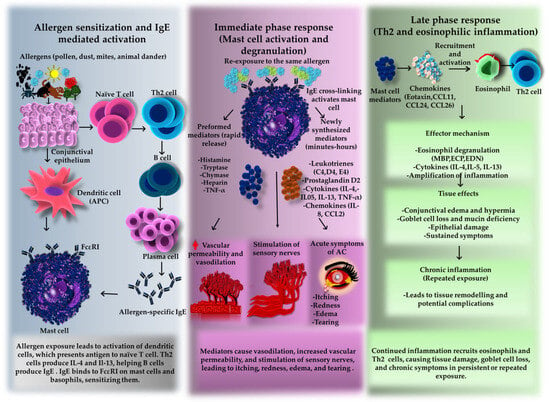
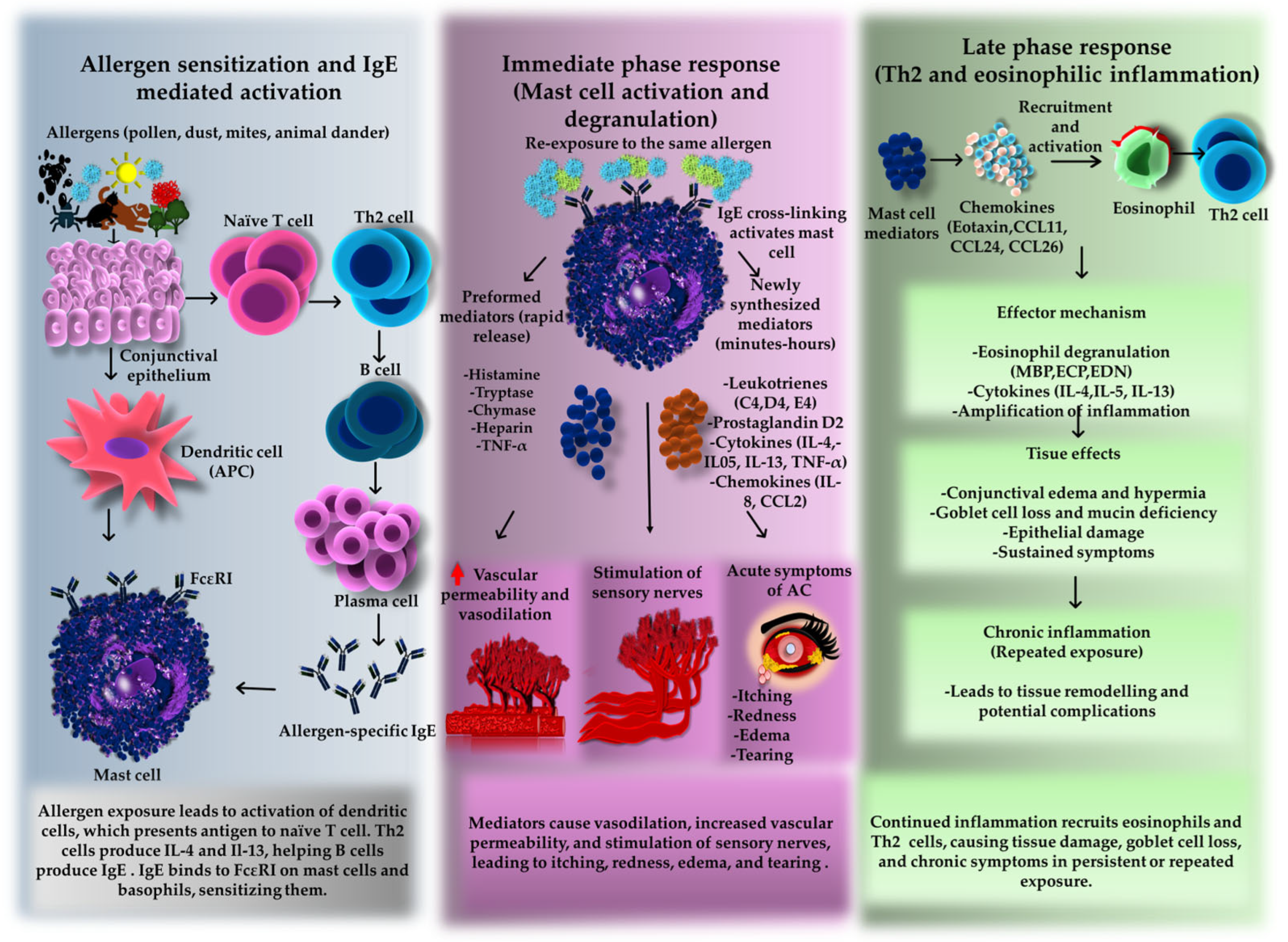
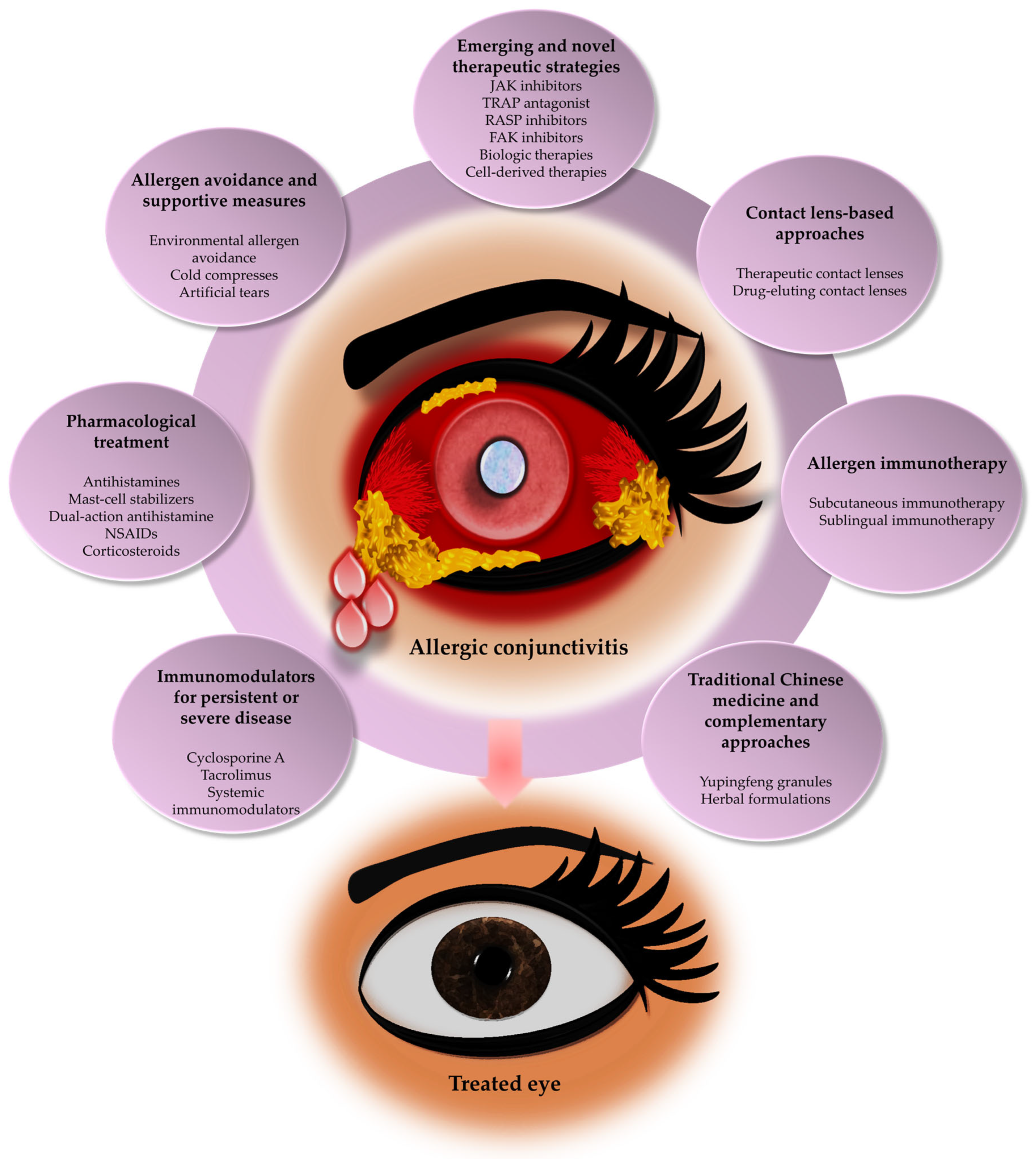
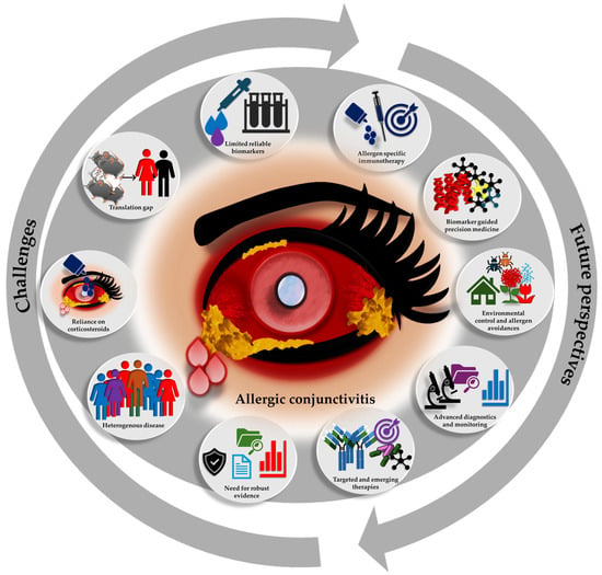
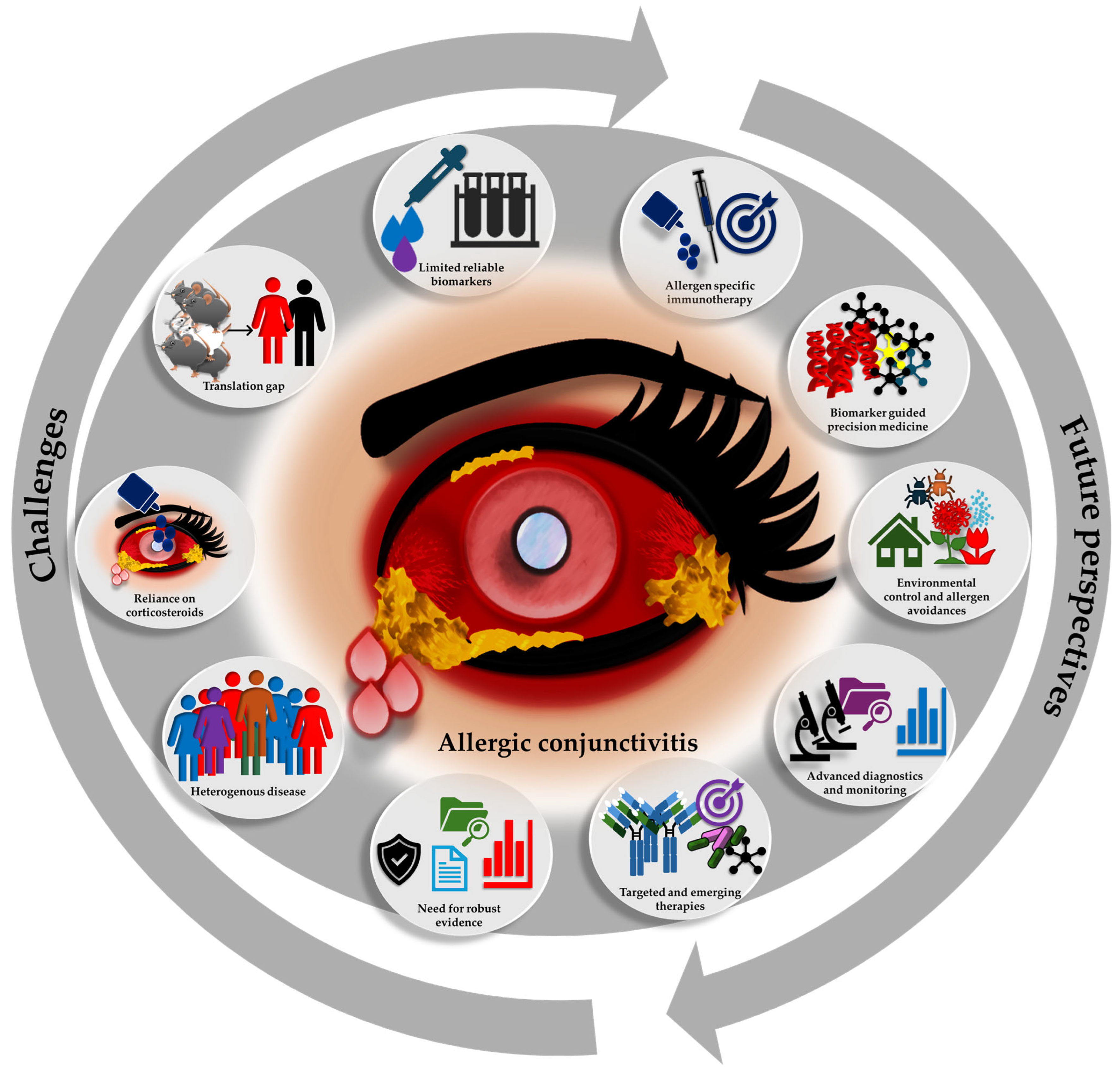

Disclaimer/Publisher’s Note: The statements, opinions and data contained in all publications are solely those of the individual author(s) and contributor(s) and not of MDPI and/or the editor(s). MDPI and/or the editor(s) disclaim responsibility for any injury to people or property resulting from any ideas, methods, instructions or products referred to in the content. |
© 2026 by the authors. Licensee MDPI, Basel, Switzerland. This article is an open access article distributed under the terms and conditions of the Creative Commons Attribution (CC BY) license.