


Abstract
The Human Leukocyte Antigen (HLA) system represents one of the most polymorphic genetic systems in humans and plays a central role in immune recognition. This review explores the complex relationship between HLA polymorphism and allergenicity. Allergic diseases are highly prevalent worldwide, and their pathogenesis is strongly influenced by genetic predisposition. HLA molecules, particularly class II alleles, shape the presentation of allergen-derived peptides to T helper cells, orchestrating immune responses that can lead to IgE-mediated hypersensitivity. Here, the molecular mechanisms underpinning allergen recognition, the structural determinants of HLA–peptide binding, and the consequences of allele-specific differences are discussed. Empirical evidence is reviewed for common allergen categories such as pollens, foods, environmental allergens, skin contact allergens, and stings. Associations between HLA alleles and allergic outcomes are often population-specific, reflecting evolutionary pressures and environmental exposures. The review also highlights computational approaches for predicting HLA–peptide interactions and their application to allergen research. Finally, some future directions including precision allergy medicine, epitope-guided immunotherapy, and integration of genomic, environmental, and clinical data are outlined. Understanding how HLA polymorphism contributes to allergenicity not only improves insight into disease mechanisms but also opens opportunities for improved diagnostics, personalized interventions, and the rational design of therapeutic strategies.
1. Introduction
The Human Leukocyte Antigen (HLA) system is the human counterpart of the major histocompatibility complex (MHC) and serves as a cornerstone of adaptive immunity. Its fundamental role lies in distinguishing self from non-self by presenting peptides to T cells, thereby orchestrating immune responses against pathogens while maintaining tolerance to host-derived molecules. HLA polymorphism, the extraordinary diversity of allelic variants across populations, is central to its immunological effectiveness. More than 44,000 alleles [1] have been described to date, each encoding unique structural features that influence peptide binding repertoires. This polymorphism, while advantageous in protecting populations against a broad spectrum of infectious diseases, comes with an evolutionary trade-off: susceptibility to autoimmunity and allergy.
Allergy is a hypersensitivity reaction of the immune system to typically harmless environmental substances, collectively termed allergens. The global prevalence of allergic diseases—including asthma, rhinitis, atopic dermatitis, food allergy, and drug hypersensitivity—has been rising steadily, constituting a major public health burden [2]. Estimates suggest that up to 40% of the global population suffers from some form of allergy [3]. Although environmental factors such as pollution, dietary habits, and microbial exposures shape allergic risk, genetic predisposition is an essential determinant of individual susceptibility. Among genetic factors, variation within the HLA region is consistently implicated as a key driver of allergy.
The influence of HLA polymorphism on allergenicity can be understood through the molecular basis of peptide presentation [4,5,6]. HLA class II molecules bind extracellularly derived peptides and display them to CD4+ T helper cells. The binding affinity, stability, and conformation of the peptide–HLA complex dictate the strength and character of T cell activation. Certain alleles preferentially bind peptides from specific allergens, thereby inducing immune responses by Th2 cells that promote IgE production. This immunological cascade underlies type I hypersensitivity reactions, the classical mechanism of allergic disease.
The first review addressing the relationship between HLA polymorphism and allergic diseases was published by de Weck back in 1977 [7]. Nearly 50 years later, this field has expanded considerably, with numerous genetic, immunological, clinical, and computational studies investigating how HLA variation influences allergen recognition, peptide presentation, T-cell responses, and susceptibility or protection from allergic diseases.
The present review synthesizes current understanding of the role of HLA polymorphism in allergenicity. It begins by describing the HLA polymorphism and its role in the mechanisms of allergen recognition, followed by an overview of empirical associations between specific HLA alleles and allergic conditions, grouped by allergen categories. Then the environmental, epigenetic, and computational aspects that interact with genetic predisposition are discussed. Finally, challenges and opportunities for translating these insights into precision allergy medicine are outlined. By integrating molecular, population-level, and computational evidence, this review highlights the pivotal role of HLA in shaping allergic disease outcomes and identifies opportunities for research and clinical innovation.
2. HLA Polymorphism
The HLA system is the most diverse genetic region in the human genome and plays a key role in how the immune system recognizes and responds to foreign substances. It is located on the short arm of chromosome 6 (6p21.3) and contains genes encoding the HLA system. These proteins present peptides originating from foreign or self-proteins to T lymphocytes, which control immune reactions [8].
The HLA system is divided into three main classes [9,10]:
- Class I (HLA-A, -B, -C) proteins are found in almost all body cells and present peptides to CD8+ T cells (Figure 1).
- Class II (HLA-DR, -DQ, -DP) proteins are found mainly in immune cells such as macrophages and dendritic cells and present peptides to CD4+ T cells (Figure 1).
- Class III proteins include complement proteins, cytokines, and other immune-related molecules.
A special feature of the HLA system is its extreme genetic diversity. Each gene has many different versions, called alleles, especially in the peptide-binding site of the class I and class II molecules. This diversity allows the immune system to recognize a wide range of pathogens while still tolerating the body’s own proteins. Most of this variation is found in the DNA parts that code for the peptide-binding site–exons 2 and 3 in class I genes and exon 2 in class II genes.
This genetic variation is developing through several natural processes such as point mutations, gene conversion, recombination, and unequal crossing-over during cell division [9,11]. These processes constantly create new alleles. The diversity is maintained by balancing selection, meaning that different versions of a gene are kept in the population because they help fight various infections. This evolutionary process is called trans-species polymorphism, since some HLA alleles are older than the human species itself and are shared with other primates like chimpanzees [12].
Figure 1.
X-ray structures of HLA class I (left, pdb code: 1K5N [13]) and class II (right, pdb code: 5NI9 [14]) molecules. The top row shows the structures from the side, and the bottom row shows them from the top. The HLA class I proteins consist of single α chain (green ribbon) assembled with β-microglobulin (blue ribbon). The peptide bound in peptide-binding site is given in orange. The HLA class II proteins consist of α chain (green ribbon) and β chain (blue ribbon). The peptide bound in peptide-binding site is given in orange.
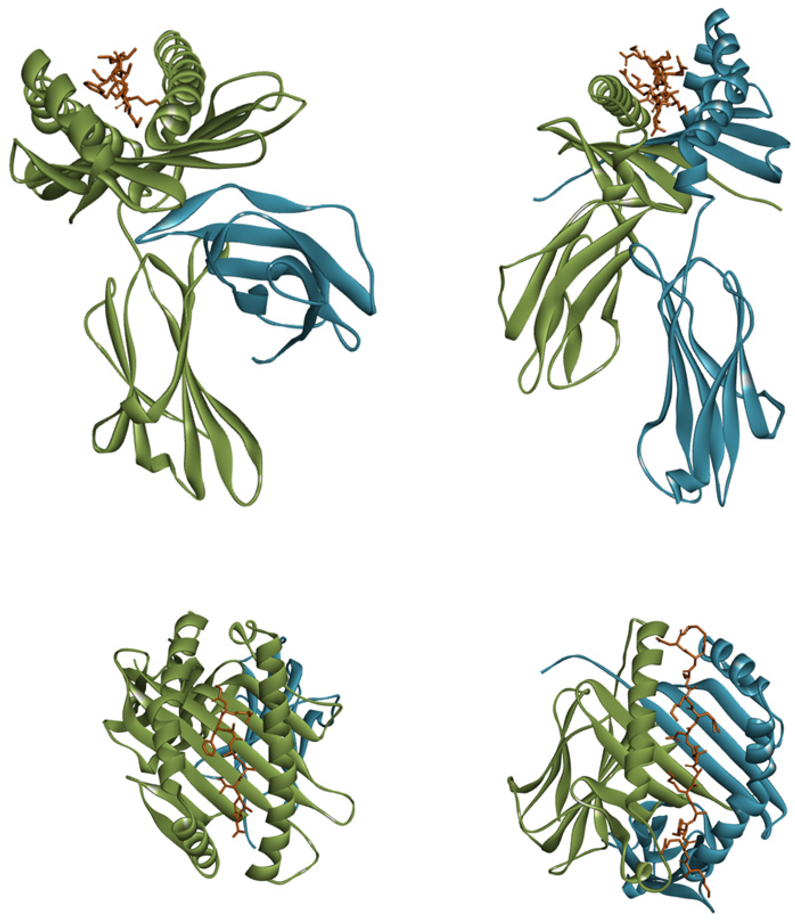
The main function of HLA molecules is to bind and present peptides to T cells, which then trigger immune responses. The shape and chemical properties of the peptide binding site determine which peptides can bind [8,15]. Even small changes in amino acids in this region can change the set of peptides that an HLA molecule can present. As a result, each person’s immune system recognizes a slightly different set of foreign antigens. This diversity helps protect populations from a wide variety of infections [16,17].
From an evolutionary point of view, this variation follows the “heterozygote advantage” principle. People who inherit two different HLA alleles can present a wider range of peptides and may therefore have better protection against infections [18]. Another important process is frequency-dependent selection, where rare alleles have an advantage because pathogens adapt to more common ones [19].
The types and frequencies of HLA alleles differ widely among human populations due to natural selection, migration, and genetic drift. Studies have shown that the pattern of HLA variation reflects adaptation to local diseases and environmental pressures [16]. For example, African populations which have historically been exposed to a greater diversity of pathogens show higher HLA diversity compared to populations from other continents [20].
In addition, groups of HLA alleles that are inherited together, known as haplotypes, vary between populations. These linkage patterns provide valuable information about population history, ancestry, and migration [21]. Understanding these differences is also important for practical applications such as organ transplantation, forensic identification, and disease association studies.
In medicine, HLA polymorphism has major implications for organ and stem cell transplantation. A close match between donor and recipient HLA types is essential to avoid graft rejection and graft-versus-host disease [22]. Because of the high number of different alleles, finding compatible donors can be very difficult, which is why large, ethnically diverse donor registries are needed worldwide.
HLA variation also affects how individuals respond to infections, vaccines, and certain drugs. Some HLA alleles are linked to autoimmune or allergic diseases, while others provide protection against specific pathogens. For example, HLA-B*57:01 is associated with slower HIV progression but also with abacavir drug hypersensitivity, making genetic testing essential before treatment [23].
3. Mechanisms of Allergenicity and the Role of HLA
Allergenicity is the intrinsic capacity of certain proteins to induce an allergic immune response. This is not a random process but depends on a complex interplay of the allergen’s structural features, the host’s immunogenetic background (especially HLA polymorphisms), and environmental factors [5,24].
Allergens typically enter the body through the respiratory tract (e.g., pollen), gastrointestinal tract (e.g., food allergens), or skin. Many allergens have protease activity, which helps them penetrate the epithelial barrier and activate the immune system [25,26]. Once inside, antigen-presenting cells (APCs), such as dendritic cells, internalize and process the allergen into smaller peptide fragments [4].
The processed allergen peptides are loaded onto HLA class II molecules (HLA-DR, -DP, -DQ) and presented on the APC’s surface. This presentation is crucial for activating Th2 cells [4,6]. The specific HLA alleles an individual possesses determine which allergen peptides can be effectively presented. A strong and stable interaction between the HLA molecule and the allergen peptide is a primary determinant of a robust T-cell response, leading to allergic sensitization [4,17]. The interaction between the T-cell receptor, the HLA molecule, and the peptide is a key immunological event.
Figure 2 illustrates the multi-step process of a type I hypersensitivity reaction, commonly known as an allergic reaction. The process begins when an APC, such as a dendritic cell, encounters an allergen. The APC takes up the allergen, breaks it down into smaller peptide fragments, and presents these fragments on its surface using HLA class II molecule. A naive T helper cell (Th0 cell) recognizes the allergen peptide presented by the HLA molecule. This interaction, along with co-stimulatory signals, activates the T cell [5]. The duration of T-cell receptor (TCR) engagement is an important quantitative parameter regulating T-cell activation and effector function. Experimental studies have demonstrated that the duration of antigenic stimulation can determine the fate of both naïve and effector T cells. Whereas naïve T cells require prolonged TCR signalling for commitment to proliferation, effector T cells can become activated after substantially shorter stimulation periods; conversely, excessive and prolonged antigenic stimulation may promote effector T-cell death rather than sustained activation [27]. The T cell then differentiates into a Th2 cell, a key player in allergic responses, driven by the presence of cytokines like interleukin-4 (IL-4). The newly activated Th2 cell interacts with a B cell, which is an immune cell responsible for producing antibodies. The Th2 cell releases additional cytokines (IL-4, IL-13) that stimulate the B cell to proliferate and switch to producing a specific type of antibody called immunoglobulin E (IgE) [27]. These IgE antibodies are specific to the allergen. The IgE antibodies released by the B cells travel through the bloodstream and bind to high-affinity receptors on the surface of mast cells. A person is now “sensitized,” meaning their body is primed for a rapid allergic reaction upon re-exposure. Upon a second exposure, the allergen directly binds to the allergen-specific IgE antibodies already attached to the mast cell. This cross-linking of IgE molecules triggers the mast cell to rapidly release a variety of inflammatory mediators, including histamine. It is the release of these chemicals that causes the characteristic symptoms of an allergic reaction, such as a runny nose, sneezing, hives, and swelling [28,29].
Figure 2.
Mechanism of a type I hypersensitivity reaction. An antigen-presenting cell (APC) presents an allergen to a naive T helper (Th0) cell. This leads to the T cell differentiating into a Th2 cell, which in turn activates a B cell. The B cell produces allergen-specific IgE antibodies that bind to mast cells. Upon re-exposure, the allergen cross-links the IgE on the mast cell, triggering the release of inflammatory mediators like histamine, which cause symptoms of an allergic reaction. The figure is generated by FigureLabs (https://www.figurelabs.ai, accessed on 2 July 2026).
The choice of peptides presented and therefore the likelihood of initiating a Th2 response depends critically on an individual’s HLA alleles. Conversely, some alleles may present peptides that promote tolerance rather than sensitization. Allergen tolerance is an active, regulated immune state in which exposure to an allergen does not result in a pathogenic immune response. It is initiated by tolerogenic antigen-presenting cells, particularly dendritic cells, which present allergen-derived peptides under conditions of limited costimulation and a regulatory cytokine environment. This promotes the induction and expansion of regulatory T cells (Tregs), which suppress allergen-specific Th2 responses through IL-10, TGF-β, and cell-contact-dependent mechanisms. Tregs also regulate B-cell responses, reducing allergen-specific IgE production while promoting IgG4, which can compete with IgE for allergen binding and thereby limit FcεRI-mediated activation of mast cells and basophils. Suppression of Th2 cytokines, particularly IL-4, IL-5, and IL-13, further reduces eosinophilic inflammation. Thus, allergen tolerance results from coordinated regulation of antigen presentation, T-cell and B-cell responses, and downstream effector-cell activation [30,31,32]. The HLA molecules serve as molecular gatekeepers of allergenicity, determining whether exposure to a substance results in tolerance, benign recognition, or a full allergic response.
4. HLA Associations with Specific Allergies
A great amount of empirical data supports associations between specific HLA alleles and distinct allergic conditions. These associations often show geographic and population-specific patterns, reflecting both genetic drift and environmental pressures. The literature search was performed in PubMed (https://pubmed.ncbi.nlm.nih.gov/, accessed on 7 September 2026) using the search strategy “HLA AND allergy NOT drug.” Additional searches using alternative combinations of keywords related to HLA, allergy, allergenicity, hypersensitivity, and sensitization were subsequently performed to maximize retrieval of relevant literature.
In this review the associations are organized according to the primary routes of allergen entry into the human body: inhalational (respiratory allergies), oral (food allergies), cutaneous (skin contact allergies), and injectable (insect stings or parenteral exposures). Drug hypersensitivity, given its unique immunogenetic mechanisms, is addressed as a separate category and will be considered elsewhere. Within each category, the studies are presented in chronological order for each specific allergen within the studied population. Only studies demonstrating a clear association between specific HLA alleles and specific allergens are included in this review.
Along the review, we will use the terms atopy and allergy. Although these two terms are often used interchangeably, they are not identical. Atopy is a genetic predisposition to produce IgE antibodies in response to low doses of common environmental allergens (such as pollen, dust mites, or food proteins). It describes a tendency or susceptibility rather than the disease itself. Atopy is usually identified by a positive skin prick test or elevated allergen-specific IgE in blood. Not all atopic individuals develop clinical symptoms. Allergy is a clinical disease resulting from an immune reaction (often but not always IgE-mediated) to an allergen. It manifests as asthma, allergic rhinitis, conjunctivitis, eczema, urticaria, or anaphylaxis. Allergy is the “realized” condition as a disease, whereas atopy is the predisposition. All atopic people are at increased risk of developing allergies, but not all of them become allergic.
In a recent EAACI position paper, Jutel et al. proposed an updated nomenclature for allergic diseases and hypersensitivity reactions that moves beyond classifications based primarily on clinical phenotype and incorporates the underlying immunological and tissue-specific mechanisms [32]. The proposed framework recognizes several distinct mechanisms of hypersensitivity and emphasizes that individual clinical conditions may involve overlapping or evolving endotypes. Importantly, this proposal represents an ongoing development in allergy nomenclature rather than an universally adopted replacement for established terminology. Therefore, the term atopy remains appropriate when referring to the established concept and to previous studies, but its use should be interpreted within the context of the evolving classification of allergic and hypersensitivity disorders.
4.1. Respiratory Allergies
In respiratory allergies, allergens enter the body by inhalation through the nose and lungs. Common triggers include pollen, dust mites, mold spores, and pet dander. Allergic reactions include allergic rhinitis (hay fever) and allergic asthma [5,28]. After crossing the respiratory epithelial barrier, allergens are captured by dendritic cells, which internalize the proteins, process them into peptide fragments, and present these peptides on HLA class II molecules (HLA-DR, -DQ and -DP) to naïve CD4+ T lymphocytes [33,34]. The strength and duration of the peptide–HLA interaction determine the efficiency of T-cell receptor engagement and strongly influence the differentiation of naïve CD4+ T cells into either Treg cells or Th2 cells [35,36]. HLA alleles that bind allergen-derived peptides with lower affinity or favor the presentation of tolerogenic epitopes may promote immune tolerance by enhancing regulatory T-cell responses, whereas susceptible alleles efficiently present immunodominant allergen peptides and facilitate Th2 polarization [37,38]. Activated Th2 cells produce the cytokines IL-4, IL-5 and IL-13, which stimulate B-cell class switching to allergen-specific IgE, promote eosinophil recruitment and survival, and sensitize mast cells and basophils through high-affinity FcεRI receptors. Upon subsequent allergen exposure, cross-linking of receptor-bound IgE induces rapid mast-cell degranulation and the release of histamine, leukotrienes and other inflammatory mediators responsible for the clinical manifestations of allergic rhinitis and allergic asthma [5,28] (Figure 2).
4.1.1. Pollen
The major perennial ryegrass pollen allergen Lol p III represents one of the earliest examples demonstrating HLA control of allergen-specific immune responses. In two independent cohorts of grass pollen-allergic subjects, Ansari et al. found that immune responsiveness to Lol p III was significantly associated with HLA-DR3 and HLA-DR5 [39]. Individuals carrying these alleles exhibited significantly stronger allergen-specific antibody responses, including increased IgE production, while no association was observed with HLA-DQ alleles, indicating that HLA-DR polymorphism plays a dominant role in the recognition of Lol p III-derived peptides. In a follow-up study, Ansari et al. showed that the association is not simply with DR3 or DR5 serotypes but with a shared EYSTS (Glu-Tyr-Ser-Thr-Ser) amino acid motif in the first hypervariable region of the HLA-DRβ chain, providing one of the first molecular explanations for HLA-restricted allergen recognition [40].
Mugwort pollen allergens are the main cause of pollinosis in late summer in Europe. Ninety-five percent of patients allergic to mugwort are sensitized to the major allergen Art v 1 [38]. In contrast to other common pollen allergens that contain multiple T-cell epitopes, Art v 1 contains only one immunodominant T-cell epitope (Art v 1 25-36). Jahn-Schmid et al. found that allergy to Art v 1 is characterized by a uniform T-cell response [38]. The disease is apparently associated with the HLA-DRB1*01 as 69% of the patients carry this allele. The dominant epitope exhibits strong HLA association because its sequence contains anchor residues that preferentially fit this particular HLA allele. Therefore, mugwort pollinosis is an ideal candidate for a peptide-based immunotherapy [38].
Gheerbrant et al. have investigated the association between HLA class-II alleles and specific IgE sensitization to a large number of respiratory allergen molecules: pollen allergens (mugwort Art v 1, olive tree Ole e 1, timothy grass Phl p 2, Phl p 5 and plantain Pla l 1), mold allergen (Alt a 1) and dust mite allergen (Der p 7) among the EGEA cohort [41]. EGEA stands for Epidemiological study on the Genetics and Environment of Asthma which is a 20-year cohort including cases with asthma (n = 388), their first-degree relatives (n = 1244) and population-based controls (n = 415) recruited in the early 90’s from five French cities [42]. Participants were thoroughly characterized in terms of environmental exposure and respiratory health, with dedicated efforts made to collect and store biological samples (ISO 9001 certified since 2006). Data and samples are available to researchers interested in establishing new scientific collaborations. The survey has already generated over a hundred publications, nearly one-third of which resulted from national and international collaborations. The strongest associations found by Gheerbrant et al. were between mugwort Art v 1 and DQB1*05:01, DQA1*01:01 and DRB1*01:01 [41].
Wang et al. genotyped at HLA class II DQA1 and DQB1 alleles 41 patients with Artemisia pollen-induced allergic rhinitis and 41 healthy controls from Beijing, China [43]. They found that the allele frequencies of HLA-DQA1*02:01, DQB1*06:02 were lower in patients with allergic rhinitis compared with the controls, while the frequency of DQA1*03:02 was higher among patients than the controls.
In a study of 176 individuals from 20 asthmatic family pedigrees, HLA class II genotypes were analyzed for associations with asthma, atopy, and specific IgE responses to six allergen extracts and six purified allergens (Der p 1, Der p 2, Fel d 1, Can f 1, Alt a 1, Phl p 5) [44]. Results indicated that HLA-DRB1*08 was negatively associated with asthma and atopy, while DRB1*15 was positively associated with asthma. A specific codon 9–12 motif in DRB1 alleles correlated with reduced atopy risk, and DPA1*0201 was negatively associated with specific IgE to grass pollen mix and Phl p 5.
In regions where birch pollen (Bet v 1) is a major cause of airborne allergies, up to 70% of affected patients also react to fruits, particularly apples. To assess HLA class II associations, Sénéchal et al. studied 42 atopic European adults (31 with asthma) and 42 healthy controls [45]. They found that HLA-DR4 or DR7 were present in 42.6% of patients compared with only 2.4% of controls. These findings confirm the link of DR4 and DR7 with susceptibility to birch pollen and apple allergy. The cross-reactivity between birch and apple sensitization could be explained by common epitopes in Bet v 1 and a 17-kd protein of apple extracts [46].
Similarly, Boehncke et al. have investigated 120 patients with grass and/or birch allergy and 80 patients with pollen-associated food allergy [47]. They have found that an increased frequency of HLA-DQB1*03:01 is associated with grass pollen allergy, HLA-DRB *08 with a sixfold higher risk for peanut allergy and -DRB1*12 with a 13-fold higher risk for carrot allergy. Cross-reactivities were found between birch pollen and hazel nut allergy for the haplotype HLA-DRB1*01, -DQA1*01:01, -DQB1*05:01, between grass pollen and peanut allergy for HLA-DRB1*08 and between birch pollen and carrot allergy for HLA-DRB1*12.
Cardaba et al. found significant associations between IgE antibody responses to the major olive pollen allergens Ole e 1 and Ole e 3 and the HLA-DQB1*02:01 allele, suggesting that this HLA class II molecule efficiently presents olive pollen-derived peptides to CD4+ T cells, thereby promoting Th2 polarization and IgE production [48]. To identify the peptides responsible for this response, the same group mapped the T-cell epitopes of Ole e 1 using overlapping synthetic peptides and peripheral blood mononuclear cells from allergic patients. Two regions, encompassing amino acid residues 91–102 and 109–130, were identified as immunodominant T-cell epitopes capable of inducing strong proliferative responses and cytokine production in sensitized individuals [49]. The peptide spanning residues 109–130 was recognized by most patients, indicating the presence of a promiscuous T-cell epitope that can be presented by multiple HLA class II molecules. The association between HLA-DQB1*02:01 and olive pollen allergy therefore most likely reflects the efficient presentation of these immunodominant Ole e 1 peptides, resulting in enhanced activation of allergen-specific Th2 cells and sustained IgE responses.
These studies demonstrate that susceptibility to pollen-induced respiratory allergies is strongly influenced by HLA class II polymorphism, particularly at the HLA-DRB1, HLA-DQA1 and HLA-DQB1 loci. Although the specific risk alleles differ among pollen species and ethnic populations, a common pattern emerges in which disease-associated HLA molecules efficiently present immunodominant allergen-derived peptides to CD4+ T cells, thereby promoting Th2 polarization, IgE production, and allergic sensitization. Conversely, several alleles, including HLA-DRB1*08, HLA-DPA1*02:01, HLA-DQA1*02:01 and HLA-DQB1*06:02, have been associated with reduced sensitization or protection in specific populations, emphasizing that HLA polymorphism can influence both susceptibility and resistance to allergic disease. The identification of dominant HLA-restricted epitopes in allergens such as Lol p III, Art v 1 and Ole e 1 has provided important mechanistic insight into the molecular basis of allergen recognition and has opened new opportunities for peptide-based allergen immunotherapy and personalized approaches guided by the patient’s HLA genotype.
4.1.2. House Dust Mites
House dust mites (HDMs) are among the most important perennial indoor allergens and represent a major cause of allergic rhinitis and allergic asthma worldwide. The predominant species, Dermatophagoides pteronyssinus and Dermatophagoides farinae, produce numerous allergens, including Der p 1, Der p 2, Der p 5, Der p 7, Der p 10, and Der f 1, which elicit strong IgE- and CD4+ T-cell-mediated immune responses [50]. Sensitization usually occurs early in life following chronic exposure to mite allergens in house dust. After uptake by dendritic cells, mite-derived proteins are processed into peptides and presented by HLA class II molecules to CD4+ T cells [51]. Differences in the peptide-binding repertoire of HLA class II alleles influence the spectrum of presented epitopes and the magnitude of allergen-specific T-cell responses, thereby contributing to Th2 polarization, IgE production, eosinophilic inflammation, and airway hyperresponsiveness [17].
Among respiratory allergens, HDM allergens have been particularly useful for studying the contribution of HLA polymorphism to allergic disease because their major CD4+ T-cell epitopes have been extensively mapped [52,53,54]. Early epitope-mapping studies identified multiple immunodominant T-cell epitopes within the major allergens Der p 1 and Der p 2 using overlapping synthetic peptides and allergen-specific T-cell clones, demonstrating considerable heterogeneity in HLA restriction. Der p 1 contains several overlapping epitopes presented by both HLA-DR and HLA-DP molecules, whereas Der p 2 peptides are presented by HLA-DR and HLA-DQ molecules, with individual epitopes exhibiting distinct HLA restriction patterns depending on the responding subject [52,53,54]. Structural and immunogenetic studies further demonstrated that Der p-derived peptides bind with different affinities to common HLA-DR and HLA-DQ molecules, and that the repertoire of presented peptides varies substantially among HLA alleles, resulting in marked inter-individual differences in allergen-specific CD4+ T-cell responses [55].
Subsequent studies specifically examined HDM-sensitive allergic rhinitis. Zhao et al. genotyped 142 Han Chinese patients with HDM-induced allergic rhinitis and 184 healthy controls using high-resolution HLA typing [56]. Two alleles showed highly significant associations with disease susceptibility: HLA-DRB1*08:03:02 and HLA-DQB1*06:01:01, both of which were more frequent among allergic patients. In contrast, HLA-DRB1*14 and HLA-DQB1*05 occurred significantly less frequently, suggesting protective effects against HDM sensitization. These findings indicate that subtle polymorphisms within the peptide-binding site influence the presentation of dominant HDM epitopes and the subsequent activation of allergen-specific CD4+ T cells.
The same research group later investigated whether HLA polymorphism also influences the outcome of allergen-specific immunotherapy (AIT), currently the only disease-modifying treatment for allergic rhinitis. Fifty-one patients receiving standardized HDM immunotherapy were followed for one year, and treatment efficacy was correlated with HLA genotype. Remarkably, patients carrying HLA-DRB1*03:01, DRB1*04:06, DRB1*14:05, DQB1*03:02:01, or DQB1*05:03:01 experienced significantly greater improvements in nasal symptoms than non-carriers [57]. Conversely, HLA-DRB1*07:01:01 and DRB1*11:01 were associated with poor therapeutic response. These observations suggest that HLA polymorphism determines not only susceptibility to sensitization but also the efficiency with which tolerogenic immune responses develop during immunotherapy. Identification of predictive HLA markers may therefore facilitate individualized treatment strategies in allergic rhinitis.
Additional support for the role of HLA polymorphism in HDM-associated allergic disease comes from paediatric asthma studies. Movahedi et al. investigated 112 Iranian children with allergic asthma and demonstrated significantly increased frequencies of HLA-DQB1*06:02 and DQB1*06:03, whereas DQB1*05:01 occurred less frequently than in healthy controls [58]. Children carrying susceptible alleles also exhibited significantly higher total serum IgE concentrations and stronger positive skin prick reactions to house dust allergens. These findings indicate that HLA class II polymorphism contributes to both sensitization and the magnitude of IgE responses.
Genome-wide analyses have further reinforced the importance of the HLA region in HDM allergy. In EGEA, Gheerbrant et al. analysed associations between HLA class II alleles and IgE responses against 26 purified respiratory allergen molecules [41]. Among HDM allergens, sensitization to Der p 7 was significantly associated with specific HLA class II haplotypes, confirming that different HDM allergens exhibit distinct HLA restriction patterns. This observation is consistent with structural studies showing that Der p proteins possess different repertoires of immunodominant T-cell epitopes and therefore interact with different peptide-binding sites.
Several major HDM allergens possess intrinsic biological activities that amplify HLA-mediated adaptive immune responses. The major allergen Der p 1 is a papain-like cysteine protease that disrupts epithelial tight junctions, thereby increasing epithelial permeability and facilitating allergen penetration into the airway mucosa [59,60]. In contrast, Der p 2 is a structural and functional homologue of myeloid differentiation protein-2 (MD-2) and enhances TLR4-dependent innate immune signalling, acting as an endogenous adjuvant during sensitization [61]. However, these innate mechanisms alone are insufficient to establish persistent allergic disease. Long-term allergen-specific Th2 immunity requires efficient processing and presentation of allergen-derived peptides by HLA class II molecules to CD4+ T lymphocytes [62,63]. Individuals carrying susceptible HLA alleles are therefore more likely to generate stable peptide–HLA complexes capable of sustained T-cell activation, resulting in persistent IgE production, eosinophilic inflammation, and chronic airway disease [53,57].
Current evidence demonstrates that HLA polymorphism is an important determinant of HDM allergy. Susceptibility alleles differ among populations, reflecting ethnic variation in allele frequencies and environmental exposure, but the underlying biological mechanism remains consistent. HLA molecules determine the repertoire of Der p- and Der f-derived peptides presented to CD4+ T lymphocytes, thereby influencing sensitization, disease severity, allergen-specific IgE production, and responsiveness to immunotherapy. Future integration of HLA genotyping with molecular allergen diagnostics may enable more accurate prediction of disease risk and facilitate personalized allergen immunotherapy.
4.1.3. Mold Spores
Allergic bronchopulmonary aspergillosis (ABPA) is a severe pulmonary hypersensitivity disorder caused by colonization of the airways by the opportunistic fungus Aspergillus fumigatus. The disease occurs predominantly in patients with persistent asthma or cystic fibrosis and is characterized by an exaggerated Th2-mediated immune response, resulting in elevated total and Aspergillus-specific IgE levels, eosinophilia, recurrent pulmonary infiltrates, and central bronchiectasis [64,65]. Genetic susceptibility plays an important role in ABPA, particularly polymorphisms within the HLA class II region that influence the presentation of A. fumigatus-derived peptides to CD4+ T lymphocytes. Early studies identified increased frequencies of HLA-DR2 and HLA-DR5 among patients with ABPA, suggesting that these alleles efficiently present fungal epitopes and promote pathogenic Th2 responses [66,67]. Aron et al. demonstrated that HLA-DR4 and HLA-DR7 were significantly overrepresented in patients with ABPA, especially in those with cystic fibrosis, supporting the hypothesis that HLA-DR polymorphism contributes to disease susceptibility by facilitating allergen presentation and enhancing IgE-mediated immune responses to A. fumigatus [68]. Subsequent genetic studies identified HLA-DRB1*15:01, DRB1*15:03 (DR2), and DRB1*11:04 (DR5) as susceptibility alleles, whereas HLA-DQB1*02:01 was associated with protection against ABPA, supporting the hypothesis that HLA polymorphism influences the quality of T-cell responses to fungal allergens [69].
Genome-wide analyses have further confirmed the importance of HLA polymorphism in mold sensitization. In EGEA, Gheerbrant et al. investigated HLA class II associations with sensitization to 26 purified respiratory allergen molecules and identified significant associations between Alternaria alternata allergen Alt a 1 and several HLA class II alleles, demonstrating that sensitization to individual fungal allergens is under allele-specific genetic control [41].
Knutsen et al. have compared 96 children with mold-sensitive moderate-severe asthma to 90 children with mild asthma and have found that the frequency of HLA-DRB1*03 and HLA-DRB1*13 alleles were increased in moderate-severe asthmatic children, while the frequency of HLA-DQB1*03 alleles was significantly decreased [70].
Although fewer association studies have been performed for mold allergens than for pollen or house dust mites, the available data consistently support a significant contribution of HLA polymorphism to fungal sensitization and asthma severity.
4.1.4. Pet Dander
Pet dander is one of the major sources of perennial indoor allergens and an important cause of allergic rhinitis and allergic asthma [71,72]. Cats and dogs account for the majority of sensitization cases, although rodents, horses, rabbits, and other mammals may also induce allergic disease [73]. Animal allergens are produced primarily in saliva, sebaceous glands, urine, and skin, and subsequently adhere to dander particles and hair that become airborne. Their small particle size enables prolonged suspension in indoor air and deep penetration into the lower respiratory tract [71]. The major cat allergen Fel d 1, a secretoglobin, accounts for more than 90% of cat sensitization, whereas the major dog allergen Can f 1 belongs to the lipocalin family [74,75].
Compared with pollen and house dust mite allergy, relatively few studies have investigated the influence of HLA polymorphism on pet dander allergy. Early family-based analyses demonstrated that HLA class II polymorphism contributes to allergen-specific IgE responses against pet allergens. Howell et al. examined members of asthmatic family pedigrees and observed that HLA-DPA1*02:01 was negatively associated with IgE responses to the major cat allergen Fel d 1, suggesting that this allele may confer protection against sensitization [44]. In the same study, HLA-DRB1*15 was associated with asthma susceptibility, whereas HLA-DRB1*08 appeared to protect against both asthma and atopy, supporting the concept that HLA polymorphism influences allergen-specific immune responses rather than generalized IgE production [44].
Subsequent immunological studies demonstrated that both Fel d 1 and Can f 1 contain multiple CD4+ T-cell epitopes presented by common HLA class II molecules. Epitope mapping of Fel d 1 identified several immunodominant peptides recognized in the context of multiple HLA-DR molecules, indicating considerable promiscuity in HLA binding and broad population coverage [76,77,78]. Likewise, studies of the major dog allergen Can f 1 demonstrated that its T-cell epitopes are presented by several HLA-DR molecules and elicit heterogeneous cytokine responses among allergic individuals [51]. Seven immunodominant T-cell epitope regions have been identified, and the corresponding peptides bind efficiently to several common HLA-DRB1 alleles, including DRB1*01:01, DRB1*03:01, DRB1*04:01, DRB1*07:01, DRB1*11:01, DRB1*13:01, and DRB1*15:01, making them broadly recognized across the population. Nevertheless, allergen-specific CD4+ T-cell responses differ considerably among individuals with respect to proliferative capacity and cytokine production, reflecting differences in T-cell receptor recognition and immune regulation rather than HLA binding alone [79,80,81].
Although convincing HLA association studies remain limited for cat and dog allergy, available evidence suggests that HLA polymorphism modulates allergen-specific T-cell activation rather than acting as the primary determinant of sensitization. In contrast to house dust mite or pollen allergens, whose susceptibility is strongly associated with specific HLA alleles, pet allergens appear to exhibit relatively promiscuous HLA binding. Consequently, environmental exposure, epithelial barrier integrity, innate immune activation, and immunoregulatory mechanisms probably play a greater role in determining whether sensitization progresses to clinically significant allergic disease. Future genome-wide association studies (GWAS) and high-resolution HLA typing in well-characterized patient cohorts are needed to clarify the contribution of HLA polymorphism to pet allergy and to identify genetic markers predictive of disease severity and response to allergen immunotherapy.
4.2. Food Allergies
Food allergies affect approximately 6–8% of children and 3–4% of adults, and their prevalence has increased substantially over the past several decades, particularly in industrialized countries [82,83]. The most common allergenic foods include cow’s milk, hen’s eggs, peanuts, tree nuts, wheat, soy, fish, and shellfish, known as the “Big Eight” [82,84], which together account for approximately 90% of IgE-mediated food allergies worldwide. Owing to the increasing prevalence and clinical significance of sesame allergy, the traditional “Big Eight” food allergens have been expanded to the “Big Nine”, with sesame now recognized as the ninth major food allergen requiring mandatory labeling in several countries, including the United States [85,86]. Clinical manifestations range from oral allergy syndrome, urticaria, angioedema, and gastrointestinal symptoms to respiratory compromise and life-threatening anaphylaxis. While allergies to milk, eggs, wheat, and soy are frequently outgrown during childhood, allergies to peanuts, tree nuts, fish, and shellfish often persist throughout life and are associated with a greater risk of severe systemic reactions [83,87,88].
Food allergies develop following the ingestion of allergenic proteins that induce sensitization within the gastrointestinal immune system. Under physiological conditions, dietary proteins promote oral tolerance, a state of antigen-specific immune unresponsiveness mediated by intestinal dendritic cells, Treg cells, and the anti-inflammatory cytokines IL-10 and transforming growth factor-β (TGF-β) [89,90]. In susceptible individuals, however, this tolerogenic mechanism fails, resulting in aberrant activation of allergen-specific CD4+ Th2 cells. Th2 cytokines, particularly IL-4, IL-5, and IL-13, stimulate B-cell class switching to allergen-specific IgE, which binds to high-affinity FcεRI receptors on mast cells and basophils. Upon subsequent allergen exposure, cross-linking of receptor-bound IgE triggers rapid release of histamine and other inflammatory mediators, leading to immediate hypersensitivity reactions [89,90,91].
The development of food allergy results from a complex interaction between environmental exposure, epithelial barrier integrity, gut microbiota, and genetic susceptibility. Among genetic factors, the HLA class II region plays a central role because it determines which food-derived peptides are processed and presented to CD4+ T lymphocytes. Differences in peptide-binding specificity among HLA-DR, HLA-DQ, and HLA-DP molecules influence the repertoire of immunodominant T-cell epitopes available for immune recognition, thereby affecting the balance between oral tolerance and allergic sensitization. Recent genetic studies indicate that the HLA region contributes to susceptibility to several food allergies, particularly peanut allergy, although the strength of association varies among different food allergens and populations [92,93]. These observations support the concept that HLA polymorphism is an important determinant of food allergen immunogenicity and provide a rationale for developing HLA-guided diagnostic and immunotherapeutic approaches.
4.2.1. Milk
Cow’s milk allergy (CMA) is the most common food allergy during infancy and early childhood, affecting approximately 2–3% of young children in developed countries [94,95]. The major milk allergens are caseins (Bos d 8), β-lactoglobulin (Bos d 5), α-lactalbumin (Bos d 4), bovine serum albumin (Bos d 6), and lactoferrin [96,97]. Although most children develop oral tolerance during the first years of life, persistent cow’s milk allergy is associated with severe IgE-mediated reactions and an increased risk of developing other atopic diseases. Like other food allergies, sensitization depends on efficient presentation of allergen-derived peptides by HLA class II molecules to CD4+ T lymphocytes, resulting in Th2 polarization, IgE production, and mast-cell sensitization.
Although HLA class II molecules are responsible for presenting cow’s milk-derived peptides to CD4+ T cells, evidence linking specific HLA alleles to susceptibility to IgE-mediated cow’s milk allergy remains limited. Early candidate-gene studies suggested that HLA class II polymorphism may influence sensitization to milk proteins, but these findings have not been consistently replicated in independent populations. Current evidence indicates that cow’s milk allergy is a complex polygenic disorder in which HLA variation represents only one component of genetic susceptibility [98].
Subsequent studies demonstrated that the association between HLA polymorphism and cow’s milk allergy is allergen-specific rather than disease-specific. Using computational prediction of peptide binding combined with experimentally validated T-cell and IgE epitopes, Dimitrov and Doytchinova showed that peptides derived from the major milk allergens are predicted to bind preferentially to HLA-DRB1*01:01, HLA-DQ7, and HLA-DQ8, whereas HLA-DRB1*03:01, DRB1*04:04, DRB1*12:01, and DRB1*15:01 exhibited markedly fewer predicted binders and were proposed to represent protective alleles [99]. Approximately 77% of experimentally verified T-cell and IgE epitopes from milk and egg allergens were correctly identified by this two-step prediction algorithm, supporting the biological relevance of differential HLA binding.
Mapping of T-cell epitopes has provided important insights into the cellular immune response to cow’s milk proteins. Several immunodominant CD4+ T-cell epitopes have been identified within αs1-casein, β-casein, κ-casein, α-lactalbumin, and β-lactoglobulin, many of which can bind multiple HLA class II molecules, indicating a high degree of HLA promiscuity [100,101]. These epitopes stimulate allergen-specific CD4+ T cells and induce Th2 cytokines, including IL-4, IL-5, and IL-13, in patients with IgE-mediated cow’s milk allergy, whereas children who naturally acquire tolerance exhibit increased production of IL-10, transforming growth factor-β (TGF-β), and other regulatory cytokines [102].
It should be emphasized that evidence directly linking specific HLA alleles to cow’s milk allergy remains limited compared with other food allergies, particularly peanut allergy. The T-cell epitope mapping and HLA-binding studies support a role for HLA polymorphism in susceptibility to cow’s milk allergy, although further large-scale genetic association studies are needed to validate these observations.
4.2.2. Eggs
Hen’s egg allergy is one of the most common food allergies in infants and young children, affecting approximately 1–2% of children during the first years of life [103,104]. The major egg allergens are found predominantly in egg white and include ovomucoid (Gal d 1), ovalbumin (Gal d 2), ovotransferrin (Gal d 3), and lysozyme (Gal d 4), whereas α-livetin (Gal d 5) is the principal allergen in egg yolk [105,106]. Ovomucoid is heat-stable and resistant to gastrointestinal digestion, making it the dominant allergen associated with persistent egg allergy [107,108]. Clinical manifestations range from mild cutaneous reactions and gastrointestinal symptoms to severe systemic anaphylaxis. Although many children outgrow egg allergy during childhood, persistent disease is associated with higher concentrations of egg-specific IgE, particularly ovomucoid-specific IgE, and sensitization to ovomucoid has become an important predictor of persistent allergy and reactivity to extensively heated egg products [106,107].
As with other IgE-mediated food allergies, sensitization to egg allergens depends on the presentation of allergen-derived peptides by HLA class II molecules to CD4+ T lymphocytes. Several studies have identified immunodominant T-cell epitopes within ovomucoid and ovalbumin that induce strong proliferative responses and Th2 cytokine production in allergic patients. Mapping of ovomucoid-derived peptides demonstrated that multiple epitopes are recognized by peripheral blood mononuclear cells from egg-allergic individuals, indicating that T-cell recognition is directed toward a limited number of immunodominant regions within the allergen [109]. More recently, component-resolved studies have confirmed that Gal d 1-specific T-cell responses correlate with persistent egg allergy and elevated IgE levels, emphasizing the importance of HLA-restricted antigen presentation in disease pathogenesis [106,107,108,110,111].
Direct association studies between HLA polymorphism and egg allergy remain relatively limited. Park et al. investigated 185 Korean children with atopic dermatitis and evaluated associations between HLA-DRB1 polymorphism and egg allergy. Although no significant association was found between individual HLA-DRB1 alleles and egg allergy, the HLA-DRB1*11:01 allele was significantly associated with susceptibility to atopic dermatitis, indicating that HLA polymorphism may influence the underlying atopic background rather than egg sensitization itself [112].
Computational analyses nevertheless support an important contribution of HLA polymorphism to the presentation of egg-derived allergens. Dimitrov and Doytchinova analyzed the binding of peptides derived from major egg allergens to the most frequent HLA-DRB1 and HLA-DQ molecules using an integrated digestion and peptide-binding prediction approach. Their study predicted that HLA-DRB1*01:01, HLA-DQ4, HLA-DQ7, and HLA-DQ8 preferentially present egg allergen-derived peptides and may therefore represent susceptibility alleles, whereas HLA-DRB1*03:01, HLA-DRB1*04:04, and HLA-DRB1*12:01 exhibited substantially fewer predicted binders and were proposed to confer protection [99]. Approximately 77% of experimentally validated T-cell and IgE epitopes originating from milk and egg allergens were correctly identified by the computational workflow, supporting the biological relevance of allele-specific peptide presentation.
The available evidence indicates that HLA polymorphism contributes to egg allergy primarily through differential presentation of immunodominant egg-derived peptides rather than through strong disease-associated alleles identified by GWAS. Further high-resolution HLA typing combined with comprehensive T-cell epitope mapping will be necessary to clarify the contribution of individual HLA class II alleles to susceptibility, persistence, and clinical severity of egg allergy.
4.2.3. Peanut
Peanut allergy is one of the most severe and persistent food allergies, affecting approximately 1–3% of children in Western countries and frequently persisting into adulthood [82,88]. It is the leading cause of food-induced anaphylaxis [82,113]. The major peanut allergens belong to the cupin and prolamin protein superfamilies and include Ara h 1, Ara h 2, Ara h 3, Ara h 6, Ara h 8, and Ara h 9, among which Ara h 2 and Ara h 6 are the most potent elicitors of IgE-mediated reactions and the best predictors of clinical peanut allergy [114,115,116].
The first GWAS of clinically well-defined food allergies, including peanut, milk, and egg allergy, was conducted by Hong et al. in 2759 participants from the Chicago Food Allergy Study [117]. Analysis of 2197 individuals of European ancestry identified peanut allergy-specific susceptibility loci within the HLA-DR/DQ region on chromosome 6p21.32, representing the strongest genetic signal detected in the study. The authors identified HLA-DQA1*01:02 and HLA-DQB1*06 as the principal risk alleles associated with peanut allergy. Fine mapping further demonstrated that an amino acid polymorphism at position β71 of HLA-DRB1, located within the peptide-binding groove, produced the strongest association signal, with the presence of Arg71 significantly reducing the risk of peanut allergy.
These findings were independently confirmed in an Australian GWAS involving 73 infants with challenge-proven IgE-mediated peanut allergy and 148 non-allergic controls. Martino et al. reproduced the association with amino acid polymorphisms at position β71 of HLA-DRB1, confirming that structural variation within the peptide-binding groove is a major determinant of peanut allergy susceptibility [118]. Since residue β71 contributes to the formation of peptide-binding pockets 4 and 7 of HLA-DR molecules, these observations strongly suggest that allele-specific differences in peptide binding influence the presentation of peanut-derived epitopes to CD4+ T lymphocytes.
Further evidence has emerged from independent GWAS performed in European populations. A German study involving 497 food-allergic patients and 2387 controls confirmed the importance of the HLA-DQB1 region while also identifying the SERPINB gene cluster as an additional susceptibility locus for food allergy [119]. Similarly, a Canadian GWAS followed by meta-analysis demonstrated that the HLA region represents a major genetic risk factor for peanut allergy independently of asthma [120]. Fine mapping of the Canadian cohort subsequently identified HLA-DQB1*02 and HLA-DQB1*06:03P as the alleles showing the strongest association with peanut allergy [121].
Although most genetic studies have focused on populations of European ancestry, recent investigations indicate that HLA associations differ among ethnic groups while converging on similar structural mechanisms. In a study of non-Hispanic Black individuals, Wu et al. identified HLA-DRB1*13:02 as the allele most strongly associated with peanut allergy, whereas HLA-DQA1*01:02 showed the strongest association with peanut-induced anaphylaxis [122]. Amino acid polymorphisms at position β71 of HLA-DRB1 again represented one of the strongest association signals, suggesting that variation in the architecture of the peptide-binding site is a common determinant of peanut allergy across different populations.
Beyond disease susceptibility, HLA polymorphism also appears to influence the induction of immune tolerance. Analysis of participants in the Learning Early About Peanut Allergy (LEAP) trial demonstrated that children carrying HLA-DQA1*01:02 who consumed peanut regularly developed significantly higher concentrations of peanut-specific IgG4 antibodies than non-carriers [123]. Because allergen-specific IgG4 competes with IgE for allergen binding and inhibits FcεRI-mediated activation of mast cells and basophils, these findings provide a mechanistic explanation for the protective effect of early peanut consumption in genetically susceptible individuals.
The strong and reproducible association between peanut allergy and HLA class II polymorphism has stimulated extensive investigation of peanut-derived CD4+ T-cell epitopes. Numerous immunodominant T-cell epitopes have been identified within Ara h 1, Ara h 2, Ara h 3, and Ara h 6, many of which bind multiple common HLA-DR and HLA-DQ molecules, demonstrating a high degree of HLA promiscuity [124,125]. Peanut-allergic individuals exhibit robust allergen-specific CD4+ T-cell responses characterized predominantly by the production of IL-4, IL-5, and IL-13, whereas tolerant individuals display increased frequencies of regulatory T cells and enhanced production of IL-10 and other immunoregulatory cytokines [126,127]. Consequently, peanut allergy represents one of the best-characterized examples of HLA-dependent allergenicity and provides an excellent model for the development of HLA-guided peptide immunotherapies and precision allergy prevention.
4.2.4. Tree Nuts
Tree nut allergies are among the most persistent and potentially severe food allergies, often continuing throughout life and representing one of the leading causes of food-induced anaphylaxis [82,128]. The most common allergenic tree nuts include hazelnut, walnut, cashew, pistachio, almond, pecan, and Brazil nut, although the prevalence of sensitization varies geographically according to dietary habits and environmental exposure [129]. Sensitization may occur through primary exposure to seed storage proteins or secondarily through cross-reactivity with homologous pollen allergens, particularly in individuals allergic to birch pollen. This pollen-food syndrome is largely attributable to structural homology between the major birch pollen allergen Bet v 1 and the hazelnut allergen Cor a 1, which share cross-reactive T-cell and IgE epitopes [46,130]. As with other IgE-mediated food allergies, allergen-derived peptides are processed by antigen-presenting cells and presented by HLA class II molecules to CD4+ T lymphocytes, initiating Th2 immune responses, allergen-specific IgE production, and subsequent mast-cell activation upon re-exposure to the allergen [131].
Compared with peanut allergy, relatively few studies have investigated the association between HLA polymorphism and tree nut allergy. One of the earliest studies was performed by Boehncke et al., who analyzed HLA class II alleles in patients with pollen allergy and pollen-associated food allergy [47]. They found that the haplotype HLA-DRB1*01–DQA1*01:01–DQB1*05:01 was significantly associated with the coexistence of birch pollen and hazelnut allergy, providing genetic evidence that HLA class II polymorphism contributes to pollen-food syndrome through the presentation of cross-reactive T-cell epitopes. The same study demonstrated that different HLA alleles predisposed to distinct pollen-associated food allergies, supporting the concept that HLA-restricted recognition of homologous allergen-derived peptides underlies clinical cross-reactivity.
The molecular basis of this association has been clarified by studies of the major birch pollen allergen Bet v 1 and its homologues in plant foods. Hazelnut contains the PR-10 protein Cor a 1, which shares extensive structural and sequence homology with Bet v 1. T-cell clones isolated from birch pollen-allergic patients recognize homologous peptides derived from both allergens, indicating that sensitization to birch pollen can drive secondary immune responses to hazelnut through HLA class II-mediated presentation of conserved epitopes [130,132]. This mechanism explains why hazelnut allergy in Northern and Central Europe is frequently associated with birch pollinosis and is generally characterized by oral allergy syndrome rather than severe systemic reactions.
In contrast, primary tree nut allergy, particularly to walnut, cashew, and pistachio, is predominantly associated with sensitization to highly stable seed storage proteins, including 2S albumins, 7S vicilins, and 11S legumins, which are resistant to heat treatment and gastrointestinal digestion and are therefore frequently associated with severe systemic reactions [97,129,133]. Several CD4+ T-cell epitopes have been identified within these major allergens, including Jug r 1 from walnut, Ana o 1, Ana o 2, and Ana o 3 from cashew, and homologous allergens from pistachio, providing evidence that these proteins can efficiently stimulate allergen-specific T-cell responses [134,135]. However, unlike peanut allergy, convincing associations between specific HLA class II alleles and primary tree nut allergy have not been consistently demonstrated. Most available studies have focused on allergen characterization, IgE epitopes, and component-resolved diagnostics rather than HLA genetics. The contribution of HLA polymorphism to primary tree nut allergy remains incompletely understood and warrants further investigation in large, ethnically diverse cohorts using high-resolution HLA typing and genome-wide approaches [82,129].
4.2.5. Wheat
Wheat allergy comprises several distinct clinical entities, including classical IgE-mediated food allergy, baker’s asthma, and wheat-dependent exercise-induced anaphylaxis (WDEIA) [136,137]. WDEIA is a severe form of food allergy in which ingestion of wheat alone is insufficient to elicit symptoms but, in combination with cofactors such as physical exercise, alcohol consumption, non-steroidal anti-inflammatory drugs, or infections, triggers systemic allergic reactions that may progress to life-threatening anaphylaxis. The major allergen responsible for WDEIA is ω5-gliadin (Tri a 19), although high- and low-molecular-weight glutenins have also been implicated [138,139]. The underlying immunopathogenesis involves IgE-mediated recognition of wheat allergens, followed by enhanced intestinal permeability and increased allergen absorption induced by exercise, leading to mast-cell activation and systemic mediator release.
The identification of genetic risk factors for WDEIA could facilitate early identification of susceptible individuals before exposure to wheat-containing foods. In the first GWAS of WDEIA, Fukunaga et al. analyzed 77 Japanese patients with WDEIA and 924 healthy controls, and confirmed their findings in an independent replication cohort of 91 patients and 435 controls [140]. The study identified HLA-DPB1*02:01:02 as the strongest genetic susceptibility allele, with the association remaining highly significant in both the replication and combined analyses. Fine-mapping of the HLA region further demonstrated that amino acid polymorphisms within the peptide-binding site of HLA-DP molecules largely accounted for the observed association, suggesting that allele-specific presentation of wheat-derived peptides contributes directly to disease susceptibility.
The predominance of HLA-DPB1*02:01:02 is biologically plausible because HLA class II molecules determine the repertoire of gluten-derived peptides presented to CD4+ T lymphocytes. Several immunodominant T-cell epitopes have been identified within ω5-gliadin and other gluten proteins, supporting the hypothesis that efficient presentation of these peptides by susceptible HLA molecules promotes Th2 polarization and allergen-specific IgE production [136,138]. Unlike celiac disease, which is strongly associated with HLA-DQ2 and HLA-DQ8 and is mediated predominantly by Th1 immune responses against deamidated gluten peptides, WDEIA represents an IgE-mediated allergic disease with a distinct HLA association centered on the HLA-DP locus [140,141]. These findings illustrate that different HLA class II loci may determine susceptibility to distinct immune responses against the same dietary proteins.
At present, the study by Fukunaga et al. remains the strongest evidence linking HLA polymorphism to wheat allergy [140]. Additional studies in ethnically diverse populations are required to determine whether the association with HLA-DPB1*02:01:02 is universally applicable or reflects population-specific genetic architecture. Furthermore, characterization of HLA-DP-restricted wheat epitopes may facilitate the development of peptide-based diagnostic tools and precision immunotherapies for WDEIA.
4.2.6. Soy
Soybean (Glycine max) is one of the major food allergens in children and is included among the “Big Eight” food allergens responsible for approximately 90% of IgE-mediated food allergies [82,142]. Clinical manifestations range from mild oral allergy syndrome to severe systemic reactions and anaphylaxis. Soy allergy exhibits two distinct sensitization patterns. Primary soy allergy, which predominates in young children, is mainly directed against the seed storage proteins Gly m 5 (β-conglycinin), Gly m 6 (glycinin), and Gly m 8 (2S albumin), whereas secondary soy allergy frequently occurs in adolescents and adults sensitized to birch pollen through cross-reactivity between the major birch pollen allergen Bet v 1 and the homologous soy allergen Gly m 4 [143,144]. Component-resolved diagnostics have demonstrated that sensitization to Gly m 4 is associated predominantly with pollen-food syndrome, whereas Gly m 5, Gly m 6, and particularly Gly m 8 are associated with primary soy allergy and more severe systemic reactions [144,145].
Despite increasing knowledge of soy allergen components and their immunological properties, evidence linking HLA polymorphism to soy allergy remains remarkably limited. To date, no large GWAS or well-powered case–control studies have identified reproducible associations between specific HLA class II alleles and susceptibility to soy allergy.
4.2.7. Fish
Fish allergy is one of the most common causes of persistent food allergy and is a frequent trigger of severe systemic reactions and anaphylaxis [82,146]. Unlike milk or egg allergy, which are often outgrown during childhood, fish allergy usually persists throughout life and affects both children and adults [147,148]. More than 170 fish species have been reported to cause allergic reactions, although the degree of cross-reactivity varies considerably among species [146,148]. The major fish allergens belong to the parvalbumin family, particularly β-parvalbumins, which are highly stable calcium-binding proteins resistant to heat treatment and gastrointestinal digestion [148,149]. Their remarkable structural conservation explains the extensive IgE cross-reactivity observed among phylogenetically related fish species.
Several immunodominant B-cell and CD4+ T-cell epitopes have been identified within fish parvalbumins, demonstrating that these proteins are efficiently processed and presented by antigen-presenting cells to CD4+ T lymphocytes, thereby promoting Th2 differentiation and allergen-specific IgE production [146,150]. However, despite increasing knowledge of fish allergen structure and epitope composition, evidence linking HLA polymorphism to fish allergy remains scarce. To date, no GWAS or well-powered case–control studies have identified reproducible associations between specific HLA class II alleles and susceptibility to IgE-mediated fish allergy. Most investigations have focused on molecular characterization of fish allergens, cross-reactivity among different fish species, and component-resolved diagnostics rather than HLA genetics [146,148].
4.2.8. Shellfish
Shellfish allergy is one of the most common causes of food-induced anaphylaxis in adolescents and adults and is generally considered a lifelong condition. Shellfish are broadly classified into crustaceans (shrimp, crab, lobster, crayfish) and mollusks (oysters, mussels, clams, squid, and scallops), with crustaceans accounting for most clinically significant allergic reactions [149]. The major shellfish allergen is tropomyosin, a highly conserved muscle protein that exhibits extensive sequence homology among crustaceans, mites, and cockroaches, thereby explaining the frequent IgE cross-reactivity observed between shellfish allergy and sensitization to house dust mites or cockroaches [151,152]. Additional clinically relevant shellfish allergens include arginine kinase, myosin light chain, sarcoplasmic calcium-binding protein, troponin C, and hemocyanin, which contribute to the heterogeneity of individual sensitization profiles [146,149].
Compared with other seafood allergies, shellfish allergy is one of the few conditions in which HLA polymorphism has been investigated by genome-wide association analysis. Khor et al. performed a GWAS of self-reported food reactions in 11,011 Japanese women and identified significant associations between shellfish allergy and polymorphisms within the HLA-DR/DQ region [153]. Fine mapping demonstrated significant associations with HLA-DRB1*04:05, HLA-DRB1*15:01, HLA-DQB1*04:01, HLA-DQB1*06:02, HLA-DPB1*06:01, and HLA-DPB1*14:01, indicating that variation in HLA class II molecules contributes to susceptibility to shrimp allergy. The associated alleles are thought to influence the repertoire of tropomyosin-derived peptides presented to CD4+ T lymphocytes, thereby modulating allergen-specific Th2 responses and IgE production.
The biological plausibility of these genetic associations is supported by immunological studies demonstrating that shrimp tropomyosin contains multiple CD4+ T-cell epitopes capable of stimulating proliferative responses in allergic individuals [152]. Because tropomyosin is highly conserved among invertebrate species, many of these epitopes are shared with homologous allergens from mites and cockroaches, providing a mechanistic explanation for the “mite–shrimp syndrome”, in which primary sensitization to inhaled mite allergens is followed by secondary allergic reactions to shellfish [149,151]. Nevertheless, despite the strong GWAS evidence from the Japanese population, comparable HLA association studies in other ethnic groups remain scarce. Replication of these findings in independent populations and identification of HLA-restricted shellfish epitopes will be essential for clarifying the role of HLA polymorphism in shellfish allergy and for developing HLA-guided diagnostic and therapeutic approaches.
4.2.9. Sesame
Sesame (Sesamum indicum) allergy has emerged as an increasingly important food allergy worldwide and is now recognized as the ninth major food allergen in the United States following the implementation of the Food Allergy Safety, Treatment, Education, and Research (FASTER) Act [85,86]. Sesame allergy is frequently persistent throughout life and can cause severe systemic reactions, including anaphylaxis [154,155].
To date, nine sesame allergens (Ses i 1–Ses i 9) have been identified. The major allergens include the seed storage proteins Ses i 1 (2S albumin), Ses i 2 and Ses i 3 (7S vicilin-like globulins), Ses i 6 and Ses i 7 (11S globulins), as well as the oleosins Ses i 4 and Ses i 5, which are associated with severe allergic reactions because of their hydrophobic nature and poor detection by conventional aqueous extracts [154].
Evidence linking HLA polymorphism directly to sesame allergy is currently lacking. Unlike peanut allergy, WDEIA, or shrimp allergy, no GWAS have identified reproducible associations between specific HLA-DR, HLA-DQ, or HLA-DP alleles and susceptibility to sesame allergy.
4.2.10. Peach
Peach allergy is one of the most common fruit allergies worldwide, although its clinical manifestations vary considerably according to geographic region and the sensitizing allergen. In Northern and Central Europe, peach allergy is predominantly associated with pollen-food syndrome resulting from cross-reactivity between the major birch pollen allergen Bet v 1 and the homologous peach allergen Pru p 1, usually causing mild oral allergy syndrome [97,156]. In contrast, in Mediterranean countries, primary sensitization is directed mainly against the non-specific lipid transfer protein Pru p 3, which is highly resistant to heat treatment and gastrointestinal digestion and is associated with severe systemic reactions and anaphylaxis [157]. Other clinically relevant peach allergens include Pru p 4 (profilin) and Pru p 7 (peamaclein), both of which contribute to regional differences in sensitization profiles and clinical severity [97].
The first GWAS investigating genetic susceptibility to food allergy identified significant associations between peach allergy and the HLA class II region. Khor et al. analyzed self-reported food allergies in a Japanese population and demonstrated strong associations between peach allergy and polymorphisms within HLA-DRB1, HLA-DQB1, and HLA-DPB1 [153]. Fine mapping identified significant associations with HLA-DRB1*09:01, DRB1*14:05, DRB1*15:01, DRB1*15:02, HLA-DQB1*03:03, DQB1*05:03, DQB1*06:01, DQB1*06:02, HLA-DPB1*09:01, and DPB1*14:01, indicating that variation in HLA class II molecules contributes substantially to genetic susceptibility to peach allergy.
The biological basis of these associations is consistent with current knowledge of peach allergen immunology. Several CD4+ T-cell epitopes have been identified within Pru p 1 and Pru p 3, demonstrating that allergen-derived peptides are efficiently processed and presented by HLA class II molecules to CD4+ T lymphocytes, thereby promoting Th2 differentiation and allergen-specific IgE production [158,159]. Because Pru p 1 shares extensive structural homology with Bet v 1, cross-reactive T-cell responses contribute to pollen-food syndrome, whereas the highly stable Pru p 3 lipid transfer protein induces primary sensitization and severe systemic allergy independently of birch pollen sensitization [97,159].
4.3. Skin Allergies
Skin allergies develop following exposure of the skin to environmental allergens, haptens, or proteins that penetrate the epidermal barrier and activate the immune system. Common triggers include plant allergens (e.g., urushiol from poison ivy), metals such as nickel and cobalt, cosmetics, topical medications, preservatives, and natural rubber latex. Depending on the mechanism involved, skin allergies manifest as atopic dermatitis, an IgE-mediated chronic inflammatory disease characterized by Th2 polarization, or allergic contact dermatitis, a delayed-type (type IV) hypersensitivity reaction mediated predominantly by allergen-specific T lymphocytes [160,161].
4.3.1. Atopic Dermatitis
Atopic dermatitis (AD) is a chronic, relapsing inflammatory skin disease resulting from a complex interaction between genetic susceptibility, epidermal barrier dysfunction, immune dysregulation, and environmental exposure. Food allergies affect approximately 35–45% of children with moderate-to-severe AD, with hen’s egg, cow’s milk, peanut, and wheat being the most common triggers [162]. Among these, egg allergy, particularly sensitization to egg white proteins, is associated with increased AD severity and a higher risk of developing additional food allergies during childhood [163].
Although HLA class II polymorphism has long been considered a potential contributor to AD susceptibility, evidence remains inconsistent. In a study of 185 Korean children with atopic dermatitis, Park et al. demonstrated that HLA-DRB1*11:01 was significantly associated with susceptibility to AD but found no association between HLA-DRB1 polymorphism and egg allergy, indicating that HLA variation may influence the underlying inflammatory skin disease rather than sensitization to individual food allergens [112]. GWAS have likewise identified the HLA class II region as one of several susceptibility loci for AD, although its effect appears smaller than that of barrier-related genes such as FLG, suggesting that impaired epithelial integrity remains the primary determinant of disease susceptibility [164,165].
4.3.2. Allergic Contact Dermatitis
Allergic contact dermatitis is a T-cell-mediated delayed hypersensitivity reaction induced by low-molecular-weight chemicals (haptens) that bind covalently to self-proteins and generate neoantigens. Nickel is the most common contact allergen worldwide, followed by chromium, cobalt, fragrances, preservatives, and rubber additives [166]. Because antigen presentation is essential for T-cell activation, HLA polymorphism has been investigated as a potential determinant of susceptibility. Several studies have reported associations between HLA-DRB1*07, HLA-DQB1*02, and nickel hypersensitivity, although these associations vary considerably among populations and have not been consistently replicated [166,167]. Overall, current evidence suggests that HLA class II molecules contribute to individual differences in hapten presentation, but their effect is modest compared with environmental exposure.
4.3.3. Latex Allergy
Natural rubber latex allergy is an IgE-mediated hypersensitivity caused by proteins derived from Hevea brasiliensis and is particularly prevalent among healthcare workers and patients with repeated latex exposure [168]. Clinical manifestations range from contact urticaria to severe systemic anaphylaxis. One of the major latex allergens is hevein (Hev b 6.02), which induces strong IgE responses in sensitized individuals.
Among skin allergies, latex allergy provides one of the clearest examples of HLA-associated allergen-specific immune responses. Rihs et al. investigated 269 healthcare workers with latex allergy and demonstrated that HLA-DQB1*03:02 (DQ8) and the HLA-DQB1*03:02–DRB1*04 (DQ8–DR4) haplotype were significantly associated with hevein-specific IgE production, indicating that these HLA class II molecules efficiently present hevein-derived peptides to CD4+ T lymphocytes [169]. Similarly, Blanco et al. reported significant associations between latex-fruit syndrome and HLA-DQB1*02:01 together with HLA-DR functional group E, suggesting that common HLA-restricted T-cell responses contribute to cross-reactivity between latex and plant-derived food allergens such as banana, avocado, kiwi, and chestnut [170].
The structural basis for this association may lie in the peptide-binding specificity of DR functional group E, which largely corresponds to the DR4 supertype [171,172]. Members of this supertype share conserved amino acid residues at positions β70, β71, and β74 that form peptide-binding pocket 4 and favor peptides containing negatively charged residues at the corresponding anchor position [172,173,174]. Efficient presentation of hevein-derived peptides by these HLA molecules may therefore contribute to enhanced Th2 responses and IgE production in genetically susceptible individuals.
HLA polymorphism appears to play a variable role across skin allergies. While associations with atopic dermatitis and allergic contact dermatitis remain relatively modest and population-dependent, latex allergy demonstrates a clear relationship between specific HLA class II alleles and allergen-specific IgE responses.
4.4. Injectable Allergies
Injectable allergies comprise allergic disorders in which sensitization and elicitation occur following the introduction of allergens directly into the skin or subcutaneous tissues through stings, bites, or injections. Excluding drug- and vaccine-induced hypersensitivity, the principal causes of injectable allergies are Hymenoptera venom, fire ant venom, tick saliva, and, less frequently, mosquito saliva and marine animal venoms [175,176]. Because allergens bypass epithelial barriers and are rapidly distributed within tissues and the circulation, injectable allergies are characterized by a high risk of systemic IgE-mediated reactions, including anaphylaxis [176]. Their development depends on a complex interplay between environmental exposure, venom composition, and host genetic factors. Among the latter, HLA class II polymorphism plays a central role by determining the repertoire of allergen-derived peptides presented to CD4+ T lymphocytes, thereby influencing T-cell polarization toward either immune tolerance or Th2-mediated allergic sensitization [4,6,30].
Compared with respiratory and food allergies, relatively few studies have examined the contribution of HLA polymorphism to injectable allergies. This is partly because venom allergy is less prevalent and because genetic studies have generally involved small cohorts. Nevertheless, available evidence indicates that HLA molecules contribute to both susceptibility to venom allergy and the immunological mechanisms underlying successful venom immunotherapy.
4.4.1. Hymenoptera Venom Allergy
Hymenoptera venom allergy is the most extensively investigated injectable allergy and is caused by stings from honeybees (Apis mellifera), bumblebees (Bombus spp.), yellow jackets (Vespula spp.), paper wasps (Polistes spp.), hornets (Vespa spp.), and ants (Solenopsis spp.) [175]. Approximately 0.3–7.5% of adults experience systemic allergic reactions following Hymenoptera stings, while large local reactions occur in up to one-quarter of the population. Insect stings remain one of the leading causes of fatal anaphylaxis in adults [175].
The allergenic components of Hymenoptera venoms have been extensively characterized. Honeybee venom contains several major allergens, including phospholipase A2 (Api m 1), hyaluronidase (Api m 2), acid phosphatase (Api m 3), dipeptidyl peptidase IV (Api m 5), and icarapin (Api m 10) [177]. Yellow jacket venom is dominated by phospholipase A1 (Ves v 1) and antigen 5 (Ves v 5), whereas paper wasp venom contains homologous proteins Pol d 1 and Pol d 5 [177]. These proteins contain multiple CD4+ T-cell epitopes whose presentation depends on HLA class II molecules.
During sensitization, venom proteins are internalized by dendritic cells, enzymatically processed into peptides, and presented by HLA-DR, HLA-DQ, and HLA-DP molecules to naïve CD4+ T cells. The peptide-binding specificity of individual HLA alleles determines which venom-derived epitopes are presented and therefore influences the magnitude and quality of the adaptive immune response [4,6]. Efficient presentation of immunodominant venom peptides promotes differentiation of Th2 lymphocytes producing IL-4, IL-5, and IL-13, which stimulate B cells to produce venom-specific IgE antibodies. These IgE molecules bind FcεRI receptors on mast cells and basophils, establishing long-term sensitization [30]. Upon subsequent stings, cross-linking of receptor-bound IgE induces rapid degranulation with release of histamine, tryptase, leukotrienes, prostaglandins, and platelet-activating factor, leading to local inflammation or systemic anaphylaxis [176,178].
Evidence linking HLA polymorphism with Hymenoptera venom allergy has accumulated over the past three decades, although the reported associations remain heterogeneous and have not been consistently replicated across populations. Early studies demonstrated that HLA class II polymorphism influences the capacity to mount IgE responses to individual honeybee venom allergens. HLA-DR4 and DQw3 were found to be significantly less frequent among individuals producing IgE against melittin and phospholipase A2 (Api m 1), suggesting that these alleles may confer relative protection by modulating allergen-specific immune responsiveness rather than directly determining susceptibility to venom allergy [179].
Subsequent investigations identified multiple HLA class II-restricted CD4+ T-cell epitopes within phospholipase A2, demonstrating that presentation of venom-derived peptides depends on the peptide-binding specificity of individual HLA molecules and providing a molecular basis for HLA-dependent variation in T-cell activation [180]. Despite these findings, no single HLA allele has been reproducibly associated with Hymenoptera venom allergy in different populations, indicating that genetic susceptibility is likely determined by the combined effects of multiple immune-related genes together with environmental exposure [180,181].
Several HLA-DR-restricted T-cell epitopes have been identified within phospholipase A2 (Api m 1), demonstrating that differences in peptide-binding specificity among HLA class II molecules modulate T-cell activation and the ensuing immune response [182,183]. However, population-based genetic association studies have produced inconsistent results, and no individual HLA-DR or HLA-DQ allele has been reproducibly associated with Hymenoptera venom allergy across different ethnic groups. These discrepancies most likely reflect differences in HLA allele frequencies, patterns of venom exposure, clinical phenotypes, and the relatively small size of most study cohorts. Hymenoptera venom allergy appears to be a complex multifactorial disorder in which HLA polymorphism represents only one component of disease risk [177,182].
HLA polymorphism may also influence the efficacy of venom immunotherapy (VIT). Successful VIT depends on repeated HLA-restricted presentation of venom peptides that promotes expansion of allergen-specific Treg and regulatory B (Breg) cells, increased secretion of IL-10 and transforming growth factor-β (TGF-β), suppression of Th2 cytokines, and production of blocking IgG4 antibodies. Although several studies have suggested that individual HLA alleles may affect the breadth of T-cell epitope recognition during immunotherapy, no allele has consistently predicted clinical outcome, and HLA typing is not currently recommended for patient selection [183,184].
4.4.2. Fire Ant Allergy
Fire ant allergy, primarily caused by Solenopsis invicta, represents another clinically important form of injectable allergy. Unlike bee and wasp venoms, fire ant venom consists predominantly of piperidine alkaloids, which are responsible for the characteristic local tissue toxicity and sterile pustule formation, whereas a relatively small protein fraction contains the major allergens responsible for IgE-mediated sensitization. Clinical manifestations range from large local reactions to generalized urticaria, angioedema, and life-threatening anaphylaxis [185,186]. During sensitization, venom proteins are processed by antigen-presenting cells and presented by HLA class II molecules to CD4+ T lymphocytes, initiating allergen-specific Th2 responses and IgE production through the same fundamental immunological mechanisms that characterize other IgE-mediated venom allergies [4,30]. However, the contribution of HLA polymorphism to fire ant allergy remains essentially unexplored. To date, no reproducible associations between specific HLA-DR, -DQ, or -DP alleles and susceptibility to fire ant allergy have been reported, and neither HLA-restricted T-cell epitopes nor genetic risk loci have been systematically characterized.
4.4.3. Tick Saliva Allergy
Tick bites introduce numerous salivary proteins with potent immunomodulatory activity into the skin. Tick saliva suppresses innate immune responses, modulates dendritic cell function, inhibits complement activation, and facilitates prolonged blood feeding, but it can also promote allergic sensitization [187,188]. The best-characterized consequence is α-Gal syndrome, in which bites from certain tick species induce IgE antibodies against the carbohydrate galactose-α-1,3-galactose (α-Gal), resulting in delayed allergic reactions occurring several hours after consumption of mammalian meat or exposure to other α-Gal-containing products [189,190].
Unlike most allergic diseases, α-Gal syndrome is directed against a carbohydrate epitope rather than a peptide antigen. Consequently, classical HLA class II-mediated peptide presentation is unlikely to represent the primary mechanism driving IgE sensitization to α-Gal. Nevertheless, tick salivary proteins are processed and presented by HLA class II molecules and may provide the T-cell help required for the development of α-Gal-specific B-cell responses. Although this possibility is biologically plausible, no reproducible associations between specific HLA alleles and α-Gal syndrome have yet been demonstrated, and the contribution of HLA polymorphism to disease susceptibility remains largely unknown [188,190].
4.4.4. Mosquito and Marine Venom Allergy
Systemic allergic reactions to mosquito saliva and marine animal venoms are uncommon. Mosquito saliva contains numerous allergenic proteins capable of inducing IgE-mediated hypersensitivity, while recurrent exposure to jellyfish, sea anemones, or stingrays may occasionally result in allergic sensitization. No convincing associations between HLA polymorphism and these rare injectable allergies have yet been established, primarily because of their low prevalence and limited patient cohorts [191].
4.4.5. Implant-Associated Metal Hypersensitivity
Recent study has found an association between HLA class II polymorphism and metal hypersensitivity in implanted patients. Langton et al. investigated 606 patients undergoing metal-on-metal hip arthroplasty and demonstrated that the development of delayed-type hypersensitivity, manifested histologically as aseptic lymphocyte-dominated vasculitis-associated lesions (ALVAL), was associated with specific HLA class II haplotypes [192]. The DQA1*02:01-DQB1*02:02-DRB1*07:01 haplotype was associated with increased risk of ALVAL, whereas DQA1*01:01-DQB1*05:01-DRB1*01:01 was associated with a lower risk. In silico analysis further showed that the DQA1*02:01-DQB1*02:02 haplotype had high predicted affinity for the N-terminal sequence of serum albumin containing metal-binding sites. Greater predicted HLA-DQ binding affinity, together with increased cobalt exposure, was associated with a higher risk of ALVAL, supporting a model in which genetic predisposition and metal exposure interact to determine the development of metal hypersensitivity. These findings provide evidence that HLA polymorphism may influence metal-induced T-cell responses through differential presentation of metal-associated self-peptides, although the findings primarily concern implant-associated DTH/ALVAL and should not be directly equated with classical allergic contact dermatitis.
Zhang et al. investigated HLA-DRB1, DRB3, DRB4, and DRB5 variation in patients evaluated for nickel sensitization in the context of joint replacement and reported a potential association between HLA-DR53 (DRB4*01) and Ni2+ sensitization [193]. Among post-implant patients, 75% of nickel-sensitized individuals were DRB4*01-positive compared with 25% of non-sensitized individuals, although this difference did not reach statistical significance. When pre- and post-implant patients were considered together, DRB4*01 was present in 69% of nickel-sensitized compared with 38% of non-sensitized subjects, again without statistical significance. The DRB4*01:03:01:02N (DR53-null variant) was observed exclusively among non-sensitized individuals. The authors proposed that presentation of nickel by HLA-DR53 may contribute to sensitization, while emphasizing that the small sample size limits definitive conclusions.
5. Environmental and Epigenetic Modifiers of HLA-Mediated Allergy
Although HLA polymorphism represents one of the strongest genetic determinants of allergen-specific T-cell responses, it accounts for only a portion of the overall risk of allergic disease. Twin studies have estimated the heritability of common allergic disorders to range from 35% to 90%, depending on the phenotype, indicating that environmental and epigenetic factors substantially influence disease development [194,195]. The interaction between inherited HLA alleles and environmental exposures determines whether allergen-derived peptides presented by HLA molecules induce immune tolerance or pathogenic Th2 responses. Consequently, individuals carrying identical HLA alleles may exhibit markedly different allergic phenotypes depending on their environmental history and epigenetic landscape [194,196].
5.1. Environmental Modifiers
Environmental exposures play a critical role in shaping immune responses throughout life, particularly during prenatal development and early childhood, when immune tolerance is established. Urbanization, industrialization, air pollution, climate change, dietary habits, microbial diversity, tobacco smoke, viral infections, and occupational exposures all influence allergen sensitization by modulating antigen presentation by HLA, epithelial barrier integrity, and adaptive immune responses [5,194,197,198].
Among environmental factors, air pollution has emerged as one of the most important modifiers of allergic disease. Diesel exhaust particles, particulate matter (PM2.5 and PM10), nitrogen dioxide (NO2), and ozone (O3) induce oxidative stress and epithelial injury, leading to the release of alarmins such as thymic stromal lymphopoietin (TSLP), IL-25, and IL-33 [5,199]. These cytokines activate dendritic cells and innate lymphoid cells (ILC2s), promoting Th2 polarization and enhancing IgE production [5,28]. Experimental studies have demonstrated that diesel exhaust particles act as adjuvants, increasing the uptake of inhaled allergens by dendritic cells and augmenting HLA class II-mediated antigen presentation. Consequently, individuals carrying HLA alleles that efficiently present pollen-derived epitopes may exhibit exaggerated immune responses when exposed simultaneously to airborne pollutants [199,200,201].
Climate change further amplifies allergen exposure by increasing atmospheric carbon dioxide concentrations, prolonging pollen seasons, and enhancing pollen production by many allergenic plants. Elevated temperatures also facilitate the geographic spread of allergenic species such as ragweed (Ambrosia artemisiifolia) and alter the expression of pollen proteins, increasing their allergenic potential [202,203,204]. In addition, extreme weather events, including thunderstorms, can fragment pollen grains into respirable particles capable of penetrating the lower airways, resulting in epidemic thunderstorm asthma in genetically susceptible individuals [205,206,207].
The hygiene hypothesis, subsequently expanded into the microbial diversity and biodiversity hypotheses, proposes that reduced exposure to microorganisms during early childhood impairs immune maturation and favors allergic sensitization [197,208,209]. Infants raised in rural environments or on traditional farms exhibit a lower prevalence of allergic diseases than children living in urban settings [210,211,212]. Exposure to diverse microbial communities promotes Treg differentiation through interactions with pattern-recognition receptors and microbial metabolites, particularly short-chain fatty acids produced by the intestinal microbiota. These regulatory pathways counterbalance HLA-mediated activation of allergen-specific Th2 cells and promote long-term immune tolerance.
Diet also influences allergic susceptibility through effects on both the immune system and the gut microbiome. Diets rich in dietary fiber promote microbial production of acetate, propionate, and butyrate, metabolites that enhance regulatory T-cell differentiation and suppress allergic inflammation through epigenetic regulation of gene expression [213,214,215]. Conversely, Western diets characterized by high fat and low fiber content have been associated with reduced microbial diversity and increased prevalence of allergic diseases [215].
Early-life viral infections, particularly respiratory syncytial virus (RSV) and rhinovirus, also interact with genetic susceptibility [194,216,217]. Severe viral bronchiolitis during infancy alters airway epithelial function and immune development, increasing the subsequent risk of asthma, especially in children carrying susceptibility loci within the HLA region and other immune-regulatory genes [216].
These observations indicate that environmental exposures do not act independently of genetic predisposition but rather modify how HLA molecules shape adaptive immune responses to allergens.
5.2. Epigenetic Regulation of HLA-Mediated Allergy
Epigenetic mechanisms provide a molecular link between environmental exposures and genetic susceptibility by regulating gene expression without altering DNA sequence. The principal epigenetic mechanisms include DNA methylation, histone modifications, chromatin remodeling, and non-coding RNAs, particularly microRNAs (miRNAs) [196,218,219]. These processes regulate expression of HLA genes, cytokines, transcription factors, and co-stimulatory molecules involved in antigen presentation and T-cell differentiation, thereby influencing whether HLA-mediated peptide presentation results in immune tolerance or allergic sensitization.
DNA methylation is the best-characterized epigenetic modification associated with allergic disease. Numerous studies have demonstrated hypomethylation of the IL4 promoter, resulting in increased IL-4 expression and enhanced Th2 differentiation [220]. Similar hypomethylation has been reported for the IL13 gene, whereas hypermethylation of the IFNG promoter suppresses Th1 responses, further skewing immunity toward allergic inflammation [220,221]. These epigenetic changes are particularly evident during early childhood and may persist in adulthood, thereby amplifying the effects of HLA alleles that efficiently present allergen-derived peptides.
Conversely, allergic individuals frequently exhibit reduced expression of the transcription factor FOXP3, which is essential for the development and maintenance of Treg cells. Hypermethylation of the FOXP3 locus decreases Treg function and impairs immune tolerance, facilitating persistent allergen-specific Th2 responses [222]. Restoration of FOXP3 expression has therefore become an important therapeutic objective in allergen immunotherapy.
Histone modifications also contribute to allergic inflammation by regulating chromatin accessibility. Increased histone acetylation at Th2 cytokine loci, including IL4, IL5, and IL13, enhances transcriptional activity, whereas histone deacetylation favors transcriptional repression [223]. Environmental pollutants, cigarette smoke, and microbial metabolites can alter histone acetyltransferase and histone deacetylase activity, thereby modifying cytokine production independently of DNA sequence [218,224].
MicroRNAs represent another important level of post-transcriptional regulation. Several miRNAs have been implicated in allergic diseases, including miR-21, miR-155, miR-146a, and the let-7 family. These small non-coding RNAs regulate dendritic-cell maturation, T-cell activation, IgE production, and cytokine expression. For example, miR-21 promotes Th2 differentiation by suppressing IL-12 signaling, whereas miR-146a negatively regulates NF-κB-mediated inflammatory pathways [225,226,227]. Altered expression of these miRNAs has been consistently observed in asthma, allergic rhinitis, and atopic dermatitis, indicating that post-transcriptional regulation contributes substantially to allergic susceptibility.
Emerging evidence further indicates that epigenetic mechanisms regulate expression of HLA class II genes themselves. DNA methylation within HLA and epigenetic regulation of the master transcriptional activator CIITA (Class II Transactivator) influence HLA class II expression on antigen-presenting cells, thereby modifying the efficiency of allergen presentation [228,229,230]. Environmental stimuli such as microbial products, interferon-γ, and inflammatory cytokines dynamically regulate these pathways, providing an additional mechanism by which environmental exposures interact with inherited HLA polymorphisms to influence allergic disease susceptibility.
5.3. Gene–Environment Interactions
The combined influence of HLA polymorphism, environmental exposures, and epigenetic regulation provides the basis for the highly heterogeneous clinical manifestations of allergic diseases [194,195,196,219]. Individuals carrying HLA alleles associated with efficient presentation of allergen-derived peptides may remain clinically tolerant when exposed to protective environmental conditions that promote regulatory immune responses. Conversely, exposure to air pollution, reduced microbial diversity, tobacco smoke, or adverse dietary patterns may induce epigenetic modifications that enhance Th2 polarization, thereby converting genetic susceptibility into overt allergic disease. This dynamic interaction explains why genetically identical individuals, including monozygotic twins, frequently display discordant allergic phenotypes despite sharing the same HLA genotype.
6. Computational Prediction of Allergenicity and HLA–Peptide Interactions
Computational approaches have revolutionized the study of HLA–peptide interactions. Experimental validation of every potential allergen epitope is impractical, but bioinformatics tools allow systematic predictions. Advances in bioinformatics, structural biology, and artificial intelligence have transformed the computational analysis of allergic diseases by enabling large-scale prediction of both protein allergenicity and HLA–peptide interactions. These computational approaches substantially reduce the number of proteins and peptides requiring experimental validation while providing mechanistic insights into antigen processing, HLA binding, and immune recognition.
Current computational methods applicable in allergenicity research can be broadly divided into two complementary categories. The first comprises whole-protein allergenicity prediction tools, which evaluate whether an entire protein is likely to induce allergic sensitization. These methods employ sequence similarity, physicochemical descriptors, structural homology, and machine-learning algorithms to compare query proteins with experimentally validated allergens and estimate their allergenic potential. Representative examples include AllerTOP, AllergenFP, AlgPred 2.0, AllerCatPro 2.0, and ALLERDET. The second category consists of HLA peptide-binding prediction tools, which identify peptides capable of binding specific HLA class I or class II molecules. Since presentation of allergen-derived peptides by HLA molecules is the critical first step in T-cell activation, these predictors are widely used to identify allergenic T-cell epitopes, investigate HLA-associated susceptibility, and support the development of peptide-based immunotherapies. The most widely used platforms include NetMHCpan, NetMHCIIpan, the IEDB Analysis Resource, MHCflurry, MARIA, SYFPEITHI, ProPred, EpiJen, EpiTOP, EpiDOCK, and preDQ. Together, these complementary computational tools provide a comprehensive framework for studying allergenicity from the level of whole proteins to individual HLA-restricted epitopes, thereby bridging protein sequence analysis with antigen presentation and adaptive immune responses.
6.1. Computational Tools for Allergenicity Prediction
The most widely used and validated web-based tools for predicting the allergenicity of whole proteins (not epitope prediction) are summarized in Table 1. These tools analyze complete protein sequences and are commonly cited in allergenicity assessment, particularly for novel foods, genetically modified proteins, enzymes, biologics, and regulatory safety evaluations. These servers can be grouped into three methodological generations:
Table 1.
Web-based tools for predicting the allergenicity of whole proteins.
- Sequence similarity-based methods: AlgPred, AllergenOnline (FAO/WHO 35% identity rule), AllerScreener;
- Alignment-free machine learning methods: AllerTOP, AllergenFP.
- Artificial intelligence and structure-based methods: AllerCatPro 2.0, ALLERDET.
This progression illustrates the evolution from simple homology searches to sophisticated AI-driven predictors that integrate sequence, structural, and functional information.
AllergenOnline is a peer-reviewed, curated database of allergenic protein sequences that provides a bioinformatic resource for evaluating the potential allergenicity and cross-reactivity of novel proteins by comparison with characterized allergens [231].
AllerCatPro 2.0 is currently considered one of the most comprehensive web servers for whole-protein allergenicity assessment. Unlike sequence-only methods, it combines amino acid sequence similarity, predicted three-dimensional structure, protein family annotation, and extensive curated allergen databases (WHO/IUIS, Allergome, COMPARE, FARRP and UniProt). The server also evaluates potential cross-reactivity and reports the clinical relevance of similar allergens. Benchmarking demonstrated improved performance over previous prediction methods, with 100% sensitivity on challenging datasets while maintaining good specificity [232].
ALLERDET is a newer artificial intelligence-based web application that combines sequence alignment with deep learning and decision-tree classification. It was developed primarily for identifying novel food allergens and has reported high predictive performance compared with earlier methods, although independent validation is still limited [233].
AlgPred employs a hybrid prediction strategy that integrates several independent criteria, including similarity to known allergens, IgE epitope matching, allergen-specific motifs, and machine-learning classifiers. It also maps known IgE epitopes onto query proteins, making it particularly useful for identifying potentially allergenic regions [234]. The updated AlgPred 2.0 incorporates larger datasets and improved machine-learning models for enhanced prediction performance [235].
AllergenFP is an alignment-free predictor developed in our Drug Design and Bioinformatics Lab (DDBL) at the Medical University of Sofia [236]. It converts proteins into descriptor fingerprints based on amino acid physicochemical properties and predicts allergenicity using fingerprint similarity rather than conventional sequence alignment. Because of its computational efficiency, it is often used for rapid screening of large protein datasets.
AllerScreener is another server for predicting allergenicity and allergen cross-reactivity based on the ability of proteins to generate peptides that bind to HLA class II molecules, developed in our Lab. The server identifies HLA-binding peptides derived from the query protein and compares them with experimentally validated HLA-binding peptides from known allergens. It returns a list of shared HLA-binding peptides together with their corresponding source proteins and species of origin. The presence of common HLA-binding peptides among proteins from different species provides a mechanistic basis for predicting potential immunological cross-reactivity [237].
AllerTOP v.1 is the first alignment-free allergenicity prediction server developed in DDBL [238]. Instead of relying on sequence similarity, proteins are encoded using Wold’s three amino acid physicochemical z-score descriptors [240] followed by auto- and cross-covariance (ACC) transformation and classified using k-nearest neighbors. Besides predicting whether a protein is allergenic, AllerTOP also predicts the most probable route of exposure (food, inhalant, or toxin).
AllerTOP v.2 [239] applies the same algorithm as AllerTOP v.1 but the proteins are described by five E descriptors, originally derived by Venkatarajan and Braun [241]. They describe amino acid hydrophobicity, molecular size, helix-forming propensity, relative abundance of amino acids, and β-strand forming propensity.
6.2. Computational Tools for HLA–Peptide Binding Prediction
The most widely used computational tools for HLA–peptide binding prediction tools are based on machine learning algorithms trained on experimentally measured peptide-binding affinities and naturally presented HLA ligands identified by mass spectrometry (Table 2). Continuous expansion of immunological datasets has substantially improved the performance of these algorithms, making computational prediction an essential component of allergy research.
Table 2.
Web-based tools for HLA–Peptide Binding Prediction.
The current gold standard for HLA class I peptide-binding prediction is NetMHCpan, developed by the Technical University of Denmark (DTU) [242]. NetMHCpan employs artificial neural networks trained on both peptide-binding affinity data and mass spectrometry-derived HLA ligands while incorporating the amino acid sequences of HLA molecules to achieve pan-specific prediction. The server can accurately predict peptide binding for virtually all known HLA-A, HLA-B, and HLA-C alleles, including rare alleles lacking experimental binding data. The output includes predicted binding affinity (IC50), percentile rank, and classification of peptides as strong or weak binders.
Its counterpart for HLA class II molecules, NetMHCIIpan, is considered the reference method for predicting peptide binding to HLA-DR, HLA-DQ, and HLA-DP molecules [242]. Unlike HLA class I molecules, whose binding groove accommodates peptides of fixed length, HLA class II molecules bind peptides of variable lengths with an open-ended binding groove, making prediction substantially more challenging. NetMHCIIpan overcomes this complexity by integrating experimentally determined peptide-binding affinities with naturally eluted HLA ligands identified by mass spectrometry. In addition to predicting binding affinity, the server identifies the most probable nine-residue binding core within longer peptides, providing valuable information for T-cell epitope mapping.
The Immune Epitope Database (IEDB) Analysis Resource provides one of the most comprehensive web platforms for epitope prediction [243]. Rather than relying on a single algorithm, the IEDB integrates multiple prediction methods, including NetMHCpan, NetMHCIIpan, artificial neural networks (ANN), stabilized matrix methods (SMM), SMM-align, and consensus predictors. In addition to HLA-binding prediction, the IEDB offers tools for proteasomal cleavage prediction, TAP transport, antigen processing, immunogenicity estimation, epitope conservancy analysis, and population coverage. Because of its broad functionality and extensive experimental database, the IEDB has become the standard immunoinformatics platform recommended for epitope discovery by the National Institute of Allergy and Infectious Diseases (NIAID) [243].
MHCflurry utilizes deep neural networks to predict peptide binding and antigen presentation by HLA class I molecules and has demonstrated excellent performance in cancer neoantigen discovery [244]. Similarly, MixMHCpred predicts naturally presented HLA class I ligands directly from mass spectrometry datasets, enabling highly accurate identification of peptides presented on the cell surface [245]. For HLA class II molecules, MARIA combines deep learning with antigen-processing information and gene expression data to predict peptide presentation by multiple HLA-II alleles, providing an alternative approach for identifying CD4+ T-cell epitopes [246].
Earlier prediction systems continue to be valuable because of their simplicity and broad accessibility. SYFPEITHI, one of the first HLA-binding prediction databases, employs motif matrices derived from experimentally identified HLA ligands and remains useful for manual analysis of peptide-binding motifs despite its lower predictive accuracy compared with modern machine-learning methods [247]. Likewise, ProPred and ProPred-I use quantitative matrix methods to predict peptide binding to multiple HLA class II and class I alleles, respectively, and have been widely applied in vaccine and epitope research [248,249].
Several prediction platforms for HLA binding prediction have been developed in DDBL. EpiJen is the first integrated web servers for MHC class I epitope prediction, modeling the entire antigen-processing pathway from proteasomal cleavage through TAP transport to HLA class I binding [250]. This integrated strategy improves the identification of naturally presented cytotoxic T-cell epitopes compared with predictions based solely on HLA binding. EpiTOP predicts peptide binding to the most frequent HLA-DR, -DQ and -DP molecules using quantitative matrices combined with machine-learning techniques [251]. The server has been applied extensively to the identification of T-cell epitopes from allergens, infectious agents, and vaccine candidates. To complement sequence-based prediction, EpiDOCK has been developed to predict peptide binding through molecular docking into HLA-DR binding grooves, providing structural insight into peptide–HLA interactions [252].
More recently, preDQ was developed to address the specific requirements of assessing gluten immunogenicity and celiac disease risk. Unlike general HLA-binding predictors, preDQ focuses exclusively on the disease-associated molecules HLA-DQ2.5 and HLA-DQ8.1 and combines multiple computational approaches, including quantitative matrix models, docking-based scoring, machine-learning regression models, and consensus voting [253]. In addition to HLA-binding prediction, preDQ simulates gastrointestinal digestion, identifies overlapping nonamers, detects characteristic celiac disease motifs, and evaluates the presence of peptides in the Food Nonamers Database. The platform is hosted by the European Food Safety Authority (EFSA) and used for the assessment of novel food proteins. It represents one of the most specialized HLA-DQ prediction systems currently available.
Despite the remarkable performance of current prediction algorithms, several challenges remain. HLA class II molecules exhibit extensive polymorphism and bind peptides of variable lengths, making accurate prediction considerably more difficult than for HLA class I molecules. Furthermore, peptide binding represents only one component of antigen presentation. Proteolytic processing, peptide transport, HLA expression levels, T-cell receptor recognition, and immune regulation all contribute to immunogenicity and allergic sensitization. Recent research has focused on integrating peptide-binding prediction with antigen processing, structural modeling, immunopeptidomics, and artificial intelligence to improve the identification of clinically relevant T-cell epitopes.
7. Challenges
Considerable progress has been made in elucidating the role of HLA polymorphism in allergic diseases; however, translating these discoveries into clinical practice remains challenging. Although numerous studies have identified associations between specific HLA alleles and susceptibility to food, respiratory, contact, and venom allergies, many reported associations have not been consistently replicated across independent populations [194,195]. This inconsistency reflects the extraordinary polymorphism of the HLA region, differences in allele frequencies among ethnic groups, variation in allergen exposure, limited sample sizes, heterogeneous clinical phenotypes, and differences in HLA typing methodologies [9,254]. Most currently identified HLA associations should be regarded as population-specific rather than universally applicable genetic markers [194,195]. A limitation of this review is the potential for selection bias arising from the emphasis on studies reporting clear HLA–allergy associations. Such an approach may preferentially capture positive findings and underrepresent studies reporting null or unsuccessful associations, particularly when these studies are less likely to be published.
One of the major limitations of current research is the incomplete characterization of HLA diversity. Many earlier association studies relied on low- or intermediate-resolution serological or molecular typing, making comparisons between studies difficult and often masking functional differences among closely related alleles [9]. The widespread adoption of next-generation sequencing (NGS)-based HLA typing now permits allele-level and even amino acid-level characterization of HLA polymorphism, providing unprecedented opportunities to identify the precise structural determinants responsible for allergen peptide presentation [255,256,257]. High-resolution HLA genotyping will likely improve the reproducibility of association studies and facilitate identification of causal HLA variants rather than merely associated haplotypes [254,255].
Another important challenge is understanding the complex interaction between genetic predisposition and environmental exposure. HLA polymorphism alone cannot explain the marked increase in allergy prevalence observed over recent decades, emphasizing the importance of environmental modifiers, including urbanization, air pollution, climate change, microbial diversity, dietary habits, and viral infections [194,199,209]. These environmental factors interact with genetic susceptibility through epigenetic mechanisms, including DNA methylation, histone modification, and microRNA regulation, which influence antigen presentation and T-cell differentiation [218,219,258]. Large prospective birth cohorts integrating HLA genotypes with longitudinal environmental exposure data will therefore be essential for elucidating the mechanisms underlying gene–environment interactions [194,212,258].
A further limitation is the underrepresentation of many global populations in genetic studies of allergy. Most HLA association studies have been conducted in European and East Asian populations, whereas relatively little information is available for individuals of African, Latin American, Middle Eastern, or Indigenous ancestry [9,194]. Given the remarkable geographic variation in HLA allele frequencies and linkage disequilibrium patterns, expanding studies to diverse populations is essential for identifying both universal and population-specific susceptibility alleles [21,259]. International collaborative consortia and standardized phenotyping protocols will be crucial for achieving this objective [195,254].
Although GWAS have identified multiple genetic loci associated with allergic diseases, including food allergy, accumulating evidence indicates that the highly polymorphic HLA region represents an important genetic determinant of susceptibility to allergic disease, particularly food allergy [118,120,121]. However, the extensive linkage disequilibrium within the HLA complicates identification of causal variants [9,254]. Integration of GWAS with fine mapping, HLA imputation, expression quantitative trait loci (eQTL) analyses, and functional genomics is expected to improve resolution of HLA-associated risk loci and clarify their biological significance [17,260].
Translation of HLA associations into routine clinical practice remains another important challenge. A successful example of pharmacogenetics is HLA-B*57:01 screening before abacavir therapy, which virtually eliminated abacavir hypersensitivity reactions and established HLA genotyping as a clinically valuable predictive tool [23]. In contrast, no HLA-based diagnostic test has yet achieved comparable clinical utility for predicting common allergic diseases such as food allergy, pollen allergy, or atopic dermatitis [17,194]. This reflects the multifactorial nature of allergic diseases, in which HLA polymorphism interacts with numerous genetic, environmental, and immunological factors [195,260]. In addition, ethical considerations, data privacy, regulatory approval, cost-effectiveness, and equitable access continue to present challenges for implementing large-scale HLA screening in allergy practice [261,262].
8. Future Directions
Rapid advances in computational immunology are expected to accelerate progress in this field. Modern machine-learning algorithms now integrate peptide sequence information, HLA amino acid polymorphisms, structural modeling, immunopeptidomics, and mass spectrometry-derived ligand datasets to improve prediction of HLA-restricted T-cell epitopes. Artificial intelligence approaches, particularly deep learning and transformer-based neural networks, have significantly improved prediction of peptide–HLA binding and are increasingly being combined with antigen processing, T-cell receptor recognition, and population genetics to generate comprehensive models of immune recognition [243].
An equally promising direction is the integration of multi-omics technologies. Simultaneous analysis of genomics, transcriptomics, epigenomics, proteomics, metabolomics, microbiomics, and immunopeptidomics is providing increasingly comprehensive insights into the molecular mechanisms underlying allergic sensitization [263,264]. Coupling these datasets with detailed environmental exposure (“exposome”) information and clinical phenotyping will enable systems biology approaches capable of identifying biomarkers of disease susceptibility, progression, and treatment response [263,265].
These advances support the emerging concept of precision allergy medicine, in which prevention, diagnosis, and therapy are tailored to the individual genetic and immunological characteristics of each patient. High-resolution HLA typing combined with computational prediction of allergen-derived T-cell epitopes may improve individual risk assessment, identify patients most likely to benefit from allergen immunotherapy, and facilitate personalized treatment strategies. Such approaches are expected to complement component-resolved diagnostics and molecular allergy testing, enabling more accurate characterization of clinically relevant sensitizations [266,267,268,269].
The development of HLA-informed allergen immunotherapy represents another important future direction. Current allergen immunotherapy relies primarily on whole allergen extracts or recombinant proteins, which may still contain IgE-binding epitopes capable of inducing adverse reactions [266]. Identification of immunodominant HLA-restricted T-cell epitopes has stimulated the development of synthetic peptide vaccines designed to induce immune tolerance without cross-linking IgE on mast cells and basophils [270,271,272]. Several peptide-based immunotherapy approaches have demonstrated encouraging results for respiratory allergies, suggesting that HLA-guided epitope selection may improve both efficacy and safety [273,274,275].
Continued progress in structural biology will further enhance understanding of HLA-mediated allergen recognition. High-resolution X-ray crystallography, cryogenic electron microscopy (cryo-EM), molecular docking, and molecular dynamics simulations are increasingly revealing the structural determinants governing peptide binding, HLA stability, and T-cell receptor recognition. These approaches provide atomic-level explanations for why certain HLA alleles preferentially present highly allergenic peptides and will support the rational design of hypoallergenic proteins and peptide-based immunotherapies with optimized HLA binding properties [4].
The future research is moving toward an integrated framework that combines high-resolution HLA genotyping, artificial intelligence, multi-omics, structural immunology, and clinical phenotyping to explain the complex mechanisms of allergic disease. As these technologies mature, HLA polymorphism is expected to become an increasingly important component of personalized allergy risk assessment, molecular diagnosis, and precision immunotherapy. Although substantial challenges remain, the convergence of genomics, computational biology, and translational immunology offers unprecedented opportunities to transform the prevention and management of allergic diseases.
9. Conclusions
HLA polymorphism profoundly shapes allergenicity by governing which peptides are presented to T cells and how immune responses are orchestrated. The diversity of associations across populations underlines the evolutionary balance between pathogen defence and allergy susceptibility. Advances in computational biology, structural analysis, and population genetics are deepening our understanding of these relationships. Moving forward, integration of genetic, environmental, and computational insights offers the potential to transform allergy diagnosis, prevention, and therapy. Personalized interventions based on HLA profiles represent a realistic future for precision allergy medicine.
Funding
This work was supported by GIANT LEAPS, HORIZON-CL6-2021-FARM2FORK-01, European Research Executive Agency 2022–2026.
Institutional Review Board Statement
Not applicable.
Informed Consent Statement
Not applicable.
Data Availability Statement
No new data were created or analyzed in this study. Data sharing is not applicable to this article.
Acknowledgments
This review was prepared in the Centre of Excellence in Informatics and ICT under Grant No. BG16RFPR002-1.014-0018-C01, funded by the Research, Innovation and Digitalization for Smart Transformation Programme, EU 2025–2029. AI-assisted tools were used for English language editing and figure generation.
Conflicts of Interest
The author declares no conflicts of interest. The funders had no role in the design of the study; in the collection, analyses, or interpretation of data; in the writing of the manuscript; or in the decision to publish the results.
Abbreviations
The following abbreviations are used in this manuscript:
| ABPA | allergic bronchopulmonary aspergillosis |
| ACD | allergic contact dermatitis |
| AD | atopic dermatitis |
| AIT | allergen-specific immunotherapy |
| APC | antigen-presenting cell |
| Breg | regulatory B cell |
| CMA | cow’s milk allergy |
| DDBL | Drug Design and Bioinformatics Lab at the Medical University of Sofia |
| EFSA | European Food Safety Authority |
| EGEA | Epidemiological study on Genetics and Environment of Asthma |
| GWAS | genome-wide association study |
| HDM | house dust mite |
| HIV | human immunodeficiency virus |
| HLA | Human Leukocyte Antigen |
| IgE | immunoglobulin E |
| IL-4 | interleukin-4 |
| MHC | Major Histocompatibility Complex |
| NGS | next-generation sequencing |
| PFS | pollen-food syndrome |
| Th0 cell | naive T helper cell |
| Th2 cell | T helper 2 cells |
| Treg | regulatory T cell |
| VIT | venom immunotherapy |
| WDEIA | wheat-dependent exercise-induced anaphylaxis |
References
- Robinson, J.; Barker, D.J.; Marsh, S.G.E. 25 years of the IPD-IMGT/HLA Database. HLA 2024, 103, e15549. [Google Scholar] [CrossRef] [Scilit] [PubMed]
- Shin, Y.H.; Hwang, J.; Kwon, R.; Lee, S.W.; Kim, M.S.; GBD 2019 Allergic Disorders Collaborators; Shin, J.I.; Yon, D.K. Global, regional, and national burden of allergic disorders and their risk factors in 204 countries and territories, from 1990 to 2019: A systematic analysis for the Global Burden of Disease Study 2019. Allergy 2023, 78, 2232–2254. [Google Scholar] [CrossRef] [Scilit] [PubMed]
- Pawankar, R. The unmet global health need of severe and complex allergies: Meeting the challenge. World Allergy Organ. J. 2012, 5, 20–21. [Google Scholar] [CrossRef] [Scilit] [PubMed]
- Roche, P.A.; Furuta, K. The ins and outs of MHC class II-mediated antigen processing and presentation. Nat. Rev. Immunol. 2015, 15, 203–216. [Google Scholar] [CrossRef] [Scilit] [PubMed]
- Lambrecht, B.N.; Hammad, H. The immunology of asthma. Nat. Immunol. 2015, 16, 45–56. [Google Scholar] [CrossRef] [Scilit] [PubMed]
- Stern, L.J.; Santambrogio, L. The melting pot of the MHC II peptidome. Curr. Opin. Immunol. 2016, 40, 70–77. [Google Scholar] [CrossRef] [Scilit] [PubMed]
- de Weck, A.L. HLA and allergy. Monogr. Allergy 1977, 11, 3–18. [Google Scholar] [PubMed]
- Neefjes, J.; Jongsma, M.L.; Paul, P.; Bakke, O. Towards a systems understanding of MHC class I and MHC class II antigen presentation. Nat. Rev. Immunol. 2011, 11, 823–836. [Google Scholar] [CrossRef] [Scilit] [PubMed]
- Trowsdale, J.; Knight, J.C. Major histocompatibility complex genomics and human disease. Annu. Rev. Genom. Hum. Genet. 2013, 14, 301–323. [Google Scholar] [CrossRef] [Scilit] [PubMed]
- Rock, K.L.; Reits, E.; Neefjes, J. Present yourself! By MHC class I and MHC class II molecules. Trends Immunol. 2016, 37, 724–737. [Google Scholar] [CrossRef] [Scilit] [PubMed]
- Parham, P.; Ohta, T. Population biology of antigen presentation by MHC class I molecules. Science 1996, 272, 67–74. [Google Scholar] [CrossRef] [Scilit] [PubMed]
- Těšický, M.; Vinkler, M. Trans-species polymorphism in immune genes: General pattern or MHC-restricted phenomenon? J. Immunol. Res. 2015, 2015, 838035. [Google Scholar] [CrossRef] [Scilit] [PubMed]
- Hülsmeyer, M.; Hillig, R.C.; Volz, A.; Rühl, M.; Schröder, W.; Saenger, W.; Ziegler, A.; Uchanska-Ziegler, B. HLA-B27 subtypes differentially associated with disease exhibit subtle structural alterations. J. Biol. Chem. 2002, 277, 47844–47853. [Google Scholar] [CrossRef] [Scilit] [PubMed]
- Pieper, J.; Dubnovitsky, A.; Gerstner, C.; James, E.A.; Rieck, M.; Kozhukh, G.; Tandre, K.; Pellegrino, S.; Gebe, J.A.; Rönnblom, L.; et al. Memory T cells specific to citrullinated alpha-enolase are enriched in the rheumatic joint. J. Autoimmun. 2018, 92, 47–56. [Google Scholar] [CrossRef] [Scilit] [PubMed]
- Bjorkman, P.J.; Parham, P. Structure, function, and diversity of class I major histocompatibility complex molecules. Annu. Rev. Biochem. 1990, 59, 253–288. [Google Scholar] [CrossRef] [PubMed]
- Prugnolle, F.; Manica, A.; Charpentier, M.; Guégan, J.F.; Guernier, V.; Balloux, F. Pathogen-driven selection and worldwide HLA class I diversity. Curr. Biol. 2005, 15, 1022–1027. [Google Scholar] [CrossRef] [Scilit] [PubMed]
- Dendrou, C.A.; Petersen, J.; Rossjohn, J.; Fugger, L. HLA variation and disease. Nat. Rev. Immunol. 2018, 18, 325–339. [Google Scholar] [CrossRef] [Scilit] [PubMed]
- Hedrick, P.W. Pathogen resistance and genetic variation at MHC loci. Evolution 2002, 56, 1902–1908. [Google Scholar] [CrossRef] [Scilit] [PubMed]
- Spurgin, L.G.; Richardson, D.S. How pathogens drive genetic diversity: MHC, mechanisms and misunderstandings. Proc. R. Soc. B Biol. Sci. 2010, 277, 979–988. [Google Scholar] [CrossRef] [Scilit] [PubMed]
- Cao, K.; Moormann, A.M.; Lyke, K.E.; Masaberg, C.; Sumba, O.P.; Doumbo, O.K.; Koech, D.; Lancaster, A.; Nelson, M.; Meyer, D.; et al. Differentiation between African populations is evidenced by the diversity of alleles and haplotypes of HLA class I loci. Tissue Antigens 2004, 63, 293–325. [Google Scholar] [CrossRef] [Scilit] [PubMed]
- Norman, P.J.; Norberg, S.J.; Guethlein, L.A.; Nemat-Gorgani, N.; Royce, T.; Wroblewski, E.E.; Dunn, T.; Mann, T.; Alicata, C.; Hollenbach, J.A.; et al. Sequences of 95 human MHC haplotypes reveal extreme coding variation in genes other than highly polymorphic HLA class I and II. Genome Res. 2017, 27, 813–823. [Google Scholar] [CrossRef] [Scilit] [PubMed]
- Petersdorf, E.W. In celebration of Ruggero Ceppellini: HLA in transplantation. HLA 2017, 89, 71–76. [Google Scholar] [CrossRef] [Scilit] [PubMed]
- Mallal, S.; Phillips, E.; Carosi, G.; Molina, J.-M.; Workman, C.; Tomazic, J.; Jägel-Guedes, E.; Rugina, S.; Kozyrev, O.; Cid, J.F.; et al. HLA-B*5701 screening for hypersensitivity to abacavir. N. Engl. J. Med. 2008, 358, 568–579. [Google Scholar] [CrossRef] [Scilit] [PubMed]
- Santos, A.F.; Brough, H.A. Making the most of in vitro tests to diagnose food allergy. J. Allergy Clin. Immunol. Pract. 2017, 5, 237–248. [Google Scholar] [CrossRef] [Scilit] [PubMed]
- Kauffman, H.F.; Tamm, M.; Timmerman, J.A.; Borger, P. House dust mite major allergens Der p 1 and Der p 5 activate human airway-derived epithelial cells by protease-dependent and protease-independent mechanisms. Clin. Mol. Allergy 2006, 4, 5. [Google Scholar] [CrossRef] [Scilit] [PubMed]
- Iezzi, G.; Karjalainen, K.; Lanzavecchia, A. The duration of antigenic stimulation determines the fate of naive and effector T cells. Immunity 1998, 8, 89–95. [Google Scholar] [CrossRef] [Scilit] [PubMed]
- Jacquet, A. Interactions of airway epithelium with protease allergens in the allergic response. Clin. Exp. Allergy 2011, 41, 305–311. [Google Scholar] [CrossRef] [Scilit] [PubMed]
- Galli, S.J.; Tsai, M.; Piliponsky, A.M. The development of allergic inflammation. Nature 2008, 454, 445–454. [Google Scholar] [CrossRef] [Scilit] [PubMed]
- Palm, N.W.; Rosenstein, R.K.; Medzhitov, R. Allergic host defences. Nature 2012, 484, 465–472. [Google Scholar] [CrossRef] [Scilit] [PubMed]
- Akdis, C.A.; Akdis, M. Mechanisms of allergen-specific immunotherapy and immune tolerance to allergens. World Allergy Organ. J. 2015, 8, 17. [Google Scholar] [CrossRef] [Scilit] [PubMed]
- Palomares, O.; Akdis, M.; Martín-Fontecha, M.; Akdis, C.A. Mechanisms of immune regulation in allergic diseases: The role of regulatory T and B cells. Immunol. Rev. 2017, 278, 219–236. [Google Scholar] [CrossRef] [Scilit] [PubMed]
- Jutel, M.; Agache, I.; Zemelka-Wiacek, M.; Akdis, M.; Chivato, T.; Del Giacco, S.; Gajdanowicz, P.; Gracia, I.E.; Klimek, L.; Lauerma, A.; et al. Nomenclature of allergic diseases and hypersensitivity reactions: Adapted to modern needs: An EAACI position paper. Allergy 2023, 78, 2851–2874. [Google Scholar] [CrossRef] [Scilit] [PubMed]
- Trombetta, E.S.; Mellman, I. Cell biology of antigen processing in vitro and in vivo. Annu. Rev. Immunol. 2005, 23, 975–1028. [Google Scholar] [CrossRef] [Scilit] [PubMed]
- Hammad, H.; Lambrecht, B.N. The basic immunology of asthma. Cell 2021, 184, 1469–1485. [Google Scholar] [CrossRef] [Scilit] [PubMed]
- Stern, L.J.; Calvo-Calle, J.M. HLA-DR: Molecular insights and vaccine design. Curr. Pharm. Des. 2009, 15, 3249–3261. [Google Scholar] [CrossRef] [Scilit] [PubMed]
- Unanue, E.R.; Turk, V.; Neefjes, J. Variations in MHC class II antigen processing and presentation in health and disease. Annu. Rev. Immunol. 2016, 34, 265–297. [Google Scholar] [CrossRef] [Scilit] [PubMed]
- Bacher, P.; Scheffold, A. The effect of regulatory T cells on tolerance to airborne allergens and allergen immunotherapy. J. Allergy Clin. Immunol. 2018, 142, 1697–1709. [Google Scholar] [CrossRef] [Scilit] [PubMed]
- Jahn-Schmid, B.; Fischer, G.F.; Bohle, B.; Faé, I.; Gadermaier, G.; Ferreira, F.; Ebner, C. Antigen presentation of the immunodominant T-cell epitope of the major mugwort pollen allergen Art v 1 is associated with the expression of HLA-DRB1*01. J. Allergy Clin. Immunol. 2005, 115, 399–404. [Google Scholar] [CrossRef] [Scilit] [PubMed]
- Ansari, A.A.; Freidhoff, L.R.; Meyers, D.A.; Bias, W.B.; Marsh, D.G. Human immune responsiveness to Lolium perenne pollen allergen Lol p III (Rye III) is associated with HLA-DR3 and DR5. Hum. Immunol. 1989, 25, 59–71. [Google Scholar] [CrossRef] [Scilit] [PubMed]
- Ansari, A.A.; Shinomiya, N.; Zwollo, P.; Marsh, D.G. HLA-D gene studies in relation to immune responsiveness to a grass allergen Lol p III. Immunogenetics 1991, 33, 24–32. [Google Scholar] [CrossRef] [Scilit] [PubMed]
- Gheerbrant, H.; Guillien, A.; Vernet, R.; Lupinek, C.; Pison, C.; Pin, I.; Demenais, F.; Nadif, R.; Bousquet, J.; Pickl, W.F.; et al. Associations between specific IgE sensitization to 26 respiratory allergen molecules and HLA class II alleles in the EGEA cohort. Allergy 2021, 76, 2575–2586. [Google Scholar] [CrossRef] [Scilit] [PubMed]
- Kauffmann, F.; Dizier, M.H.; Annesi-Maesano, I.; Bousquet, J.; Charpin, D.; Demenais, F.; Ecochard, D.; Feingold, J.; Gormand, F.; Grimfeld, A.; et al. EGEA (Epidemiological Study on the Genetics and Environment of Asthma, bronchial hyperresponsiveness and atopy): Descriptive characteristics. Clin. Exp. Allergy 1999, 29, 17–21. [Google Scholar] [PubMed]
- Wang, M.; Xing, Z.M.; Yu, D.L.; Yan, Z.; Yu, L.S. Association between HLA class II locus and the susceptibility to Artemisia pollen-induced allergic rhinitis in the Chinese population. Otolaryngol. Head Neck Surg. 2004, 130, 192–196. [Google Scholar] [CrossRef] [Scilit] [PubMed]
- Howell, W.M.; Standring, P.; Warner, J.A.; Warner, J.O. HLA class II genotype, HLA-DR B-cell surface expression and allergen-specific IgE production in atopic and non-atopic members of asthmatic family pedigrees. Clin. Exp. Allergy 1999, 29, 35–38. [Google Scholar] [PubMed]
- Sénéchal, H.; Geny, S.; Desvaux, F.X.; Busson, M.; Mayer, C.; Aron, Y.; Oster, J.P.; Bessot, J.C.; Peltre, G.; Pauli, G.; et al. Genetics and specific immune response in allergy to birch pollen and food: Evidence of a strong, positive association between atopy and the HLA class II allele HLA-DR7. J. Allergy Clin. Immunol. 1999, 104, 395–401. [Google Scholar] [CrossRef] [Scilit] [PubMed]
- Ebner, C.; Birkner, T.; Valenta, R.; Rumpold, H.; Breitenbach, M.; Scheiner, O.; Kraft, D. Common epitopes of birch pollen and apples: Studies by Western and Northern blot. J. Allergy Clin. Immunol. 1991, 88, 588–594. [Google Scholar] [CrossRef] [Scilit] [PubMed]
- Boehncke, W.H.; Loeliger, C.; Kuehnl, P.; Kalbacher, H.; Böhm, B.O.; Gall, H. Identification of HLA-DR and -DQ alleles conferring susceptibility to pollen allergy and pollen-associated food allergy. Clin. Exp. Allergy 1998, 28, 434–441. [Google Scholar] [CrossRef] [Scilit] [PubMed]
- Cardaba, B.; Cortegano, I.; Florido, F.; Arrieta, I.; Aceituno, E.; del Pozo, V.; Gallardo, S.; Rojo, M.; Palomino, P.; Lahoz, C. Genetic restrictions in olive pollen allergy. J. Allergy Clin. Immunol. 2000, 105, 292–298. [Google Scholar] [CrossRef] [Scilit] [PubMed]
- Cárdaba, B.; del Pozo, V.; Jurado, A.; Gallardo, S.; Cortegano, I.; Arrieta, I.; del Amo, A.; Tramón, P.; Florido, F.; Sastre, J.; et al. Olive pollen allergy: Searching for immunodominant T-cell epitopes on the Ole e 1 molecule. Clin. Exp. Allergy 1998, 28, 413–422. [Google Scholar] [CrossRef] [Scilit] [PubMed]
- Thomas, W.R. Hierarchy and molecular properties of house dust mite allergens. Allergol. Int. 2015, 64, 304–311. [Google Scholar] [CrossRef] [Scilit] [PubMed]
- Gregory, L.G.; Lloyd, C.M. Orchestrating house dust mite-associated allergy in the lung. Trends Immunol. 2011, 32, 402–411. [Google Scholar] [CrossRef] [Scilit] [PubMed]
- Verhoef, A.; Higgins, J.A.; Thorpe, C.J.; Marsh, S.G.E.; Hayball, J.D.; Lamb, J.R.; O’Hehir, R.E. Clonal analysis of the atopic immune response to the group 2 allergen of Dermatophagoides spp. Identification of HLA-DR- and HLA-DQ-restricted T-cell epitopes. Int. Immunol. 1993, 5, 1589–1597. [Google Scholar] [CrossRef] [Scilit] [PubMed]
- Higgins, J.A.; Thorpe, C.J.; Hayball, J.D.; O’Hehir, R.E.; Lamb, J.R. Overlapping T-cell epitopes in the group I allergen of Dermatophagoides species restricted by HLA-DP and HLA-DR class II molecules. J. Allergy Clin. Immunol. 1994, 93, 891–899. [Google Scholar] [CrossRef] [Scilit] [PubMed]
- O’Brien, R.M.; Thomas, W.R.; Tait, B.D. An immunogenetic analysis of T-cell reactive regions on the major allergen from the house dust mite, Der p I, with recombinant truncated fragments. J. Allergy Clin. Immunol. 1994, 93, 628–634. [Google Scholar] [CrossRef] [Scilit] [PubMed]
- Prickett, S.R.; Rolland, J.M.; O’Hehir, R.E. Immunoregulatory T-cell epitope peptides: The new frontier in allergy therapy. Clin. Exp. Allergy 2015, 45, 1015–1026. [Google Scholar] [CrossRef] [Scilit] [PubMed]
- Zhao, Y.; Zhao, Y.; Li, J.; Zhang, Y.; Zhang, L. HLA-DRB1*08:03:02 and HLA-DQB1*06:01:01 are associated with house dust mite-sensitive allergic rhinitis in Chinese subjects. Int. Forum Allergy Rhinol. 2016, 6, 854–861. [Google Scholar] [CrossRef] [Scilit] [PubMed]
- Zhao, Y.; Zhao, Y.; Zhang, Y.; Zhang, L. HLA-II genes are associated with outcomes of specific immunotherapy for allergic rhinitis. Int. Forum Allergy Rhinol. 2019, 9, 1311–1317. [Google Scholar] [CrossRef] [Scilit] [PubMed]
- Movahedi, M.; Moin, M.; Gharagozlou, M.; Aghamohammadi, A.; Dianat, S.; Moradi, B.; Nicknam, M.H.; Nikbin, B.; Amirzargar, A. Association of HLA class II alleles with childhood asthma and total IgE levels. Iran. J. Allergy Asthma Immunol. 2008, 7, 215–220. [Google Scholar] [PubMed]
- Wan, H.; Winton, H.L.; Soeller, C.; Tovey, E.R.; Gruenert, D.C.; Thompson, P.J.; Stewart, G.A.; Taylor, G.W.; Garrod, D.R.; Cannell, M.B.; et al. Der p 1 facilitates transepithelial allergen delivery by disruption of tight junctions. J. Clin. Investig. 1999, 104, 123–133. [Google Scholar] [CrossRef] [Scilit] [PubMed]
- Reithofer, M.; Jahn-Schmid, B. Allergens with protease activity from house dust mites. Int. J. Mol. Sci. 2017, 18, 1368. [Google Scholar] [CrossRef] [Scilit] [PubMed]
- Trompette, A.; Divanovic, S.; Visintin, A.; Blanchard, C.; Hegde, R.S.; Madan, R.; Thorne, P.S.; Wills-Karp, M.; Gioannini, T.L.; Weiss, J.P.; et al. Allergenicity resulting from functional mimicry of a Toll-like receptor complex protein. Nature 2009, 457, 585–588. [Google Scholar] [CrossRef] [Scilit] [PubMed]
- Lamb, J.R.; Eckels, D.D.; Lake, P.; Woody, J.N.; Green, N. Human T-cell clones recognize chemically synthesized peptides of influenza haemagglutinin. Nature 1982, 300, 66–69. [Google Scholar] [CrossRef] [Scilit] [PubMed]
- O’Brien, R.M.; Thomas, W.R.; Nicholson, I.; Lamb, J.R.; Tait, B.D. An immunogenetic analysis of the T-cell recognition of the major house dust mite allergen Der p 2: Identification of high- and low-responder HLA-DQ alleles and localization of T-cell epitopes. Immunology 1995, 86, 176–182. [Google Scholar] [PubMed]
- Stevens, D.A.; Moss, R.B.; Kurup, V.P.; Knutsen, A.P.; Greenberger, P.; Judson, M.A.; Denning, D.W.; Crameri, R.; Brody, A.S.; Light, M.; et al. Participants in the Cystic Fibrosis Foundation Consensus Conference. Allergic bronchopulmonary aspergillosis in cystic fibrosis—State of the art. Clin. Infect. Dis. 2003, 37, S225–S264. [Google Scholar] [CrossRef] [Scilit] [PubMed]
- Agarwal, R.; Chakrabarti, A.; Shah, A.; Gupta, D.; Meis, J.F.; Guleria, R.; Moss, R.; Denning, D.W.; ABPA Complicating Asthma ISHAM Working Group. Allergic bronchopulmonary aspergillosis: Review of literature and proposal of new diagnostic and classification criteria. Clin. Exp. Allergy 2013, 43, 850–873. [Google Scholar] [CrossRef] [Scilit] [PubMed]
- Chauhan, B.; Santiago, L.; Hutcheson, P.S.; Schwartz, H.J.; Spitznagel, E.; Castro, M.; Slavin, R.G.; Bellone, C.J.; Knutsen, A.P. Association of HLA-DR alleles and T-cell activation with allergic bronchopulmonary aspergillosis. J. Immunol. 1997, 159, 4072–4076. [Google Scholar] [CrossRef] [Scilit]
- Chauhan, B.; Santiago, L.; Kirschmann, D.A.; Hauptfeld, V.; Knutsen, A.P.; Hutcheson, P.S.; Slavin, R.G.; Bellone, C.J. Evidence for the involvement of two different MHC class II regions in susceptibility or protection in allergic bronchopulmonary aspergillosis. J. Allergy Clin. Immunol. 2000, 106, 723–729. [Google Scholar] [CrossRef] [Scilit] [PubMed]
- Aron, Y.; Bienvenu, T.; Hubert, D.; Dusser, D.; Dall’Ava, J.; Polla, B.S. HLA-DR polymorphism in allergic bronchopulmonary aspergillosis. J. Allergy Clin. Immunol. 1999, 104, 891–892. [Google Scholar] [CrossRef] [Scilit] [PubMed]
- Chauhan, B.; Hutcheson, P.S.; Slavin, R.G.; Bellone, C.J. MHC restriction in allergic bronchopulmonary aspergillosis. Front. Biosci. 2003, 8, S140–S148. [Google Scholar] [CrossRef] [Scilit] [PubMed]
- Knutsen, A.P.; Vijay, H.M.; Kumar, V.; Kariuki, B.; Santiago, L.A.; Graff, R.; Wofford, J.D.; Shah, M.R. Mold sensitivity in children with moderate-to-severe asthma is associated with HLA-DR and HLA-DQ. Allergy 2010, 65, 1367–1375. [Google Scholar] [CrossRef] [Scilit] [PubMed]
- Custovic, A.; Green, R.; Taggart, S.C.O.; Smith, A.; Pickering, C.A.C.; Chapman, M.D.; Woodcock, A. Domestic allergens in public places. II: Dog (Can f1) and cockroach (Bla g 2) allergens in dust and mite, cat, dog and cockroach allergens in the air in public buildings. Clin. Exp. Allergy 1996, 26, 1246–1252. [Google Scholar] [CrossRef] [Scilit]
- Ling, M.; Long, A.A. Pet dander and difficult-to-control asthma: Therapeutic options. Allergy Asthma Proc. 2010, 31, 385–391. [Google Scholar] [CrossRef] [Scilit] [PubMed]
- Konradsen, J.R.; Fujisawa, T.; van Hage, M.; Hedlin, G.; Hilger, C.; Kleine-Tebbe, J.; Matsui, E.C.; Roberts, G.; Rönmark, E.; Platts-Mills, T.A.E. Allergy to furry animals: New insights, diagnostic approaches, and challenges. J. Allergy Clin. Immunol. 2015, 135, 616–625. [Google Scholar] [CrossRef] [Scilit] [PubMed]
- Grönlund, H.; Saarne, T.; Gafvelin, G.; van Hage, M. The major cat allergen, Fel d 1, in diagnosis and therapy. Int. Arch. Allergy Immunol. 2010, 151, 265–274. [Google Scholar] [CrossRef] [Scilit] [PubMed]
- Nilsson, O.B.; van Hage, M.; Grönlund, H. Mammalian-derived respiratory allergens: Implications for diagnosis and therapy of individuals allergic to furry animals. Methods 2014, 66, 86–95. [Google Scholar] [CrossRef] [Scilit] [PubMed]
- van Neerven, R.J.; van de Pol, M.M.; van Milligen, F.J.; Jansen, H.M.; Aalberse, R.C.; Kapsenberg, M.L. Characterization of cat dander-specific T lymphocytes from atopic patients. J. Immunol. 1994, 152, 4203–4210. [Google Scholar] [CrossRef] [Scilit] [PubMed]
- Reefer, A.J.; Carneiro, R.M.; Custis, N.J.; Platts-Mills, T.A.; Sung, S.S.; Hammer, J.; Woodfolk, J.A. A role for IL-10-mediated HLA-DR7-restricted T-cell-dependent events in development of the modified Th2 response to cat allergen. J. Immunol. 2004, 172, 2763–2772. [Google Scholar] [CrossRef] [Scilit] [PubMed]
- Kwok, W.W.; Roti, M.; DeLong, J.H.; Tan, V.; Wambre, E.; James, E.A.; Robinson, D. Direct ex vivo analysis of allergen-specific CD4+ T cells. J. Allergy Clin. Immunol. 2010, 125, 1407–1409.e1. [Google Scholar] [CrossRef] [Scilit] [PubMed]
- Liukko, A.L.K.; Kinnunen, T.T.; Rytkönen-Nissinen, M.A.; Kailaanmäki, A.H.; Randell, J.T.; Maillère, B.; Virtanen, T.I. Human CD4+ T-cell responses to the dog major allergen Can f 1 and its human homologue tear lipocalin resemble each other. PLoS ONE 2014, 9, e98461. [Google Scholar] [CrossRef] [Scilit] [PubMed]
- Immonen, A.; Farci, S.; Taivainen, A.; Partanen, J.; Poulsen, L.K.; Rytkönen-Nissinen, M.; Virtanen, T.; Kinnunen, T. T-cell epitope-containing peptides of the major dog allergen Can f 1 as candidates for allergen immunotherapy. J. Immunol. 2005, 175, 3614–3620. [Google Scholar] [CrossRef] [Scilit] [PubMed]
- Juntunen, R.; Liukko, A.; Taivainen, A.; Närvänen, A.; Durand, G.; Kauppinen, A.; Nieminen, A.; Rytkönen-Nissinen, M.; Saarelainen, S.; Maillère, B.; et al. Suboptimal recognition of a T-cell epitope of the major dog allergen Can f 1 by human T cells. Mol. Immunol. 2009, 46, 3320–3327. [Google Scholar] [CrossRef] [Scilit] [PubMed]
- Sicherer, S.H.; Sampson, H.A. Food allergy: A review and update on epidemiology, pathogenesis, diagnosis, prevention, and management. J. Allergy Clin. Immunol. 2018, 141, 41–58. [Google Scholar] [CrossRef] [Scilit] [PubMed]
- Loh, W.; Tang, M.L.K. The epidemiology of food allergy in the global context. Int. J. Environ. Res. Public Health 2018, 15, 2043. [Google Scholar] [CrossRef] [Scilit] [PubMed]
- Boyce, J.A.; Assa’ad, A.; Burks, A.W.; Jones, S.M.; Sampson, H.A.; Wood, R.A.; Plaut, M.; Cooper, S.F.; Fenton, M.J.; Arshad, S.H.; et al. Guidelines for the diagnosis and management of food allergy in the United States: Report of the NIAID-sponsored Expert Panel. J. Allergy Clin. Immunol. 2010, 126, S1–S58. [Google Scholar] [CrossRef] [Scilit] [PubMed]
- Food Allergy Safety, Treatment, Education, and Research (FASTER) Act of 2021; Public Law No. 117-11; U.S. Food and Drug Administration: Silver Spring, MD, USA, 2023.
- Warren, C.M.; Jiang, J.; Gupta, R.S. Epidemiology and burden of sesame allergy in the United States. JAMA Netw. Open 2019, 2, e199144. [Google Scholar] [CrossRef] [Scilit] [PubMed]
- Gargano, D.; Appanna, R.; Santonicola, A.; De Bartolomeis, F.; Stellato, C.; Cianferoni, A.; Casolaro, V.; Iovino, P. Food allergy and intolerance: A narrative review on nutritional concerns. Nutrients 2021, 13, 1638. [Google Scholar] [CrossRef] [Scilit] [PubMed]
- Bartha, I.; Almulhem, N.; Santos, A.F. Feast for thought: A comprehensive review of food allergy 2021–2023. J. Allergy Clin. Immunol. 2024, 153, 576–594. [Google Scholar] [CrossRef] [Scilit] [PubMed]
- Tordesillas, L.; Berin, M.C.; Sampson, H.A. Immunology of Food Allergy. Immunity 2017, 47, 32–50. [Google Scholar] [CrossRef] [Scilit] [PubMed]
- Ramanan, D.; Pratama, A.; Zhu, Y.; Venezia, O.; Sassone-Corsi, M.; Chowdhary, K.; Galván-Peña, S.; Sefik, E.; Brown, C.; Gélineau, A.; et al. Regulatory T cells in the face of the intestinal microbiota. Nat. Rev. Immunol. 2023, 23, 749–762. [Google Scholar] [CrossRef] [Scilit] [PubMed]
- Tordesillas, L.; Mondoulet, L.; Blazquez, A.B.; Benhamou, P.H.; Sampson, H.A.; Berin, M.C. Epicutaneous immunotherapy induces gastrointestinal LAP+ regulatory T cells and prevents food-induced anaphylaxis. J. Allergy Clin. Immunol. 2017, 139, 189–201.e4. [Google Scholar] [CrossRef] [Scilit] [PubMed]
- Kanchan, K.; Clay, S.; Irizar, H.; Bunyavanich, S.; Mathias, R.A. Current insights into the genetics of food allergy. J. Allergy Clin. Immunol. 2021, 147, 15–28. [Google Scholar] [CrossRef] [Scilit] [PubMed]
- Arnau-Soler, A.; Tremblay, B.L.; Sun, Y.; Madore, A.M.; Simard, M.; Kersten, E.T.G.; Ghauri, A.; Marenholz, I.; Eiwegger, T.; Simons, E.; et al. Food allergy genetics and epigenetics: A review of genome-wide association studies. Allergy 2025, 80, 106–131. [Google Scholar] [CrossRef] [Scilit] [PubMed]
- Host, A. Frequency of cow’s milk allergy in childhood. Ann. Allergy Asthma Immunol. 2002, 89, 33–37. [Google Scholar] [CrossRef] [Scilit] [PubMed]
- Fiocchi, A.; Brozek, J.; Schünemann, H.; Bahna, S.L.; von Berg, A.; Beyer, K.; Bozzola, M.; Bradsher, J.; Compalati, E.; Ebisawa, M.; et al. World Allergy Organization (WAO) diagnosis and rationale for action against cow’s milk allergy (DRACMA) guidelines. World Allergy Organ. J. 2010, 3, 57–161. [Google Scholar] [CrossRef] [Scilit] [PubMed]
- Wal, J.-M. Bovine milk allergenicity. Ann. Allergy Asthma Immunol. 2004, 93, S2–S11. [Google Scholar] [CrossRef] [Scilit] [PubMed]
- Matricardi, P.M.; Kleine-Tebbe, J.; Hoffmann, H.J.; Valenta, R.; Hilger, C.; Hofmaier, S.; Aalberse, R.C.; Agache, I.; Asero, R.; Ballmer-Weber, B.; et al. EAACI Molecular Allergology User’s Guide. Pediatr. Allergy Immunol. 2016, 27, 1–250. [Google Scholar] [CrossRef] [Scilit] [PubMed]
- Fiocchi, A.; Bognanni, A.; Brożek, J.; Ebisawa, M.; Schünemann, H.; WAO DRACMA Guideline Group. World Allergy Organization (WAO) diagnosis and rationale for action against cow’s milk allergy (DRACMA) guidelines update—I—Plan and definitions. World Allergy Organ. J. 2022, 15, 100609. [Google Scholar] [CrossRef] [Scilit] [PubMed]
- Dimitrov, I.; Doytchinova, I. Associations between milk and egg allergens and the HLA-DRB1/DQ polymorphism: A bioinformatics approach. Int. Arch. Allergy Immunol. 2016, 169, 33–39. [Google Scholar] [CrossRef] [Scilit] [PubMed]
- Schade, R.P.; van Ieperen-Van Dijk, A.G.; van Reijsen, F.C.; Versluis, C.; Kimpen, J.L.L.; Bruijnzeel-Koomen, C.A.F.M.; Knol, E.F. Differences in antigen-specific T-cell responses between infants with atopic dermatitis with and without cow’s milk allergy: The role of TH2 cytokines. J. Allergy Clin. Immunol. 2000, 106, 1155–1162. [Google Scholar] [CrossRef] [Scilit] [PubMed]
- Järvinen, K.M.; Beyer, K.; Vila, L.; Chatchatee, P.; Busse, P.J.; Sampson, H.A. B-cell epitopes as a screening instrument for persistent cow’s milk allergy. J. Allergy Clin. Immunol. 2010, 125, 131–136.e1. [Google Scholar] [CrossRef] [Scilit] [PubMed]
- Nowak-Węgrzyn, A.; Fiocchi, A. Is oral immunotherapy the cure for food allergies? Curr. Opin. Allergy Clin. Immunol. 2010, 10, 214–219. [Google Scholar] [CrossRef] [Scilit] [PubMed]
- Peters, R.L.; Koplin, J.J.; Gurrin, L.C.; Dharmage, S.C.; Wake, M.; Ponsonby, A.-L.; Tang, M.L.K.; Lowe, A.J.; Matheson, M.; Dwyer, T.; et al. The prevalence of food allergy and other allergic diseases in early childhood in a population-based study: HealthNuts age 4-year follow-up. J. Allergy Clin. Immunol. 2017, 140, 145–153.e8. [Google Scholar] [CrossRef] [Scilit] [PubMed]
- Savage, J.; Johns, C.B. Food allergy: Epidemiology and natural history. Immunol. Allergy Clin. N. Am. 2015, 35, 45–59. [Google Scholar] [CrossRef] [Scilit] [PubMed]
- Kovacs-Nolan, J.; Zhang, J.W.; Hayakawa, S.; Mine, Y. Immunochemical and structural analysis of pepsin-digested egg white ovomucoid. J. Agric. Food Chem. 2000, 48, 6261–6266. [Google Scholar] [CrossRef] [Scilit] [PubMed]
- Caubet, J.C.; Wang, J. Current understanding of egg allergy. Pediatr. Clin. N. Am. 2011, 58, 427–443. [Google Scholar] [CrossRef] [Scilit] [PubMed]
- Järvinen, K.M.; Beyer, K.; Vila, L.; Bardina, L.; Mishoe, M.; Sampson, H.A. Specificity of IgE antibodies to sequential epitopes of hen’s egg ovomucoid as a marker for persistence of egg allergy. Allergy 2007, 62, 758–765. [Google Scholar] [CrossRef] [Scilit] [PubMed]
- Benhamou, A.H.; Caubet, J.C.; Eigenmann, P.A.; Nowak-Wegrzyn, A.; Marcos, C.P.; Reche, M.; Urisu, A. State of the art and new horizons in the diagnosis and management of egg allergy. Allergy 2010, 65, 283–289. [Google Scholar] [CrossRef] [Scilit] [PubMed]
- Elsayed, S.; Apold, J. Immunochemical analysis of hen egg allergens with identification of immunologically active regions of ovomucoid. Int. Arch. Allergy Appl. Immunol. 1987, 83, 355–362. [Google Scholar] [CrossRef] [Scilit] [PubMed]
- Eigenmann, P.A.; Huang, S.K.; Sampson, H.A. Characterization of ovomucoid-specific T-cell lines and clones from egg-allergic subjects. Pediatr. Allergy Immunol. 1996, 7, 12–21. [Google Scholar] [CrossRef] [Scilit] [PubMed]
- Suzuki, K.; Inoue, R.; Sakaguchi, H.; Aoki, M.; Kato, Z.; Kaneko, H.; Matsushita, S.; Kondo, N. Correlation between ovomucoid-derived peptides, HLA class II molecules and TCR-CDR3 compositions in patients with egg-white allergy. Clin. Exp. Allergy 2002, 32, 1223–1230. [Google Scholar] [CrossRef] [Scilit] [PubMed]
- Park, H.; Ahn, K.; Park, M.H.; Lee, S.I. The HLA-DRB1 polymorphism is associated with atopic dermatitis, but not egg allergy in Korean children. Allergy Asthma Immunol. Res. 2012, 4, 143–149. [Google Scholar] [CrossRef] [Scilit] [PubMed]
- Turner, P.J.; Baumert, J.L.; Beyer, K.; Boyle, R.J.; Chan, C.-H.; Clark, A.T.; Crevel, R.W.R.; DunnGalvin, A.; Fernández-Rivas, M.; Gowland, M.H.; et al. Can we identify patients at risk of life-threatening allergic reactions to food? Allergy 2016, 71, 1241–1255. [Google Scholar] [CrossRef] [Scilit] [PubMed]
- Koppelman, S.J.; Wensing, M.; Ertmann, M.; Knulst, A.C.; Knol, E.F. Relevance of Ara h 1, Ara h 2 and Ara h 3 in peanut-allergic patients, as determined by immunoglobulin E Western blotting, basophil-histamine release and intracutaneous testing: Ara h 2 is the most important peanut allergen. Clin. Exp. Allergy 2004, 34, 583–590. [Google Scholar] [CrossRef] [Scilit] [PubMed]
- Klemans, R.J.B.; Broekman, H.C.H.P.; Knol, E.F.; Bruijnzeel-Koomen, C.A.F.M.; Otten, H.G.; Pasmans, S.G.M.A.; Knulst, A.C. Ara h 2 is the best predictor for peanut allergy in adults. J. Allergy Clin. Immunol. Pract. 2015, 3, 632–638.e1. [Google Scholar] [CrossRef] [Scilit] [PubMed]
- Hemmings, O.; du Toit, G.; Radulovic, S.; Lack, G.; Santos, A.F. Ara h 2 is the dominant peanut allergen despite similarities with Ara h 6. J. Allergy Clin. Immunol. 2020, 146, 621–630.e5. [Google Scholar] [CrossRef] [Scilit] [PubMed]
- Hong, X.; Hao, K.; Ladd-Acosta, C.; Hansen, K.D.; Tsai, H.J.; Liu, X.; Xu, X.; Thornton, T.A.; Caruso, D.; Keet, C.A.; et al. Genome-wide association study identifies peanut allergy-specific loci and evidence of epigenetic mediation in US children. Nat. Commun. 2015, 6, 6304. [Google Scholar] [CrossRef] [Scilit] [PubMed]
- Martino, D.J.; Ashley, S.; Koplin, J.; Ellis, J.; Saffery, R.; Dharmage, S.C.; Gurrin, L.; Matheson, M.C.; Kalb, B.; Marenholz, I.; et al. Genome-wide association study of peanut allergy reproduces association with amino acid polymorphisms in HLA-DRB1. Clin. Exp. Allergy 2017, 47, 217–223. [Google Scholar] [CrossRef] [Scilit] [PubMed]
- Marenholz, I.; Grosche, S.; Kalb, B.; Rüschendorf, F.; Blümchen, K.; Schlags, R.; Harandi, N.; Price, M.; Hansen, G.; Seidenberg, J.; et al. Genome-wide association study identifies the SERPINB gene cluster as a susceptibility locus for food allergy. Nat. Commun. 2017, 8, 1056. [Google Scholar] [CrossRef] [Scilit] [PubMed]
- Asai, Y.; Eslami, A.; van Ginkel, C.D.; Akhabir, L.; Wan, M.; Yin, D.; Ellis, G.; Ben-Shoshan, M.; Marenholz, I.; Martino, D.; et al. A Canadian genome-wide association study and meta-analysis confirm HLA as a risk factor for peanut allergy independent of asthma. J. Allergy Clin. Immunol. 2018, 141, 1513–1516. [Google Scholar] [CrossRef] [Scilit] [PubMed]
- Madore, A.M.; Vaillancourt, V.T.; Asai, Y.; Alizadehfar, R.; Ben-Shoshan, M.; Michel, D.L.; Kozyrskyj, A.L.; Becker, A.; Chan-Yeung, M.; Clarke, A.E.; et al. HLA-DQB1*02 and DQB1*06:03P are associated with peanut allergy. Eur. J. Hum. Genet. 2013, 21, 1181–1184. [Google Scholar] [CrossRef] [Scilit] [PubMed]
- Wu, B.; Yang, M.; Hu, D.; Zhang, R.; Liu, B.; Hochstadt, S.; Lanfear, D.E.; Witonsky, J.; Kumar, R.; Hollenbach, J.A.; et al. HLA variation associated with peanut allergy and anaphylaxis among non-Hispanic Black individuals. J. Allergy Clin. Immunol. Glob. 2025, 4, 100485. [Google Scholar] [CrossRef] [Scilit] [PubMed]
- Kanchan, K.; Grinek, S.; Bahnson, H.T.; Ruczinski, I.; Shankar, G.; Larson, D.; du Toit, G.; Barnes, K.C.; Sampson, H.A.; Suarez-Farinas, M.; et al. HLA alleles and sustained peanut consumption promote IgG4 responses in subjects protected from peanut allergy. J. Clin. Investig. 2022, 132, e152070. [Google Scholar] [CrossRef] [Scilit] [PubMed]
- Prickett, S.R.; Voskamp, A.L.; Phan, T.; Dacumos-Hill, A.; Mannering, S.I.; Rolland, J.M.; O’Hehir, R.E. Ara h 1 CD4+ T cell epitope-based peptides: Candidates for a peanut allergy therapeutic. Clin. Exp. Allergy 2013, 43, 684–697. [Google Scholar] [CrossRef] [Scilit] [PubMed]
- DeLong, J.H.; Simpson, K.H.; Wambre, E.; James, E.A.; Robinson, D.; Kwok, W.W. Ara h 1-reactive T cells in individuals with peanut allergy. J. Allergy Clin. Immunol. 2011, 127, 1211–1218.e3. [Google Scholar] [CrossRef] [Scilit] [PubMed]
- Wambre, E.; DeLong, J.H.; James, E.A.; LaFond, R.E.; Robinson, D.; Kwok, W.W. Differentiation stage determines pathologic and protective allergen-specific CD4+ T-cell outcomes during specific immunotherapy. J. Allergy Clin. Immunol. 2012, 129, 544–551.e5517. [Google Scholar] [CrossRef] [Scilit] [PubMed]
- Satitsuksanoa, P.; Jansen, K.; Głobińska, A.; van de Veen, W.; Akdis, M. Regulatory immune mechanisms in tolerance to food allergy. Front. Immunol. 2018, 9, 2939. [Google Scholar] [CrossRef] [Scilit] [PubMed]
- McWilliam, V.; Koplin, J.; Lodge, C.; Tang, M.; Dharmage, S.; Allen, K. The prevalence of tree nut allergy: A systematic review. Curr. Allergy Asthma Rep. 2015, 15, 54. [Google Scholar] [CrossRef] [Scilit] [PubMed]
- Costa, J.; Bavaro, S.L.; Benedé, S.; Diaz-Perales, A.; Bueno-Diaz, C.; Gelencser, E.; Klueber, J.; Larré, C.; Lozano-Ojalvo, D.; Lupi, R.; et al. Are physicochemical properties shaping the allergenic potency of tree nuts? Clin. Rev. Allergy Immunol. 2022, 62, 37–63. [Google Scholar] [CrossRef] [Scilit]
- Hofmann, C.; Scheurer, S.; Rost, K.; Graulich, E.; Jamin, A.; Foetisch, K.; Saloga, J.; Vieths, S.; Steinbrink, K.; Adler, H.S. Cor a 1-reactive T cells and IgE are predominantly cross-reactive to Bet v 1 in patients with birch pollen-associated food allergy to hazelnut. J. Allergy Clin. Immunol. 2013, 131, 1384–1392.e6. [Google Scholar] [CrossRef] [Scilit] [PubMed]
- Pajno, G.B.; Fernandez-Rivas, M.; Arasi, S.; Roberts, G.; Akdis, C.A.; Alvaro-Lozano, M.; Beyer, K.; Bindslev-Jensen, C.; Burks, W.; Ebisawa, M.; et al. EAACI guidelines on allergen immunotherapy: IgE-mediated food allergy. Allergy 2018, 73, 799–815. [Google Scholar] [CrossRef] [Scilit] [PubMed]
- Ebner, C.; Ferreira, F.; Hoffmann, K.; Hirschwehr, R.; Schenk, S.; Szépfalusi, Z.; Breiteneder, H.; Parronchi, P.; Romagnani, S.; Scheiner, O. T-cell clones specific for Bet v I, the major birch pollen allergen, cross-react with the major allergens of hazel, Cor a I, and alder, Aln g I. Mol. Immunol. 1993, 30, 1323–1329. [Google Scholar] [CrossRef] [Scilit] [PubMed]
- Weinberger, T.; Sicherer, S. Current perspectives on tree nut allergy: A review. J. Asthma Allergy 2018, 11, 41–51. [Google Scholar] [CrossRef] [Scilit] [PubMed]
- Robotham, J.M.; Wang, F.; Seamon, V.; Teuber, S.S.; Sathe, S.K.; Sampson, H.A.; Beyer, K.; Seavy, M.; Roux, K.H. Ana o 3, an important cashew nut (Anacardium occidentale L.) allergen of the 2S albumin family. J. Allergy Clin. Immunol. 2005, 115, 1284–1290. [Google Scholar] [CrossRef] [Scilit] [PubMed]
- Lange, L.; Lasota, L.; Finger, A.; Vlajnic, D.; Büsing, S.; Meister, J.; Broekaert, I.; Pfannenstiel, C.; Friedrichs, F.; Price, M.; et al. Ana o 3-specific IgE is a good predictor of clinically relevant cashew allergy in children. Allergy 2017, 72, 598–603. [Google Scholar] [CrossRef] [Scilit] [PubMed]
- Tatham, A.S.; Shewry, P.R. Allergens to wheat and related cereals. Clin. Exp. Allergy 2008, 38, 1712–1726. [Google Scholar] [CrossRef] [Scilit] [PubMed]
- Dramburg, S.; Hilger, C.; Santos, A.F.; de Las Vecillas, L.; Aalberse, R.C.; Acevedo, N.; Aglas, L.; Altmann, F.; Arruda, L.K.; Asero, R.; et al. EAACI Molecular Allergology User’s Guide 2.0. Pediatr. Allergy Immunol. 2023, 34, e13854. [Google Scholar] [CrossRef] [Scilit] [PubMed]
- Palosuo, K.; Alenius, H.; Varjonen, E.; Koivuluhta, M.; Mikkola, J.; Keskinen, H.; Kalkkinen, N.; Reunala, T. A novel wheat gliadin as a cause of exercise-induced anaphylaxis. J. Allergy Clin. Immunol. 1999, 103, 912–917. [Google Scholar] [CrossRef] [Scilit] [PubMed]
- Matsuo, H.; Morita, E.; Tatham, A.S.; Morimoto, K.; Horikawa, T.; Osuna, H.; Ikezawa, Z.; Kaneko, S.; Kohno, K.; Dekio, S. Identification of the IgE-binding epitope in omega-5 gliadin, a major allergen in wheat-dependent exercise-induced anaphylaxis. J. Biol. Chem. 2004, 279, 12135–12140. [Google Scholar] [CrossRef] [Scilit] [PubMed]
- Fukunaga, K.; Chinuki, Y.; Hamada, Y.; Fukutomi, Y.; Sugiyama, A.; Kishikawa, R.; Fukunaga, A.; Oda, Y.; Ugajin, T.; Yokozeki, H.; et al. Genome-wide association study reveals an association between the HLA-DPB1*02:01:02 allele and wheat-dependent exercise-induced anaphylaxis. Am. J. Hum. Genet. 2021, 108, 1540–1548. [Google Scholar] [CrossRef] [Scilit] [PubMed]
- Sollid, L.M.; Jabri, B. Triggers and drivers of autoimmunity: Lessons from coeliac disease. Nat. Rev. Immunol. 2013, 13, 294–302. [Google Scholar] [CrossRef] [Scilit] [PubMed]
- Savage, J.H.; Kaeding, A.J.; Matsui, E.C.; Wood, R.A. The natural history of soy allergy. J. Allergy Clin. Immunol. 2010, 125, 683–686. [Google Scholar] [CrossRef] [Scilit] [PubMed]
- Holzhauser, T.; Wackermann, O.; Ballmer-Weber, B.K.; Bindslev-Jensen, C.; Scibilia, J.; Perono-Garoffo, L.; Utsumi, S.; Poulsen, L.K.; Vieths, S. Soybean (Glycine max) allergy in Europe: Gly m 5 (β-conglycinin) and Gly m 6 (glycinin) are potential diagnostic markers for severe allergic reactions to soy. J. Allergy Clin. Immunol. 2009, 123, 452–458. [Google Scholar] [CrossRef] [Scilit] [PubMed]
- Klemans, R.J.B.; Knol, E.F.; Michelsen-Huisman, A.; Pasmans, S.G.; de Kruijf-Broekman, W.; Bruijnzeel-Koomen, C.A.; van Hoffen, E.; Knulst, A.C. Components in soy allergy diagnostics: Gly m 2S albumin has the best diagnostic value in adults. Allergy 2013, 68, 1396–1402. [Google Scholar] [CrossRef] [Scilit] [PubMed]
- Ito, K.; Sjölander, S.; Sato, S.; Movérare, R.; Tanaka, A.; Söderström, L.; Borres, M.; Poorafshar, M.; Ebisawa, M. IgE to Gly m 5 and Gly m 6 is associated with severe allergic reactions to soybean in Japanese children. J. Allergy Clin. Immunol. 2011, 128, 673–675. [Google Scholar] [CrossRef] [Scilit] [PubMed]
- Ruethers, T.; Taki, A.C.; Johnston, E.B.; Nugraha, R.; Le, T.T.K.; Kalic, T.; McLean, T.R.; Kamath, S.D.; Lopata, A.L. Seafood allergy: A comprehensive review of fish and shellfish allergens. Mol. Immunol. 2018, 100, 28–57. [Google Scholar] [CrossRef] [Scilit] [PubMed]
- Sampson, H.A. Update on food allergy. J. Allergy Clin. Immunol. 2004, 113, 805–819. [Google Scholar] [CrossRef] [PubMed]
- Kuehn, A.; Swoboda, I.; Arumugam, K.; Hilger, C.; Hentges, F. Fish allergens at a glance: Variable allergenicity of parvalbumins, the major fish allergens. Front. Immunol. 2014, 5, 179. [Google Scholar] [CrossRef] [Scilit] [PubMed]
- Lopata, A.L.; O’Hehir, R.E.; Lehrer, S.B. Shellfish allergy. Clin. Exp. Allergy 2010, 40, 850–858. [Google Scholar] [CrossRef] [Scilit] [PubMed]
- Swoboda, I.; Bugajska-Schretter, A.; Linhart, B.; Verdino, P.; Keller, W.; Schulmeister, U.; Sperr, W.R.; Valent, P.; Peltre, G.; Quirce, S.; et al. A recombinant hypoallergenic parvalbumin mutant for immunotherapy of IgE-mediated fish allergy. J. Immunol. 2007, 178, 6290–6296. [Google Scholar] [CrossRef] [Scilit] [PubMed]
- Leung, P.S.; Chu, K.H.; Chow, W.K.; Ansari, A.; Bandea, C.I.; Kwan, H.S.; Nagy, S.M.; Gershwin, M.E. Cloning, expression, and primary structure of Metapenaeus ensis tropomyosin, the major heat-stable shrimp allergen. J. Allergy Clin. Immunol. 1994, 94, 882–890. [Google Scholar] [CrossRef] [Scilit] [PubMed]
- Ayuso, R.; Lehrer, S.B.; Reese, G. Identification of continuous allergenic regions of the major shrimp allergen Pen a 1 (tropomyosin). Int. Arch. Allergy Immunol. 2002, 127, 27–37. [Google Scholar] [CrossRef] [Scilit] [PubMed]
- Khor, S.S.; Morino, R.; Nakazono, K.; Kamitsuji, S.; Akita, M.; Kawajiri, M.; Yamasaki, T.; Kami, A.; Hoshi, Y.; Tada, A.; et al. Genome-wide association study of self-reported food reactions in Japanese identifies shrimp- and peach-specific loci in the HLA-DR/DQ gene region. Sci. Rep. 2018, 8, 1069. [Google Scholar] [CrossRef] [Scilit] [PubMed]
- Gangur, V.; Kelly, C. The global rise and the complexity of sesame allergy. Allergies 2021, 1, 1–21. [Google Scholar] [CrossRef] [Scilit]
- Kaman, K.; Factor, J.M. A practical focus on sesame allergy and a brief review of other seed allergies. J. Food Allergy 2022, 4, 151–157. [Google Scholar] [CrossRef] [Scilit] [PubMed]
- Vieths, S.; Scheurer, S.; Ballmer-Weber, B. Current understanding of cross-reactivity of food allergens and pollen. Ann. N. Y. Acad. Sci. 2002, 964, 47–68. [Google Scholar] [CrossRef] [Scilit] [PubMed]
- Fernández-Rivas, M.; Bolhaar, S.; González-Mancebo, E.; Asero, R.; van Leeuwen, A.; Bohle, B.; Ma, Y.; Ebner, C.; Rigby, N.; Sancho, A.I.; et al. Apple allergy across Europe: How allergen sensitization profiles determine the clinical expression of allergies to plant foods. J. Allergy Clin. Immunol. 2006, 118, 481–488. [Google Scholar] [CrossRef] [Scilit] [PubMed]
- Pastorello, E.A.; Robino, A.M. Clinical role of lipid transfer proteins in food allergy. Mol. Nutr. Food Res. 2004, 48, 356–362. [Google Scholar] [CrossRef] [Scilit] [PubMed]
- Schulten, V.; Radakovics, A.; Hartz, C.; Mari, A.; Vazquez-Cortes, S.; Fernandez-Rivas, M.; Lauer, I.; Jahn-Schmid, B.; Eiwegger, T.; Scheurer, S.; et al. Characterization of the allergic T-cell response to Pru p 3, the nonspecific lipid transfer protein in peach. J. Allergy Clin. Immunol. 2009, 124, 100–107. [Google Scholar] [CrossRef] [Scilit] [PubMed]
- Langan, S.M.; Irvine, A.D.; Weidinger, S. Atopic dermatitis. Lancet 2020, 396, 345–360. [Google Scholar] [CrossRef] [Scilit] [PubMed]
- Kaplan, D.H.; Igyártó, B.Z.; Gaspari, A.A. Early immune events in the induction of allergic contact dermatitis. Nat. Rev. Immunol. 2012, 12, 114–124. [Google Scholar] [CrossRef] [Scilit] [PubMed]
- Eigenmann, P.A.; Sicherer, S.H.; Borkowski, T.A.; Cohen, B.A.; Sampson, H.A. Prevalence of IgE-mediated food allergy among children with atopic dermatitis. Pediatrics 1998, 101, E8. [Google Scholar] [CrossRef] [Scilit] [PubMed]
- Eggesbø, M.; Botten, G.; Halvorsen, R.; Magnus, P. The prevalence of allergy to egg: A population-based study in young children. Allergy 2001, 56, 403–411. [Google Scholar] [CrossRef] [Scilit] [PubMed]
- Paternoster, L.; Standl, M.; Waage, J.; Baurecht, H.; Hotze, M.; Strachan, D.P.; Curtin, J.A.; Bønnelykke, K.; Tian, C.; Takahashi, A.; et al. Multi-ancestry genome-wide association study of 21,000 cases and 95,000 controls identifies new risk loci for atopic dermatitis. Nat. Genet. 2015, 47, 1449–1456. [Google Scholar] [CrossRef] [Scilit] [PubMed]
- Weidinger, S.; Novak, N. Atopic dermatitis. Lancet 2016, 387, 1109–1122. [Google Scholar] [CrossRef] [Scilit] [PubMed]
- Schram, S.E.; Warshaw, E.M.; Laumann, A.E. Nickel hypersensitivity: A clinical review and call for research. Int. J. Dermatol. 2010, 49, 115–125. [Google Scholar] [CrossRef] [Scilit]
- Emtestam, L.; Zetterquist, H.; Olerup, O. HLA-DR, -DQ and -DP alleles in nickel-, chromium-, and/or cobalt-sensitive individuals: Genomic analysis based on restriction fragment length polymorphisms. J. Investig. Dermatol. 1993, 100, 271–274. [Google Scholar] [CrossRef] [Scilit] [PubMed]
- Nucera, E.; Aruanno, A.; Rizzi, A.; Centrone, M. Latex allergy: Current status and future perspectives. J. Asthma Allergy 2020, 13, 385–398. [Google Scholar] [CrossRef] [Scilit] [PubMed]
- Rihs, H.P.; Chen, Z.; Ruëff, F.; Cremer, R.; Raulf-Heimsoth, M.; Baur, X.; Moneret-Vautrin, D.A.; Brüning, T. HLA-DQ8 and the HLA-DQ8-DR4 haplotype are positively associated with the hevein-specific IgE immune response in health care workers with latex allergy. J. Allergy Clin. Immunol. 2002, 110, 507–514. [Google Scholar] [CrossRef] [Scilit] [PubMed]
- Blanco, C.; Sánchez-García, F.; Torres-Galván, M.J.; Dumpierrez, A.G.; Almeida, L.; Figueroa, J.; Ortega, N.; Castillo, R.; Gallego, M.D.; Carrillo, T. Genetic basis of the latex-fruit syndrome: Association with HLA class II alleles in a Spanish population. J. Allergy Clin. Immunol. 2004, 114, 1070–1076. [Google Scholar] [CrossRef] [Scilit] [PubMed]
- Ou, D.; Mitchell, L.A.; Tingle, A.J. A new categorization of HLA-DR alleles on a functional basis. Hum. Immunol. 1998, 59, 665–676. [Google Scholar] [CrossRef] [Scilit] [PubMed]
- Doytchinova, I.A.; Flower, D.R. In silico identification of supertypes for class II major histocompatibility complexes. J. Immunol. 2005, 174, 7085–7095. [Google Scholar] [CrossRef] [Scilit] [PubMed]
- Stern, L.J.; Brown, J.H.; Jardetzky, T.S.; Gorga, J.C.; Urban, R.G.; Strominger, J.L.; Wiley, D.C. Crystal structure of the human class II MHC protein HLA-DR1 complexed with an influenza virus peptide. Nature 1994, 368, 215–221. [Google Scholar] [CrossRef] [Scilit] [PubMed]
- Ghosh, P.; Amaya, M.; Mellins, E.; Wiley, D.C. The structure of an intermediate in class II MHC maturation: CLIP bound to HLA-DR3. Nature 1995, 378, 457–462. [Google Scholar] [CrossRef] [Scilit] [PubMed]
- Bilò, M.B.; Bonifazi, F. The natural history and epidemiology of insect venom allergy: Clinical implications. Clin. Exp. Allergy 2009, 39, 1467–1476. [Google Scholar] [CrossRef] [Scilit] [PubMed]
- Golden, D.B.K.; Demain, J.; Freeman, T.; Graft, D.; Tankersley, M.; Tracy, J.; Blessing-Moore, J.; Bernstein, D.; Dinakar, C.; Greenhawt, M.; et al. Stinging insect hypersensitivity: A practice parameter update 2016. Ann. Allergy Asthma Immunol. 2017, 118, 28–54. [Google Scholar] [CrossRef] [Scilit] [PubMed]
- Blank, S.; Grosch, J.; Ollert, M.; Bilò, M.B. Precision medicine in Hymenoptera venom allergy: Diagnostics, biomarkers, and therapy of different endotypes and phenotypes. Front. Immunol. 2020, 11, 579409. [Google Scholar] [CrossRef] [Scilit] [PubMed]
- Reber, L.L.; Hernandez, J.D.; Galli, S.J. The pathophysiology of anaphylaxis. J. Allergy Clin. Immunol. 2017, 140, 335–348. [Google Scholar] [CrossRef] [Scilit] [PubMed]
- Lympany, P.; Kemeny, D.M.; Welsh, K.I.; Lee, T.H. An HLA-associated nonresponsiveness to melittin: A component of bee venom. J. Allergy Clin. Immunol. 1990, 86, 160–170. [Google Scholar] [CrossRef] [Scilit] [PubMed]
- Carballido, J.M.; Carballido-Perrig, N.; Kägi, M.K.; Meloen, R.H.; Wüthrich, B.; Heusser, C.H.; Blaser, K. T-cell epitope specificity in human allergic and nonallergic subjects to bee venom phospholipase A2. J. Immunol. 1993, 150, 3582–3591. [Google Scholar] [CrossRef] [Scilit] [PubMed]
- Texier, C.; Pouvelle, S.; Busson, M.; Hervé, M.; Charron, D.; Ménez, A.; Maillère, B. HLA-DR-restricted peptide candidates for bee venom immunotherapy. J. Immunol. 2000, 164, 3177–3184. [Google Scholar] [CrossRef] [Scilit] [PubMed]
- Dhillon, M.; Roberts, C.; Nunn, T.; Kuo, M. Mapping human T-cell epitopes on phospholipase A2: The major bee venom allergen. J. Allergy Clin. Immunol. 1992, 90, 42–51. [Google Scholar] [CrossRef] [Scilit] [PubMed]
- Sturm, G.J.; Varga, E.-M.; Roberts, G.; Mosbech, H.; Bilò, M.B.; Akdis, C.A.; Antolín-Amérigo, D.; Cichocka-Jarosz, E.; Gawlik, R.; Jakob, T.; et al. EAACI guidelines on allergen immunotherapy: Hymenoptera venom allergy. Allergy 2018, 73, 744–764. [Google Scholar] [CrossRef] [Scilit] [PubMed]
- Blank, S.; Bilò, M.B.; Ollert, M. Component-resolved diagnostics to direct venom immunotherapy: Important steps towards precision medicine. Clin. Exp. Allergy 2018, 48, 354–364. [Google Scholar] [CrossRef] [Scilit] [PubMed]
- Hoffman, D.R. Hymenoptera venom allergens. Clin. Rev. Allergy Immunol. 2006, 30, 109–128. [Google Scholar] [CrossRef] [Scilit] [PubMed]
- deShazo, R.D.; Williams, D.F.; Moak, E.S. Fire ant attacks on residents in health care facilities: A report of two cases. Ann. Intern. Med. 1999, 131, 424–429. [Google Scholar] [CrossRef] [Scilit] [PubMed]
- Kazimírová, M.; Štibrániová, I. Tick salivary compounds: Their role in modulation of host defences and pathogen transmission. Front. Cell. Infect. Microbiol. 2013, 3, 43. [Google Scholar] [CrossRef] [Scilit] [PubMed]
- Chmelar, J.; Kotal, J.; Karim, S.; Kopacek, P.; Francischetti, I.M.B.; Pedra, J.H.F.; Kotsyfakis, M. Sialomes and mialomes: A systems biology view of tick tissues and tick-host interactions. Trends Parasitol. 2016, 32, 242–254. [Google Scholar] [CrossRef] [Scilit] [PubMed]
- Commins, S.P. Diagnosis and management of alpha-gal syndrome: Lessons from 2500 patients. Expert Rev. Clin. Immunol. 2020, 16, 667–677. [Google Scholar] [CrossRef] [Scilit] [PubMed]
- Platts-Mills, T.A.E.; Li, R.C.; Keshavarz, B.; Smith, A.R.; Wilson, J.M. Diagnosis and management of patients with the α-Gal syndrome. J. Allergy Clin. Immunol. Pract. 2020, 8, 15–23.e1. [Google Scholar] [CrossRef] [Scilit] [PubMed]
- Peng, Z.; Simons, F.E.R. Mosquito allergy: Immune mechanisms and recombinant salivary allergens. Int. Arch. Allergy Immunol. 2004, 133, 198–209. [Google Scholar] [CrossRef] [Scilit] [PubMed]
- Langton, D.J.; Bhalekar, R.M.; Joyce, T.J.; Rushton, S.P.; Wainwright, B.J.; Nargol, M.E.; Shyam, N.; Lie, B.A.; Pabbruwe, M.B.; Stewart, A.J.; et al. The influence of HLA genotype on the development of metal hypersensitivity following joint replacement. Commun. Med. 2022, 2, 73. [Google Scholar] [CrossRef] [Scilit] [PubMed]
- Zhang, Y.; Anderson, K.M.; Freed, B.M.; Dai, S.; Pacheco, K.A. HLA-DR53 (DRB4∗01) associates with nickel sensitization. Ann. Allergy Asthma Immunol. 2020, 125, 614–616. [Google Scholar] [CrossRef] [Scilit] [PubMed]
- Ober, C.; Yao, T.-C. The genetics of asthma and allergic disease: A 21st century perspective. Immunol. Rev. 2011, 242, 10–30. [Google Scholar] [CrossRef] [Scilit] [PubMed]
- Thomsen, S.F. Genetics of asthma: An introduction for the clinician. Eur. Clin. Respir. J. 2015, 2, 24643. [Google Scholar] [CrossRef] [Scilit] [PubMed]
- Lima, D.D.S.; de Morais, R.V.; Rechenmacher, C.; Michalowski, M.B.; Goldani, M.Z. Epigenetics, hypersensibility and asthma: What do we know so far? Clinics 2023, 78, 100296. [Google Scholar] [CrossRef] [Scilit] [PubMed]
- Strachan, D.P. Hay fever, hygiene, and household size. Br. Med. J. 1989, 299, 1259–1260. [Google Scholar] [CrossRef] [Scilit] [PubMed]
- Prescott, S.L. The influence of early environmental exposures on immune development and subsequent risk of allergic disease. Allergy 2011, 66, 4–6. [Google Scholar] [CrossRef] [Scilit] [PubMed]
- Guarnieri, M.; Balmes, J.R. Outdoor air pollution and asthma. Lancet 2014, 383, 1581–1592. [Google Scholar] [CrossRef] [Scilit] [PubMed]
- Diaz-Sanchez, D.; Tsien, A.; Fleming, J.; Saxon, A. Combined diesel exhaust particulate and ragweed allergen challenge markedly enhances human in vivo IgE production. J. Immunol. 1997, 158, 2406–2413. [Google Scholar] [CrossRef] [Scilit] [PubMed]
- Nel, A. Air pollution-related illness: Effects of particles. Science 2005, 308, 804–806. [Google Scholar] [CrossRef] [Scilit] [PubMed]
- Ziska, L.H.; Caulfield, F.A. Rising carbon dioxide and pollen production of common ragweed (Ambrosia artemisiifolia), a known allergy-inducing species: Implications for public health. Aust. J. Plant Physiol. 2000, 27, 893–898. [Google Scholar] [CrossRef] [Scilit]
- Beggs, P.J. Adaptation to impacts of climate change on aeroallergens and allergic respiratory diseases. Int. J. Environ. Res. Public Health 2010, 7, 3006–3021. [Google Scholar] [CrossRef] [Scilit] [PubMed]
- Lake, I.R.; Jones, N.R.; Agnew, M.; Goodess, C.M.; Giorgi, F.; Hamaoui-Laguel, L.; Semenov, M.A.; Solomon, F.; Storkey, J.; Vautard, R.; et al. Climate change and future pollen allergy in Europe. Environ. Health Perspect. 2017, 125, 385–391. [Google Scholar] [CrossRef] [Scilit] [PubMed]
- D’Amato, G.; Cecchi, L.; Bonini, S.; Nunes, C.; Annesi-Maesano, I.; Behrendt, H.; Liccardi, G.; Popov, T.; van Cauwenberge, P. Allergenic pollen and pollen allergy in Europe. Allergy 2007, 62, 976–990. [Google Scholar] [CrossRef] [Scilit] [PubMed]
- D’Amato, G.; Vitale, C.; De Martino, A.; Viegi, G.; Lanza, M.; Molino, A.; Sanduzzi, A.; Vatrella, A.; Annesi-Maesano, I. Effects on asthma and respiratory allergy of climate change and air pollution. Multidiscip. Respir. Med. 2015, 10, 39. [Google Scholar] [CrossRef] [Scilit] [PubMed]
- Thien, F.; Beggs, P.J.; Csutoros, D.; Darvall, J.; Hew, M.; Davies, J.M.; Bardin, P.G.; Bannister, T.; Barnes, S.; Bellomo, R.; et al. The Melbourne epidemic thunderstorm asthma event 2016: An investigation of environmental triggers, effect on health services, and patient risk factors. Lancet Planet. Health 2018, 2, e255–e263. [Google Scholar] [CrossRef] [Scilit] [PubMed]
- Rook, G.A.W. Hygiene hypothesis and autoimmune diseases. Clin. Rev. Allergy Immunol. 2012, 42, 5–15. [Google Scholar] [CrossRef] [Scilit] [PubMed]
- Haahtela, T.; Holgate, S.; Pawankar, R.; Akdis, C.A.; Benjaponpitak, S.; Caraballo, L.; Demain, J.G.; Portnoy, J.; von Hertzen, L.; WAO Special Committee on Climate Change and Biodiversity. The biodiversity hypothesis and allergic disease: World Allergy Organization position statement. World Allergy Organ. J. 2013, 6, 3. [Google Scholar] [CrossRef] [Scilit] [PubMed]
- Braun-Fahrländer, C.; Gassner, M.; Grize, L.; Neu, U.; Sennhauser, F.H.; Varonier, H.S.; Vuille, J.C.; Wüthrich, B.; SCARPOL Team. Prevalence of hay fever and allergic sensitization in farmers’ children and their peers living in the same rural community. Clin. Exp. Allergy 1999, 29, 28–34. [Google Scholar] [CrossRef] [Scilit]
- Ege, M.J.; Mayer, M.; Normand, A.C.; Genuneit, J.; Cookson, W.O.C.M.; Braun-Fahrländer, C.; Heederik, D.; Piarroux, R.; von Mutius, E.; GABRIELA Transregio 22 Study Group. Exposure to environmental microorganisms and childhood asthma. N. Engl. J. Med. 2011, 364, 701–709. [Google Scholar] [CrossRef] [Scilit] [PubMed]
- von Mutius, E.; Vercelli, D. Farm living: Effects on childhood asthma and allergy. Nat. Rev. Immunol. 2010, 10, 861–868. [Google Scholar] [CrossRef] [Scilit] [PubMed]
- Furusawa, Y.; Obata, Y.; Fukuda, S.; Endo, T.A.; Nakato, G.; Takahashi, D.; Nakanishi, Y.; Uetake, C.; Kato, K.; Kato, T.; et al. Commensal microbe-derived butyrate induces the differentiation of colonic regulatory T cells. Nature 2013, 504, 446–450. [Google Scholar] [CrossRef] [Scilit] [PubMed]
- Arpaia, N.; Campbell, C.; Fan, X.; Dikiy, S.; van der Veeken, J.; de Roos, P.; Liu, H.; Cross, J.R.; Pfeffer, K.; Coffer, P.J.; et al. Metabolites produced by commensal bacteria promote peripheral regulatory T-cell generation. Nature 2013, 504, 451–455. [Google Scholar] [CrossRef] [Scilit] [PubMed]
- Trompette, A.; Gollwitzer, E.S.; Yadava, K.; Sichelstiel, A.K.; Sprenger, N.; Ngom-Bru, C.; Blanchard, C.; Junt, T.; Nicod, L.P.; Harris, N.L.; et al. Gut microbiota metabolism of dietary fiber influences allergic airway disease and hematopoiesis. Nat. Med. 2014, 20, 159–166. [Google Scholar] [CrossRef] [Scilit] [PubMed]
- Jackson, D.J.; Gangnon, R.E.; Evans, M.D.; Roberg, K.A.; Anderson, E.L.; Pappas, T.E.; Printz, M.C.; Lee, W.-M.; Shult, P.A.; Reisdorf, E.; et al. Wheezing rhinovirus illnesses in early life predict asthma development in high-risk children. Am. J. Respir. Crit. Care Med. 2008, 178, 667–672. [Google Scholar] [CrossRef] [Scilit] [PubMed]
- Jartti, T.; Gern, J.E. Role of viral infections in the development and exacerbation of asthma in children. J. Allergy Clin. Immunol. 2017, 140, 895–906. [Google Scholar] [CrossRef] [Scilit] [PubMed]
- Yang, I.V.; Schwartz, D.A. Epigenetic mechanisms and the development of asthma. J. Allergy Clin. Immunol. 2012, 130, 1243–1255. [Google Scholar] [CrossRef] [Scilit] [PubMed]
- Kabesch, M.; Tost, J. Recent findings in the genetics and epigenetics of asthma and allergy. Semin. Immunopathol. 2020, 42, 43–60. [Google Scholar] [CrossRef] [Scilit] [PubMed]
- Kwon, N.H.; Kim, J.S.; Lee, J.Y.; Oh, M.J.; Choi, D.C. DNA methylation and the expression of IL-4 and IFN-γ promoter genes in patients with bronchial asthma. J. Clin. Immunol. 2008, 28, 139–146. [Google Scholar] [CrossRef] [Scilit] [PubMed]
- Lovinsky-Desir, S.; Miller, R.L. Epigenetics, asthma, and allergic diseases: A review of the latest advancements. Curr. Allergy Asthma Rep. 2012, 12, 211–220. [Google Scholar] [CrossRef] [Scilit] [PubMed]
- Lal, G.; Bromberg, J.S. Epigenetic mechanisms of regulation of Foxp3 expression. Blood 2009, 114, 3727–3735. [Google Scholar] [CrossRef] [Scilit] [PubMed]
- Wilson, C.B.; Rowell, E.; Sekimata, M. Epigenetic control of T-helper-cell differentiation. Nat. Rev. Immunol. 2009, 9, 91–105. [Google Scholar] [CrossRef] [Scilit] [PubMed]
- Seumois, G.; Ramírez-Suástegui, C.; Schmiedel, B.J.; Liang, S.; Peters, B.; Sette, A.; Vijayanand, P. Single-cell transcriptomic analysis of allergen-specific T cells in allergy and asthma. Sci. Immunol. 2020, 5, eaba6087. [Google Scholar] [CrossRef] [Scilit] [PubMed]
- Lu, T.X.; Munitz, A.; Rothenberg, M.E. MicroRNA-21 is up-regulated in allergic airway inflammation and regulates IL-12p35 expression. J. Immunol. 2009, 182, 4994–5002. [Google Scholar] [CrossRef] [Scilit] [PubMed]
- Panganiban, R.P.; Pinkerton, M.H.; Maru, S.Y.; Jefferson, S.J.; Roff, A.N.; Ishmael, F.T. Differential microRNA expression in asthma and the role of microRNAs in regulating immune responses. Am. J. Clin. Exp. Immunol. 2012, 1, 154–165. [Google Scholar] [PubMed]
- Lu, T.X.; Rothenberg, M.E. MicroRNA. J. Allergy Clin. Immunol. 2018, 141, 1202–1207. [Google Scholar] [CrossRef] [Scilit] [PubMed]
- Ting, J.P.-Y.; Trowsdale, J. Genetic control of MHC class II expression. Cell 2002, 109, S21–S33. [Google Scholar] [CrossRef] [Scilit] [PubMed]
- Boss, J.M.; Jensen, P.E. Transcriptional regulation of the MHC class II antigen presentation pathway. Curr. Opin. Immunol. 2003, 15, 105–111. [Google Scholar] [CrossRef] [Scilit] [PubMed]
- van den Elsen, P.J. Expression regulation of major histocompatibility complex class I and class II encoding genes. Front. Immunol. 2011, 2, 48. [Google Scholar] [CrossRef] [Scilit] [PubMed]
- Goodman, R.E.; Ebisawa, M.; Ferreira, F.; Sampson, H.A.; van Ree, R.; Vieths, S.; Baumert, J.L.; Bohle, B.; Lalithambika, S.; Wise, J.; et al. AllergenOnline: A peer-reviewed, curated allergen database to assess novel food proteins for potential cross-reactivity. Mol. Nutr. Food Res. 2016, 60, 1183–1198. [Google Scholar] [CrossRef] [Scilit] [PubMed]
- Nguyen, M.N.; Krutz, N.L.; Limviphuvadh, V.; Lopata, A.L.; Gerberick, G.F.; Maurer-Stroh, S. AllerCatPro 2.0: A web server for predicting protein allergenicity potential. Nucleic Acids Res. 2022, 50, W36–W43. [Google Scholar] [CrossRef] [Scilit] [PubMed]
- Garcia-Moreno, F.M.; Gutiérrez-Naranjo, M.A. ALLERDET: A novel web app for prediction of protein allergenicity. J. Biomed. Inform. 2022, 135, 104217. [Google Scholar] [CrossRef] [Scilit] [PubMed]
- Saha, S.; Raghava, G.P.S. AlgPred: Prediction of allergenic proteins and mapping of IgE epitopes. Nucleic Acids Res. 2006, 34, W202–W209. [Google Scholar] [CrossRef] [Scilit] [PubMed]
- Sharma, N.; Patiyal, S.; Dhall, A.; Pande, A.; Arora, C.; Raghava, G.P.S. AlgPred 2.0: An improved method for predicting allergenic proteins and mapping of IgE epitopes. Brief. Bioinform. 2021, 22, bbaa294. [Google Scholar] [CrossRef] [Scilit] [PubMed]
- Dimitrov, I.; Naneva, L.; Doytchinova, I.; Bangov, I. AllergenFP: Allergenicity prediction by descriptor fingerprints. Bioinformatics 2014, 30, 846–851. [Google Scholar] [CrossRef] [Scilit] [PubMed]
- Dimitrov, I.; Atanasova, M. AllerScreener—A server for allergenicity and cross-reactivity prediction. Cybern. Inf. Technol. 2020, 20, 175–184. [Google Scholar] [CrossRef] [Scilit]
- Dimitrov, I.; Flower, D.R.; Doytchinova, I. AllerTOP—A server for in silico prediction of allergens. BMC Bioinform. 2013, 14, S4. [Google Scholar] [CrossRef] [Scilit] [PubMed]
- Dimitrov, I.; Bangov, I.; Flower, D.R.; Doytchinova, I. AllerTOP v.2—A server for in silico prediction of allergens. J. Mol. Model. 2014, 20, 2278. [Google Scholar] [CrossRef] [Scilit] [PubMed]
- Hellberg, S.; Sjöström, M.; Skagerberg, B.; Wold, S. Peptide quantitative structure–activity relationships: A multivariate approach. J. Med. Chem. 1987, 30, 1126–1135. [Google Scholar] [CrossRef] [Scilit] [PubMed]
- Venkatarajan, M.; Braun, W. New quantitative descriptors of amino acids based on multidimensional scaling of a large number of physicochemical properties. J. Mol. Model. 2001, 7, 445–453. [Google Scholar] [CrossRef] [Scilit]
- Reynisson, B.; Alvarez, B.; Paul, S.; Peters, B.; Nielsen, M. NetMHCpan-4.1 and NetMHCIIpan-4.0: Improved predictions of MHC antigen presentation by concurrent motif deconvolution and integration of MS MHC-eluted ligand data. Nucleic Acids Res. 2020, 48, W449–W454. [Google Scholar] [CrossRef] [Scilit] [PubMed]
- Vita, R.; Mahajan, S.; Overton, J.A.; Dhanda, S.K.; Martini, S.; Cantrell, J.R.; Wheeler, D.K.; Sette, A.; Peters, B. The Immune Epitope Database (IEDB): 2018 update. Nucleic Acids Res. 2019, 47, D339–D343. [Google Scholar] [CrossRef] [Scilit] [PubMed]
- O’Donnell, T.J.; Rubinsteyn, A.; Laserson, U. MHCflurry 2.0: Improved pan-allele prediction of MHC class I-presented peptides by incorporating antigen processing. Cell Syst. 2020, 11, 42–48.e7. [Google Scholar] [CrossRef] [Scilit] [PubMed]
- Gfeller, D.; Guillaume, P.; Michaux, J.; Pak, H.S.; Daniel, R.T.; Racle, J.; Coukos, G.; Bassani-Sternberg, M. The length distribution and multiple specificity of naturally presented HLA-I ligands. J. Immunol. 2018, 201, 3705–3716. [Google Scholar] [CrossRef] [Scilit] [PubMed]
- Chen, B.; Khodadoust, M.S.; Olsson, N.; Wagar, L.E.; Fast, E.; Liu, C.L.; Muftuoglu, Y.; Sworder, B.J.; Diehn, M.; Levy, R.; et al. Predicting HLA class II antigen presentation through integrated deep learning. Nat. Biotechnol. 2019, 37, 1332–1343. [Google Scholar] [CrossRef] [Scilit] [PubMed]
- Rammensee, H.G.; Bachmann, J.; Emmerich, N.P.N.; Bachor, O.A.; Stevanović, S. SYFPEITHI: Database for MHC ligands and peptide motifs. Immunogenetics 1999, 50, 213–219. [Google Scholar] [CrossRef] [Scilit] [PubMed]
- Singh, H.; Raghava, G.P.S. ProPred: Prediction of HLA-DR binding sites. Bioinformatics 2001, 17, 1236–1237. [Google Scholar] [CrossRef] [Scilit] [PubMed]
- Singh, H.; Raghava, G.P.S. ProPred1: Prediction of promiscuous MHC class I binding sites. Bioinformatics 2003, 19, 1009–1014. [Google Scholar] [CrossRef] [Scilit] [PubMed]
- Doytchinova, I.A.; Guan, P.; Flower, D.R. EpiJen: A server for multistep T-cell epitope prediction. BMC Bioinform. 2006, 7, 131. [Google Scholar] [CrossRef] [Scilit] [PubMed]
- Dimitrov, I.; Garnev, P.; Flower, D.R.; Doytchinova, I. EpiTOP—A proteochemometric tool for MHC class II binding prediction. Bioinformatics 2010, 26, 2066–2068. [Google Scholar] [CrossRef] [Scilit] [PubMed]
- Atanasova, M.; Patronov, A.; Dimitrov, I.; Flower, D.R.; Doytchinova, I. EpiDOCK: A molecular docking-based tool for MHC class II binding prediction. Protein Eng. Des. Sel. 2013, 26, 631–634. [Google Scholar] [CrossRef] [Scilit] [PubMed]
- Doytchinova, I.; Dimitrov, I.; Atanasova, M. preDQ—A software tool for peptide binding prediction to HLA-DQ2 and HLA-DQ8. EFSA Support. Publ. 2023, 20, e8108E. [Google Scholar] [CrossRef] [Scilit]
- Petersdorf, E.W. The major histocompatibility complex: A model for understanding graft-versus-host disease. Blood 2013, 122, 1863–1872. [Google Scholar] [CrossRef] [Scilit] [PubMed]
- Shiina, T.; Hosomichi, K.; Inoko, H.; Kulski, J.K. The HLA genomic loci map: Expression, interaction, diversity and disease. J. Hum. Genet. 2009, 54, 15–39. [Google Scholar] [CrossRef] [Scilit] [PubMed]
- Lind, C.; Ferriola, D.; Mackiewicz, K.; Heron, S.; Rogers, M.; Slavich, L.; Walker, R.; Hsiao, T.; McLaughlin, L.; D’Arcy, M.; et al. Next-generation sequencing: The solution for high-resolution, unambiguous human leukocyte antigen typing. Hum. Immunol. 2010, 71, 1033–1042. [Google Scholar] [CrossRef] [Scilit] [PubMed]
- Erlich, H. HLA DNA typing: Past, present, and future. Tissue Antigens 2012, 80, 1–11. [Google Scholar] [CrossRef] [Scilit] [PubMed]
- Martino, D.; Prescott, S. Epigenetics and prenatal influences on asthma and allergic airways disease. Chest 2011, 139, 640–647. [Google Scholar] [CrossRef] [Scilit] [PubMed]
- Solberg, O.D.; Mack, S.J.; Lancaster, A.K.; Single, R.M.; Tsai, Y.; Sanchez-Mazas, A.; Thomson, G. Balancing selection and heterogeneity across the classical human leukocyte antigen loci: A meta-analytic review of 497 population studies. Hum. Immunol. 2008, 69, 443–464. [Google Scholar] [CrossRef] [Scilit] [PubMed]
- Ferreira, M.A.; Vonk, J.M.; Baurecht, H.; Marenholz, I.; Tian, C.; Hoffman, J.D.; Helmer, Q.; Tillander, A.; Ullemar, V.; van Dongen, J.; et al. Shared genetic origin of asthma, hay fever and eczema elucidates allergic disease biology. Nat. Genet. 2017, 49, 1752–1757. [Google Scholar] [CrossRef] [Scilit] [PubMed]
- Martin, M.A.; Hoffman, J.M.; Freimuth, R.R.; Klein, T.E.; Dong, B.J.; Pirmohamed, M.; Hicks, J.K.; Wilkinson, M.R.; Haas, D.W.; Kroetz, D.L.; et al. Clinical Pharmacogenetics Implementation Consortium guidelines for HLA-B genotype and abacavir dosing: 2014 update. Clin. Pharmacol. Ther. 2014, 95, 499–500. [Google Scholar] [CrossRef] [Scilit] [PubMed]
- Phillips, E.J.; Sukasem, C.; Whirl-Carrillo, M.; Müller, D.J.; Dunnenberger, H.M.; Chantratita, W.; Goldspiel, B.; Chen, Y.T.; Carleton, B.C.; George, A.L., Jr.; et al. Clinical Pharmacogenetics Implementation Consortium guideline for HLA genotype and use of carbamazepine and oxcarbazepine: 2017 update. Clin. Pharmacol. Ther. 2018, 103, 574–581. [Google Scholar] [CrossRef] [Scilit] [PubMed]
- Hasin, Y.; Seldin, M.; Lusis, A. Multi-omics approaches to disease. Genome Biol. 2017, 18, 83. [Google Scholar] [CrossRef] [Scilit] [PubMed]
- Haas, R.; Zelezniak, A.; Iacovacci, J.; Kamrad, S.; Townsend, S.; Ralser, M. Designing and interpreting multi-omic experiments that may change our understanding of biology. Curr. Opin. Syst. Biol. 2017, 6, 37–45. [Google Scholar] [CrossRef] [Scilit] [PubMed]
- Agache, I.; Akdis, C.A.; Akdis, M.; Canonica, G.W.; Casale, T.; Chivato, T.; Corren, J.; Chu, D.K.; Del Giacco, S.; Eiwegger, T.; et al. EAACI Biologicals Guidelines—Recommendations for severe asthma. Allergy 2021, 76, 14–44. [Google Scholar] [CrossRef] [Scilit] [PubMed]
- Valenta, R.; Campana, R.; Marth, K.; van Hage, M. Allergen-specific immunotherapy: From therapeutic vaccines to prophylactic approaches. J. Intern. Med. 2012, 272, 144–157. [Google Scholar] [CrossRef] [Scilit] [PubMed]
- Valenta, R.; Karaulov, A.; Niederberger, V.; Gattinger, P.; van Hage, M.; Flicker, S.; Linhart, B.; Campana, R.; Focke-Tejkl, M.; Curin, M.; et al. Molecular aspects of allergens and allergy. Adv. Immunol. 2018, 138, 195–256. [Google Scholar] [CrossRef] [Scilit] [PubMed]
- Valenta, R.; Karaulov, A.; Niederberger, V.; Zhernov, Y.; Elisyutina, O.; Campana, R.; Focke-Tejkl, M.; Curin, M.; Namazova-Baranova, L.; Wang, J.Y.; et al. Allergen extracts for in vivo diagnosis and treatment of allergy: Is there a future? J. Allergy Clin. Immunol. Pract. 2018, 6, 1845–1855.e2. [Google Scholar] [CrossRef] [Scilit] [PubMed]
- Curin, M.; Khaitov, M.; Karaulov, A.; Namazova-Baranova, L.; Campana, R.; Garib, V.; Valenta, R. Next-generation of allergen-specific immunotherapies: Molecular approaches. Curr. Allergy Asthma Rep. 2018, 18, 39. [Google Scholar] [CrossRef] [Scilit] [PubMed]
- Larché, M. Peptide therapy for allergic diseases: Basic mechanisms and new clinical approaches. Pharmacol. Ther. 2005, 108, 353–361. [Google Scholar] [CrossRef] [Scilit] [PubMed]
- Larché, M. Update on the current status of peptide immunotherapy. J. Allergy Clin. Immunol. 2007, 119, 906–909. [Google Scholar] [CrossRef] [Scilit] [PubMed]
- Wraith, D.C.; Krishna, M.T. Peptide allergen-specific immunotherapy for allergic airway diseases: State of the art. Clin. Exp. Allergy 2021, 51, 751–769. [Google Scholar] [CrossRef] [Scilit] [PubMed]
- Oldfield, W.L.; Larché, M.; Kay, A.B. Effect of T-cell peptides derived from Fel d 1 on allergic reactions and cytokine production in patients sensitive to cats: A randomized controlled trial. Lancet 2002, 360, 47–53. [Google Scholar] [CrossRef] [Scilit] [PubMed]
- O’Hehir, R.E.; Prickett, S.R.; Rolland, J.M. T-cell epitope peptide therapy for allergic diseases. Curr. Allergy Asthma Rep. 2016, 16, 14. [Google Scholar] [CrossRef] [Scilit] [PubMed]
- Ramchandani, R.; Hossenbaccus, L.; Ellis, A.K. Immunoregulatory T-cell epitope peptides for the treatment of allergic disease. Immunotherapy 2021, 13, 1283–1291. [Google Scholar] [CrossRef] [Scilit] [PubMed]
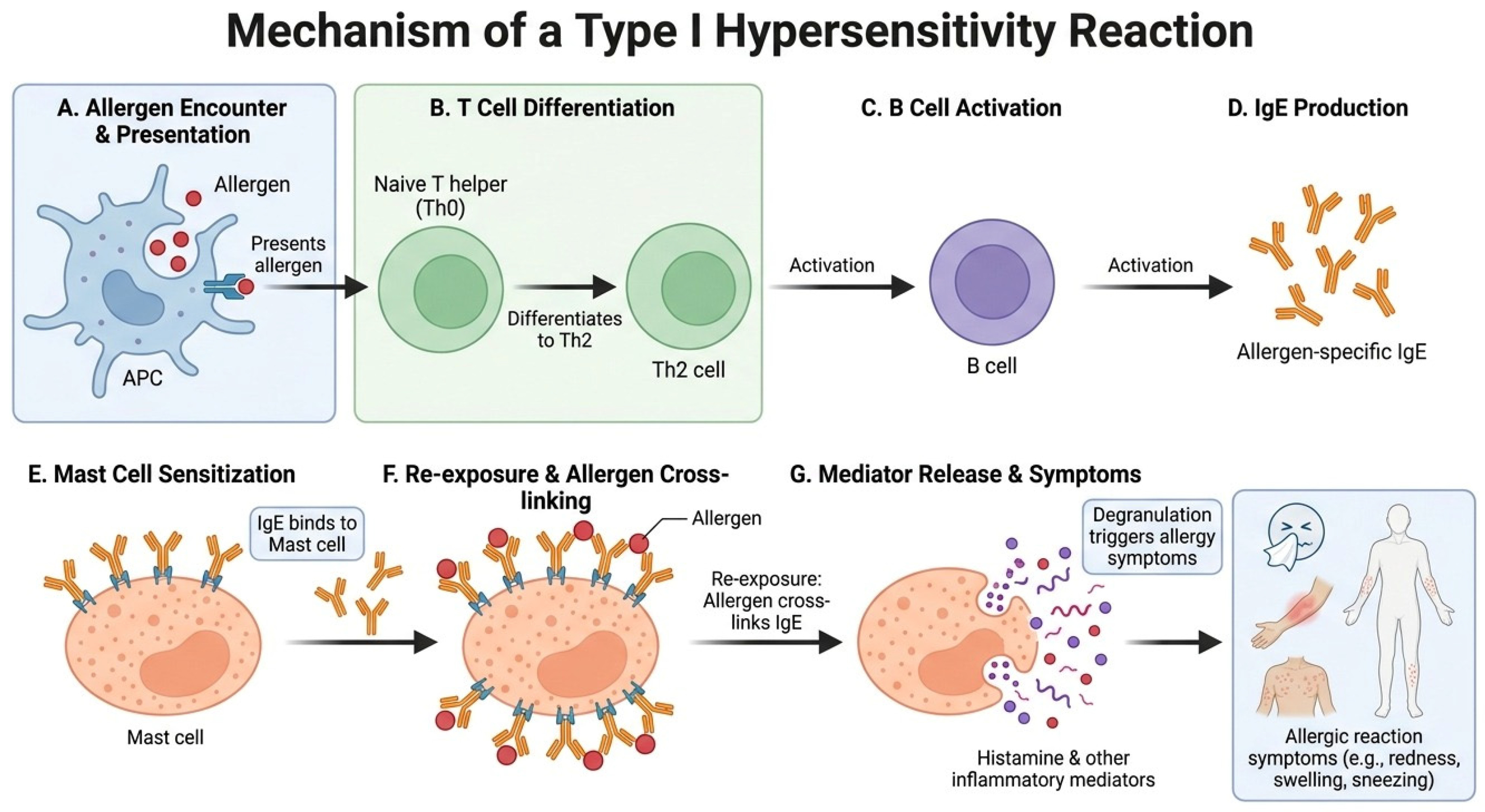

Disclaimer/Publisher’s Note: The statements, opinions and data contained in all publications are solely those of the individual author(s) and contributor(s) and not of MDPI and/or the editor(s). MDPI and/or the editor(s) disclaim responsibility for any injury to people or property resulting from any ideas, methods, instructions or products referred to in the content. |
© 2026 by the author. Licensee MDPI, Basel, Switzerland. This article is an open access article distributed under the terms and conditions of the Creative Commons Attribution (CC BY) license.