


Abstract
Eosinophilia is a heterogeneous clinical finding ranging from benign reactive states to clonal hematologic disorders associated with significant morbidity. This narrative, practical clinical review provides a structured, mechanism-based approach to the evaluation and management of peripheral eosinophilia by integrating contemporary consensus definitions, epidemiologic data, and diagnostic frameworks. Eosinophilia is defined as an absolute eosinophil count of ≥500 cells/µL, with hypereosinophilia defined as ≥1500 cells/µL. Etiologies are broadly classified as primary (clonal) or secondary (reactive). Peripheral eosinophil counts do not reliably predict organ involvement. Evaluation requires systematic assessment, including clinical history, laboratory testing, and targeted investigations. Management is guided by etiology and the presence of organ damage, with corticosteroids as first-line therapy in many cases and targeted therapies for clonal disease. The objective of this narrative, practical clinical review is to provide a structured, mechanism-based approach to the evaluation and management of peripheral eosinophilia, with emphasis on common reactive causes, recognition of organ involvement, and identification of high-risk clonal or hypereosinophilic presentations.
1. Introduction
Eosinophils are specialized granulocytes involved in immune defense and inflammatory regulation [1]. Although detectable in peripheral blood, they are found mainly in tissues, particularly within mucosal and lymphoid environments such as the gastrointestinal tract and respiratory epithelium [1]. Their biologic activity is mediated through cytoplasmic granules containing cytotoxic proteins, lipid mediators, and cytokines. Upon activation, eosinophils release these mediators, influencing immune signaling, epithelial integrity, and tissue remodeling [2]. While traditionally associated with parasitic infection and allergic disease, eosinophils are now recognized to have broader roles in immune homeostasis and pathologic inflammation [3].
Eosinophil differentiation is driven primarily by interleukin-5, with contributions from interleukin-3 and granulocyte–macrophage colony-stimulating factor. Tissue recruitment is mediated through chemokine pathways, particularly eotaxin–CCR3 signaling [2]. Dysregulation of these pathways may result in eosinophilia and, in some cases, clinically significant organ involvement [4].
2. Methods
This article was developed as a narrative, practical clinical review intended to provide a structured approach to the evaluation and management of peripheral eosinophilia. Relevant literature was identified through PubMed and major clinical references using combinations of the terms “eosinophilia,” “hypereosinophilia,” “hypereosinophilic syndrome,” “secondary eosinophilia,” “clonal eosinophilia,” “drug-induced eosinophilia,” “parasitic eosinophilia,” “eosinophilic gastrointestinal disease,” “eosinophilic lung disease,” and “eosinophilia management.” Priority was given to consensus statements, clinical guidelines, major reviews, and recent publications relevant to diagnostic classification, evaluation, and treatment. Additional references were selected from bibliographies of key articles when clinically relevant. Because this is a narrative clinical review rather than a systematic review, formal risk-of-bias assessment and meta-analysis were not performed.
3. Definitions and Conceptual Framework
Eosinophilia is defined as an absolute eosinophil count (AEC) of ≥500 cells/µL. Hypereosinophilia is defined as a persistent AEC of ≥1500 cells/µL and represents a clinically important threshold prompting further evaluation [1,5]. The degree of eosinophilia, however, does not reliably predict disease severity or organ involvement.
Hypereosinophilic syndrome (HES) is characterized by sustained hypereosinophilia in conjunction with eosinophil-mediated tissue injury after exclusion of secondary causes [1,5]. Tissue involvement may be present even in patients without extreme elevations in circulating eosinophils. Diagnosis requires confirmation of sustained eosinophilia, assessment for end-organ damage, and systematic exclusion of reactive causes. The differential diagnosis includes atopic disease, asthma, drug hypersensitivity, parasitic infection, allergic bronchopulmonary aspergillosis, eosinophilic gastrointestinal or pulmonary disease, eosinophilic granulomatosis with polyangiitis, autoimmune disease, adrenal insufficiency, solid tumors, lymphoma, and primary hematologic neoplasms such as chronic eosinophilic leukemia or myeloid/lymphoid neoplasms with eosinophilia [6].
HES is further classified into myeloid/clonal, lymphocytic-variant, overlap/organ-restricted, and idiopathic forms. This distinction is clinically important because clonal disease may involve rearrangements such as PDGFRA or PDGFRB, which can respond to targeted therapy, while lymphocytic-variant HES reflects aberrant T-cell cytokine production driving eosinophilia [6].
Tissue eosinophilia may be demonstrated by histopathologic infiltration, increased marrow eosinophils, or deposition of eosinophil-derived proteins [1,5]. Given the limited correlation between peripheral counts and tissue injury, evaluation must incorporate both laboratory and clinical findings.
4. Epidemiology
Hypereosinophilic disorders are rare, heterogeneous conditions whose epidemiology varies by underlying mechanism, geography, referral setting, and diagnostic criteria. True population-level estimates remain limited because many cases are identified in specialty settings rather than through population-based surveillance. HES is uncommon, but its true burden may be underestimated because milder, organ-restricted, or incompletely classified cases may not be recognized [7]. Reactive eosinophilia is far more common than primary or clonal eosinophilic disease and may occur in allergic rhinitis, asthma, atopic dermatitis, drug hypersensitivity, parasitic infection, autoimmune disease, eosinophilic gastrointestinal disorders, and eosinophilic pulmonary disease. The relative frequency of these causes varies by age, environmental exposure, medication use, travel history, and local prevalence of helminthic disease [8].
Primary or clonal eosinophilic disorders represent a smaller but clinically important subset because they may be associated with hematologic malignancy, progressive organ injury, and targetable molecular abnormalities. Myeloid and lymphoid neoplasms with eosinophilia may involve rearrangements in PDGFRA, PDGFRB, FGFR1, JAK2, or related pathways [6,8,9]. Among these, FIP1L1::PDGFRA-positive disease is the most frequently recognized molecular subtype and is notable for its strong response to imatinib [6,10,11]. Lymphocytic-variant HES is another important subtype, driven by aberrant T-cell populations that produce eosinophil-promoting cytokines, particularly interleukin-5 [8]. Distinguishing reactive, organ-restricted, lymphocytic, idiopathic, and clonal forms is essential because prognosis, treatment, and risk of irreversible tissue injury differ substantially [6].
5. Pathophysiology and Tissue Injury
Eosinophil-mediated disease reflects complex interactions among circulating eosinophils, tissue microenvironments, epithelial barriers, endothelial cells, fibroblasts, mast cells, lymphocytes, macrophages, and local cytokine networks. Eosinophils are not merely terminal effector cells; they participate in immune regulation, host defense, tissue remodeling, and inflammatory amplification [12]. Under physiologic conditions, eosinophils are present in selected tissues, especially the gastrointestinal tract, where they contribute to mucosal immune homeostasis. In disease states, excessive eosinophil recruitment, activation, and survival can disrupt this balance and promote tissue injury [12].
Eosinophil development and survival are driven primarily by type 2 inflammatory pathways, especially interleukin-5, with additional contributions from interleukin-3 and granulocyte–macrophage colony-stimulating factor. Tissue recruitment is mediated by chemokines such as eotaxins, adhesion molecules, and epithelial-derived cytokines, including thymic stromal lymphopoietin, interleukin-25, and interleukin-33. Once eosinophils enter tissues, they interact with mast cells, T lymphocytes, macrophages, and fibroblasts, creating self-amplifying inflammatory circuits [12].
Tissue injury occurs through direct cytotoxicity and indirect remodeling. Activated eosinophils release granule proteins, including major basic protein, eosinophil cationic protein, eosinophil peroxidase, and eosinophil-derived neurotoxin, which can damage epithelial and endothelial surfaces, increase vascular permeability, activate platelets, and promote thrombosis [13]. Eosinophils also release cytokines, lipid mediators, and growth factors that recruit additional inflammatory cells, activate fibroblasts, and stimulate extracellular matrix deposition. Over time, these processes may lead to fibrosis and irreversible organ damage [3,4]. Importantly, peripheral eosinophil count does not reliably predict disease severity, so evaluation should include symptoms, biomarkers, imaging, histopathology, and molecular testing when appropriate [1,4,8].
6. Mechanisms and Causes of Eosinophilia
Eosinophilia, defined as an increased eosinophil count in peripheral blood or tissues, is not a diagnosis in itself but a clinical sign that should prompt a structured differential diagnosis. In practice, the most useful first step is to determine whether the eosinophilia is primary (clonal) or secondary (reactive), because this distinction directs both urgency and testing. Primary eosinophilia is uncommon and results from intrinsic hematopoietic abnormalities causing clonal eosinophil proliferation. These disorders arise at the level of hematopoietic stem cells or myeloid precursors and may be associated with cytogenetic or molecular abnormalities such as FIP1L1::PDGFRA and other rearrangements involving PDGFRA, PDGFRB, or FGFR1. Clinically, clonal eosinophilia should be suspected when eosinophilia is marked, persistent, and unexplained and accompanied by constitutional symptoms, splenomegaly, abnormal blood counts, elevated vitamin B12 or tryptase, or evidence of a myeloid neoplasm [6,8,14].
Far more often, eosinophilia is secondary and reflects a reactive, polyclonal expansion of eosinophils driven largely by interleukin-5, interleukin-3, and granulocyte–macrophage colony-stimulating factor. From a practical clinical standpoint, reactive eosinophilia is best approached by reviewing the major categories that commonly present in outpatient and inpatient settings: allergic or atopic disease, medications, infections, autoimmune or inflammatory disorders, malignancy, and organ-specific eosinophilic syndromes. The history often provides the key. A careful timeline of symptoms, medication exposures, travel, dietary history, animal contact, asthma or sinus disease, rash, weight loss, fever, lymphadenopathy, and gastrointestinal or pulmonary symptoms is usually more informative than repeating eosinophil counts alone [8,14].
Allergic and atopic disorders are among the most frequent causes of mild eosinophilia in routine clinical practice. Allergic rhinitis, chronic rhinosinusitis, asthma, and atopic dermatitis commonly produce modest blood eosinophilia, usually not exceeding 1500/µL. When eosinophilia is substantially higher, clinicians should avoid attributing it too quickly to uncomplicated atopy and instead look for a more specific eosinophilic disorder. In patients with asthma, for example, eosinophilia may identify a type 2 inflammatory phenotype and may help guide biologic therapy, but marked eosinophilia should also raise concern for allergic bronchopulmonary aspergillosis, eosinophilic granulomatosis with polyangiitis, or chronic eosinophilic pneumonia. Thus, the presence of asthma does not end the evaluation; it often broadens it [8,14].
Medication-related eosinophilia is a major clinical category and may be missed unless deliberate medication reconciliation is performed. Nearly any prescription medication, over-the-counter preparation, herbal product, or supplement may be implicated. Recent and nonessential exposures should be reviewed carefully, particularly drugs started within the preceding days to weeks, while recognizing that latency may vary. Drug reaction with eosinophilia and systemic symptoms (DRESS) is especially important because it may initially resemble infection or a viral syndrome and can worsen even after the culprit drug is discontinued. Fever, rash, facial edema, lymphadenopathy, hepatitis, nephritis, pulmonary infiltrates, and hematologic abnormalities should heighten suspicion. Acute interstitial nephritis should also be considered when eosinophilia accompanies kidney injury [8,14,15].
An increasingly recognized medication-associated pattern is elevation in blood eosinophil count after anti-IL-4/IL-13 pathway blockade with dupilumab. This may reflect reduced eosinophil trafficking from blood into tissues rather than primary eosinophilic disease. The rise is often transient and asymptomatic, but rare eosinophil-associated complications, including eosinophilic pneumonia and eosinophilic granulomatosis with polyangiitis-like presentations, have been reported. Recent or current dupilumab exposure should therefore be documented during eosinophilia evaluation [16,17].
Parasitic infection remains a classic cause of eosinophilia and must be assessed thoughtfully, especially before starting systemic corticosteroids. In clinical practice, the question is not simply whether the patient has traveled internationally, but whether there has been epidemiologic exposure to helminths through residence, immigration history, freshwater exposure, barefoot soil contact, undercooked meat, or endemic environments. Strongyloides deserves special attention because untreated infection may disseminate after corticosteroid therapy. Depending on the exposure history, stool ova and parasite testing, serologies, and targeted infectious disease evaluation may be necessary. Other infectious causes, including fungal infection, HIV, scabies, and myiasis, should be considered in the appropriate clinical setting [8,15].
When eosinophilia is associated with pulmonary, dermatologic, gastrointestinal, cardiac, or neurologic symptoms, the clinician should think beyond generic “reactive eosinophilia” and evaluate for organ-specific eosinophilic disease. Pulmonary eosinophilic syndromes may present with cough, wheeze, dyspnea, fever, and infiltrates. Acute eosinophilic pneumonia can present dramatically and may initially occur without peripheral eosinophilia, whereas chronic eosinophilic pneumonia often follows a more indolent course. Dermatologic disorders such as bullous pemphigoid, Wells syndrome, eosinophilic panniculitis, chronic urticaria, and pregnancy-related dermatoses may also be associated with eosinophilia. Gastrointestinal involvement may occur in eosinophilic gastrointestinal diseases, inflammatory bowel disease, celiac disease, and hepatobiliary eosinophilic disorders. In these cases, symptoms and organ-directed evaluation should guide the workup rather than eosinophil count alone [8,14].
Autoimmune and rheumatologic diseases are additional considerations, especially when eosinophilia occurs with multisystem findings. Eosinophilic granulomatosis with polyangiitis is the prototypical example and should be suspected in patients with asthma, sinus disease, neuropathy, pulmonary infiltrates, or cardiac involvement. Other immune-mediated conditions, including IgG4-related disease, systemic sclerosis, Sjögren disease, systemic lupus erythematosus, and Behçet syndrome, may also be associated with eosinophilia, although eosinophilia is rarely the dominant feature.
Episodic angioedema with eosinophilia, also known as Gleich’s syndrome, is a rare but important cause of recurrent eosinophilia. It is characterized by periodic episodes of angioedema, urticaria, fever, weight gain, and marked hypereosinophilia, often recurring every 3 to 4 weeks. Although its pathogenesis remains incompletely defined, cyclic increases in interleukin-5 and eosinophil activation appear to play important roles. Gleich’s syndrome may resemble allergic angioedema, hereditary or acquired angioedema, eosinophilic granulomatosis with polyangiitis, or idiopathic HES; however, it is typically distinguished by its episodic pattern, recurrent peripheral eosinophilia, and generally favorable prognosis without progressive end-organ damage. Recognition of this entity is important to avoid misclassification as idiopathic HES or clonal eosinophilic disease [18].
Unexplained eosinophilia should also prompt consideration of hematologic malignancy or occult solid tumor, particularly when accompanied by systemic symptoms, abnormal smear findings, cytopenias, thrombocytosis, or organomegaly. In children, the broad categories overlap with adults, but atopy remains the most common cause of mild-to-moderate eosinophilia. Also, inborn errors of immunity assume greater importance in young children with eosinophilia plus severe dermatitis, recurrent or unusual infections, diarrhea, failure to thrive, autoimmunity, lymphadenopathy, hepatosplenomegaly, or markedly elevated IgE. Relevant examples include hyper-IgE syndromes (autosomal dominant STAT-3 mutation, DOCK-8 deficiency), Omenn syndrome, Wiskott–Aldrich syndrome, IPEX syndrome, autoimmune lymphoproliferative syndrome, and other combined immunodeficiencies with immune dysregulation. These conditions may resemble severe allergic disease but require immunologic evaluation and, when appropriate, genetic testing [15,19].
In adults, cholesterol crystal embolization syndrome should be considered after angiography, vascular surgery, endovascular intervention, or anticoagulation, especially with kidney injury, livedo reticularis, blue-toe syndrome, distal ischemic changes, systemic inflammation, and eosinophilia [20].
Finally, some patients have persistent hypereosinophilia of undetermined significance, defined as eosinophilia at or above 1500/µL without an identified cause and without evidence of eosinophil-mediated organ damage. This is a diagnosis of exclusion and should be assigned only after an appropriate evaluation has ruled out reactive and clonal causes. Clinically, these patients require careful follow-up, periodic reassessment for evolving organ involvement, and individualized management rather than reflexive treatment. Overall, eosinophilia should be approached systematically: confirm persistence, assess severity, look for organ injury, identify common secondary causes, and escalate to molecular and bone marrow evaluation when the presentation suggests a clonal or otherwise high-risk process [1,4].
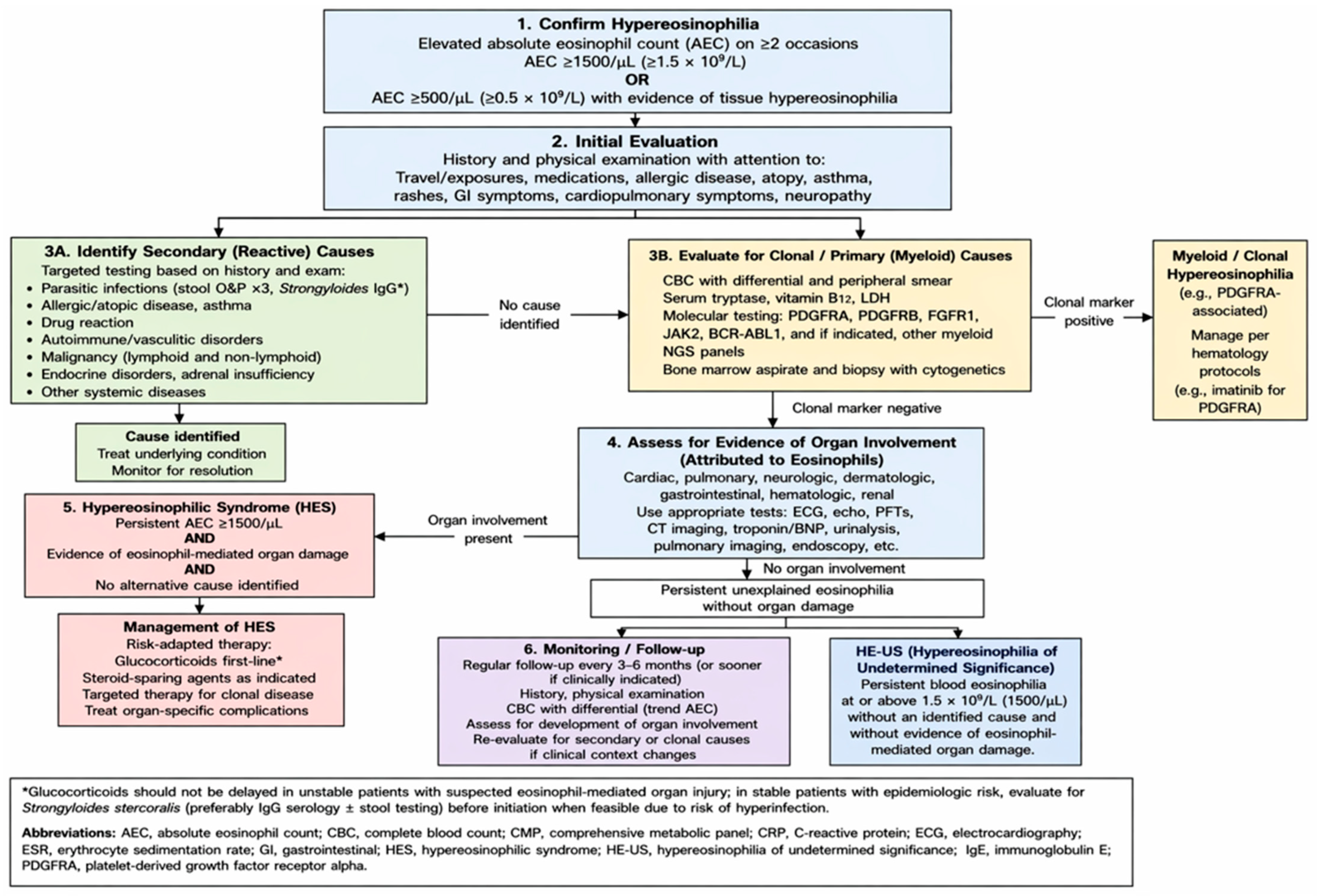
7. Evaluation of Peripheral Eosinophilia (Figure 1)
7.1. Overview and Diagnostic Rationale
Peripheral eosinophilia represents a heterogeneous clinical finding ranging from transient reactive eosinophilia to hypereosinophilic syndromes associated with irreversible organ injury. Because the absolute eosinophil count does not consistently correlate with disease severity, evaluation should not rely on eosinophil count alone. Tissue injury may occur with modest or intermittent eosinophilia, whereas some patients with marked eosinophilia remain asymptomatic. Diagnostic assessment should therefore integrate eosinophil persistence, symptoms, end-organ involvement, medication and exposure history, and evidence of clonal or hematologic disease [1,4].
Figure 1.
Proposed diagnostic algorithm synthesizing current approaches to hypereosinophilia and hypereosinophilic disorders.
7.2. Confirmation of Persistent Eosinophilia
The first step is confirmation of true and persistent eosinophilia with repeat complete blood count with differential and review of the peripheral smear. Transient eosinophilia may occur with allergic disease, infection, medication exposure, or inflammatory conditions. Persistent hypereosinophilia, generally defined as an absolute eosinophil count ≥1500/µL on repeated testing, should prompt a structured evaluation for secondary causes, end-organ involvement, and primary eosinophilic disorders [1,4].
7.3. Clinical History and Secondary Causes
Clinical evaluation should include a detailed history focused on allergic disease, asthma, atopic dermatitis, chronic rhinosinusitis, medication or supplement exposure, travel and migration history, parasitic risk, animal exposure, occupational or environmental exposures, autoimmune symptoms, constitutional symptoms, thrombotic events, and malignancy-associated features. Drug-induced eosinophilia is an important consideration because it may range from isolated laboratory eosinophilia to severe systemic disease, including drug reaction with eosinophilia and systemic symptoms. Fever, rash, lymphadenopathy, hepatitis, nephritis, pulmonary infiltrates, or recent medication initiation should raise suspicion. Infectious causes should be considered before immunosuppressive therapy, particularly helminthic infections such as Strongyloides stercoralis, Toxocara, and Ascaris. Strongyloides testing is especially important in at-risk patients before systemic corticosteroids because untreated infection may progress to hyperinfection syndrome after immunosuppression [1,4].
7.4. Baseline Laboratory Evaluation
Because eosinophil-mediated injury may be clinically silent early, evaluation should actively assess for organ involvement. Baseline studies may include a comprehensive metabolic panel, liver and renal function testing, urinalysis, inflammatory markers, serum IgE, vitamin B12, serum tryptase, lactate dehydrogenase, and peripheral smear examination. Elevated vitamin B12, tryptase, lactate dehydrogenase, cytopenias, dysplastic eosinophils, circulating blasts, thrombocytopenia, thrombocytosis, or splenomegaly should raise concern for a myeloid or clonal eosinophilic disorder [1,4].
7.5. Organ-Directed Assessment
Organ-directed testing should be guided by symptoms and examination. Pulmonary complaints may warrant chest imaging and pulmonary function testing. Gastrointestinal symptoms may require endoscopic evaluation with tissue biopsies. Cardiac involvement requires particular attention because eosinophilic myocarditis, intracardiac thrombus, and endomyocardial fibrosis may be life-threatening. Troponin, electrocardiography, echocardiography, and cardiac MRI should be considered when there is chest pain, dyspnea, abnormal cardiac biomarkers, thromboembolism, or persistent unexplained hypereosinophilia. Neurologic symptoms, neuropathy, or thrombotic events should prompt evaluation for vascular or neurologic complications [4].
7.6. Evaluation for Primary or Clonal Eosinophilic Disorders
If secondary causes are not identified, or if hematologic features are present, evaluation should proceed toward primary or clonal eosinophilic disorders. Features warranting hematology referral include persistent AEC ≥ 1500/µL, rapidly rising eosinophil count, AEC ≥ 50,000/µL, abnormal smear, cytopenias, circulating blasts, elevated vitamin B12 or tryptase, splenomegaly, lymphadenopathy, constitutional symptoms, thrombosis, or unexplained cardiac involvement. Molecular and cytogenetic testing should assess for recurrent rearrangements associated with myeloid and lymphoid neoplasms with eosinophilia, including FIP1L1::PDGFRA, PDGFRB, FGFR1, JAK2, and related tyrosine kinase abnormalities. Identification of these alterations is clinically important because PDGFRA- and PDGFRB-rearranged disease may respond dramatically to imatinib [6,10,11].
7.7. Bone Marrow, Cytogenetic, and Lymphocytic-Variant Evaluation
Bone marrow aspiration and biopsy should be considered when clonal disease is suspected. Marrow evaluation may reveal increased eosinophils, dysplasia, increased blasts, fibrosis, mast cell aggregates, or features of myeloid/lymphoid neoplasia. Cytogenetics, fluorescence in situ hybridization, polymerase chain reaction, and next-generation sequencing can identify cryptic rearrangements or myeloid mutations not evident on routine morphology. Lymphocytic-variant HES should also be considered, especially in patients with prominent skin disease, elevated IgE, lymphadenopathy, or steroid-responsive eosinophilia without a myeloid driver. Flow cytometry for aberrant T-cell populations and T-cell receptor gene rearrangement studies may be useful in this setting [1,4].
7.8. Diagnostic Pitfalls and Uncertainties
Several diagnostic pitfalls should be emphasized. Absence of marked peripheral eosinophilia does not exclude tissue eosinophilic disease, particularly when eosinophils are compartmentalized within affected organs. Corticosteroids may rapidly suppress eosinophilia and obscure diagnosis if given before infectious, hematologic, or tissue evaluation. Negative stool ova and parasite testing does not fully exclude helminthic infection; serologic testing may be required. Elevated IgE is nonspecific and may be seen in atopic disease, parasitic infection, lymphocytic-variant HES, and other inflammatory conditions. Finally, distinction among idiopathic HES, overlap eosinophilic disorders, and early clonal disease may remain uncertain, requiring longitudinal reassessment when eosinophilia persists or new organ involvement develops [1,4].
7.9. Urgent Evaluation and Diagnostic Pathway Integration
Urgent evaluation is warranted when eosinophilia is associated with cardiac symptoms, thromboembolism, neurologic deficits, pulmonary compromise, rapidly rising eosinophil count, AEC ≥ 50,000/µL, cytopenias, circulating blasts, or acute organ dysfunction. Overall, the diagnostic pathway should move from confirmation of persistent eosinophilia to exclusion of common secondary causes to assessment for end-organ involvement and, finally, to targeted hematologic, molecular, and marrow evaluation when primary or clonal disease is suspected [1,4,8].
8. Treatment and Management of Peripheral Eosinophilia
8.1. Initial Approach
For all patients:
- Stop nonessential or suspect medications.
- Treat reversible secondary causes such as infection, allergy, or autoimmune disease.
- Perform baseline organ assessment as indicated (e.g., troponin, echocardiography, pulmonary function testing).
Patients who are clinically unstable or have suspected organ injury should receive immediate therapy, often before the full diagnostic workup is complete [4].
8.2. Corticosteroids
Systemic corticosteroids are commonly used when eosinophilia is associated with clinically significant inflammation, suspected eosinophil-mediated organ injury, or high-risk hypereosinophilic presentations, but treatment should be directed whenever possible at the underlying cause. Typical regimens include prednisone 0.5 to 1 mg/kg/day with intravenous steroids or pulse methylprednisolone for severe or life-threatening diseases. Response is usually rapid, but gradual tapering is required, and relapse is common. Long-term use is limited by adverse effects, so steroid-sparing strategies should be considered early [4].
8.3. Management of Secondary Causes
Early management should prioritize treatment of secondary eosinophilia, including the following:
- Drug reactions;
- Parasitic infections;
- Allergic and atopic disease;
- Autoimmune or vasculitic conditions.
In patients at risk for Strongyloides, empiric ivermectin should be given before corticosteroids to prevent hyperinfection [4,21]. Optimization of comorbid conditions such as asthma or eosinophilic gastrointestinal disease is essential.
8.4. Targeted and Steroid-Sparing Therapies
Treatment should be tailored based on underlying etiology:
- Clonal eosinophilia: Imatinib is the first-line treatment for FIP1L1::PDGFRA-associated disease and is highly effective, often at low doses. Other molecular abnormalities may require alternative targeted therapies [6].
- Idiopathic or lymphocytic-variant HES: Corticosteroids remain the first-line treatment. For steroid-refractory or -dependent disease, options include hydroxyurea, interferon-α, or immunosuppressive agents [1,4].
Biologic therapies targeting the IL-5 pathway have become central in management. Mepolizumab is approved for HES and reduces steroid requirements [22]. Other biologics, such as dupilumab, may be useful in selected patients [16].
9. Follow-Up and Monitoring
Follow-up should be individualized based on etiology and disease severity [6]. Patients with persistent hypereosinophilia require closer monitoring, while those with mild or transient eosinophilia may be reassessed at longer intervals. Resolution of eosinophilia generally reduces the need for ongoing surveillance [6].
10. Limitations
This review may also be limited by its clinically dense, checklist-style structure. Although this format may be useful for experienced clinicians managing patients with eosinophilia, it may be less accessible to trainees, generalists, or clinicians who are less familiar with eosinophilic disorders and may require additional guidance in prioritizing diagnostic steps, interpreting specialized testing, and determining when subspecialty referral is warranted.
11. Conclusions
Eosinophilia represents a heterogeneous clinical entity encompassing a wide spectrum of disorders. Accurate diagnosis requires integration of clinical context with laboratory and molecular data [6]. The degree of eosinophilia alone does not reliably predict disease severity, emphasizing the importance of comprehensive evaluation [6]. Advances in targeted and biologic therapies have improved outcomes, but individualized clinical management remains essential.
Funding
This research received no external funding.
Institutional Review Board Statement
Not applicable.
Informed Consent Statement
Not applicable.
Data Availability Statement
No new data were created or analyzed in this study. Data sharing is not applicable to this article.
Acknowledgments
The author thanks Amra Osmanovic, RRT, for providing logistical support in the preparation of this manuscript.
Conflicts of Interest
The author declares no conflicts of interest.
Abbreviations
The following abbreviations are used in this manuscript:
| AEC | Absolute eosinophil count |
| HES | Hypereosinophilic syndrome |
References
- Valent, P.; Klion, A.D.; Roufosse, F.; Simon, D.; Metzgeroth, G.; Leiferman, K.M.; Schwaab, J.; Butterfield, J.H.; Sperr, W.R.; Sotlar, K.; et al. Proposed refined diagnostic criteria and classification of eosinophil disorders and related syndromes. Allergy 2023, 78, 47–59. [Google Scholar] [CrossRef] [Scilit]
- Davoine, F.; Lacy, P. Eosinophil cytokines, chemokines, and growth factors: Emerging roles in immunity. Front. Immunol. 2014, 5, 570. [Google Scholar] [CrossRef] [Scilit] [PubMed]
- Gurtner, A.; Crepaz, D.; Arnold, I.C. Emerging functions of tissue-resident eosinophils. J. Exp. Med. 2023, 220, e20221435. [Google Scholar] [CrossRef] [Scilit] [PubMed]
- Klion, A.D. How I treat hypereosinophilic syndromes. Blood 2015, 126, 1069–1077. [Google Scholar] [CrossRef] [Scilit] [PubMed]
- Simon, H.U.; Rothenberg, M.E.; Bochner, B.S.; Weller, P.F.; Wardlaw, A.J.; Wechsler, M.E.; Rosenwasser, L.J.; Roufosse, F.; Gleich, G.J.; Klion, A.D. Refining the definition of hypereosinophilic syndrome. J. Allergy Clin. Immunol. 2010, 126, 45–49. [Google Scholar] [CrossRef] [Scilit] [PubMed]
- Shomali, W.; Gotlib, J. World Health Organization and International Consensus Classification of eosinophilic disorders: 2024 update on diagnosis, risk stratification, and management. Am. J. Hematol. 2024, 99, 946–968. [Google Scholar] [CrossRef] [Scilit] [PubMed]
- Crane, M.M.; Chang, C.M.; Kobayashi, M.G.; Weller, P.F. Incidence of myeloproliferative hypereosinophilic syndrome in the United States and an estimate of all hypereosinophilic syndrome incidence. J. Allergy Clin. Immunol. 2010, 126, 179–181. [Google Scholar] [CrossRef] [Scilit] [PubMed]
- Roufosse, F.; Weller, P.F. Practical approach to the patient with hypereosinophilia. J. Allergy Clin. Immunol. 2010, 126, 39–44. [Google Scholar] [CrossRef] [Scilit] [PubMed]
- Pozdnyakova, O.; Orazi, A.; Kelemen, K.; King, R.; Reichard, K.K.; Craig, F.E.; Quintanilla-Martinez, L.; Rimsza, L.; George, T.I.; Horny, H.P.; et al. Myeloid/lymphoid neoplasms associated with eosinophilia and rearrangements of PDGFRA, PDGFRB, or FGFR1 or with PCM1-JAK2. Am. J. Clin. Pathol. 2021, 155, 160–178. [Google Scholar] [CrossRef] [Scilit] [PubMed]
- Cools, J.; DeAngelo, D.J.; Gotlib, J.; Stover, E.H.; Legare, R.D.; Cortes, J.; Kutok, J.; Clark, J.; Galinsky, I.; Griffin, J.D.; et al. A tyrosine kinase created by fusion of the PDGFRA and FIP1L1 genes as a therapeutic target of imatinib in idiopathic hypereosinophilic syndrome. N. Engl. J. Med. 2003, 348, 1201–1214. [Google Scholar] [CrossRef] [Scilit] [PubMed]
- Rohmer, J.; Couteau-Chardon, A.; Trichereau, J.; Panel, K.; Gesquière, C.; Ben Abdelali, R.; Bidet, A.; Bladé, J.S.; Cayuela, J.M.; Cony-Makhoul, P.; et al. Epidemiology, clinical picture and long-term outcomes of FIP1L1-PDGFRA-positive myeloid neoplasm with eosinophilia: Data from 151 patients. Am. J. Hematol. 2020, 95, 1314–1323. [Google Scholar] [CrossRef] [Scilit] [PubMed]
- Rosenberg, H.F.; Dyer, K.D.; Foster, P.S. Eosinophils: Changing perspectives in health and disease. Nat. Rev. Immunol. 2013, 13, 9–22. [Google Scholar] [CrossRef] [Scilit] [PubMed]
- Acharya, K.R.; Ackerman, S.J. Eosinophil granule proteins: Form and function. J. Biol. Chem. 2014, 289, 17406–17415. [Google Scholar] [CrossRef] [Scilit] [PubMed]
- Kuang, F.L. Approach to patients with eosinophilia. Med. Clin. N. Am. 2020, 104, 1–14. [Google Scholar] [CrossRef] [Scilit] [PubMed]
- Butt, N.M.; Lambert, J.; Ali, S.; Beer, P.A.; Cross, N.C.; Duncombe, A.; Ewing, J.; Harrison, C.N.; Knapper, S.; McLornan, D.; et al. Guideline for the investigation and management of eosinophilia. Br. J. Haematol. 2017, 176, 553–572. [Google Scholar] [CrossRef] [Scilit] [PubMed]
- Ezekwe, E.A.D., Jr.; Weskamp, A.L.; Rahim, R.; Makiya, M.A.; Wetzler, L.; Ware, J.M.; Nelson, C.; Castillo, P.A.; Riley, C.A.; Brown, T.; et al. Dupilumab use in patients with hypereosinophilic syndromes: A multicenter case series and review of the literature. J. Allergy Clin. Immunol. Pract. 2025, 13, 167–175.e6. [Google Scholar] [CrossRef] [Scilit] [PubMed]
- Portacci, A.; Poto, R.; Varricchi, G.; Carpagnano, G.E. Dupilumab and blood eosinophilia: A disease-specific phenomenon? Allergy 2025, 80, 1811–1814. [Google Scholar] [CrossRef] [Scilit] [PubMed]
- Mormile, I.; Petraroli, A.; Loffredo, S.; Rossi, F.W.; Mormile, M.; Del Mastro, A.; Spadaro, G.; de Paulis, A.; Bova, M. Episodic angioedema with hypereosinophilia (Gleich’s syndrome): A case report and extensive review of the literature. J. Clin. Med. 2021, 10, 1442. [Google Scholar] [CrossRef] [Scilit] [PubMed]
- Williams, K.W.; Milner, J.D.; Freeman, A.F. Eosinophilia associated with disorders of immune deficiency or immune dysregulation. Immunol. Allergy Clin. N. Am. 2015, 35, 523–544. [Google Scholar] [CrossRef] [Scilit] [PubMed]
- Ozkok, A. Cholesterol-embolization syndrome: Current perspectives. Vasc. Health Risk Manag. 2019, 15, 209–220. [Google Scholar] [CrossRef] [Scilit] [PubMed]
- Nutman, T.B. Evaluation and differential diagnosis of marked, persistent eosinophilia. Immunol. Allergy Clin. N. Am. 2007, 27, 529–549. [Google Scholar] [CrossRef] [Scilit] [PubMed]
- Rothenberg, M.E.; Klion, A.D.; Roufosse, F.E.; Kahn, J.E.; Weller, P.F.; Simon, H.U.; Schwartz, L.B.; Rosenwasser, L.J.; Ring, J.; Griffin, E.F.; et al. Treatment of patients with the hypereosinophilic syndrome with mepolizumab. N. Engl. J. Med. 2008, 358, 1215–1228. [Google Scholar] [CrossRef] [Scilit] [PubMed]
Disclaimer/Publisher’s Note: The statements, opinions and data contained in all publications are solely those of the individual author(s) and contributor(s) and not of MDPI and/or the editor(s). MDPI and/or the editor(s) disclaim responsibility for any injury to people or property resulting from any ideas, methods, instructions or products referred to in the content. |
© 2026 by the author. Licensee MDPI, Basel, Switzerland. This article is an open access article distributed under the terms and conditions of the Creative Commons Attribution (CC BY) license.