


Abstract
Type 2 inflammation is a central immunopathogenic driver of chronic immune-mediated disease, characterized by IgE polarization, eosinophilia, epithelial barrier disruption, and tissue remodeling. In veterinary medicine, especially in canine atopic and allergic disorders, the absence of validated functional endpoints hampers disease monitoring and therapeutic assessment. In humans, this endotype underlies a spectrum of linked conditions, including asthma, allergic rhinitis, atopic dermatitis, and chronic rhinosinusitis with nasal polyps (CRSwNP), where olfactory dysfunction is common, quantifiable, and clinically meaningful. In this narrative review, we examine the relationship between type 2 inflammation and olfactory outcomes in human disease and consider its translational relevance in dogs within a reverse translational One Health framework. Evidence from human studies, particularly in CRSwNP, shows a strong association between type 2 inflammatory burden and olfactory loss, while biologics targeting these pathways yield clinically meaningful gains in smell function. Comparable clinical evidence is still lacking in dogs. Olfactory function should therefore be regarded as a promising, yet unvalidated, translational endpoint in canine type 2 disease and a priority for prospective clinical investigation and methodological validation.
1. Introduction
Despite advances in the understanding of Type 2 inflammation in human diseases, its clinical translation to veterinary medicine remains limited. In dogs, particularly in conditions such as canine atopic dermatitis and other Th2-like disorders, therapeutic strategies are increasingly available; however, validated functional endpoints to monitor disease activity and treatment response are lacking. This represents a critical gap, as current assessments rely predominantly on clinical or behavioral parameters that may not fully capture the underlying immunological dynamics. Addressing this gap is essential to improving both disease stratification and therapeutic monitoring in veterinary medicine.
1.1. Immunological Basis of Type 2 (Th2) Inflammation
Type 2 (Th2) immunity is a distinct adaptive response that primarily arises at epithelial barriers, where the host interacts with the external environment. In response to allergenic stimuli or tissue injury, epithelial and resident dendritic cells release alarmin cytokines—most notably IL-33, IL-25, and thymic stromal lymphopoietin (TSLP)—which shape the local immunological milieu and promote polarization toward a Th2 profile [1]. In this context, IL-4 functions as the key differentiation signal, activating the transcription factors STAT6 and GATA3 to drive naïve CD4+ T cells toward a Th2 phenotype and coordinate Type 2 cytokine production.
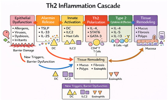
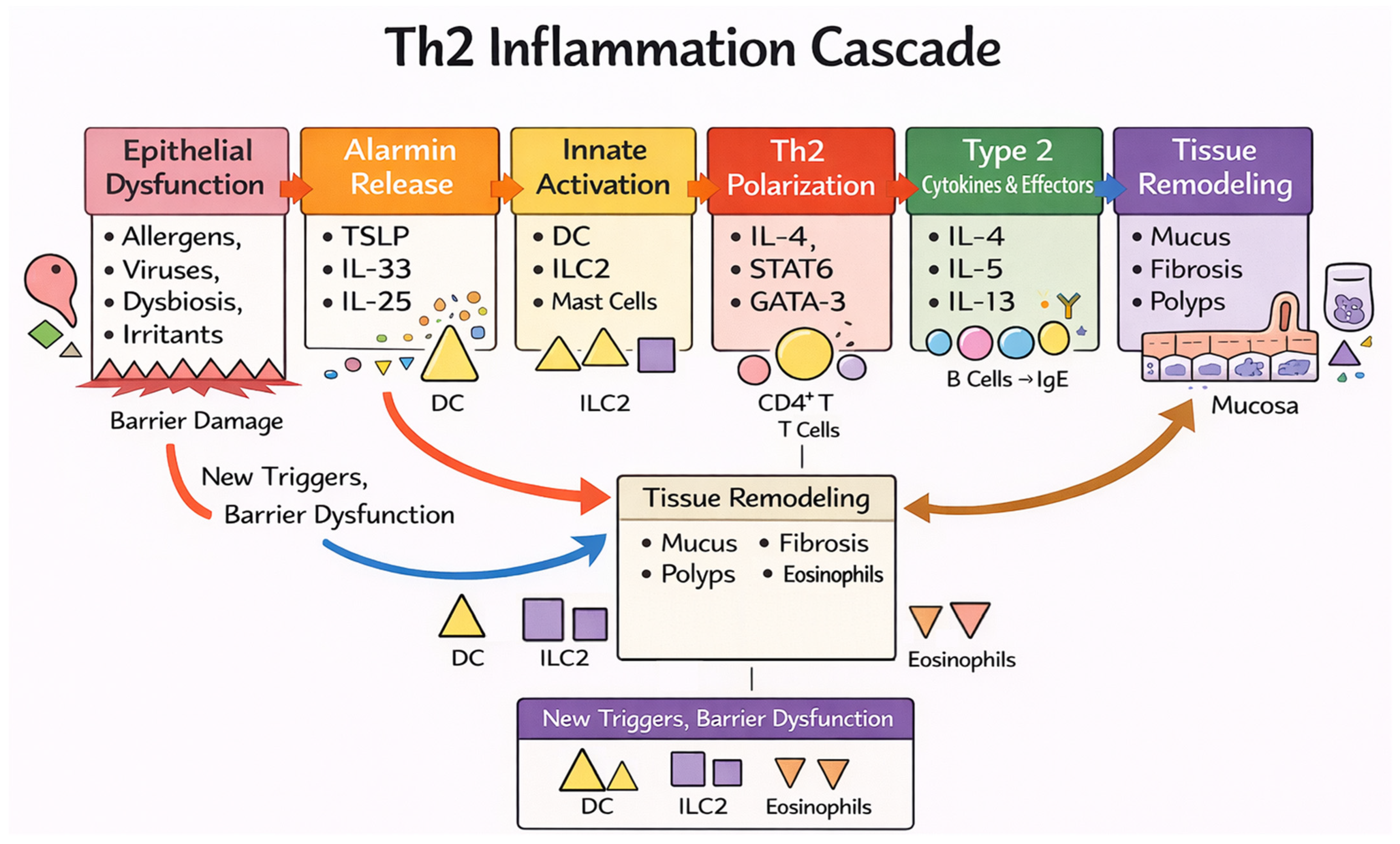
Once differentiated, Th2 cells, together with group 2 innate lymphoid cells (ILC2s), orchestrate a cytokine network dominated by IL-4, IL-5, and IL-13. IL-4 promotes B-cell class switching toward IgE production, IL-5 supports eosinophil maturation and recruitment, and IL-13 contributes to epithelial barrier dysfunction and tissue remodeling processes [2]. Collectively, these mechanisms generate an eosinophil-dominated inflammatory profile that physiologically defends against extracellular pathogens. Comparable Th2-driven pathways have been described in both humans and dogs, with species-specific variations in the magnitude and regulation of the response [3,4], as shown in Figure 1.
Figure 1.
Th2 inflammation cascade. Epithelial barrier dysfunction triggers alarmin release (TSLP, IL-33, IL-25), activating innate immune cells and promoting Th2 polarization. Type-2 cytokines (IL-4, IL-5, IL-13) drive IgE production, eosinophilic inflammation, and tissue remodeling (mucus, fibrosis, polyps), sustaining a self-perpetuating inflammatory loop. This schematic highlights the key immunological steps linking epithelial dysfunction to downstream Type 2-mediated tissue and functional alterations.
Beyond cytokine signaling, epigenetic mechanisms contribute to the stabilization and chronicity of Type 2 immune responses. In CD4+ T cells, sustained epigenetic remodeling of the Type 2 cytokine locus supports persistent expression of IL-4, IL-5, and IL-13, while alarmin-driven signals, particularly IL-33, further reinforce Th2 cytokine production and long-term maintenance of allergic immune programs [5]. At epithelial barrier sites, allergen exposure induces persistent activation states characterized by enhanced IL-33 expression, promoting feed-forward epithelial–immune amplification of Type 2 inflammation [6,7]. Environmental exposures such as air pollutants can further exacerbate these processes by inducing epigenetic alterations that skew immune balance toward a Th2 phenotype [8].
1.2. Th2-Driven Diseases: Shared Core Immunopathological Pathways with Species-Specific Variation
Persistent or dysregulated activation of Th2 pathways can shift the response from protective to chronically pathological, characterized by mucus hypersecretion, structural alterations, and progressive tissue remodeling culminating in fibrosis [2,9].
In humans, this paradigm is well established in conditions such as asthma, atopic dermatitis, allergic rhinitis, and chronic rhinosinusitis with nasal polyps (CRSwNP). In dogs, the strongest evidence concerns canine atopic dermatitis (CAD), whereas respiratory Th2-like phenotypes appear more limited and heterogeneous, partly reflecting anatomical differences and a lower degree of model standardization.
Based on this premise, the present work builds a translational rationale linking human and canine models, proposing olfactory function as a candidate functional endpoint of Type 2 inflammatory activity within a One Health framework—an integrated, multidisciplinary approach recognizing the interconnected health of humans, animals, and their shared environment—and adopts a reverse translational perspective that leverages robust human evidence to inform hypothesis generation in veterinary medicine.
1.3. Olfactory Function as a Functional Endpoint of Type 2 Inflammation
Olfactory dysfunction is a clinically relevant feature of chronic Type 2-predominant inflammatory diseases that directly or indirectly affect the nasal mucosa. It represents a sensitive functional indicator of inflammatory status and therapeutic response, reflecting the degree of epithelial and neuroepithelial impairment induced by Type 2 cytokines. Activation of IL-4- and IL-13-mediated pathways promotes mucosal barrier dysfunction, tissue remodeling, and loss of olfactory function [10].
Although these alterations are most prominent in CRSwNP, overlapping epithelial dysfunctions have also been documented in other Th2-predominant conditions, including allergic rhinitis, asthma, and atopic dermatitis. This pattern suggests the presence of shared immuno-epithelial mechanisms across Type 2-associated diseases; however, the extent, anatomical context, and functional consequences of epithelial dysfunction differ substantially across the aforementioned diseases [11].
Taken together, these observations suggest that olfactory function may serve as a functional endpoint of Type 2 inflammation. In the human model, olfactory performance provides a complementary functional readout to molecular and histological markers, integrating the overall impact of inflammation on the mucosal microenvironment. From this perspective, olfaction could represent a relevant indicator of inflammatory activity in other species affected by Th2-driven diseases, including dogs, thereby enriching the clinical assessment of allergic conditions in veterinary medicine.
2. Methods
This narrative review aimed to explore the mechanistic and clinical links between Type 2 (Th2) inflammation and olfactory dysfunction in humans, and to evaluate the translational relevance of these findings in dogs within a One Health framework. Given its narrative design, this review does not adhere to a formal systematic review protocol.
A targeted literature search was conducted in PubMed/MEDLINE and Scopus up to March 2026, complemented by manual screening of reference lists from key publications. The search strategy combined keywords related to Type 2 inflammation (e.g., “type 2 inflammation”, “Th2”, “IL-4”, “IL-13”, “IL-5”, “IL-33”, “TSLP”, “eosinophils”, “epithelial barrier”), olfactory function (e.g., “olfactory dysfunction”, “hyposmia”, “smell loss”, “olfactory epithelium”, “olfactory mucosa”), major human disease phenotypes (e.g., chronic rhinosinusitis with nasal polyps, allergic rhinitis, asthma, atopic dermatitis), and comparative or veterinary contexts (e.g., dog, canine, atopic dermatitis, One Health), using Boolean operators (AND/OR).
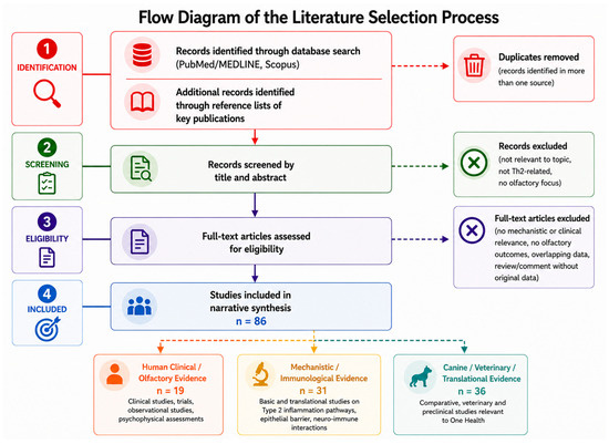
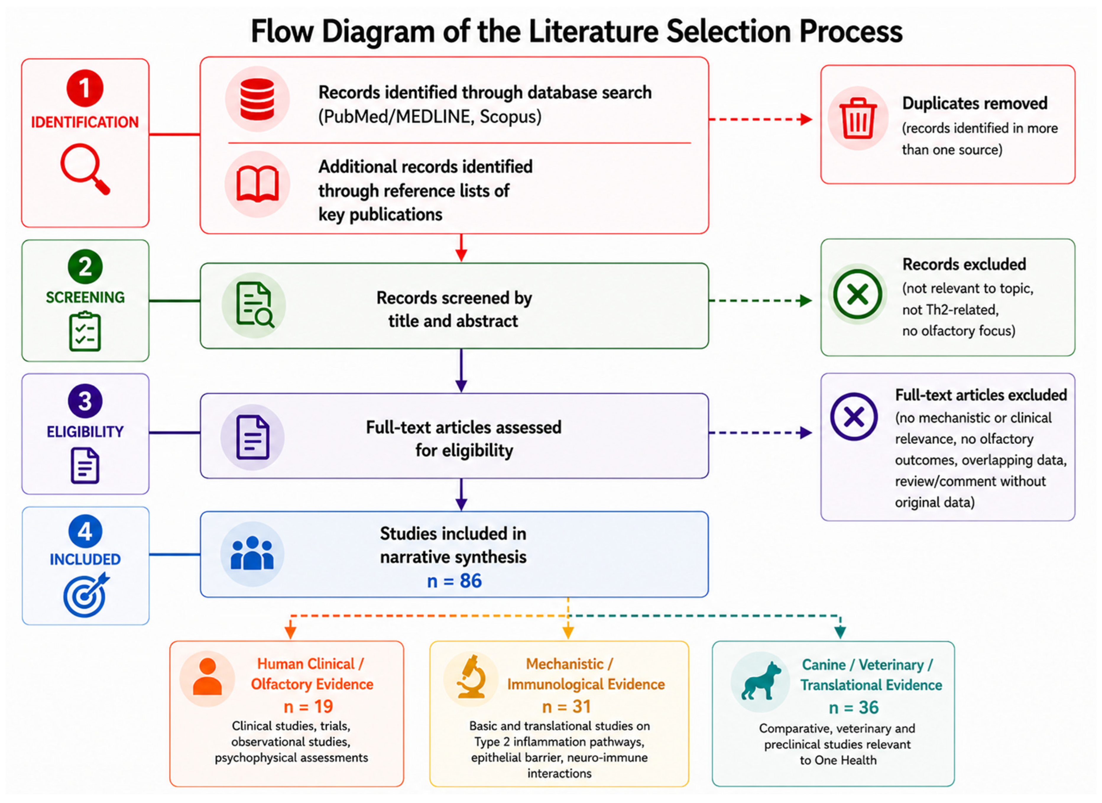
Studies were considered eligible if they addressed immunological mechanisms of Type 2 inflammation and/or investigated olfactory function in airway diseases, including both human and relevant canine data within a translational perspective. Articles were screened by title and abstract, followed by full-text evaluation when appropriate. To enhance transparency, the study selection process is summarized in a simplified and adapted flow diagram shown in Figure 2.
Figure 2.
Flow diagram of the literature selection process. This simplified and adapted flow diagram summarizes the study selection process used in this narrative review. It does not follow a formal PRISMA protocol and no quantitative tracking of records was performed during identification, screening, or exclusion stages. Studies included in the qualitative synthesis are grouped according to their predominant role as human clinical/olfactory evidence, mechanistic/immunological evidence, or canine/veterinary/translational evidence.
Priority was given to high-level evidence (systematic reviews, meta-analyses, consensus statements, and pivotal clinical trials), while additional original and translational studies were included to capture mechanistic insights. Screening and selection were performed by the authors, with discrepancies resolved through discussion. Only articles published in English were included. Conference abstracts were considered only when providing unique mechanistic information not available in full-text publications. When overlapping data were identified, priority was given to primary studies to minimize duplication.
Evidence was synthesized qualitatively and organized into thematic domains, including epithelial barrier dysfunction, Type 2 immune signaling, eosinophilic inflammation, olfactory neuro-immune interactions, clinical olfactory outcomes, and therapeutic modulation. No formal risk-of-bias assessment or quantitative meta-analysis was performed, in line with the narrative nature of the review.
The strength of evidence varies across domains. Robust data are mainly available in chronic rhinosinusitis with nasal polyps (CRSwNP), where randomized trials using validated psychophysical measures consistently report improvement in olfactory function following biologic therapies targeting Type 2 pathways, despite heterogeneity in patient characteristics and outcome definitions. In other Type 2-associated conditions, evidence is more heterogeneous and often indirect, with olfactory outcomes inconsistently assessed and rarely used as primary endpoints. Real-world data provide complementary insights but remain limited by potential bias and lack of standardization.
In dogs, available evidence is scarce and largely indirect. Although key Type 2 immunological pathways are partially conserved, no clinical studies have directly evaluated olfactory function in canine Th2-driven diseases. Therefore, the translational extension of human findings is based on biological plausibility rather than direct clinical validation, and the selection of studies may be subject to bias without ensuring completeness of the evidence base.
3. Type 2 (Th2) Diseases in Humans
3.1. Pathogenetic Mechanisms of Th2 Inflammation in Humans
As described in the Introduction, Th2 axis activation is an adaptive mechanism that protects against extracellular pathogens but, when dysregulated, drives chronic allergic disease. In the human model, the initiating event typically occurs at mucosal barrier surfaces, where epithelial damage or allergen exposure induces the release of alarmin cytokines (IL-25, IL-33, and TSLP), thereby triggering the coordinated activation of dendritic cells and group 2 innate lymphoid cells (ILC2s) [12]. This pro-Th2 microenvironment promotes lymphocyte differentiation and the coordinated production of IL-4, IL-5, and IL-13 [13], which mediate the characteristic immunological and epithelial effects of Type 2 inflammation.
In pathological settings, persistent allergen exposure and impaired regulatory control lead to a chronic effector phase in which mast cell degranulation and eosinophil activity amplify epithelial damage [14]. Toxic eosinophil-derived proteins, such as major basic protein (MBP) and eosinophil cationic protein (ECP), compromise mucosal integrity, while secondary-wave epithelial cytokines, including IL-24 and members of the IL-20 family, reduce the expression of barrier-associated proteins [15,16].
Persistent IL-13 signaling, together with TGF-β activity, ultimately drives the transition toward chronic tissue remodeling, characterized by fibrosis, hyperplasia, and functional epithelial alterations [17], consolidating the role of the epithelium as an active and self-amplifying node in Type 2 inflammation.
3.2. Major Human Th2-Driven Diseases
Considering the pathogenetic mechanisms outlined above, persistent activation results in distinct yet biologically interconnected clinical phenotypes, reflecting a shared immuno-epithelial dysfunction. This group includes asthma, CRSwNP, atopic dermatitis, and allergic rhinitis, in which inflammation is dominated by IL-4, IL-5, and IL-13 produced by Th2 lymphocytes and ILC2s [18].
This pathobiological convergence has driven the development of biologic therapies targeting Type 2 pathways, capable of modulating different clinical manifestations through shared molecular targets [19]. Within this context, olfactory function represents a particularly informative functional endpoint, as it integrates the impact of inflammation on the mucosal microenvironment beyond structural clinical markers alone.
3.3. Effects of Th2 Inflammation on Human Olfaction
Olfactory dysfunction represents a clinically relevant manifestation of Th2-driven diseases and may arise from both conductive mechanisms (related to airflow limitation and obstruction) and neurosensory mechanisms (related to inflammation-induced damage of the olfactory neuroepithelium), which are distinct, non-interchangeable processes that may contribute differently depending on the disease context. In CRSwNP, hyposmia is among the most frequent symptoms and correlates with the intensity of mucosal inflammation. Histopathological studies have shown that eosinophil density within the olfactory neuroepithelium is directly associated with the severity of olfactory loss, suggesting eosinophil-mediated neuronal damage and disruption of normal cellular turnover [20,21].
Chronic inflammation alters both the morphology and function of the olfactory neuroepithelium, leading to loss of sensory neurons and disorganization of regenerative basal cell populations. Inflammatory cytokines, together with eosinophil-derived mediators, promote neuronal apoptosis and ineffective regeneration [22].
Comparable alterations have also been described in other Th2-driven conditions, such as allergic rhinitis and atopic dermatitis, in which olfactory impairment affects a substantial proportion of patients [23,24].
3.4. Olfactory Outcomes as Clinical Endpoints in Biologic Therapies
Within this framework, recovery of olfactory function emerges as a sensitive and early indicator of response to anti-Type 2 biologic therapies.
In CRSwNP, treatment with the monoclonal antibody dupilumab has been associated with rapid and sustained improvement in olfactory performance, often observed independently of reductions in polyp volume, suggesting an effect directly related to modulation of Type 2 inflammation [25,26]. Similarly, other anti-Type 2 biologics—including omalizumab, mepolizumab, and benralizumab—have also been associated with improvements in olfactory function in both clinical trials and real-world studies, suggesting a potential shared immunomodulatory effect on the olfactory mucosa that extends beyond structural disease reduction. [27,28].
Notably, improvements in olfactory performance have frequently been reported to precede reductions in endoscopic scores and patient-reported outcomes such as SNOT-22 [29]; however, this observation should be interpreted with caution, as it may depend on study design, patient selection, baseline disease severity, and the specific assessment tools used.
Overall, meta-analyses and real-world evidence confirm that olfactory function represents a clinically relevant endpoint in the evaluation of anti-Type 2 biologic therapies [28,30].
Therefore, the use of olfactory function as a functional endpoint should be interpreted within the context of disease-specific characteristics and methodological variability.
3.5. Validated Tools for Assessing Olfactory Function in Humans
Assessment of olfactory function in humans relies primarily on standardized psychophysical tests, which are recommended by international guidelines as reference tools for both clinical practice and research [31].
Among these, Sniffin’ Sticks and the University of Pennsylvania Smell Identification Test (UPSIT) enable reproducible quantification of the main components of olfactory perception and provide composite scores that are widely used as functional endpoints in clinical trials [32,33,34].
In addition to psychophysical testing, self-reported questionnaires such as the Sino-Nasal Outcome Test-22 (SNOT-22) and the Questionnaire of Olfactory Disorders assess the subjective impact of olfactory dysfunction on quality of life [35,36]; however, they represent patient-reported outcome measures reflecting symptom burden and quality of life rather than direct assessments of olfactory function.
Electrophysiological techniques and neuroimaging may complement assessment in research settings or selected cases, but they do not represent first-line tools in routine clinical practice.
Taken together, these observations position the human olfactory system as a privileged model for understanding how Type 2 inflammation translates into measurable functional outcomes, providing a conceptual and methodological reference for translational exploration in other species. This framework offers a foundation from which, with appropriate species-specific adaptations, an operational approach to olfactory assessment in dogs may be derived.
Despite the strong mechanistic and clinical evidence in humans, the relative weight of mechanistic insights compared to direct clinical evidence should be considered when interpreting the translational implications.
4. Type 2 (Th2) Diseases in Dogs
4.1. The Th2 Axis in the Canine Immune Response
In dogs, Th2 responses represent a component of adaptive immunity involved in the control of extracellular pathogens and in the regulation of allergic inflammation.
The major effector cytokines described in humans have also been identified in the canine species, with levels of sequence homology consistent with functional conservation of key signaling pathways. Cloning and characterization studies have documented the conservation of IL-13 and its canine receptors (IL-13Rα1 and IL-13Rα2), supporting the presence of receptor-mediated mechanisms and downstream signaling comparable to those in humans [37]. Consistently, STAT6 activation in response to IL-4 has also been demonstrated in canine cellular models, indicating a functionally active IL-4/STAT6 axis [38].
Within the immune compartment, canine dendritic cells can produce Th2-related mediators in response to pro-inflammatory stimuli, suggesting an endogenous contribution to Type 2 polarization [39]. Moreover, canine T-cell populations with a Th2 cytokine profile and responsiveness to IL-33/ST2 signaling have been described, in line with an epithelial–Th2 amplification circuit recognized in other species [40,41].
Overall, available evidence supports the presence in dogs of a conserved Th2 axis in its fundamental elements, including coordinated activation of intracellular pathways such as JAK/STAT and modulation of IgE-mediated responses, albeit within a context of inter-individual and breed-related variability that may influence immunological phenotypes [42].
4.2. Th2-Driven Diseases in Dogs
Canine atopic dermatitis (CAD) is the most extensively studied condition and represents the main spontaneous model of Type 2 inflammation in dogs. At the histological and molecular levels, cutaneous lesions show lymphocytic infiltrates and a cytokine profile consistent with Th2 activation, including increased expression of polarization-associated markers (STAT6, GATA-3) and effector cytokines [43].
IL-31 emerges as a key mediator in the clinical phenotype of CAD, consistent with the central role of pruritus and cutaneous inflammation. In parallel, the IL-33/ST2 system has been implicated in Type 2 response modulation, with findings compatible with an amplification circuit associated with lesion chronicity [40,44].
With respect to respiratory Th2-like diseases (e.g., eosinophilic bronchitis, asthma-like phenotypes, allergic rhinitis), evidence is more limited and fragmented. Some studies suggest activation of a conserved IL-4/IL-13 axis in dogs, analogous to humans, capable of promoting allergen-specific IgE production and eosinophilic inflammation [45]. Concomitant expression of IL-4 and IL-31 in allergic contexts further suggests interactions between Th2 immunity, epithelial barrier function, and the nervous system, consistent with functional parallels to human Type 2 diseases [46].
From a morphological standpoint, canine eosinophilic bronchitis shows diffuse and nodular inflammatory patterns with high specificity, but without direct counterparts to human CRSwNP [47].
Compared with CAD, evidence for respiratory Type 2-like disease in dogs remains less standardized, which limits translational interpretation, particularly for olfactory outcomes.
4.3. Potential Impact of Type 2 Inflammation on Canine Olfaction
Although olfaction is biologically central in dogs, it is not routinely assessed in chronic inflammatory disease. To date, no direct clinical studies have evaluated olfactory function in dogs with Th2-driven disease. Any link between Type 2 inflammation and canine olfactory performance therefore remains hypothesis-driven. Operationally, one may hypothesize that dogs with CAD and/or Th2-like respiratory phenotypes could show longitudinal changes in detection or discrimination performance in relation to inflammatory burden and treatment response.
By analogy with experimental models, chronic IL-4- and IL-13-mediated signaling could impair epithelial integrity and regenerative processes, with possible effects on olfactory epithelial turnover [48]. In dogs, however, exposure of the neuroepithelium to inflammatory mediators may be modified by species-specific anatomy. The caudo-dorsal distribution of the olfactory epithelium and the complexity of the turbinates may reduce direct contact with particulate matter and irritants compared with more exposed configurations [49].
Histological and biological observations also suggest substantial regenerative capacity of the canine olfactory epithelium, supported by resident progenitors and sustentacular/supporting cells in the caudal nasal mucosa and frontal sinus, with possible implications for neuronal repair after injury [50].
Interpretation is further complicated by marked inter-individual and inter-breed variability in the canine olfactory receptor repertoire. More than 800 functional genes have been described, with substantial breed-related variability, and many variants involve amino acid changes that may influence odor sensitivity [51]. Functional analyses support the complexity of combinatorial olfactory coding, with broad and differentiated responses to odorants even within the same receptor family [52]. In addition, polymorphisms in olfactory receptor genes have been associated with performance in detection dogs, suggesting a link between receptor genetics and behavioral output [53].
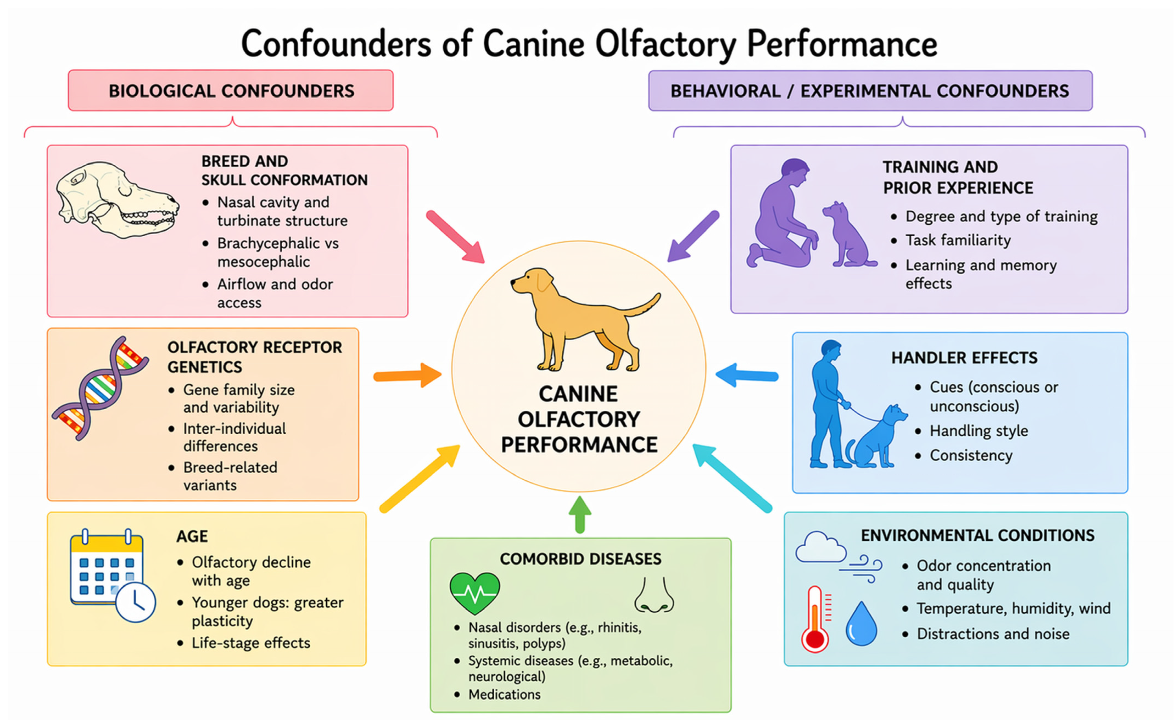
Accordingly, breed, skull conformation, training, motivation, age, concurrent disease, and handler-related effects should be controlled or stratified in study design, as summarized in Figure 3. At present, canine olfaction should be regarded as a candidate functional endpoint of translational interest, but one that remains unvalidated and hypothesis-generating.
Figure 3.
Main confounding factors influencing olfactory function in dogs. Schematic overview of biological and behavioral/experimental variables that may affect canine olfactory function independently of Type 2 (Th2) inflammation, representing potential sources of bias in olfactory assessment is a figure.
4.4. Biologic and Targeted Therapies in Veterinary Medicine
The introduction of targeted therapies addressing Type 2 pathways has substantially changed the management of canine atopic dermatitis. Major innovations include anti-IL-31 monoclonal antibodies (lokivetmab) and Janus kinase (JAK) inhibitors such as oclacitinib.
Lokivetmab rapidly reduces pruritus and skin lesions in dogs with CAD, with a favorable safety profile even during prolonged treatment [54,55], and may improve animal- and owner-reported quality-of-life indicators [56]. Lokivetmab primarily targets IL-31 and should therefore be distinguished from biologic therapies that directly modulate core Type 2 pathways, such as IL-4/IL-13 or IL-5 signaling, limiting its comparability with human anti-Type 2 biologics. However, transcriptomic data suggest that IL-31 blockade may not substantially modulate other components of the Type 2 network, such as IL-4 or IL-13, indicating a partial effect on the broader inflammatory circuit [57].
Oclacitinib, through inhibition of JAK-dependent pathways, reduces pruritus and lesions by interfering with cytokine signaling relevant to the Th2 phenotype, and indirect effects on skin barrier markers have been reported [58].
Taken together, these therapies support the clinical relevance of pathways shared with human medicine [59], although their impact on olfactory function has not yet been studied.
4.5. Potential Approaches to Olfactory Testing in Dogs
Assessment of olfactory function in dogs is a developing field but currently lacks clinically standardized instruments. In this context, behavioral tests represent the main operational approach, as they allow quantification of performance domains such as detection, discrimination, and olfactory generalization under controlled conditions. Among these, habituation–dishabituation paradigms may represent the most feasible approach for early validation studies, given their relatively low training requirements and adaptability to clinical settings, compared to more complex techniques such as functional MRI or controlled olfactometry. Experimental paradigms have documented differences across breeds and across dogs with different functional selection (e.g., working olfaction), supporting the feasibility of quantitative measures [60].
Approaches requiring minimal training, such as habituation–dishabituation paradigms, have been proposed as promising experimental options to estimate sensitivity and thresholds, although they require broader validation [61].
Other protocols use discriminative conditioning and controlled olfactometry to increase reproducibility and stimulus concentration control, but these methods remain largely confined to experimental or specialized settings [62].
Finally, fMRI studies in awake, unrestrained dogs demonstrate the possibility of linking cerebral responses to odor stimuli, providing a neurophysiological basis useful for translational research; however, costs and complexity limit clinical applicability [63,64].
Overall, most available approaches remain research tools requiring controlled settings, specialized equipment, and variable degrees of training, and are not currently suitable for routine clinical application. Human reference tests and proposed canine paradigms are summarized in Table 1, which highlights the gap between validated human tools and currently proposed canine approaches.
Table 1.
Olfactory assessment tools: validated human tests and proposed canine paradigms.
4.6. Criteria for Endpoint Validation in Canine Olfactory Assessment
If olfactory function is to be considered a potential endpoint in canine Type 2-driven disease, its validity should be assessed against standard methodological criteria.
Construct validity requires that olfactory performance reflect biological processes related to Type 2 inflammation, such as epithelial barrier dysfunction and neuroepithelial damage. While this relationship is supported in humans, it has not yet been demonstrated in dogs.
Responsiveness refers to the ability of the endpoint to detect clinically meaningful changes over time or after treatment. In humans, olfactory improvement has been reported after biologic therapies; comparable longitudinal data are not yet available in dogs.
Reproducibility and reliability require standardized, repeatable protocols with limited inter- and intra-subject variability. At present, canine olfactory testing lacks widely validated tools suitable for routine clinical use.
Feasibility concerns the practical implementation of olfactory testing in clinical and research settings, including time, training, and resource requirements.
Control of confounding is essential because canine olfactory performance may be influenced by breed, training status, age, motivation, and environmental conditions.
Finally, clinical relevance requires that changes in olfactory function correlate with meaningful outcomes or disease activity, a relationship that has not yet been established in dogs.
Taken together, these criteria indicate that olfactory function in dogs is, at present, a promising but unvalidated endpoint that requires formal methodological validation before clinical implementation. These validation domains are summarized in Table 2.
Table 2.
Proposed criteria for validation of olfactory function as an endpoint in dogs.
5. Translational Rationale: From the Human to the Canine Model
5.1. Immunological Analogy of Type 2 (Th2) Inflammation
The aim of this section is not to establish interspecies equivalence between humans and dogs, but to evaluate whether sufficient biological grounds exist to propose comparable functional endpoints within a translational framework. From this perspective, Type 2 inflammation represents an immunopathological paradigm that shows conserved elements across the two species, particularly within the IL-4/IL-5/IL-13 cytokine network and the alarmin IL-33/ST2 axis.
In humans, IL-4/IL-13 signaling promotes Th2 polarization and epithelial barrier dysfunction, with mucus hypersecretion and tissue remodeling [65], whereas IL-33 amplifies the response through ST2 by sustaining IL-5 and IL-13 production and the downstream eosinophilic cascade [66].
In dogs, transcriptomic and cellular evidence indicates a coherent circuit: activation of the IL-33/ST2 receptor in Th2 lymphocytes induces transcription of IL-5 and IL-13 [40], and IL-5/IL-5RA expression has been associated with eosinophilic responses and IgE production in canine atopic dermatitis [67], although the evidence remains limited and should be interpreted with caution. The presence of convergent tissue responses—such as eosinophilia, barrier dysfunction, and remodeling—supports the plausibility of a translational approach between humans and dogs; however, these similarities do not imply direct functional equivalence across species. Taken together, these elements support a testable translational hypothesis rather than an established mechanism, suggesting that analogous processes may affect olfactory function in dogs and requiring dedicated validation in canine models before any clinical applicability can be assumed.
5.2. Anatomical–Functional and Neuro-Immune Comparison of the Human and Canine Olfactory System
The olfactory system constitutes a microenvironment in which neuronal and immune components coexist and interact, making it plausible to consider olfactory function as an integrated readout of mucosal and neuroepithelial processes [68]. In humans, activation of Th2 pathways can modulate regeneration and function of the olfactory neuroepithelium; specifically, IL-4Rα/IL-13 signaling alters the neuroepithelial microenvironment and has been associated with loss of olfactory function in experimental and clinical contexts [48,69].
In chronic conditions, reorganization of basal cell compartments has also been described, with reduced regenerative capacity and persistence of an inflammatory state [70]. Consistently, in humans, the association between Type 2 inflammation within the olfactory region and functional deficits indicates that olfactory performance may reflect mucosal inflammatory burden [71,72].
From a comparative standpoint, the canine olfactory system displays a more extensive organization and anatomical–functional features that may influence exposure and epithelial vulnerability, including a larger sensory surface area and turbinate complexity that can optimize airflow dynamics [73]. Proposed mechanisms of relative protection include the caudo-dorsal localization of olfactory epithelium and specific cellular features, such as glial components with immunoregulatory properties, potentially relevant to modulating the effects of chronic inflammation [50,74]. While these anatomical features are well established, their proposed functional implications in terms of protection against inflammatory damage remain hypothetical and require dedicated experimental validation. Additional histological differences, including epithelial populations with distinct maturation and proliferative profiles, may contribute to recovery capacity after injury [75].
These differences do not invalidate the translational hypothesis but suggest that cross-species comparability should be defined in terms of functional endpoints rather than direct biological overlap. Within this framework, key processes—such as altered neurogenesis, epithelial damage, and neuronal apoptosis—can be considered partially shared, although differing in extent and regenerative capacity [76,77].
5.3. Proposed Stepwise Mechanistic Framework Across Species
To improve conceptual clarity, the relationship between Type 2 inflammation and olfactory dysfunction can be described as a stepwise mechanistic cascade.
In humans, strong evidence supports the upstream components of this pathway. Type 2 cytokines, particularly IL-4 and IL-13, promote epithelial barrier dysfunction and tissue remodeling, leading to mucus hypersecretion and structural alterations of the nasal mucosa. These processes are associated with inflammatory damage to the olfactory neuroepithelium, including eosinophil-mediated toxicity, altered basal cell function, and impaired neurogenesis. As a result, measurable olfactory dysfunction is consistently observed and supported by psychophysical testing.
In dogs, the initial steps of this cascade are partially supported. Type 2-associated cytokine pathways and epithelial barrier alterations have been documented in canine inflammatory conditions, suggesting the biological plausibility of similar upstream mechanisms. However, direct evidence linking these processes to olfactory neuroepithelial damage is currently lacking.
The downstream components—namely, the impact of Type 2 inflammation on olfactory neurogenesis and the resulting measurable olfactory impairment—remain largely hypothetical in the canine model. No clinical or experimental studies have directly demonstrated a causal relationship between Th2-driven inflammation and olfactory dysfunction in dogs.
Accordingly, this cross-species pathway should be interpreted as a stepwise translational hypothesis, in which upstream immunological mechanisms are partially supported, whereas downstream functional consequences remain to be validated.
5.4. Methodological Limits and Challenges of Interspecies Translation
Transposing immuno-olfactory models from humans to dogs requires a cautious approach, given conceptual, methodological, and ethical constraints. The complexity of Type 2 responses and interspecies differences in mucosal architecture, neuroplasticity, and receptor repertoires make it difficult to establish full functional equivalence between the two species.
Methodologically, key challenges include genetic heterogeneity across canine breeds, limited standardization of olfactory testing, and variability in assessment protocols and experimental conditions—factors that constrain endpoint comparability and reproducibility [78,79]. Immunological endpoints may also vary according to exposure route and species, with potential discrepancies between induced models and spontaneous disease, representing a relevant source of bias in comparative research [80,81,82].
From an interpretative standpoint, the principal risk is conflating molecular analogy with functional identity: conservation of IL-4/IL-5/IL-13 pathways or the IL-33/ST2 axis does not necessarily imply phenotypic or clinical equivalence. Accordingly, a One Health approach should integrate species-specific immunobiology, physiology, and behavior, prioritizing biological plausibility over assumptions of experimental equivalence [83].
A rigorous translational framework requires validated assays, shared endpoints, and recognition of species-specific constraints. The proposed model should therefore be considered a testable operational hypothesis rather than a claim of functional equivalence. Current evidence does not show a direct association between Type 2 inflammation and olfactory dysfunction in dogs, and the framework remains hypothesis-generating, requiring prospective controlled validation before any clinical application. Alignment between biological plausibility, methodological rigor, and clinically meaningful outcomes will be essential to ensure robustness and veterinary relevance.
5.5. Proposed Translational Study Design Within a One Health Framework
To operationalize the proposed One Health approach, a structured translational study design is required to evaluate olfactory function as a potential endpoint in canine Type 2-driven diseases.
The target population would include dogs affected by Type 2-associated conditions, primarily canine atopic dermatitis, with or without concomitant airway involvement. Control groups should include healthy dogs matched for breed, age, and training status, as well as disease controls with non-Type 2 inflammatory conditions where appropriate.
Olfactory assessment should be based on standardized and reproducible behavioral paradigms, such as detection and discrimination tasks, or habituation–dishabituation protocols requiring minimal training. Where feasible, controlled olfactometry or simplified threshold-based testing could be incorporated to improve quantitative resolution.
Parallel assessment of inflammatory status should include relevant biomarkers of Type 2 inflammation, such as circulating or tissue eosinophil counts, IgE levels, and cytokine profiling, alongside clinical severity indices appropriate for the underlying disease.
A longitudinal study design is essential to evaluate responsiveness, with repeated olfactory and immunological assessments performed before and after therapeutic intervention (e.g., biologics or targeted therapies), allowing analysis of temporal associations between inflammatory modulation and olfactory performance.
Key outcomes should include changes in olfactory performance metrics (accuracy, detection thresholds, response time), their correlation with inflammatory biomarkers, and their association with clinical disease activity and quality-of-life indicators where available. Within this framework, olfactory function should be regarded as a hypothesis-driven and exploratory endpoint, to be validated through controlled and standardized study designs before any clinical implementation.
6. Conclusions and Future Directions
Current evidence supports the biological plausibility of a translational framework linking Type 2 (Th2) inflammation and olfactory dysfunction across species, but does not demonstrate direct clinical equivalence between humans and dogs. In humans, the association between Type 2 inflammatory burden and olfactory impairment is supported by mechanistic, clinical, and therapeutic data, whereas in dogs the same relationship remains hypothetical and requires direct validation. Within a One Health perspective, this comparative framework is valuable not because it establishes interspecies equivalence, but because it identifies a coherent and testable research question grounded in shared immunopathological principles [84,85].
The most immediate priority is therefore methodological rather than clinical. Future studies should focus on the development of standardized canine olfactory testing paradigms, the control of major biological and behavioral confounders, and the integration of olfactory measures with immunological, histological, and clinical indicators of Type 2 inflammation. Longitudinal designs will be essential to determining whether olfactory performance is reproducible, responsive to changes in inflammatory burden, and clinically informative as a functional outcome.
If validated, olfactory function could become a complementary endpoint in veterinary studies of Th2-driven disease, adding a functional dimension to conventional measures such as lesion severity, pruritus, and quality of life. In this context, combining biological and functional parameters may improve characterization of disease activity and therapeutic response, as already suggested in human inflammatory research [86].
At present, however, the principal implication of the proposed model is the definition of a feasible and scientifically grounded translational agenda, rather than immediate application in veterinary clinical practice.
Author Contributions
Conceptualization, A.S. (Alessandro Serrone); methodology, A.S. (Alessandro Serrone), C.R., G.L.F. and G.C. (Giovanni Cavallo); formal analysis, A.S. (Alessandro Serrone) and C.R.; investigation, A.S. (Alessandro Serrone), C.R., A.M. and A.S. (Andrea Serrone); data curation, A.S. (Alessandro Serrone), A.S. (Andrea Serrone), L.M., G.C. (Giorgia Cavallo) and G.L.F.; writing—original draft preparation, A.S. (Alessandro Serrone); writing—review and editing, A.S. (Alessandro Serrone), C.R. and A.M.; visualization, A.S. (Alessandro Serrone) and C.R.; supervision, C.R., G.L.F. and G.C. (Giovanni Cavallo); project administration, A.S. (Alessandro Serrone). All authors have read and agreed to the published version of the manuscript.
Funding
This research received no external funding.
Institutional Review Board Statement
Not applicable (this manuscript is a review and did not involve new studies with human participants or animals).
Informed Consent Statement
Not applicable (this manuscript is a review and did not involve human participants or identifiable personal data).
Data Availability Statement
No new data were created or analyzed in this study.
Acknowledgments
During the preparation of this manuscript, the authors used an AI system to assist in language refinement, manuscript structuring, and the graphical preparation of figures. The figures were created by the authors using a digital drawing tablet with AI-assisted graphic tools. The authors have reviewed and edited all outputs and take full responsibility for the content of this publication.
Conflicts of Interest
The authors declare no conflicts of interest.
Abbreviations
The following abbreviations are used in this manuscript:
| CAD | Canine Atopic Dermatitis |
| CD4+ | Cluster of Differentiation 4 |
| CRSwNP | Chronic Rhinosinusitis with Nasal Polyps |
| ECP | Eosinophil Cationic Protein |
| fMRI | Functional Magnetic Resonance Imaging |
| GATA3 | GATA Binding Protein 3 |
| IgE | Immunoglobulin E |
| IL-4 | Interleukin 4 |
| IL-4Rα | Interleukin 4 Receptor Alpha |
| IL-5 | Interleukin 5 |
| IL-5RA | Interleukin 5 Receptor Alpha |
| IL-13 | Interleukin 13 |
| IL-13Rα1 | Interleukin 13 Receptor Alpha 1 |
| IL-13Rα2 | Interleukin 13 Receptor Alpha 2 |
| IL-20 | Interleukin 20 |
| IL-24 | Interleukin 24 |
| IL-25 | Interleukin 25 |
| IL-31 | Interleukin 31 |
| IL-33 | Interleukin 33 |
| ILC2s | Group 2 Innate Lymphoid Cells |
| JAK | Janus Kinase |
| JAK/STAT | Janus Kinase/Signal Transducer and Activator of Transcription |
| MBP | Major Basic Protein |
| SNOT-22 | Sino-Nasal Outcome Test-22 |
| ST2 | Suppression of Tumorigenicity 2 (IL-33 Receptor) |
| STAT6 | Signal Transducer and Activator of Transcription 6 |
References
- Zhu, J. T Helper 2 (Th2) Cell Differentiation, Type 2 Innate Lymphoid Cell (ILC2) Development and Regulation of Interleukin-4 (IL-4) and IL-13 Production. Cytokine 2015, 75, 14–24. [Google Scholar] [CrossRef] [Scilit] [PubMed]
- Goetzl, E.J. Th2 Cells in Rapid Immune Responses and Protective Avoidance Reactions. FASEB J. 2024, 38, e23485. [Google Scholar] [CrossRef] [Scilit]
- Chow, L.; Wheat, W.; Ramirez, D.; Impastato, R.; Dow, S. Direct Comparison of Canine and Human Immune Responses Using Transcriptomic and Functional Analyses. Sci. Rep. 2024, 14, 2207. [Google Scholar] [CrossRef] [Scilit] [PubMed]
- Gazi, U.; Taylan-Ozkan, A.; Mumcuoglu, K.Y. Immune Mechanisms in Human and Canine Demodicosis: A Review. Parasite Immunol. 2019, 41, e12673. [Google Scholar] [CrossRef] [Scilit] [PubMed]
- Louten, J.; Rankin, A.L.; Li, Y.; Murphy, E.E.; Beaumont, M.; Moon, C.; Bourne, P.; McClanahan, T.K.; Pflanz, S.; De Waal Malefyt, R. Endogenous IL-33 Enhances Th2 Cytokine Production and T-Cell Responses during Allergic Airway Inflammation. Int. Immunol. 2011, 23, 307–315. [Google Scholar] [CrossRef] [Scilit]
- Hristova, M.; Habibovic, A.; Veith, C.; Janssen-Heininger, Y.M.W.; Dixon, A.E.; Geiszt, M.; Van der Vliet, A. Airway Epithelial Dual Oxidase 1 Mediates Allergen-Induced IL-33 Secretion and Activation of Type 2 Immune Responses. J. Allergy Clin. Immunol. 2016, 137, 1545–1556.e11. [Google Scholar] [CrossRef] [Scilit]
- Altman, M.C.; Lai, Y.; Nolin, J.D.; Long, S.; Chen, C.-C.; Piliponsky, A.M.; Altemeier, W.A.; Larmore, M.; Frevert, C.W.; Mulligan, M.S.; et al. Airway Epithelium-Shifted Mast Cell Infiltration Regulates Asthmatic Inflammation via IL-33 Signaling. J. Clin. Investig. 2019, 129, 4979–4991. [Google Scholar] [CrossRef] [Scilit]
- Wang, C.; Wang, J.; Zheng, X.; Zhang, J.; Zhang, J.; Qiao, G.; Liu, H.; Zhao, H.; Bai, J.; Zhang, H.; et al. Epigenetic Regulation Is Involved in Traffic-Related PM2.5 Aggravating Allergic Airway Inflammation in Rats. Clin. Immunol. 2022, 234, 108914. [Google Scholar] [CrossRef] [Scilit]
- Lott, J.M.; Sumpter, T.L.; Turnquist, H.R. New Dog and New Tricks: Evolving Roles for IL-33 in Type 2 Immunity. J. Leukoc. Biol. 2015, 97, 1037–1048. [Google Scholar] [CrossRef] [Scilit]
- Bachert, C.; Hicks, A.; Gane, S.; Peters, A.T.; Gevaert, P.; Nash, S.; Horowitz, J.E.; Sacks, H.; Jacob-Nara, J.A. The Interleukin-4/Interleukin-13 Pathway in Type 2 Inflammation in Chronic Rhinosinusitis with Nasal Polyps. Front. Immunol. 2024, 15, 1356298. [Google Scholar] [CrossRef] [Scilit]
- Steelant, B.; Seys, S.; Boeckxstaens, G.E.; Akdis, C.; Ceuppens, J.; Hellings, P. Restoring Airway Epithelial Barrier Dysfunction: A New Therapeutic Challenge in Allergic Airway Disease. Rhinology 2017, 54, 195–205. [Google Scholar] [CrossRef] [Scilit]
- Gordon, E.D.; Locksley, R.M.; Fahy, J.V. Cross-Talk between Epithelial Cells and Type 2 Immune Signaling: The Role of IL-25. Am. J. Respir. Crit. Care Med. 2016, 193, 935–936. [Google Scholar] [CrossRef] [Scilit]
- Humphry, N. Epithelial Barrier Dysfunction in Type 2 Inflammatory Diseases. EMJ Dermatol. 2019, 7, 44–51. [Google Scholar] [CrossRef] [Scilit]
- Dong, X.; Ding, M.; Zhang, J.; Ogülür, I.; Pat, Y.; Akdis, M.; Gao, Y.; Akdis, C.A. Involvement and Therapeutic Implications of Airway Epithelial Barrier Dysfunction in Type 2 Inflammation of Asthma. Chin. Med. J. 2022, 135, 519. [Google Scholar] [CrossRef] [Scilit] [PubMed]
- Zhang, S.; Wu, W.; Gu, M.; Zhao, Y.; Wang, L.; Liu, K.; Yu, Z. House Dust Mite Induced Mucosal Barrier Dysfunction and Type 2 Inflammatory Responses via the MAPK/AP-1/IL-24 Signaling Pathway in Allergic Rhinitis. Int. Immunopharmacol. 2025, 148, 113972. [Google Scholar] [CrossRef] [Scilit]
- Kaymak, T.; Kaya, B.; Wuggenig, P.; Nuciforo, S.; Göldi, A.; Swiss EoE Cohort Study Group (SEECS); Oswald, F.; Roux, J.; Noti, M.; Melhem, H.; et al. IL-20 Subfamily Cytokines Impair the Oesophageal Epithelial Barrier by Diminishing Filaggrin in Eosinophilic Oesophagitis. Gut 2023, 72, 821–833. [Google Scholar] [CrossRef] [Scilit] [PubMed]
- Nakayama, T.; Tsunemi, Y.; Kashiwagi, T. Epithelial Cell Dynamics: Key Drivers of Type 2 Inflammation in Eosinophilic Chronic Rhinosinusitis. Auris Nasus Larynx 2025, 52, 354–361. [Google Scholar] [CrossRef] [Scilit] [PubMed]
- Gao, Y.; Wang, Z.; Ogulur, I.; Li, S.; Yazici, D.; Li, X.; Pat, Y.; Zheng, Y.; Babayev, H.; Zeyneloglu, C.; et al. The Evolution, Immunopathogenesis and Biomarkers of Type 2 Inflammation in Common Allergic Disorders. Allergy 2025, 80, 1848–1877. [Google Scholar] [CrossRef] [Scilit]
- Ghezzi, M.; Pozzi, E.; Abbattista, L.; Lonoce, L.; Zuccotti, G.V.; D’Auria, E. Barrier Impairment and Type 2 Inflammation in Allergic Diseases: The Pediatric Perspective. Children 2021, 8, 1165. [Google Scholar] [CrossRef] [Scilit]
- Yee, K.K.; Pribitkin, E.A.; Cowart, B.J.; Vainius, A.A.; Klock, C.T.; Rosen, D.; Feng, P.; McLean, J.; Hahn, C.-G.; Rawson, N.E. Neuropathology of the Olfactory Mucosa in Chronic Rhinosinusitis. Am. J. Rhinol. Allergy 2010, 24, 110–120. [Google Scholar] [CrossRef] [Scilit]
- Hauser, L.J.; Chandra, R.K.; Li, P.; Turner, J.H. Role of Tissue Eosinophils in Chronic Rhinosinusitis–Associated Olfactory Loss. Int. Forum Allergy Rhinol. 2017, 7, 957–962. [Google Scholar] [CrossRef] [Scilit]
- Chen, Y.; Li, M.; Lu, J. Apoptosis and Turnover Disruption of Olfactory Sensory Neurons in Eosinophilic Chronic Rhinosinusitis. Front. Cell. Neurosci. 2024, 18, 1371587. [Google Scholar] [CrossRef] [Scilit]
- Stuck, B.A.; Hummel, T. Olfaction in Allergic Rhinitis: A Systematic Review. J. Allergy Clin. Immunol. 2015, 136, 1460–1470. [Google Scholar] [CrossRef] [Scilit]
- Passali, F.M.; Passali, G.C.; Passali, D.; Ciprandi, G. Smell Impairment in Patients with Allergic Rhinitis. Int. Forum Allergy Rhinol. 2021, 11, 1031–1032. [Google Scholar] [CrossRef] [Scilit] [PubMed]
- Cantone, E.; de Corso, E.; Ricciardiello, F.; Di Nola, C.; Grimaldi, G.; Allocca, V.; Motta, G. Olfaction Recovery Following Dupilumab Is Independent of Nasal Polyp Reduction in CRSwNP. J. Pers. Med. 2022, 12, 1215. [Google Scholar] [CrossRef] [Scilit] [PubMed]
- Otten, J.J.; van der Lans, R.J.L.; Elzinga, H.B.E.; Adriaensen, G.F.J.P.M.; Benoist, L.B.L.; Hoven, R.D.; Seys, S.; Fokkens, W.J.; Reitsma, S. More than Half of Chronic Rhinosinusitis with Nasal Polyps (CRSwNP) Patients Treated with Dupilumab Experience Early and Fast Olfactory Improvement within 28 Days. Allergy 2024, 79, 3166–3168. [Google Scholar] [CrossRef] [Scilit]
- Barroso, B.; Valverde-Monge, M.; Betancor, D.; Gómez-López, A.; Villalobos-Vildas, C.; González-Cano, B.; Sastre, J. Improvement in Smell Using Monoclonal Antibodies Among Patients with Chronic Rhinosinusitis with Nasal Polyps: A Systematic Review. J. Investig. Allergol. Clin. Immunol. 2023, 33, 419–430. [Google Scholar] [CrossRef] [Scilit] [PubMed]
- Cai, S.; Xu, S.; Zhao, Y.; Zhang, L. Efficacy and Safety of Biologics for Chronic Rhinosinusitis with Nasal Polyps: A Meta-Analysis of Real-World Evidence. Allergy 2025, 80, 1256–1270. [Google Scholar] [CrossRef] [Scilit]
- Stilo, G.; Messina, G.; Faro, C.L.; Ruta, S.; Frangipane, S.; Mariut, D.; Giunta, G.; Distefano, M.E.; Zappalà, A.G.; Maniaci, A.; et al. Correlation Between Smell Recovery and Nasal Polyp Score in Patients Treated with Dupilumab: A Real-Life Retrospective, Observational, Monocentric Study. J. Pers. Med. 2025, 15, 164. [Google Scholar] [CrossRef] [Scilit]
- Hellings, P.W.; Verhoeven, E.; Fokkens, W.J. State-of-the-Art Overview on Biological Treatment for CRSwNP. Rhinology 2021, 59, 151–163. [Google Scholar] [CrossRef] [Scilit]
- Hummel, T.; Whitcroft, K.L.; Andrews, P.; Altundag, A.; Cinghi, C.; Costanzo, R.M.; Damm, M.; Frasnelli, J.; Gudziol, H.; Gupta, N.; et al. Position Paper on Olfactory Dysfunction. Rhinology 2017, 54, 1–30. [Google Scholar] [CrossRef] [Scilit] [PubMed]
- Kim, W.S.; Jang, D.P.; Kim, I.Y. The Current Status of Evaluation Technologies for the Function of Human Olfaction. Hanyang Med. Rev. 2014, 34, 120. [Google Scholar] [CrossRef] [Scilit]
- Doty, R.L. Psychophysical Testing of Human Olfactory Function. In Springer Handbook of Odor; Doty, R.L., Shaman, P., Eds.; Springer: Cham, Switzerland, 2017; pp. 59–60. [Google Scholar] [CrossRef] [Scilit]
- Su, B.; Bleier, B.; Wei, Y.; Wu, D. Clinical Implications of Psychophysical Olfactory Testing: Assessment, Diagnosis, and Treatment Outcome. Front. Neurosci. 2021, 15, 646956. [Google Scholar] [CrossRef] [Scilit] [PubMed]
- Thomas, A.J.; Mace, J.C.; Ramakrishnan, V.R.; Alt, J.A.; Mattos, J.L.; Schlosser, R.J.; Soler, Z.M.; Smith, T.L. Quality-of-Life and Olfaction Changes Observed with Short-Term Medical Management of Chronic Rhinosinusitis. Int. Forum Allergy Rhinol. 2020, 10, 656–664. [Google Scholar] [CrossRef] [Scilit]
- Cardella, A.; Riva, G.; Preti, A.; Albera, A.; Luzi, L.; Albera, R.; Cadei, D.; Motatto, G.M.; Omenetti, F.; Pecorari, G.; et al. Italian Version of the Brief Questionnaire of Olfactory Disorders (Brief-IT-QOD). ACTA Otorhinolaryngol. Ital. 2023, 43, 252–261. [Google Scholar] [CrossRef] [Scilit]
- Yang, S.; Boroughs, K.L.; McDermott, M.J. Canine Interleukin-13: Molecular Cloning of Full-Length cDNA and Expression of Biologically Active Recombinant Protein. J. Interferon Cytokine Res. 2000, 20, 779–785. [Google Scholar] [CrossRef] [Scilit]
- Rachakonda, P.S.; Rai, M.F.; Manning, K.; Schmidt, M.F.G. Expression of Canine Interleukin-4 in Canine Chondrocytes Inhibits Inflammatory Cascade through STAT6. Cytokine 2008, 44, 179–184. [Google Scholar] [CrossRef] [Scilit]
- Wang, Y.-S.; Chi, K.-H.; Chu, R.-M. Cytokine Profiles of Canine Monocyte-Derived Dendritic Cells as a Function of Lipopolysaccharide- or Tumor Necrosis Factor-Alpha-Induced Maturation. Vet. Immunol. Immunopathol. 2007, 118, 186–198. [Google Scholar] [CrossRef] [Scilit]
- Asahina, R.; Nishida, H.; Kamishina, H.; Maeda, S. Transcriptional Analysis of the IL-33 Receptor Suppression of Tumourigenicity 2 and Its Effects on Canine Type 2 T Helper Cells: A Preliminary Study. Vet. Dermatol. 2018, 29, 112-e45. [Google Scholar] [CrossRef] [Scilit]
- Protschka, M.; Di Placido, D.; Moore, P.F.; Büttner, M.; Alber, G.; Eschke, M. Canine Peripheral Non-Conventional TCRαβ+ CD4−CD8α− Double-Negative T Cells Show T Helper 2-like and Regulatory Properties. Front. Immunol. 2024, 15, 1400550. [Google Scholar] [CrossRef] [Scilit]
- Fung, K.L.; Breiner, M.L.; Mitchell, J.B.; Parchment, R.E. Abstract 2837: Conservation of JAK-STAT Signaling in Canine Lymphocytes Responding to Human IL-6, IL-7, and IL-1 Alpha. Cancer Res. 2024, 84, 2837. [Google Scholar] [CrossRef] [Scilit]
- Plager, D.A.; Torres, S.; Koch, S.; Kita, H. Gene Transcription Abnormalities in Canine Atopic Dermatitis and Related Human Eosinophilic Allergic Diseases. Vet. Immunol. Immunopathol. 2012, 149, 136–142. [Google Scholar] [CrossRef] [Scilit]
- Borek, F.; Nagashima, S.; Villalobos, W.R.; Gmyterco, V.C.; Sell, T.; de Farias, M.R.; Bechara, G.H. Immunoexpression of IL-33 in the Different Clinical Aspects of Canine Atopic Dermatitis. Vet. Immunol. Immunopathol. 2024, 273, 110786. [Google Scholar] [CrossRef] [Scilit] [PubMed]
- Tang, L.; Boroughs, K.L.; Morales, T.; Stedman, K.; Sellins, K.; Clarke, K.; McDermott, M.; Yang, S.; McCall, C. Recombinant Canine IL-13 Receptor α2-Fc Fusion Protein Inhibits Canine Allergen-Specific IgE Production In Vitro by Peripheral Blood Mononuclear Cells from Allergic Dogs. Vet. Immunol. Immunopathol. 2001, 83, 115–122. [Google Scholar] [CrossRef] [Scilit]
- McCandless, E.E.; Rugg, C.A.; Fici, G.J.; Messamore, J.E.; Aleo, M.M.; Gonzales, A.J. Allergen-Induced Production of IL-31 by Canine Th2 Cells and Identification of Immune, Skin, and Neuronal Target Cells. Vet. Immunol. Immunopathol. 2014, 157, 42–48. [Google Scholar] [CrossRef] [Scilit]
- Bottero, E.; Ruggiero, P.; Benvenuti, E.; Mussi, E.; Falcioni, D.; De Lorenzi, D.; Di Girolamo, N. Presence of Bronchial Nodules, Younger Age, and Heavier Body Weight Are Associated with a Diagnosis of Eosinophilic Lung Disease in Dogs with Cough. J. Am. Vet. Med. Assoc. 2022, 260, 414–421. [Google Scholar] [CrossRef] [Scilit] [PubMed]
- Saraswathula, A.; Liu, M.M.; Kulaga, H.; Lane, A.P. Chronic Interleukin-13 Expression in Mouse Olfactory Mucosa Results in Regional Aneuronal Epithelium. Int. Forum Allergy Rhinol. 2023, 13, 230–241. [Google Scholar] [CrossRef] [Scilit]
- Craven, B.; Paterson, E.; Settles, G. The Fluid Dynamics of Canine Olfaction: Unique Nasal Airflow Patterns as an Explanation of Macrosmia. J. R. Soc. Interface 2010, 7, 933–943. [Google Scholar] [CrossRef] [Scilit]
- Skinner, A.P.C.; Pachnicke, S.; Lakatos, A.; Franklin, R.J.M.; Jeffery, N.D. Nasal and Frontal Sinus Mucosa of the Adult Dog Contain Numerous Olfactory Sensory Neurons and Ensheathing Glia. Res. Vet. Sci. 2005, 78, 9–15. [Google Scholar] [CrossRef] [Scilit]
- Robin, S.; Tacher, S.; Rimbault, M.; Vaysse, A.; Dréano, S.; André, C.; Hitte, C.; Galibert, F. Genetic Diversity of Canine Olfactory Receptors. BMC Genom. 2009, 10, 21. [Google Scholar] [CrossRef] [Scilit]
- Benbernou, N.; Tacher, S.; Robin, S.; Rakotomanga, M.; Senger, F.; Galibert, F. Functional Analysis of a Subset of Canine Olfactory Receptor Genes. J. Hered. 2007, 98, 500–505. [Google Scholar] [CrossRef] [Scilit]
- Lesniak, A.; Walczak, M.; Jezierski, T.; Sacharczuk, M.; Gawkowski, M.; Jaszczak, K. Canine Olfactory Receptor Gene Polymorphism and Its Relation to Odor Detection Performance by Sniffer Dogs. J. Hered. 2008, 99, 518–527. [Google Scholar] [CrossRef] [Scilit]
- Moyaert, H.; Van Brussel, L.; Borowski, S.; Escalada, M.; Mahabir, S.P.; Walters, R.R.; Stegemann, M.R. A Blinded, Randomized Clinical Trial Evaluating the Efficacy and Safety of Lokivetmab Compared to Ciclosporin in Client-Owned Dogs with Atopic Dermatitis. Vet. Dermatol. 2017, 28, 593-e145. [Google Scholar] [CrossRef] [Scilit] [PubMed]
- Gober, M.; Amodie, D.; Mellencamp, M.; Hillier, A. Long-Term Use of Lokivetmab (Cytopoint®) in Atopic Dogs. BMC Vet. Res. 2025, 21, 203. [Google Scholar] [CrossRef] [Scilit] [PubMed]
- Marques, V.S.; Celesso, J.R.; Bicalho, A.P.d.C.V. Evaluation of Quality of Life in Dogs with Atopic Dermatitis and Their Owners after Lokivetmab Therapy. Res. Soc. Dev. 2021, 10, e392101119775. [Google Scholar] [CrossRef] [Scilit]
- Tamamoto-Mochizuki, C.; Crawford, N.; Eder, J.M.; Gonzales, A.J.; Olivry, T. Cytokine Transcriptome Profiling in Acute Experimental Canine Atopic Dermatitis Skin Lesions after IL-31 Inhibition with Lokivetmab. Vet. Dermatol. 2023, 34, 327–338. [Google Scholar] [CrossRef] [Scilit]
- Villalobos, W.R.; Ferreira, T.; Borek, F.; Santoro, D.; Ferrer, L.; Farias, M. Evaluation of Filaggrin 2 Expression in Dogs with Atopic Dermatitis before and after Oclacitinib Maleate Administration. Vet. Dermatol. 2025, 36, 453–461. [Google Scholar] [CrossRef] [Scilit]
- Banovic, F. Updated Insights into the Molecular Pathogenesis of Canine Atopic Dermatitis. Vet. Dermatol. 2025, 36, 375–384. [Google Scholar] [CrossRef] [Scilit]
- Polgár, Z.; Kinnunen, M.; Újváry, D.; Miklósi, Á.; Gácsi, M. A Test of Canine Olfactory Capacity: Comparing Various Dog Breeds and Wolves in a Natural Detection Task. PLoS ONE 2016, 11, e0154087. [Google Scholar] [CrossRef] [Scilit]
- Moser, A.Y.; Brown, W.Y.; Bizo, L.A. Use of a Habituation–Dishabituation Test to Determine Canine Olfactory Sensitivity. J. Exp. Anal. Behav. 2022, 118, 316–326. [Google Scholar] [CrossRef] [Scilit] [PubMed]
- DeChant, M.T.; Bunker, P.C.; Hall, N.J. Stimulus Control of Odorant Concentration: Pilot Study of Generalization and Discrimination of Odor Concentration in Canines. Animals 2021, 11, 326. [Google Scholar] [CrossRef] [Scilit]
- Jia, H.; Pustovyy, O.M.; Waggoner, P.; Beyers, R.J.; Schumacher, J.; Wildey, C.; Barrett, J.; Morrison, E.; Salibi, N.; Denney, T.S.; et al. Functional MRI of the Olfactory System in Conscious Dogs. PLoS ONE 2014, 9, e86362. [Google Scholar] [CrossRef] [Scilit]
- Berns, G.S.; Brooks, A.M.; Spivak, M. Scent of the Familiar: An fMRI Study of Canine Brain Responses to Familiar and Unfamiliar Human and Dog Odors. Behav. Process. 2015, 110, 37–46. [Google Scholar] [CrossRef] [Scilit]
- Stolarski, B.; Kurowska-Stolarska, M.; Kewin, P.; Xu, D.; Liew, F.Y. IL-33 Exacerbates Eosinophil-Mediated Airway Inflammation. J. Immunol. 2010, 185, 3472–3480. [Google Scholar] [CrossRef] [Scilit]
- Lee, H.Y.; Rhee, C.K.; Kang, J.Y.; Byun, J.H.; Choi, J.Y.; Kim, S.J.; Kim, Y.K.; Kwon, S.S.; Lee, S.Y. Blockade of IL-33/ST2 Ameliorates Airway Inflammation in a Murine Model of Allergic Asthma. Exp. Lung Res. 2014, 40, 66–76. [Google Scholar] [CrossRef] [Scilit]
- Tavener, S.K.; Badri, D.V.; Panickar, K.S. Role of IL-5/IL-5RA Axis in Eosinophilic Allergic Response in Canines with Dermatological Problems. FASEB J. 2022, 36, R4853. [Google Scholar] [CrossRef] [Scilit]
- Dietz, A.; Senf, K.; Neuhaus, E.M. ACKR3 in Olfactory Glia Cells Shapes the Immune Defense of the Olfactory Mucosa. Glia 2024, 72, 1183–1200. [Google Scholar] [CrossRef] [Scilit]
- Hara, Y.; Jha, M.K.; Huang, J.Y.; Han, Y.; Langohr, I.M.; Gaglia, G.; Zhu, C.; Piepenhagen, P.; Gayvert, K.; Lim, W.K.; et al. The IL-4–IL-4Rα Axis Modulates Olfactory Neuroimmune Signaling to Induce Loss of Smell. Allergy 2025, 80, 440–461. [Google Scholar] [CrossRef] [Scilit]
- Chen, M.; Reed, R.R.; Lane, A.P. Chronic Inflammation Directs an Olfactory Stem Cell Functional Switch from Neuroregeneration to Immune Defense. Cell Stem Cell 2019, 25, 501–513.e5. [Google Scholar] [CrossRef] [Scilit]
- Schlosser, R.J.; Mulligan, J.K.; Hyer, J.M.; Karnezis, T.T.; Gudis, D.A.; Soler, Z.M. Mucous Cytokine Levels in Chronic Rhinosinusitis–Associated Olfactory Loss. JAMA Otolaryngol. Head Neck Surg. 2016, 142, 731–737. [Google Scholar] [CrossRef] [Scilit]
- Lavin, J.; Min, J.; Lidder, A.K.; Huang, J.H.; Kato, A.; Lam, K.; Meen, E.; Chmiel, J.S.; Norton, J.; Suh, L.; et al. Superior Turbinate Eosinophilia Correlates with Olfactory Deficit in Chronic Rhinosinusitis Patients. Laryngoscope 2017, 127, 2210–2218. [Google Scholar] [CrossRef] [Scilit] [PubMed]
- Barrios, A.W.; Sánchez-Quinteiro, P.; Salazar, I. Dog and Mouse: Toward a Balanced View of the Mammalian Olfactory System. Front. Neuroanat. 2014, 8, 106. [Google Scholar] [CrossRef] [Scilit]
- Jiang, Y.; Guo, J.; Tang, X.; Wang, X.; Hao, D.; Yang, H. The Immunological Roles of Olfactory Ensheathing Cells in the Treatment of Spinal Cord Injury. Front. Immunol. 2022, 13, 881162. [Google Scholar] [CrossRef] [Scilit]
- Bock, P.; Rohn, K.; Beineke, A.; Baumgärtner, W.; Wewetzer, K. Site-Specific Population Dynamics and Variable Olfactory Marker Protein Expression in the Postnatal Canine Olfactory Epithelium. J. Anat. 2009, 215, 377–387. [Google Scholar] [CrossRef] [Scilit]
- Rouyar, A.; Classe, M.; Gorski, R.; Bock, M.-D.; Le-Guern, J.; Roche, S.; Fourgous, V.; Remaury, A.; Paul, P.; Ponsolles, C.; et al. Type 2/Th2-Driven Inflammation Impairs Olfactory Sensory Neurogenesis in Mouse Chronic Rhinosinusitis Model. Allergy 2019, 74, 549–559. [Google Scholar] [CrossRef] [Scilit]
- Kikuta, S.; Nagayama, S.; Hasegawa-Ishii, S. Structures and Functions of the Normal and Injured Human Olfactory Epithelium. Front. Neural Circuits 2024, 18, 1406218. [Google Scholar] [CrossRef] [Scilit] [PubMed]
- Dearman, R.J.; Kimber, I. Animal Models of Protein Allergenicity: Potential Benefits, Pitfalls and Challenges. Clin. Exp. Allergy 2009, 39, 458–468. [Google Scholar] [CrossRef] [Scilit]
- Bøgh, K.L.; van Bilsen, J.; Głogowski, R.; López-Expósito, I.; Bouchaud, G.; Blanchard, C.; Bodinier, M.; Smit, J.; Pieters, R.; Bastiaan-Net, S.; et al. Current Challenges Facing the Assessment of the Allergenic Capacity of Food Allergens in Animal Models. Clin. Transl. Allergy 2016, 6, 21. [Google Scholar] [CrossRef] [Scilit] [PubMed]
- Van Gramberg, J.L.; de Veer, M.J.; O’Hehir, R.E.; Meeusen, E.N.T.; Bischof, R.J. Use of Animal Models to Investigate Major Allergens Associated with Food Allergy. J. Allergy 2013, 2013, 635695. [Google Scholar] [CrossRef] [Scilit]
- Madhuranga, H.D.T.; Alles, C. Experimental In Vivo Animal Models for Asthma/Allergic Asthma: Importance and Documented Parameters—A Review. J. Phytopharmacol. 2024, 13, 49–63. [Google Scholar] [CrossRef] [Scilit]
- Santoro, D.; Marsella, R. Animal Models of Allergic Diseases. Vet. Sci. 2014, 1, 192–212. [Google Scholar] [CrossRef] [Scilit]
- Montoya-Alonso, J.A.; Balmori-de la Puente, A.; Costa-Rodríguez, N.; Matos, J.I.; Carretón, E.; Morchón, R. A One Health Perspective on Heartworm Disease: Allergy Risk in Owners of Infected Dogs in Gran Canaria (Spain). Animals 2025, 15, 3084. [Google Scholar] [CrossRef] [Scilit] [PubMed]
- Bongiovanni, L.; Brachelente, C.; Dow, S.; Bergman, P.J. Editorial: Canine Melanoma in Comparative Oncology: Translate Research Advances to Develop New Diagnostic and Therapeutic Options. Front. Vet. Sci. 2023, 9, 1127527. [Google Scholar] [CrossRef] [Scilit]
- Regan, D.; Garcia, K.; Thamm, D. Clinical, Pathological, and Ethical Considerations for the Conduct of Clinical Trials in Dogs with Naturally Occurring Cancer: A Comparative Approach to Accelerate Translational Drug Development. ILAR J. 2018, 59, 99–110. [Google Scholar] [CrossRef] [Scilit]
- Todd, L.L.; Sivakumar, R.; Lynch, S.G.; Diebolt, J.H.; White, J.; Villwock, J.A. Longitudinal Olfactory Patterns in Multiple Sclerosis: A Scoping Review and Implication for Use in Management of Disease. Int. J. MS Care 2023, 25, 131–136. [Google Scholar] [CrossRef] [Scilit] [PubMed]

Disclaimer/Publisher’s Note: The statements, opinions and data contained in all publications are solely those of the individual author(s) and contributor(s) and not of MDPI and/or the editor(s). MDPI and/or the editor(s) disclaim responsibility for any injury to people or property resulting from any ideas, methods, instructions or products referred to in the content. |
© 2026 by the authors. Licensee MDPI, Basel, Switzerland. This article is an open access article distributed under the terms and conditions of the Creative Commons Attribution (CC BY) license.