Next Issue
Volume 11, April
Previous Issue
Volume 11, February


J. Imaging, Volume 11, Issue 3 (March 2025) – 24 articles

Cover Story (view full-size image): 
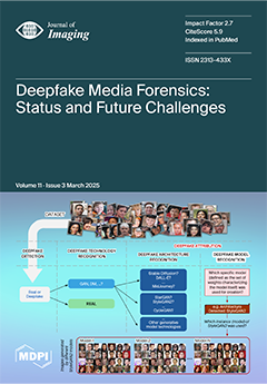
The rapid evolution of AI-generated media, or deepfakes, presents both opportunities and threats to digital communication, entertainment, and cybersecurity. Leveraging cutting-edge generative models such as GANs and diffusion models, deepfakes turn out to be hyper-realistic but fake content, raising serious concerns about misinformation and media reliability. The FF4ALL research project addresses deepfake detection, forensic attribution, and media authentication by developing innovative methodologies to fight against the illegal use of deepfakes. Analyzing current methodologies, challenges, and future directions, this study proposes solutions to improve the integrity of digital content and fight emerging threats in synthetic media. View this paper
  • Issues are regarded as officially published after their release is announced to the table of contents alert mailing list.
  • You may sign up for e-mail alerts to receive table of contents of newly released issues.
  • PDF is the official format for papers published in both, html and pdf forms. To view the papers in pdf format, click on the "PDF Full-text" link, and use the free Adobe Reader to open them.
Order results
Result details
Section
Select all
Export citation of selected articles as:
57 pages, 8107 KiB  
Review
Machine Learning for Human Activity Recognition: State-of-the-Art Techniques and Emerging Trends
by Md Amran Hossen and Pg Emeroylariffion Abas
J. Imaging 2025, 11(3), 91; https://doi.org/10.3390/jimaging11030091 - 20 Mar 2025
Cited by 1 | Viewed by 897
Abstract
Human activity recognition (HAR) has emerged as a transformative field with widespread applications, leveraging diverse sensor modalities to accurately identify and classify human activities. This paper provides a comprehensive review of HAR techniques, focusing on the integration of sensor-based, vision-based, and hybrid methodologies. [...] Read more.
Human activity recognition (HAR) has emerged as a transformative field with widespread applications, leveraging diverse sensor modalities to accurately identify and classify human activities. This paper provides a comprehensive review of HAR techniques, focusing on the integration of sensor-based, vision-based, and hybrid methodologies. It explores the strengths and limitations of commonly used modalities, such as RGB images/videos, depth sensors, motion capture systems, wearable devices, and emerging technologies like radar and Wi-Fi channel state information. The review also discusses traditional machine learning approaches, including supervised and unsupervised learning, alongside cutting-edge advancements in deep learning, such as convolutional and recurrent neural networks, attention mechanisms, and reinforcement learning frameworks. Despite significant progress, HAR still faces critical challenges, including handling environmental variability, ensuring model interpretability, and achieving high recognition accuracy in complex, real-world scenarios. Future research directions emphasise the need for improved multimodal sensor fusion, adaptive and personalised models, and the integration of edge computing for real-time analysis. Additionally, addressing ethical considerations, such as privacy and algorithmic fairness, remains a priority as HAR systems become more pervasive. This study highlights the evolving landscape of HAR and outlines strategies for future advancements that can enhance the reliability and applicability of HAR technologies in diverse domains. Full article
Show Figures

Figure 1

18 pages, 5545 KiB  
Article
The Effect of Simulated Dose Reduction on the Performance of Artificial Intelligence in Chest Radiography
by Hendrik Erenstein, Wim P. Krijnen, Annemieke van der Heij-Meijer and Peter van Ooijen
J. Imaging 2025, 11(3), 90; https://doi.org/10.3390/jimaging11030090 - 19 Mar 2025
Viewed by 269
Abstract
Chest imaging plays a pivotal role in screening and monitoring patients, and various predictive artificial intelligence (AI) models have been developed in support of this. However, little is known about the effect of decreasing the radiation dose and, thus, image quality on AI [...] Read more.
Chest imaging plays a pivotal role in screening and monitoring patients, and various predictive artificial intelligence (AI) models have been developed in support of this. However, little is known about the effect of decreasing the radiation dose and, thus, image quality on AI performance. This study aims to design a low-dose simulation and evaluate the effect of this simulation on the performance of CNNs in plain chest radiography. Seven pathology labels and corresponding images from Medical Information Mart for Intensive Care datasets were used to train AI models at two spatial resolutions. These 14 models were tested using the original images, 50% and 75% low-dose simulations. We compared the area under the receiver operator characteristic (AUROC) of the original images and both simulations using DeLong testing. The average absolute change in AUROC related to simulated dose reduction for both resolutions was <0.005, and none exceeded a change of 0.014. Of the 28 test sets, 6 were significantly different. An assessment of predictions, performed through the splitting of the data by gender and patient positioning, showed a similar trend. The effect of simulated dose reductions on CNN performance, although significant in 6 of 28 cases, has minimal clinical impact. The effect of patient positioning exceeds that of dose reduction. Full article
Show Figures

Graphical abstract

15 pages, 4518 KiB  
Article
Synergy of Art, Science, and Technology: A Case Study of Augmented Reality and Artificial Intelligence in Enhancing Cultural Heritage Engagement
by Ailin Chen, Rui Jesus and Márcia Vilarigues
J. Imaging 2025, 11(3), 89; https://doi.org/10.3390/jimaging11030089 - 19 Mar 2025
Viewed by 360
Abstract
In recent years, there has been growing interest in taking advantage of the technological progress in information technology and computer science to enhance the synergy between multidisciplinary organisations with a mutual objective of improving scientific knowledge and engaging society in cultural activities. Such [...] Read more.
In recent years, there has been growing interest in taking advantage of the technological progress in information technology and computer science to enhance the synergy between multidisciplinary organisations with a mutual objective of improving scientific knowledge and engaging society in cultural activities. Such an example of collaboration networks includes those where governmental, scientific and cultural institutions work in unison to provide services that support research through the use of technology while disseminating information and promoting cultural heritage. Here, we present a case study implementing the results of the work between multidisciplinary departments of the NOVA University Lisbon and third-party cultural heritage organisations. In particular, a mobile and desktop PC application uses augmented reality to showcase results obtained from analysis of artwork by Amadeo de Souza-Cardoso using artificial intelligence. The mobile application is intended to be used to enhance museum visitors’ experience and strengthen the link between scientific, governmental, and heritage organisations. Full article
Show Figures

Figure 1

18 pages, 1483 KiB  
Article
Recovering Image Quality in Low-Dose Pediatric Renal Scintigraphy Using Deep Learning
by Marta Arsénio, Ricardo Vigário and Ana M. Mota
J. Imaging 2025, 11(3), 88; https://doi.org/10.3390/jimaging11030088 - 19 Mar 2025
Viewed by 271
Abstract
The objective of this study is to propose an advanced image enhancement strategy to address the challenge of reducing radiation doses in pediatric renal scintigraphy. Data from a public dynamic renal scintigraphy database were used. Based on noisier images, four denoising neural networks [...] Read more.
The objective of this study is to propose an advanced image enhancement strategy to address the challenge of reducing radiation doses in pediatric renal scintigraphy. Data from a public dynamic renal scintigraphy database were used. Based on noisier images, four denoising neural networks (DnCNN, UDnCNN, DUDnCNN, and AttnGAN) were evaluated. To evaluate the quality of the noise reduction, with minimal detail loss, the kidney signal-to-noise ratio (SNR) and multiscale structural similarity (MS-SSIM) were used. Although all the networks reduced noise, UDnCNN achieved the best balance between SNR and MS-SSIM, leading to the most notable improvements in image quality. In clinical practice, 100% of the acquired data are summed to produce the final image. To simulate the dose reduction, we summed only 50%, simulating a proportional decrease in radiation. The proposed deep-learning approach for image enhancement ensured that half of all the frames acquired may yield results that are comparable to those of the complete dataset, suggesting that it is feasible to reduce patients’ exposure to radiation. This study demonstrates that the neural networks evaluated can markedly improve the renal scintigraphic image quality, facilitating high-quality imaging with lower radiation doses, which will benefit the pediatric population considerably. Full article
(This article belongs to the Special Issue Advances in Medical Imaging and Machine Learning)
Show Figures

Figure 1

25 pages, 5077 KiB  
Review
Advances in Optical Contrast Agents for Medical Imaging: Fluorescent Probes and Molecular Imaging
by Divya Tripathi, Mayurakshi Hardaniya, Suchita Pande and Dipak Maity
J. Imaging 2025, 11(3), 87; https://doi.org/10.3390/jimaging11030087 - 18 Mar 2025
Viewed by 385
Abstract
Optical imaging is an excellent non-invasive method for viewing visceral organs. Most importantly, it is safer as compared to ionizing radiation-based methods like X-rays. By making use of the properties of photons, this technique generates high-resolution images of cells, molecules, organs, and tissues [...] Read more.
Optical imaging is an excellent non-invasive method for viewing visceral organs. Most importantly, it is safer as compared to ionizing radiation-based methods like X-rays. By making use of the properties of photons, this technique generates high-resolution images of cells, molecules, organs, and tissues using visible, ultraviolet, and infrared light. Moreover, optical imaging enables real-time evaluation of soft tissue properties, metabolic alterations, and early disease markers in real time by utilizing a variety of techniques, including fluorescence and bioluminescence. Innovative biocompatible fluorescent probes that may provide disease-specific optical signals are being used to improve diagnostic capabilities in a variety of clinical applications. However, despite these promising advancements, several challenges remain unresolved. The primary obstacle includes the difficulty of developing efficient fluorescent probes, and the tissue autofluorescence, which complicates signal detection. Furthermore, the depth penetration restrictions of several imaging modalities limit their use in imaging of deeper tissues. Additionally, enhancing biocompatibility, boosting fluorescent probe signal-to-noise ratios, and utilizing cutting-edge imaging technologies like machine learning for better image processing should be the main goals of future research. Overcoming these challenges and establishing optical imaging as a fundamental component of modern medical diagnoses and therapeutic treatments would require cooperation between scientists, physicians, and regulatory bodies. Full article
(This article belongs to the Section Medical Imaging)
Show Figures

Figure 1

24 pages, 14035 KiB  
Article
Analysis of Dynamic Changes in Sedimentation in the Coastal Area of Amir-Abad Port Using High-Resolution Satellite Images
by Ali Sam-Khaniani, Giacomo Viccione, Meisam Qorbani Fouladi and Rahman Hesabi-Fard
J. Imaging 2025, 11(3), 86; https://doi.org/10.3390/jimaging11030086 - 18 Mar 2025
Viewed by 295
Abstract
Sediment transport and shoreline changes causing shoreline morphodynamic evolution are key indicators of a coastal structure’s operational continuity. To reduce the computational costs associated with sediment transport modelling tools, a novel procedure based on the combination of a support vector machine for image [...] Read more.
Sediment transport and shoreline changes causing shoreline morphodynamic evolution are key indicators of a coastal structure’s operational continuity. To reduce the computational costs associated with sediment transport modelling tools, a novel procedure based on the combination of a support vector machine for image classification and a trained neural network to extrapolate the shore evolution is presented here. The current study focuses on the coastal area over the Amir-Abad port, using high-resolution satellite images. The real conditions of the study domain between 2004 and 2023 are analysed, with the aim of investigating changes in the shore area, shoreline position, and sediment appearance in the harbour basin. The measurements show that sediment accumulation increases by approximately 49,000 m2/y. A portion of the longshore sediment load is also trapped and deposited in the harbour basin, disrupting the normal operation of the port. Afterwards, satellite images were used to quantitatively analyse shoreline changes. A neural network is trained to predict the remaining time until the reservoir is filled (less than a decade), which is behind the west arm of the rubble-mound breakwaters. Harbour utility services will no longer be offered if actions are not taken to prevent sediment accumulation. Full article
(This article belongs to the Section AI in Imaging)
Show Figures

Graphical abstract

20 pages, 8880 KiB  
Article
Automatic Segmentation of Plants and Weeds in Wide-Band Multispectral Imaging (WMI)
by Sovi Guillaume Sodjinou, Amadou Tidjani Sanda Mahama and Pierre Gouton
J. Imaging 2025, 11(3), 85; https://doi.org/10.3390/jimaging11030085 - 18 Mar 2025
Viewed by 332
Abstract
Semantic segmentation in deep learning is a crucial area of research within computer vision, aimed at assigning specific labels to each pixel in an image. The segmentation of crops, plants, and weeds has significantly advanced the application of deep learning in precision agriculture, [...] Read more.
Semantic segmentation in deep learning is a crucial area of research within computer vision, aimed at assigning specific labels to each pixel in an image. The segmentation of crops, plants, and weeds has significantly advanced the application of deep learning in precision agriculture, leading to the development of sophisticated architectures based on convolutional neural networks (CNNs). This study proposes a segmentation algorithm for identifying plants and weeds using broadband multispectral images. In the first part of this algorithm, we utilize the PIF-Net model for feature extraction and fusion. The resulting feature map is then employed to enhance an optimized U-Net model for semantic segmentation within a broadband system. Our investigation focuses specifically on scenes from the CAVIAR dataset of multispectral images. The proposed algorithm has enabled us to effectively capture complex details while regulating the learning process, achieving an impressive overall accuracy of 98.2%. The results demonstrate that our approach to semantic segmentation and the differentiation between plants and weeds yields accurate and compelling outcomes. Full article
(This article belongs to the Section Color, Multi-spectral, and Hyperspectral Imaging)
Show Figures

Figure 1

20 pages, 4006 KiB  
Article
Deep Learning-Based Semantic Segmentation for Objective Colonoscopy Quality Assessment
by Radu Alexandru Vulpoi, Adrian Ciobanu, Vasile Liviu Drug, Catalina Mihai, Oana Bogdana Barboi, Diana Elena Floria, Alexandru Ionut Coseru, Andrei Olteanu, Vadim Rosca and Mihaela Luca
J. Imaging 2025, 11(3), 84; https://doi.org/10.3390/jimaging11030084 - 18 Mar 2025
Viewed by 709
Abstract
Background: This study aims to objectively evaluate the overall quality of colonoscopies using a specially trained deep learning-based semantic segmentation neural network. This represents a modern and valuable approach for the analysis of colonoscopy frames. Methods: We collected thousands of colonoscopy frames extracted [...] Read more.
Background: This study aims to objectively evaluate the overall quality of colonoscopies using a specially trained deep learning-based semantic segmentation neural network. This represents a modern and valuable approach for the analysis of colonoscopy frames. Methods: We collected thousands of colonoscopy frames extracted from a set of video colonoscopy files. A color-based image processing method was used to extract color features from specific regions of each colonoscopy frame, namely, the intestinal mucosa, residues, artifacts, and lumen. With these features, we automatically annotated all the colonoscopy frames and then selected the best of them to train a semantic segmentation network. This trained network was used to classify the four region types in a different set of test colonoscopy frames and extract pixel statistics that are relevant to quality evaluation. The test colonoscopies were also evaluated by colonoscopy experts using the Boston scale. Results: The deep learning semantic segmentation method obtained good results, in terms of classifying the four key regions in colonoscopy frames, and produced pixel statistics that are efficient in terms of objective quality assessment. The Spearman correlation results were as follows: BBPS vs. pixel scores: 0.69; BBPS vs. mucosa pixel percentage: 0.63; BBPS vs. residue pixel percentage: −0.47; BBPS vs. Artifact Pixel Percentage: −0.65. The agreement analysis using Cohen’s Kappa yielded a value of 0.28. The colonoscopy evaluation based on the extracted pixel statistics showed a fair level of compatibility with the experts’ evaluations. Conclusions: Our proposed deep learning semantic segmentation approach is shown to be a promising tool for evaluating the overall quality of colonoscopies and goes beyond the Boston Bowel Preparation Scale in terms of assessing colonoscopy quality. In particular, while the Boston scale focuses solely on the amount of residual content, our method can identify and quantify the percentage of colonic mucosa, residues, and artifacts, providing a more comprehensive and objective evaluation. Full article
(This article belongs to the Section Medical Imaging)
Show Figures

Figure 1

21 pages, 6447 KiB  
Article
Battle Royale Optimization for Optimal Band Selection in Predicting Soil Nutrients Using Visible and Near-Infrared Reflectance Spectroscopy and PLSR Algorithm
by Jagadeeswaran Ramasamy, Anand Raju, Kavitha Krishnasamy Ranganathan, Muthumanickam Dhanaraju, Backiyathu Saliha, Kumaraperumal Ramalingam and Sathishkumar Samiappan
J. Imaging 2025, 11(3), 83; https://doi.org/10.3390/jimaging11030083 - 17 Mar 2025
Viewed by 360
Abstract
An attempt was made to quantify soil properties using hyperspectral remote-sensing techniques and machine-learning algorithms. In total, 100 soil samples representing various locations and soil-nutrient statuses were collected, and the samples were analyzed for soil pH, EC, soil organic carbon, available nitrogen (AN), [...] Read more.
An attempt was made to quantify soil properties using hyperspectral remote-sensing techniques and machine-learning algorithms. In total, 100 soil samples representing various locations and soil-nutrient statuses were collected, and the samples were analyzed for soil pH, EC, soil organic carbon, available nitrogen (AN), available phosphorus (AP), and available potassium (AK) by following standard methods. Soil had a wide range of properties, i.e., pH varied from 5.62 to 8.49, EC varied from 0.08 to 1.78 dS/m, soil organic carbon varied from 0.23 to 0.94%, available nitrogen varied from 154 to 344 kg/ha, available phosphorus varied from 9.5 to 25.5 kg/ha, and available potassium varied from 131 to 747 kg/ha. The same set of soil samples were subjected to spectral reflectance measurement using SVC GER 1500 Spectroradiometer (spectral range: 350 to 1050 nm). The measured spectral signatures of various soils were organized for developing a spectral library and for deriving various spectral indices to correlate with soil properties to quantify the nutrients. The soil samples were partitioned into 60:40 ratios for training and validation, respectively. In order to select optimum bands (wavelength) from the soil spectra, we have employed metaheuristic algorithms i.e., Particle Swarm Optimization (PSO), Moth–Flame optimization (MFO), Flower Pollination Optimization (FPO), and Battle Royale Optimization (BRO) algorithm. Further partial least square regression (PLSR) was used to find the latent variable and to evaluate various algorithms for their performance in predicting soil properties. The results indicated that nutrients could be quantified from spectral reflectance measurement with fair to good accuracy through the Battle Royale Optimization technique with a R2 value of 0.45, 0.32, 0.48, 0.21, 0.71, and 0.35 for pH, EC, soil organic carbon, available-N, available-P, and available-K, respectively. Full article
(This article belongs to the Special Issue Imaging Applications in Agriculture)
Show Figures

Figure 1

20 pages, 1980 KiB  
Article
Evaluation of the First Metacarpal Bone Head and Distal Radius Bone Architecture Using Fractal Analysis of Adolescent Hand–Wrist Radiographs
by Kader Azlağ Pekince and Adem Pekince
J. Imaging 2025, 11(3), 82; https://doi.org/10.3390/jimaging11030082 - 13 Mar 2025
Viewed by 411
Abstract
The purpose of this study was to investigate changes in bone trabecular structure during adolescence using the fractal analysis (FA) method on hand–wrist radiographs (HWRs) and to evaluate the relationship of these changes with pubertal growth stages. HWRs of healthy individuals aged 8–18 [...] Read more.
The purpose of this study was to investigate changes in bone trabecular structure during adolescence using the fractal analysis (FA) method on hand–wrist radiographs (HWRs) and to evaluate the relationship of these changes with pubertal growth stages. HWRs of healthy individuals aged 8–18 years were included (N = 600). Pubertal stages were determined by the Fishman method and divided into 10 groups (early puberty [EP], pre-peak [PRPK], peak [PK], post-peak [PTPK], late puberty [LP]). FA was performed using FIJI (ImageJ) software and the BoneJ plugin on circular regions of interest (ROIs) selected from the first metacarpal bone head and distal radius. Image processing steps were applied according to the White and Rudolph method. Differences between groups were statistically evaluated. Fractal dimension (FD) values of the distal radius (RAFAM) and metacarpal bone head (MAFAM) showed significant differences according to pubertal growth stages (p < 0.05). The highest FD value was observed in the LP group, and the lowest FD value was observed in the EP group (except MAFAM in females). FD generally increased from EP to LP in the whole population, but a significant decrease was observed in all groups during the PK period. This decrease was more pronounced in RAFAM of males. These findings suggest a potential decrease of bone mechanical properties in the PK, which is found the be more suitable for orthodontic treatment in the literature. FA on HWRs is a useful and sensitive tool for quantitatively assessing pubertal changes in trabecular bone microarchitecture. The findings demonstrate a significant decrease in FD in both bone regions during the pubertal growth spurt, particularly at the peak period. This may indicate a temporary reduction in bone mechanical strength during this critical stage and could contribute to increased distal radius fracture incidence. Clinically, the relationship between FD and pubertal stages suggests this method could serve as a valuable biomarker in orthodontic treatment planning, allowing for optimized timing of interventions. Furthermore, it may aid in pediatric fracture risk assessment, potentially leading to preventative strategies for high-risk individuals. Full article
(This article belongs to the Special Issue Advances and Challenges in Bone Imaging)
Show Figures

Figure 1

18 pages, 6362 KiB  
Article
Distributed Sparse Manifold-Constrained Optimization Algorithm in Linear Discriminant Analysis
by Yuhao Zhang, Xiaoxiang Chen, Manlong Feng and Jingjing Liu
J. Imaging 2025, 11(3), 81; https://doi.org/10.3390/jimaging11030081 - 13 Mar 2025
Viewed by 423
Abstract
In the field of video image processing, high definition is one of the main directions for future development. Faced with the curse of dimensionality caused by the increasingly large amount of ultra-high-definition video data, effective dimensionality reduction techniques have become increasingly important. Linear [...] Read more.
In the field of video image processing, high definition is one of the main directions for future development. Faced with the curse of dimensionality caused by the increasingly large amount of ultra-high-definition video data, effective dimensionality reduction techniques have become increasingly important. Linear discriminant analysis (LDA) is a supervised learning dimensionality reduction technique that has been widely used in data preprocessing for dimensionality reduction and video image processing tasks. However, traditional LDA methods are not suitable for the dimensionality reduction and processing of small high-dimensional samples. In order to improve the accuracy and robustness of linear discriminant analysis, this paper proposes a new distributed sparse manifold constraint (DSC) optimization LDA method, called DSCLDA, which introduces L2,0-norm regularization for local sparse feature representation and manifold regularization for global feature constraints. By iterating the hard threshold operator and transforming the original problem into an approximate non-convex sparse optimization problem, the manifold proximal gradient (ManPG) method is used as a distributed iterative solution. Each step of the algorithm has an explicit solution. Simulation experiments have verified the correctness and effectiveness of this method. Compared with several advanced sparse linear discriminant analysis methods, this method effectively improves the average classification accuracy by at least 0.90%. Full article
Show Figures

Figure 1

14 pages, 16304 KiB  
Article
Morphodynamic Features of Contrast-Enhanced Mammography and Their Correlation with Breast Cancer Histopathology
by Claudio Ventura, Marco Fogante, Elisabetta Marconi, Barbara Franca Simonetti, Silvia Borgoforte Gradassi, Nicola Carboni, Enrico Lenti and Giulio Argalia
J. Imaging 2025, 11(3), 80; https://doi.org/10.3390/jimaging11030080 - 13 Mar 2025
Viewed by 417
Abstract
Contrast-enhanced mammography (CEM) combines morphological and functional imaging, enhancing breast cancer (BC) diagnosis. This study investigates the relationship between CEM morphodynamic features and histopathological characteristics of BC. In this prospective study, 50 female patients (mean age: 57.2 ± 13.7 years) with BI-RADS 4–5 [...] Read more.
Contrast-enhanced mammography (CEM) combines morphological and functional imaging, enhancing breast cancer (BC) diagnosis. This study investigates the relationship between CEM morphodynamic features and histopathological characteristics of BC. In this prospective study, 50 female patients (mean age: 57.2 ± 13.7 years) with BI-RADS 4–5 lesions underwent CEM followed by surgical excision between December 2022 and May 2024. Low-energy and recombined CEM images were analyzed for breast composition, lesion characteristics, and enhancement patterns, while histopathological evaluation included tumor size, histotype, grade, lymphovascular invasion, and immunophenotype. Spearman rank correlation and multivariable regression analysis were used to evaluate the relationship between CEM findings and histopathological characteristics. Tumor size on CEM strongly correlated with histopathological tumor size (ρ = 0.788, p < 0.001) and was associated with high-grade lesions (p = 0.017). Non-circumscribed margins were linked to a Luminal-B subtype (p = 0.001), while high lesion conspicuity was associated with Luminal-B and triple-negative BC (p = 0.001) and correlated with larger tumors (ρ = 0.517, p < 0.001). Background parenchymal enhancement was negatively correlated with age (ρ = −0.286, p = 0.049). CEM provides critical insights into BC, demonstrating significant relationship between imaging features and histopathological characteristics. These findings highlight CEM’s potential as a reliable tool for tumor size estimation, subtype characterization, and prognostic assessment, suggesting its role as an alternative to MRI, particularly for patients with contraindications. Full article
(This article belongs to the Section Medical Imaging)
Show Figures

Figure 1

12 pages, 926 KiB  
Article
Establishing Diagnostic Reference Levels for Mammography Digital Breast Tomosynthesis, Contrast Enhance, Implants, Spot Compression, Magnification and Stereotactic Biopsy in Dubai Health Sector
by Entesar Z. Dalah, Maryam K. Alkaabi, Nisha A. Antony and Hashim M. Al-Awadhi
J. Imaging 2025, 11(3), 79; https://doi.org/10.3390/jimaging11030079 - 7 Mar 2025
Viewed by 537
Abstract
The aim of this patient dose review is to establish a thorough diagnostic reference level (DRL) system. This entails calculating a DRL value for each possible image technique/view considered to perform a diagnostic mammogram in our practice. Diagnostic mammographies from a total of [...] Read more.
The aim of this patient dose review is to establish a thorough diagnostic reference level (DRL) system. This entails calculating a DRL value for each possible image technique/view considered to perform a diagnostic mammogram in our practice. Diagnostic mammographies from a total of 1191 patients who underwent a diagnostic mammogram study in our designated diagnostic mammography center were collected and retrospectively analyzed. The DRL representing our health sector was set as the median of the mean glandular dose (MGD) for each possible image technique/view, including the 2D standard bilateral craniocaudal (LCC/RCC) and mediolateral oblique (LMLO/RMLO), the 2D bilateral spot compression CC and MLO (RSCC/LSCC and RSMLO/LSMLO), the 2D bilateral spot compression with magnification (RMSCC/LMSCC and RMSMLO/LMSMLO), the 3D digital breast tomosynthesis CC and MLO (RCC/LCC and RMLO/LMLO), the 2D bilateral implant CC and MLO (RIMCC/LIMCC and RIMMLO/LIMMLO), the 2D bilateral contrast enhanced CC and MLO (RCECC/LCECC and RCEMLO/LCEMLO) and the 2D bilateral stereotactic biopsy guided CC (SBRCC/SBLCC). This patient dose review revealed that the highest MGD was associated with the 2D bilateral spot compression with magnification (MSCC/MSMLO) image view. For the compressed breast thickness (CBT) group 60–69 mm, the median and 75th percentile of the MGD values obtained were MSCC: 3.35 and 3.96, MSMLO: 4.14 and 5.25 mGy respectively. Obvious MGD variations were witnessed across the different possible views even for the same CBT group. Our results are in line with the published DRLs when using same statistical quantity and CBT group. Full article
(This article belongs to the Special Issue Tools and Techniques for Improving Radiological Imaging Applications)
Show Figures

Figure 1

17 pages, 2294 KiB  
Article
A Novel Method to Compute the Contact Surface Area Between an Organ and Cancer Tissue
by Alessandra Bulanti, Alessandro Carfì, Paolo Traverso, Carlo Terrone and Fulvio Mastrogiovanni
J. Imaging 2025, 11(3), 78; https://doi.org/10.3390/jimaging11030078 - 6 Mar 2025
Viewed by 600
Abstract
The contact surface area (CSA) quantifies the interface between a tumor and an organ and is a key predictor of perioperative outcomes in kidney cancer. However, existing CSA computation methods rely on shape assumptions and manual annotation. We propose a novel approach using [...] Read more.
The contact surface area (CSA) quantifies the interface between a tumor and an organ and is a key predictor of perioperative outcomes in kidney cancer. However, existing CSA computation methods rely on shape assumptions and manual annotation. We propose a novel approach using 3D reconstructions from computed tomography (CT) scans to provide an accurate CSA estimate. Our method includes a segmentation protocol and an algorithm that processes reconstructed meshes. We also provide an open-source implementation with a graphical user interface. Tested on synthetic data, the algorithm showed minimal error and was evaluated on data from 82 patients. We computed the CSA using both our approach and Hsieh’s method, which relies on subjective CT scan measurements, in a double-blind study with two radiologists of different experience levels. We assessed the correlation between our approach and the expert radiologist’s measurements, as well as the deviation of both our method and the less experienced radiologist from the expert’s values. While the mean and variance of the differences between the less experienced radiologist and the expert were lower, our method exhibited a slight deviation from the expert’s, demonstrating its reliability and consistency. These findings are further supported by the results obtained from synthetic data testing. Full article
Show Figures

Figure 1

15 pages, 9352 KiB  
Article
Detection of Chips on the Threaded Part of Cosmetic Glass Bottles
by Daiki Tomita and Yue Bao
J. Imaging 2025, 11(3), 77; https://doi.org/10.3390/jimaging11030077 - 4 Mar 2025
Cited by 1 | Viewed by 498
Abstract
Recycled glass has been the focus of attention owing to its role in reducing plastic waste and further increasing the demand for glass containers. Cosmetics glass bottles require strict quality inspections because of the frequent handling, safety concerns, and other factors. During manufacturing, [...] Read more.
Recycled glass has been the focus of attention owing to its role in reducing plastic waste and further increasing the demand for glass containers. Cosmetics glass bottles require strict quality inspections because of the frequent handling, safety concerns, and other factors. During manufacturing, glass bottles sometimes develop chips on the top surface, rim, or screw threads of the bottle mouth. Conventionally, these chips are visually inspected by inspectors; however, this process is time consuming and prone to inaccuracies. To address these issues, automatic inspection using image processing has been explored. Existing methods, such as dynamic luminance value correction and ring-shaped inspection gates, have limitations: the former relies on visible light, which is strongly affected by natural light, and the latter acquires images directly from above, resulting in low accuracy in detecting chips on the lower part of screw threads. To overcome these challenges, this study proposes a method that combines infrared backlighting and image processing to determine the range of screw threads and detect chips accurately. Experiments were conducted in an experimental environment replicating an actual factory production line. The results confirmed that the detection accuracy of chipping was 99.6% for both good and defective bottles. This approach reduces equipment complexity compared to conventional methods while maintaining high inspection accuracy, contributing to the productivity and quality control of glass bottle manufacturing. Full article
(This article belongs to the Section Image and Video Processing)
Show Figures

Figure 1

27 pages, 23884 KiB  
Article
GM-CBAM-ResNet: A Lightweight Deep Learning Network for Diagnosis of COVID-19
by Junjiang Zhu, Yihui Zhang, Cheng Ma, Jiaming Wu, Xuchen Wang and Dongdong Kong
J. Imaging 2025, 11(3), 76; https://doi.org/10.3390/jimaging11030076 - 3 Mar 2025
Viewed by 956
Abstract
COVID-19 can cause acute infectious diseases of the respiratory system, and may probably lead to heart damage, which will seriously threaten human health. Electrocardiograms (ECGs) have the advantages of being low cost, non-invasive, and radiation free, and is widely used for evaluating heart [...] Read more.
COVID-19 can cause acute infectious diseases of the respiratory system, and may probably lead to heart damage, which will seriously threaten human health. Electrocardiograms (ECGs) have the advantages of being low cost, non-invasive, and radiation free, and is widely used for evaluating heart health status. In this work, a lightweight deep learning network named GM-CBAM-ResNet is proposed for diagnosing COVID-19 based on ECG images. GM-CBAM-ResNet is constructed by replacing the convolution module with the Ghost module (GM) and adding the convolutional block attention module (CBAM) in the residual module of ResNet. To reveal the superiority of GM-CBAM-ResNet, the other three methods (ResNet, GM-ResNet, and CBAM-ResNet) are also analyzed from the following aspects: model performance, complexity, and interpretability. The model performance is evaluated by using the open ‘ECG Images dataset of Cardiac and COVID-19 Patients’. The complexity is reflected by comparing the number of model parameters. The interpretability is analyzed by utilizing Gradient-weighted Class Activation Mapping (Grad-CAM). Parameter statistics indicate that, on the basis of ResNet19, the number of model parameters of GM-CBAM-ResNet19 is reduced by 45.4%. Experimental results show that, under less model complexity, GM-CBAM-ResNet19 improves the diagnostic accuracy by approximately 5% in comparison with ResNet19. Additionally, the interpretability analysis shows that CBAM can suppress the interference of grid backgrounds and ensure higher diagnostic accuracy under lower model complexity. This work provides a lightweight solution for the rapid and accurate diagnosing of COVD-19 based on ECG images, which holds significant practical deployment value. Full article
(This article belongs to the Topic Machine Learning and Deep Learning in Medical Imaging)
Show Figures

Figure 1

18 pages, 10958 KiB  
Article
Reduction of Uneven Brightness and Ghosts of Aerial Images Using a Prism in a Micromirror Array Plate
by Kaito Shoji, Yuto Osada, Atsutoshi Kurihara and Yue Bao
J. Imaging 2025, 11(3), 75; https://doi.org/10.3390/jimaging11030075 - 3 Mar 2025
Viewed by 566
Abstract
A micro-mirror array plate is a type of aerial image display that allows an observer to touch the aerial image directly. The problem with this optical element is that it produces stray light, called a ghost, which reduces the visibility of the aerial [...] Read more.
A micro-mirror array plate is a type of aerial image display that allows an observer to touch the aerial image directly. The problem with this optical element is that it produces stray light, called a ghost, which reduces the visibility of the aerial image. Conventional methods can suppress the occurrence of ghosts; however, depending on the observation position, uneven luminance is produced in aerial images. Therefore, in this study, we proposed a method for eliminating ghosts while suppressing the unevenness in the luminance of an aerial image using a prism. In the proposed device, a prism is placed between the liquid crystal display and the diffuser, which is the light source of the aerial display. The experimental results showed that the proposed method can suppress the unevenness in the luminance of aerial images better than the conventional ghost removal methods and can reduce the formation of ghosts better than the micromirror array plate alone. Therefore, the proposed method can be shown to be a ghost removal method that can suppress unevenness in the brightness of aerial images. Full article
Show Figures

Figure 1

11 pages, 1878 KiB  
Article
Typical Diagnostic Reference Levels of Radiation Exposure on Neonates Under 1 kg in Mobile Chest Imaging in Incubators
by Ioannis Antonakos, Matina Patsioti, Maria-Eleni Zachou, George Christopoulos and Efstathios P. Efstathopoulos
J. Imaging 2025, 11(3), 74; https://doi.org/10.3390/jimaging11030074 - 28 Feb 2025
Viewed by 556
Abstract
The purpose of this study is to determine the typical diagnostic reference levels (DRLs) of radiation exposure values for chest radiographs in neonates (<1 kg) in mobile imaging at a University Hospital in Greece and compare these values with the existing DRL values [...] Read more.
The purpose of this study is to determine the typical diagnostic reference levels (DRLs) of radiation exposure values for chest radiographs in neonates (<1 kg) in mobile imaging at a University Hospital in Greece and compare these values with the existing DRL values from the literature. Patient and dosimetry data, including age, sex, weight, tube voltage (kV), tube current (mA), exposure time (s), exposure index of a digital detector (S), and dose area product (DAP) were obtained from a total of 80 chest radiography examinations performed on neonates (<1 kg and <30 days old). All examinations were performed in a single X-ray system, and all data (demographic and dosimetry data) were collected from the PACS of the hospital. Typical radiation exposure values were determined as the median value of DAP and ESD distribution. Afterward, these typical values were compared with DRL values from other countries. Three radiologists reviewed the images to evaluate image quality for dose optimization in neonatal chest radiography. From all examinations, the mean value and standard deviation of DAP was 0.13 ± 0.11 dGy·cm2 (range: 0.01–0.46 dGy·cm2), and ESD was measured at 11.55 ± 4.96 μGy (range: 4.01–30.4 μGy). The typical values in terms of DAP and ESD were estimated to be 0.08 dGy·cm2 and 9.87 μGy, respectively. The results show that the DAP value decreases as the exposure index increases. This study’s typical values were lower than the DRLs reported in the literature because our population had lower weight and age. From the subjective evaluation of image quality, it was revealed that the vast majority of radiographs (over 80%) met the criteria for being diagnostic as they received an excellent rating in terms of noise levels, contrast, and sharpness. This study contributes to the recording of typical dose values in a sensitive and rare category of patients (neonates weighing <1 kg) as well as information on the image quality of chest X-rays that were performed in this group. Full article
(This article belongs to the Special Issue Learning and Optimization for Medical Imaging)
Show Figures

Figure 1

42 pages, 10351 KiB  
Article
Deepfake Media Forensics: Status and Future Challenges
by Irene Amerini, Mauro Barni, Sebastiano Battiato, Paolo Bestagini, Giulia Boato, Vittoria Bruni, Roberto Caldelli, Francesco De Natale, Rocco De Nicola, Luca Guarnera, Sara Mandelli, Taiba Majid, Gian Luca Marcialis, Marco Micheletto, Andrea Montibeller, Giulia Orrù, Alessandro Ortis, Pericle Perazzo, Giovanni Puglisi, Nischay Purnekar, Davide Salvi, Stefano Tubaro, Massimo Villari and Domenico Vitulanoadd Show full author list remove Hide full author list
J. Imaging 2025, 11(3), 73; https://doi.org/10.3390/jimaging11030073 - 28 Feb 2025
Cited by 1 | Viewed by 3283
Abstract
The rise of AI-generated synthetic media, or deepfakes, has introduced unprecedented opportunities and challenges across various fields, including entertainment, cybersecurity, and digital communication. Using advanced frameworks such as Generative Adversarial Networks (GANs) and Diffusion Models (DMs), deepfakes are capable of producing highly realistic [...] Read more.
The rise of AI-generated synthetic media, or deepfakes, has introduced unprecedented opportunities and challenges across various fields, including entertainment, cybersecurity, and digital communication. Using advanced frameworks such as Generative Adversarial Networks (GANs) and Diffusion Models (DMs), deepfakes are capable of producing highly realistic yet fabricated content, while these advancements enable creative and innovative applications, they also pose severe ethical, social, and security risks due to their potential misuse. The proliferation of deepfakes has triggered phenomena like “Impostor Bias”, a growing skepticism toward the authenticity of multimedia content, further complicating trust in digital interactions. This paper is mainly based on the description of a research project called FF4ALL (FF4ALL-Detection of Deep Fake Media and Life-Long Media Authentication) for the detection and authentication of deepfakes, focusing on areas such as forensic attribution, passive and active authentication, and detection in real-world scenarios. By exploring both the strengths and limitations of current methodologies, we highlight critical research gaps and propose directions for future advancements to ensure media integrity and trustworthiness in an era increasingly dominated by synthetic media. Full article
Show Figures

Figure 1

25 pages, 9497 KiB  
Article
Concealed Weapon Detection Using Thermal Cameras
by Juan D. Muñoz, Jesus Ruiz-Santaquiteria, Oscar Deniz and Gloria Bueno
J. Imaging 2025, 11(3), 72; https://doi.org/10.3390/jimaging11030072 - 26 Feb 2025
Viewed by 1352
Abstract
In an era where security concerns are ever-increasing, the need for advanced technology to detect visible and concealed weapons has become critical. This paper introduces a novel two-stage method for concealed handgun detection, leveraging thermal imaging and deep learning, offering a potential real-world [...] Read more.
In an era where security concerns are ever-increasing, the need for advanced technology to detect visible and concealed weapons has become critical. This paper introduces a novel two-stage method for concealed handgun detection, leveraging thermal imaging and deep learning, offering a potential real-world solution for law enforcement and surveillance applications. The approach first detects potential firearms at the frame level and subsequently verifies their association with a detected person, significantly reducing false positives and false negatives. Alarms are triggered only under specific conditions to ensure accurate and reliable detection, with precautionary alerts raised if no person is detected but a firearm is identified. Key contributions include a lightweight algorithm optimized for low-end embedded devices, making it suitable for wearable and mobile applications, and the creation of a tailored thermal dataset for controlled concealment scenarios. The system is implemented on a chest-worn Android smartphone with a miniature thermal camera, enabling hands-free operation. Experimental results validate the method’s effectiveness, achieving an mAP@50-95 of 64.52% on our dataset, improving state-of-the-art methods. By reducing false negatives and improving reliability, this study offers a scalable, practical solution for security applications. Full article
(This article belongs to the Special Issue Object Detection in Video Surveillance Systems)
Show Figures

Figure 1

20 pages, 46708 KiB  
Article
Sowing, Monitoring, Detecting: A Possible Solution to Improve the Visibility of Cropmarks in Cultivated Fields
by Filippo Materazzi
J. Imaging 2025, 11(3), 71; https://doi.org/10.3390/jimaging11030071 - 25 Feb 2025
Viewed by 479
Abstract
This study explores the integration of UAS-based multispectral remote sensing and targeted agricultural practises to improve cropmark detection in buried archaeological contexts. The research focuses on the Vignale plateau, part of the pre-Roman city of Falerii (Viterbo, Italy), where traditional remote sensing methods [...] Read more.
This study explores the integration of UAS-based multispectral remote sensing and targeted agricultural practises to improve cropmark detection in buried archaeological contexts. The research focuses on the Vignale plateau, part of the pre-Roman city of Falerii (Viterbo, Italy), where traditional remote sensing methods face challenges due to complex environmental and archaeological conditions. As part of the Falerii Project at Sapienza Università di Roma, a field was cultivated with barley (Hordeum vulgare L.), selected for its characteristics, enabling a controlled experiment to maximise cropmark visibility. The project employed high-density sowing, natural cultivation practises, and monitoring through a weather station and multispectral imaging to observe crop growth and detect anomalies. The results demonstrated enhanced crop uniformity, facilitating the identification and differentiation of cropmarks. Environmental factors, particularly rainfall and temperature, were shown to significantly influence crop development and cropmark formation. This interdisciplinary approach also engaged local stakeholders, including students from the Istituto Agrario Midossi, fostering educational opportunities and community involvement. The study highlights how tailored agricultural strategies, combined with advanced remote sensing technologies, can significantly improve the precision and efficiency of non-invasive archaeological investigations. These findings suggest potential developments for refining the methodology, offering a sustainable and integrative model for future research. Full article
Show Figures

Figure 1

13 pages, 3005 KiB  
Article
Evaluation of Radiation Dose and Image Quality in Clinical Routine Protocols from Three Different CT Scanners
by Thawatchai Prabsattroo, Jiranthanin Phaorod, Piyaphat Tathuwan, Khanitta Tongluan, Puengjai Punikhom, Tongjit Maharantawong and Waraporn Sudchai
J. Imaging 2025, 11(3), 70; https://doi.org/10.3390/jimaging11030070 - 25 Feb 2025
Viewed by 860
Abstract
Computed tomography examination plays a vital role in imaging and its use has rapidly increased in radiology diagnosis. This study aimed to assess radiation doses of routine CT protocols of the brain, chest, and abdomen in three different CT scanners, together with a [...] Read more.
Computed tomography examination plays a vital role in imaging and its use has rapidly increased in radiology diagnosis. This study aimed to assess radiation doses of routine CT protocols of the brain, chest, and abdomen in three different CT scanners, together with a qualitative image quality assessment. Methods: A picture archiving and communication system (PACS) and Radimetrics software version 3.4.2 retrospectively collected patients’ radiation doses. Radiation doses were recorded as the CTDIvol, dose length product, and effective dose. CT images were acquired using the Catphan700 phantom to evaluate image quality. Results: The findings revealed that median values for the CTDIvol and DLP across the brain, chest, and abdomen protocols were lower than the national and international DRLs. Effective doses for brain, chest, and abdomen protocols were also below the median value of R. Smith-Bindman. Neusoft achieved higher spatial frequencies in brain protocols, while Siemens outperformed others in chest protocols. Neusoft consistently exhibited superior high-contrast resolution. Siemens and Neusoft outperformed low-contrast detectability, while Siemens also outperformed the contrast-to-noise ratio. In addition, Siemens had the lowest image noise in brain protocols and high uniformity in chest and abdomen protocols. Neusoft showed the lowest noise in chest and abdomen protocols and high uniformity in the brain protocol. The noise power spectrum revealed that Philips had the highest noise magnitude with different noise textures across protocols and scanners. Conclusions: This study provides a comprehensive evaluation of radiation doses and image quality for three different CT scanners using standard clinical protocols. Almost all CT protocols exhibited radiation doses below the DRLs and demonstrated varying image qualities across each protocol and scanner. Selecting the right CT scanner for each protocol is essential to ensure that the CT images exhibit the best quality among a wide range of CT machines. The MTF, HCR, LCD, CNR, NPS, noise, and uniformity are suitable parameters for evaluating and monitoring image quality. Full article
(This article belongs to the Special Issue Tools and Techniques for Improving Radiological Imaging Applications)
Show Figures

Figure 1

17 pages, 2454 KiB  
Article
Improving Object Detection in High-Altitude Infrared Thermal Images Using Magnitude-Based Pruning and Non-Maximum Suppression
by Yajnaseni Dash, Vinayak Gupta, Ajith Abraham and Swati Chandna
J. Imaging 2025, 11(3), 69; https://doi.org/10.3390/jimaging11030069 - 24 Feb 2025
Viewed by 823
Abstract
The advancement of technology has ushered in remote sensing with the adoption of high-altitude infrared thermal object detection to leverage the distinct advantages of high-altitude platforms. These new technologies readily capture the thermal signatures of objects from an elevated point, generally unmanned aerial [...] Read more.
The advancement of technology has ushered in remote sensing with the adoption of high-altitude infrared thermal object detection to leverage the distinct advantages of high-altitude platforms. These new technologies readily capture the thermal signatures of objects from an elevated point, generally unmanned aerial vehicles or drones, and thus allow for the enhancement of the detection and monitoring of extensive areas. This study explores the application of YOLOv8’s advanced architecture, as well as dynamic magnitude-based pruning techniques paired with non-maximum suppression for high-altitude infrared thermal object detection using UAVs. The current research addresses the complexities of processing high-resolution thermal imagery, where traditional methods fall short. We converted dataset annotations from the COCO and PASCAL VOC formats to YOLO’s required format, enabling efficient model training and inference. The results demonstrate the proposed architecture’s superior speed and accuracy, effectively handling thermal signatures and object detection. Precision–recall metrics indicate robust performance, though some misclassification, particularly for persons, suggests areas for further refinement. This work highlights the advanced architecture of YOLOv8’s potential in enhancing UAV-based thermal imaging applications, paving the way for more effective real-time object detection solutions. Full article
(This article belongs to the Section Computer Vision and Pattern Recognition)
Show Figures

Figure 1

24 pages, 3013 KiB  
Article
Machine Learning-Driven Radiomics Analysis for Distinguishing Mucinous and Non-Mucinous Pancreatic Cystic Lesions: A Multicentric Study
by Neus Torra-Ferrer, Maria Montserrat Duh, Queralt Grau-Ortega, Daniel Cañadas-Gómez, Juan Moreno-Vedia, Meritxell Riera-Marín, Melanie Aliaga-Lavrijsen, Mateu Serra-Prat, Javier García López, Miguel Ángel González-Ballester, Maria Teresa Fernández-Planas and Júlia Rodríguez-Comas
J. Imaging 2025, 11(3), 68; https://doi.org/10.3390/jimaging11030068 - 20 Feb 2025
Viewed by 738
Abstract
The increasing use of high-resolution cross-sectional imaging has significantly enhanced the detection of pancreatic cystic lesions (PCLs), including pseudocysts and neoplastic entities such as IPMN, MCN, and SCN. However, accurate categorization of PCLs remains a challenge. This study aims to improve PCL evaluation [...] Read more.
The increasing use of high-resolution cross-sectional imaging has significantly enhanced the detection of pancreatic cystic lesions (PCLs), including pseudocysts and neoplastic entities such as IPMN, MCN, and SCN. However, accurate categorization of PCLs remains a challenge. This study aims to improve PCL evaluation by developing and validating a radiomics-based software tool leveraging machine learning (ML) for lesion classification. The model categorizes PCLs into mucinous and non-mucinous types using a custom dataset of 261 CT examinations, with 156 images for training and 105 for external validation. Three experienced radiologists manually delineated the images, extracting 38 radiological and 214 radiomic features using the Pyradiomics module in Python 3.13.2. Feature selection was performed using Least Absolute Shrinkage and Selection Operator (LASSO) regression, followed by classification with an Adaptive Boosting (AdaBoost) model trained on the optimized feature set. The proposed model achieved an accuracy of 89.3% in the internal validation cohort and demonstrated robust performance in the external validation cohort, with 90.2% sensitivity, 80% specificity, and 88.2% overall accuracy. Comparative analysis with existing radiomics-based studies showed that the proposed model either outperforms or performs on par with the current state-of-the-art methods, particularly in external validation scenarios. These findings highlight the potential of radiomics-driven machine learning approaches in enhancing PCL diagnosis across diverse patient populations. Full article
(This article belongs to the Section Medical Imaging)
Show Figures

Figure 1

Previous Issue
Next Issue
Back to TopTop