


Abstract
ERF/AP2 is a family of transcription factors that plays a broad role in plant growth and development and in responses to various environmental stresses. In our previous studies, we found that the transcription factor LoERF4 indirectly induces the breaking of dormancy in lily bulbs by regulating its downstream gene, LoXTH23. To further investigate the function of LoERF4, we overexpressed it in Arabidopsis thaliana. Paraffin section analysis revealed that root cells in OE-LoERF4 transgenic Arabidopsis thaliana lines exhibited significantly longer average cell lengths compared to the wild type. In the overexpression lines, the expression of multiple modified genes, including AtXTHs and AtEXPAs was significantly upregulated, and these lines exhibited earlier lateral root emergence and a significant increase in primary root length. Under 100 mM sodium chloride treatment, the overexpression lines exhibited significantly higher numbers of lateral roots, true leaves, and primary root length compared with the wild type (WT). In the OE-LoERF4 line, antioxidant enzyme (SOD, POD, CAT) activity was enhanced, oxidative damage was reduced (decreased MDA content), and root survival rate was improved (as reflected by TTC reduction). This confirms that LoERF4 may promote root development in the overexpression line by positively regulating downstream AtXTHs and AtEXPAs, while simultaneously enhancing the salt tolerance of the overexpression line.
1. Introduction
Lilium spp. (lily) represents one of the most economically important ornamental bulbous crops worldwide, valued for their cut flowers, potted plants, and garden landscaping [1]. However, lily cultivation faces significant challenges from soil salinization, which has emerged as a major abiotic stress limiting productivity in many production regions [2]. Salt stress adversely affects plant growth and development through multiple mechanisms, including osmotic stress, ion toxicity, and oxidative damage [3]. Park Y.’s studies have demonstrated that high salinity significantly reduces photosynthetic performance and nutrient uptake in lily, leading to decreased bulb quality and ornamental value [4]. Ruiqin Bai, et al. [5] reported that stomatal limitation is a key factor in photosynthetic inhibition under salt stress. Under 140 mM NaCl treatment, both the photosynthetic rate (Ps) and stomatal conductance (gs) of lily hybrids decreased significantly [3]. These physiological disturbances ultimately compromise bulb development and storage organ quality, which are critical determinants of commercial lily production [4].
To cope with such adverse environmental conditions, plants have evolved sophisticated regulatory mechanisms mediated by various transcription factor families [6,7]. Among these, the AP2/ERF (APETALA2/Ethylene-Responsive Factor) superfamily represents one of the largest and most conserved transcription factor families in plants, playing crucial roles in growth regulation and stress responses [8,9]. The ERF subfamily, characterized by a single AP2 domain, serves as a critical link between ethylene signaling and downstream gene regulation, participating in various physiological processes including seed germination, organ morphogenesis, and stress adaptation [10,11]. ERF transcription factors bind to the GCC box or DRE/CRT elements in the promoters of target genes and have been shown to regulate plant tolerance by modulating downstream gene expression in response to high salinity, drought, and extreme temperatures [12,13].
However, ERF transcription factors are not limited to transcriptional repression; some members could also function as transcriptional activators during stress responses and developmental processes. For instance, AtERF74 and AtERF75 positively regulate drought tolerance in Arabidopsis by activating ROS-scavenging genes [14]. In tomato, SlERF84 enhances salt tolerance through direct activation of peroxidase genes [15]. OsERF48 improves drought tolerance by regulating the calmodulin-like protein gene OsCML16 [16]. Wheat TaERF3 positively regulates salt and drought tolerance through activation of stress-responsive genes [17]. These examples demonstrate that ERF factors can mediate transcriptional activation through diverse mechanisms, typically involving direct binding to promoter elements or interaction with transcriptional co-activators. Notably, certain ERF proteins exhibit dual functions depending on cellular context, post-translational modifications, or specific downstream target genes.
The XTH (xyloglucan endotransglucosylase/hydrolase) gene family encodes enzymes that catalyze the cleavage and re-ligation of xyloglucan chains, thereby remodeling cell wall architecture to facilitate cell expansion during plant growth and development [18]. Beyond their roles in organ morphogenesis, XTH genes have emerged as important regulators of abiotic stress responses. In Arabidopsis, AtXTH19 and AtXTH23 are directly activated by the brassinosteroid-responsive transcription factor BES1 to promote lateral root development under salt stress [19]. TaXTH17 responds to salt, drought, and multiple plant hormones [20]. In lily, the XTH gene family has undergone significant expansion, with a LaXTH positively regulating bulb development [21].
Accumulating evidence indicates that ERF transcription factors directly regulate XTH gene expression, thereby linking ethylene signaling to cell wall remodeling. In Arabidopsis, AtERF34 directly binds to and activates the promoters of multiple XTH genes, thereby influencing cell wall organization and cell elongation in the root apical meristem [22]. OsERF71 influences root system architecture by regulating cell wall-related genes, including XTH family members [23]. These findings suggest that the ERF-XTH regulatory module may represent a conserved mechanism coordinating growth and stress responses across plant species.
Working in concert with XTH are expansions (EXP), another gene family involved in cell wall modification. Expansins induce acid-induced cell wall loosening by disrupting hydrogen bonds between wall polysaccharides, enabling cell wall extension under turgor pressure [24,25]. While XTH primarily mediates covalent remodeling of xyloglucan chains, EXP initiates cell wall loosening through non-covalent mechanisms, creating initial space for subsequent cell expansion [26]. The synergistic action of these two gene families is considered a key rate-limiting step in cell wall remodeling and cell elongation. ERF transcription factors directly regulate EXP gene expression, connecting ethylene signaling to cell expansion processes. In Arabidopsis, AtERF4 participates in the MKK3-MPK7-ERF4 module regulating seed dormancy release by activating downstream EXPAs genes [27]. In tobacco, NtERF172 increases plant height and leaf area through upregulation of expansion and xyloglucan endotransglucosylase genes [26].
Despite accumulating evidence linking ERF transcription factors to cell wall remodeling and stress responses, the functional characterization of ERF4 homologs has predominantly focused on their repressive roles in developmental processes. Whether an ERF4 family member containing a canonical EAR motif can simultaneously function as a transcriptional activator to coordinate root development and salt tolerance remains largely unknown. Furthermore, while ERF-XTH regulatory modules have been documented in various species, the potential of a single ERF factor to orchestrate multiple downstream cell wall remodeling genes under both normal and stress conditions has not been well established. In the present study, we addressed these knowledge gaps by heterologously overexpressing the lily LoERF4 gene in Arabidopsis thaliana, demonstrating its dual role in promoting root growth through cell elongation and enhancing salt tolerance via coordinated upregulation of XTH/EXP genes and antioxidant enzymes.
2. Materials and Methods
2.1. Plant Materials and Growth Conditions
2.1.1. Arabidopsis thaliana
All Arabidopsis seeds were surface-sterilized with 70% ethanol for 1.5 min, rinsed three times with sterile water, followed by treatment with 4% NaClO for 7 min and five additional washes with sterile water. The seeds were sown on a modified half-strength MS basal medium containing 1% sucrose [28,29]. Plants were grown at 23 ± 2 °C under a 16 h light/8 h dark photoperiod and 60% relative humidity. For salt-stress treatment, NaCl was added to the medium to a final concentration of 100 mM. Seed sterilization and subsequent culture conditions for the salt-stress experiments were identical to those described for the standard MS medium [28,29].
2.1.2. Lily ‘Siberia’ Bulbs
Bulbs of the lily cultivar ‘Siberia’, sourced from Wode Plant Co. (Kunming, China), were cold treated at 4 °C for 60 days. Following this treatment, the outer and middle scales were excised and used for the transient transformation assays.
2.1.3. Bulblets
Non-germinated bulblets of Lilium ‘Siberia’ were harvested and immersed in solutions containing distilled water (control) and 200 mM NaCl. Bulblets were collected at 0, 6, 12, and 24 h after treatment, immediately frozen in liquid nitrogen, and stored at −80 °C for further analysis.
2.2. Transgenic Plasmid Construction and Plant Transformation
For vector construction, we cloned the complete cDNA of LoERF4 from lily ‘Siberia’, then made it into a PBWA(V)BS vector under the double CaMV35S promoter to generate 35S::LoERF4. The construct was transformed into the GV3101 Agrobacterium tumefaciens strain and introduced into Wild-type Arabidopsis plants using the floral dip method [30]. Transformants were selected on MS medium containing 15 mg/L glufosinate (commercial herbicide Basta). The VIGS vector for LoERF4 was constructed using a standard protocol [31,32]. A 250 bp gene-specific fragment (Figure S1) was amplified and cloned into the pTRV2 vector. The resulting construct, designated pTRV2-LoERF4, was verified by sequencing for subsequent silencing assays, then transformed into the GV3101 Agrobacterium tumefaciens strain.
With modifications adapted from Stange Klein, C. [33], transient transformation was carried out in bulblets which were freshly excised from bulbs. After successfully transforming GV3101 Agrobacterium tumefacien with the recombinant plasmids 35S::LoERF4 and pTRV-LoERF4, culture the suspension at 28 °C for 2 h until the OD600 = 0.6. Immerse the microbulbs in the bacterial suspension, perform vacuum filtration for 30 s, and then incubate in the dark for 7 days.
2.3. RNA Isolation and qRT-PCR Analysis
2.3.1. Arabidopsis thaliana
Total RNA was extracted using the FastPure Universal Plant Total RNA Extraction Kit (Vazyme, Nanjing, China) from 8-day-old seedlings grown on modified MS medium without sodium chloride and from 17-day-old seedlings grown on modified MS medium containing sodium chloride, respectively.
2.3.2. Lily ‘Siberia’
For lily bulblets subjected to 200 mM NaCl treatment, small bulblets were collected at 0 h, 6 h, 12 h, and 24 h respectively for RNA extraction. Transient transformation was performed in bulblets as described in section Transgenic plasmid construction and plant transformation above. On the fifth day, tissue samples (2 mm in size) were excised from the wound sites of each bulblets (created during the initial cutting for infiltration) for RNA extraction. All materials mentioned above using the Fast Pure Universal Plant Total RNA Isolation Kit (Vazyme, Nanjing, China) as described previously.
2.3.3. qRT-PCR Analysis
First-strand cDNA was synthesized from 1 µg of total RNA using the HiScript II Q RT SuperMix for qPCR with oligo (dT) primers. qRT-PCR was performed on a Loche96 using ChamQ Universal SYBR qPCR Master Mix (Vazyme, Nanjing, China). Gene-specific primers were designed using Primer Premier 6.0. The relative expression level of target genes was calculated using the 2−ΔΔCt method. AtUBQ10 [34,35] and LoACTIN [36] were used as internal controls for normalization in Arabidopsis and lily, respectively. Three technical replicates were run for each biological sample.
2.4. Preparation of Paraffin Sections
Root tips from 5-day-old and 8-day-old Arabidopsis plants (both overexpression and wild-type lines) were harvested, softened, dehydrated, embedded in paraffin, sectioned, and dried. The sections were then stained with eosin and hematoxylin, and scanned using a Panoramic MIDI digital tissue scanner (3DHISTECH, Budapest, Hungary).
2.5. Phenotypic Measurement and Statistical Analysis
2.5.1. Phenotypic Measurement
The physiological indicators, including catalase (CAT), peroxidase (POD), malondialdehyde (MDA), superoxide dismutase (SOD), and triphenyltetrazolium chloride (TTC), were measured using assay kits purchased from Beijing Solarbio Science & Technology Co., Ltd. All measurements were performed following the manufacturer’s instructions. Root samples were collected on the 17th day after treatment with 100 mM NaCl. For each sample, a mixed root sample was collected from approximately 20 individual plants for ROS system part and fresh roots for TTC, and at least three technical replicates were conducted for each mixed root sample replicate depending on the specific indicator. Statistical analysis was performed using GraphPad Prism 10 (Dotmatics, Boston, MA, USA) software. All data were expressed as mean ± SD (standard deviation). Comparisons between two groups were analyzed using Student’s t-test, while comparisons among multiple groups were performed using one-way analysis of variance (ANOVA) followed by Tukey’s post hoc test. A difference with p < 0.05 was considered statistically significant.
The following homogenates were obtained:
Wild type (WT): Weight (W) = 4.24 g, Volume (Vt) = 18 mL.
Overexpression (OE) lines: W = 8.45 g, Vt = 17 mL.
2.5.2. CAT Activity Assay
Wild-type (WT) and OE-LoERF4 Arabidopsis plants were harvested 17 days after salt treatment, and catalase (CAT) content was measured.
where ΔA is the change in absorbance per minute, Vrt is the total reaction volume (1.035 mL), ε is the extinction coefficient for H2O2 (43.6 L·mol−1·cm−1), d is the light path length (1 cm), Vs is the sample volume added to the reaction (0.035 mL), T is the reaction time (1 min), W is the fresh weight (g), and Vt is the total extraction volume (mL).
CAT (U/g) = [ΔA × Vrt/(ε × d) × 106]/(W × Vs/Vt)/T
2.5.3. POD Activity Assay
Wild-type (WT) and OE-LoERF4 Arabidopsis plants were harvested 17 days after salt treatment, and peroxidase (POD) content was measured.
where ΔA is the change in absorbance at 470 nm, Vrt is the total reaction volume (1.05 mL), Vs is the sample volume added to the reaction (0.005 mL), Vt is the total extraction volume (17 mL for OE-lines and 18 mL for WT lines), T is the reaction time (1 min), and W is the fresh weight (g) of the sample.
POD (U/g) = [ΔA × Vrt/(W × Vs/Vt)]/(0.01 × T)
2.5.4. SOD Activity Assay
Wild-type (WT) and OE-LoERF4 Arabidopsis plants were harvested 17 days after salt treatment, and dismutase (SOD) content was measured.
The inhibition percentage was calculated as:
Inhibition (%) = (ΔAb − ΔAs)/ΔAb × 100%
SOD activity based on sample fresh weight was calculated using the following formula:
where ΔA is the change in absorbance at 560 nm, ΔAb is the absorbance change in the blank control, ΔAs is the absorbance change in the sample, Vrt is the total reaction volume (1 mL), Vs is the sample volume added to the reaction (0.09 mL), Vt is the total extraction volume (1 mL), W is the fresh weight (g) of the sample, F is the dilution factor (1 mL stock solution: 1 mL reaction system).
SOD (U/g) = [[Inhibition (%)/(1 − Inhibition (%)) × Vrt]/[1 mL × (W × Vs/Vt)]] × F
2.5.5. MDA Activity Assay
Malondialdehyde (MDA) content was calculated based on sample fresh weight using the following formula:
where ΔA is the absorbance change at 532 nm, Vrt is the total reaction volume (0.001 L), α is the molar extinction coefficient for MDA (1.55 × 10⁵ L·mol−1·cm−1) d is the light path length (1 cm), 109 is the conversion factor from mol to nmol, W is the fresh weight (g) of the sample, Vs is the sample volume added to the reaction (0.2 mL), Vt is the total extraction volume (mL), F is the dilution factor.
MDA content (nmol/g) = [ΔA × Vrt/(α × d) × 109]/(W × Vs/Vt) × F
2.5.6. TTC Activity Assay
Root samples from wild-type (WT) plants (weighing 1.93 g) and overexpression lines (weighing 3.21 g) were collected for analysis. Triphenyltetrazolium chloride (TTC) reducing activity was calculated based on root fresh weight using the following formula:
where ΔAs is the absorbance change in the sample at 485 nm, Cs is the concentration of the standard (12.5 μg/mL), Vt is the total extraction volume (1.25 mL, representing the volume of Reagent 4, i.e., the homogenate volume), W is the fresh weight (g) of the root sample, T is the reaction time (4 h).
TTC reducing activity [μg TTC/(g·h)] = (ΔAs ×Cs× Vt)/(ΔAs × W × T)
2.6. Subcellular Localization
The tobacco material used was Nicotiana benthamiana. E. coli DH5α competent cells were employed for plasmid cloning and amplification, while Agrobacterium GV3101 was used to mediate transient transformation of the target gene. The vector used for screening was Super1300-LoERF4-GFP, which contains an enhanced green fluorescent protein tag. Using a 1 mL syringe, a suspension of Agrobacterium GV3101 containing both the target and non-target fragments was injected onto the underside of tobacco leaves. After 2 days of cultivation in the dark, the leaves were excised with a scalpel, the epidermal tissue was peeled off from the underside, and placed on microscope slides. Following staining, the samples were observed under an Olympus inverted fluorescence microscope (Olympus, Tokyo, Japan), and corresponding images were captured.
3. Results
3.1. Bioinformatics Analysis of LoERF4
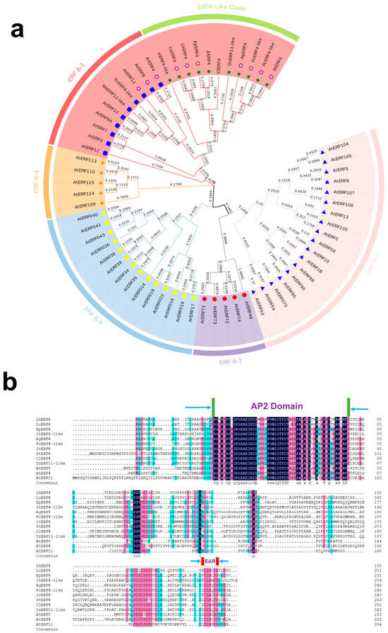
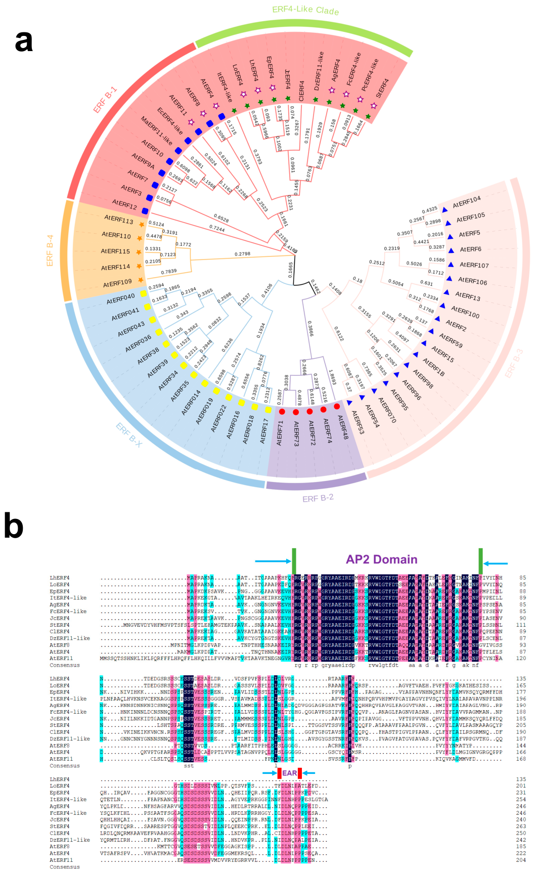
We constructed a phylogenetic tree using protein sequences from LoERF4 sequence of lily and the ERF gene family in species such as Arabidopsis thaliana, Euphorbia peplus, and Ipomoea triloba. Phylogenetically, LoERF4 was classified within the ERF B-1 subfamily (ERF4-like clade) of the AP2/ERF superfamily (Figure 1a). It contains a conserved AP2 domain and includes a C-terminal EAR repressor motif, suggesting it may possess transcriptional repressor activity (Figure 1b).
Figure 1.
Phylogenetic analysis and conserved domain architecture of LoERF4. (a) The phylogenetic tree was constructed using the maximum likelihood method based on the full-length protein sequences of AP2/ERF members mainly from Arabidopsis thaliana, lily (Lilium spp.), Euphorbia peplus, Ipomoea triloba, etc. The numbers at the nodes indicate bootstrap values (%) from 1000 replicates. LoERF4 is highlighted with a white-filled purple star. The scale bar represents genetic distance. (b) Protein sequences obtained from NCBI were analyzed using DNAMAN for multiple sequences alignment and conserved domain visualization. Amino acid positions are shown on the far right. The conserved domains “AP2 Domain” and “EAR” are marked with blue arrows.
3.2. Subcellular Localization of LoERF4 and Its Expression Under Ethylene and Salt Treatment
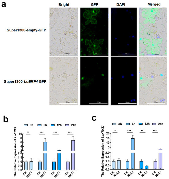
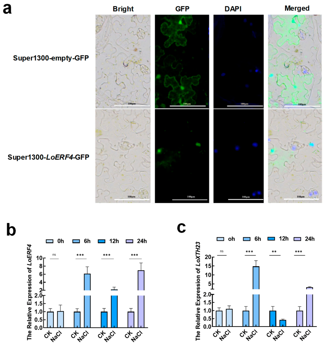
Subcellular localization of LoERF4. The LoERF4 protein is localized exclusively in the nucleus, confirming its role as a nuclear transcription factor (Figure 2a). Following treatment of ‘Siberia’ lily seedlings with 100 mM NaCl treatment, the transcript levels of LoERF4 were rapidly and significantly induced. Expression increased markedly at 6 h post-treatment, decreased significantly at 12 h, and then rebounded at 24 h to levels comparable to those observed at 6 h (Figure 2b). Similarly, LoXTH23 expression exhibited a fluctuating pattern characterized by an initial increase, followed by a transient decrease, and a subsequent rise, with transcript levels remaining significantly higher than those in the wild-type control throughout the time course (Figure 2c).
Figure 2.
Subcellular Localization of LoERF4 and Its Expression under Salt Treatment. (a) Subcellular localization of LoERF4 in the nucleus. Each row represents a sample: Super1300-empty-GFP (empty vector control) and Super1300-LoERF4-GFP (fusion protein expression vector). Each column displays a different imaging channel: Bright: Bright-field image, used to observe cell contours and overall morphology. GFP: Green fluorescent protein channel, showing the distribution of the GFP fusion protein. DAPI: Blue fluorescence channel, stained with DAPI to label the nucleus. Merged: Overlay image, combining the bright-field and fluorescence channels to show the relative localization of the protein within the cell structure. (b,c) Expression analysis of LoERF4 and LoXTH23 at 0, 6, 12, and 24 h under 100 mM NaCl treatment in bulblets of lily ‘Siberia’. In part (b,c), data represent mean ± SD. Asterisks indicate significant differences compared to WT (ns represent not significant, ** p < 0.01, *** p < 0.001; Student’s t-test).
3.3. Overexpression of LoERF4 Alters Root Architecture and Modulates the Expression of Cell Wall-Modifying Genes in Arabidopsis
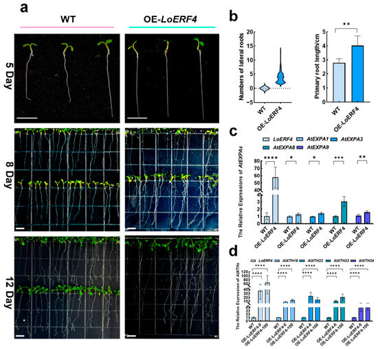
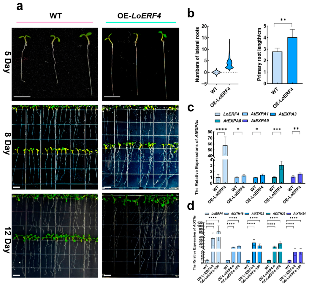
To further investigate the physiological function of LoERF4, we heterologously expressed it in Arabidopsis thaliana (OE-LoERF4 lines) and examined its effect on root development. Compared to the WT, the OE-LoERF4 seedlings exhibited a pronounced alteration in root architecture at both 5, 8 and 12 days post-germination. By day 8, the WT plants exhibited no visible lateral roots, whereas the OE-LoERF4 lines had already developed an average of 5 lateral roots per plant. Regarding primary root development, the mean primary root length of WT seedlings was approximately 2.8 cm, while that of the OE-LoERF4 lines reached an average of 4 cm (Figure 3a,b). To investigate the underlying causes of the pronounced differences in lateral root and primary root development between WT and OE-LoERF4 lines at day 8, we performed quantitative real-time PCR analysis of cell wall modification-related genes. The results revealed that the expression levels of AtEXPA and AtXTH genes, particularly AtXTHs, were significantly upregulated in the overexpression lines. These findings suggest that LoERF4 likely promotes lateral root and primary root development through positive regulation of AtXTHs and AtEXPAs (Figure 3c,d).
Figure 3.
Roots growth performance of WT and OE-LoERF4 Arabidopsis lines. (a) Representative images of WT and OE-LoERF4 seedlings at 5, 8, and 12 days after germination. Scale bar, 1 cm. (b) Quantification of lateral root number and primary root length in WT and transgenic lines (n = 100). The dashed line represents the reference line without lateral roots. (c) Expression analysis of AtEXPA genes in WT and OE-LoERF4 lines on day 8. (d) Expression analysis of AtXTH genes in WT and OE-LoERF4 lines including two treatments: OE-LoERF4-0 (treated with water) and OE-LoERF4-100 (treated with 200 mM NaCl) on day 8. AtEXPAs and AtXTHs transcript levels were quantified by RT-qPCR and normalized to AtUBQ10. Data represents mean ± SD. Asterisks indicate significant differences compared to WT (* p < 0.05, ** p < 0.01, *** p < 0.001, **** p < 0.0001; Student’s t-test).
3.4. LoERF4 Positively Regulates Root System Architecture in Overexpression Lines by Promoting Root Cell Elongation in Arabidopsis
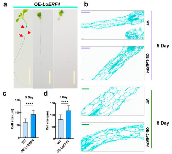
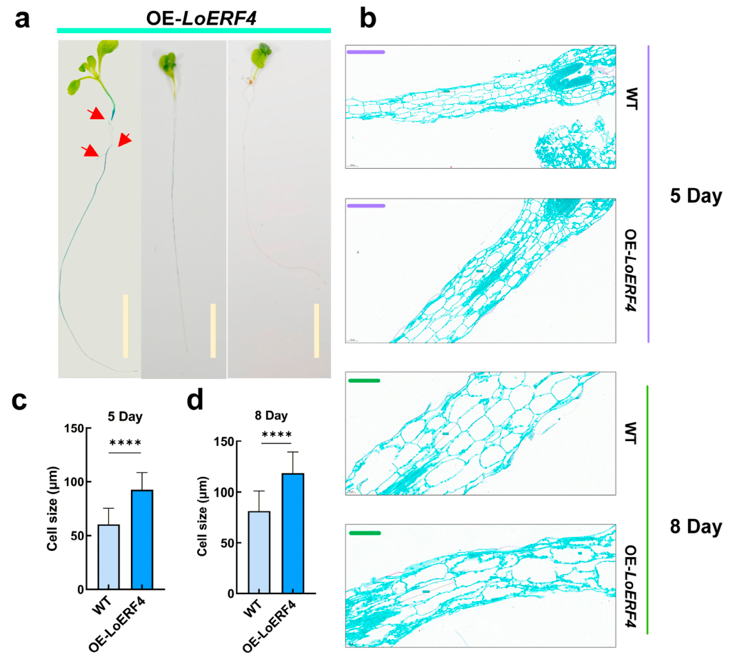
LoERF4 positively influences the expression of cell wall modification-related genes and promotes both lateral root and primary root development (Figure 3a–d). We observed that differences in primary root length between WT and OE-LoERF4 lines were already evident by day 5 after germination, and by day 8, the differences in lateral root development became highly significant. GUS staining revealed that on day 8, LoERF4 expression levels were closely correlated with lateral root number; specifically, darker GUS staining corresponded to greater lateral root production (Figure 4a).
Figure 4.
LoERF4 promotes lateral root elongation and root cell elongation. (a) Histochemical GUS staining of OE-LoERF4 8-day-old seedlings after germination. Darker staining indicates faster lateral root emergence. Red arrows indicate lateral root sites. Scale bar, 1 cm. (b) Paraffin sections of longitudinal root segments from WT and OE-LoERF4 lines at 5 and 8 days after germination. The purple scale bar represents 150 μm (5 days); the green scale bar represents 60 μm (8 days). (c) Quantification of root cortical cell length in WT and OE-LoERF4 lines at day 5 (n = 30). (d) Quantification of root cortical cell length in WT and OE-LoERF4 lines at day 8 (n = 30). In part (c,d), the centerline represents the mean, and the error bars represent the standard deviation of three biological replicates. statistical significance was determined by Student’s t-test (**** p < 0.0001).
To elucidate the cellular mechanism underlying LoERF4-mediated root growth promotion, we performed longitudinal section analysis of roots from 5-day-old and 8-day-old seedlings. On day 5, the average root cell length in wild-type plants was 60.3 μm, whereas that in OE-LoERF4 lines reached 92.73 μm (Figure 4b,c). On day 8, wild-type root cells averaged 81.3 μm in length, compared to 118.4 μm in the overexpression lines (Figure 4b,d), indicating that OE-LoERF4 plants maintained significantly longer root cells at both time points. These results demonstrate that LoERF4 positively regulates lateral root and primary root development by promoting root cell elongation in Arabidopsis. This conclusion further supports the notion that LoERF4 influences root system architecture through positive regulation of cell wall modification genes, including AtXTHs and AtEXPAs.
3.5. Overexpression of LoERF4 Enhances Salt Tolerance in Arabidopsis by Modulating Root Architecture and Antioxidant Capacity
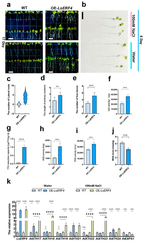
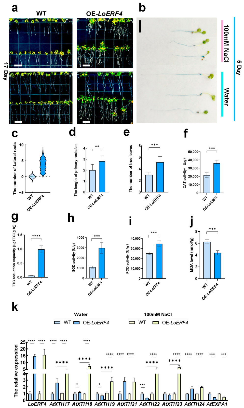
Based on the finding that LoERF4 responds to both ethylene and NaCl through the LoERF4-LoXTH23 regulatory module in lily, we performed similar validation experiments in Arabidopsis. The results demonstrated that LoERF4 also positively responds to salt signals in Arabidopsis and enhances the salt tolerance of overexpression lines. Under 100 mM NaCl treatment, both WT and OE-LoERF4 lines exhibited typical physiological symptoms of salt stress, including leaf yellowing, increased root hair formation, and reduced growth rate. However, notable differences were observed between genotypes. At 17 days post-treatment, the overexpression lines maintained more lateral roots, longer primary roots, and a greater number of rosette leaves, indicating better overall growth performance under saline conditions (Figure 5a,c–e). GUS staining revealed that LoERF4 expression remained localized predominantly in root tissues under salt stress (Figure 5b). To more accurately assess whether the salt tolerance of overexpression lines was genuinely enhanced, we measured several physiological parameters at 17 days under salt treatment. The activities of antioxidant enzymes including catalase (CAT), superoxide dismutase (SOD), and peroxidase (POD) were significantly higher in OE-LoERF4 lines compared to WT (Figure 5f,h,i). Additionally, the overexpression lines exhibited lower malondialdehyde (MDA) content, indicating reduced oxidative damage (Figure 5j), and higher root viability as determined by TTC assay (Figure 5g).
Figure 5.
LoERF4 enhances salt tolerance in overexpression lines. (a) Phenotypes of WT and OE-LoERF4 seedlings under 100 mM NaCl treatment at 17 days after treatment. Scale bar, 1 cm. (b) GUS staining of 5-day-old OE-LoERF4 seedlings under control (water) and 100 mM NaCl treatment conditions. Scale bar, 1 cm. (c–e) Quantification of lateral root number, primary root length, and rosette leaf number in WT and OE-LoERF4 lines under salt treatment (n = 60). (f,h,i) Catalase (CAT), superoxide dismutase (SOD), and peroxidase (POD) activities in WT and OE-LoERF4 lines at 17 days under salt treatment. (g) Root viability (measured by TTC reduction) in WT and OE-LoERF4 lines at 17 days under salt treatment. (j) Malondialdehyde (MDA) content in WT and OE-LoERF4 lines at 17 days under salt treatment. (k) Expression analysis of AtXTHs and AtEXPA1 in WT and OE-LoERF4 lines at 17 days under control (water) and 100 mM NaCl treatment conditions. Datas are presented as mean ± SD. Asterisks indicate significant differences compared with WT (* p < 0.05, ** p < 0.01, *** p < 0.001, **** p < 0.0001; Student’s t-test).
After 17 days under 100 mM NaCl and water treatment, we performed quantitative real-time PCR analysis to examine the expression levels of AtXTHs and AtEXPA1. The results revealed that the expressions of AtXTH17, AtXTH21 and AtXTH24 genes were significantly upregulated in LoERF4 compared to WT (Figure 5k). Additionally, AtXTHs maintained high expression levels under salt stress (Figure 5k). Specifically, AtXTH17, AtXTH18, AtXTH19, AtXTH22, and AtXTH23 exhibited significantly higher transcript abundance under salt treatment compared to control (water) conditions. These findings suggest that, under salt stress, LoERF4 may still participate in salt stress responses in Arabidopsis through a LoERF4-AtXTH regulatory module, thereby contributing to the enhanced salt tolerance observed in the overexpression lines.
4. Discussion
The present study demonstrates that heterologous overexpression of LoERF4 in Arabidopsis thaliana confers two distinct phenotypic advantages: enhanced root system architecture under normal conditions and improved salt tolerance under stress. Mechanistically, these phenotypes are underpinned by interconnected molecular pathways. Under optimal conditions, LoERF4 promotes lateral root development and primary root elongation through transcriptional activation of cell wall remodeling genes (AtXTHs and AtEXPAs), leading to enhanced cell elongation. Under salt stress, LoERF4 enhances tolerance through a dual mechanism: it maintains elevated expression of cell wall remodeling genes to sustain root growth under adverse conditions, while simultaneously bolstering the reactive oxygen species (ROS) scavenging system by increasing antioxidant enzyme activities (SOD, POD, CAT), thereby reducing oxidative damage.
4.1. LoERF4 Drives Root Elongation Through Promoting Cell Expansion Rather than Cell Division
ERF4, a member of the ERF subfamily, has been functionally characterized in multiple species primarily as a transcriptional repressor. In Arabidopsis thaliana, AtERF4 contains an EAR (ERF-associated amphiphilic repression) motif and participates in the MKK3-MPK7 signaling module to regulate seed dormancy release [27]. It also interacts with MYB52 to modulate pectin demethylesterification and controls organ size by suppressing endoreduplication inhibitors [37]. In apple, MdERF4 reduces ethylene synthesis through EAR motif-mediated transcriptional repression, thereby affecting fruit firmness [38]. In lily, LhERF4 negatively regulates anthocyanin biosynthesis by suppressing LhMYBSPLATTER expression [39].
Histological analysis of longitudinal sections from OE-LoERF4 lines revealed a key cytological phenotype: at both 5 days post-germination (prior to lateral root emergence) and 8 days post-germination (when lateral roots became visible), cortical and epidermal cells were significantly longer in transgenic lines compared to wild type. This observation indicates that LoERF4 primarily drives root elongation through promoting cell expansion rather than cell division, consistent with the upregulation of multiple AtXTHs and AtEXPAs genes in OE lines. Both gene families encode proteins that facilitate cell wall loosening: XTH enzymes remodel the xyloglucan-cellulose network by cleaving and re-ligating xyloglucan chains [18], while expansins disrupt hydrogen bonds between wall polysaccharides [24]. The coordinated upregulation of these gene families provides a molecular basis for the enhanced cell elongation observed in root cortical tissues.
Notably, enhanced cell elongation was evident prior to lateral root emergence and persisted after lateral root formation, suggesting that LoERF4-mediated cell wall remodeling facilitates both primary root elongation and lateral root primordium development. During lateral root formation, localized cell wall loosening in overlying parental tissues is required for primordia emergence [40]. The expanded cells in OE-LoERF4 roots may create a more favorable mechanical environment for lateral root initiation, explaining the increased lateral root density in transgenic lines. Similar mechanisms have been reported for other ERF transcription factors; for instance, OsERF71 influences rice root system architecture by regulating cell wall-related genes [23].
While ERF-XTH regulatory modules have been characterized in several species, their existence in Lilium and functional conservation across monocot-dicot boundaries remained unexplored. Bioinformatic analysis provided initial evidence supporting such conservation: LoXTH23, a lily XTH gene positively regulated by LoERF4 (Figure S2), clusters phylogenetically with AtXTH23 at Group I/II boundary and contains a canonical GH16 catalytic domain essential for cell wall remodeling activity (Figure S3). This conserved structural architecture suggests that LoXTH23 likely retains the capacity to modulate cell wall expansion, a function relevant to both lily bulblet development and Arabidopsis root growth. The positive regulation of LoXTHs by LoERF4 was further validated in transient assays using Lilium ‘Siberia’ (Figure S2), indicating that the ERF-XTH module is functionally active in the native lily context.
4.2. LoERF4 Enhances Salt Tolerance Through Parallel Mechanisms of Cell Wall Remodeling and ROS Scavenging
Beyond its effects on root architecture, LoERF4 overexpression significantly enhanced the antioxidant capacity under salt stress. Elevated SOD, POD, and CAT activities, coupled with reduced MDA accumulation, indicate that OE-LoERF4 lines experienced less oxidative damage under saline conditions, while TTC reduction assays confirmed higher root metabolic activity in transgenic lines. Beyond its effects on root architecture, LoERF4 overexpression significantly enhanced antioxidant capacity under salt stress. The role of ERF transcription factors in modulating ROS homeostasis has been documented in multiple species: CaERF2 in pepper maintains ROS balance through activation of antioxidant enzymes [41], OPBP1 from tobacco enhances salt tolerance in rice by upregulating glutathione S-transferase genes [42], and DcERF3 in Dendrobium officinale simultaneously activates the antioxidant system and promotes lateral root development [43]—echoing the dual effects observed in the present study.
The relationship between root system architecture and stress tolerance is well established. Lateral roots expand the absorptive surface area, improving water and nutrient acquisition under resource-limited conditions [44]. In saline environments, an extensive lateral root system enables plants to explore low-salt microdomains and maintain ionic homeostasis [45]. Our observation that OE-LoERF4 lines maintained longer primary roots and more lateral roots under 100 mM NaCl stress suggests that LoERF4-mediated alterations in root architecture directly contribute to improved growth performance under stress. Importantly, the cell elongation phenotype observed under normal conditions was maintained under salt stress, indicating that LoERF4-mediated cell wall remodeling provides a structural basis for sustained root growth under adverse conditions.
4.3. The Dual Function of LoERF4: Reconciling the EAR Motif with Transcriptional Activation
ERF transcription factors containing EAR motifs have been widely characterized as transcriptional repressors in Arabidopsis. Canonical EAR motif-containing ERFs such as AtERF3, AtERF4, AtERF7, AtERF8, AtERF9, AtERF10, AtERF11 and AtERF12 have been experimentally verified to possess active repression activities [9,46,47]. However, LoERF4—despite harboring a typical EAR motif—acts as a transcriptional activator in both root development and salt stress response when ectopically expressed in Arabidopsis. Phylogenetic analysis placed LoERF4 in close proximity to AtERF4, AtERF8 and AtERF11 (Figure 1a), three well-characterized Class II (B1 subgroup) ERF repressors in Arabidopsis [9]. Interestingly, alignment of EAR motif sequences revealed substantial divergence between LoERF4 and these three Arabidopsis homologs (Figure 1b). Such sequence variations within the EAR motif can alter transcriptional activity; for instance, point mutations in the AtERF4 EAR motif (LDLNL to PDPNL or PDLNL) abolish its ability to bind downstream target gene promoters and eliminate its repressive function [48]. The distinct EAR motif architecture in LoERF4 may thus account for the absence of growth inhibition phenotypes in OE-LoERF4 lines.
Although LoERF4 contains a canonical EAR motif, our results suggest it may function as an activator in the context of Arabidopsis root development and salt tolerance. However, we cannot rule out that it acts by relieving repression of other factors. Future transcriptional activity assays are needed to directly test this hypothesis. Ding [37] demonstrated that AtERF4 interacts with TCP15 to repress CYCA2;3 expression, thereby promoting endoreduplication and leading to enlarged cells and organs in overexpression lines. However, such growth-promoting effects—paradoxically arising from ERF4-mediated repression—were not observed in LoERF4-overexpressing plants. This absence further supports the notion that LoERF4 has diverged functionally from its Arabidopsis homologs.
Several mechanisms could explain how an EAR motif-containing protein functions as an activator: (1) LoERF4 might relieve repression of positive regulators through competitive binding or co-repressor sequestration; (2) post-translational modifications such as phosphorylation could convert LoERF4 from a repressor to an activator; or (3) LoERF4 may recruit different transcriptional co-factors depending on cellular context or target gene promoter architecture. The observation that LoERF4 promotes cell expansion—a process requiring active gene expression—further supports its role as a transcriptional activator in the context of cell wall remodeling. Based on these findings, we propose that the divergent EAR motif of LoERF4 likely prevents it from exerting its native repressive functions when transferred into the Arabidopsis context.
4.4. Conserved and Divergent Functions of LoERF4 Between Lily and Arabidopsis
The functional conservation of the ERF-XTH module across monocot and dicot species raises the question of whether LoERF4 plays analogous roles in its native lily context. To explore this, we generated LoERF4-overexpressing chimeric lily lines, given the technical challenges associated with stable transformation and phenotypic observation in this species. GUS staining localized transgene expression predominantly to the basal plate region of micro bulbs (Figure S4), a meristematic zone critical for bulb development where emerging scales converge. Transcript analysis of this region confirmed upregulation of both LoERF4 and LoXTH23, consistent with our transient assay findings. Notably, genes involved in sucrose metabolism—including those encoding sucrose synthases and invertases—were also significantly upregulated in these tissues.
The co-regulation of cell wall remodeling and sucrose metabolism genes by LoERF4 in lily basal plates may reflect a conserved function adapted to the specific physiological demands of bulb development. Sucrose metabolism plays a pivotal role in bulbous species: sucrose cleavage by cell wall invertase promotes bulblet initiation by enhancing apo plasmic sugar unloading [49], while a high hexose-to-sucrose ratio supports cell division during early vegetative propagation [50,51]. In Lilium lancifolium, the transcription factor LlbHLH35 directly activates LlSusy1 in response to auxin signaling, linking hormonal control to sucrose metabolism during bulbil formation [52]. Furthermore, active sugars such as sucrose and fructose accumulate significantly during dormancy release and bud development in Lilium brownii [53]. Beyond development, sucrose metabolism also contributes to stress tolerance: under salt stress, maintaining high sugar content through enhanced sucrose synthetase activity supports photosynthetic performance and energy homeostasis [54], and SWEET family sugar transporters are dynamically regulated under salinity, implicating sugar partitioning in abiotic stress adaptation [50]. These observations suggest that LoERF4 may coordinate cell wall remodeling and carbon partitioning during bulb development—a function partially overlapping with, yet distinct from, its role in Arabidopsis root growth.
Although phenotypic assessment remains limited in lily, the convergence of LoERF4-mediated transcriptional regulation on both cell wall and sucrose metabolic genes reinforces the idea that ERF-XTH modules are embedded in broader developmental and stress-responsive networks. The fact that a lily ERF transcription factor with a divergent EAR motif acts as a transcriptional activator in Arabidopsis—while retaining regulatory connections to XTH genes in both species—reveals both functional conservation and evolutionary plasticity within this protein family.
4.5. Implications for Molecular Breeding in Lily and Other Recalcitrant Species
The technical challenges of lily research, including difficulties in genetic transformation, unclear developmental patterns of bulb growth, and limited accessibility for phenotypic observation—have long hindered functional genomics and molecular breeding in this economically important ornamental crop. Our findings demonstrate that functional insights gained from a tractable heterologous system like Arabidopsis can inform molecular breeding strategies in lily, even when direct genetic manipulation remains challenging. In this experiment, the ERF transcription factor from lily was found to activate conserved downstream targets (XTHs) under salt stress conditions, conferring beneficial traits (enhanced root architecture and salt tolerance) in Arabidopsis. This suggests that key regulatory modules may remain conserved across species, and their functions can be validated in model systems.
Our previous research on the oriental lily ‘Siberia’ identified an ethylene-induced AP2/ERF transcription factor gene, LoERF4, and demonstrated its regulation of the XTH family member LoXTH23 [28]. Specifically, LoERF4 and LoXTH23 exhibited rapid and coordinated induction following ethylene treatment. Virus-induced gene silencing (VIGS) significantly reduced LoERF4 expression while concurrently decreasing LoXTH23 expression, whereas transient overexpression of LoERF4 upregulated LoXTH23 expression, confirming a positive regulatory relationship between these two genes (Figure S2). Bioinformatics analysis revealed that LoERF4 contains both a conserved AP2 DNA-binding domain and a C-terminal EAR motif, suggesting potential transcriptional repressor activity. However, its positive regulation of LoXTH23 indicates it may function as an activator in specific contexts. These observations suggest that LoERF4 may link ethylene signaling to cell wall remodeling during lily bulb dormancy release. Nevertheless, the broader functions of LoERF4 in root system architecture and abiotic stress adaptation, as well as its potential regulatory effects on other cell wall modification-related downstream genes, remain to be explored.
A limitation of this study is the lack of loss-of-function mutants in Arabidopsis or lily. Therefore, we cannot conclude that LoERF4 is essential for root development or salt tolerance. Our conclusions are based on gain-of-function evidence only. Furthermore, given that this study involved only technical replicates and lacked independent biological replicates, the results may be subject to error and should be considered exploratory only.
In an era of rapidly advancing molecular breeding, functional divergence of versatile transcription factors across evolutionarily distant species offers a promising avenue for crop improvement. By exploiting or engineering variations in key functional domains such as the EAR motif, breeders may selectively eliminate undesirable transcriptional outputs while preserving traits of interest in target crops. Future studies employing chromatin immunoprecipitation sequencing (ChIP-seq) and yeast two-hybrid screening will be necessary to identify direct target genes and interacting proteins of LoERF4, and to determine how its regulatory activity is modulated across different species and developmental contexts.
5. Conclusions
In this study, we demonstrated that LoERF4 functions as a positive regulator of root development and salt tolerance in Arabidopsis through dual mechanisms involving cell wall remodeling and ROS scavenging. Under salt stress, OE-LoERF4 lines exhibited enhanced antioxidant enzyme activities, reduced oxidative damage, sustained root growth, and increased root viability. LoERF4 positively regulates LoXTHs, potentially coordinating sucrose metabolic pathways during bulb development. The finding that a lily ERF transcription factor with a divergent EAR motif acts as a transcriptional activator in a heterologous system reveals previously unrecognized functional plasticity within this protein family, with practical implications for molecular breeding of stress-resilient crops—particularly in species like lily where direct genetic manipulation remains challenging.
Supplementary Materials
The following supporting information can be downloaded at: https://www.mdpi.com/article/10.3390/horticulturae12050512/s1.
Author Contributions
Conceptualization, M.P., L.S. and C.J.; methodology, M.P.; investigation, M.P.; data curation, M.P.; formal analysis, C.S.; writing—original draft preparation, M.P. and C.S.; writing—review and editing, M.Z., C.S., C.J. and L.S.; supervision, C.J., Y.W. and F.L.; project administration, Y.W.; funding acquisition, Y.W. and F.L. All authors have read and agreed to the published version of the manuscript.
Funding
This research was funded by the National Natural Science Foundation of China (Regional Science Foundation Project, No. 32060690); and the Yunnan Xingdian Talents—Special Selection Project for High-level Scientific and Technological Talents and Innovation Teams—Team Specific Project, Xingdian Talent Support Project (No. CYRC2020004). The APC was funded by Youguo Wang.
Data Availability Statement
The original contributions presented in this study are included in the article/Supplementary Material. Further inquiries can be directed to the corresponding authors.
Conflicts of Interest
The authors declare no conflicts of interest.
References
- Tang, Y.; Tan, B.; Liu, H.; Liu, Y.; Zhang, L.; Zhang, P.; Sun, M. A comprehensive review of physiological and molecular responses to stress of lilies (genus Lilium). Hortic. Res. 2026, 13, uhaf347. [Google Scholar] [CrossRef] [Scilit] [PubMed]
- Shahid, S.A.; Zaman, M.; Heng, L. Soil salinity: Historical perspectives and a world overview of the problem. In Guideline for Salinity Assessment, Mitigation and Adaptation Using Nuclear and Related Techniques; Zaman, M., Shahid, S.A., Heng, L., Eds.; Springer International Publishing: Cham, Switzerland, 2018; pp. 43–53. [Google Scholar]
- Munns, R.; Tester, M. Mechanisms of Salinity Tolerance. Annu. Rev. Plant Biol. 2008, 59, 651–681. [Google Scholar] [CrossRef] [Scilit] [PubMed]
- Park, Y.; Jo, W.J.; Choi, H.; Im, N.H.; Lee, H.B. Effects of Salinity Stress on Bulb Growth and Photosynthetic Capacity of Lilium LA Hybrid ‘Serrada’. Flower Res. J. 2024, 32, 68–76. [Google Scholar] [CrossRef] [Scilit]
- Bai, R.; Lin, Y.; Jiang, Y. Diverse genotypic variations of photosynthetic capacity, transpiration and antioxidant enzymes of lily hybrids to increasing salinity stress. Sci. Hortic. 2021, 280, 109939. [Google Scholar] [CrossRef] [Scilit]
- Golldack, D.; Lüking, I.; Yang, O. Plant tolerance to drought and salinity: Stress regulating transcription factors and their functional significance in the cellular transcriptional network. Plant Cell Rep. 2011, 30, 1383–1391. [Google Scholar] [CrossRef] [Scilit]
- Singh, D.; Laxmi, A. Transcriptional regulation of drought response: A tortuous network of transcriptional factors. Front. Plant Sci. 2015, 6, 895. [Google Scholar] [CrossRef] [Scilit]
- Licausi, F.; Ohme-Takagi, M.; Perata, P. APETALA 2/Ethylene Responsive Factor (AP2/ERF) transcription factors: Mediators of stress responses and developmental programs. New Phytol. 2013, 199, 639–649. [Google Scholar] [CrossRef] [Scilit]
- Nakano, T.; Suzuki, K.; Fujimura, T.; Shinshi, H. Genome-Wide Analysis of the ERF Gene Family in Arabidopsis and Rice. Plant Physiol. 2006, 140, 411–432. [Google Scholar] [CrossRef] [Scilit]
- Feng, K.; Hou, X.L.; Xing, G.M.; Liu, J.X.; Duan, A.Q.; Xu, Z.S.; Li, M.Y.; Zhuang, J.; Xiong, A.S. Advances in AP2/ERF super-family transcription factors in plant. Crit. Rev. Biotechnol. 2020, 40, 750–776. [Google Scholar] [CrossRef] [Scilit]
- Müller, M.; Munné-Bosch, S. Ethylene Response Factors: A Key Regulatory Hub in Hormone and Stress Signaling. Plant Physiol. 2015, 169, 32–41. [Google Scholar] [CrossRef] [Scilit] [PubMed]
- Mizoi, J.; Shinozaki, K.; Yamaguchi-Shinozaki, K. AP2/ERF family transcription factors in plant abiotic stress responses. Biochim. Biophys. Acta (BBA)—Gene Regul. Mech. 2012, 1819, 86–96. [Google Scholar] [CrossRef] [Scilit] [PubMed]
- Xie, Z.; Nolan, T.M.; Jiang, H.; Yin, Y. AP2/ERF Transcription Factor Regulatory Networks in Hormone and Abiotic Stress Responses in Arabidopsis. Front. Plant Sci. 2019, 10, 228. [Google Scholar] [CrossRef] [Scilit] [PubMed]
- Yao, Y.; He, R.J.; Xie, Q.L.; Zhao, X.H.; Deng, X.M.; He, J.B.; Song, L.; He, J.; Marchant, A.; Chen, X.; et al. ETHYLENE RESPONSE FACTOR 74 (ERF74) plays an essential role in controlling a respiratory burst oxidase homolog D (RbohD)-dependent mechanism in response to different stresses in Arabidopsis. New Phytol. 2016, 213, 1667–1681. [Google Scholar] [CrossRef] [Scilit]
- Li, Z.; Tian, Y.; Xu, J.; Fu, X.; Gao, J.; Wang, B.; Han, H.; Wang, L.; Peng, R.; Yao, Q. A tomato ERF transcription factor, SlERF84, confers enhanced tolerance to drought and salt stress but negatively regulates immunity against Pseudomonas syringae pv. Tomato DC3000. Plant Physiol. Biochem. 2018, 132, 683–695. [Google Scholar] [CrossRef] [Scilit]
- Jung, H.; Chung, P.J.; Park, S.; Redillas, M.C.F.R.; Kim, Y.S.; Suh, J.; Kim, J. Overexpression of OsERF48 causes regulation of OsCML16, a calmodulin-like protein gene that enhances root growth and drought tolerance. Plant Biotechnol. J. 2017, 15, 1295–1308. [Google Scholar] [CrossRef] [Scilit]
- Rong, W.; Qi, L.; Wang, A.; Ye, X.; Du, L.; Liang, H.; Xin, Z.; Zhang, Z. The ERF transcription factor TaERF3 promotes tolerance to salt and drought stresses in wheat. Plant Biotechnol. J. 2014, 12, 468–479. [Google Scholar] [CrossRef] [Scilit]
- Rose, J.K.C.; Braam, J.; Fry, S.C.; Nishitani, K. The XTH Family of Enzymes Involved in Xyloglucan Endotransglucosylation and Endohydrolysis: Current Perspectives and a New Unifying Nomenclature. Plant Cell Physiol. 2002, 43, 1421–1435. [Google Scholar] [CrossRef] [Scilit]
- Xu, P.; Fang, S.; Chen, H.; Cai, W. The brassinosteroid-responsive xyloglucan endotransglucosylase/hydrolase 19 (XTH19) and XTH23 genes are involved in lateral root development under salt stress in Arabidopsis. Plant J. 2020, 104, 59–75. [Google Scholar] [CrossRef] [Scilit]
- Bi, H.; Liu, Z.; Liu, S.; Qiao, W.; Zhang, K.; Zhao, M.; Wang, D. Genome-wide analysis of wheat xyloglucan endotransglucosylase/hydrolase (XTH) gene family revealed TaXTH17 involved in abiotic stress responses. BMC Plant Biol. 2024, 24, 640. [Google Scholar] [CrossRef] [Scilit]
- Liang, Y.; Gao, Q.; Li, F.; Du, Y.; Wu, J.; Pan, W.; Wang, S.; Zhang, X.; Zhang, M.; Song, X.; et al. The giant genome of lily provides insights into the hybridization of cultivated lilies. Nat. Commun. 2025, 16, 45. [Google Scholar] [CrossRef] [Scilit] [PubMed]
- Zhang, J.; Liu, Z.; Sakamoto, S.; Mitsuda, N.; Ren, A.; Persson, S.; Zhang, D. ETHYLENE RESPONSE FACTOR 34 promotes secondary cell wall thickening and strength of rice peduncles. Plant Physiol. 2022, 190, 1806–1820. [Google Scholar] [CrossRef] [Scilit] [PubMed]
- Lee, D.K.; Jung, H.; Jang, G.; Jeong, J.S.; Kim, Y.S.; Ha, S.H.; Choi, Y.D.; Kim, J.K. Overexpression of the OsERF71 Transcription Factor Alters Rice Root Structure and Drought Resistance. Plant Physiol. 2016, 172, 575–588. [Google Scholar] [CrossRef] [Scilit]
- Lee, Y.; Choi, D.; Kende, H. Expansins: Ever-expanding numbers and functions. Curr. Opin. Plant Biol. 2001, 4, 527–532. [Google Scholar] [CrossRef] [Scilit] [PubMed]
- Sharova, E.I. Expansins: Proteins involved in cell wall softening during plant growth and morphogenesis. Russ. J. Plant Physiol. 2007, 54, 713–727. [Google Scholar] [CrossRef] [Scilit]
- Cosgrove, D.J. Plant expansins: Diversity and interactions with plant cell walls. Curr. Opin. Plant Biol. 2015, 25, 162–172. [Google Scholar] [CrossRef] [Scilit]
- Zhao, Q.; Hu, R.; Liu, D.; Liu, X.; Wang, J.; Xiang, X.; Li, Y. The AP2 transcription factor NtERF172 confers drought resistance by modifying NtCAT. Plant Biotechnol. J. 2020, 18, 2444–2455. [Google Scholar] [CrossRef] [Scilit]
- Zhao, G.; Sun, L.; Jia, W.; Li, X.; Wang, Y. Transcriptional regulation analysis of LoXTH23 and LoERF4 transcription factors in Oriental lily ‘Siberia’. J. Zhejiang Univ. (Agric. Life Sci.) 2025, 51, 904–915. [Google Scholar]
- Nagamani, J.E.; Indu, B.K.; Subbalakshmi, G.; Anuradha, M. Practical Plant Tissue Culture: Techniques and Protocols. In In Vitro Production of Plant Secondary Metabolites: Theory and Practice; Springer: Singapore, 2025; pp. 57–70. [Google Scholar]
- Clough, S.J.; Bent, A.F. Floral dip: A simplified method for Agrobacterium-mediated transformation of Arabidopsis thaliana. Plant J. 1998, 16, 735–743. [Google Scholar] [CrossRef] [Scilit]
- Liu, Y.; Schiff, M.; Dinesh-Kumar, S.P. Virus-induced gene silencing in tomato. Plant J. 2002, 31, 777–786. [Google Scholar] [CrossRef] [Scilit]
- Liu, Y.; Schiff, M.; Marathe, R.; Dinesh-Kumar, S.P. Tobacco Rar1, EDS1 and NPR1/NIM1 like genes are required for N-mediated resistance to tobacco mosaic virus. Plant J. 2002, 30, 415–429. [Google Scholar] [CrossRef] [Scilit] [PubMed]
- Stange Klein, C. (Ed.) Agrobacterium: Methods and Protocols; Springer: New York, NY, USA, 2025. [Google Scholar]
- Sun, C.W.; Callis, J. Independent modulation of Arabidopsis thaliana polyubiquitin mRNAs in different organs and in response to environmental changes. Plant J. 1997, 11, 1017–1027. [Google Scholar] [CrossRef] [Scilit]
- Czechowski, T.; Stitt, M.; Altmann, T.; Udvardi, M.K.; Scheible, W.-R. Genome-wide identification and testing of superior reference genes for transcript normalization in Arabidopsis. Plant Physiol. 2005, 139, 5–17. [Google Scholar] [CrossRef] [Scilit]
- Yamagishi, M. High temperature enhances anthocyanin coloration in Asiatic hybrid lily flowers via upregulation of the MYB12 positive regulator. Hortic. Plant J. 2022, 8, 769–776. [Google Scholar] [CrossRef] [Scilit]
- Ding, A.; Xu, C.; Xie, Q.; Zhang, M.; Yan, N.; Dai, C.; Lv, J.; Cui, M.; Wang, W.; Sun, Y. ERF4 interacts with and antagonizes TCP15 in regulating endoreduplication and cell growth in Arabidopsis. J. Integr. Plant Biol. 2022, 64, 1673–1689. [Google Scholar] [CrossRef] [Scilit]
- Hu, Y.; Han, Z.; Sun, Y.; Wang, S.; Wang, T.; Wang, Y.; Xu, K.; Zhang, X.; Xu, X.; Han, Z.; et al. ERF4 affects fruit firmness through TPL4 by reducing ethylene production. Plant J. 2020, 103, 937–950. [Google Scholar] [CrossRef] [Scilit]
- Cao, Y.; Song, M.; Bi, M.; Yang, P.; He, G.; Wang, J.; Yang, Y.; Xu, L.; Ming, J. Lily (Lilium spp.) LhERF4 negatively affects anthocyanin biosynthesis by suppressing LhMYBSPLATTER transcription. Plant Sci. 2024, 342, 112026. [Google Scholar] [CrossRef] [Scilit]
- Péret, B.; De Rybel, B.; Casimiro, I.; Benková, E.; Swarup, R.; Laplaze, L.; Beeckman, T.; Bennett, M.J. Arabidopsis lateral root development: An emerging story. Trends Plant Sci. 2009, 14, 399–408. [Google Scholar] [CrossRef] [Scilit]
- Zhao, J.; Huang, M.; Liu, J.; Cai, J.; He, Y.; Zhao, W.; Liu, C.; Wu, Y. Pepper (Capsicum annuum L.) AP2/ERF transcription factor, CaERF2 enhances salt stress tolerance through ROS scavenging. Theor. Appl. Genet. 2025, 138, 44. [Google Scholar] [CrossRef] [Scilit] [PubMed]
- Chen, X.; Guo, Z. Tobacco OPBP1 Enhances Salt Tolerance and Disease Resistance of Transgenic Rice. Int. J. Mol. Sci. 2008, 9, 2601–2613. [Google Scholar] [CrossRef] [Scilit] [PubMed]
- Zhu, H.; Chen, R.; Xu, Y.; Gong, W.; Miao, M.; Sun, Y.; Mei, J. An ERF Gene DcERF3 of Dendrobium catenatum Improves Salt Tolerance in Arabidopsis. Mol. Biotechnol. 2025, 68, 896–906. [Google Scholar] [CrossRef] [Scilit] [PubMed]
- Alshareef, N.O.; Melino, V.J.; Saber, N.; De Rosa, A.; Rey, E.; Wang, J.Y.; Al Babili, S.; Byrt, C.; Tester, M.A.; Julkowska, M.M. Root Remodeling Mechanisms and Salt Tolerance Trade-Offs: The Roles of HKT1, TMAC2, and TIP2;2 in Arabidopsis. Plant Biol. 2024, 21, e1011713. [Google Scholar] [CrossRef] [Scilit]
- Zhang, H.; Tian, X.; Yu, W.; Wang, Y.; Du, Q.; Li, R. The specific HbHAK2 promoter from halophytic Hordeum brevisubulatum regulates root development under salt stress. Agric. Commun. 2024, 2, 100050. [Google Scholar] [CrossRef] [Scilit]
- Kagale, S.; Links, M.G.; Rozwadowski, K. Genome-Wide Analysis of Ethylene-Responsive Element Binding Factor-Associated Amphiphilic Repression Motif-Containing Transcriptional Regulators in Arabidopsis. Plant Physiol. 2010, 152, 1109–1134. [Google Scholar] [CrossRef] [Scilit]
- Ohta, M.; Matsui, K.; Hiratsu, K.; Shinshi, H.; Ohme-Takagi, M. Repression Domains of Class II ERF Transcriptional Repressors Share an Essential Motif for Active Repression. Plant Cell 2001, 13, 1959–1968. [Google Scholar] [CrossRef] [Scilit] [PubMed]
- Liu, W.; Karemera, N.J.U.; Wu, T.; Yang, Y.; Zhang, X.; Xu, X.; Wang, Y.; Han, Z. The ethylene response factor AtERF4 negatively regulates the iron deficiency response in Arabidopsis thaliana. PLoS ONE 2017, 12, e0186580. [Google Scholar] [CrossRef] [Scilit]
- Gao, C.; Li, S.; Xu, Y.; Liu, Y.; Xia, Y.; Ren, Z.; Wu, Y. Molecular cloning, characterization and promoter analysis of LbgCWIN1 and its expression profiles in response to exogenous sucrose during in vitro bulblet initiation in lily. Hortic. Plant J. 2024, 10, 545–555. [Google Scholar] [CrossRef] [Scilit]
- Gautam, T.; Dutta, M.; Jaiswal, V.; Zinta, G.; Gahlaut, V.; Kumar, S. Emerging Roles of SWEET Sugar Transporters in Plant Development and Abiotic Stress Responses. Cells 2022, 11, 1303. [Google Scholar] [CrossRef] [Scilit]
- Ren, Z.; Zhang, D.; Jiao, C.; Li, D.; Wu, Y.; Wang, X.; Gao, C.; Lin, Y.; Ruan, Y.; Xia, Y. Comparative transcriptome and metabolome analyses identified the mode of sucrose degradation as a metabolic marker for early vegetative propagation in bulbs of Lycoris. Plant J. 2022, 112, 115–134. [Google Scholar] [CrossRef] [Scilit]
- Xin, Y.; Chen, X.; Liang, J.; Wang, S.; Pan, W.; Wu, J.; Zhang, M.; Zaccai, M.; Yu, X.; Zhang, X.; et al. Auxin regulates bulbil initiation by mediating sucrose metabolism in Lilium lancifolium. Hortic. Res. 2024, 11, uhae054. [Google Scholar] [CrossRef] [Scilit]
- Zhu, H.; Chen, Z.; Zhang, K.; Cui, J. The key role of sugar metabolism in the dormancy release and bud development of Lilium brownii var. Viridulum Baker bulbs. Sci. Hortic. 2024, 336, 113453. [Google Scholar] [CrossRef] [Scilit]
- Yang, Z.; Li, J.L.; Liu, L.N.; Xie, Q.; Sui, N. Photosynthetic Regulation Under Salt Stress and Salt-Tolerance Mechanism of Sweet Sorghum. Front. Plant Sci. 2020, 10, 1722. [Google Scholar] [CrossRef] [Scilit] [PubMed]
Disclaimer/Publisher’s Note: The statements, opinions and data contained in all publications are solely those of the individual author(s) and contributor(s) and not of MDPI and/or the editor(s). MDPI and/or the editor(s) disclaim responsibility for any injury to people or property resulting from any ideas, methods, instructions or products referred to in the content. |
© 2026 by the authors. Licensee MDPI, Basel, Switzerland. This article is an open access article distributed under the terms and conditions of the Creative Commons Attribution (CC BY) license.