


Abstract
Valsa canker, caused by Valsa mali (Cytospora mali) or V. pyri, is a destructive fungal disease affecting apple and pear production. NAC transcription factors have been shown to participate in multiple immune signaling pathways. However, the key members of this family involved in Valsa canker resistance remain largely unknown. In this study, we demonstrate that PbeNAC72, an NAC transcription factor responsive to Valsa canker signals in Pyrus betulifolia, positively regulates resistance in apple and pear fruits. In ‘Duli-G03’ (P. betulifolia) suspension cells, overexpression of PbeNAC72 significantly enhanced resistance to Valsa canker and induced reactive oxygen species bursts, as well as the expression of salicylic acid and jasmonic acid-related genes. Subsequent weighted gene co-expression network analysis revealed that PbeNAC72 is primarily linked to genes associated with “DNA replication” and “microtubule-based movement.” We therefore suggest that PbeNAC72 activates genes related to core signaling pathways and fundamental cellular processes, which further contribute to the positive regulation of Valsa canker resistance. Our results provide novel insights into the resistance mechanism and identify a candidate gene for future molecular breeding efforts.
1. Introduction
Valsa canker, also known as Cytospora canker or Leucostoma canker, is a highly destructive fungal disease that primarily infects tree species in the Rosaceae and Salicaceae families [1]. Valsa canker of apple and pear, caused by Valsa mali (Cytospora mali) and V. pyri, respectively, is widespread in China and Eastern Asia, severely affecting production and fruit quality [2]. The fungal conidia are dispersed by wind and rain, with rapid transmission occurring during the spring rainy season. The disease mainly damages the bark of the main trunk, primary branches, and lateral branches. Symptoms include branch cankers and branch dieback [3]. Various chemical, horticultural, and biological methods have been employed to control the occurrence and spread of the disease. However, due to the extension of the mycelium into the xylem, the effectiveness of these treatments remains unsatisfactory [4]. To advance resistance breeding as a profound, enduring, and effective strategy, it is urgent to screen resistant germplasm and investigate the underlying resistance mechanisms and potential genes.
During the prolonged evolutionary struggle against plant pathogens, plants have developed two layers of innate immune systems, PAMP-triggered immunity (PTI) and effector-triggered immunity (ETI) [5]. For successful colonization, pathogens secrete pathogen-associated molecular patterns (PAMPs), which are recognized by membrane localized pattern recognition receptors (PRRs) of host cell and trigger plant basal resistance [6]. To further infection, pathogenic microbes release effector proteins in host cells to overcome PTI, but the signals can be detected by resistance proteins and induce ETI, a type of robust, durable and race-specific resistance responses [7]. Both PTI and ETI are linked to signaling molecules, such as Ca2+, reactive oxygen species (ROS), jasmonic acid (JA) and salicylic acid (SA) [8,9,10]. Transcription factors, such as some members of the WRKY, MYB, and NAC families, play a central regulatory role via activating or repressing the expression of defense-related genes to coordinate precise immune signaling [11].
The NAC transcription factors constitute a plant-specific family of transcriptional regulators. The acronym NAC is derived from the first identified genes encoding No Apical Meristem (NAM), Arabidopsis Transcription Activation Factor 1/2 (ATAF1/2), and Cup-Shaped Cotyledon 2 (CUC2) [12]. Typically, NAC members contain an NAC domain (approximately 150–160 amino acids in length) at the N-terminus and a divergent transcriptional activation region (TAR) at the C-terminus [13]. Multiple members of this family regulate various biological processes of the plant life cycle, such as organogenesis, fruit ripening, secondary wall synthesis, and adjustment of biotic and abiotic stresses. In rice, overexpression of NAC6 enhanced host resistance against multiple abiotic stresses (drought and high salinity) and infection of the blast fungus Magnaporthe oryzae [14]. Other NAC members, such as DgNAC1, TaNAC48, OsNAC5, AtNAC019, AtNAC055, AtNAC072 and AtNAC096, also involve activation of plant immune responses or play a crucial role in host immunity [15,16,17,18,19,20]. As principal regulators for plant immunity, some members were suggested as candidates for resistance improvement of crops [21].
Furthermore, NAC transcription factors integrate multiple hormonal signals, such as SA, JA, and ethylene, or directly respond to pathogen invasion [22,23]. Overexpression of TaNAC1 in Arabidopsis led to the suppression of resistance-related genes (PR1 and PR2) in the SA signaling pathway and AtWRKY70, a key integrator of JA and SA signaling [24]. DNA replication and microtubule-based movement are increasingly recognized as critical components of plant immunity. Activation of DNA damage response pathways in plants enhances biotic stress tolerance by directly driving the transcriptional reprogramming of defense-related genes and the SA biosynthesis pathway [25]. Concurrently, the microtubule cytoskeleton facilitates the targeted transport of antimicrobial compounds to infection sites, contributing to the execution of defense responses [26]. NAC transcription factors may coordinate these processes to optimize resource allocation during defense [11]. These findings collectively underscore the central role of NAC transcription factors in the plant immune regulatory network and their complex, diverse mechanisms of action.
Our previous genome-wide analysis of Pyrus betulaefolia (birchleaf pear), a wild relative of the cultivated pear, revealed that the NAC transcription factor PbeNAC72, encoded by the gene located at position Chr3.g18343, exhibits enhanced expression upon infection by the Valsa canker pathogen. To elucidate the role of this factor in plant immune mechanisms, this study aims to investigate the function of PbeNAC72 in regulating plant defense responses against V. pyri. At present, we investigated preliminary functions on Valsa canker resistance by using transient expression in fruits and stable transformation of suspension cells. Furthermore, we also explored the potential signals involved in the contribution of PbeNAC72 to resistance. Our results enrich the theoretical understanding of immune regulation in fruit trees and provide gene targets for resistance breeding in the future.
2. Materials and Methods
2.1. Materials
The experimental materials, consisting of fruits from the Huangguan pear (Pyrus bretschneideri) and Fuji apple (Malus × domestica) cultivars, were procured from a commercial supermarket at Gansu Agricultural University. The P. betulifolia suspension cell line, designated as Duli-G03, was induced and maintained in our laboratory according to established protocols [27].
2.2. Pathogen and Its Metabolites
Strain Vp-P-007 of V. pyri was originally isolated from infected pear tree tissue and is maintained in the culture collection of our laboratory. Following a 3-day cultivation period on potato dextrose agar (PDA), the strain was utilized in subsequent experiments. V. pyri metabolites (VpMs) were obtained by culturing 15 agar plugs (5 mm in diameter) of Vp-P-007 in 150 mL of potato dextrose broth for 3 days. The supernatant was filtered with a 13 mm PES syringe with 0.22 μm filter and counter ring to obtain the metabolites, which were subsequently diluted using MS medium. [28].
2.3. Vector Construction
The full-length sequence of PbeNAC72 was amplified by PCR using the following primers containing restriction enzyme sites: upstream primer F1(CGCGGATCC ATGGGTGTGCCGGAAACCGACCCTC), which includes an AscI site (CGCGG), and downstream primer R1(TGCTCTAGATTACTGCCAGAGCCCGAACACGCTG), which includes a AvrII site (TGCTCTAGA). The PCR product was cloned into the expression vector pFGC5941 via double digestion with AscI and AvrII, followed by ligation using T4 DNA ligase. The resulting recombinant plasmid was first transformed into Escherichia coli DH5α competent cells for propagation. The recombinant plasmid and the empty pFGC-5941 vector plasmid were then extracted and introduced into Agrobacterium tumefaciens strain GV3101 via the freeze–thaw method. Putative transformants were screened by PCR to confirm the presence of the target gene or the empty vector, yielding Agrobacterium transformants carrying either the target gene or the empty vector control. The PbeNAC72-GFP recombinant vector and the empty pCAMBIA1300-GFP vector were co-transformed into A. tumefaciens GV3101. The coding sequence of PbeNAC72 lacking the stop codon was amplified by PCR using the following primers containing restriction enzyme sites: upstream primer F1 (GAGAACGGGGGACGAGCTCATGGGTGTGCCGGAAACCGACCCTC), which includes an SacI site (GAGAACGGGGGACGAGCTC), and downstream primer R1 (CTTGCTCACCATGGTGTCGACAGATTGAATTGGTACCCAGCAACCT), which includes an SaII site (CTTGCTCACCATGGTGTCGAC).
2.4. Subcellular Localization of PbeNAC72
A. tumefaciens strain GV3101 harboring the target gene construct (PbeNAC72-GFP) or the empty vector control (pCAMBIA1300-GFP) was cultured in 15 mL Luria–Bertani (LB) broth containing 100 μg/mL kanamycin and 50 μg/mL rifampicin at 28 °C with a shaking speed of 180 rpm. When the culture reached an optical density (OD) of 0.6–1.0 at 600 nm, bacterial cells were harvested by centrifugation at 5000 rpm for 8 min. The resulting pellet was resuspended in induction buffer (composed of 10 mM MES, pH 5.2, containing 100 μM acetosyringone and 25 mM MgCl2) to restore the final optical density to 0.6–1.0. This A. tumefaciens suspension was then incubated at 4 °C for 3–4 h. Healthy Nicotiana benthamiana plants were then selected, and their leaf discs were injected on both sides using sterile syringes (designated as the control group and experimental group, respectively). Following injection, discs were cultured in darkness for 24 h to restore their normal photoperiod. After 48 h, observations were conducted using a laser confocal microscope with an excitation wavelength of 488 nm and an emission wavelength of 507 nm.
2.5. Exogenous V. pyri Metabolites (VpMs) and Hormone Treatment
P. betulifolia-G03 suspension cells were prepared as follows. For wild-type cells, larger cell aggregates were filtered out using a 40-mesh cell strainer. The filtered cells were inoculated into Murashige and Skoog (MS) liquid medium (pH 5.8) and cultured in the dark at 25 °C and 120 rpm for 3 days. The final volume was adjusted using 1.5 mL sterile centrifuge tubes for subsequent experiments. For cell treatment with VpMs, the metabolite suspension was diluted to 20% (v/v) concentration in MS medium. Treatment durations were set at 1 h, 3 h, and 6 h (as longer exposure times resulted in cell death), with untreated wild-type cells serving as the control group.
For hormone cell treatments, the following exogenous hormone concentrations were used: abscisic acid (ABA) at 100 μmol/L, SA at 100 μmol/L, and JA at 50 μmol/L (We observed that a concentration of 100 μmol/L caused cell death during the experiment). P. betulifolia-G03 suspension cells were prepared as described above. Hormone treatment durations were selected as 3 h, 6 h, 12 h, 24 h, and 48 h, with untreated wild-type cells serving as the CK control group.
2.6. Transient Gene Expression in Fruit
A. tumefaciens GV3101 harboring either the target gene construct (pFGC5941-PbeNAC72) or the empty vector control (pFGC-5941) was cultured in 15 mL of LB liquid medium supplemented with 100 μg/mL kanamycin and 50 μg/mL rifampicin at 28 °C with shaking at 180 rpm. When OD600 reached 0.6–1.0, the bacterial cells were harvested by centrifugation at 5000 rpm for 8 min. The pellet was resuspended in an induction buffer (10 mM MES, pH 5.2, containing 100 μM acetosyringone) to a final OD600 of approximately 0.6–1.0. The bacterial suspension was then incubated at 4 °C for 3–4 h. Subsequently, a 0.2 mL aliquot of the induced A. tumefaciens suspension was injected into Huangguan pear and Fuji apple fruits using a sterile syringe. Fruits injected with the empty vector strain served as controls. All injected fruits were maintained at 25 °C for 3 days to allow transgene expression prior to pathogen challenge. For the pathogenicity assay, mycelial plugs (5 mm in diameter) of V. pyri were placed onto the injection sites of both apple and pear fruits. Lesion diameters were measured at 36, 48, 60, and 72 h post-inoculation (hpi) during disease progression. The entire experiment included five biological replicates.
2.7. Functional Characterization of Disease Resistance in Transgenic P. betulifolia-G03 Suspension Cells via Genetic Transformation
The P. betulifolia-G03 suspension cells were filtered through a 40-mesh cell strainer to collect small cell clusters. The filtration was then subjected to oscillating culture in the dark at 110 rpm for 3 days prior to co-culture. A. tumefaciens harboring the target gene was activated, expanded, and resuspended following the same protocol as described previously. The bacterial suspension and P. betulifolia-G03 suspension cells were mixed at a volume ratio of 1:10 and allowed to stand for 5 min. After removing the excess A. tumefaciens suspension, the mixture was subjected to static co-culture in the dark for 48 h. Subsequently, cephalosporin was added to eliminate the remaining A. tumefaciens. The cells were then transferred to MS medium supplemented with appropriate antibiotics and cultured for approximately 20 days. Newly emerged cell clusters were selected for subculture and expansion. Putative transformed cell lines were screened using polymerase chain reaction (PCR) and quantitative real-time PCR (qRT-PCR) to confirm the integration and expression of the target gene.
Three transgenic cell lines were selected that exhibited significantly higher expression levels than the wild-type control and demonstrated robust cell growth for resistance analysis against V. pyri (Vp) and its metabolites (VpMs). The resistance of suspension cells to the pathogen was assessed as follows: cell aggregates (1 mL packed cell volume) from both wild-type and overexpressing cell lines were evenly spread onto MS solid medium. A mycelial plug (5 mm in diameter) of V. pyri was inoculated at the center of each plate. Lesion diameters were measured at 36, 48, 60 and 72 hpi. At 72 hpi, cell viability was evaluated using 3-(4,5-dimethyl-2-thiazolyl)-2,5-diphenyl-2-H-tetrazolium bromide (MTT) staining [28].
2.8. Determination of ROS Production
Wild-type ‘Duli-G03’ suspension cells and PbeNAC72 overexpression cells were treated with 10% and 20% VpM for 0, 1, 3, and 6 h. These samples were subsequently collected and incubated with the ROS-specific fluorescent dye, 2′,7′-Dichlorodihydrofluorescein diacetate (H2DCFDA), for 10 min at 37 °C in the dark. Finally, the labeled cells were quantified using a Spark microplate reader (Tecan Group Ltd., Männedorf, Switzerland). The excitation wavelength of this fluorescent dye was 488 nm, and the emission wavelength was 525 nm.
2.9. RNA Extraction and qRT-PCR Analysis
Total RNA from wild-type ‘Duli-G03’ suspension cells and PbeNAC72 overexpression cells was isolated using RNAprep Pure Plant kit (Tiangen, Beijing, China), cDNA reverse transcription, and qRT-PCR, which were performed as described by Zhao et al. [29]. Primers were designed using online software primer3plus (https://www.primer3plus.com/ accessed on 20 March 2025). The relative expression analysis was performed by the 2−ΔΔCt method. The PbeTUB-b2 gene was used as the housekeeping gene. We analyzed the expression patterns of 15 genes associated with immune responses, including PTI, SA, JA, ET, and phytoalexin pathways. The primers used for qRT-PCR are listed in Supplementary Table S1.
2.10. Bioinformatics Analysis
The genomic sequences utilized in this study were sourced from three major repositories: the National Center for Biotechnology Information (NCBI; https://www.ncbi.nlm.nih.gov/, accessed on 20 March 2025), the Arabidopsis Information Resource (TAIR; http://www.arabidopsis.org, accessed on 1 April 2025), and the Genome Database for Rosaceae (GDR; https://www.rosaceae.org/, accessed on 1 April 2025). A phylogenetic tree was constructed using the maximum likelihood method as implemented in FastTree (2.1.11) [30], with the JTT model and 1000 bootstrap replicates for estimating genetic distances. The phylogenetic tree was visualized, manipulated, and annotated using the Interactive Tree of Life (iTOL) online platform (https://itol.embl.de/, accessed on 3 April 2025). Protein domain prediction was performed using the Simple Modular Architecture Research Tool (SMART; http://smart.embl.de/, accessed on 3 April 2025) [31]. The 2000 bp promoter sequence upstream of the PbeNAC72 gene coding region was extracted using the “GXF Sequences Extract” function in TBtools (2.366). The extracted promoter sequence was then submitted to the online database PlantCARE (http://bioinformatics.psb.ugent.be/webtools/plantcare/html/, accessed on 3 April 2025) for the prediction of cis-acting regulatory elements.
2.11. Weighted Gene Co-Expression Network Analysis (WGCNA)
We used the method described by Mao et al. [32] to pre-culture suspension cells and collect V. pyri metabolites (VpMs). We treated wild-type Duli-G03 suspension cells with VpM solution and collected the samples at 0, 1, 3 and 6 h, respectively. The data for PRJNA829646 were obtained by placing Duli-G03 suspension cells on a plate of V. pyri and collecting samples at 0, 1, 3, and 6 h. Total RNA extraction was performed as described above. The first strand of cDNA was synthesized using random hexameric primers; the second strand of cDNA was synthesized using RNase H and DNA polymerase I. T4 DNA polymerase was used to modify the cDNA ends, and Klenow 3′→5′ polymerase was used to add a single adenine (A) nucleotide to the 3′ end. T4 ligase was employed to join sequencing adapters to cDNA fragments. After fragment enrichment, cDNA was amplified using PCR on an ABI StepOnePlus instrument (Applied Biosystems, Waltham, MA, USA). The resulting cDNA library was then subjected to high-throughput sequencing. This experiment was carried out three times. We generated RNA-seq data using the Illumina HiSeq 2000 system at Suzhou Biomedical Technology Co., Ltd. (Suzhou, China). We mapped the clean reads to the P. betulifolia genome using the STAR algorithm for further analysis of gene expression [33]. We used the R package (4.4.0) WGCNA (https://CRAN.R-project.org/package=WGCNA, accessed on 1 November 2025) to identify modules with highly similar expression patterns. Based on the transcriptomic data, we calculated pairwise correlations between PbeNAC72 and all other detected genes using the Spearman correlation coefficient. Genes exhibiting an absolute correlation coefficient |r| > 0.35 with PbeNAC72 were subsequently identified as significantly co-expressed and used to construct the co-expression network. All analyses were performed in the R programming environment, and network visualization was implemented using Cytoscape software (3.10.4).
2.12. Gene Ontology (GO) Enrichment Analysis of Co-Expressed Genes
GO annotations for the proteins showing significant co-expression with PbeNAC72 were retrieved from the publicly available eggNOG database (https://github.com/eggnogdb/eggnog-mapper, accessed on 3 April 2025) [34]. This was achieved by uploading the full-length protein sequence, retrieving the gene annotation background file, and conducting enrichment analysis using the R package clusterProfiler (4.10.0) [35]. Additionally, leveraging the RNA Sequencing (RNA-Seq) data previously generated in the laboratory, an expression matrix was constructed. The R package pheatmap (1.0.12), which includes ggplot2 and dplyr (https://CRAN.Rproject.org/package=pheatmap, accessed on 3 April 2025), was employed to generate a heatmap for visualizing the expression patterns. These analyses were performed to identify the set of genes that are co-expressed with PbeNAC72.
2.13. Statistical Analysis
Data were initially organized using Microsoft Excel 2016. Significant differences between groups were assessed by Student’s t-test, with statistical significance defined at * p< 0.05 and ** p< 0.01.
3. Results
3.1. PbeNAC72 Is a Typical NAC Transcription Factor
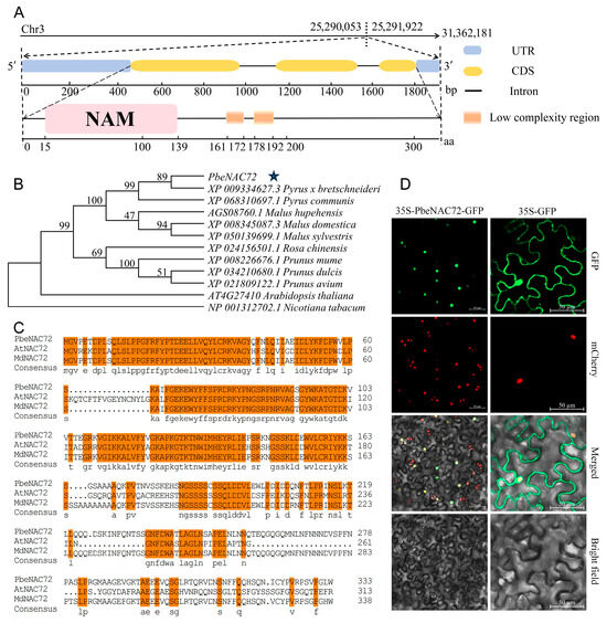
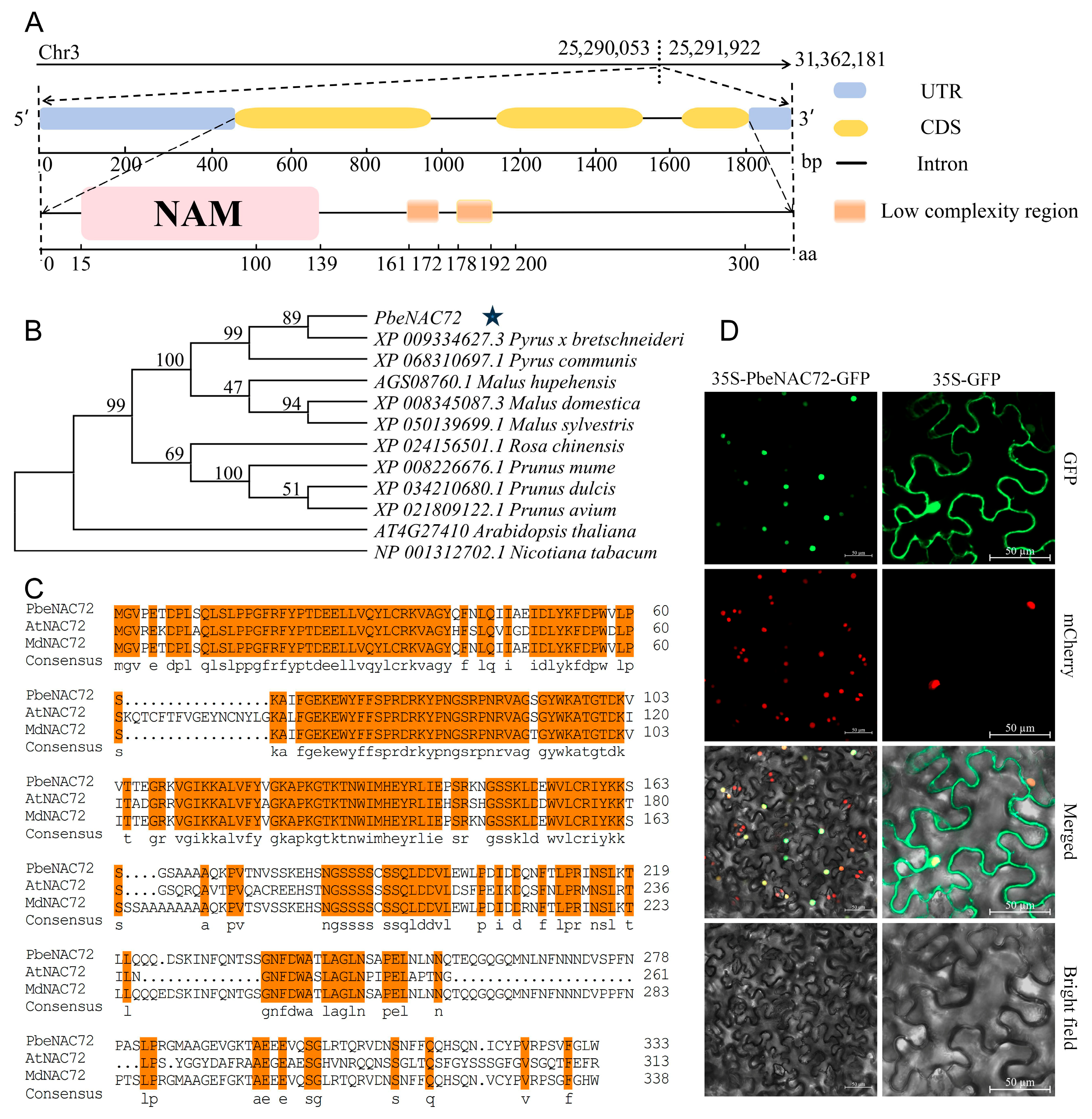
GWHTAAYT036729 (PbeNAC72) is located on chromosome 3 and contains a 1066 bp open reading frame (ORF), which is flanked by untranslated regions (UTRs) at both ends. It comprises three exons and two introns. Domain analysis via SMART revealed that this gene encodes a typical NAC protein, which possesses an NAM domain at the N-terminus (Figure 1A). We further performed multiple sequence alignment and constructed a phylogenetic tree of the target protein and its homologs in model plants and several plant species within the Rosaceae (Figure 1B,C). The results indicated that it was a homolog of Arabidopsis thaliana AtNAC72 and it was therefore named PbeNAC72. Predictions using the online tools DeepLoc 2.1 and UniProt revealed nuclear localization of PbeNAC72 (Supplemental Figures S1 and S2). To confirm this, we transiently expressed the recombinant plasmid PbeNAC72-GFP and an empty vector in Nicotiana benthamiana leaves. The results showed that GFP fluorescence from the PbeNAC72–GFP fusion protein was exclusively localized in the nucleus, whereas the signal from the empty vector was distributed throughout the entire cells (Figure 1D). These results confirm that PbeNAC72 is a typical NAC transcription factor.
Figure 1.
The basic structure, subcellular localization, evolutionary analysis, and multiple sequence alignment of PbeNAC72 from Pyrus betulifolia. (A) Basic structure of PbeNAC72 (UTR: Untranslated Region; CDS: Coding Sequence). (B) Evolutionary analysis of PbeNAC72. The star symbol indicated that it was a homolog of Arabidopsis thaliana AtNAC72 and it was therefore named PbeNAC72. (C) Multiple sequence alignment of PbeNAC72. (D) Subcella localization of the PbeNAC72 protein in Nicotiana benthamiana leaves (PbeNAC72–GFP localizes to the cell nucleus. The green fluorescence signal represents the PbeNAC72–GFP fusion protein, the red fluorescence signal indicates the nuclear marker protein, and the yellow fluorescence in the merged image demonstrates that the target protein colocalizes with the cell nucleus).
3.2. Expression of PbeNAC72 Is Increased by VpM and Stresses-Related Signals
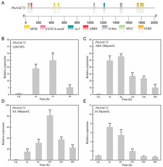
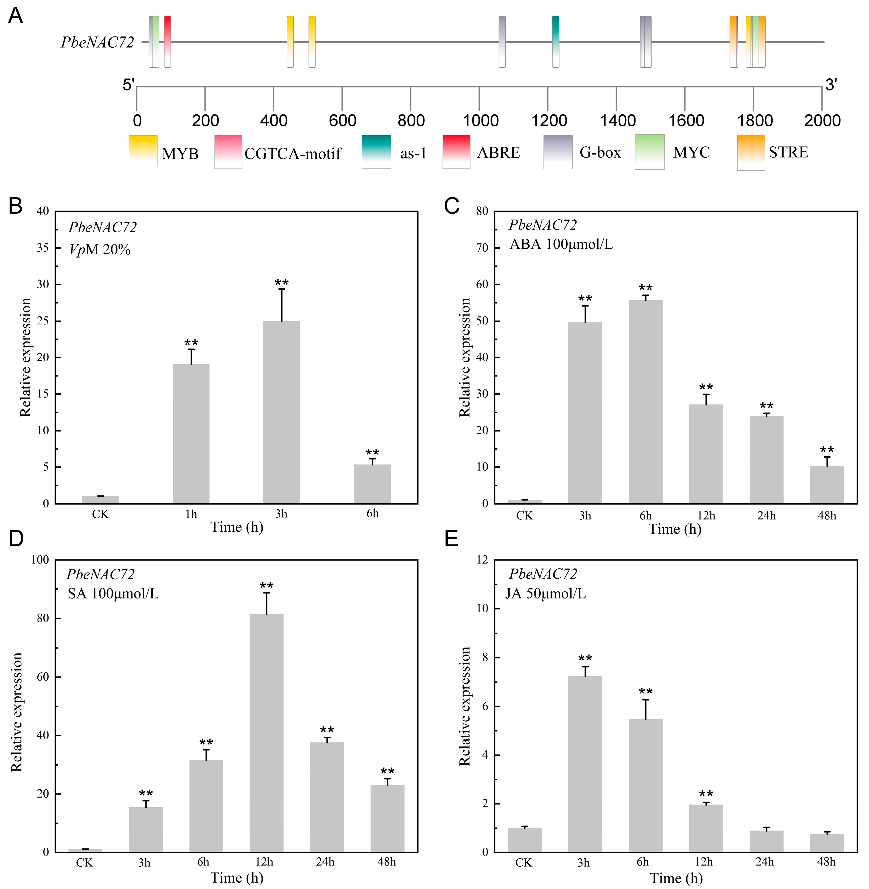
To investigate the role of PbeNAC72 in stress responses, we first extracted the sequences on the promoter region (2 kb sequence upstream of the coding sequence) and predicted cis-elements using the online database PlantCARE. The results revealed a large number of cis-elements responsive to signals of stress-associated hormone signals. For instance, ABRE, as-1, and CGTCA-motif respond to JA, SA and ABA signals, respectively (Figure 2A). To validate this prediction, we analyzed the expression pattern of PbeNAC72 in Duli-G03 cells treated with VpMs or stress-related hormones (Figure 2B–E). As expected, PbeNAC72 expression was strongly induced by both VpM and stress-related hormone treatments. Compared to the control, the expression levels were upregulated by 25.8-, 55-, and 80.6-fold in cells treated with VpMs at 3 h, ABA at 6 h, and SA at 12 h, respectively. These results indicate that PbeNAC72 is involved in plant responses to stress signals and may play a role in plant immunity.
Figure 2.
Analysis of promoters and stress induction of PbeNAC72 in Pyrus betulifolia. (A) Analysis of promoter of PbeNAC72. (B) Expression patterns in response to secondary metabolites of the canker pathogen. (C–E) Expression patterns in response to abscisic acid (ABA), salicylic acid (SA), and jasmonic acid (JA) treatments. CK represents the expression level of PbeNAC72 in untreated wild-type cells. Prolonged exposure times can lead to cell death, so analyses were only performed at 1 h, 3 h, and 6 h. Data in (B–E) are means (±SD), n = 3. Significant differences compared with CK were determined using Students t-test: ** p < 0.01.
3.3. PbeNAC72 Positively Regulates Valsa Canker Resistance of Apple and Pear Fruits
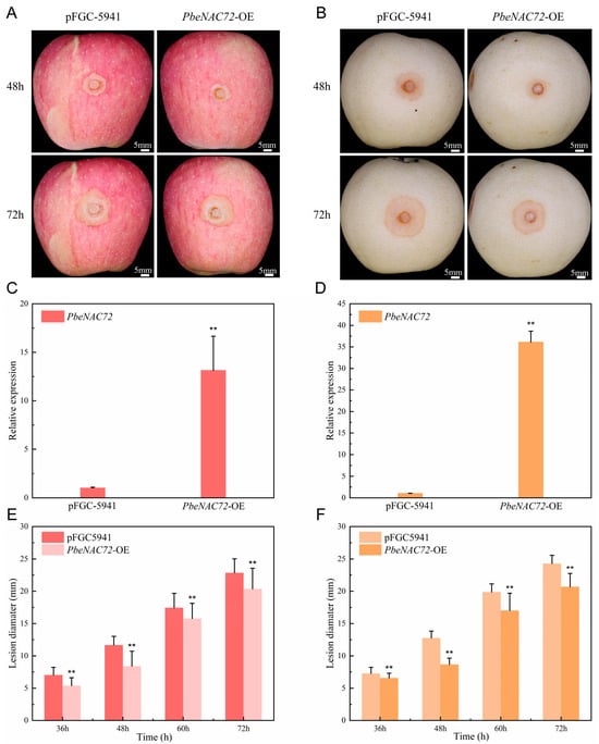
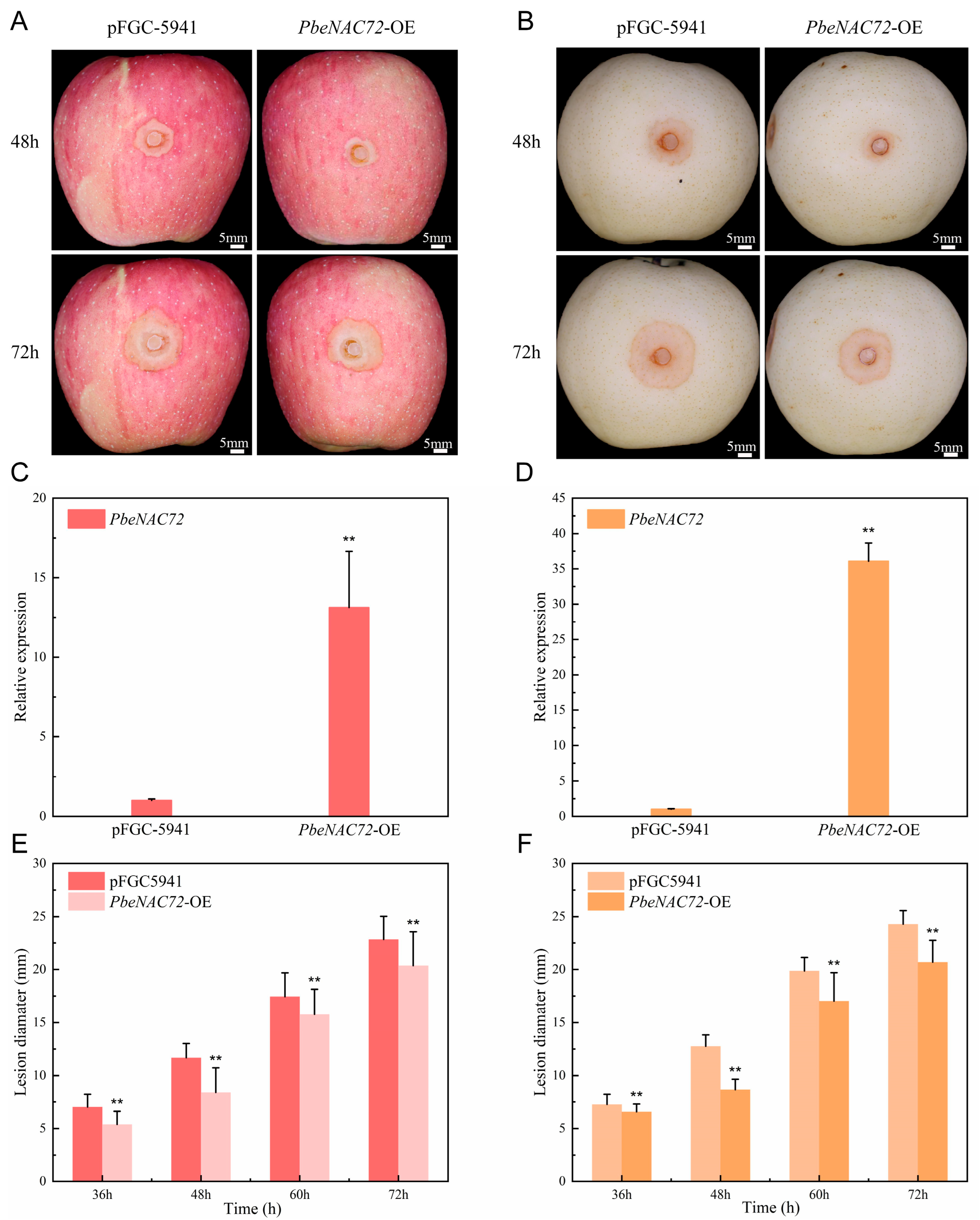
To elucidate the function of PbeNAC72, we performed transient expression assays in Huangguan pear and Fuji apple fruits using A. tumefaciens-mediated transformation with the constructs pFGC5941 (empty vector control) and pFGC5941–PbeNAC72 (recombinant vector) (Figure 3A,B). In both apple and pear fruits, the expression levels of the target genes increased to 13-fold and 36-fold, respectively, compared to the wild-type control group at 72 h (Figure 3C,D). The Valsa canker pathogen was inoculated at the infiltration sites at this time point. At 36, 48, 60, and 72 hpi, the lesion diameters on apple fruits with transient overexpression were recorded as 75%, 71%, 90%, and 89% of the control, respectively. The test in pear fruits displayed similar results to that in apple (Figure 3E,F). These findings indicate that transient overexpression of PbeNAC72 in pear and in apple fruits significantly enhance resistance to V. pyri.
Figure 3.
Validation of PbeNAC72-mediated resistance to infection by Valsa pyri at the fruit level. (A) Resistance to infection conferred by PbeNAC72 overexpression in Fuji apple fruits. (B) Resistance to infection conferred by PbeNAC72 overexpression in Huangguan pear fruits. (C,D) Expression levels of PbeNAC72 in overexpressing fruits (both apple and pear fruits exhibit transient overexpression). (E,F) Lesion diameter in PbeNAC72 overexpressing fruits after inoculation. Data in (C–F) are means (±SD), n = 3 and significant differences were determined using Student’s t-test: ** p < 0.01.
3.4. PbeNAC72 Positively Regulates Valsa Canker Resistance of Duli-G03 Cells
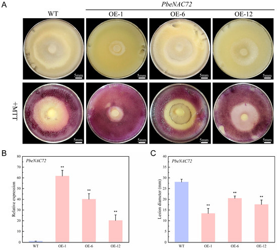
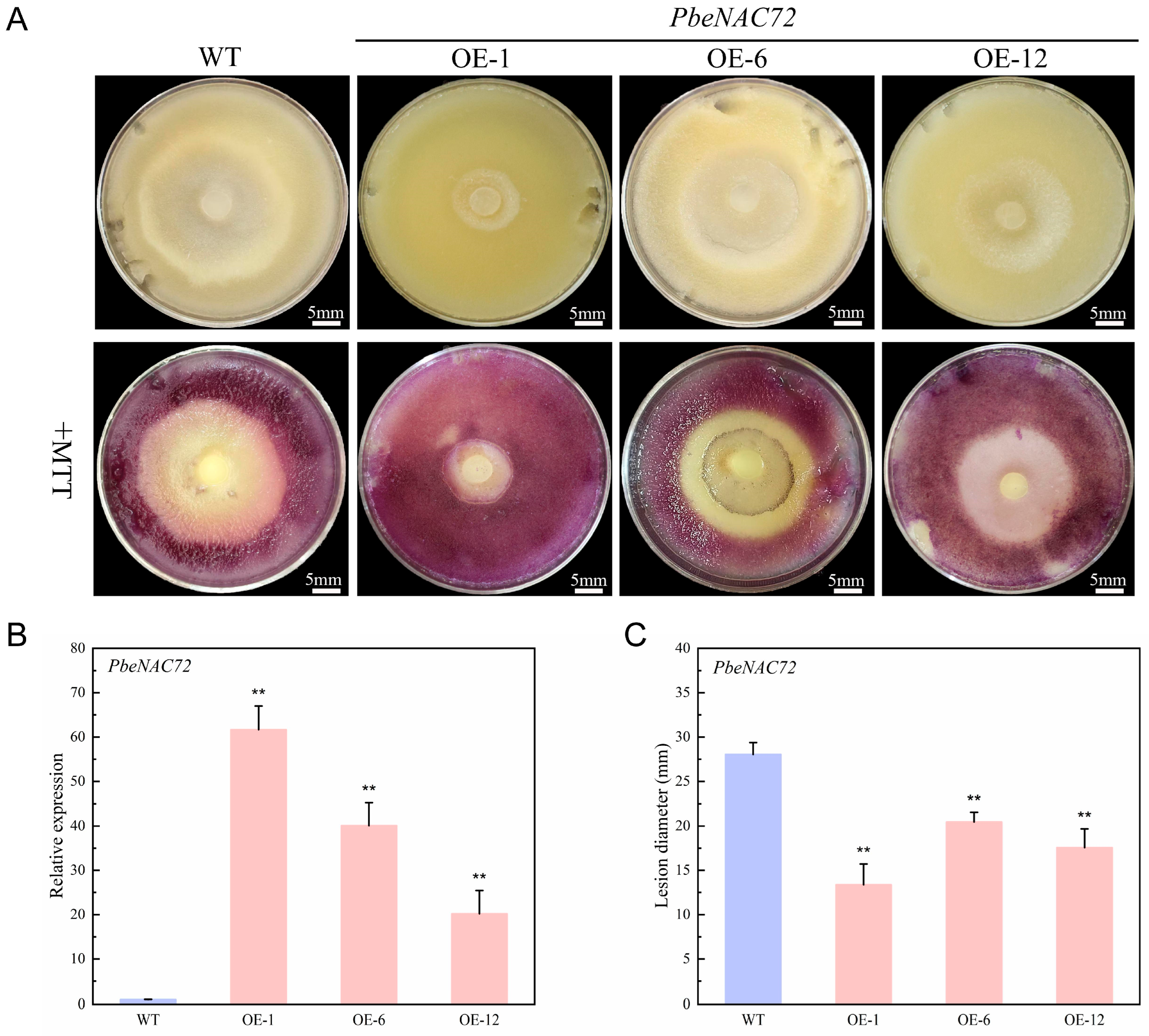
To elucidate the role of PbeNAC72 in resistance to Valsa canker, the target gene was stably expressed in suspension cells of P. betulifolia. In the overexpression cell lines OE-1, OE-6, and OE-12, the expression levels of the target gene were upregulated by 63-, 40-, and 21-fold of that in the wild-type cells (WT), respectively (Figure 4B). Three days post-inoculation with a V. pyri mycelial plug, the pathogen colony produced on WT P. betulifolia cells was significantly larger than those on the overexpression cell lines (Figure 4A,C). These findings indicate that overexpression of the PbeNAC72 gene significantly inhibits the growth of V. pyri on Duli-G03 suspension cells.
Figure 4.
PbeNAC72 positively regulates Tolerance to Valsa pyri in Pyrus betulifolia (A) Phenotype of P. betulaefolia suspension cells 3 days after inoculation with a mycelial plug of V. pyri, stained with MTT to visualize viable cells (live cells can be stained by MTT, while dead cells cannot be stained). (B) Relative expression level of PbeNAC72. (C) Lesion diameter in suspension cells 3 days after inoculation. Data in (B,C) are means (±SD), n = 3. Significant differences compared with the wild type (WT) were determined using Student’s t-test: ** p < 0.01.
3.5. PbeNAC72 Positively Regulates Tolerance of Duli-G03 Cells to V. pyri Metabolites
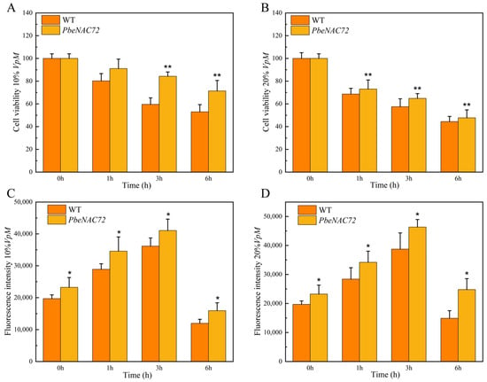
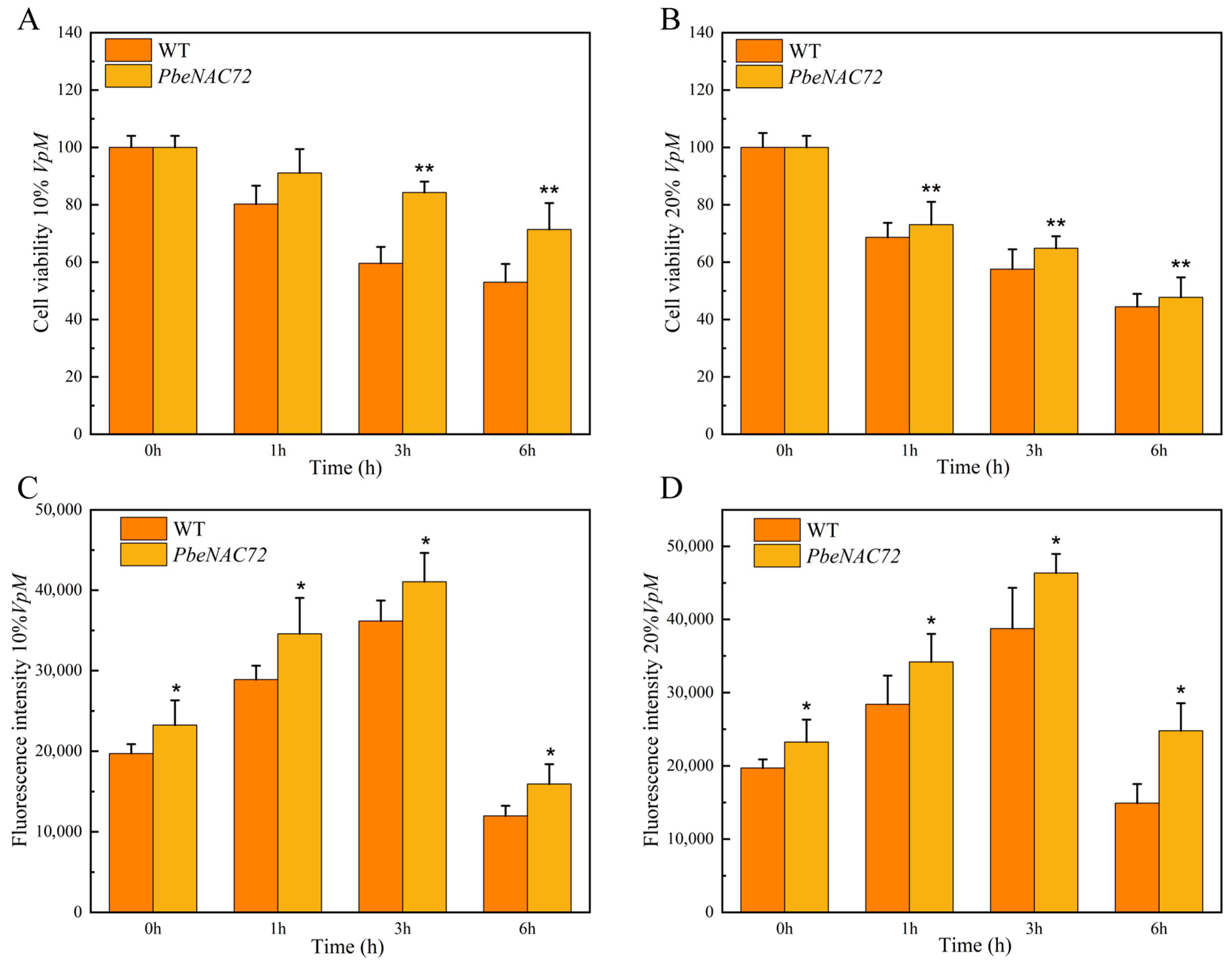
Since the pathogen causing Valsa canker is a typical necrotrophic microbe, we further assessed the influence of PbeNAC72 on cell viability when treated with V. pyri metabolites (VpM). Compared to the control, the viability of PbeNAC72-overexpressing cell lines (OE-1) was significantly higher than that of the wild type when treated with 20% VpM at 1, 3, and 6 h (Figure 5A,B). Subsequently, we investigated the accumulation of ROS, a signal molecule for plant immunity, in WT and overexpressing cell lines. The results demonstrated that for both 10% and 20% VpM treatments, the PbeNAC72-overexpressing suspension cells exhibited a more pronounced ROS burst compared to the WT during the early response to pathogenic signals (Figure 5C,D). Consequently, we hypothesize that PbeNAC72 overexpression may enhance resistance to pathogen invasion through early ROS accumulation and potentially activate downstream disease resistance signaling pathways.
Figure 5.
Cell viability (%) and reactive oxygen species (ROS) in Pyrus betulifolia suspension cells under treatment with 10% and 20% secondary metabolites of Valsa pyri. (A,B) Cell viability under treatment with 10% and 20% secondary metabolites at 0, 1, 3, and 6 h. (C,D) ROS under treatment with 10% and 20% secondary metabolites at 0, 1, 3, and 6 h. Data in (A–D) are means (±SD), n = 3. Significant differences compared with the wild type (WT) were determined using Student’s t-test: * p < 0.05, ** p < 0.01.
3.6. Effect of PbeNAC72 on the Expression of Defense-Related Genes
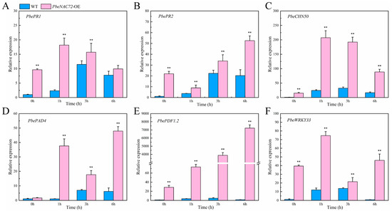
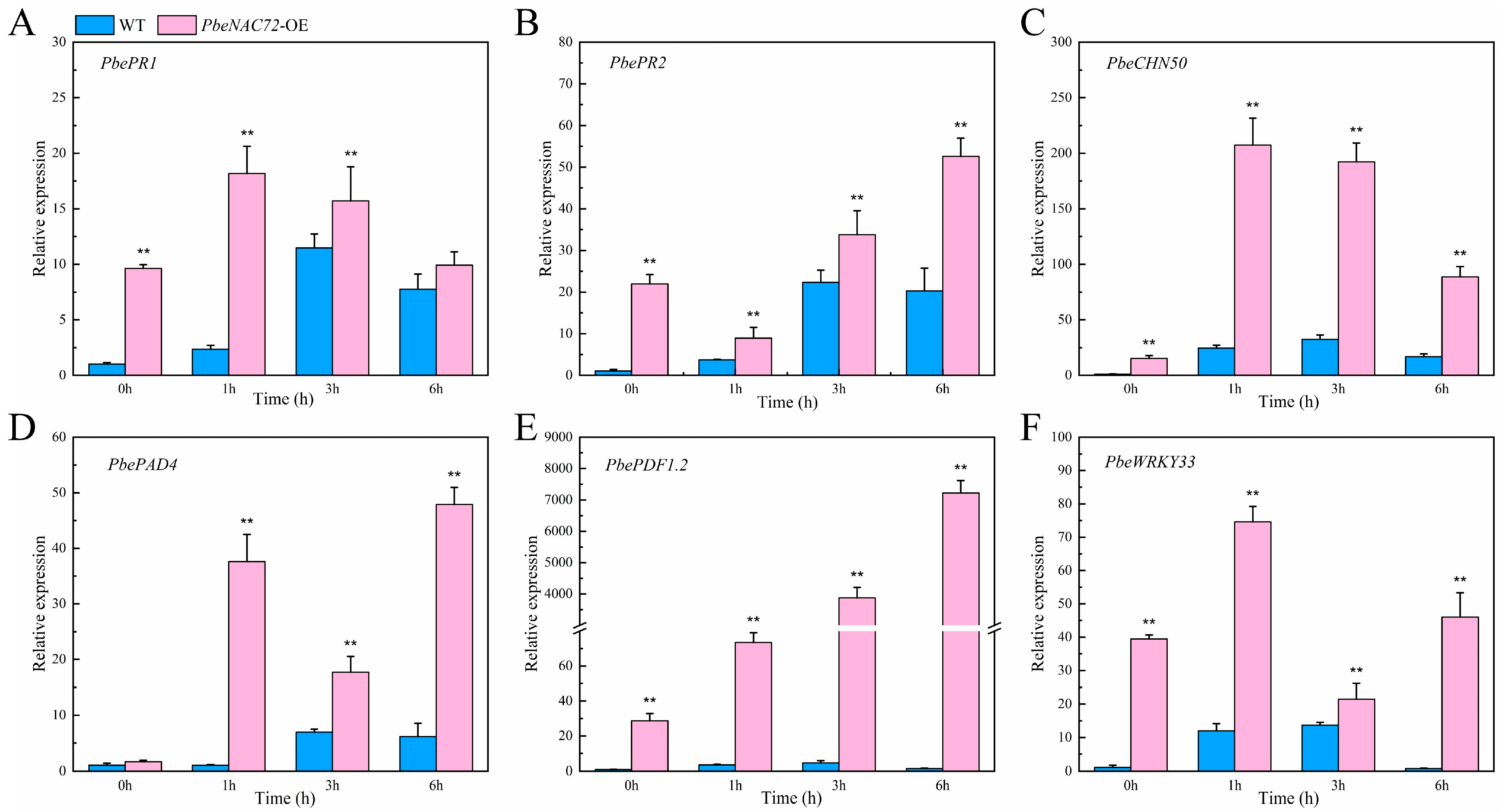
After confirming the role of PbeNAC72 in Valsa canker resistance, we selected the cell line with the most pronounced phenotype among the three transgenic cell lines (OE-1) for subsequent defense gene expression analysis, conducting three biological replicates. Compared to WT, the expression levels of the three SA-related genes PbePR1, PbePR2, and PbeCHN50 were significantly elevated in the OE-1 line. (Figure 6A,B). Strikingly, the expression of PbeCHN50 in OE-1 was upregulated to 15, 8.4-, 5.9-, and 5.3-fold of that in WT at 0, 1, 3 and 6 h, respectively (Figure 6C). In addition to SA-related genes, the expressions of the R-gene PbePAD4 (Figure 6D), JA-related gene PbePDF1.2, and phytoalexin-related gene PbeWRKY33 were also increased in OE-1 compared to WT. It is interesting that the expression levels of PbePDF1.2 were upregulated 24.3- to 7200-fold in OE1 compared with WT (Figure 6E). Collectively, these findings demonstrate that overexpression of PbeNAC72 activates the expression of genes related to multiple immune signals such as SA, JA, R-genes, and phytoalexins.
Figure 6.
Expression analysis of defense genes in PbeNAC72-overexpressing Pyrus betulifolia suspension cells treated with 20% dilution of Valsa pyri secondary metabolites. (A–C) Expression analysis of salicylic acid (SA) pathway-related genes PbePR1, PbePR2, and PbeCHN50. (D) Expression analysis of PbePAD4 in relation to the R-gene pathway. (E) Expression analysis of PbePDF1.2 in relation to the jasmonic acid (JA) pathway. (F) Expression analysis of PbeWRKY33 in relation to the phytoalexin biosynthesis pathway. Data in (A–F) are means (±SD), n = 3. Significant differences compared with the wild type (WT) were determined using Student’s t-test: ** p < 0.01.
3.7. Co-Expression Network, GO Enrichment, and Expression Level Validation of PbeNAC72 in P. betulifolia
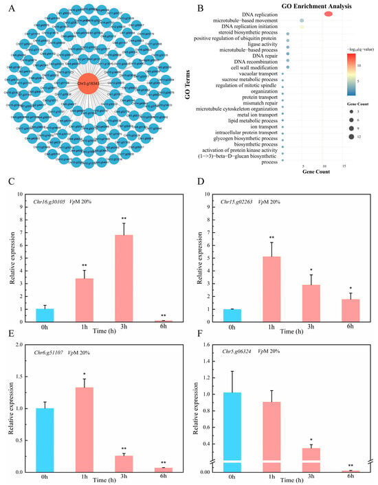
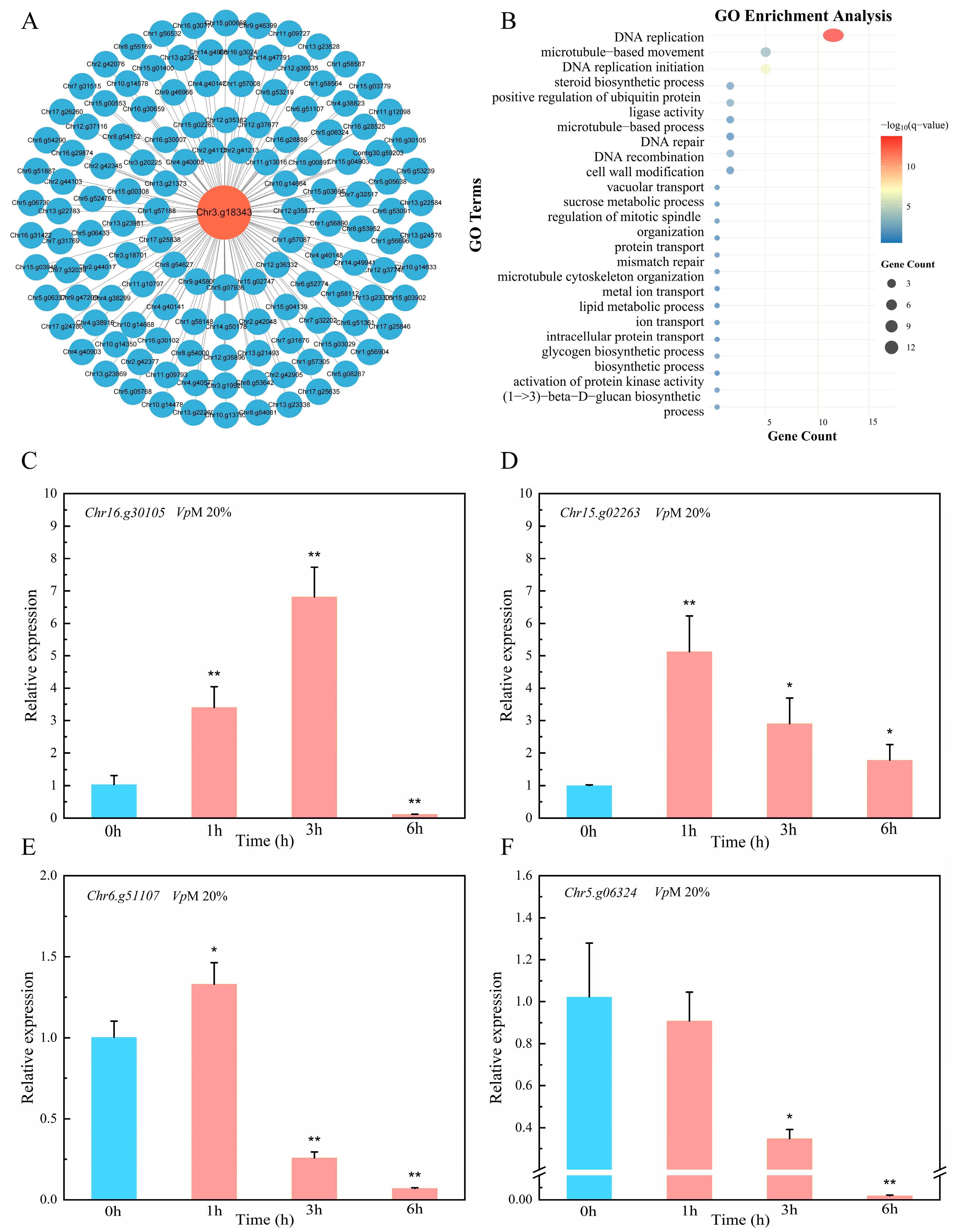
To elucidate the potential mechanism by which PbeNAC72 regulates the plant’s resistance to V. pyri, we performed a systematic analysis of transcriptome data and identified a gene module significantly co-expressed with PbeNAC72 (|r| > 0.35) (Figure 7A). The results demonstrated that under stimulation by VpMs, a total of 134 genes exhibited co-expression patterns with PbeNAC72. Then, we performed GO enrichment analysis on the identified gene set. (Figure 7B). The results revealed significant enrichment in terms related to “DNA replication”, “microtubule-based movement”, and “DNA replication initiation”, among others. The term “DNA replication” was the most prominently enriched, encompassing 12 genes. Secondly, 5 genes were assigned to the “microtubule-based movement” category. From this pool of 17 genes, four with the highest absolute correlation coefficients—microtubule-based movement Chr16.g30105 (|r| = 0.37), DNA replication Chr15.g02263 (|r| = 0.39), DNA replication Chr6.g51107 (|r| = 0.38), and DNA replication Chr5.g06324 (|r| = 0.38)—were selected for subsequent validation by qRT-PCR (Figure 7C–F). qRT-PCR analysis revealed that the expressions of Chr15.g02263 and Chr16.g30105 were in line with predictions. However, Chr6.g51107 was induced at 1 h, and Chr5.g06324 remained uninduced. This observation is largely consistent with the predictions derived from GO enrichment analysis, thereby further validating the reliability of these bioinformatics analyses.
Figure 7.
Co-expression network and Gene Ontology (GO) enrichment analysis of PbeNAC72 in Duli-G03 suspension cells under 20% VpM, along with qRT-PCR validation. (A) Co-expression network analysis of PbeNAC72. (B) GO enrichment analysis of genes from the co-expression network. (C–F) qRT-PCR analysis of Chr16.g30105, Chr15.g02263, Chr6.g51107 and Chr5.g06324. All data in the graphs are means (±SD), n = 3, and significant differences were determined using Student’s t-test: * p < 0.05, ** p < 0.01.
4. Discussion
Based on functional investigations, we demonstrated that the canonical NAC transcription factor PbeNAC72 positively regulates Valsa canker resistance in both apple and pear. Subsequent investigations revealed that overexpression of PbeNAC72 promotes the expression of SA- and JA-related genes in ‘Duli-G03’ suspension cells in response to VpMs. Furthermore, weighted gene co-expression network analysis (WGCNA) showed that PbeNAC72 is primarily associated with genes involved in “DNA replication” and “microtubule-based movement”.
The endogenous gene of P. betulifolia-G03 suspension cells may only be expressed at low levels under pathogen infection induction, or its basal expression may be insufficient to produce an observable disease-resistant phenotype. Upon overexpression, however, the abundance of this protein within plant cells significantly increases, directly enhancing its ability to recognize pathogens and thereby amplifying the response of downstream disease-resistance signaling pathways.
The functional roles of NAC72 homologs in plant disease resistance have been validated across various species. In apple, MpNAC72 has been identified as a positive regulator conferring resistance to the obligate parasitic rust fungus Gymnosporangium yamadae by interacting with MpERF105 to activate the anthocyanin biosynthesis pathway, thereby enhancing disease resistance [36]. Conversely, in grapevine, VvNAC72 negatively regulates resistance against the obligate parasitic downy mildew pathogen by directly suppressing the expression of glyoxalase I-4 (VvGLYI-4) [37]. Currently, direct experimental evidence demonstrating the role of NAC72 homologs in plant resistance against necrotrophic fungi remains limited. Our functional validation results indicate that overexpression of PbeNAC72 significantly enhances the resistance to Valsa canker of Duli-03 suspension cells. This discovery expands our understanding of the functional spectrum of NAC72 transcription factors, highlighting their potential role in defense against necrotrophic pathogens.
ROS burst is a key strategy for plants to limit pathogen spread, serving not only as a direct chemical barrier against pathogens but also as an important secondary messenger activating the expression of downstream defense-related genes [38]. In A. thaliana, the expression of AtNAC72 has been clearly demonstrated to be strongly induced by oxidative stress, indicating its involvement in ROS signal transduction [39]. We observed that overexpression conferred enhanced disease resistance, a finding that is attributable to a marked ROS burst and consistent with established knowledge.
Plant hormones play a pivotal role in orchestrating plant defense responses against pathogens [40]. It has been established that AtNAC72 acts as a key downstream transcription factor within the JA signaling pathway in A. thaliana [41]. ATAF1, an important homologous protein to AtNAC72, is capable of the direct regulation of genes such as PR1 [42]. Our results show that PbeNAC72 simultaneously activates key genes in both the SA and JA signaling pathways. Although SA and JA signaling often act antagonistically to optimize resource allocation, growing evidence indicates that they can function synergistically under specific contexts [43]. We speculate that the activation of the SA pathway may enhance systemic alertness and reactive capacity, such as the synthesis of defense proteins within cells, while JA pathway activation potentially promotes the reinforcement of physical barriers such as cell wall thickening.
Furthermore, plants employ a mechanism wherein the activation of the DNA damage response (DDR) pathway for damage repair triggers the expression of downstream defense genes to suppress pathogens [44]. The importance of microtubules, a primary cytoskeletal component essential for cell shape, intracellular transport, and division, lies in their ability to provide directional tracks for the transport of defense molecules during immune responses [45]. The repertoire of downstream genes activated by immunity-related NAC transcription factors is likely to encompass those encoding components of the vesicle transport machinery and cytoskeletal organizers [46]. Based on co-expression networks, GO enrichment analysis, and subsequent qRT-PCR results, the activation of relevant genes indicates that PbeNAC72 is closely associated with cell cycle progression and cytoskeletal reorganization processes. Therefore, we hypothesize that the role of PbeNAC72 may extend beyond the direct regulation of defense genes and could involve maintaining and optimizing the internal cellular environment to support an effective immune response [11].
We employed WT pear cells for the WGCNA, while the validation of the SA and JA pathways was conducted using cell lines overexpressing PbeNAC72. Therefore, the lack of gene association may be attributed to differences in PbeNAC72 expression levels.
In conclusion, this study identifies PbeNAC72 as a positive regulatory transcription factor in pear trees responding to V. pyri infection. Overexpression of PbeNAC72 significantly enhanced the resistance of Duli-03 suspension cells to Valsa canker. This enhancement was achieved through the rapid activation of the ROS burst, the upregulation of key genes in defense signaling pathways such as SA and JA, and the potential activation of genes associated with DNA replication and cytoskeletal reorganization. These findings provide novel theoretical insights into the role of NAC transcription factors in pear resistance to Valsa canker and establish a crucial foundation for the molecular breeding of pear trees.
Supplementary Materials
The following supporting information can be downloaded at: https://www.mdpi.com/article/10.3390/horticulturae12020171/s1, Supplementary Table S1. Gene and primer information associated with qRT-PCR analysis. Supplementary Figure S1. Predicted localization of PbeNAC72 in the nucleus based on annotations from the UniProt database. Supplementary Figure S2. Subcellular localization probabilities for the candidate protein PbeNAC72 predicted using the DeepLoc 2.1 webserver.
Author Contributions
C.Z. (Cunwu Zuo) conceived, designed, and coordinated this study. W.W. and H.H. performed the experiments. Y.L., C.Z. (Cunliang Zuo), M.C., E.S. and H.P. performed, collected, analyzed, and deposited the data. C.Z. (Cunwu Zuo) and X.L. proofread the final draft. All authors have read and agreed to the published version of the manuscript.
Funding
This research was funded by Gansu Province Technology Innovation Guidance Program Project, grant number 23CXNA0003, Gansu Provincial University Industry Support Program Project, grant number 2025CYZC-035, Gansu Provincial Science and Technology Major Special Project, grant number 22ZD6NA045 and 2024 Gansu Provincial Natural Science Foundation-Excellent Doctoral Student Program, grant number 24JRRA678.
Data Availability Statement
The original contributions presented in this study are included in the article and Supplementary Material. Further inquiries can be directed to the corresponding authors.
Acknowledgments
The authors would like to thank China National Knowledge Infrastructure (CNKI) for providing access to e-resources.
Conflicts of Interest
The authors declare no conflicts of interest.
References
- Yin, Z.; Liu, H.; Li, Z.; Ke, X.; Dou, D.; Gao, X.; Song, N.; Dai, Q.; Wu, Y.; Xu, J.R.; et al. Genome sequence of Valsa canker pathogens uncovers a potential adaptation of colonization of woody bark. New Phytol. 2015, 208, 1202–1216. [Google Scholar] [CrossRef] [Scilit]
- Wang, X.; Wei, J.; Huang, L.; Kang, Z. Re-evaluation of pathogens causing Valsa canker on apple in China. Mycologia 2011, 103, 317–324. [Google Scholar] [CrossRef] [Scilit]
- Abe, K.; Kotoda, N.; Kato, H.; Soejima, J. Resistance sources to Valsa canker (Valsa ceratosperma) in a germplasm collection of diverse Malus species. Plant Breed. 2007, 126, 449–453. [Google Scholar] [CrossRef] [Scilit]
- Yuan, H.; Shi, B.; Huang, T.; Zhou, Z.; Wang, L.; Hou, H.; Tu, H. Biological Control of Pear Valsa Canker Caused by Valsa pyri Using Penicillium citrinum. Horticulturae 2021, 7, 198. [Google Scholar] [CrossRef] [Scilit]
- Wu, S.; Shan, L.; He, P. Microbial signature-triggered plant defense responses and early signaling mechanisms. Plant Sci. 2014, 228, 118–126. [Google Scholar] [CrossRef] [Scilit]
- Jones, J.D.; Dangl, J.L. The plant immune system. Nature 2006, 444, 323–339. [Google Scholar] [CrossRef] [Scilit]
- Ngou, B.P.M.; Ahn, H.K.; Ding, P.; Jones, J.D.G. Mutual potentiation of plant immunity by cell-surface and intracellular receptors. Nature 2021, 592, 110–115. [Google Scholar] [CrossRef] [Scilit]
- Tsuda, K.; Sato, M.; Stoddard, T.; Glazebrook, J.; Katagiri, F. Network properties of robust immunity in plants. PLoS Genet. 2009, 5, e1000772. [Google Scholar] [CrossRef] [Scilit] [PubMed]
- Torres, M.A.; Jones, J.D.; Dangl, J.L. Reactive oxygen species signaling in response to pathogens. Plant Physiol. 2006, 141, 373–378. [Google Scholar] [CrossRef] [Scilit]
- Hirschi, K.D. The calcium conundrum. Both versatile nutrient and specific signal. Plant Physiol. 2004, 136, 2438–2442. [Google Scholar] [CrossRef] [Scilit]
- Dong, B.; Liu, Y.; Huang, G.; Song, A.; Chen, S.; Jiang, J.; Chen, F.; Fang, W. Plant NAC transcription factors in the battle against pathogens. BMC Plant Biol. 2024, 24, 958. [Google Scholar] [CrossRef] [Scilit]
- Olsen, A.N.; Ernst, H.A.; Leggio, L.L.; Skriver, K. NAC transcription factors: Structurally distinct, functionally diverse. Trends Plant Sci. 2005, 10, 79–87. [Google Scholar] [CrossRef] [Scilit] [PubMed]
- Puranik, S.; Sahu, P.P.; Srivastava, P.S.; Prasad, M. NAC proteins: Regulation and role in stress tolerance. Trends Plant Sci. 2012, 17, 369–381. [Google Scholar] [CrossRef] [Scilit]
- Nakashima, K.; Tran, L.S.; Van Nguyen, D.; Fujita, M.; Maruyama, K.; Todaka, D.; Ito, Y.; Hayashi, N.; Shinozaki, K.; Yamaguchi-Shinozaki, K. Functional analysis of a NAC-type transcription factor OsNAC6 involved in abiotic and biotic stress-responsive gene expression in rice. Plant J. 2007, 51, 617–630. [Google Scholar] [CrossRef] [Scilit] [PubMed]
- Zhao, Q.; Zhong, M.; He, L.; Wang, B.; Liu, Q.L.; Pan, Y.Z.; Jiang, B.B.; Zhang, L. Overexpression of a chrysanthemum transcription factor gene DgNAC1 improves drought tolerance in chrysanthemum. Plant Cell Tissue Organ Cult. 2018, 135, 119–132. [Google Scholar] [CrossRef] [Scilit]
- Chen, J.; Gong, Y.; Gao, Y.; Zhou, Y.B.; Chen, M.; Xu, Z.S.; Guo, C.H.; Ma, Y.Z. TaNAC48 positively regulates drought tolerance and ABA responses in wheat (Triticum aestivum L.). Crop J. 2021, 9, 785–793. [Google Scholar] [CrossRef] [Scilit]
- Takasaki, H.; Maruyama, K.; Kidokoro, S.; Ito, Y.; Fujita, Y.; Shinozaki, K.; Yamaguchi-Shinozaki, K.; Nakashima, K. The abiotic stress-responsive NAC-type transcription factor OsNAC5 regulates stress-inducible genes and stress tolerance in rice. Mol. Genet. Genom. 2010, 284, 173–183. [Google Scholar] [CrossRef] [Scilit]
- Xu, Y.; Zou, S.; Zeng, H.; Wang, W.; Wang, B.; Wang, H.; Tang, D. A NAC Transcription Factor TuNAC69 Contributes to ANK-NLR-WRKY NLR-Mediated Stripe Rust Resistance in the Diploid Wheat Triticum urartu. Int. J. Mol. Sci. 2022, 23, 564. [Google Scholar] [CrossRef] [Scilit]
- Tran, L.S.; Nakashima, K.; Sakuma, Y.; Simpson, S.D.; Fujita, Y.; Maruyama, K.; Fujita, M.; Seki, M.; Shinozaki, K.; Yamaguchi-Shinozaki, K. Isolation and functional analysis of Arabidopsis stress-inducible NAC transcription factors that bind to a drought-responsive cis-element in the early responsive to dehydration stress 1 promoter. Plant Cell 2004, 16, 2481–2498. [Google Scholar] [CrossRef] [Scilit]
- Xu, Z.-Y.; Kim, S.Y.; Hyeon, D.Y.; Kim, D.H.; Dong, T.; Park, Y.; Jin, J.B.; Joo, S.-H.; Kim, S.-K.; Hong, J.C.; et al. The Arabidopsis NAC Transcription Factor ANAC096 Cooperates with bZIP-Type Transcription Factors in Dehydration and Osmotic Stress Responses. Plant Cell 2013, 25, 4708–4724. [Google Scholar] [CrossRef] [Scilit]
- Han, K.; Zhao, Y.; Sun, Y.; Li, Y. NACs, generalist in plant life. Plant Biotechnol. J. 2023, 21, 2433–2457. [Google Scholar] [CrossRef] [Scilit]
- Forlani, S.; Mizzotti, C.; Masiero, S. The NAC side of the fruit: Tuning of fruit development and maturation. BMC Plant Biol. 2021, 21, 238. [Google Scholar] [CrossRef] [Scilit]
- Jensen, M.K.; Kjaersgaard, T.; Petersen, K.; Skriver, K. NAC genes: Time-specific regulators of hormonal signaling in Arabidopsis. Plant Signal. Behav. 2010, 5, 907–910. [Google Scholar] [CrossRef] [Scilit]
- Wang, F.; Lin, R.; Feng, J.; Chen, W.; Qiu, D.; Xu, S. TaNAC1 acts as a negative regulator of stripe rust resistance in wheat, enhances susceptibility to Pseudomonas syringae, and promotes lateral root development in transgenic Arabidopsis thaliana. Front. Plant Sci. 2015, 6, 108. [Google Scholar] [CrossRef] [Scilit]
- Nisa, M.U.; Huang, Y.; Benhamed, M.; Raynaud, C. The Plant DNA Damage Response: Signaling Pathways Leading to Growth Inhibition and Putative Role in Response to Stress Conditions. Front. Plant Sci. 2019, 10, 653. [Google Scholar] [CrossRef] [Scilit]
- Jeon, H.S.; Jang, E.; Kim, J.; Kim, S.H.; Lee, M.H.; Nam, M.H.; Tobimatsu, Y.; Park, O.K. Pathogen-induced autophagy regulates monolignol transport and lignin formation in plant immunity. Autophagy 2023, 19, 597–615. [Google Scholar] [CrossRef] [Scilit]
- Sun, E.; Yu, H.; Chen, Z.; Cai, M.; Mao, X.; Li, Y.; Zuo, C. Enhanced Valsa canker resistance conferred by expression of MdLecRK-S.4.3 in Pyrus betulifolia is largely suppressed by PbePUB36. J. Exp. Bot. 2023, 74, 3998–4013. [Google Scholar] [CrossRef] [Scilit] [PubMed]
- Yu, H.; Sun, E.; Mao, X.; Chen, Z.; Xu, T.; Zuo, L.; Jiang, D.; Cao, Y.; Zuo, C. Evolutionary and functional analysis reveals the crucial roles of receptor-like proteins in resistance to Valsa canker in Rosaceae. J. Exp. Bot. 2023, 74, 162–177. [Google Scholar] [CrossRef] [Scilit] [PubMed]
- Zhao, D.; Tian, Y.Z.; Yu, H.Q.; Mao, X.; Wang, C.; Duo, H.; Sun, E.; Zuo, C.W. Establishment of the ‘Valsa pyri metabolites (VpM)-suspension cell’-based system to study the response of pears to VpM. Physiol. Mol. Plant Pathol. 2022, 120, 101850. [Google Scholar] [CrossRef] [Scilit]
- Price, M.N.; Dehal, P.S.; Arkin, A.P. FastTree: Computing large minimum evolution trees with profiles instead of a distance matrix. Mol. Biol. Evol. 2009, 26, 1641–1650. [Google Scholar] [CrossRef] [Scilit]
- Letunic, I.; Khedkar, S.; Bork, P. SMART: Recent updates, new developments and status in 2020. Nucleic Acids Res. 2021, 49, D458–D460. [Google Scholar] [CrossRef] [Scilit]
- Mao, X.; Wang, C.; Lü, Q.; Tian, Y.; Wang, D.; Chen, B.; Mao, J.; Li, W.; Chu, M.; Zuo, C. Cyclic nucleotide gated channel genes (CNGCs) in Rosaceae: Genome-wide annotation, evolution and the roles on Valsa canker resistance. Plant Cell Rep. 2021, 40, 2369–2382. [Google Scholar] [CrossRef] [Scilit]
- Dobin, A.; Davis, C.A.; Schlesinger, F.; Drenkow, J.; Zaleski, C.; Jha, S.; Batut, P.; Chaisson, M.; Gingeras, T.R. STAR: Ultrafast universal RNA-seq aligner. Bioinformatics 2013, 29, 15–21. [Google Scholar] [CrossRef] [Scilit]
- Cantalapiedra, C.P.; Hernández-Plaza, A.; Letunic, I.; Bork, P.; Huerta-Cepas, J. eggNOG-mapper v2: Functional Annotation, Orthology Assignments, and Domain Prediction at the Metagenomic Scale. Mol. Biol. Evol. 2021, 38, 5825–5829. [Google Scholar] [CrossRef] [Scilit]
- Yu, G.; Wang, L.G.; Han, Y.; He, Q.Y. clusterProfiler: An R package for comparing biological themes among gene clusters. OMICS 2012, 16, 284–287. [Google Scholar] [CrossRef] [Scilit]
- Wang, Y.; An, H.; Yang, Y.; Yi, C.; Duan, Y.; Wang, Q.; Guo, Y.; Yao, L.; Chen, M.; Meng, J.; et al. The MpNAC72/MpERF105-MpMYB10b module regulates anthocyanin biosynthesis in Malus ‘Profusion’ leaves infected with Gymnosporangium yamadae. Plant J. 2024, 118, 1569–1588. [Google Scholar] [CrossRef] [Scilit] [PubMed]
- Li, T.; Cheng, X.; Wang, X.; Li, G.; Wang, B.; Wang, W.; Zhang, N.; Han, Y.; Jiao, B.; Wang, Y.; et al. Glyoxalase I-4 functions downstream of NAC72 to modulate downy mildew resistance in grapevine. Plant J. 2021, 108, 394–410. [Google Scholar] [CrossRef] [Scilit] [PubMed]
- Jin, H.; Axtell, M.J.; Dahlbeck, D.; Ekwenna, O.; Zhang, S.; Staskawicz, B.; Baker, B. NPK1, an MEKK1-like mitogen-activated protein kinase kinase kinase, regulates innate immunity and development in plants. Dev. Cell 2002, 3, 291–297. [Google Scholar] [CrossRef] [Scilit] [PubMed]
- Huang, J.-C.; Piater, L.A.; Dubery, I.A. The NAC transcription factor gene ANAC072 is differentially expressed in Arabidopsis thaliana in response to microbe-associated molecular pattern (MAMP) molecules. Physiol. Mol. Plant Pathol. 2012, 80, 19–27. [Google Scholar] [CrossRef] [Scilit]
- Pieterse, C.M.; Leon-Reyes, A.; Van der Ent, S.; Van Wees, S.C. Networking by small-molecule hormones in plant immunity. Nat. Chem. Biol. 2009, 5, 308–316. [Google Scholar] [CrossRef] [Scilit]
- Kazan, K.; Manners, J.M. MYC2: The master in action. Mol. Plant 2013, 6, 686–703. [Google Scholar] [CrossRef] [Scilit] [PubMed]
- Wang, X.; Basnayake, B.M.; Zhang, H.; Li, G.; Li, W.; Virk, N.; Mengiste, T.; Song, F. The Arabidopsis ATAF1, a NAC transcription factor, is a negative regulator of defense responses against necrotrophic fungal and bacterial pathogens. Mol. Plant Microbe Interact. 2009, 22, 1227–1238. [Google Scholar] [CrossRef] [Scilit] [PubMed]
- Pieterse, C.M.; Van der Does, D.; Zamioudis, C.; Leon-Reyes, A.; Van Wees, S.C. Hormonal modulation of plant immunity. Annu. Rev. Cell Dev. Biol. 2012, 28, 489–521. [Google Scholar] [CrossRef] [Scilit] [PubMed]
- Dewitte, W.; Murray, J.A. The plant cell cycle. Annu. Rev. Plant Biol. 2003, 54, 235–264. [Google Scholar] [CrossRef] [Scilit]
- Yagi, N.; Fujita, S.; Nakamura, M. Plant microtubule nucleating apparatus and its potential signaling pathway. Curr. Opin. Plant Biol. 2024, 82, 102624. [Google Scholar] [CrossRef] [Scilit]
- Ma, J.; Wang, L.Y.; Dai, J.X.; Wang, Y.; Lin, D. The NAC-type transcription factor CaNAC46 regulates the salt and drought tolerance of transgenic Arabidopsis thaliana. BMC Plant Biol. 2021, 21, 11. [Google Scholar] [CrossRef] [Scilit]
Disclaimer/Publisher’s Note: The statements, opinions and data contained in all publications are solely those of the individual author(s) and contributor(s) and not of MDPI and/or the editor(s). MDPI and/or the editor(s) disclaim responsibility for any injury to people or property resulting from any ideas, methods, instructions or products referred to in the content. |
© 2026 by the authors. Licensee MDPI, Basel, Switzerland. This article is an open access article distributed under the terms and conditions of the Creative Commons Attribution (CC BY) license.