


Abstract
Lance asiabell roots (LAR; Codonopsis lanceolata) undergo substantial physicochemical and biochemical changes during prolonged storage, but their responses to different low-temperature conditions remain unclear. This study investigated the effects of storage temperature on the long-term quality of LAR stored at −4, −2, 0, and 4 °C for up to 5 months. Weight loss increased throughout storage and was highest at −2 °C after 5 months, whereas the lowest decay rate was observed at 0 °C. Storage at −4 °C resulted in a marked decrease in firmness and greater changes in cross-sectional color, while shoot emergence occurred only at 4 °C during the later storage period. Total phenolic content and DPPH and ABTS radical scavenging activities generally increased during the middle stage of storage and subsequently declined. Tangshenoside I and lobetyolin showed different temperature-dependent responses, with relatively higher tangshenoside I at 4 °C and lobetyolin at 0 and 4 °C. PCA and correlation analyses indicated that physical deterioration, internal color changes, and antioxidant-related responses were interrelated during storage. These findings provide practical information for commercial storage and distribution of LAR under different low-temperature conditions.
1. Introduction
Lance asiabell (Codonopsis lanceolata) is a perennial medicinal and edible plant widely consumed in East Asia [1,2]. Lance asiabell roots (LAR) are consumed as fresh vegetables and are also used in processed foods and traditional herbal preparations because of their characteristic flavor and functional properties [1,3]. LAR contain various bioactive compounds, including phenolic compounds, polyacetylenes, and saponins [4,5], which have been associated with antioxidant [5], anti-inflammatory [6], immunomodulatory [7], anti-obesity [8], antidiabetic [9], and anticancer activities [10]. The contents of these bioactive constituents may change during prolonged storage depending on storage conditions, thereby affecting the nutritional and functional quality of LAR [11,12].
After harvest, root crops, including lance asiabell, remain metabolically active and undergo continuous physicochemical and biochemical changes during storage [13,14]. These changes are strongly influenced by storage conditions, particularly temperature, and may result in water loss, texture deterioration, discoloration, and decay, thereby reducing marketability and limiting long-term distribution [14,15]. In addition to these physical changes, biochemical alterations during storage may affect the nutritional and functional quality of LAR [11,12]. Accordingly, information on temperature-dependent quality changes during prolonged storage is important for understanding the postharvest behavior of LAR.
Low-temperature storage is widely used to suppress respiration, water loss, microbial growth, and other metabolic processes in horticultural products [13,16]. However, the response to low temperature varies depending on the commodity and storage duration [13,17]. Excessively high temperatures can accelerate moisture loss, discoloration, and senescence, whereas inappropriate subzero temperatures may also cause physiological or structural damage of agricultural crops [13,16,17]. Thus, successful long-term storage requires a temperature range that sufficiently slows metabolic activity without causing low-temperature injury [13].
Previous studies have shown that storage temperature significantly affects the postharvest quality of several medicinal root crops. In American ginseng roots, low-temperature storage at 4 °C combined with antimicrobial coating and modified atmosphere packaging maintained texture, color, and appearance with minimal weight loss during storage for up to 38 weeks [18]. In sweet potato roots stored at 4, 13, and 20 °C, markedly different physiological responses were observed, with storage at 4 °C inducing browning and chilling injury as storage progressed [14]. In radish roots, prolonged cold storage for 4 months resulted in substantial decreases in sulforaphene content and myrosinase activity, indicating that bioactive properties may also change during long-term storage [19]. In LAR, storage under different temperature and duration conditions affected total phenolic and flavonoid contents as well as antioxidant activities [11]. In another study, LAR stored at 5, 15, 25, 35, and 45 °C for up to 90 days showed changes in xanthine oxidase inhibitory activity, reducing power, immune activity, and cytotoxicity [12]. Long-term low-temperature storage has been investigated in other medicinal root crops; for example, fresh ginseng has been stored at 0 °C for up to 5 months, and subzero temperatures down to −4 °C have also been evaluated during storage and distribution [20,21]. In contrast, previous studies on LAR have generally examined shorter storage periods or a narrower range of storage temperatures [11,12].
Therefore, to evaluate the effects of storage temperature on the long-term quality of LAR, roots were stored at −4, −2, 0, and 4 °C for up to 5 months in this study, and changes in visual quality, weight loss, firmness, color characteristics, antioxidant properties, and bioactive compounds were investigated. The results of this study may provide practical information for selecting appropriate storage temperatures to maintain the quality of LAR during long-term storage.
2. Materials and Methods
2.1. Sample Preparation
Three-year-old lance asiabell roots (Codonopsis lanceolata) were harvested on 15 September 2025, in Hoengseong, Gangwon-do, Republic of Korea for the storage experiment. The initial moisture content of the roots was 75.4 ± 2.2%. On the day after harvest, the roots were packed in polypropylene (PP) woven sacks at 5 kg per sack (approximately 110 roots per sack) and stored at −4, −2, 0, and 4 °C and 70% relative humidity for up to 5 months. For each storage temperature and sampling time, three PP woven sacks were prepared. After assessing decay and shoot emergence, marketable roots from the three sacks were pooled, and 10 roots were randomly selected for analysis. Five roots were used for measurements of firmness, soluble solids content, and cross-sectional color, while the remaining five roots were freeze-dried and ground separately for the determination of total phenolic content, antioxidant activity, and tangshenoside I and lobetyolin contents. Individual roots were considered independent biological replicates.
2.2. Measurement of Weight Loss, Decay Rate, and Shoot Emergence Rate
Weight loss was determined monthly using PP woven sacks containing 5 kg of LAR. Three replicate sacks were used for each storage temperature, and weight loss was calculated as the difference between the initial and measured weights at each sampling time, expressed as a percentage of the initial weight.
Decay rate was determined using three replicate PP woven sacks for each storage temperature and expressed as the percentage of decayed roots relative to the total number of roots in each sack. Roots showing visible fungal mycelia or apparent softening associated with decay were considered decayed.
Shoot emergence rate was also determined using three replicate PP woven sacks for each storage temperature. A root was considered to have undergone shoot emergence when the shoot emerging from the root crown reached ≥1 cm in length. Shoot emergence rate was calculated as the percentage of roots showing shoot emergence relative to the total number of roots in each sack.
2.3. Analysis of Quality Characteristics
Soluble solids content (SSC) was measured using five individual roots per treatment and sampling time (n = 5). For each biological replicate, one whole root was used for juice extraction. The peeled root body from each root was cut into small pieces, wrapped in two layers of gauze, and pressed five times using a juice extractor (Goojung Engineering Co., Ltd., Seoul, Republic of Korea). The extracted juice was homogenized, and 2 mL was used for measurement using a digital refractometer (PAL-1, Atago Co., Ltd., Tokyo, Japan) [22].
Firmness was measured in five individual roots (n = 5) using a texture analyzer (Lloyd Instrument BG/TA Plus, Ametek, Inc., Fareham, UK). Firmness was measured at 3 cm below the root crown after removing the outer skin from the measurement area to minimize variation associated with differences in root thickness. Firmness was measured once per root using a 3 mm diameter probe with a depression limit of 7 mm, test speed of 2 mm/s, and trigger force of 0.98 N [23].
Cross-sectional color was measured in 10 roots using a colorimeter (CR-400, Konica Minolta Co., Tokyo, Japan). Each root was transversely cut 2–3 cm below the root crown, and color was measured near the vascular tissue. The mean of five measurements taken at randomly selected points near the vascular tissue was used as the representative color value for each root. Color characteristics were expressed as CIELAB values, including L* (lightness), a* (redness/greenness), and b* (yellowness/blueness). Chroma and hue angle were also calculated. Browning index (BI) and whiteness index (WI) were calculated from the color values using the following equations [24]:
2.4. Analysis of Total Phenolic Content and Antioxidant Activities
Briefly, 1 g of sample was extracted with 10 mL of 70% (v/v) aqueous methanol by sonication for 30 min at 25 °C. The extract was centrifuged at 3000× g for 10 min at 25 °C, and the resulting supernatant was used to determine total phenolic content (TPC) and antioxidant activities (n = 5).
TPC was determined using the Folin–Ciocalteu method with slight modifications [25]. Briefly, 20 μL of the sample extract was mixed with 10 μL of Folin–Ciocalteu reagent and allowed to react for 5 min. Subsequently, 30 μL of 20% sodium carbonate solution was added, and the mixture was incubated for 60 min at 25 °C. The absorbance was measured at 760 nm. TPC was calculated using a gallic acid calibration curve and expressed as mg gallic acid equivalents (GAE)/g dry sample.
2,2-diphenyl-1-picrylhydrazyl (DPPH) radical scavenging activity was determined according to the method of Blois with slight modifications [26]. Briefly, 10 μL of the sample extract was mixed with 200 μL of 0.2 mM DPPH solution. The mixture was allowed to react for 30 min in the dark at 25 °C, and the absorbance was measured at 517 nm. DPPH radical scavenging activity was calculated using a Trolox calibration curve and expressed as mg Trolox equivalents (TE)/g dry sample.
2,2′-Azinobis(3-ethylbenzothiazoline-6-sulfonic acid) (ABTS) radical scavenging activity was determined using the ABTS radical cation decolorization assay with slight modifications [27]. The ABTS radical solution was prepared by mixing 14 mM ABTS solution with 4.9 mM potassium persulfate and incubating the mixture in the dark for 12 h at room temperature. Before use, the ABTS radical solution was diluted to obtain an absorbance of 1.0 at 734 nm. Subsequently, 10 μL of the sample extract was mixed with 200 μL of the ABTS radical solution and allowed to react for 10 min. The absorbance was measured at 734 nm, and ABTS radical scavenging activity was calculated using a Trolox calibration curve and expressed as mg TE/g dry sample.
2.5. Analysis of Tahgshenoside I and Lobetyolin Content
Briefly, 0.1 g of each sample was mixed with 2 mL of 70% methanol (v/v) and extracted by shaking for 30 min at room temperature. The mixture was then centrifuged at 13,000× g for 10 min at 4 °C, and the resulting supernatant was subjected to HPLC analysis using a diode array detector (Agilent Technologies, Santa Clara, CA, USA). Chromatographic separation was performed on a XBridge® C18 column (250 × 4.6 mm, 5 µm; Waters Corp., Milford, MA, USA) maintained at 40 °C. Water containing 0.1% formic acid and acetonitrile were used as mobile phases A and B, respectively, under the following gradient program: 0 min, 45% B; 15 min, 45% B; 17 min, 90% B; and 19 min, 90% B. The flow rate was set at 1 mL/min. Tangshenoside I and lobetyolin were monitored at 210 nm. Compound identification was based on comparison of retention times with authentic standards, and quantification was performed using external calibration curves prepared for each standard compound [28].
2.6. Statistical Analysis
Differences among experimental groups were analyzed using two-way ANOVA with storage temperature and storage period as factors, including their interaction, followed by Tukey’s HSD test (p < 0.05). Principal component analysis (PCA) and Pearson correlation analysis were also performed using the raw data for all measured variables with z-score standardization applied prior to PCA. All analyses were conducted using GraphPad Prism version 11.1.0 (GraphPad Software, San Diego, CA, USA).
3. Results
3.1. Changes in Appearance, Weight Loss, and Decay Rate
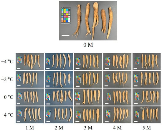
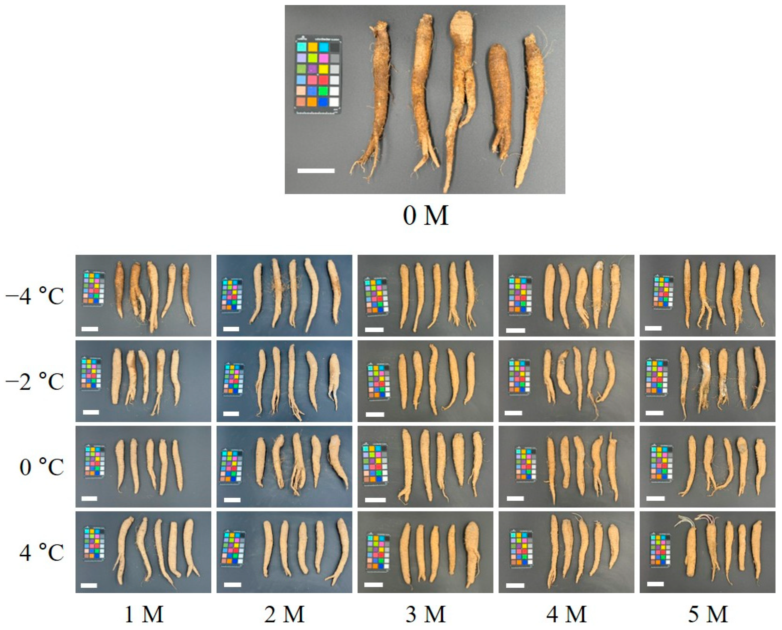
The appearance of LAR gradually changed during storage, with the extent of deterioration differing among storage temperatures (Figure 1). At −2 and 0 °C, partial decay was observed after 2 months of storage, followed by visible surface wrinkling after 3 months. At −4 °C, decay became apparent after 4 months of storage. At 4 °C, distinct shoot emergence was observed after 4 months of storage.
Figure 1.
Appearance of lance asiabell roots during 5 months of storage at different temperatures. The white scale bar at the bottom left of each image represents 5 cm.
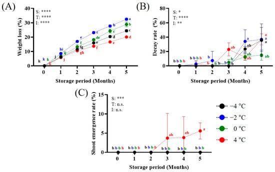
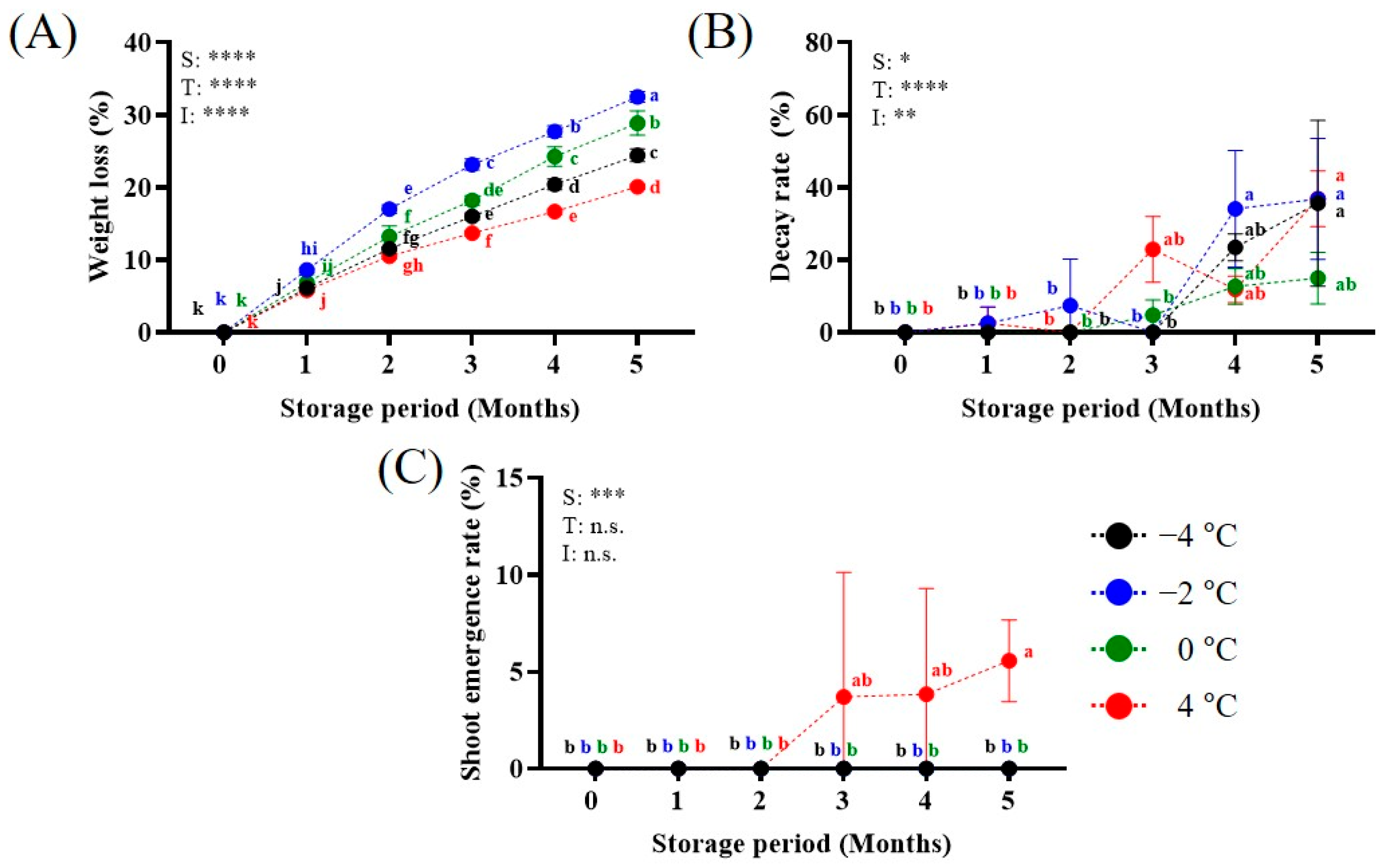
Weight loss increased continuously with storage period at all temperatures and was significantly affected by storage temperature, storage period, and their interaction (Figure 2A). After 5 months of storage, weight loss was highest at −2 °C (32.5 ± 0.8%), followed by 0 °C (28.9 ± 1.7%), −4 °C (24.4 ± 0.9%), and 4 °C (20.1 ± 0.3%). Decay was rarely observed during the early storage period but increased markedly after 3–4 months, with significant effects of storage temperature, storage period, and their interaction (Figure 2B). After 5 months of storage, the decay rate was lowest at 0 °C (15.0 ± 7.1%), while those at −4, −2, and 4 °C were approximately 36%. Shoot emergence was observed only in samples stored at 4 °C after 3 months of storage (Figure 2C).
Figure 2.
Effects of storage temperature and storage period on weight loss (A), decay rate (B), and shoot emergence rate (C) of lance asiabell roots. Different letters indicate significant differences among storage temperature × storage period combinations by Tukey’s HSD test following two-way ANOVA (p < 0.05). Asterisks indicate the effects of storage temperature (S), storage period (T), and their interaction (I) (* p < 0.05, ** p < 0.01, *** p < 0.001, and **** p < 0.0001; n.s., not significant).
3.2. Changes in Soluble Solids Content, Firmness, and Cross-Sectional Color
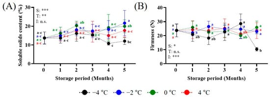
SSC showed an increasing trend during storage at −2 and 0 °C, although the changes were not statistically significant (Figure 3A). After 5 months, SSC at −2 °C (21.6%) was significantly higher than that at −4 °C (12.6%) (p < 0.05). Firmness remained relatively unchanged during storage, except at −4 °C, where it decreased to 10.3 N after 5 months (Figure 3B).
Figure 3.
Effects of storage temperature and storage period on soluble solids content (A) and firmness (B) of lance asiabell roots. Different letters indicate significant differences among storage temperature × storage period combinations by Tukey’s HSD test following two-way ANOVA (p < 0.05). Asterisks indicate the effects of storage temperature (S), storage period (T), and their interaction (I) (* p < 0.05, ** p < 0.01, *** p < 0.001; n.s., not significant).
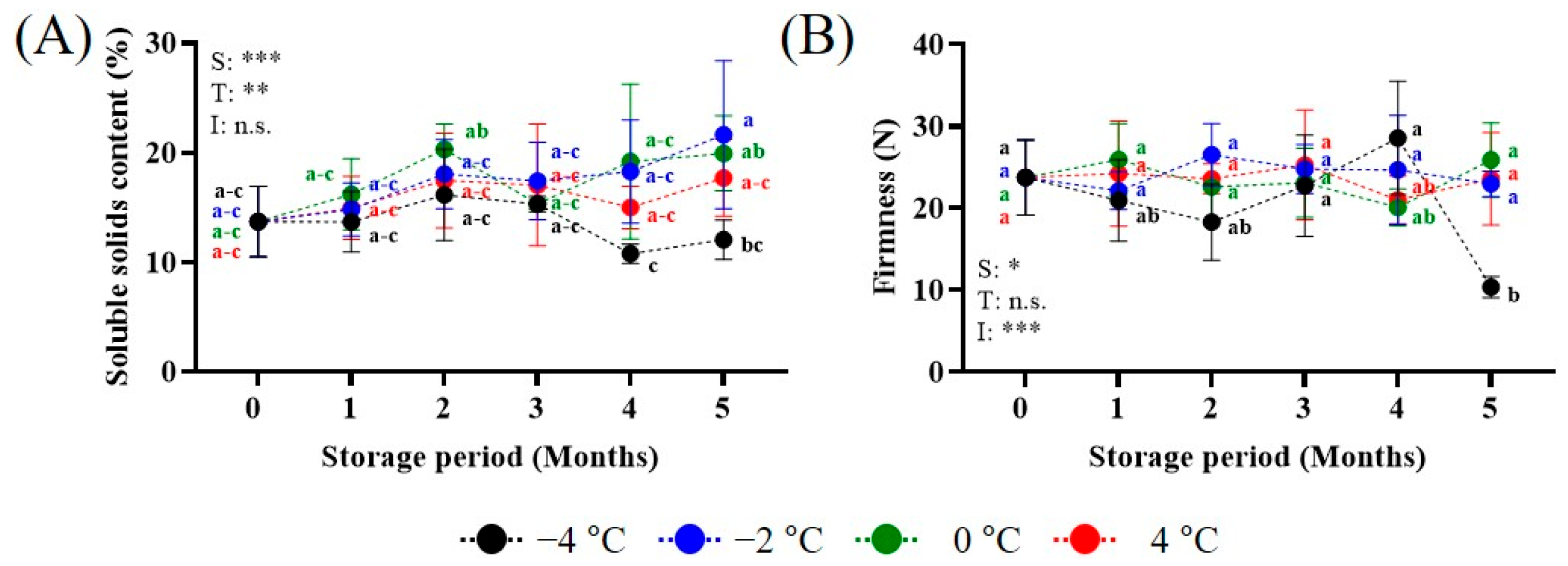
Cross-sectional L* decreased markedly at −4 °C from 1 month onward and remained generally lower than those at the other temperatures (Figure 4A). The a* value decreased during the first 2 months, particularly at −4 °C, and subsequently increased (Figure 4B). The b* value and chroma showed no significant differences among storage temperatures (Figure 4C,D). Hue angle increased during the first 2 months at −4 and −2 °C and subsequently decreased from 3 months onward (Figure 4E). Browning index increased significantly during storage at −4 and −2 °C (Figure 4F), whereas whiteness index decreased significantly only at −4 °C (Figure 4G).
Figure 4.
Effects of storage temperature and storage period on the cross-sectional color characteristics of lance asiabell roots. Different letters indicate significant differences among storage temperature × storage period combinations by Tukey’s HSD test following two-way ANOVA (p < 0.05). Asterisks indicate the effects of storage temperature (S), storage period (T), and their interaction (I) (* p < 0.05, *** p < 0.001, and **** p < 0.0001; n.s., not significant). (A) Root cross-section (B) L*; (C) a*; (D) b*; (E) chroma; (F) hue angle; (G) browning index; (H) whiteness index.
3.3. Changes in Total Phenolic Content and Antioxidant Activities
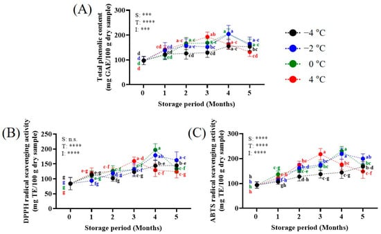
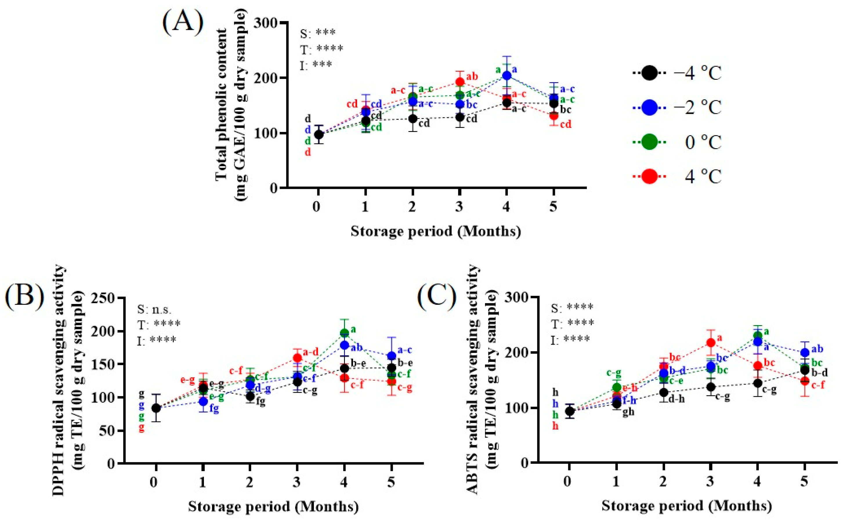
TPC generally increased during storage, although the extent of increase differed among storage temperatures (Figure 5A). The highest TPC values were observed at −2 and 0 °C after 4 months of storage (204.3 ± 34.9 and 204.5 ± 20.3 mg GAE/100 g dry sample, respectively), while TPC at 4 °C reached a maximum after 3 months (192.9 ± 19.3 mg GAE/100 g dry sample) and subsequently decreased. DPPH radical scavenging activity generally increased during storage and was highest at 0 °C after 4 months (197.0 ± 20.8 mg TE/100 g dry sample) (Figure 5B). Similarly, ABTS radical scavenging activity increased during storage, with the highest values observed at 0 and −2 °C after 4 months (219.7 ± 22.1 and 230.6 ± 17.9 mg TE/100 g dry sample, respectively) (Figure 5C).
Figure 5.
Effects of storage temperature and storage period on total phenolic content (A), DPPH radical scavenging activity (B), and ABTS radical scavenging activity (C) of lance asiabell roots. Different letters indicate significant differences among storage temperature × storage period combinations by Tukey’s HSD test following two-way ANOVA (p < 0.05). Asterisks indicate the effects of storage temperature (S), storage period (T), and their interaction (I) (*** p < 0.001, and **** p < 0.0001; n.s., not significant).
3.4. Changes in Tangshenoside I and Lobetyolin Content
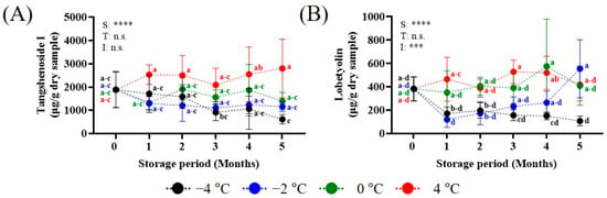
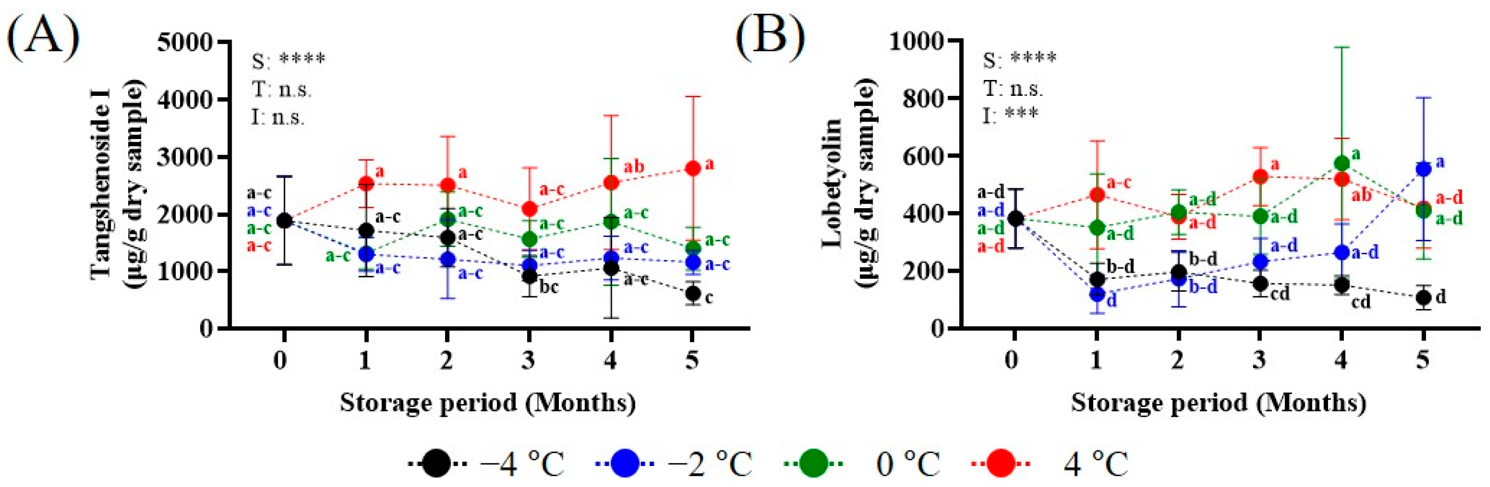
Tangshenoside I content was significantly affected by storage temperature but not by storage period or their interaction (Figure 6A). After 1 month of storage, tangshenoside I content remained relatively higher at 4 °C than at the other storage temperatures. Lobetyolin content was significantly affected by storage temperature and the interaction between storage temperature and period (Figure 6B). Lobetyolin content remained relatively higher at 0 and 4 °C than at the other storage temperatures during storage. At −2 °C, it remained relatively low for 1–4 months and increased at 5 months. At −2 °C, it remained relatively low for 1–4 months and increased at 5 months.
Figure 6.
Effects of storage temperature and storage period on major bioactive compound contents ((A), tangshenoside I; (B), lobetyolin) of lance asiabell roots. Different letters indicate significant differences among storage temperature × storage period combinations by Tukey’s HSD test following two-way ANOVA (p < 0.05). Asterisks indicate the effects of storage temperature (S), storage period (T), and their interaction (I) (*** p < 0.001, and **** p < 0.0001; n.s., not significant).
3.5. Relationships Among Quality Characteristics During Storage at Different Temperatures
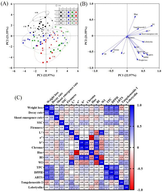
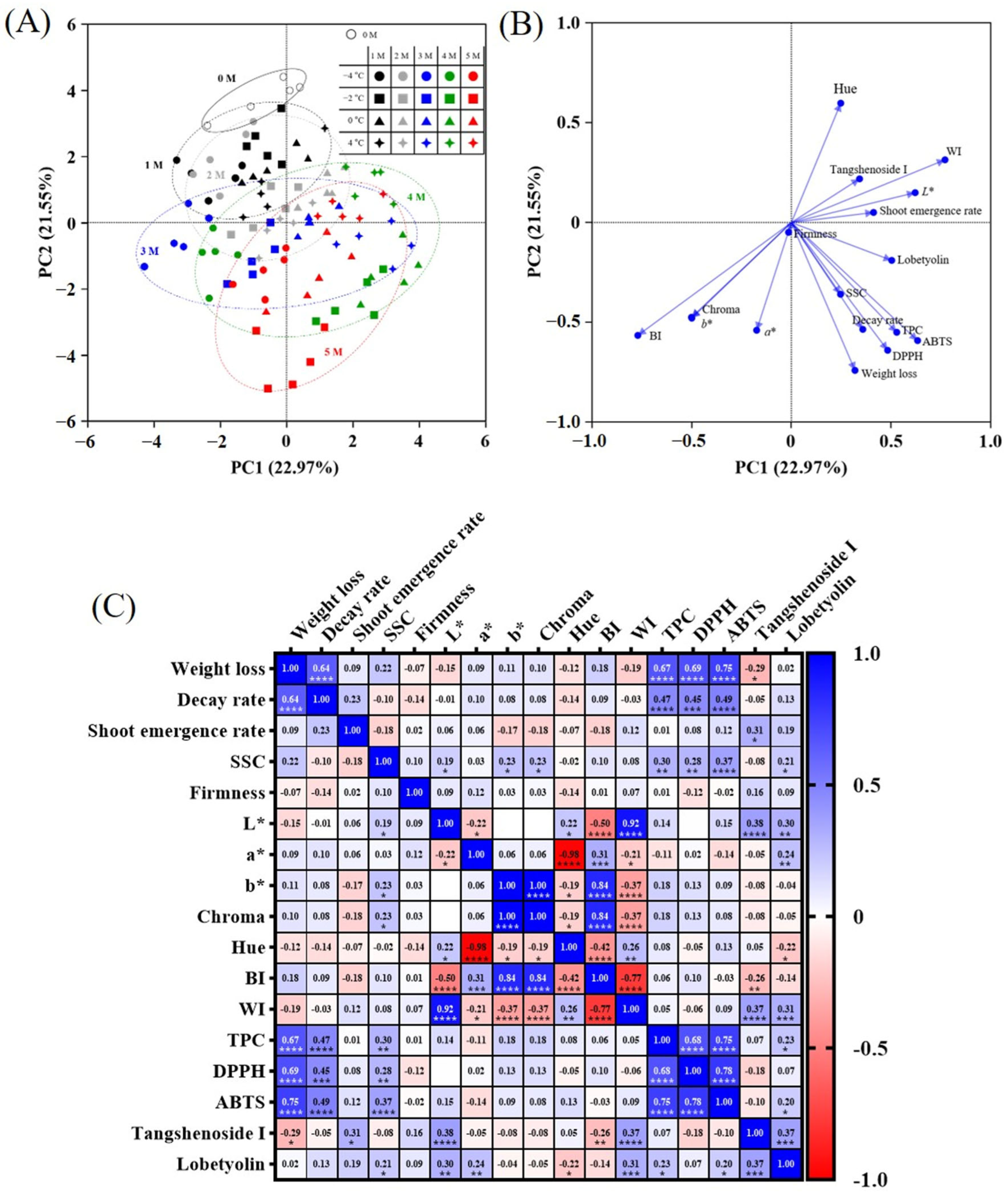
The PCA score plot showed differentiation of samples according to storage period and temperature based on PC1 (22.97%) and PC2 (21.55%). Initial samples were mainly distributed on the positive side of PC2, whereas samples from longer storage periods were generally positioned on the negative side of PC2. Samples stored at higher temperatures also tended to be distributed on the positive side of PC1 (Figure 7A). PCA loading analysis showed that weight loss and decay rate were closely associated with TPC, DPPH, and ABTS, while L*, WI, and hue were positioned in the opposite direction along PC2 (Figure 7B). Chroma, b*, and BI were also closely grouped.
Figure 7.
PCA score plot (A), PCA loading plot (B), and Pearson correlation matrix (C) of quality characteristics, antioxidant activities, and major bioactive compounds of lance asiabell roots during storage. Values in the correlation matrix represent Pearson correlation coefficients, and asterisks indicate significant correlations (* p < 0.05, ** p < 0.01, *** p < 0.001, and **** p < 0.0001). SSC, soluble solids content; L*, lightness; a*, redness; b*, yellowness; BI, browning index; WI, whiteness index; TPC, total phenolic content; DPPH, 2,2-diphenyl-1-picrylhydrazyl radical scavenging activity; ABTS, 2,2′-azinobis(3-ethylbenzothiazoline-6-sulfonic acid) radical scavenging activity.
Pearson correlation analysis supported these relationships (Figure 7C). Weight loss was positively correlated with decay rate (r = 0.64), TPC (r = 0.67), DPPH (r = 0.69), and ABTS (r = 0.75) (p < 0.0001). Decay rate was also positively correlated with TPC (r = 0.47), DPPH (r = 0.45), and ABTS (r = 0.49) (p < 0.0001). TPC was positively correlated with DPPH (r = 0.68) and ABTS (r = 0.75), while DPPH and ABTS showed a strong positive correlation (r = 0.78) (p < 0.0001). Among the color parameters, L* was strongly positively correlated with WI (r = 0.92) and negatively correlated with BI (r = −0.50), whereas BI was negatively correlated with WI (r = −0.77) (p < 0.0001).
4. Discussion
Storage temperature influenced the progression of physical deterioration in LAR, with increasing weight loss and decay accompanied by temperature-dependent changes in shoot emergence, firmness, and cross-sectional color (Figure 1 and Figure 2). Water loss during storage is generally associated with continued transpiration and respiration after harvest, while prolonged storage can increase the occurrence of tissue deterioration and decay [13,29]. Similar responses have been reported in other root crops, in which low-temperature storage reduced metabolic activity, whereas prolonged exposure to temperatures below the tolerance range of the tissue caused physiological injury [13,14]. In the present study, the decrease in firmness together with lower L* and whiteness index and higher browning index at −4 °C may be related to changes in tissue integrity during prolonged subzero storage (Figure 4A,G). Low-temperature injury in root and tuber crops has been associated with membrane damage and loss of cellular compartmentalization, which may promote contact between phenolic compounds and oxidative enzymes and contribute to tissue browning [30,31]. Recent studies in ginseng have also associated quality deterioration during cold storage with oxidative stress, membrane lipid peroxidation, and changes in lipid and phenylpropanoid metabolism [32,33]. The increase in browning index at −2 °C may also indicate that prolonged subzero storage affected internal tissue quality, although the changes were less pronounced than those observed at −4 °C (Figure 4F). By contrast, shoot emergence occurred only at 4 °C during the later storage period. Sprouting of harvested medicinal roots is strongly influenced by storage temperature and dormancy status (Figure 2C). In ginseng seedlings, lower storage temperatures suppressed sprout development during prolonged storage [22]. Bud dormancy in ginseng is also closely associated with hormonal and stress-related signaling, particularly ABA-related pathways, which regulate the transition between dormancy and active growth [34]. This may partly explain the shoot emergence observed in LAR stored at 4 °C during the later storage period.
In addition to the physical changes, TPC and antioxidant activities generally increased during the middle stage of storage, although the extent and timing of these increases differed among storage temperatures (Figure 5). In harvested plant tissues, phenolic compounds can increase in response to storage-related stresses such as water loss, low temperature, and oxidative stress [35,36]. These responses are often associated with activation of phenolic metabolism and antioxidant defense mechanisms, which may help reduce oxidative stress during storage [37,38]. Similar increases in phenolic content and antioxidant activity have been reported in root crops under postharvest stress or prolonged storage [39,40]. In the present study, the similar changes in TPC, DPPH, and ABTS suggest that the increase in antioxidant activity may have been partly associated with changes in phenolic compounds. The earlier peaks observed at 4 °C than at 0 and −2 °C may reflect a more rapid progression of storage-related metabolic and stress responses at the higher temperature [41,42]. However, TPC decreased after reaching relatively high levels at several storage temperatures. This later decline may be associated with phenolic oxidation or consumption during enzymatic browning as tissue aging and damage progressed [41,43]. A similar increase followed by a decrease in phenolic content was reported in rutabaga roots during long-term cold storage [37]. Phenolic compounds may undergo oxidation or metabolic degradation during prolonged storage, particularly as tissue senescence and cellular damage progress, and may also participate in enzymatic browning reactions [39]. These processes may partly account for the decrease in TPC observed during the later storage period.
In addition to phenolic compounds, C. lanceolata contains various bioactive constituents, including phenylpropanoids, polyacetylenes, and triterpenoid saponins [40]. Among these compounds, tangshenoside I and lobetyolin have been used as marker constituents for the quality evaluation of C. lanceolata [28,44]. In the present study, tangshenoside I remained relatively higher at 4 °C, whereas lobetyolin tended to be maintained at relatively higher levels at 0 and 4 °C than under the lower subzero conditions (Figure 6). At −2 °C, however, lobetyolin remained low during storage and increased at 5 months (Figure 6B). Changes in lobetyolin content during postharvest storage have also been reported in C. pilosula, with its retention differing among storage temperatures [41]. In addition, postharvest rubbing–sweating treatment increased lobetyolin content in C. pilosula, accompanied by changes in stress-responsive pathways [45]. Similar responses have been reported for bioactive polyacetylenes, such as falcarinol and falcarindiol-3-acetate, in carrot roots following postharvest wound-healing treatment [46].
PCA and correlation analyses indicated that the physical and biochemical changes in LAR during storage were closely related rather than occurring independently (Figure 7). Similar relationships among physical deterioration, antioxidant activity, active compounds, and physiological responses have been reported in medicinal root crops. In C. pilosula, weight loss, phenolic content, antioxidant activity, oxidative damage, and active compounds were closely associated during storage at different temperatures [41], while similar relationships between deterioration, oxidative stress, and antioxidant responses have been reported in American ginseng [32]. Multivariate approaches have also been applied to medicinal roots to examine relationships among multiple storage-related quality traits [23,41]. In the present study, the combined PCA and correlation results suggest that storage temperature influenced several interrelated aspects of LAR quality, indicating that physical deterioration, internal color, and antioxidant-related responses may be considered together when interpreting storage-induced changes.
5. Conclusions
Storage temperature markedly influenced the physical and biochemical characteristics of LAR during prolonged storage. Among the tested conditions, storage at −2 °C resulted in the greatest weight loss after 5 months, whereas 0 °C showed the lowest decay rate. Storage at −4 °C was associated with a marked decrease in firmness and greater changes in cross-sectional color, while shoot emergence occurred only at 4 °C during the later storage period. TPC and antioxidant activities generally increased during the middle stage of storage and subsequently declined, while tangshenoside I and lobetyolin showed different temperature-dependent responses. PCA and correlation analyses further indicated that physical deterioration, internal color changes, and antioxidant-related responses were interrelated during storage. Considering the overall quality changes, storage at 0 °C may provide a relatively favorable balance among the tested conditions. However, the responses of individual biochemical properties varied with storage temperature and were not always consistent with changes in overall quality. Although we did not investigate other bioactive constituents, including triterpenoid saponins, the physiological mechanisms underlying their changes, or the effects of different packaging materials, our findings provide useful insights into the relationships among physicochemical quality, antioxidant responses, bioactive compounds, and storage temperature in LAR. These results may serve as practical reference information for commercial storage and distribution of LAR, particularly when considering 4 °C as a storage temperature according to the quality attributes to be maintained within the conditions evaluated in this study.
Author Contributions
Conceptualization, N.K. and D.-S.K.; methodology, J.W.C., J.-H.L., D.-G.P., J.L., and D.-S.K.; formal analysis, S.L., Y.E.Y., Y.J.J., J.L., and D.-S.K.; investigation, N.K., J.L., S.L., Y.E.Y., and Y.J.J.; data curation, J.W.C., J.-H.L., and D.-G.P.; writing—original draft preparation, N.K., and D.-S.K.; writing—review and editing, D.-S.K.; supervision, N.K. and D.-S.K.; project administration, N.K.; funding acquisition, N.K. All authors have read and agreed to the published version of the manuscript.
Funding
This study was funded by the Cooperative Research Program for Agriculture, Science, and Technology (Project No PJ01733401) in the Rural Development Administration of the Republic of Korea.
Institutional Review Board Statement
Not applicable.
Informed Consent Statement
Not applicable.
Data Availability Statement
The datasets generated and analyzed during the current study are not publicly available due to the sensitivity of personal data but are available from the corresponding author on reasonable request.
Conflicts of Interest
The authors declare no conflicts of interest.
References
- Hossen, M.J.; Kim, M.-Y.; Kim, J.-H.; Cho, J.-Y. Codonopsis lanceolata: A Review of Its Therapeutic Potentials. Phytother. Res. 2016, 30, 347−356. [Google Scholar] [CrossRef] [Scilit]
- Sanjay; Sood, R.; Lee, H.-J. Pharmacological Insights into Codonopsis lanceolata: A Review of Its Potential in Disease Prevention and Therapy. Molecules 2026, 31, 2327. [Google Scholar] [CrossRef] [Scilit]
- Cho, Y.R.; Hong, J.-H.; Kang, D.-M.; Ko, Y.-J.; Ahn, M.-J.; Kim, K.H. Deodeokaloid, a New Indole Alkaloid N-Glycoside and Bioactive Phenolic Compounds from the Roots of Codonopsis lanceolata. Plants 2024, 13, 3243. [Google Scholar] [CrossRef] [Scilit]
- Du, Y.-E.; Lee, J.-S.; Kim, H.-M.; Ahn, J.-H.; Jung, I.-H.; Ryu, J.-H.; Choi, J.-H.; Jang, D.S. Chemical constituents of the roots of Codonopsis lanceolata. Arch. Pharm. Res. 2018, 41, 1082–1091. [Google Scholar] [CrossRef] [Scilit]
- Nam, M.; Jo, S.r.; Kim, Y.-C.; Kim, M.-S. UPLC-QTOF-MS-Based Metabolomics and Antioxidant Capacity of Codonopsis lanceolata from Different Geographical Origins. Foods 2023, 12, 267. [Google Scholar] [CrossRef] [Scilit]
- Kim, J.; Yang, W.S.; Kim, J.H.; Park, J.G.; Kim, H.G.; Ko, J.; Hong, Y.D.; Rho, H.S.; Shin, S.S.; Sung, G.-H.; et al. Lancemaside A from Codonopsis lanceolata Modulates the Inflammatory Responses Mediated by Monocytes and Macrophages. Mediat. Inflamm. 2014, 2014, 405158. [Google Scholar] [CrossRef] [Scilit]
- Lee, Y.G.; Kim, J.Y.; Lee, J.Y.; Byeon, S.E.; Hong, E.K.; Lee, J.; Rhee, M.H.; Park, H.J.; Cho, J.Y. Regulatory effects of Codonopsis lanceolata on macrophage-mediated immune responses. J. Ethnopharmacol. 2007, 112, 180−188. [Google Scholar] [CrossRef] [Scilit]
- Lee, J.S.; Kim, K.-J.; Kim, Y.-H.; Kim, D.-B.; Shin, G.-H.; Cho, J.-H.; Kim, B.K.; Lee, B.-Y.; Lee, O.-H. Codonopsis lanceolata Extract Prevents Diet-Induced Obesity in C57BL/6 Mice. Nutrients 2014, 6, 4663–4677. [Google Scholar] [CrossRef] [Scilit]
- Zhang, Y.; Wang, H.; Zhang, L.; Yuan, Y.; Yu, D. Codonopsis lanceolata polysaccharide CLPS alleviates high fat/high sucrose diet-induced insulin resistance via anti-oxidative stress. Int. J. Biol. Macromol. 2020, 145, 944−949. [Google Scholar] [CrossRef] [Scilit]
- Wang, L.; Xu, M.L.; Hu, J.H.; Rasmussen, S.K.; Wang, M.-H. Codonopsis lanceolata extract induces G0/G1 arrest and apoptosis in human colon tumor HT-29 cells—Involvement of ROS generation and polyamine depletion. Food Chem. Toxicol. 2011, 49, 149−154. [Google Scholar] [CrossRef] [Scilit]
- Boo, H.-O.; Kwom, S.-J.; Kim, H.-H. Comparison of Total Polyphenol, Total Flavonoid Content and Antioxidant Activity of Codonopsis lanceolata Extracts Stored at Different Temperatures and for Different Durations. Korean J. Community Living Sci. 2018, 29, 361−370. [Google Scholar] [CrossRef] [Scilit]
- Boo, H.-O.; Shin, J.-H.; Kim, H.-H.; Kwon, S.-J.; Lee, M.-S.; Ku, Y.-G. Evaluation of Cytotoxicity on Human Cancer Cells and Antioxidant and Immune Cell Activities of Codonopsis lanceolata Root Extracts Stored at Different Temperatures and for Different Durations. Hortic. Sci. Technol. 2019, 37, 130−139. [Google Scholar] [CrossRef] [Scilit]
- Hoffmann, T.G.; Souza, C.K.; Sonawane, A.D.; Praeger, U.; Büchele, F.; Neuwald, D.A.; Jedermann, R.; Linke, M.; Sturm, B.; Mahajan, P.V. Challenges and future perspectives in postharvest cold storage: Technological review on sustainable and efficient cold storage of fresh produce. Food Res. Int. 2025, 221, 117406. [Google Scholar] [CrossRef] [Scilit]
- Ge, W.; Zhou, Q.; Meng, Z.; Gao, J.; Chen, G.; Li, Y.; Yang, H. Breath of the chill: Transcriptomic exploration of respiration and energy metabolism in sweet potato roots during cold storage. Postharvest Biol. Technol. 2025, 224, 113483. [Google Scholar] [CrossRef] [Scilit]
- Haran, Y.; Homaida, M.A.; Elkhedir, A.E.; Chen, X.; Li, J.; Shoulei, Y.; Huang, Q. Effects of chilling injury on the physical, cooking, textural, and water mobility properties of sweet potatoes. J. Sci. Food Agric. 2025, 105, 5301–5311. [Google Scholar] [CrossRef] [Scilit]
- Gidado, M.J.; Gunny, A.A.N.; Gopinath, S.C.B.; Ali, A.; Wongs-Aree, C.; Salleh, N.H.M. Challenges of postharvest water loss in fruits: Mechanisms, influencing factors, and effective control strategies—A comprehensive review. J. Agric. Food Res. 2024, 17, 101249. [Google Scholar] [CrossRef] [Scilit]
- Kang, H.; Thomas, H.R.; Xia, X.; Shi, H.; Zhang, L.; Hong, J.; Shi, K.; Zhou, J.; Yu, J.; Zhou, Y. An integrative overview of cold response and regulatory pathways in horticultural crops. J. Integr. Plant Biol. 2025, 67, 1028−1059. [Google Scholar] [CrossRef] [Scilit]
- Jin, T.Z.; Huang, M.; Niemira, B.A.; Cheng, L. Shelf life extension of fresh ginseng roots using sanitizer washing, edible antimicrobial coating and modified atmosphere packaging. Int. J. Food Sci. Technol. 2016, 51, 2132−2139. [Google Scholar] [CrossRef] [Scilit]
- Lim, S.; Lee, E.-J.; Kim, J. Decreased sulforaphene concentration and reduced myrosinase activity of radish (Raphanus sativus L.) root during cold storage. Postharvest Biol. Technol. 2015, 100, 219−225. [Google Scholar] [CrossRef] [Scilit]
- Hu, W.Z.; Jiang, A.L.; Qi, H.P. Physiological behavior and quality of fresh ginseng stored in modified atmospheres generated by several package films. J. Food Sci. Technol. 2014, 51, 3862−3869. [Google Scholar] [CrossRef] [Scilit]
- Lee, J.H.; Choi, J.W.; Hong, Y.P.; Kim, G.S. Effects of Storage Temperature on Quality of Fresh Ginseng during Distribution. Korean J. Med. Crop Sci. 2015, 23, 431−438. [Google Scholar] [CrossRef] [Scilit]
- Chang, E.H.; Lee, J.H.; Choi, J.W.; Lim, S.; Shin, I.S. Effects of storage temperature and film treatment on sprouting and growth of ginseng seedlings. Korean J. Food Preserv. 2020, 27, 534−543. [Google Scholar] [CrossRef] [Scilit]
- Park, J.-J.; Choi, J.-H.; Park, K.-J.; Cho, J.-S.; Yun, D.-Y.; Lin, J.-H. Shelf-life prediction of fresh ginseng packaged with plastic films based on a kinetic model and multivariate accelerated shelf-life testing. Korean J. Food Preserv. 2023, 30, 573–588. [Google Scholar] [CrossRef] [Scilit]
- Kim, D.-S.; Lee, J.G.; Park, D.-G.; Lee, J.; Yun, Y.E.; Jang, Y.J.; Lim, S.; Cho, J.-H.; Lee, J.; Han, J.-W. Comparative analysis of storage quality and metabolite profiles in onion bulbs according to cultivar and cultivation region. Food Sci. Preserv. 2025, 32, 776−789. [Google Scholar] [CrossRef] [Scilit]
- Singleton, V.L.; Orthofer, R.; Lamuela-Raventós, R.M. Analysis of Total Phenols and Other Oxidation Substrates and Antioxidants by Means of Folin-Ciocalteu Reagent. Methods Enzymol. 1999, 299, 152–178. [Google Scholar] [CrossRef] [Scilit]
- Blois, M.S. Antioxidant Determinations by the Use of a Stable Free Radical. Nature 1958, 181, 1199–1200. [Google Scholar] [CrossRef] [Scilit]
- Re, R.; Pellegrini, N.; Proteggente, A.; Pannala, A.; Yang, M.; Rive-Evans, C. Antioxidant activity applying an improved ABTS radical cation decolorization assay. Free Radic. Biol. Med. 1999, 26, 1231–1237. [Google Scholar] [CrossRef] [Scilit]
- Park, Y.; Son, H.; Lee, H.; Heo, S.; Ko, J.; Park, N.I. The Analysis of Functional Components and Physiological Activities in Different Parts of Korean and Chinese Deodeok (Codonopsis lanceolata). J. Agric. Life Environ. Sci. 2023, 35, 564–572. [Google Scholar] [CrossRef]
- Zou, X.; Wang, J.; Li, Y.; Zhang, J.; Wu, Z.; Jin, P.; Cao, S.; Zheng, Y. Recent advances in high relative humidity strategy for preservation of postharvest fruits and vegetables: A comprehensive review. Food Chem. 2025, 481, 144130. [Google Scholar] [CrossRef] [Scilit]
- Cheng, J.; Zhang, X.; Miao, Z.; Wang, H.; Pang, L.; Pan, Y. Hot air treatment alleviates chilling injury of sweet potato tuberous roots by regulating osmoregulatory substances and inducing antioxidant defense system. Food Chem. 2024, 459, 140393. [Google Scholar] [CrossRef] [Scilit]
- Lin, Z.; Yao, R.; Pang, X.; Liang, H.; Li, Y.; Wu, X.; Yan, X.; Yuan, S.; Xu, X.; Jiang, A.; et al. ClO2 fumigation of yam tubers prior to storage inhibits enzymatic Browning in fresh-cut yams: Impact of ClO2 on phenylpropanoid metabolism and membrane integrity. Food Chem. 2026, 501, 147567. [Google Scholar] [CrossRef] [Scilit]
- Ren, Z.; Wang, D.; Deng, X.; Yang, X.; Jia, L. Ozone (O3) enhances the storability and antioxidant quality of fresh American ginseng. Food Qual. Saf. 2026, 10, fyaf062. [Google Scholar] [CrossRef] [Scilit]
- Qu, D.; Chen, S.; Sha, J.; Tian, R.; Cai, D.; He, Y.; Wen, L.; Peng, Y. Non-targeted metabolomics reveals the metabolic mechanisms and changes in potential marker compounds of fresh ginseng during cold storage. Future Foods 2026, 13, 101042. [Google Scholar] [CrossRef] [Scilit]
- Hong, J.; Han, S.; Geem, K.R.; Bae, W.; Kim, J.; Jee, M.-G.; Lee, J.-W.; Kim, J.-W.; Lee, G.; Joo, Y.; et al. Identification of a key signaling network regulating perennating bud dormancy in Panax ginseng. J. Ginseng Res. 2024, 48, 511−519. [Google Scholar] [CrossRef] [Scilit]
- Cao, Y.; Hu, W.; Yuan, J.; Zhang, J.; Zhu, L.; Xu, X.; Pan, S. Cyclic variable temperature conditioning-maintained storage quality of sweet potatoes by enhancing antioxidant activity. Food Biosci. 2024, 61, 104689. [Google Scholar] [CrossRef] [Scilit]
- Zhang, X.; Guo, L.; Hong, C.; Wu, P.; Tuly, J.; Ma, H. Accumulation of phenolic in fresh-cut lotus roots induced by thermosonication: Regulation of phenylpropanoid pathway and reactive oxygen species metabolism. Food Chem. 2025, 467, 142206. [Google Scholar] [CrossRef] [Scilit]
- Jakopic, J.; Veberic, R.; Slatnar, A. Changes in quality parameters in rutabaga (Brassica napus var. napobrassica) roots during long term storage. LWT-Food Sci. Technol. 2021, 147, 111587. [Google Scholar] [CrossRef] [Scilit]
- Valerga, L.; González, R.E.; Mauricci, M.; Concellón, A.; Cavagnaro, P.F. Use of UVC radiation as a postharvest stressor to increase phenolic compounds concentration and antioxidant status in purple, orange, and white carrots. Postharvest Biol. Technol. 2024, 211, 112817. [Google Scholar] [CrossRef] [Scilit]
- Maghoumi, M.; Fatchurrahman, D.; Amodio, M.L.; Quinto, M.; Cisneros-Zevallos, L.; Colelli, G. Is pomegranate husk scald during storage induced by water loss and mediated by ABA signaling? J. Sci. Food Agric. 2023, 103, 2914–2925. [Google Scholar] [CrossRef] [Scilit]
- Luo, C.; Duan, Z.; Kikuchi, T.; Otsuki, K.; Zhang, M.; Kambayashi, R.; Ito, K.; Sugiyama, A.; Li, W. Comprehensive Chemical Analysis of Codonopsis lanceolata Roots Using Ultra-High-Performance Liquid Chromatography–Quadrupole-Exactive–Orbitrap Mass Spectrometry. Chemistry 2025, 7, 4. [Google Scholar] [CrossRef] [Scilit]
- Hwang, B.S.; Kim, J.Y.; Jang, M.; Kim, G.-C.; Park, Y.-H.; Hwang, I.G. Quantitative Analysis of Tangshenoside I and Lobetyolin from Korean Deoduk (Codonopsis lanceolata). Korean J. Food Nutr. 2018, 31, 957−963. [Google Scholar] [CrossRef]
- Fugate, K.K.; Eide, J.D.; Lafta, A.M.; Tehseen, M.M.; Chu, C.; Khan, M.F.R.; Finger, F.L. Transcriptomic and metabolomic changes in postharvest sugarbeet roots reveal widespread metabolic changes in storage and identify genes potentially responsible for respiratory sucrose loss. Front. Plant Sci. 2024, 15, 1320705. [Google Scholar] [CrossRef] [Scilit]
- Tilley, A.; McHenry, M.P.; McHenry, J.A.; Solah, V.; Bayliss, K. Enzymatic browning: The role of substrates in polyphenol oxidase mediated browning. Curr. Res. Food Sci. 2023, 7, 100623. [Google Scholar] [CrossRef] [Scilit]
- Wen, L.; Wang, Y.; Song, P.; Wang, Z.; Tang, Z.; Guo, Y.; Yu, H.; Hu, F. Exploration of Postharvest Conditions for Codonopsis pilosula Nannf. var. modesta (Nannf.) L. T. Shen Roots Based on Sensory Quality, Active Components, Antioxidant Capacity and Physiological Changes at Different Storage Temperatures. Foods 2023, 12, 4418. [Google Scholar] [CrossRef] [Scilit]
- Liang, W.; Bai, G.; Sun, J.; Tao, W.; Li, Q.; Dong, P.; Wang, H.; Cheng, J.; Guo, F.; Chen, Y. Effects of post-harvest ‘rubbing-sweating ‘drying treatment on the accumulation of bioactive compounds in Codonopsis pilosula: A transcriptomic analysis. Front. Plant Sci. 2025, 16, 1650787. [Google Scholar] [CrossRef] [Scilit]
- Heltoft, P.; Trop, T.; Wold, A.-B.; Edelenbos, M.; Thomsen, M.G. Effect of temperatures strategies in the early storage period on weightloss, respiration rate and disease development in carrot (Daucus carota subsp. sativus). Postharvest Biol. Technol. 2026, 231, 113911. [Google Scholar] [CrossRef] [Scilit]
Disclaimer/Publisher’s Note: The statements, opinions and data contained in all publications are solely those of the individual author(s) and contributor(s) and not of MDPI and/or the editor(s). MDPI and/or the editor(s) disclaim responsibility for any injury to people or property resulting from any ideas, methods, instructions or products referred to in the content. |
© 2026 by the authors. Licensee MDPI, Basel, Switzerland. This article is an open access article distributed under the terms and conditions of the Creative Commons Attribution (CC BY) license.