


Abstract
Powdery mildew is one of the major diseases of peach. To clarify the relationships between peach powdery mildew resistance and leaf structure and defensive enzyme activities, this study used the resistant genotype ‘Zhou Xing Shan Tao’ (Prunus davidiana) and the susceptible genotype ‘Shache Lürou No.3’ (Prunus ferganensis) as a control. Resistance was evaluated through artificial inoculation with powdery mildew conidia (Podosphaera tridactyla). Key indicators including leaf wax content, stomatal characteristics, leaf transverse structure, and the activities of three defense enzymes were assessed and the relationship between these traits and resistance was analyzed. The results showed that ‘Zhou Xing Shan Tao’ exhibited stronger resistance to powdery mildew than ‘Shache Lürou No.3’. ‘Zhou Xing Shan Tao’ leaves exhibited a significantly higher (p < 0.01) wax content and a significantly lower stomatal density relative to ‘Shache Lürou No.3’. However, the size of individual stomata (long diameter, short diameter, circumference, area) did not differ significantly between the two genotypes. Microstructurally, ‘Zhou Xing Shan Tao’ leaves had significantly greater fenestrated tissue, lower epidermis, palisade cell layers, and overall leaf thickness. Its leaf tissue structure was also significantly more compact, with a higher compactness-to-openness ratio, and significantly less cellular laxity than ‘Shache Lürou No.3’. Following inoculation with powdery mildew, ‘Zhou Xing Shan Tao’ leaves accumulated hydrogen peroxide (H2O2) and malondialdehyde (MDA) more rapidly and to a greater extent. The activities of the superoxide dismutase (SOD), peroxidase (POD), and catalase (CAT) defense enzymes also increased faster and were significantly higher than in ‘Shache Lürou No.3’. In conclusion, leaf structural traits such as wax content and stomatal density combined with pathogenicity assessments serve as effective phenotypic indicators for evaluating powdery mildew resistance in Prunus species.
Keywords:
peach; powdery mildew; leaf structure; enzyme activity; resistance; Podosphaera tridactyla 1. Introduction
Peach (Prunus persica L.) is an important economic fruit tree in China. Peach constitutes a significant source of income for fruit growers, while playing a vital role in promoting regional economic development. However, peach trees are susceptible to various diseases during their growth, among which peach powdery mildew is particularly prevalent and causes severe damage during the seedling stage. This disease primarily infects tender tissues such as leaves and fruits, severely impairing tree growth and development as well as fruit appearance and quality, thereby leading to economic losses. This disease is primarily caused by infections from Podosphaera pannosa and Podosphaera tridactyla [1]. Previously considered a single species, P. tridactyla is now considered a species complex with multiple members [2]. Currently, the control of peach powdery mildew relies mainly on chemical agents. However, the heavy application of fungicides can readily induce fungicide resistance in the powdery mildew pathogen, potentially triggering new disease problems. Therefore, breeding for peach varieties resistant to powdery mildew represents a crucial approach for fundamentally controlling this disease.
Over long-term evolution, plants have developed a complex and sophisticated defense system to counter pathogen invasion. This system relies not only on fundamental physical structural barriers but also involves highly regulated biochemical reaction networks. As organs directly exposed to the external environment, leaves typically develop various stress-resistant adaptive morphologies. Among these, leaf wax content serves as the outermost physical defense [3,4], in some cases hindering pathogen spore adhesion, germination, and penetration [5]. Meanwhile, leaf stomatal density, morphology, and aperture may directly regulate pathogen entry points [6]. Their structural characteristics (such as density and size) [7] and closure dynamics constitute key morphological indicators of disease resistance. Internal cellular structures within leaves, such as the compactness of epidermal cell arrangement and the thickness of the lower epidermis, form an internal physical barrier that resists pathogen expansion, thereby enhancing plant disease resistance. However, the plant’s disease resistance mechanisms extend beyond physical isolation. When pathogens successfully breach these physical barriers, plants activate their intrinsic physiological and biochemical defense mechanisms [8,9], manifested by significant changes in the activity of defense enzymes. This includes enhanced activity of defense enzymes such as peroxidase (POD) and catalase (CAT) [10,11,12], along with the burst of reactive oxygen species (ROS) [13], thereby inhibiting further colonization and spread of pathogens. Currently, few studies have reported on the relationship between peach leaf structure, defense enzyme activity, and powdery mildew resistance.
As the center of origin of the peach and several of its wild relatives, China possesses the world’s most abundant Prunus germplasm resources [14,15]. Among these, Prunus davidiana and Prunus ferganensis are wild relatives of the peach [14], sharing a very close genetic relationship with it [16]. During 2022 and 2023, while investigating peach germplasm resources in Kashgar, Xinjiang, our research group observed that P. davidiana exhibited resistance to powdery mildew, while P. ferganensis was identified as susceptible. Therefore, this study used ‘Zhou Xing Shan Tao’ (P. davidiana) and ‘Shache Lürou No.3’ (P. ferganensis) as experimental materials to analyze their leaf structure, along with their physiological and biochemical changes at different time points after inoculation with powdery mildew. This study lays the foundation for understanding the relationship between leaf structure, defense enzyme activity, and disease resistance, as well as for advancing powdery mildew resistance research and variety breeding in peaches.
2. Materials and Methods
2.1. Plant and Fungal Material
The susceptible ‘Shache Lürou No.3’ (P. ferganensis) germplasm [17] was obtained from Kashgar region, Xinjiang, and disease resistant ‘Zhou Xing Shan Tao’ (P. davidiana) germplasm was obtained from the National Horticultural Germplasm Resource Center, Chinese Academy of Agricultural Sciences (CAAS). In November 2024, stratification treatments were conducted in this laboratory. In January 2025, the two genotypes were transplanted to the group’s smart greenhouse for subsequent experiments.
Podosphaera tridactyla was collected from cultures maintained on plants in a growth chamber, and species-level identification was confirmed by rDNA-ITS sequencing using primers ITS1 and ITS4. The resulting sequence (567 bp. GenBank accession no. PX588431) shared 100% similarity with sequences of P. tridactyla reported in GenBank (accession no. MH271213.1, OP935133.1).
2.2. Inoculation and Disease Assessment
Fresh P. tridactyla conidia were collected and placed in a 6.0% glucose solution at a concentration of ~1 × 10⁷ spores/mL. One-year-old seedlings with uniform growth were selected and placed in the greenhouse (temperature 25–26 °C, relative humidity 60–80%). Prior to inoculation, the peach plants were sprayed with sterile water until runoff and then air-dried. Uniform foliar spraying was performed on the leaves of ‘Shache Lürou No.3’ and ‘Zhou Xing Shan Tao’ seedlings between 19:00 and 20:00 applying about 7 mL of spore suspension per plant. Each treatment group included five biological replicates. Disease was assessed at 17 days post inoculation (dpi). A Disease Index was developed following Cao et al. [18], with some modifications. The disease rating scale was defined as: grade 0 (no infection), grade 1 (0.1–5.0% of total leaves infected), grade 3 (5.1–10.0% of total leaves infected), grade 5 (10.1–20.0% of total leaves infected), grade 7 (20.1–40.0% of total leaves infected), and grade 9 (>40.1% of total leaves infected). The Disease Index (DI) was calculated as 100% × [∑(Number of leaves in each grade × value of the grade)]/(Total number of leaves surveyed × value of the highest grade).
2.3. Leaf Wax Content
Chloroform extraction was employed to quantify leaf wax content [19]. Individual plants exhibiting consistent growth were selected from each species, and leaves were collected from the middle section of their branches. Fresh leaf samples (approximately 2.0 g) were cut into pieces and transferred into 50 mL centrifuge tubes. Following the addition of 40 mL chloroform, samples were allowed to stand for 1 min before filtration into glass Petri dishes. The Petri dishes were then incubated in a fume hood until complete evaporation of chloroform. Post-evaporation, samples were reweighed to determine wax content, expressed as milligrams per gram of fresh weight. This procedure was replicated five times for each species.
2.4. Leaf Stomatal Observation
Stomatal characteristics were observed on the leaf underside using the nail polish film method [20]. For each species, ten fields of view were randomly selected, and stomatal counts were recorded in each field (area: 435.2 μm × 326.4 μm, 200× magnification). Stomatal density was calculated based on these counts. Subsequently, five stomata were randomly selected per field of view as measurement targets, totaling 25 stomata per species. Morphometric analyses, including stomatal length diameter, width diameter, density, circumference, and area, were performed using CaseViewer 2.3 software. Calculations were executed according to the following equations [21]:
Stomatal density = number of stomata/area of field of view;
Stomatal area = π × (stomatal width/2) × (stomatal length/2);
Stomatal perimeter = 1.57 × [1.5 × (stomatal width + stomatal length) − stomatal length + stomatal width].
2.5. Leaf Anatomical Structure
The apical penultimate fifth to sixth healthy leaves of the two peach genotypes were selected, and the surface of the leaves was wiped clean with a degreasing cotton pad, retaining a 4 cm length in the middle of the leaf. The leaf section was immediately put into 50% or 70% FAA fixative and placed in darkness for fixation. After fixation, the leaf blades were sent to Wuhan Servicebio Technology Co., Ltd. (Wuhan, China) for the production of electron microscope scanning sections (Regulus 8100, Hitachi High-Tech Corporation, Tokyo, Japan), and 30 replicates were performed for each species. The leaf and internal tissue thickness was measured using the software CaseViewer 2.3 [22], and leaf tissue structural tightness (CTR), cellular structural sparsity (SR), and palisade-spongy ratio (PS) were calculated according to the following equation [23]:
CTR = Palisade mesophyll thickness/Leaf thickness;
PS = Palisade mesophyll thickness/Spongy mesophyll thickness;
SR = Spongy mesophyll thickness/Leaf thickness.
2.6. Physiological and Biochemical Measurements
One-year-old peach seedlings of cultivars ‘Shache Lürou No.3’ and ‘Zhou Xing Shan Tao’ were inoculated with conidia of P. tridactyla as described in Section 2.2 above. Following inoculation, plants were grown under natural sunlight conditions in a smart glasshouse. Each treatment comprised 15 plants and was replicated three times. At 0 h, 0.5 days, 1 day, 5 days, 10 days, and 21 days post-treatment, leaf samples were collected and immediately frozen in liquid nitrogen for subsequent preservation. Key physiological indicators, including antioxidant substances and defense enzyme activities, were measured at each time point. The activities of CAT, POD, and SOD, as well as the content of MDA and H2O2, were quantified using commercial assay kits from Jiangsu ADS Biotechnology Co., Ltd. (Yancheng, China) (catalog numbers: ADS-W-KY002, ADS-W-KY003, ADS-W-KY011, ADS-W-YH002, and ADS-W-YH015, respectively). Calculations were performed according to standard protocols derived from these kits.
2.7. Statistical Analysis
All collected data were analyzed using SPSS Statistics 27.0 software (IBM, Armonk, NY, USA). After verifying variance homogeneity with Levene’s test (p > 0.05), statistical analyses were performed using one-way ANOVA coupled with Tukey’s multiple range test, or Student’s t-test for comparisons between two groups.
3. Results
3.1. Resistance Identification
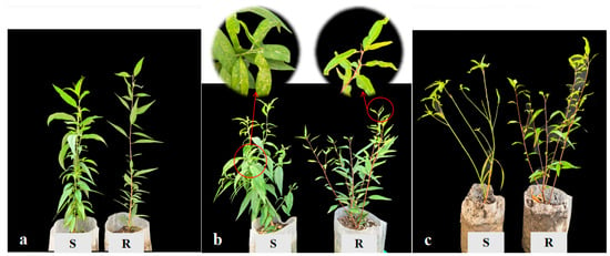
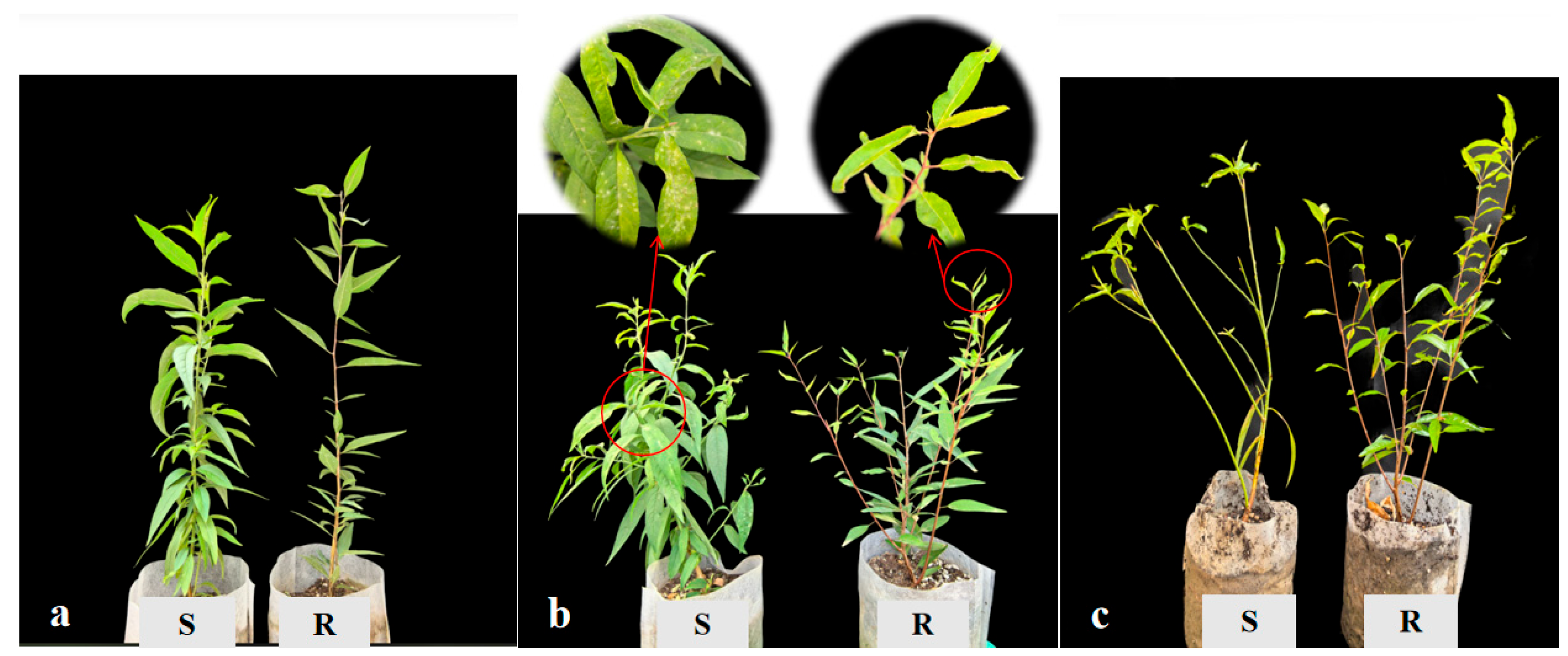
Following 24 days of inoculation, leaves of ‘Zhou Xing Shan Tao’ exhibited minimal powdery mildew signs on the leaf blades, characterized by small and discrete colony areas, indicating a high level of disease resistance. In contrast, leaves of ‘Shache Lürou No.3’ showed signs of fungal infection as early as 17 days after inoculation, with powdery mildew covering approximately 20% of the total leaf area (Figure 1). Compared to ‘Zhou Xing Shan Tao’, the colony areas on ‘Shache Lürou No.3’ were significantly larger. The disease index revealed that ‘Zhou Xing Shan Tao’ showed resistance (DI = 1.61 ± 0.11%), whereas ‘Shache Lürou No.3’ showed susceptibility (DI = 21.79 ± 1.78%). By the late infection stage (31 dpi), susceptible plants showed extensive leaf loss, a phenotype demonstrating weak pathogen resistance (Figure 1).
Figure 1.
Comparison of disease phenotypes in two peach genotypes after inoculation with powdery mildew. (a) Pre-treatment; (b) 24 days post inoculation; (c) 31 days post inoculation. Susceptible germplasm (S) ‘Shache Lürou No.3’. Resistant germplasm (R) ‘Zhou Xing Shan Tao’.
3.2. Comparison of Wax Content and Stomatal Structure
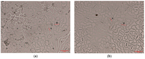
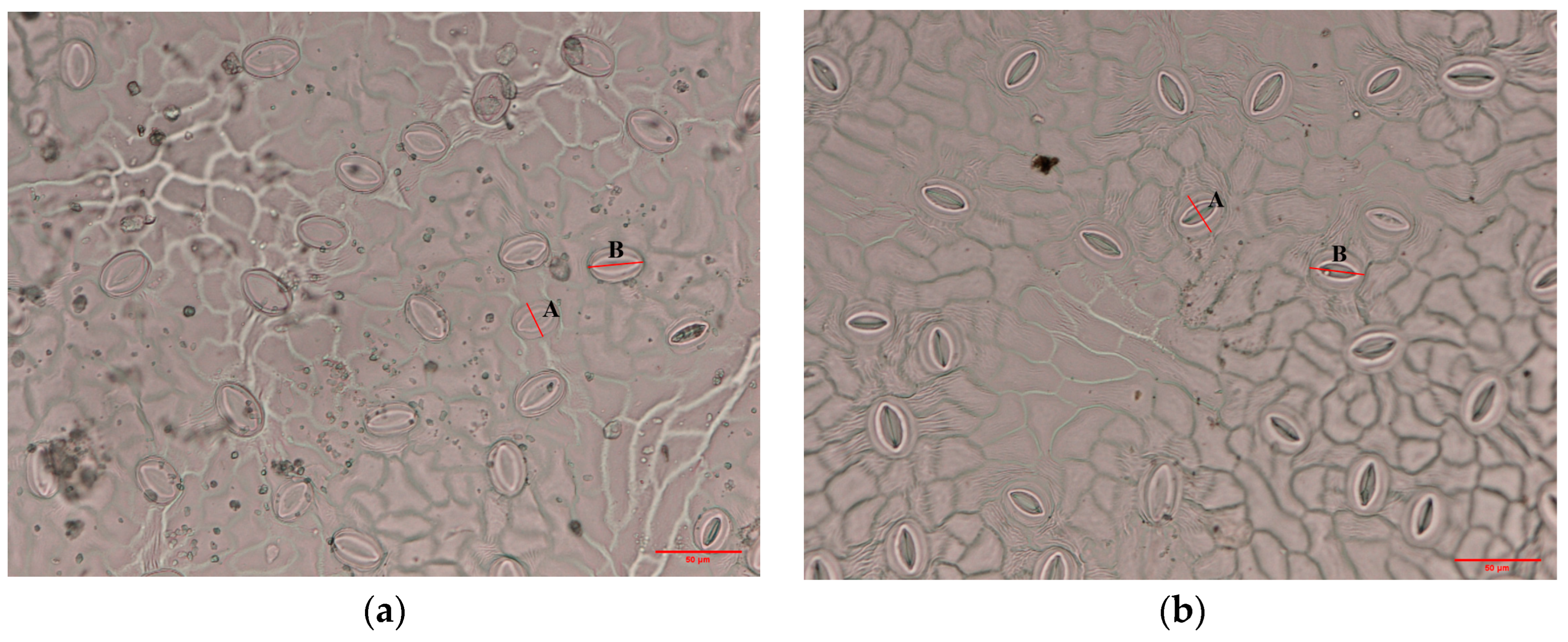
The microstructural characteristics of leaf stomata in the two accessions with distinct disease resistance were analyzed using microscopic observations (Figure 2). The results indicated that stomata in peach leaves were predominantly localized in the lower epidermis and composed of two kidney-shaped guard cells, which predominantly exhibited ovoid or elongated ovoid morphology.
Figure 2.
Microscopic images of the lower epidermal stomata from leaves of two peach genotypes differing in resistance to powdery mildew. (a) Resistant ‘Zhou Xing Shan Tao’; (b) Susceptible ‘Shache Lürou No.3’. A. Stomatal width; B. Stomatal length (scale bars: 50 μm).
Comparisons of leaf wax content and stomatal structure between the two genotypes are shown in Table 1. Leaf wax content varied significantly between the genotypes, with ‘Zhou Xing Shan Tao’ and ‘Shache Lürou No.3’ displaying values of 80.92 ± 4.81 mg/g and 55.33 ± 10.17 mg/g, respectively. Notably, ‘Zhou Xing Shan Tao’ exhibited a highly significantly higher wax content compared to ‘Shache Lürou No.3’ (p < 0.01). In terms of stomatal density, ‘Zhou Xing Shan Tao’ had a value of 136.8 ± 31.86 stomata/mm2, which was highly significantly lower than that of ‘Shache Lürou No.3’ (262.5 ± 23.77 stomata/mm2). However, no significant differences were observed between ‘Zhou Xing Shan Tao’ and ‘Shache Lürou No.3’ in stomatal length diameter, short diameter, circumference, or area.
Table 1.
Comparison of wax content and stomatal structure in leaves of two peach genotypes differing in resistance to powdery mildew (‘Zhou Xing Shan Tao’ = resistant, ‘Shache Lürou No.3’ = susceptible).
3.3. Features of Leaf Microstructure
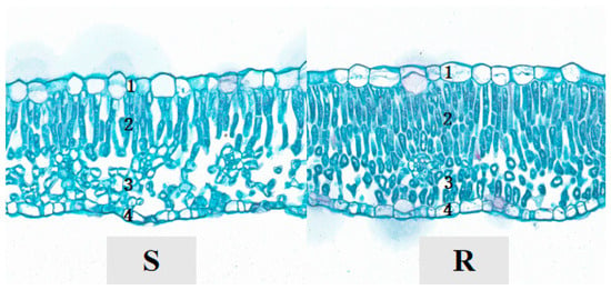
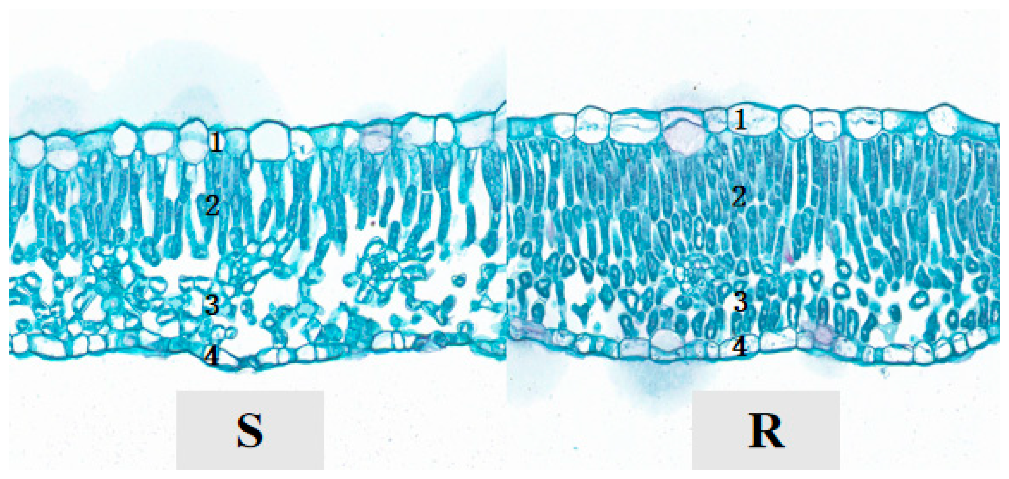
Comparative analysis of leaf cross-sections between ‘Zhou Xing Shan Tao’ and ‘Shache Lürou No.3’ revealed that both genotypes possess single-layered, irregularly shaped epidermal cells (Figure 3). Both exhibit 2–3 layers of palisade parenchyma and 3–4 layers of spongy parenchyma. However, in ‘Zhou Xing Shan Tao’, the palisade and spongy parenchyma demonstrate a more compact and orderly arrangement, whereas in ‘Shache Lürou No.3’, these tissues exhibit increased intercellular spaces and a looser cellular organization.
Figure 3.
Cross-sectional microscopic images of leaves from two peach genotypes differing in resistance to powdery mildew. 1. Upper epidermis; 2. Palisade tissue; 3. Spongy tissue; 4. Lower epidermis. Susceptible genotype (S) ‘Shache Lürou No.3’; Resistant genotype (R) ‘Zhou Xing Shan Tao’.
‘Zhou Xing Shan Tao’ exhibited significantly greater thickness in palisade tissue, lower epidermis, and overall leaf thickness compared to ‘Shache Lürou No.3’ (Table 2). Regarding CTR and PS, the resistant genotype demonstrated markedly higher values than the susceptible germplasm (p < 0.01), whereas SR in the resistant genotype was highly significantly lower than in susceptible germplasm. These structural differences suggest that the resistant ‘Zhou Xing Shan Tao’ possesses a more compact leaf tissue organization, tighter cellular arrangement, higher palisade-to-spongy tissue ratio, thicker palisade parenchyma, and thicker lower epidermis.
Table 2.
Comparison of leaf microstructure of two peach genotypes differing in resistance to powdery mildew (‘Zhou Xing Shan Tao’ = resistant, ‘Shache Lürou No.3’ = susceptible).
3.4. Effect of Powdery Mildew Infection on H2O2 and MDA Content
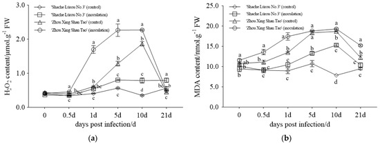
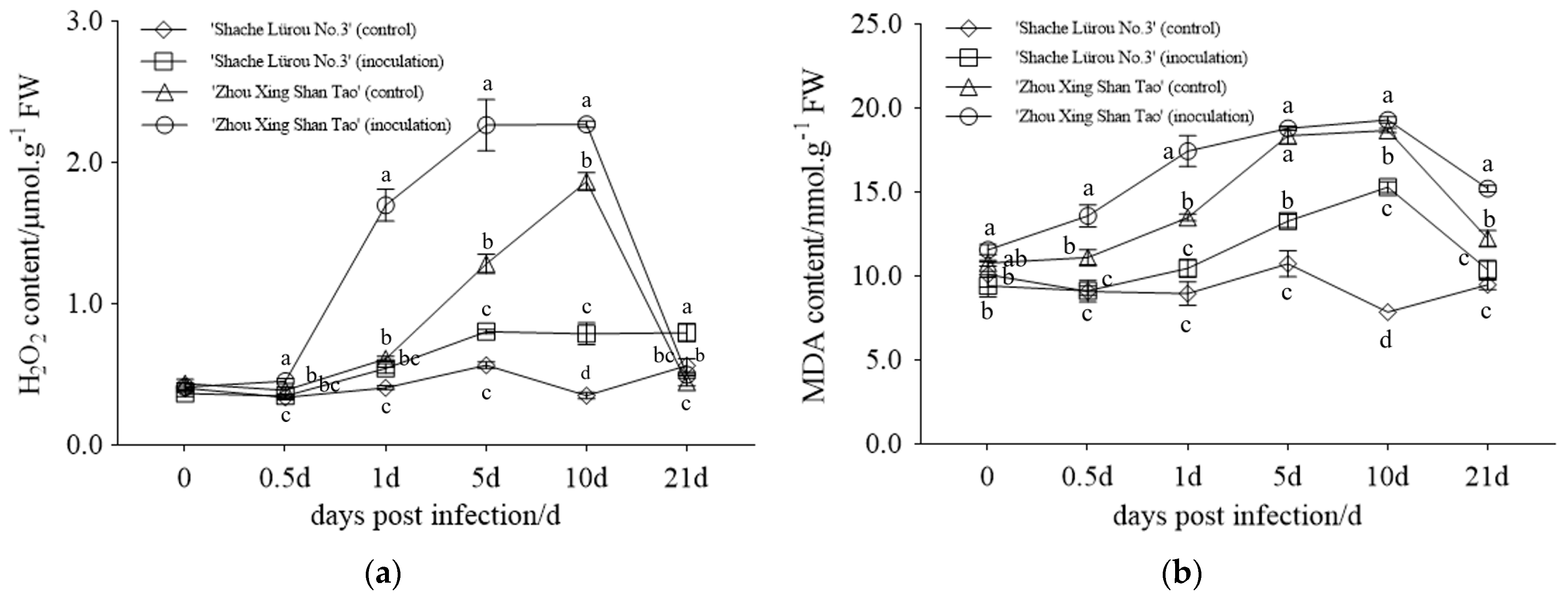
As shown in Figure 4a, the H2O2 content in peach seedling leaves of ‘Zhou Xing Shan Tao’ (water-treated and inoculated groups) exhibited an initial increase followed by a subsequent decline with prolonged post-treatment duration. The highest H2O2 accumulation occurred at 5 and 10 dpi, reaching 2.27 ± 0.18 nmol·g−1 and 2.27 ± 0.02 nmol·g−1, respectively, representing increases of 77.34% and 21.72% compared to the water treatment. In contrast, the H2O2 content in leaves of ‘Shache Lürou No.3’ seedlings under inoculation treatment showed a gradual upward trend, with peak accumulations of 0.80 ± 0.01 μmol·g−1 (at 5 days) and 0.80 ± 0.06 μmol·g−1 (at 21 days), corresponding to 40.35% and 42.86% increases relative to the water treatment group. Comparative analysis among the two genotypes revealed that, during 0.5–10 dpi, the H2O2 content in ‘Zhou Xing Shan Tao’ was significantly higher than that in ‘Shache Lürou No.3’ (p < 0.05), and the magnitude of H2O2 elevation was greater in ‘Zhou Xing Shan Tao’.
Figure 4.
The effect of powdery mildew infection on hydrogen peroxide (H2O2) and malondialdehyde (MDA) content of two peach genotypes differing in resistance to powdery mildew (‘Zhou Xing Shan Tao’ = resistant, ‘Shache Lürou No.3’ = susceptible). (a) H2O2 content; (b) MDA content. Intergroup differences were analyzed by one-way ANOVA with Tukey’s multiple range test for multiple comparisons. Error bars represent the standard deviation (SD), n = 3. Lowercase letters indicate significant differences at p < 0.05.
The MDA content in the two genotypes under both water treatment and inoculation treatment exhibited a trend of initial increase followed by a decrease with increasing inoculation time (Figure 5b). Between 0 and 21 days, the MDA content in inoculated ‘Zhou Xing Shan Tao’ remained significantly higher than that in ‘Shache Lürou No.3’ (p < 0.05). The maximum MDA content in ‘Zhou Xing Shan Tao’ was 19.30 nmol·g−1 at 10 dpi, while in ‘Shache Lürou No.3’ it peaked at 15.29 nmol·g−1 at the same time point. The MDA content in the disease-resistant genotype was 1.26 times that of the disease-susceptible genotype. These results indicate that ‘Zhou Xing Shan Tao’ exhibits a more intense oxidative stress response to pathogen challenge compared to ‘Shache Lürou No.3’.
Figure 5.
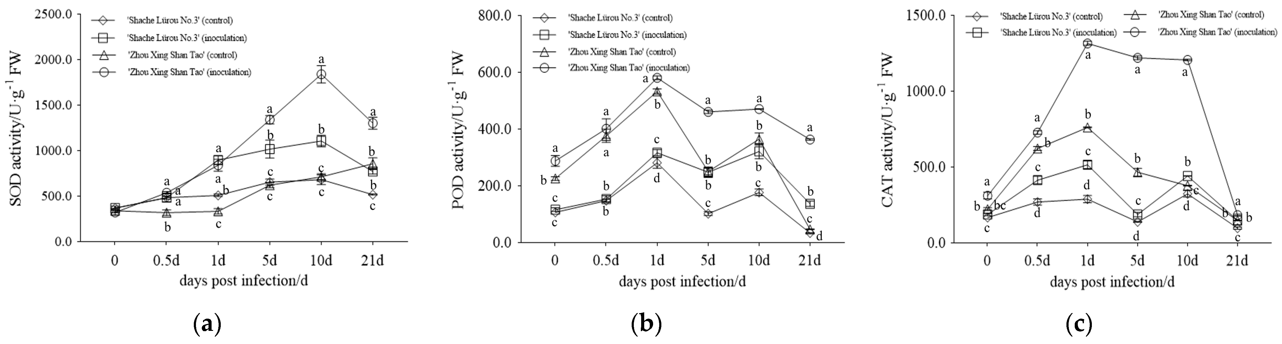
Effect of powdery mildew infection on superoxide dismutase (SOD), peroxidase (POD), and catalase (CAT) activity of two peach genotypes differing in resistance to powdery mildew (‘Zhou Xing Shan Tao’ = resistant, ‘Shache Lürou No.3’ = susceptible). (a) SOD activity; (b) POD activity; (c) CAT activity. Intergroup differences were analyzed by one-way ANOVA with Tukey’s multiple range test for multiple comparisons. Error bars represent the standard deviation (SD) of the mean, n = 3. Lowercase letters indicate significant differences at p < 0.05.
3.5. Effect of Powdery Mildew Infection on Defense-Related Enzyme Activity
The metabolic reactions of defense-related enzymes are closely associated with plant disease resistance. In this study, the physiological and biochemical responses of ‘Zhou Xing Shan Tao’ and ‘Shache Lürou No.3’ were evaluated in both inoculation and water control groups (Figure 5). Following inoculation, SOD activity in ‘Zhou Xing Shan Tao’ demonstrated an initial increase followed by a decline, with significantly higher levels observed compared to the susceptible genotype between 5 and 21 dpi (p < 0.05). The maximum SOD activity reached 1839.85 U/g on day 10, representing an increase of 733.05 U/g over ‘Shache Lürou No.3’. As a whole, POD showed an overall trend of increasing, then decreasing, then increasing and then decreasing in the resistant genotype after inoculation with powdery mildew.
At 0.5–21 days, the enzyme activity of POD in ‘Zhou Xing Shan Tao’ was significantly higher than that in the susceptible genotype (p < 0.05), and it reached a maximum value of 580.82 U/g at 1 day, which was 263.90 U/g higher than that of the susceptible genotype. When inoculated with powdery mildew, the CAT enzyme activity of the disease-resistant genotype showed a trend of increasing and then decreasing, and that of susceptible materials showed a trend of increasing and then decreasing, then increasing and then decreasing. The CAT enzyme activity of ‘Zhou Xing Shan Tao’ was significantly higher than that of the susceptible genotype from 0 to 21 days (p < 0.05), and it reached a maximum of 1316.14 U/g in 1 day of ‘Zhou Xing Shan Tao’, which was higher than that of the susceptible ‘Shache Lürou No.3’ by 800.41 U/g.
Overall, it can be seen that when seedlings of the resistant ‘Zhou Xing Shan Tao’ were inoculated with the powdery mildew fungus, the changes in SOD, POD and CAT defense enzyme activity responses were significantly higher than those of ‘Shache Lürou No.3’, which showed a correlation with varietal resistance.
4. Discussion
4.1. Peach Leaf Structure in Relation to Peach Powdery Mildew Resistance
As the first physical line of defense against the invasion of pathogenic fungi, leaf wax and stomata play a key role in plant immunity [7,24]. Leaf wax can effectively prevent pathogen spores from adhering to and penetrating the leaf surface. A previous study showed that the leaf wax content of different bayberry genotypes was positively correlated with its resistance to twig dieback, and the higher the wax content, the stronger the disease resistance [25]. In another study, Cao et al. demonstrated that the leaf wax content was significantly greater in powdery mildew-resistant pear varieties than in susceptible ones [3].
In addition, the density, morphology, and opening degree of leaf stomata directly regulate the pathways for pathogen invasion. Yang et al. showed that walnut leaf stomatal length and stomatal area were correlated with resistance to walnut blight, which was not the case for leaf stomatal width and stomatal density, while Wang et al. demonstrated that stomatal density and epidermal wax content are positively associated with resistance against citrus canker [26,27]. It can thus be seen that the relationship between leaf stomatal characteristics and disease resistance varied, which may be related to the invasion mode of different types of pathogens. In this study, through the determination of the wax content of ‘Zhou Xing Shan Tao’ and ‘Shache Lürou No.3’ and the observation of leaf stomatal characteristics, it was found that the leaf wax content of ‘Zhou Xing Shan Tao’ was significantly higher than that of ‘Shache Lürou No.3’, and the number of stomata of ‘Shache Lürou No.3’ was significantly larger than that of ‘Zhou Xing Shan Tao’. The study thus revealed that these two leaf traits are associated with resistance to peach powdery mildew, consistent with the findings of Wang et al. [27]. Therefore, leaf wax content and stomatal density can be used as reference indexes for peach powdery mildew resistance.
It has been shown that leaf thickness [28], cellular compactness, and the alignment of mesophyll tissue contribute to resistance against pathogen infection and invasion, serving as a secondary physical barrier in plant innate defense mechanisms. For instance, studies on oil tea identified significant correlations between anthracnose resistance and specific structural traits, including spongy mesophyll thickness, palisade-to-spongy tissue ratio, cellular laxity, and lower epidermal thickness [29]. Similarly, correlations were observed between melon leaf structural compactness and resistance to powdery mildew [30]. While these findings collectively suggest an association between leaf anatomical features (specifically mesophyll thickness, compactness, and laxity) and disease resistance, contradictory evidence also exists. For instance, studies by Cao et al. and Zheng et al. on different cultivars of pear and apple, respectively, did not find a significant correlation between leaf microstructure and susceptibility to powdery mildew [3,7]. These findings indicate that pear and apple resistance to powdery mildew may be determined by physiological-biochemical resistance mechanisms (such as defense-related enzyme activity and hormone signaling pathways), rather than by leaf physical structure. Comparative analysis of foliar microstructures in two different peach genotypes in the present study revealed that the powdery mildew-resistant ‘Zhou Xing Shan Tao’ exhibits greater thickness in palisade tissue and lower epidermal tissues, small intercellular spaces, enhanced tissue compactness, and a higher palisade-to-spongy ratio relative to susceptible ‘Shache Lürou No.3’. These structural traits align with resistance-associated characteristics documented in prior studies [31] and demonstrate a correlation with the observed disease resistance in ‘Zhou Xing Shan Tao’. However, given this investigation’s limitation to only two genotypes, the generalizability of foliar structural features as morphological markers for powdery mildew resistance evaluation across peach germplasm remains undetermined. Further validation through expanded germplasm screening is warranted to establish their utility in resistance phenotyping.
4.2. Relationship Between Antioxidant Systems and Peach Resistance to Powdery Mildew
When plants are subjected to pathogen invasion, H2O2 within the plant system exerts dual functional roles in disease resistance. On one hand, H2O2 inhibits the proliferation of pathogenic fungi through its oxidative stress-inducing properties [32]. On the other hand, it safeguards plant cells from oxidative damage by activating a suite of defense mechanisms [33]. Zhou et al., in studies examining the pathological and physiological effects of powdery mildew inoculation on diverse pumpkin varieties with varying resistance levels, demonstrated that H2O2 accumulation exhibited a positive correlation with host disease resistance [34]. In the present study, following inoculation of peach seedlings with powdery mildew conidia, H2O2 concentrations in leaves displayed a dynamic pattern of initial increase followed by decline. Notably, during the 0.5–10-day post-inoculation period, the disease-resistant genotype accumulated H2O2 more rapidly and at higher levels compared to the susceptible one, which showed slower and lower accumulation, indicating that disease resistance levels in different plant materials may be correlated with the extent of H2O2 bursts. This finding aligns with previous studies by Ru et al. [35] and Qin et al. [36], further confirming the central role of H2O2 in plant immune responses.
Reactive oxygen species (ROS) are a core component of the plant immune system [37]. ROS comprises a diverse array of species, including superoxide anions, hydroxyl radicals, and peroxides, which interact with lipids to induce oxidative damage. MDA, as the terminal product of lipid peroxidation, serves as a critical biomarker for assessing the extent of cellular oxidative stress [38]. Its accumulation is directly proportional to the degree of membrane lipid peroxidation and indirectly reflects alterations in membrane permeability, soluble sugar content, and overall oxidative damage dynamics within plant cells [39]. Notably, MDA levels have been demonstrated to exhibit a significant correlation with plant resistance, rendering it a valuable indicator for screening disease-resistant materials. This study revealed that following powdery mildew infection, the MDA content in ‘Zhou Xing Shan Tao’ leaves exhibited a trend of initially increasing and then decreasing. Notably, the resistant genotype accumulated significantly higher levels of MDA compared to the susceptible one, indicating that the degree of cellular oxidative stress during the resistance response is more pronounced in resistant materials. These results are consistent with the conclusions reported by Han et al. [40] and Feng et al. [41], thus underscoring the role of MDA-mediated lipid peroxidation in modulating plant-pathogen interactions and disease resistance.
Defense enzyme responses represent a fundamental reaction by which plants combat pathogen invasion. Among these, defense enzymes such as superoxide dismutase (SOD), peroxidase (POD), and catalase (CAT) play crucial roles in plant disease resistance [42]. Our experimental results revealed that defense enzyme activities—SOD, POD, and CAT —dynamically fluctuated during the natural growth of resistant and susceptible materials. Upon infection with powdery mildew, the resistant genotype exhibited a greater enhancement in the activities of three defense enzymes compared to the susceptible one. This positive correlation between enzyme activity and resistance aligns with findings in Cucurbita species [41]. Similarly, in Zhang et al.’s research on the resistance of sponge gourd to powdery mildew, the activity of defense enzymes in its leaves exhibited a trend of first increasing and then decreasing, with the increase being greater in resistant varieties than in susceptible ones [12]. Thus, defense-related enzymes in resistant materials exhibit strong responsiveness to pathogen infection and demonstrate significant differences in activity compared to susceptible materials. Although susceptible materials exhibited elevated defense enzyme activities following pathogen inoculation, these responses were insufficient to suppress pathogen proliferation.
5. Conclusions
This study investigated the physiological basis of resistance of the wild peach ‘Zhou Xing Shan Tao’ (Prunus davidiana) to powdery mildew by comparing leaf structure and physiological responses with a susceptible species, ‘Shache Lürou No.3’ (Prunus ferganensis). Key findings include: (1) After inoculation, the resistant ‘Zhou Xing Shan Tao’ exhibited minimal powdery mildew colonies on its leaves, while the susceptible ‘Shache Lürou No.3’ developed extensive colonies, with most leaves wilting and dying by day 31. (2) Leaf wax content and stomatal density were found to correlate with resistance, potentially serving as reference indicators. (3) Resistant ‘Zhou Xing Shan Tao’ seedlings accumulated higher levels of H2O2 and MDA, and showed faster increases in the activities of defense enzymes (SOD, POD, CAT) after inoculation, in contrast to the susceptible genotype. Changes in defense enzyme activity may be preliminarily used as physiological indicators for identifying peach resistance to powdery mildew.
Author Contributions
Conceptualization, L.W. and J.Y.; methodology, Y.L., C.W. and L.W.; software, Y.L.; validation, Z.Y. and J.Y.; formal analysis, J.Y. and Y.L.; data curation, J.Y. and Y.L.; writing—original draft preparation, J.Y. and Y.L.; writing—review and editing, J.Y., C.W. and L.W.; visualization, Z.Y. and J.Y.; project administration, L.W. and C.W. All authors have read and agreed to the published version of the manuscript.
Funding
This research was funded by the Xinjiang Tianshan Talent Program-Young Top-notch Talent Project (2024TSYCCX0049), National Key Research and Development Program (2023YFE0105400), National Key Research and Development Program (2021YFD1200202), National Natural Science Foundation of China (32341042), Agricultural Science and Technology Innovation Program of Western Research Institute, CAAS, and Scientific Research Innovation Project of postgraduate in Xinjiang Production and Construction Corps (2023–2024).
Data Availability Statement
The original contributions presented in the study are included in the article, further inquiries can be directed to the corresponding authors.
Conflicts of Interest
The authors declare no conflicts of interest.
Abbreviations
The following abbreviations are used in this manuscript:
| H2O2 | hydrogen peroxide |
| MDA | malondialdehyde |
| SOD | superoxide dismutase |
| POD | peroxidase |
| CAT | catalase |
| R | Resistant germplasm ‘Zhou Xing Shan Tao’ |
| S | Susceptible germplasm ‘Shache Lürou No.3’ |
| dpi | days post infection |
References
- Zheng, R.Y.; Yu, Y.N. Flora Fungorum Sinicorum Vol. 1 Erysiphales; Science Press: Beijing, China, 1987. [Google Scholar]
- Jamjan, M.; Susumu, T.; Uwe, B. Morphophylogenetic analyses revealed that Podosphaera tridactyla constitutes a species complex. Mycologia 2020, 112, 244–266. [Google Scholar] [CrossRef] [Scilit]
- Cao, S.F.; Wang, W.; Zhao, M.X.; Cao, G.; Li, H.X. Relationship between the leaf structure and its resistance to powdery mildew in pear. J. Fruit Sci. 2021, 38, 2148–2155. (In Chinese) [Google Scholar] [CrossRef]
- Kang, L.K.; Qi, F.K.; Xu, X.Y.; Li, J.F. Relationship between tomato leaf wax and cutin layers with infection by Helminthosporium carposaprum. China Veget. 2010, 47–50. (In Chinese) [Google Scholar] [CrossRef]
- Inada, N.; Savory, A.E. Inhibition of prepenetration processes of the powdery mildew Golovinomyces orontii on host inflorescence stems is reduced in the Arabidopsis cuticular mutant cer3 but not in cer1. J. Gen. Plant Pathol. 2011, 77, 273–281. [Google Scholar] [CrossRef] [Scilit]
- Hou, S.G.; Rodrigues, O.; Liu, Z.Y.; Shan, L.B.; He, P. Small holes, big impact: Stomata in plant-pathogen-climate epic trifecta. Mol. Plant 2023, 17, 26–49. [Google Scholar] [CrossRef] [Scilit] [PubMed]
- Zheng, W.; Wu, Y.W.; Wang, B.; Song, S.; Luo, C.G. Preliminary study on correlation between leaf structure and powdery mildew resistance in Apple. Southwest China J. Agric Sci. 2017, 30, 2108–2112. (In Chinese) [Google Scholar] [CrossRef]
- Zhu, L.M.; Luo, F.X. Relationship of soluble protein, chlorophyll and soluble sugar contents in lily leaves to gray mold resistance. Jiangsu Agric. Sci. 2011, 39, 134–136. (In Chinese) [Google Scholar] [CrossRef]
- Cheng, P.; Xu, P.F.; Fan, S.J.; Jin, L.M.; Wu, J.J.; Wang, J.S.; Zhang, S.Z. Response of POD activity in glycine soja inoculated by Phytophthora sojae. Soybean Sci. 2013, 32, 197–201. Available online: https://api.semanticscholar.org/CorpusID:203859713 (accessed on 28 November 2025). (In Chinese)
- Diao, Q.Q.; Cao, Y.Y.; Jiang, X.J.; Chen, Y.Y.; Zhang, Y.P. Effect of powdery mildew on physiological and biochemical indexes of different melon varieties. Mol. Plant Breed. 2021, 19, 2346–2353. [Google Scholar] [CrossRef] [Scilit]
- Gao, P.; Xu, N.; Zhang, M.L.; Liu, T.; Ding, Z.; Cui, H.N.; Zhang, T.F. Histopathology and expression analysis of ROS metabolism-related enzyme genes in leaves of melon with different powdery mildew resistance after inoculation. J. Northeast Agric. Univ. 2022, 53, 44–53. (In Chinese) [Google Scholar] [CrossRef]
- Zhang, Z.H.; Tian, S.B.; Yang, X.F.; Chen, C.H. Effects of powdery mildew pathogen on physiological and biochemical indexes of different resistance to Lagenaria siceraria. Mol. Plant Breed. 2024, 22, 3028–3035. (In Chinese) [Google Scholar] [CrossRef]
- Kim, C.Y.; Song, H.; Lee, Y.H. Ambivalent response in pathogen defense: A double-edged sword? Plant Commn. 2022, 3, 100415. [Google Scholar] [CrossRef] [Scilit] [PubMed]
- Wang, L.R.; Zhu, G.R.; Fang, W.C. Peach Genetic Resources in China; Agriculture Press: Beijing, China, 2012. [Google Scholar]
- Yu, M.L.; Wang, L.R.; Wang, Z.Q.; Peng, F.T.; Zhang, F.; Ye, Z.W. Fruit scientific research in New China in the past 70 years: Peach. J. Fruit Sci. 2019, 36, 1283–1291. (In Chinese) [Google Scholar] [CrossRef]
- Jiang, X.S.; Lan, H.Y.; Liao, M.K. Study on the genetic relationship between Prunus persica ssp. ferganensis Kost. et Riab. and P. persica (L.) Batsch by rDNA ITS sequence. J. Xinjiang Univ. (Nat. Sci. Edit.) 2013, 30, 95–99. Available online: https://api.semanticscholar.org/CorpusID:88327306 (accessed on 28 November 2025). (In Chinese)
- Lu, C.H.; Li, Y.; Wu, J.L.; Xu, M.Q.; Yu, J.; Zhu, G.R.; Wang, L.R. Phenotypic and physiological evaluation of drought resistance in seedling populations of Xinjiang native peach germplasm. J. Fruit Sci. 2025, 42, 1148–1161. (In Chinese) [Google Scholar] [CrossRef]
- Cao, S.F.; Cao, J.L.; Ding, H.; Zhu, S.C.; Cao, G.; Chen, L.; Tian, J.; Zhao, M.X.; Wang, W.; Li, H.X. Investigation on the occurrence and control status of pear powdery mildew in Gansu Province. China Fruits (In Chinese) 2025, 113–117. [Google Scholar] [CrossRef]
- Wang, B.B.; Tian, J. Association analysis of leaf structure and defense enzyme activity with fire blight resistance in different pear germplasms. China Fruits 2024, 113–123. (In Chinese) [Google Scholar] [CrossRef]
- Wu, S.C.; Zhao, B.Y. Using clear nail polish to make arabidopsis epidermal impressions for measuring the change of stomatal aperture size in immune response. Methods Mol Biol. 2017, 1578, 243–248. [Google Scholar] [CrossRef] [Scilit]
- Pompelli, M.F.; Martins, S.C.V.; Celin, E.F.; Ventrella, M.C.; Damatta, F.M. What is the influence of ordinary epidermal cells and stomata on the leaf plasticity of coffee plants grown under fullsun and shady conditions. Braz. J. Biol. 2010, 70, 1083–1088. [Google Scholar] [CrossRef] [Scilit]
- Mahmoud, R.M.; El Sheekh, M.M.; ElKelish, A.; Adawy, A.A.; Yassein, A.A.; Mohamed, L.A.; Teleb, A.A. Marine vs. freshwater cyanobacteria as biostimulants for wheat under salt stress: Morphophysiological, anatomical and phytochemical perspectives. BMC Plant Biol. 2025, 25, 1289. [Google Scholar] [CrossRef] [Scilit]
- Dong, Z.H.; Yin, Y.M.; Xu, J.; Li, X.H.; Qu, S.H.; Xin, J.; Chang, X.Y.; Xin, P.Y. Analysis of the anatomical differences of stems and leavesbetween female and male plants of Populus yunnanensis Dode. J. Yunnan Agric. Univ. 2020, 35, 502–510. Available online: https://link.cnki.net/urlid/53.1044.S.20200623.1506.004 (accessed on 28 November 2025). (In Chinese)
- Silva, M.D.C.; Várzea, V.; Guerra-guimarães, L.; Azinheira, H. Coffee resistance to the main diseases: Leaf rust and coffee berry disease. Braz. J. Plant Physiol. 2006, 18, 119–147. [Google Scholar] [CrossRef] [Scilit]
- Wang, Y.; Ni, H.Z.; Wang, P.; Cheng, Y.F.; Yan, B.G.; Chen, F.Y. Relationship between the leaf structure and resistance to twig dieback disease in bayberry. Mol. Plant Breed. 2025, 1–12. Available online: https://link.cnki.net/urlid/46.1068.S.20230918.1627.016 (accessed on 28 November 2025). (In Chinese)
- Yang, H.B.; Han, S.; He, D.J.; Jiang, S.J.; Cao, G.L.; Wan, X.Q.; Chen, L.H.; Xiao, J.J.; Zhu, P. Resistance evaluation of walnut (Juglans spp.) against Xanthomonas arboricola and the correlation between leaf structure and resistance. For. Pathol. 2021, 51, e12659. [Google Scholar] [CrossRef] [Scilit]
- Wang, Y.; Fu, X.Z.; Liu, J.H.; Ni, H. Differential structure and physiological response to canker challenge between ‘Meiwa’kumquat and ‘Newhall’ navel orange with contrasting resistance. Sci. Hortic. 2011, 128, 115–123. [Google Scholar] [CrossRef] [Scilit]
- Ebrahim, S.; Usha, K.; Singh, B. Plant architectural traits and their role in defense mechanism against malformation in mango (Mangifera indica L.). Sci. Hortic. 2012, 139, 25–31. [Google Scholar] [CrossRef] [Scilit]
- Wang, W.F.; Qiao, X.P.; Hu, X.M.; Chen, Y.Y.; Bu, F.J.; Zhang, D.Q.; Yang, C.C. Relationship between leaf structureand inclusions of Camellia oleifera and anthracnose resistance. Nonwood Forest Res. 2024, 42, 103–111+121. (In Chinese) [Google Scholar] [CrossRef]
- Ding, Z. Physiological and biochemical differences analysis of resistance and susceptible melon lines in response to Podosphaera xanthii infection. Northeast Agric. Univ. 2021. (In Chinese) [Google Scholar] [CrossRef]
- Feng, L.Z.; Liu, Y.B.; Guo, S.Z.; Huang, R.H.; Guo, W.S. Anatomical structure of eucalyptus leaves in relation to their resistance to scorch disease. J. Chin. Electron Microsc. Soc. 2008, 27, 229–234. (In Chinese) [Google Scholar]
- Li, Z.L.; Zhou, X.P.; Li, F.F. H2O2-driven plant immunity requires post-translational modification as a switch. Trends Plant Sci. 2025, 30, 591–593. [Google Scholar] [CrossRef] [Scilit]
- Cao, L.J.; Karapetyan, S.; Yoo, H.; Chen, T.Y.; Mwimba, M.; Zhang, X.; Dong, X.N. H2O2 sulfenylates CHE, linking local infection to the establishment of systemic acquired resistance. Science 2024, 385, 1211–1217. [Google Scholar] [CrossRef] [Scilit]
- Zhou, W.; Li, C.X.; Wang, F.; Xu, X.F.; Shi, Z.Q. Changes of histopathology and isozymes in susceptible and resistant pumpkin cultivars penetrated by Podosphaera xanthii. J. Anhui Agric. Sci. 2007, 35, 1711–1713+1755. (In Chinese) [Google Scholar] [CrossRef]
- Ru, N.C.; Jiang, S.; Jiu, M.; Wang, L.J.; Ren, L.N. H2O2 Responses in tobacco plants induced by whitefly bemisia tabaci and its Co-infection with tomato yellow leaf curl China virus. Anhui Agric. Sci. Bull. 2023, 29, 119–124. (In Chinese) [Google Scholar] [CrossRef]
- Qin, H.K.; Li, S.; Ma, D.; Guo, W.L. Active oxygen outbreaks of pumpkin infected with powdery mildew. Chin. Agric. Sci. Bull. 2020, 36, 121–128. Available online: https://www.casb.org.cn/CN/Y2020/V36/I25/121 (accessed on 28 November 2025). (In Chinese)
- Wu, B.Y.; Fan, Q.; Liang, Y. Fuels for ROS signaling in plant immunity. Trends Plant Sci. 2023, 28, 1124–1131. [Google Scholar] [CrossRef] [Scilit] [PubMed]
- Liang, W.J.; Ma, X.L.; Wan, P.; Liu, L.Y. Plant salt-tolerance mechanism: A review. Biochem Bioph Res Co. 2018, 495, 286–291. [Google Scholar] [CrossRef] [Scilit]
- Zheng, D.F.; Liang, X.L.; Zuo, Y.H.; Zhang, Y.X.; Feng, N.J.; Du, J.D.; Ding, X.W. Pathogen (Fusarium oxysporum) of soybean root rot impacting on biochemical and physiological indexes of soybean seedling. Chin. J. Oil Crop Sci. 2004, 26, 57–61. (In Chinese) [Google Scholar] [CrossRef]
- Han, Q.D.; Hu, X.J.; Huang, K.Y.; Yang, M.J.; Zhou, X.Y.; Yan, L. Effects in activities of defense enzymes and contents of MDA in wheat leaf infected by powdery mildew. Mol. Plant Breed. 2016, 14, 2803–2811. (In Chinese) [Google Scholar] [CrossRef]
- Guan, F.; Shi, B.; Zhang, J.Y.; Wan, X.J. Transcriptome analysis provides insights into lignin synthesis and MAPK signaling pathway that strengthen the resistance of bitter gourd (Momordica charantia) to Fusarium wilt. Genomics 2023, 115, 110538. [Google Scholar] [CrossRef] [Scilit]
- Guo, Z.G.; Wang, M.X.; Cui, L.; Han, B.Y. Research progress on the underlying mechanisms of plant defense enzymes in response to pest stress. Chin. J. Appl. Ecol. 2018, 29, 4248–4258. (In Chinese) [Google Scholar] [CrossRef]

Disclaimer/Publisher’s Note: The statements, opinions and data contained in all publications are solely those of the individual author(s) and contributor(s) and not of MDPI and/or the editor(s). MDPI and/or the editor(s) disclaim responsibility for any injury to people or property resulting from any ideas, methods, instructions or products referred to in the content. |
© 2025 by the authors. Licensee MDPI, Basel, Switzerland. This article is an open access article distributed under the terms and conditions of the Creative Commons Attribution (CC BY) license (https://creativecommons.org/licenses/by/4.0/).