


Abstract
Inflammatory bowel disease (IBD) is a chronic non-specific gut inflammatory disease. Conventional treatments are insufficient to support long-term disease remission, so there is an urgent need to search for new IBD treatment options. This study evaluated the alleviation effect of Lactobacillus (L.) helveticus H9 on dextran sodium sulfate (DSS)-induced colitis rats. We found that administering L. helveticus H9 significantly reduced DAI (disease activity index) scores (p ≤ 0.05), spleen weights (p ≤ 0.05), and colonic tissue damage in the colitis rats, while increasing the body weight gain rate. Compared with the DSS group, L. helveticus H9-receiving rats had less serum tumor necrosis factor-α (p ≤ 0.05) and interleukin-1β (p < 0.01), a higher gut microbiota diversity, and significantly more Alistipes senegalensis (p ≤ 0.05) and Parabacteroides distasonis (p < 0.01). On the metabolomics level, 65 significantly differential metabolites (VIP > 1, p < 0.05, fold change > 2 or <0.5) were identified between the probiotic and DSS groups. Some significantly enriched pathways were identified in the fecal metabolome of the probiotic group, including Sphingolipid metabolism, Linoleic acid metabolism, alpha-Linolenic acid metabolism, Arachidonic acid metabolism, Glycerophospholipid metabolism, Pyrimidine metabolism, Tryptophan metabolism, Fatty acid biosynthesis and Steroid hormone biosynthesis metabolic pathways, and lecithin is an important differential metabolite interconnecting several of the enriched pathways. In summary, administering L. helveticus H9 modulated the gut microbiota, metabolome, and host immune response, which could relate to symptom alleviation in the colitis rats. Our study provides interesting data regarding the use of probiotics in managing chronic colitis.
1. Introduction
Inflammatory bowel disease (IBD) is a chronic and recurrent inflammatory disease of the gastrointestinal tract mediated by non-specific immunity [1]. The main types of IBD include Crohn’s disease (CD; lesions may affect any part of the gastrointestinal tract, the most common site being the terminal ileum) and ulcerative colitis (UC; lesions are limited to the mucosal layer of the rectum and colon region) [2]. The clinical symptoms of IBD are abdominal pain, diarrhea, weight loss, and vomiting, and the stool is often accompanied by blood and mucus [3], seriously affecting patients’ quality of life.
Since the 1990s, IBD has had a high incidence in developed Western countries, with approximately 40–50 cases per 100,000 people. It is a relatively common gastrointestinal disease in North America, Europe and other developed countries [4]. After entering the 21st century, with rapid urbanization and industrialization, as well as the increase in population density, the incidence of IBD has continued to rise in emerging industrial countries in Asia, Africa and the Middle East [5]. Currently, China has one of the highest incidence rates in Asia, and by 2050, its age-standardized incidence and prevalence rates are projected to increase (1.69 and 11.50 per 100,000, respectively) [6]. The rapidly evolving epidemiology of IBD brings tremendous pressure, and the prevention and treatment of IBD has become a great challenge.
The etiology of IBD is still not entirely clear. Recent studies have shown that various factors, such as individual genetic susceptibility, external environment, intestinal mucosal barrier, gut microbiota and immune response, are closely related to the development of IBD [7,8,9]. Currently, IBD is mostly managed clinically with biologic agents, such as mesalazine, prednisone and infliximab [10], but these medications have significant side effects, and a significant proportion of patients do not respond to them [11]. Therefore, there is an urgent need to find safe and effective treatments with low side effects that target the underlying mechanisms driving the disease, such as effective strategies to maintain the gut microbiota homeostasis and patients’ immune balance.
Probiotics are defined as live microorganisms that exert beneficial effects on the host when ingested in sufficient doses [12]. Improving gastrointestinal disorders including IBD with probiotics is currently a hot research topic. Several studies have found that there are three main mechanisms of action of probiotics in alleviating IBD: (1) Regulation of gut microbiota balance. Gut dysbiosis plays an important role in IBD development, and the diversity and abundance of gut microbiota in IBD patients have been found to be significantly different from healthy subjects, characterized by a reduced microbiota diversity, fewer beneficial bacteria (e.g., Bifidobacterium and Lactobacillus species), and more harmful bacteria (e.g., Escherichia coli and Bacteroides species) [13]. Wu et al. (2022) found that Lactiplantibacillus plantarum HNU082 supplementation significantly enhanced the gut microbiota diversity and enriched the beneficial microbes (such as Bifidobacterium pseudolongum, Parabacteroides distasonis), compared to the colitis model group [14]. (2) Reducing inflammation in the intestinal mucosa. The intestinal immune system maintains a delicate balance between immune activation and tolerance, and once the balance is disrupted, it can lead to the development of intestinal inflammatory diseases, such as IBD [15]. Probiotics can reduce intestinal inflammation and alleviate IBD symptoms by regulating the secretion of anti-inflammatory and pro-inflammatory cytokines. In one study, intra-gastric gavage of Limosilactobacillus fermentum KBL374 and KBL375 to mice with DSS-induced colitis significantly reduced the levels of Th1-, Th2- and Th17-related cytokines in the colon, while increasing the number of CD4+CD25+Foxp3+Treg cells in mesenteric lymph nodes [16]. (3) Enhancement of intestinal barrier function. Impaired intestinal mucosal barrier function has become an important marker of IBD. Previous studies have shown that probiotics can increase mucus layer thickness and reduce intestinal permeability by inhibiting apoptosis of intestinal epithelial cells and promoting tight junction protein synthesis [17,18]. For example, Lacticaseibacillus rhamnosus SHA113 could upregulate the expression of mucins (Muc1-4) and tight junction protein ZO-1, effectively improving the intestinal barrier function [19].
Lactobacillus (L.) helveticus H9 (GenBank: CP002427) is a probiotic, which was isolated from kurut in the Tibet area [20]. It showed good tolerance to gastrointestinal fluids and has ACE-inhibitory activity [21]. Previous studies have shown that L. helveticus H9 is a reliable probiotic bacterium with a clear origin and genetic background. The genetic material of the H9 strain has been completely sequenced and mapped. In this study, we established an IBD rat model induced by sodium dextran sulfate to explore the changes in apparent indexes, intestinal flora, cellular immune factors and fecal metabolites of L. helveticus H9 in colitis rats, and reveal the mechanism of L. helveticus H9 in relieving inflammatory bowel disease from different angles.
2. Materials and Methods
2.1. Animals and Bacterial Strain
The present study received ethical clearance from the Ethics Committee of Inner Mongolia Agricultural University (approval no. NND2022099, 12 August 2022). A total of 24 specific pathogen-free male Wistar rats were purchased from Beijing Vital River Laboratory Animal Technology Co., Ltd. (Beijing, China). The animals were housed under a 12 h light/dark cycle at 22 ± 2 °C and 45 ± 10% relative humidity, with unrestricted access to standard chow and drinking water. All procedures involving animals complied with the guidelines established by the Experimental Animal Care and Ethics Committee of Inner Mongolia Agricultural University.
The bacterial strain used in this study, L. helveticus H9, was kindly supplied by the Key Laboratory of Dairy Biotechnology and Engineering, Ministry of Education, Inner Mongolia Agricultural University, China.
2.2. Trial Design
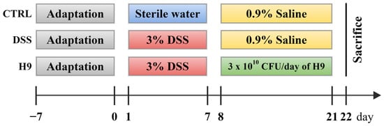
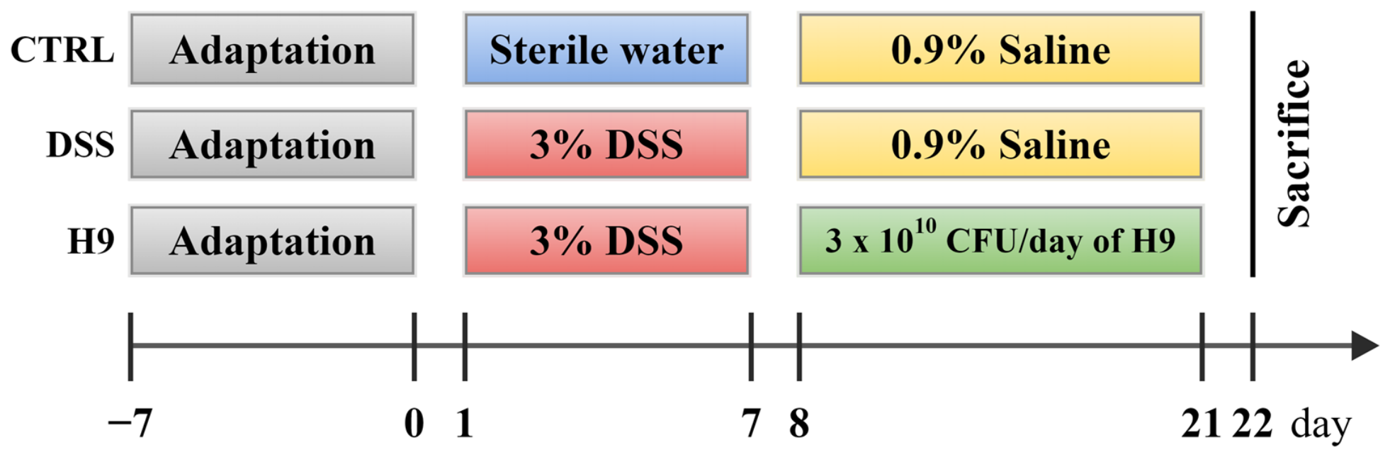
The experimental workflow is illustrated in Figure 1. Following a 7-day acclimation period, the animals were randomly assigned to three groups (n = 8 each): CTRL, DSS, and H9. “CTRL”, “DSS”, and “H9” groups represent the normal control, dextran sodium sulfate-induced, and L. helveticus H9 intervention groups, respectively. To establish the IBD model, rats in the DSS and H9 groups were given drinking water containing 3% (w/w) DSS (molecular weight, 36,000–50,000 Da; MP Biomedicals, LLC, Santa Ana, CA, USA) for 7 consecutive days. Over the subsequent 14 days, the CTRL and DSS groups received 0.9% saline (2 mL/rat/day) by oral gavage, whereas the H9 group was gavaged with L. helveticus H9 (3 × 10¹⁰ CFU suspended in 2 mL of 0.9% saline per rat per day). For preparation of the bacterial powder, L. helveticus H9 was fermented at a constant 37 °C until the pH reached 4.5, after which the culture was homogenized aseptically at 25 MPa. The resulting broth was then spray-dried using an SZ-2000 spray dryer (Shanghai Shunzhi Instrument Manufacturing Co., Ltd., Shanghai, China) with inlet and outlet temperatures of 210 °C and 88 °C, respectively, to yield L. helveticus H9 powder.
Figure 1.
Experimental design. “CTRL”, “DSS”, and “H9” groups represent the normal control, dextran sodium sulfate (DSS)-induced, and Lactobacillus helveticus H9 (H9) groups, respectively.
Throughout the trial, body weight, general health status (diarrhea, hematochezia, and activity), and mortality were monitored and documented daily. On day 21, fresh fecal samples were collected and kept at −80 °C. The rats were euthanized with volatile isoflurane and dissected to harvest the colon. After removal of colonic contents, colon length and weight were recorded. Each colon was then divided into two segments: the distal portion was fixed in 4% paraformaldehyde, and the remaining tissue was stored at −80 °C.
2.3. Disease Activity Index (DAI) Assessment
The DAI is used to assess the severity of colitis, which was the sum score of weight loss, diarrhea, and hematochezia [22]. The specific scoring rules for the body weight loss (BWL) rate was: 0 = 0%—increase weight; 1 = 0% < BWL ≤ 5%; 2 = 5 < BWL ≤ 10%; 3 = 10% < BWL ≤ 15%; 4 = body weight over 15%. The fecal occult blood was determined by the Pyramidon kit (Baso, Zhuhai, China). Fecal occult blood represented invisible blood in the stool, which was scored according to the following guidelines: 0 = negative hemoccult test, no blood; 1 = 60 < positive hemoccult test ≤ 120 s; 2 =30 < positive hemoccult test ≤ 60 s; 3 = 10 < positive hemoccult test ≤ 30 s; 4 = 0 < positive hemoccult test ≤ 10 s; 5 = gross blood observable on the slide. The stool consistency scoring guidelines were: 0 = normal; 1 = moist/sticky stool; 2 = soft stool; 3 = diarrhea.
2.4. Histological Analysis
Colon tissues were fixed in paraformaldehyde and then processed through an ascending ethanol gradient to absolute ethanol. After paraffin infiltration and blocking, the specimens were cut into thin sections. These sections were subjected to HE staining and subsequently coverslipped using neutral balsam. Stained slides were viewed under a light microscope, and representative fields were photographed. The resulting digital images were then analyzed with CaseViewer (3DHISTECH Ltd., Budapest, Hungary). For histological assessment, the grading criteria described by a previous study were used [23]. In brief, four parameters were evaluated: intestinal epithelial loss, connective tissue proliferation, inflammatory cell infiltration, and cellular edema.
2.5. Serum Cytokine Analysis
Rat blood samples were collected. Sera were obtained by centrifugation (3000× g for 10 min at 4 °C). The serum levels of interleukin (IL)-1β, IL-6, IL-10, and tumor necrosis factor (TNF)-α were measured by using ELISA kits (R&D system, Minneapolis, MN, USA) according to the manufacturer’s instructions [24].
2.6. Fecal Metabolomics Analysis
Metabolomics analysis of mouse fecal samples was performed according to the method described by Zhao et al. [25]. The 24 fecal samples (8 per group) stored at −80 °C were thawed naturally at room temperature; 0.06 g of the sample was weighed into a sterile centrifuge tube containing 1 mL of pre-cooled (ice bath) methanol–water solution (4:1, v/v), shaken at high speed for 30 s and sonicated for 30 min. The sample was centrifuged at 4000× g for 10 min at 4 °C, and the supernatant was aspirated into a new 2 mL centrifuge tube. The above methanol extraction procedure was repeated again. The supernatants collected from the two centrifuges were pooled and centrifuged at 10,000× g for 5 min at 4 °C. The supernatant was passed through a 0.22 μm nylon microporous membrane filter for further analysis. A quality control (QC) sample was prepared by pooling an equal amount of each sample (the final volume of the QC sample was approximately 1 mL) for assessing the reproducibility and stability of the LC-MS platform. The UPLC-Q-TOF system was used for the analysis, and the Acquity UPLC (Acquity, Waters) system was coupled to a Q-TOFMSE mass spectrometer (Xevo G2, Waters). Chromatographic separation was performed using an ACQUITY UPLC HSS T3 column (2.1 mm × 100 mm, 1.8 μm; Waters Corporation, Milford, MA, USA), maintained at 40 °C. The mobile phases consisted of (A) ultrapure water containing 0.1% formic acid for the positive ion mode or 5 mM ammonium formate in water for the negative ion mode, and (B) acetonitrile with 0.1% formic acid (positive mode) or pure acetonitrile (negative mode). The flow rate was set at 0.3 mL/min, and the injection volume was 2 μL. A gradient elution program was applied for both ionization modes as follows: 0–1 min, 8% B (isocratic); 1–8 min, linear increase from 8% to 98% B; 8–10 min, 98% B (hold); 10–10.1 min, ramp down to 8% B; and 10.1–12 min, 8% B (re-equilibration). This gradient ensured efficient separation of a broad range of polar and nonpolar metabolites within a short runtime while maintaining excellent peak resolution and reproducibility.
Mass spectrometric analysis was carried out on an Orbitrap Exploris 120 mass spectrometer (Thermo Fisher Scientific, Inc., Waltham, MA, USA) coupled with an electrospray ionization source. Both positive and negative ion modes were employed, and polarity switching was activated to achieve broader metabolite coverage. The ion source was optimized with the following settings: a spray voltage of 3.50 kV in positive mode and −2.50 kV in negative mode; sheath gas and auxiliary gas flow rates of 40 and 10 arbitrary units, respectively; and a capillary temperature of 325 °C.
Mass spectra data were collected and, after peak extraction, peak alignment, deconvolution and normalization of the downstream data were analyzed using Progenesis QI (Waters Corporation, Milford, MA, USA). Multivariate data analysis was performed. Significant different metabolites were annotated through the Kyoto Encyclopedia of Genes and Genomes database to screen for differential metabolic pathways.
2.7. Fecal DNA Extraction, Sequencing, and Analysis
Metagenomic sequencing and analysis of mouse fecal samples were performed according to the method described by Zhang et al. [26]. Fecal DNA was isolated from 24 samples (8 per group) using the sodium dodecyl sulfate protocol [27]. DNA integrity was verified by agarose gel electrophoresis, and DNA concentration was determined with a Qubit® 2.0 Fluorometer (Thermo Fisher Scientific Inc., Waltham, MA, USA). Approximately 1 μg of DNA per sample served as the starting material for library construction. Libraries were constructed with the NEBNext® Ultra™ DNA Library Prep Kit for Illumina (New England Biolabs Inc., Ipswich, MA, USA) following the supplier’s protocol. Unique index codes were incorporated to allow sample demultiplexing.
In brief, DNA was sheared by sonication to approximately 350 bp, followed by end repair, A-tailing, and adapter ligation. The adaptor-ligated products were then amplified by PCR for Illumina sequencing. PCR products were cleaned up with the AMPure XP system (Beckman Coulter Inc., Brea, CA, USA). Fragment size distribution was evaluated on an Agilent 2100 Bioanalyzer (Agilent Inc., Santa Clara, CA, USA), and library concentration was quantified by real-time PCR. Paired-end sequencing (PE150) was carried out on an Illumina NovaSeq platform at Beijing Novogene Bioinformatics Technology Co., Ltd. (Beijing, China), yielding approximately 3 Gb of metagenomic data per sample.
Raw metagenomic reads were first filtered to eliminate low-quality sequences and potential contaminants. Following quality control, HUMAnN 2.0 [https://huttenhower.sph.harvard.edu/humann2/ (accessed on 12 July 2024)] was employed for downstream analysis, with MetaPhlAn 2.0 used for taxonomic assignment based on a curated set of clade-specific marker genes, allowing reads to be classified at the species level.
2.8. Statistical Analysis
BWL, circulating cytokine levels, the colon length-to-weight ratio, and histological as well as DAI scores were processed with GraphPad Prism 8.0 (GraphPad Software Inc., San Diego, CA, USA). Metabolite statistics, covering Student’s t tests along with pathway and enrichment analyses, were conducted in MetaboAnalyst (www.metaboanalyst.ca (accessed on 12 July 2024)). Group-wise comparisons of metagenomic datasets were performed in R (version 3.5.2). Values are shown as mean ± standard deviation. For comparisons involving three or more groups, Tukey’s test was adopted, with an adjusted p < 0.05 taken as the threshold for significance.
3. Results
3.1. Lactobacillus helveticus H9 Alleviated the Symptoms of Colitis in Rats
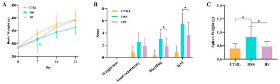
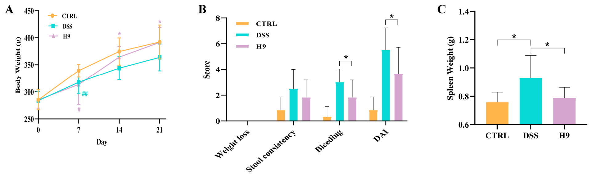
During the 3% DSS treatment, the body weight of the three groups continued to increase in varying degrees (Figure 2A). However, the rat weight growth rate in DSS and H9 group was lower than the CTRL group. On day 7, the body weight of rats in the DSS group and H9 group was significantly lower than that in the normal group (p < 0.01 and p < 0.05, respectively), accompanied by severe diarrhea and bloody stool. The probiotic intervention obviously reversed the weight loss (Figure 2A), and the body weight of probiotic-administered colitis rats was significantly higher than that of the DSS group on days 14 and 21 (p < 0.05). At the end of the trial, the probiotic group had less severe bleeding and a significantly lower DAI score and spleen weight compared with DSS group (p < 0.05 in all cases; Figure 2B,C).
Figure 2.
Differences in body weight and clinical parameters between normal control (CTRL), dextran sodium sulfate (DSS)-induced, and Lactobacillus helveticus H9 (H9) groups. (A) Body weight. (B) Weight loss, stool consistency, bleeding, and disease activity index (DAI), and (C) spleen weight on day 21. Error bars represent standard deviation of the mean. Tukey’s multiple comparison tests were used for statistical analysis. In Figure 2A, # p < 0.05 represents a significant difference between CTRL and H9 groups; ## p < 0.01 represents a significant difference between CTRL and DSS groups; * p < 0.05 represents a significant difference between H9 and DSS groups. In Figure 2B, * p < 0.05 represents a significant difference between the specified groups. In Figure 2C, * p < 0.05 represents a significant difference between CTRL and DSS groups, and between DSS and H9 groups.
3.2. Lactobacillus helveticus H9 Improved Intestinal Mucosal Injury in Rats with Colitis
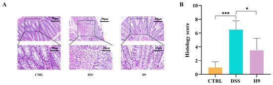
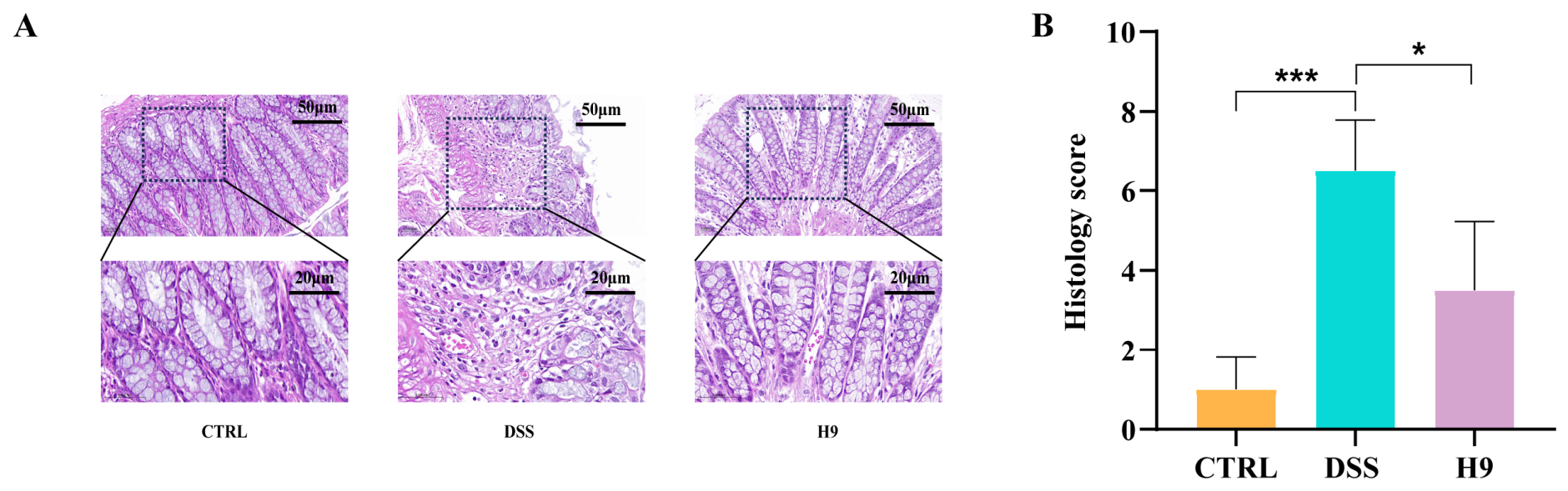
The results of hematoxylin–eosin staining of rat colonic tissues are shown in Figure 3A. The colonic tissue of the CTRL group had an intact intestinal epithelial structure in the mucosal layer, normal epithelial cell morphology and structure, and abundant intestinal glands in the lamina propria, without obvious inflammatory cell infiltration. The epithelial cells of the colonic tissue of the rats in the DSS group were shed after colitis induction, and the number of intestinal glands in the lamina propria was reduced, accompanied by inflammatory cell infiltration. After fourteen days of L. helveticus H9 intervention, all of the above pathological impairments improved, with an increase in the number of cupped cells and a decrease in the area of inflammatory cell infiltration. Histological scores were consistent with histopathology results (p < 0.05; Figure 3B), suggesting probiotic administration could mitigate the tissue damage in the colitis rats.
Figure 3.
Differences in histopathology between the normal control (CTRL), dextran sodium sulfate (DSS)-induced, and Lactobacillus helveticus H9 (H9) groups. (A) Hematoxylin and eosin staining microscopic images (20× and 40× magnification) of colon tissues. (B) Histology score. Error bars represent standard deviation of the mean. Tukey’s multiple comparison tests were used for statistical analysis (*, p < 0.05; ***, p < 0.001).
3.3. Lactobacillus helveticus H9 Reduced Serum Pro-Inflammatory Cytokine Levels in Colitis Rats
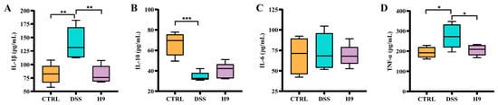
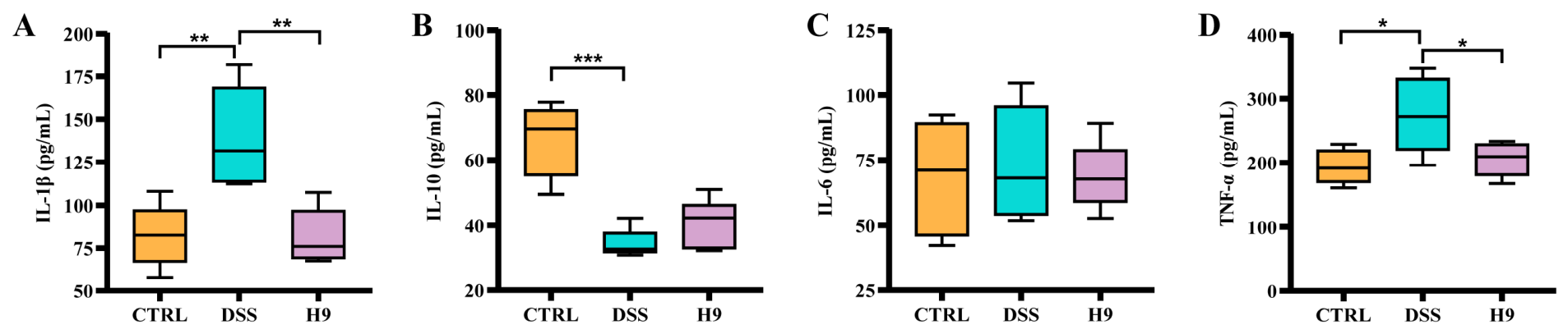
The serum levels of TNF-α, IL-1β, IL-6, and IL-10 were measured on day 21 (Figure 4A–D). The results showed that the DSS group had significantly higher serum IL-1β and TNF-α levels, but lower IL-10 compared with the CTRL group; L. helveticus H9 could significantly reduce the serum levels of IL-1β and TNF-α compared with the DSS group (IL-1β: 82.31 ± 18.01 pg/mL in CTRL group, 139.22 ± 29.40 pg/mL in DSS group, 81.71 ± 16.07 pg/mL in H9 groups; DSS vs. CTRL, p < 0.01; DSS vs. H9, p < 0.01; IL-10: 66.16 ± 11.17 pg/mL in CTRL group, 34.33 ± 4.54 pg/mL in DSS group, 41.01 ± 7.19 pg/mL in H9 group; DSS vs. CTRL, p < 0.001; DSS vs. H9, p > 0.05; TNF-α: 193.98 ± 26.74 pg/mL in CTRL group, 275.03 ± 59.12 pg/mL in DSS group, 205.78 ± 26.45 pg/mL in H9 group; DSS vs. CTRL, p < 0.05; DSS vs. H9, p < 0.05; IL-6: 68.33 ± 21.98 pg/mL in CTRL group, 73.56 ± 22.02 pg/mL in DSS group, 68.71 ± 13.07 pg/mL in H9 group; DSS vs. CTRL, p > 0.05; DSS vs. H9, p > 0.05; Figure 4A–D).
Figure 4.
Differences in serum cytokines levels between normal control (CTRL), dextran sodium sulfate (DSS)-induced, and Lactobacillus helveticus H9 (H9) groups. The levels of (A) interleukin (IL)-1β, (B) IL-10, (C) IL-6, and (D) tumor necrosis factor (TNF)-α. Error bars represent standard deviation of the mean. Tukey’s multiple comparison tests were used to evaluate differences between groups (*, p < 0.05; **, p < 0.01; ***, p < 0.001).
3.4. Lactobacillus helveticus H9 Regulated Gut Microbiota Composition in the Colitis Rats
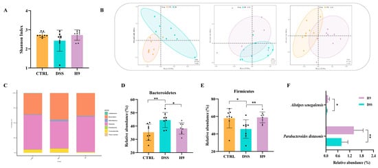
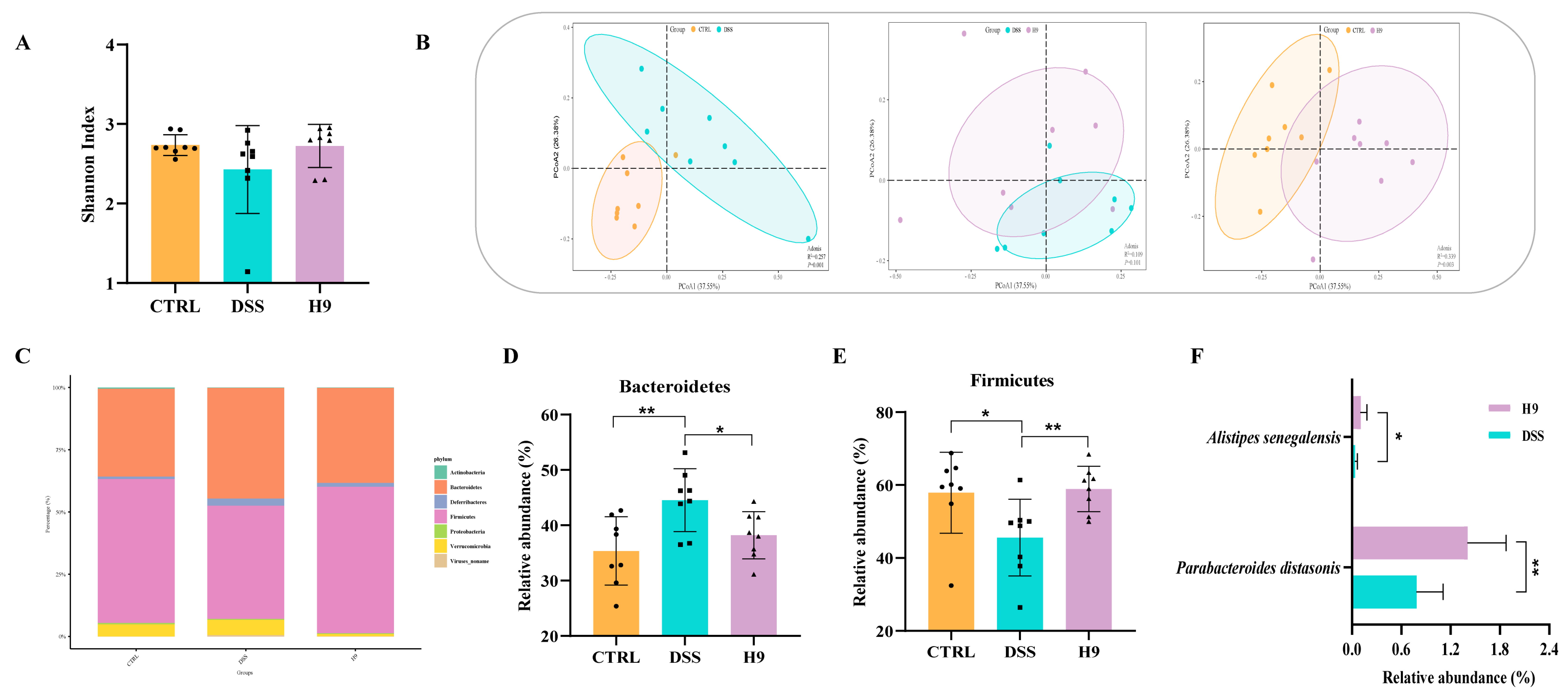
The gut microbiota of the three groups of rats was investigated by diversity and composition analysis (Figure 5). Alpha diversity, assessed by the Shannon index, reflects the microbial diversity (species richness and evenness) within a single sample. Beta diversity evaluates the differences in microbial community composition between different samples or groups [28]. In this experiment, no significant difference was found in the Shannon index between the three groups (Figure 5A); however, the Shannon index was non-significantly lower in the DSS group compared with the CTRL group, which was partly reversed by probiotic consumption. Principal coordinates analyses (PCoA; Bray–Curtis distance) were performed to analyze the β-diversity between each two groups of rats, revealing a significant difference in the gut microbiota between the DSS and CTRL groups (R2 = 0.257, p = 0.001; Figure 5B), but not H9 and DSS groups (R2 = 0.109, p = 0.101; Figure 5B). The gut microbiota comprised four main phyla, including Bacteroidetes, Firmicutes, Proteobacteria, and Verrucomicrobia (Figure 5C). The induction by DSS caused a disturbance in the rat gut microbiota, causing a significant increase in Bacteroidetes (p < 0.01; Figure 5D) and a significant decrease in Firmicutes (p < 0.05; Figure 5E) in the DSS group compared with the CTRL group. In contrast, L. helveticus H9 supplementation significantly increased Firmicutes (p < 0.01; Figure 5E) but decreased Bacteroidetes (p < 0.05; Figure 5D), compared with the DSS group. On the species level, further screening identified two differential species between the DSS and H9 groups, namely Alistipes senegalensis and Parabacteroides distasonis (p < 0.05 and p < 0.01, respectively; Figure 5F); both were significantly enriched in the H9 group compared with the DSS group.
Figure 5.
Gut microbiota in normal control (CTRL), dextran sodium sulfate (DSS)-induced, and Lactobacillus helveticus H9 (H9) groups. (A) Shannon index. (B) Principal coordinate analysis (PCoA; Bray–Curtis distance) score plots of rat gut microbiota: CTRL versus DSS group; DSS versus H9 group; and CTRL versus H9 group, respectively (from left to right). (C) Phylum-level gut microbiota profile. The relative abundance of (D) Bacteroidetes, (E) Firmicutes, (F) Alistipes senegalensis, and Parabacteroides distasonis. Error bars represent standard deviation of the mean. Tukey’s multiple comparison tests were used for statistical analysis (*, p < 0.05; **, p < 0.01).
3.5. Effect of Lactobacillus helveticus H9 on Fecal Metabolites in Rats with Colitis
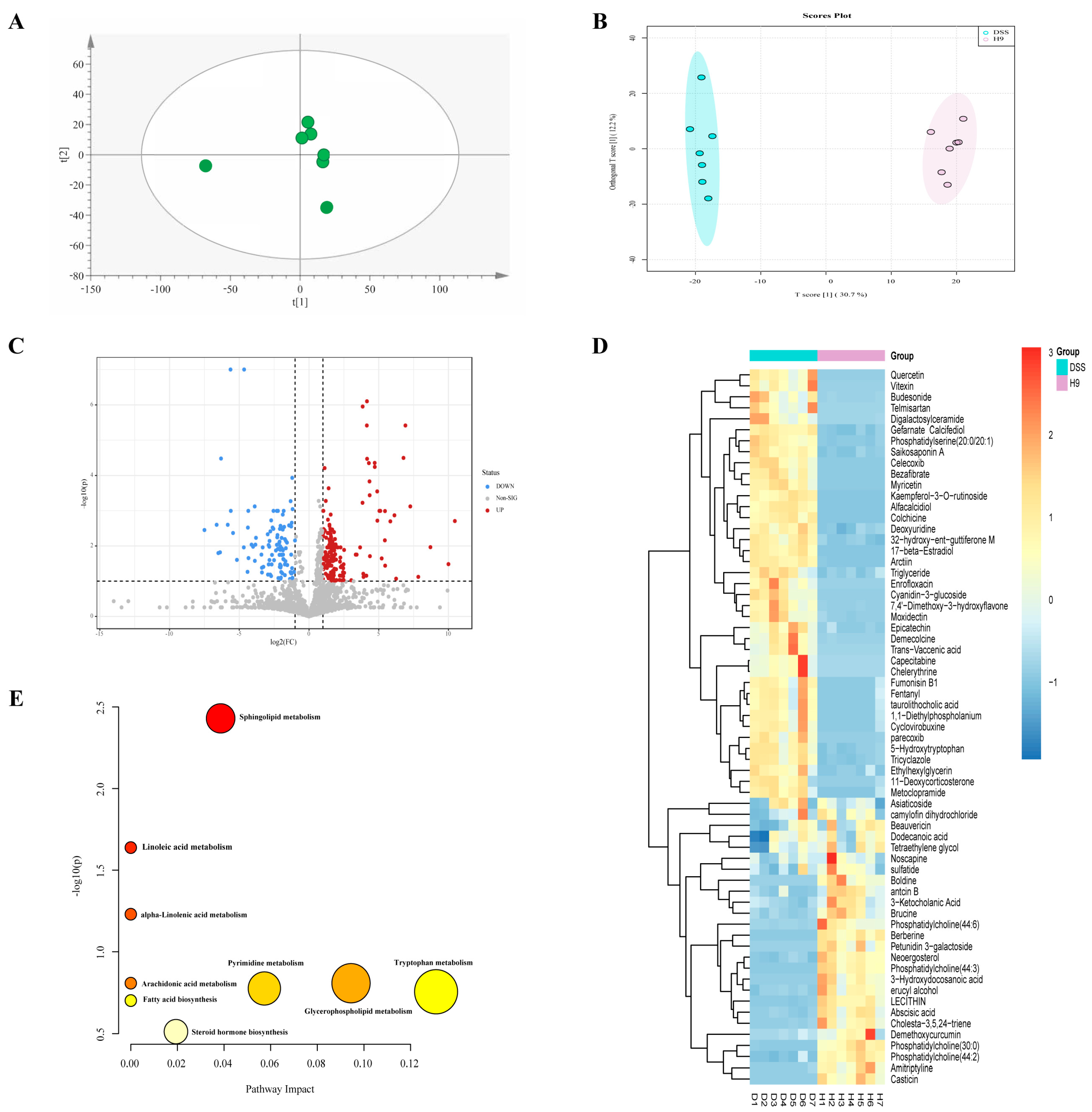
On the principal component analysis (PCA) plot, symbols representing the QC samples were located closely together, suggesting a relatively stable instrumental and chromatographic condition during the analysis (Figure 6A). Then, an orthogonal partial least-squares discrimination analysis was performed based on the fecal metabolomics data from the DSS and H9 groups, revealing significant differences between these groups (Figure 6B). Differential metabolites between DSS and H9 groups were presented in a volcano plot and identified by a one-way t-test and coefficient of variation method (cut-off level: p < 0.05, fold change > 2 or <0.5; Figure 6C). A total of 266 differential metabolites were found, including 161 upregulated and 105 downregulated ones. The screen criteria were further strengthened by applying an additional condition of variable importance in projection (VIP) > 1, narrowing to 65 significantly differential metabolites (Figure 6D). As shown in the relative abundance heatmap, lecithin metabolites were significantly enhanced and deprived in the DSS group compared with the H9 group, respectively (Figure 6D). Pathway analysis based on these metabolites yielded a total of nine significant differential pathways, including Sphingolipid metabolism, Linoleic acid metabolism, alpha-Linolenic acid metabolism, Arachidonic acid metabolism, Glycerophospholipid metabolism, Pyrimidine metabolism, Tryptophan metabolism, Fatty acid biosynthesis and Steroid hormone biosynthesis (Figure 6E).
Figure 6.
Fecal metabolites changes in normal control (CTRL), dextran sodium sulfate (DSS)-induced, and Lactobacillus helveticus H9 (H9) groups. (A) The principal component analysis score plot of the quality control samples. (B) Orthogonal partial least-squares discrimination analysis (OPLS-DA) score plot of fecal metabolomes of DSS and H9 groups. (C) Volcano plot of detected fecal metabolites of DSS and H9 groups. Blue and red dots represent significantly decreased (DOWN) and increased (UP) fecal metabolites in the DSS group compared with the H9 group (cut-off levels: p < 0.05, fold change > 2 or <0.5, VIP score > 1), while gray dots represent non-significantly different metabolites. (D) Cluster heatmap of the differential metabolites in DSS and H9 groups. The scale bar represents relative abundance of the metabolites, ranging between 3 (high abundance) and -3 (low abundance). (E) Pathway analysis of identified differential metabolites between DSS and H9 groups.
4. Discussion
Inflammatory bowel disease is a chronic systemic inflammatory disease, which has shown a rapid increase in recent years, and the medical burden associated with IBD will be a major public health challenge [29]. Probiotics have been used to manage IBD [30,31] by improving mucosal barrier function, inhibiting the growth of pathogenic microorganisms, and regulating the inflammatory response [32]. This study constructed a rat IBD model using 3% DSS, through which the beneficial effect of intake of L. helveticus H9 was evaluated. Our study found that administering L. helveticus H9 could effectively improve IBD-related symptoms and inflammation, as well as improving DSS-disrupted gut dysbiosis.
Dextran sodium sulfate is a water-soluble sulfate polysaccharide that has a direct toxic effect on intestinal epithelial cells and disrupts the integrity of the intestinal barrier [33]. It is widely used to induce colitis models due to its ease of use and high success rate in constructing a colitis model [34]. After DSS treatment, experimental animals exhibited obvious signs of colitis, including diarrhea, blood in stool and weight loss [35]. The present study found that the weight of rats in the DSS group continued to increase in the course of model construction (Figure 2A), and such results are similar to those of Zhu et al. [36]. To some extent, body weight changes can reflect the health status of colitis animals [37], but factors such as feeding environment, daily food intake and energy expenditure due to animal activities can have an impact on body weight changes. After starting DSS treatment, the rats showed a reduced daily activity level, exhibiting a depressed and lazy state, though no significant change was observed in food intake. Thus, the energy intake could be greater than energy expenditure, resulting in no decrease in body weight. The DAI is a comprehensive index calculated based on the change in body weight, fecal morphology and fecal occult blood [38], which can comprehensively evaluate the health status of colitis rats. Administering L. helveticus H9 could increase the rate of weight gain, improve the symptoms of loose stools and bloody stools, and significantly reduce the DAI score in rats with colitis (Figure 2B). Histopathology is an important clinical parameter for assessing IBD, and the improvement in histopathology scores further confirmed that IBD-related colonic tissue damage was mitigated by probiotic intake [39].
Cytokines play a key role in the development of colitis. Although the pathogenesis of colitis is not entirely clear, it is widely believed that colitis development is related to immune system abnormalities [40]. A key cytokine, TNF-α, plays a part in intestinal inflammation, regulating cell differentiation, growth, apoptosis, production of inflammatory factors and chemokines, upregulation of various endothelial cell adhesion molecules, and the increase in gut barrier permeability [41]. Curciarello et al. (2021) found that administering Lactobacillus kefiri CIDCA 8348 could inhibit the release of TNF-αin adult patients with IBD [42]. Interleukin-1β stimulates neutrophils to release inflammatory proteins and inflammatory mediators, which are directly involved in IBD onset and correlate with the severity of IBD [40]. Our study found that administering L. helveticus H9 significantly reduced the serum TNF-α and IL-1β levels (Figure 4A,D), indicating that the intake of this probiotic could mitigate colitis.
Gut dysbiosis is common in IBD. An imbalanced gut microbiota may lead to gut inflammation. On the contrary, intestinal inflammation has also been found to aggravate the extent of gut dysbiosis in the host [43]. We observed a decrease in the Shannon diversity index of rat gut microbiota in the DSS group, suggesting that DSS treatment could disrupt the gut microbiota by diminishing its diversity (Figure 5A), but, similar to the findings of other studies, probiotic intervention showed a tendency to increase the gut microbiota diversity, although this did not reach statistical significance [14,44]. In addition, the results of β diversity analysis showed that there was a significant difference in the structure of gut microbiota between the DSS group and the CTRL group, and the intervention of L. helveticus H9 could partially reverse the gut-microbiota-altering effect caused by DSS treatment (Figure 5B). The Firmicutes-to-Bacteroidetes (F/B) ratio is a well-recognized dysbiosis indicator reflecting the gut microbiota status in patients with colitis [45]. In our study, intervention with L. helveticus H9 increased this ratio, suggesting that the strain effectively reverses DSS-induced dysbiosis. However, due to limitations such as inter-individual variability, disease specificity, and its reliance on the overall effects of the microbiome, the F/B ratio alone cannot serve as a diagnostic criterion for a specific disease. Thus, it must be evaluated in conjunction with other indicators, such as inflammatory cytokines and clinical parameters, to comprehensively assess its efficacy in alleviating the clinical symptoms of IBD [46]. A final taxonomic gut microbiota compositional analysis revealed the increases in the relative abundances of Alistipes senegalensis and Parabacteroides distasonis post-probiotic treatment compared with the DSS group (Figure 5F). Alistipes are Gram-negative bacteria under the Bacteroidetes phylum. This genus can stimulate TNF-α production and exert anti-inflammatory properties directly and indirectly through short-chain fatty acid production [47]. A strong correlation has been found between ecological dysregulation of Alistipes and IBD [48]. The fecal samples of Winnie, a model mouse for IBD, were sequenced by 16S rRNA. The results showed that the abundance of Alistipes senegalensis in Winnie was significantly lower than that in wild-type mice [49]. Moreover, it was found that Alistipes senegalensis was significantly lower in the gut microbiota of CD patients compared to the healthy population, and these findings suggest that Alistipes senegalensis is a protective strain against colitis [50]. T helper 17 (Th17) cells are immune cells that promote inflammatory responses [51], and Parabacteroides distasonis could inhibit the differentiation of Th17 and reduce the production of inflammatory factors [52]. Oral gavage of Parabacteroides distasonis significantly reduced the severity of intestinal inflammation in DSS-induced acute and chronic colitis mouse models, inhibiting excessive production of pro-inflammatory cytokines, such as TNF-a and IL-6 [53]. In conclusion, our results showed that administering L. helveticus H9 could alleviate IBD in rats by increasing the gut microbiota diversity, while enriching intestinal Alistipes senegalensis and Parabacteroides distasonis.
On the metabolomics level, administering L. helveticus H9 was associated with significant changes in the abundance of some fecal metabolites and their related pathways. We identified a total of nine differential metabolic pathways between H9 and DSS groups (Figure 6E). Interestingly, the metabolite, lecithin, was found to be involved in four of the differential metabolic pathways, namely Linoleic acid metabolism, alpha-Linolenic acid metabolism, Arachidonic acid metabolism, and Glycerophospholipid metabolism. Lecithin is an important protective component of colonic mucus, accounting for more than 70% of the total phospholipids in the intestinal mucus layer [54], and its low levels tend to reduce the hydrophobic barrier function of intestinal mucus, allowing colonic bacteria to cross the intestinal mucus barrier and causing intestinal inflammation and ulceration [55,56]. Colares et al. (2016) found that lecithin was effective in reducing intestinal tissue damage in colitis mice [57]. A meta-analysis found that the use of lecithin could improve clinical and endoscopic outcomes in patients with ulcerative colitis [58]. Our study found that administering L. helveticus H9 could significantly increase the amount of lecithin in the stool (Figure 6E), which may be part of the protective mechanism against colitis-associated tissue damage.
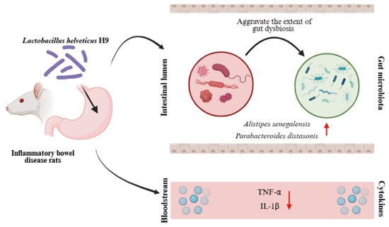
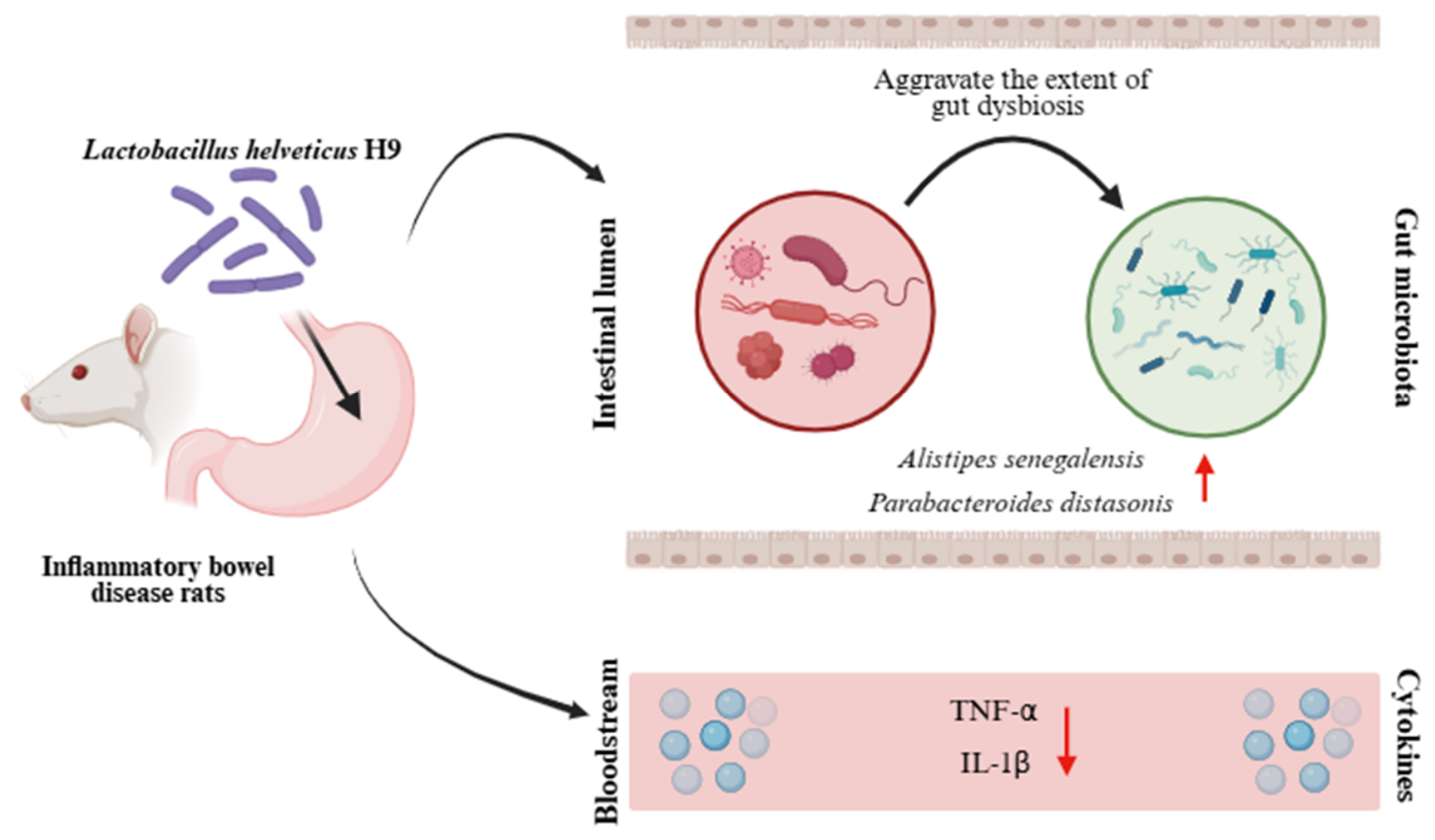
In conclusion, this study demonstrated that L. helveticus H9 could alleviate DSS-induced ulcerative colitis in rats, possibly through mitigating intestinal inflammation, modulating the cytokine profile, and regulating the gut microbiota and its metabolites (Figure 7). These findings support the development of microbiome-targeted strategies for combating inflammatory bowel disease. Future studies should employ fecal microbiota transplantation and metabolite supplementation to establish causality, focus on the microbiota–metabolite–inflammation axis, and elucidate the molecular mechanisms by which L. helveticus H9 ameliorates IBD, including but not limited to specific gut bacterial metabolites and inflammatory pathways. Moreover, these findings are based on a relatively small animal study, and larger-scale studies as well as eventual human trials are still needed to validate these effects. Such work will be essential to translate these findings into clinical applications.
Figure 7.
Schematic illustration of the effect of probiotic Lactobacillus helveticus H9-ameliorated colitis in a rat model. Dextran sulfate sodium (DSS) induced the development of ulcerative colitis, which was inhibited by probiotic Lactobacillus helveticus H9 treatment, presumably through increasing the abundance of Alistipes senegalensis and Parabacteroides distasonis.
Author Contributions
W.Z.: designed the study, analyzed the experimental data, and drafted the manuscript. C.F.: conducted the animal experiments. T.Z.: guided the trial design. L.-Y.K.: writing—review and editing. Q.H.: conceptualization, funding acquisition, project administration, and supervision. All authors have read and agreed to the published version of the manuscript.
Funding
This work was supported by the Scientific Research Foundation for High-level Talents of Huangshan University (2024xkjq017), the Major Project of Anhui Provincial Collaborative Technology Service Center for Rural Revitali-zation (Huangshan University) (XCZXZD2502) and the National Natural Science Foundation of China (32001711).
Institutional Review Board Statement
All animal procedures were approved by the Animal Ethics Committee of Inner Mongolia Agricultural University (approval no. NND2022099, 12 August 2022). All procedures followed the ARRIVE guidelines.
Informed Consent Statement
Not applicable.
Data Availability Statement
The original contributions presented in this study are included in the article. Further inquiries can be directed to the corresponding author.
Conflicts of Interest
The authors declare that they have no known competing financial interest or. personal relationships that could have appeared to influence the work reported in this paper.
References
- Ramos, G.P.; Papadakis, K.A. Mechanisms of Disease: Inflammatory Bowel Diseases. Mayo Clin. Proc. 2019, 94, 155–165. [Google Scholar] [CrossRef] [Scilit] [PubMed]
- Chang, J.T. Pathophysiology of inflammatory bowel diseases. N. Engl. J. Med. 2020, 383, 2652–2664. [Google Scholar] [CrossRef] [Scilit] [PubMed]
- Perler, B.K.; Ungaro, R.; Baird, G.; Mallette, M.; Bright, R.; Shah, S.; Shapiro, J.; Sands, B.E. Presenting symptoms in inflammatory bowel disease: Descriptive analysis of a community-based inception cohort. BMC Gastroenterol. 2019, 19, 47. [Google Scholar] [CrossRef] [Scilit] [PubMed]
- Kaplan, G.G.; Windsor, J.W. The four epidemiological stages in the global evolution of inflammatory bowel disease. Nat. Rev. Gastroenterol. Hepatol. 2021, 18, 56–66. [Google Scholar] [CrossRef] [Scilit] [PubMed]
- Ng, S.C.; Kaplan, G.G.; Tang, W.; Banerjee, R.; Adigopula, B.; Underwood, F.E.; Tanyingoh, D.; Wei, S.-C.; Lin, W.-C.; Lin, H.-H.; et al. Population Density and Risk of Inflammatory Bowel Disease: A Prospective Population-Based Study in 13 Countries or Regions in Asia-Pacific. Am. J. Gastroenterol. 2019, 114, 107–115. [Google Scholar] [CrossRef] [Scilit] [PubMed]
- Cheng, B.; Yan, J.; Huang, Y.; Fang, J.; Zhou, X.; Chen, X. Inflammatory bowel disease burden in China and the United States from 1990–2023 and prediction to 2050. Intern. Emerg. Med. 2026, 21, 1669–1683. [Google Scholar] [CrossRef] [Scilit] [PubMed]
- Lee, S.H.; eun Kwon, J.; Cho, M.-L. Immunological pathogenesis of inflammatory bowel disease. Intest. Res. 2018, 16, 26. [Google Scholar] [CrossRef] [Scilit] [PubMed]
- Nishida, A.; Inoue, R.; Inatomi, O.; Bamba, S.; Naito, Y.; Andoh, A. Gut microbiota in the pathogenesis of inflammatory bowel disease. Clin. J. Gastroenterol. 2017, 11, 1–10. [Google Scholar] [CrossRef] [Scilit] [PubMed]
- Piovani, D.; Danese, S.; Peyrin-Biroulet, L.; Nikolopoulos, G.K.; Lytras, T.; Bonovas, S. Environmental Risk Factors for Inflammatory Bowel Diseases: An Umbrella Review of Meta-analyses. Gastroenterology 2019, 157, 647–659.e4. [Google Scholar] [CrossRef] [Scilit] [PubMed]
- Colombel, J.-F.; D’haens, G.; Lee, W.-J.; Petersson, J.; Panaccione, R. Outcomes and Strategies to Support a Treat-to-target Approach in Inflammatory Bowel Disease: A Systematic Review. J. Crohn’s Colitis 2019, 14, 254–266. [Google Scholar] [CrossRef] [Scilit] [PubMed]
- Seyedian, S.S.; Nokhostin, F.; Malamir, M.D. A review of the diagnosis, prevention, and treatment methods of inflammatory bowel disease. J. Med. Life 2019, 12, 113. [Google Scholar] [CrossRef] [Scilit] [PubMed]
- Zendeboodi, F.; Khorshidian, N.; Mortazavian, A.M.; da Cruz, A.G. Probiotic: Conceptualization from a new approach. Curr. Opin. Food Sci. 2020, 32, 103–123. [Google Scholar] [CrossRef] [Scilit]
- Matsuoka, K.; Kanai, T. The gut microbiota and inflammatory bowel disease. Semin. Immunopathol. 2015, 37, 47–55. [Google Scholar] [CrossRef] [Scilit] [PubMed]
- Wu, Y.; Jha, R.; Li, A.; Liu, H.; Zhang, Z.; Zhang, C.; Zhai, Q.; Zhang, J. Probiotics (Lactobacillus plantarum HNU082) Supplementation Relieves Ulcerative Colitis by Affecting Intestinal Barrier Functions, Immunity-Related Gene Expression, Gut Microbiota, and Metabolic Pathways in Mice. Microbiol. Spectr. 2022, 10, e0165122. [Google Scholar] [CrossRef] [Scilit] [PubMed]
- Di Giovangiulio, M.; Verheijden, S.; Bosmans, G.; Stakenborg, N.; Boeckxstaens, G.E.; Matteoli, G. The neuromodulation of the intestinal immune system and its relevance in inflammatory bowel disease. Front. Immunol. 2015, 6, 590. [Google Scholar] [CrossRef] [Scilit] [PubMed]
- Jang, Y.J.; Kim, W.-K.; Han, D.H.; Lee, K.; Ko, G. Lactobacillus fermentum species ameliorate dextran sulfate sodium-induced colitis by regulating the immune response and altering gut microbiota. Gut Microbes 2019, 10, 696–711. [Google Scholar] [CrossRef] [Scilit] [PubMed]
- Bron, P.A.; Kleerebezem, M.; Brummer, R.-J.; Cani, P.D.; Mercenier, A.; MacDonald, T.T.; Garcia-Ródenas, C.L.; Wells, J.M. Can probiotics modulate human disease by impacting intestinal barrier function? Br. J. Nutr. 2017, 117, 93–107. [Google Scholar] [CrossRef] [Scilit] [PubMed]
- Rose, E.C.; Odle, J.; Blikslager, A.T.; Ziegler, A.L. Probiotics, Prebiotics and Epithelial Tight Junctions: A Promising Approach to Modulate Intestinal Barrier Function. Int. J. Mol. Sci. 2021, 22, 6729. [Google Scholar] [CrossRef] [Scilit] [PubMed]
- Pang, B.; Jin, H.; Liao, N.; Li, J.; Jiang, C.; Shao, D.; Shi, J. Lactobacillus rhamnosus from human breast milk ameliorates ulcerative colitis in mice via gut microbiota modulation. Food Funct. 2021, 12, 5171–5186. [Google Scholar] [CrossRef] [Scilit] [PubMed]
- Chen, Y.; Zhang, W.; Sun, Z.; Meng, B.; Zhang, H. Complete genome sequence of Lactobacillus helveticus H9, a probiotic strain originated from kurut. J. Biotechnol. 2015, 194, 37–38. [Google Scholar] [CrossRef] [Scilit] [PubMed]
- Chen, Y.; Zhong, Z.; Xue, J.; Yang, J.; Zhang, Q.; Yue, J.; Zhang, H. DNA microarray analysis of the antihypertensive effect of milk fermented by Lactobacillus helveticus H9 on spontaneously hypertensive rats. Dairy Sci. Technol. 2015, 95, 321–330. [Google Scholar] [CrossRef] [Scilit][Green Version]
- Liu, Y.; Chen, N.; He, H.; Liu, L.; Sun, S. Sodium butyrate alleviates DSS-induced inflammatory bowel disease by inhibiting ferroptosis and modulating ERK/STAT3 signaling and intestinal flora. Ann. Med. 2025, 57, 2470958. [Google Scholar] [CrossRef] [Scilit] [PubMed]
- Kim, W.-K.; Han, D.H.; Jang, Y.J.; Park, S.; Jang, S.J.; Lee, G.; Han, H.S.; Ko, G. Alleviation of DSS-induced colitis via Lactobacillus acidophilus treatment in mice. Food Funct. 2020, 12, 340–350. [Google Scholar] [CrossRef] [Scilit] [PubMed]
- Feng, C.; Zhao, Y.; He, Q.; Yu, J.; Sun, Z. Bifidobacterium breve B2798 and Its Heat-Killed Cells Alleviate Inflammation in Rats with DSS Model by Modulating Gut Microbiota. Probiotics Antimicrob. Proteins 2025, 18, 4118–4131. [Google Scholar] [CrossRef] [Scilit] [PubMed]
- Zhao, Y.; Kwok, L.-Y.; Gao, G.; Li, N.; Zhao, C.; Sun, Z.; Yu, J. Bifidobacterium breve B2798 ameliorates high-fat diet-induced obesity by reshaping gut microbiota and modulating host metabolic profiles. Food Sci. Hum. Wellness 2026. [Google Scholar] [CrossRef] [Scilit]
- Zhang, T.; Zhang, W.; Feng, C.; Kwok, L.-Y.; He, Q.; Sun, Z. Stronger gut microbiome modulatory effects by postbiotics than probiotics in a mouse colitis model. npj Sci. Food 2022, 6, 53. [Google Scholar] [CrossRef] [Scilit] [PubMed]
- Kumar, J.; Kumar, M.; Gupta, S.; Ahmed, V.; Bhambi, M.; Pandey, R.; Chauhan, N.S. An Improved Methodology to Overcome Key Issues in Human Fecal Metagenomic DNA Extraction. Genom. Proteom. Bioinform. 2016, 14, 371–378. [Google Scholar] [CrossRef] [Scilit] [PubMed]
- Magurran, A.E. Measuring biological diversity. Curr. Biol. 2021, 31, R1174–R1177. [Google Scholar] [CrossRef] [Scilit] [PubMed]
- Buie, M.J.; Quan, J.; Windsor, J.W.; Coward, S.; Hansen, T.M.; King, J.A.; Kotze, P.G.; Gearry, R.B.; Ng, S.C.; Mak, J.W.; et al. Global Hospitalization Trends for Crohn’s Disease and Ulcerative Colitis in the 21st Century: A Systematic Review with Temporal Analyses. Clin. Gastroenterol. Hepatol. 2022, 21, 2211–2221. [Google Scholar] [CrossRef] [Scilit] [PubMed]
- Abraham, B.P.; Quigley, E.M. Probiotics in inflammatory bowel disease. Gastroenterol. Clin. 2017, 46, 769–782. [Google Scholar] [CrossRef] [Scilit] [PubMed]
- Jakubczyk, D.; Leszczyńska, K.; Górska, S. The effectiveness of probiotics in the treatment of inflammatory bowel disease (IBD)—A critical review. Nutrients 2020, 12, 1973. [Google Scholar] [CrossRef] [Scilit] [PubMed]
- Plaza-Diaz, J.; Ruiz-Ojeda, F.J.; Gil-Campos, M.; Gil, A. Mechanisms of action of probiotics. Adv. Nutr. 2019, 10, S49–S66. [Google Scholar] [CrossRef] [Scilit] [PubMed]
- Eichele, D.D.; Kharbanda, K.K. Dextran sodium sulfate colitis murine model: An indispensable tool for advancing our understanding of inflammatory bowel diseases pathogenesis. World J. Gastroenterol. 2017, 23, 6016. [Google Scholar] [CrossRef] [Scilit] [PubMed]
- Perse, M.; Cerar, A. Dextran Sodium Sulphate Colitis Mousemodel: Traps And Tricks; Hindawi Publishing Corporation: Cairo, Egypt, 2012. [Google Scholar]
- Das, S.; Batra, S.K.; Rachagani, S. Mouse model of dextran sodium sulfate (DSS)-induced colitis. Bio-protocol 2017, 7, e2515. [Google Scholar] [CrossRef] [Scilit] [PubMed]
- Zhu, L.; Gu, P.; Shen, H. Protective effects of berberine hydrochloride on DSS-induced ulcerative colitis in rats. Int. Immunopharmacol. 2019, 68, 242–251. [Google Scholar] [CrossRef] [Scilit] [PubMed]
- Feng, C.; Zhang, W.; Zhang, T.; Li, B.; He, Q.; Kwok, L.-Y.; Zhang, H. Oral administration of pasteurized probiotic fermented milk alleviates dextran sulfate sodium-induced inflammatory bowel disease in rats. J. Funct. Foods 2022, 94, 105140. [Google Scholar] [CrossRef] [Scilit]
- Ahl, D.; Liu, H.; Schreiber, O.; Roos, S.; Phillipson, M.; Holm, L. Lactobacillus reuteri increases mucus thickness and ameliorates dextran sulphate sodium-induced colitis in mice. Acta Physiol. 2016, 217, 300–310. [Google Scholar] [CrossRef] [Scilit] [PubMed]
- Mosli, M.H.; Parker, E.C.; Nelson, A.S.; Baker, A.K.; MacDonald, J.K.; Zou, G.Y.; Feagan, B.G.; Khanna, R.; Levesque, B.G.; Jairath, V. Histologic scoring indices for evaluation of disease activity in ulcerative colitis. Cochrane Database Syst. Rev. 2017, 2017, CD011256. [Google Scholar] [CrossRef] [Scilit] [PubMed]
- Neurath, M.F. Cytokines in inflammatory bowel disease. Nat. Rev. Immunol. 2014, 14, 329–342. [Google Scholar] [CrossRef] [Scilit] [PubMed]
- Guan, Q.; Zhang, J. Recent advances: The imbalance of cytokines in the pathogenesis of inflammatory bowel disease. Mediat. Inflamm. 2017, 2017, 4810258. [Google Scholar] [CrossRef] [Scilit] [PubMed]
- Curciarello, R.; Canziani, K.E.; Salto, I.; Romero, E.B.; Rocca, A.; Doldan, I.; Peton, E.; Brayer, S.; Sambuelli, A.M.; Goncalves, S.; et al. Probiotic Lactobacilli Isolated from Kefir Promote Down-Regulation of Inflammatory Lamina Propria T Cells from Patients with Active IBD. Front. Pharmacol. 2021, 12, 658026. [Google Scholar] [CrossRef] [Scilit] [PubMed]
- Ni, J.; Wu, G.D.; Albenberg, L.; Tomov, V.T. Gut microbiota and IBD: Causation or correlation? Nat. Rev. Gastroenterol. Hepatol. 2017, 14, 573–584. [Google Scholar] [CrossRef] [Scilit] [PubMed]
- Xu, L.; Liu, B.; Huang, L.; Li, Z.; Cheng, Y.; Tian, Y.; Pan, G.; Li, H.; Xu, Y.; Wu, W.; et al. Probiotic Consortia and Their Metabolites Ameliorate the Symptoms of Inflammatory Bowel Diseases in a Colitis Mouse Model. Microbiol. Spectr. 2022, 10, e0065722. [Google Scholar] [CrossRef] [Scilit] [PubMed]
- Stojanov, S.; Berlec, A.; Štrukelj, B. The influence of probiotics on the firmicutes/bacteroidetes ratio in the treatment of obesity and inflammatory bowel disease. Microorganisms 2020, 8, 1715. [Google Scholar] [CrossRef] [Scilit] [PubMed]
- Wei, S.; Bahl, M.I.; Baunwall, S.M.D.; Hvas, C.L.; Licht, T.R. Determining Gut Microbial Dysbiosis: A Review of Applied Indexes for Assessment of Intestinal Microbiota Imbalances. Appl. Environ. Microbiol. 2021, 87, e00395-21. [Google Scholar] [CrossRef] [Scilit] [PubMed]
- Parker, B.J.; Wearsch, P.A.; Veloo, A.C.M.; Rodriguez-Palacios, A. The Genus Alistipes: Gut Bacteria with Emerging Implications to Inflammation, Cancer, and Mental Health. Front. Immunol. 2020, 11, 906. [Google Scholar] [CrossRef] [Scilit] [PubMed]
- Dziarski, R.; Park, S.Y.; Kashyap, D.R.; Dowd, S.E.; Gupta, D. Pglyrp-Regulated Gut Microflora Prevotella falsenii, Parabacteroides distasonis and Bacteroides eggerthii Enhance and Alistipes finegoldii Attenuates Colitis in Mice. PLoS ONE 2016, 11, e0146162. [Google Scholar] [CrossRef] [Scilit] [PubMed]
- Talà, A.; Guerra, F.; Resta, S.C.; Calcagnile, M.; Barca, A.; Tredici, S.M.; De Donno, M.D.; Vacca, M.; Liso, M.; Chieppa, M.; et al. Phenotyping of Fecal Microbiota of Winnie, a Rodent Model of Spontaneous Chronic Colitis, Reveals Specific Metabolic, Genotoxic, and Pro-inflammatory Properties. Inflammation 2022, 45, 2477–2497. [Google Scholar] [CrossRef] [Scilit] [PubMed]
- Hu, S.; Png, E.; Gowans, M.; Ong, D.E.H.; de Sessions, P.F.; Song, J.; Nagarajan, N. Ectopic gut colonization: A metagenomic study of the oral and gut microbiome in Crohn’s disease. Gut Pathog. 2021, 13, 13. [Google Scholar] [CrossRef] [Scilit] [PubMed]
- Oukka, M. Th17 cells in immunity and autoimmunity. Ann. Rheum. Dis. 2008, 67, iii26–iii29. [Google Scholar] [CrossRef] [Scilit] [PubMed]
- Sun, H.; Guo, Y.; Wang, H.; Yin, A.; Hu, J.; Yuan, T.; Zhou, S.; Xu, W.; Wei, P.; Yin, S.; et al. Gut commensal Parabacteroides distasonis alleviates inflammatory arthritis. Gut 2023, 72, 1664–1677. [Google Scholar] [CrossRef] [Scilit] [PubMed]
- Kverka, M.; Zakostelska, Z.; Klimesova, K.; Sokol, D.; Hudcovic, T.; Hrncir, T.; Rossmann, P.; Mrazek, J.; Kopecny, J.; Verdu, E.F.; et al. Oral administration of Parabacteroides distasonis antigens attenuates experimental murine colitis through modulation of immunity and microbiota composition. Clin. Exp. Immunol. 2010, 163, 250–259. [Google Scholar] [CrossRef] [Scilit] [PubMed]
- Schneider, H.; Braun, A.; Füllekrug, J.; Stremmel, W.; Ehehalt, R. Lipid Based Therapy for Ulcerative Colitis—Modulation of Intestinal Mucus Membrane Phospholipids as a Tool to Influence Inflammation. Int. J. Mol. Sci. 2010, 11, 4149–4164. [Google Scholar] [CrossRef] [Scilit] [PubMed]
- Parveen, F.; Bender, D.; Law, S.-H.; Mishra, V.K.; Chen, C.-C.; Ke, L.-Y. Role of Ceramidases in Sphingolipid Metabolism and Human Diseases. Cells 2019, 8, 1573. [Google Scholar] [CrossRef] [Scilit] [PubMed]
- Stremmel, W.; Gauss, A. Lecithin as a therapeutic agent in ulcerative colitis. Dig. Dis. 2013, 31, 388–390. [Google Scholar] [CrossRef] [Scilit] [PubMed]
- Colares, J.R.; Schemitt, E.G.; Hartmann, R.M.; Moura, R.M.; Morgan-Martins, M.I.; Fillmann, H.S.; Fillmann, L.; Marroni, N.P. Effect of lecithin on oxidative stress in an experimental model of rats colitis induced by acetic acid. J. Coloproctology 2016, 36, 97–103. [Google Scholar] [CrossRef] [Scilit]
- Stremmel, W.; Vural, H.; Evliyaoglu, O.; Weiskirchen, R. Delayed-Release Phosphatidylcholine Is Effective for Treatment of Ulcerative Colitis: A Meta-Analysis. Dig. Dis. 2021, 39, 508–515. [Google Scholar] [CrossRef] [Scilit] [PubMed]
Disclaimer/Publisher’s Note: The statements, opinions and data contained in all publications are solely those of the individual author(s) and contributor(s) and not of MDPI and/or the editor(s). MDPI and/or the editor(s) disclaim responsibility for any injury to people or property resulting from any ideas, methods, instructions or products referred to in the content. |
© 2026 by the authors. Licensee MDPI, Basel, Switzerland. This article is an open access article distributed under the terms and conditions of the Creative Commons Attribution (CC BY) license.