Next Issue
Volume 8, October
Previous Issue
Volume 8, June


Non-Coding RNA, Volume 8, Issue 4 (August 2022) – 18 articles

Cover Story (view full-size image): 
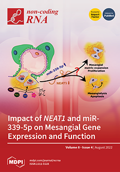
In diabetic kidney disease when various cellular functions are impaired, mesangial cells are crucial for the maintenance of the glomerular environment. However, diabetes-evoked dysregulation can result in both mesangial proliferation and apoptosis. It still needs to be elucidated which factors cause the decision for one or the other way. Here, we analyzed and discussed the relevance of the interplay of the lncRNA NEAT1 and the miRNA miR-339-5p for mesangial cell physiology in several injury models. While NEAT1 is mainly upregulated in cancer cells promoting, e.g., proliferation, we observed a reduced mesangial NEAT1 expression. Silencing NEAT1 resulted in differential expression of various coding genes and miRNAs, especially miR-339-5p, which was upregulated. Overexpression of miR-339-5p caused a similar phenotype, such as a knockdown of NEAT1. View this paper
  • Issues are regarded as officially published after their release is announced to the table of contents alert mailing list.
  • You may sign up for e-mail alerts to receive table of contents of newly released issues.
  • PDF is the official format for papers published in both, html and pdf forms. To view the papers in pdf format, click on the "PDF Full-text" link, and use the free Adobe Reader to open them.
Order results
Result details
Section
Select all
Export citation of selected articles as:
9 pages, 1860 KiB  
Article
Knockdown of lncRNA TP53TG1 Enhances the Efficacy of Sorafenib in Human Hepatocellular Carcinoma Cells
by Qingchun Lu, Mingyang Xin, Qian Guo, Brad S. Rothberg, Ana M. Gamero and Ling Yang
Non-Coding RNA 2022, 8(4), 61; https://doi.org/10.3390/ncrna8040061 - 10 Aug 2022
Cited by 1 | Viewed by 1977
Abstract
The multikinase inhibitor, sorafenib, is a first-line treatment for hepatocellular carcinoma (HCC), but its limited efficacy, drug resistance and toxicity are a concern. In this study, we investigated the role of lncRNA TP53TG1 in the efficacy of sorafenib in HCC cells. We found [...] Read more.
The multikinase inhibitor, sorafenib, is a first-line treatment for hepatocellular carcinoma (HCC), but its limited efficacy, drug resistance and toxicity are a concern. In this study, we investigated the role of lncRNA TP53TG1 in the efficacy of sorafenib in HCC cells. We found that treatment with sorafenib increased the expression of TP53TG1 in HCC cells. Knockdown of TP53TG1 sensitized tumor cells to the antiproliferative effects of sorafenib. Furthermore, TP53TG1 knockdown had an additive inhibitory effect on HCC cell proliferation and migration in the presence of sorafenib. The combination of TP53TG1 knockdown and sorafenib drastically inhibited the activation of the ERK pathway. This work demonstrates that TP53TG1 deficiency enhances the efficacy of sorafenib in HCC. Combining TP53TG1 knockdown with sorafenib may be an optimal form of therapy for HCC treatment. Full article
(This article belongs to the Special Issue Women’s Special Issue Series: Noncoding RNAs and Diseases)
Show Figures

Figure 1

26 pages, 1264 KiB  
Review
Liquid Biopsy and Circulating Biomarkers for the Diagnosis of Precancerous and Cancerous Oral Lesions
by Giuseppe Gattuso, Salvatore Crimi, Alessandro Lavoro, Roberta Rizzo, Giorgia Musumarra, Simona Gallo, Flavia Facciponte, Sabrina Paratore, Angela Russo, Roberto Bordonaro, Gaetano Isola, Alberto Bianchi, Massimo Libra and Luca Falzone
Non-Coding RNA 2022, 8(4), 60; https://doi.org/10.3390/ncrna8040060 - 10 Aug 2022
Cited by 15 | Viewed by 4821
Abstract
Oral cancer is one of the most common malignancies worldwide, accounting for 2% of all cases annually and 1.8% of all cancer deaths. To date, tissue biopsy and histopathological analyses are the gold standard methods for the diagnosis of oral cancers. However, oral [...] Read more.
Oral cancer is one of the most common malignancies worldwide, accounting for 2% of all cases annually and 1.8% of all cancer deaths. To date, tissue biopsy and histopathological analyses are the gold standard methods for the diagnosis of oral cancers. However, oral cancer is generally diagnosed at advanced stages with a consequent poor 5-year survival (~50%) due to limited screening programs and inefficient physical examination strategies. To address these limitations, liquid biopsy is recently emerging as a novel minimally invasive tool for the early identification of tumors as well as for the evaluation of tumor heterogeneity and prognosis of patients. Several studies have demonstrated that liquid biopsy in oral cancer could be useful for the detection of circulating biomarkers including circulating tumor DNA (ctDNA), microRNAs (miRNAs), proteins, and exosomes, thus improving diagnostic strategies and paving the way to personalized medicine. However, the application of liquid biopsy in oral cancer is still limited and further studies are needed to better clarify its clinical impact. The present manuscript aims to provide an updated overview of the potential use of liquid biopsy as an additional tool for the management of oral lesions by describing the available methodologies and the most promising biomarkers. Full article
(This article belongs to the Special Issue Feature Papers from Non-coding RNA Reviewers)
Show Figures

Figure 1

15 pages, 1858 KiB  
Review
Long Non-Coding RNAs: The New Frontier into Understanding the Etiology of Alcohol Use Disorder
by Allie N. Denham, John Drake, Matthew Gavrilov, Zachary N. Taylor, Silviu-Alin Bacanu and Vladimir I. Vladimirov
Non-Coding RNA 2022, 8(4), 59; https://doi.org/10.3390/ncrna8040059 - 04 Aug 2022
Cited by 4 | Viewed by 3592
Abstract
Alcohol use disorder (AUD) is a complex, chronic, debilitating condition impacting millions worldwide. Genetic, environmental, and epigenetic factors are known to contribute to the development of AUD. Long non-coding RNAs (lncRNAs) are a class of regulatory RNAs, commonly referred to as the “dark [...] Read more.
Alcohol use disorder (AUD) is a complex, chronic, debilitating condition impacting millions worldwide. Genetic, environmental, and epigenetic factors are known to contribute to the development of AUD. Long non-coding RNAs (lncRNAs) are a class of regulatory RNAs, commonly referred to as the “dark matter” of the genome, with little to no protein-coding potential. LncRNAs have been implicated in numerous processes critical for cell survival, suggesting that they play important functional roles in regulating different cell processes. LncRNAs were also shown to display higher tissue specificity than protein-coding genes and have a higher abundance in the brain and central nervous system, demonstrating a possible role in the etiology of psychiatric disorders. Indeed, genetic (e.g., genome-wide association studies (GWAS)), molecular (e.g., expression quantitative trait loci (eQTL)) and epigenetic studies from postmortem brain tissues have identified a growing list of lncRNAs associated with neuropsychiatric and substance use disorders. Given that the expression patterns of lncRNAs have been associated with widespread changes in the transcriptome, including methylation, chromatin architecture, and activation or suppression of translational activity, the regulatory nature of lncRNAs may be ubiquitous and an innate component of gene regulation. In this review, we present a synopsis of the functional impact that lncRNAs may play in the etiology of AUD. We also discuss the classifications of lncRNAs, their known functional roles, and therapeutic advancements in the field of lncRNAs to further clarify the functional relationship between lncRNAs and AUD. Full article
(This article belongs to the Special Issue Feature Papers from Non-coding RNA Reviewers)
Show Figures

Figure 1

18 pages, 2254 KiB  
Review
Polymeric Carriers for Delivery of RNA Cancer Therapeutics
by Sofía Mirón-Barroso, Joana S. Correia, Adam E. Frampton, Mark P. Lythgoe, James Clark, Laura Tookman, Silvia Ottaviani, Leandro Castellano, Alexandra E. Porter, Theoni K. Georgiou and Jonathan Krell
Non-Coding RNA 2022, 8(4), 58; https://doi.org/10.3390/ncrna8040058 - 02 Aug 2022
Cited by 5 | Viewed by 2795
Abstract
As research uncovers the underpinnings of cancer biology, new targeted therapies have been developed. Many of these therapies are small molecules, such as kinase inhibitors, that target specific proteins; however, only 1% of the genome encodes for proteins and only a subset of [...] Read more.
As research uncovers the underpinnings of cancer biology, new targeted therapies have been developed. Many of these therapies are small molecules, such as kinase inhibitors, that target specific proteins; however, only 1% of the genome encodes for proteins and only a subset of these proteins has ‘druggable’ active binding sites. In recent decades, RNA therapeutics have gained popularity due to their ability to affect targets that small molecules cannot. Additionally, they can be manufactured more rapidly and cost-effectively than small molecules or recombinant proteins. RNA therapeutics can be synthesised chemically and altered quickly, which can enable a more personalised approach to cancer treatment. Even though a wide range of RNA therapeutics are being developed for various indications in the oncology setting, none has reached the clinic to date. One of the main reasons for this is attributed to the lack of safe and effective delivery systems for this type of therapeutic. This review focuses on current strategies to overcome these challenges and enable the clinical utility of these novel therapeutic agents in the cancer clinic. Full article
(This article belongs to the Special Issue The Importance of Non-coding RNAs in Epithelial Cancers)
Show Figures

Figure 1

18 pages, 8742 KiB  
Article
SV40 miR-S1 and Cellular miR-1266 Sequester Each Other from Their Targets, Enhancing Telomerase Activity and Viral Replication
by Tetsuyuki Takahashi, Hirona Ichikawa, Yukiko Okayama, Manami Seki and Takao Hijikata
Non-Coding RNA 2022, 8(4), 57; https://doi.org/10.3390/ncrna8040057 - 28 Jul 2022
Cited by 2 | Viewed by 1995
Abstract
Virus-encoded microRNAs (miRNAs) target viral and host mRNAs to repress protein production from viral and host genes, and regulate viral persistence, cell transformation, and evasion of the immune system. The present study demonstrated that simian virus 40 (SV40)-encoded miRNA miR-S1 targets a cellular [...] Read more.
Virus-encoded microRNAs (miRNAs) target viral and host mRNAs to repress protein production from viral and host genes, and regulate viral persistence, cell transformation, and evasion of the immune system. The present study demonstrated that simian virus 40 (SV40)-encoded miRNA miR-S1 targets a cellular miRNA miR-1266 to derepress their respective target proteins, namely, T antigens (Tags) and telomerase reverse transcriptase (TERT). An in silico search for cellular miRNAs to interact with viral miR-S1 yielded nine potential miRNAs, five of which, including miR-1266, were found to interact with miR-S1 in dual-luciferase tests employing reporter plasmids containing the miRNA sequences with miR-S1. Intracellular bindings of miR-1266 to miR-S1 were also verified by the pull-down assay. These miRNAs were recruited into the Ago2-associated RNA-induced silencing complex. Intracellular coexpression of miR-S1 with miR-1266 abrogated the downregulation of TERT and decrease in telomerase activity induced by miR-1266. These effects of miR-S1 were also observed in miR-1266-expressing A549 cells infected with SV40. Moreover, the infected cells contained more Tag, replicated more viral DNA, and released more viral particles than control A549 cells infected with SV40, indicating that miR-S1-induced Tag downregulation was antagonized by miR-1266. Collectively, the present results revealed an interplay of viral and cellular miRNAs to sequester each other from their respective targets. This is a novel mechanism for viruses to manipulate the expression of viral and cellular proteins, contributing to not only viral lytic and latent replication but also cell transformation observed in viral infectious diseases including oncogenesis. Full article
Show Figures

Graphical abstract

16 pages, 4905 KiB  
Article
Systematic Analysis of Long Non-Coding RNA Genes in Nonalcoholic Fatty Liver Disease
by Mirolyuba Ilieva, James Dao, Henry E. Miller, Jens Hedelund Madsen, Alexander J. R. Bishop, Sakari Kauppinen and Shizuka Uchida
Non-Coding RNA 2022, 8(4), 56; https://doi.org/10.3390/ncrna8040056 - 22 Jul 2022
Cited by 9 | Viewed by 3845
Abstract
The largest solid organ in humans, the liver, performs a variety of functions to sustain life. When damaged, cells in the liver can regenerate themselves to maintain normal liver physiology. However, some damage is beyond repair, which necessitates liver transplantation. Increasing rates of [...] Read more.
The largest solid organ in humans, the liver, performs a variety of functions to sustain life. When damaged, cells in the liver can regenerate themselves to maintain normal liver physiology. However, some damage is beyond repair, which necessitates liver transplantation. Increasing rates of obesity, Western diets (i.e., rich in processed carbohydrates and saturated fats), and cardiometabolic diseases are interlinked to liver diseases, including non-alcoholic fatty liver disease (NAFLD), which is a collective term to describe the excess accumulation of fat in the liver of people who drink little to no alcohol. Alarmingly, the prevalence of NAFLD extends to 25% of the world population, which calls for the urgent need to understand the disease mechanism of NAFLD. Here, we performed secondary analyses of published RNA sequencing (RNA-seq) data of NAFLD patients compared to healthy and obese individuals to identify long non-coding RNAs (lncRNAs) that may underly the disease mechanism of NAFLD. Similar to protein-coding genes, many lncRNAs are dysregulated in NAFLD patients compared to healthy and obese individuals, suggesting that understanding the functions of dysregulated lncRNAs may shed light on the pathology of NAFLD. To demonstrate the functional importance of lncRNAs in the liver, loss-of-function experiments were performed for one NAFLD-related lncRNA, LINC01639, which showed that it is involved in the regulation of genes related to apoptosis, TNF/TGF, cytokine signaling, and growth factors as well as genes upregulated in NAFLD. Since there is no lncRNA database focused on the liver, especially NAFLD, we built a web database, LiverDB, to further facilitate functional and mechanistic studies of hepatic lncRNAs. Full article
(This article belongs to the Special Issue Methods and Tools in RNA Biology)
Show Figures

Figure 1

17 pages, 4805 KiB  
Review
miR-218: A Stress-Responsive Epigenetic Modifier
by Grant Schell, Bhaskar Roy, Kevin Prall and Yogesh Dwivedi
Non-Coding RNA 2022, 8(4), 55; https://doi.org/10.3390/ncrna8040055 - 21 Jul 2022
Cited by 4 | Viewed by 3444
Abstract
Understanding the epigenetic role of microRNAs (miRNAs) has been a critical development in the field of neuropsychiatry and in understanding their underlying pathophysiology. Abnormalities in miRNA expression are often seen as key to the pathogenesis of many stress-associated mental disorders, including major depressive [...] Read more.
Understanding the epigenetic role of microRNAs (miRNAs) has been a critical development in the field of neuropsychiatry and in understanding their underlying pathophysiology. Abnormalities in miRNA expression are often seen as key to the pathogenesis of many stress-associated mental disorders, including major depressive disorder (MDD). Recent advances in omics biology have further contributed to this understanding and expanded the role of miRNAs in networking a diverse array of molecular pathways, which are essentially related to the stress adaptivity of a healthy brain. Studies have highlighted the role of many such miRNAs in causing maladaptive changes in the brain’s stress axis. One such miRNA is miR-218, which is debated as a critical candidate for increased stress susceptibility. miR-218 is expressed throughout the brain, notably in the hippocampus and prefrontal cortex (PFC). It is expressed at various levels through life stages, as seen by adolescent and adult animal models. Until now, a minimal number of studies have been conducted on human subjects to understand its role in stress-related abnormalities in brain circuits. However, several studies, including animal and cell-culture models, have been used to understand the impact of miR-218 on stress response and hypothalamic-pituitary-adrenal (HPA) axis function. So far, expression changes in this miRNA have been found to regulate signaling pathways such as glucocorticoid signaling, serotonergic signaling, and glutamatergic signaling. Recently, the developmental role of miR-218 has generated interest, given its increasing expression from adolescence to adulthood and targeting the Netrin-1/DCC signaling pathway. Since miR-218 expression affects neuronal development and plasticity, it is expected that a change in miR-218 expression levels over the course of development may negatively impact the process and make individuals stress-susceptible in adulthood. In this review, we describe the role of miR-218 in stress-induced neuropsychiatric conditions with an emphasis on stress-related disorders. Full article
Show Figures

Figure 1

22 pages, 2943 KiB  
Review
Current Status of Regulatory Non-Coding RNAs Research in the Tritryp
by Rafael Sebastián Fort, Santiago Chavez, Juan M. Trinidad Barnech, Carolina Oliveira-Rizzo, Pablo Smircich, José Roberto Sotelo-Silveira and María Ana Duhagon
Non-Coding RNA 2022, 8(4), 54; https://doi.org/10.3390/ncrna8040054 - 18 Jul 2022
Cited by 4 | Viewed by 3047
Abstract
Trypanosomatids are protozoan parasites that cause devastating vector-borne human diseases. Gene expression regulation of these organisms depends on post-transcriptional control in responding to diverse environments while going through multiple developmental stages of their complex life cycles. In this scenario, non-coding RNAs (ncRNAs) are [...] Read more.
Trypanosomatids are protozoan parasites that cause devastating vector-borne human diseases. Gene expression regulation of these organisms depends on post-transcriptional control in responding to diverse environments while going through multiple developmental stages of their complex life cycles. In this scenario, non-coding RNAs (ncRNAs) are excellent candidates for a very efficient, quick, and economic strategy to regulate gene expression. The advent of high throughput RNA sequencing technologies show the presence and deregulation of small RNA fragments derived from canonical ncRNAs. This review seeks to depict the ncRNA landscape in trypanosomatids, focusing on the small RNA fragments derived from functional RNA molecules observed in RNA sequencing studies. Small RNA fragments derived from canonical ncRNAs (tsRNAs, snsRNAs, sdRNAs, and sdrRNAs) were identified in trypanosomatids. Some of these RNAs display changes in their levels associated with different environments and developmental stages, demanding further studies to determine their functional characterization and potential roles. Nevertheless, a comprehensive and detailed ncRNA annotation for most trypanosomatid genomes is still needed, allowing better and more extensive comparative and functional studies. Full article
(This article belongs to the Special Issue Feature Papers from Non-coding RNA Reviewers)
Show Figures

Figure 1

12 pages, 1195 KiB  
Commentary
Long Non-Coding RNAs in Cardiac and Pulmonary Fibroblasts and Fibrosis
by Mirolyuba Ilieva and Shizuka Uchida
Non-Coding RNA 2022, 8(4), 53; https://doi.org/10.3390/ncrna8040053 - 15 Jul 2022
Cited by 2 | Viewed by 2433
Abstract
The cardiopulmonary system delivers oxygen throughout the body via blood circulation. It is an essential part of the body to sustain the lives of organisms. The integral parts of the cardiopulmonary system—the heart and lungs—are constantly exposed to damaging agents (e.g., dust, viruses), [...] Read more.
The cardiopulmonary system delivers oxygen throughout the body via blood circulation. It is an essential part of the body to sustain the lives of organisms. The integral parts of the cardiopulmonary system—the heart and lungs—are constantly exposed to damaging agents (e.g., dust, viruses), and can be greatly affected by injuries caused by dysfunction in tissues (e.g., myocardial infarction). When damaged, mesenchymal cells, such as fibroblasts, are activated to become myofibroblasts to initiate fibrosis as part of a regenerative mechanism. In diseased states, the excess accumulation of extracellular matrices secreted by myofibroblasts results in further dysfunction in the damaged organs. These fibrotic tissues cannot easily be removed. Thus, there is a growing interest in understanding the fibrotic process, as well as finding biomolecules that can be targets for slowing down or potentially stopping fibrosis. Among these biomolecules, the interest in studying long non-coding RNAs (lncRNAs; any non-protein-coding RNAs longer than 200 nucleotides) has intensified in recent years. In this commentary, we summarize the current status of lncRNA research in the cardiopulmonary system by focusing on cardiac and pulmonary fibrosis. Full article
(This article belongs to the Special Issue Feature Papers from Non-coding RNA Reviewers)
Show Figures

Figure 1

19 pages, 2467 KiB  
Article
The Interplay of NEAT1 and miR-339-5p Influences on Mesangial Gene Expression and Function in Various Diabetic-Associated Injury Models
by Simone Reichelt-Wurm, Matthias Pregler, Tobias Wirtz, Markus Kretz, Kathrin Holler, Bernhard Banas and Miriam C. Banas
Non-Coding RNA 2022, 8(4), 52; https://doi.org/10.3390/ncrna8040052 - 13 Jul 2022
Cited by 4 | Viewed by 2588
Abstract
Mesangial cells (MCs), substantial cells for architecture and function of the glomerular tuft, take a key role in progression of diabetic kidney disease (DKD). Despite long standing researches and the need for novel therapies, the underlying regulatory mechanisms in MCs are elusive. This [...] Read more.
Mesangial cells (MCs), substantial cells for architecture and function of the glomerular tuft, take a key role in progression of diabetic kidney disease (DKD). Despite long standing researches and the need for novel therapies, the underlying regulatory mechanisms in MCs are elusive. This applies in particular to long non-coding RNAs (lncRNA) but also microRNAs (miRNAs). In this study, we investigated the expression of nuclear paraspeckle assembly transcript 1 (NEAT1), a highly conserved lncRNA, in several diabetes in-vitro models using human MCs. These cells were treated with high glucose, TGFβ, TNAα, thapsigargin, or tunicamycin. We analyzed the implication of NEAT1 silencing on mesangial cell migration, proliferation, and cell size as well as on mRNA and miRNA expression. Here, the miRNA hsa-miR-339-5p was not only identified as a potential interaction partner for NEAT1 but also for several coding genes. Furthermore, overexpression of hsa-miR-339-5p leads to a MC phenotype comparable to a NEAT1 knockdown. In-silico analyses also underline a relevant role of NEAT1 and hsa-miR-339-5p in mesangial physiology, especially in the context of DKD. Full article
Show Figures

Graphical abstract

27 pages, 18377 KiB  
Article
Pan-Cancer Analysis Reveals the Prognostic Potential of the THAP9/THAP9-AS1 Sense–Antisense Gene Pair in Human Cancers
by Richa Rashmi and Sharmistha Majumdar
Non-Coding RNA 2022, 8(4), 51; https://doi.org/10.3390/ncrna8040051 - 08 Jul 2022
Cited by 1 | Viewed by 2666
Abstract
Human THAP9, which encodes a domesticated transposase of unknown function, and lncRNA THAP9-AS1 (THAP9-antisense1) are arranged head-to-head on opposite DNA strands, forming a sense and antisense gene pair. We predict that there is a bidirectional promoter that potentially regulates the expression of THAP9 [...] Read more.
Human THAP9, which encodes a domesticated transposase of unknown function, and lncRNA THAP9-AS1 (THAP9-antisense1) are arranged head-to-head on opposite DNA strands, forming a sense and antisense gene pair. We predict that there is a bidirectional promoter that potentially regulates the expression of THAP9 and THAP9-AS1. Although both THAP9 and THAP9-AS1 are reported to be involved in various cancers, their correlative roles on each other’s expression has not been explored. We analyzed the expression levels, prognosis, and predicted biological functions of the two genes across different cancer datasets (TCGA, GTEx). We observed that although the expression levels of the two genes, THAP9 and THAP9-AS1, varied in different tumors, the expression of the gene pair was strongly correlated with patient prognosis; higher expression of the gene pair was usually linked to poor overall and disease-free survival. Thus, THAP9 and THAP9-AS1 may serve as potential clinical biomarkers of tumor prognosis. Further, we performed a gene co-expression analysis (using WGCNA) followed by a differential gene correlation analysis (DGCA) across 22 cancers to identify genes that share the expression pattern of THAP9 and THAP9-AS1. Interestingly, in both normal and cancer samples, THAP9 and THAP9-AS1 often co-express; moreover, their expression is positively correlated in each cancer type, suggesting the coordinated regulation of this H2H gene pair. Full article
Show Figures

Figure 1

14 pages, 1673 KiB  
Review
Circular RNAs Activity in the Leukemic Bone Marrow Microenvironment
by Francesca Liccardo, Alessia Iaiza, Martyna Śniegocka, Silvia Masciarelli and Francesco Fazi
Non-Coding RNA 2022, 8(4), 50; https://doi.org/10.3390/ncrna8040050 - 01 Jul 2022
Cited by 3 | Viewed by 2304
Abstract
Acute myeloid leukemia (AML) is a hematological malignancy originating from defective hematopoietic stem cells in the bone marrow. In spite of the recent approval of several molecular targeted therapies for AML treatment, disease recurrence remains an issue. Interestingly, increasing evidence has pointed out [...] Read more.
Acute myeloid leukemia (AML) is a hematological malignancy originating from defective hematopoietic stem cells in the bone marrow. In spite of the recent approval of several molecular targeted therapies for AML treatment, disease recurrence remains an issue. Interestingly, increasing evidence has pointed out the relevance of bone marrow (BM) niche remodeling during leukemia onset and progression. Complex crosstalk between AML cells and microenvironment components shapes the leukemic BM niche, consequently affecting therapy responsiveness. Notably, circular RNAs are a new class of RNAs found to be relevant in AML progression and chemoresistance. In this review, we provided an overview of AML-driven niche remodeling. In particular, we analyzed the role of circRNAs and their possible contribution to cell–cell communication within the leukemic BM microenvironment. Understanding these mechanisms will help develop a more effective treatment for AML. Full article
Show Figures

Figure 1

15 pages, 3687 KiB  
Article
The G3BP1-UPF1-Associated Long Non-Coding RNA CALA Regulates RNA Turnover in the Cytoplasm
by Luisa Kirchhof, Youssef Fouani, Andrea Knau, Galip S. Aslan, Andreas W. Heumüller, Ilka Wittig, Michaela Müller-McNicoll, Stefanie Dimmeler and Nicolas Jaé
Non-Coding RNA 2022, 8(4), 49; https://doi.org/10.3390/ncrna8040049 - 30 Jun 2022
Cited by 2 | Viewed by 2690
Abstract
Besides transcription, RNA decay accounts for a large proportion of regulated gene expression and is paramount for cellular functions. Classical RNA surveillance pathways, like nonsense-mediated decay (NMD), are also implicated in the turnover of non-mutant transcripts. Whereas numerous protein factors have been assigned [...] Read more.
Besides transcription, RNA decay accounts for a large proportion of regulated gene expression and is paramount for cellular functions. Classical RNA surveillance pathways, like nonsense-mediated decay (NMD), are also implicated in the turnover of non-mutant transcripts. Whereas numerous protein factors have been assigned to distinct RNA decay pathways, the contribution of long non-coding RNAs (lncRNAs) to RNA turnover remains unknown. Here we identify the lncRNA CALA as a potent regulator of RNA turnover in endothelial cells. We demonstrate that CALA forms cytoplasmic ribonucleoprotein complexes with G3BP1 and regulates endothelial cell functions. A detailed characterization of these G3BP1-positive complexes by mass spectrometry identifies UPF1 and numerous other NMD factors having cytoplasmic G3BP1-association that is CALA-dependent. Importantly, CALA silencing impairs degradation of NMD target transcripts, establishing CALA as a non-coding regulator of RNA steady-state levels in the endothelium. Full article
Show Figures

Figure 1

10 pages, 1552 KiB  
Commentary
Deciphering the Molecular Mechanism of Incurable Muscle Disease by a Novel Method for the Interpretation of miRNA Dysregulation
by David Israeli, Ai Vu Hong, Guillaume Corre, Quentin Miagoux and Isabelle Richard
Non-Coding RNA 2022, 8(4), 48; https://doi.org/10.3390/ncrna8040048 - 30 Jun 2022
Cited by 2 | Viewed by 1876
Abstract
It is now well-established that microRNA dysregulation is a hallmark of human diseases, and that aberrant expression of miRNA is not randomly associated with human pathologies but plays a causal role in the pathological process. Investigations of the molecular mechanism that links miRNA [...] Read more.
It is now well-established that microRNA dysregulation is a hallmark of human diseases, and that aberrant expression of miRNA is not randomly associated with human pathologies but plays a causal role in the pathological process. Investigations of the molecular mechanism that links miRNA dysregulation to pathophysiology can therefore further the understanding of human diseases. The biological effect of miRNA is thought to be mediated principally by miRNA target genes. Consequently, the target genes of dysregulated miRNA serve as a proxy for the biological interpretation of miRNA dysregulation, which is performed by target gene pathway enrichment analysis. However, this method unfortunately often fails to provide testable hypotheses concerning disease mechanisms. In this paper, we describe a method for the interpretation of miRNA dysregulation, which is based on miRNA host genes rather than target genes. Using this approach, we have recently identified the perturbations of lipid metabolism, and cholesterol in particular, in Duchenne muscular dystrophy (DMD). The host gene-based interpretation of miRNA dysregulation therefore represents an attractive alternative method for the biological interpretation of miRNA dysregulation. Full article
Show Figures

Graphical abstract

16 pages, 1618 KiB  
Article
The Profile of Circulating Blood microRNAs in Outpatients with Vulnerable and Stable Atherosclerotic Plaques: Associations with Cardiovascular Risks
by Andrey N. Rozhkov, Dmitry Yu. Shchekochikhin, Yaroslav I. Ashikhmin, Yulia O. Mitina, Veronika V. Evgrafova, Andrey V. Zhelankin, Daria G. Gognieva, Anna S. Akselrod and Philippe Yu. Kopylov
Non-Coding RNA 2022, 8(4), 47; https://doi.org/10.3390/ncrna8040047 - 29 Jun 2022
Cited by 7 | Viewed by 2308
Abstract
Non-coding RNAs reflect many biological processes in the human body, including athero-sclerosis. In a cardiology outpatient department cohort (N = 83), we aimed to compare the levels of circulating microRNAs in groups with vulnerable plaques (N = 22), stable plaques (N = 23) [...] Read more.
Non-coding RNAs reflect many biological processes in the human body, including athero-sclerosis. In a cardiology outpatient department cohort (N = 83), we aimed to compare the levels of circulating microRNAs in groups with vulnerable plaques (N = 22), stable plaques (N = 23) and plaque-free (N = 17) depending on coronary computed tomography angiography and to evaluate associations of microRNA levels with calculated cardiovascular risks (CVR), based on the SCORE2 (+OP), ACC/AHA, ATP-III and MESA scales. Coronary computed tomography was performed on a 640-slice computed tomography scanner. Relative plasma levels of microRNA were assessed via a real-time polymerase chain reaction. We found significant differences in miR-143-3p levels (p = 0.0046 in plaque-free vs. vulnerable plaque groups) and miR-181b-5p (p = 0.0179 in stable vs. vulnerable plaques groups). Analysis of microRNA associations with CVR did not show significant differences for SCORE2 (+OP) and ATPIII scales. MiR-126-5p and miR-150-5p levels were significantly higher (p < 0.05) in patients with ACC/AHA risk >10% and miR-145-5p had linear relationships with ACC/AHA score (adjusted p = 0.0164). The relative plasma level of miR-195 was higher (p < 0.05) in patients with MESA risk > 7.5% and higher (p < 0.05) in patients with zero coronary calcium index (p = 0.036). A linear relationship with coronary calcium was observed for miR-126-3p (adjusted p = 0.0484). A positive correlation with high coronary calcium levels (> 100 Agatson units) was found for miR-181-5p (p = 0.036). Analyzing the biological pathways of these microRNAs, we suggest that miR-143-3p and miR-181-5p can be potential markers of the atherosclerosis process. Other miRNAs (miR-126-3p, 126-5p, 145-5p, 150-5p, 195-5p) can be considered as potential cardiovascular risk modifiers, but it is necessary to validate our results in a large prospective trial. Full article
Show Figures

Figure 1

14 pages, 1839 KiB  
Article
Staphylococcus aureus Small RNAs Possess Dephospho-CoA 5′-Caps, but No CoAlation Marks
by Christian Löcherer, Nadja Bühler, Pascal Lafrenz and Andres Jäschke
Non-Coding RNA 2022, 8(4), 46; https://doi.org/10.3390/ncrna8040046 - 28 Jun 2022
Cited by 2 | Viewed by 2417
Abstract
Novel features of coenzyme A (CoA) and its precursor, 3′-dephospho-CoA (dpCoA), recently became evident. dpCoA was found to attach to 5′-ends of small ribonucleic acids (dpCoA-RNAs) in two bacterial species (Escherichia coli and Streptomyces venezuelae). Furthermore, CoA serves, in addition to [...] Read more.
Novel features of coenzyme A (CoA) and its precursor, 3′-dephospho-CoA (dpCoA), recently became evident. dpCoA was found to attach to 5′-ends of small ribonucleic acids (dpCoA-RNAs) in two bacterial species (Escherichia coli and Streptomyces venezuelae). Furthermore, CoA serves, in addition to its well-established coenzymatic roles, as a ubiquitous posttranslational protein modification (‘CoAlation’), thought to prevent the irreversible oxidation of cysteines. Here, we first identified and quantified dpCoA-RNAs in the small RNA fraction of the human pathogen Staphylococcus aureus, using a newly developed enzymatic assay. We found that the amount of dpCoA caps was similar to that of the other two bacteria. We furthermore tested the hypothesis that, in the environment of a cell, the free thiol of the dpCoA-RNAs, as well as other sulfur-containing RNA modifications, may be oxidized by disulfide bond formation, e.g., with CoA. While we could not find evidence for such an ‘RNA CoAlation’, we observed that CoA disulfide reductase, the enzyme responsible for reducing CoA homodisulfides in S. aureus, did efficiently reduce several synthetic dpCoA-RNA disulfides to dpCoA-RNAs in vitro. This activity may imply a role in reversing RNA CoAlation. Full article
(This article belongs to the Collection Research on RNA Modification)
Show Figures

Graphical abstract

17 pages, 2714 KiB  
Article
DisiMiR: Predicting Pathogenic miRNAs Using Network Influence and miRNA Conservation
by Kevin R. Wang and Michael J. McGeachie
Non-Coding RNA 2022, 8(4), 45; https://doi.org/10.3390/ncrna8040045 - 23 Jun 2022
Cited by 2 | Viewed by 2075
Abstract
MiRNAs have been shown to play a powerful regulatory role in the progression of serious diseases, including cancer, Alzheimer’s, and others, raising the possibility of new miRNA-based therapies for these conditions. Current experimental methods, such as differential expression analysis, can discover disease-associated miRNAs, [...] Read more.
MiRNAs have been shown to play a powerful regulatory role in the progression of serious diseases, including cancer, Alzheimer’s, and others, raising the possibility of new miRNA-based therapies for these conditions. Current experimental methods, such as differential expression analysis, can discover disease-associated miRNAs, yet many of these miRNAs play no functional role in disease progression. Interventional experiments used to discover disease causal miRNAs can be time consuming and costly. We present DisiMiR: a novel computational method that predicts pathogenic miRNAs by inferring biological characteristics of pathogenicity, including network influence and evolutionary conservation. DisiMiR separates disease causal miRNAs from merely disease-associated miRNAs, and was accurate in four diseases: breast cancer (0.826 AUC), Alzheimer’s (0.794 AUC), gastric cancer (0.853 AUC), and hepatocellular cancer (0.957 AUC). Additionally, DisiMiR can generate hypotheses effectively: 78.4% of its false positives that are mentioned in the literature have been confirmed to be causal through recently published research. In this work, we show that DisiMiR is a powerful tool that can be used to efficiently and flexibly to predict pathogenic miRNAs in an expression dataset, for the further elucidation of disease mechanisms, and the potential identification of novel drug targets. Full article
(This article belongs to the Special Issue Methods and Tools in RNA Biology)
Show Figures

Figure 1

17 pages, 10277 KiB  
Article
LncRNA-Based Classification of Triple Negative Breast Cancer Revealed Inherent Tumor Heterogeneity and Vulnerabilities
by Radhakrishnan Vishnubalaji, Ramesh Elango and Nehad M. Alajez
Non-Coding RNA 2022, 8(4), 44; https://doi.org/10.3390/ncrna8040044 - 21 Jun 2022
Cited by 6 | Viewed by 2502
Abstract
Triple negative breast cancer (TNBC) represents a diverse group of cancers based on their gene expression profiles. While the current mRNA-based classification of TNBC has contributed to our understanding of the heterogeneity of this disease, whether such heterogeneity can be resolved employing a [...] Read more.
Triple negative breast cancer (TNBC) represents a diverse group of cancers based on their gene expression profiles. While the current mRNA-based classification of TNBC has contributed to our understanding of the heterogeneity of this disease, whether such heterogeneity can be resolved employing a long noncoding RNA (lncRNA) transcriptome has not been established thus far. Herein, we used iterative clustering and guide-gene selection (ICGS) and uniform manifold approximation and projection (UMAP) dimensionality reduction analysis on a large cohort of TNBC transcriptomic data (TNBC = 360, normal = 88) and classified TNBC into four main clusters: LINC00511-enriched, LINC00393-enriched, FIRRE-enriched, and normal tissue-like. Delving into associated gene expression profiles revealed remarkable differences in canonical, casual, upstream, and functional categories among different lncRNA-derived TNBC clusters, suggesting functional consequences for altered lncRNA expression. Correlation and survival analysis comparing mRNA- and lncRNA-based clustering revealed similarities and differences between the two classification approaches. To provide insight into the potential role of the identified lncRNAs in TNBC biology, CRISPR-Cas9 mediated LINC00511 promoter deletion reduced colony formation and enhanced the sensitivity of TNBC cells to paclitaxel, suggesting a role for LINC00511 in conferring tumorigenicity and resistance to therapy. Our data revealed a novel lncRNA-based classification of TNBC and suggested their potential utilization as disease biomarkers and therapeutic targets. Full article
Show Figures

Figure 1

Previous Issue
Next Issue
Back to TopTop