


Abstract
Background/Objectives: Outer membrane vesicles (OMVs) are membrane-encapsulated spherical structures ~120 nm in diameter derived from Gram-negative bacterial cell envelopes. OMVs are primarily generated by outer membrane blebbing but contain proteins, DNA, and RNAs at concentrations distinct from that of the intracellular complement. OMVs have been associated with a number of different cellular functions including intercellular communication and resistance to phage. Methods: As bacterial small RNAs (sRNAs) also participate in bacteriophage defense and are specifically delivered to and enriched in OMVs, we recently elected to examine the effects of P22 infection on Salmonella cytosolic and OMV sRNA abundance by employing RNA sequencing. Results: We find that P22 infection triggers a global reduction in sRNAs (with Salmonella sRNA expression levels averaging only 15.6% those observed in noninfected cells) coupled with a reciprocal 72.7% global increase in Salmonella tRNA expression levels. Additionally, of note, while OMV small noncoding RNA (sncRNA) abundance is normally ~1/10 that found in the cytosol, we find that P22 infection triggers active OMV encapsulation and secretion of: (1) a subset of sRNAs, (2) all Salmonella tRNAs including one highly complementary to the P22 genome, and, much to our surprise, (3) ten distinct sRNAs expressed from P22. Conclusions: In summary, the work presented here identifies several Salmonella sncRNA cytosolic and/or OMV abundances significantly altered during P22 infection, and to our knowledge, this constitutes the first reported characterization of bacteriophage-encoded sRNAs being actively secreted within host OMVs.
1. Introduction
The P22 bacteriophage, a member of the Caudoviricetes class and genus of Lederbergvirus, specifically targets Salmonella Typhimurium (a Gram-negative, rod-shaped, non-spore-forming, facultative anaerobe belonging to the Enterobacteriaceae family) [1]. Sharing genetic and regulatory similarities with bacteriophage λ, P22 is classified as a temperate double-stranded lambdoid DNA phage [2]. Based on the multiplicity of infection (MOI), P22 can follow either a lytic or lysogenic growth pathway. The lytic pathway leads to immediate viral replication and cell lysis, while the lysogenic pathway involves the integration of the phage chromosome into the host genome. The versatility of P22 makes it an indispensable asset in the exploration of bacterial genetics and the gold standard for bacteriophage studies of Salmonella Typhimurium [3,4]. Additionally, understanding phage gene expression is crucial for comprehending how phages regulate their development within bacterial cells and for gaining insights into the genetic information they transfer. This is particularly important for phages that transfer key virulence genes, which can convert non-pathogenic bacterial hosts into virulent strains or enhance the virulence of pathogenic bacteria by providing them with more effective mechanisms of attachment, invasion, or evasion of the host immune defense [5].
Similarly involved in information transfer, outer membrane vesicle (OMV) release by Salmonella is now well documented [6]. Approximately 120 nm in diameter [7], bacterial OMVs are extruded from the cell and largely constitute miniature fragments of the cell surface. Containing an array of macromolecules, these bilayered, membranous structures facilitate communication by delivering a range of biomolecules—including proteins, lipids, and nucleic acids—to other bacteria and/or host cells [8,9,10,11]. Of note, Salmonella OMVs can significantly inhibit P22 bacteriophage infection, and these OMV-driven bacteriophage inhibitions are largely thought to occur through OMVs acting as decoys, binding to and neutralizing phages [12]. Because OMVs contain the same surface receptors as the cell membrane, phages conceivably bind to the vesicles instead of the bacteria, effectively reducing the efficacy of phage infection.
Likewise relevant to the work presented herein, bacterial small RNAs (sRNAs) also participate in bacteriophage defense. As an example, Vibrio cholerae-encoded sRNAs directly antagonize the replication of the lysogenic phage VP882 by downregulating phage mRNA expression levels. Interestingly, to counteract this during activation, VP882 expresses its own Hfq-binding sRNAs in order to outcompete host-encoded sRNAs. At least one of these VP882-encoded Hfq-binding sRNAs, VpdS, has been shown to directly promote phage replication by regulating both host and phage mRNAs [13].
As (1) Salmonella OMVs significantly inhibit P22 bacteriophage infection [12], (2) some bacterial sRNAs also directly participate in bacteriophage defense [13], (3) OMVs can be taken up by neighboring cells allowing for the delivery of encapsulated RNAs [8,9,10,11], and (4) bacterial sRNAs are specifically delivered to and enriched in OMVs via Hfq [14], we recently elected to determine the effects of P22 infection on Salmonella cytosolic and OMV sRNA abundance. To examine this, we infected Salmonella with the P22 bacteriophage then sequenced distinct small RNA populations isolated from whole cell lysates and OMVs.
2. Results
2.1. Small Noncoding RNA (sncRNA) Sequencing
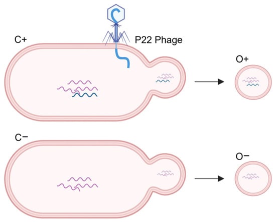
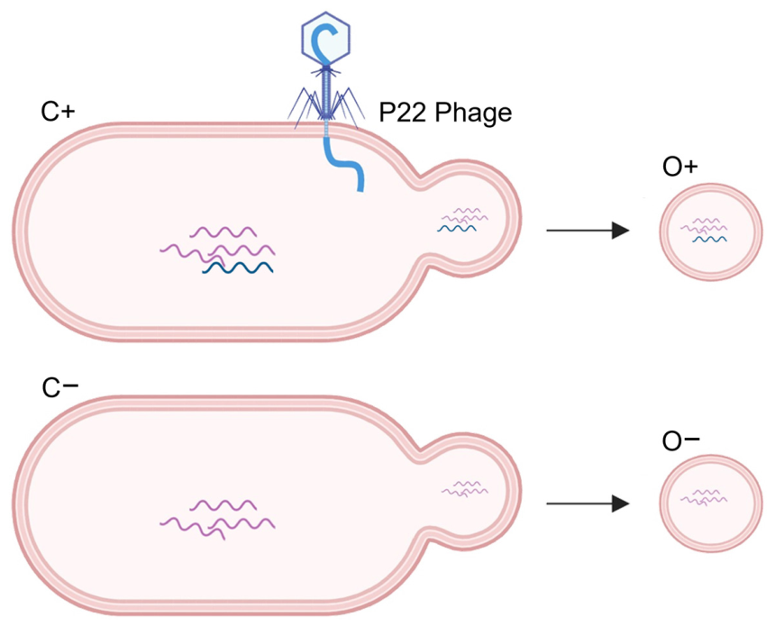
To identify sncRNAs expressed by Salmonella P22 during infection, four separate RNA isolates were collected from Salmonella enterica serovar Typhimurium (strain LT2) cultures on three separate occasions, and ~20 million high-quality reads were generated from each. As illustrated in Figure 1, C+ RNA refers to cytosolic RNA isolated from Salmonella cells infected with the P22 bacteriophage; O+ RNA refers to RNA isolated from OMVs secreted from Salmonella infected with P22; C −RNA refers to cellular RNA isolated from Salmonella cells not infected with P22; and O− RNA refers to RNA isolated from OMVs secreted from Salmonella not infected with P22. In agreement with a recent examination of E. coli OMVs finding comparable OMVs averaging 123.9 nm in diameter [7], the ZetaView visualization of our O−OMV isolates identified a single, similarly sized particle pool averaging 122.7 nm in diameter. Notably, in addition to observing a particle pool averaging ~120 nm in O+ OMV isolates, distinct particle pools averaging 70.1 nm and 208.6 were also detected, and based on previous analyses [15], they were presumed to correspond to P22 phage and OMV:P22 associated particles, respectively (Supplemental Scheme S1).
Figure 1.
Cartoon of RNA isolates. C+ indicates RNA isolated from Salmonella cells infected with P22 bacteriophage; O+ indicates RNA isolated from outer membrane vesicles secreted from Salmonella cells infected with P22; C− indicates RNA isolated from Salmonella cells not infected with P22; O− indicates RNA isolated from outer membrane vesicles secreted from Salmonella cells not infected with P22. Purple wavy lines indicate Salmonella RNAs. Blue wavy lines indicate RNAs expressed from P22.
2.2. Salmonella sncRNAs Affected by P22
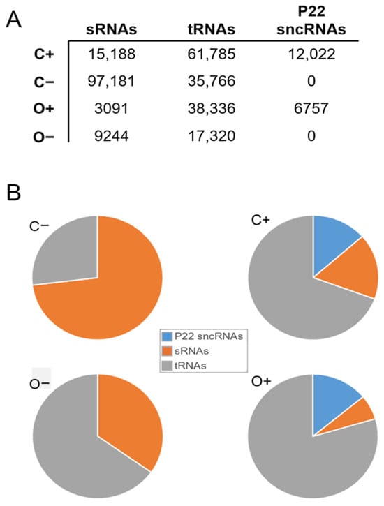
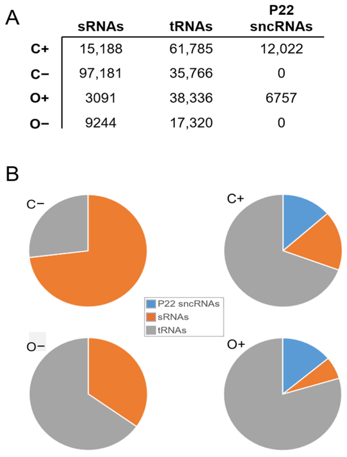
All reads were independently aligned to the LT2 genome. Whereas nearly 10% (97,181 reads per million (RPM)) of C− reads corresponded to one of the 929 Salmonella sRNAs reported to date, the percentage of reads aligning to annotated sRNAs was reduced to only 1.5% (15,188 RPM) during P22 infection. Reads corresponding to Salmonella sRNAs were similarly reduced in OMV isolates during P22 infection, from 9244 RPM in O− to 3091 RPM in O+. The cytosolic expression of only one appreciably expressed (>25 RPM) sRNA, FinP, was increased by ≥100% during P22 infection. Conversely, the cytosolic expression levels of 151 sRNAs appreciably expressed (>25 RPM) in noninfected cells decreased to less than half their normal expression levels during P22 infection (Table 1). In stark contrast to sRNA effects, P22 infection resulted in an approximate doubling of tRNA abundance in both the cytosol (from 35,766 RPM in C− to 61,785 RPM in C+) and OMVs (from 17,320 RPM in O− to 38,336 RPM in O+) (Figure 2, Table 1). Strikingly, the cytosolic expression levels of all tRNAs were increased by at least 50% during P22 infection. Interestingly, however, while most tRNA OMV abundances were also increased during P22 infection, the expression levels of tRNAAsp and tRNAMet were markedly depleted in OMVs during infection (Table 1). Similarly, we found that 37 of 45 sRNAs with appreciable OMV expression levels (>25 RPM) decreased to less than half their normal expression levels during P22 infection. Finally, we observed clear differences between cytosolic and OMV sRNA profiles potentially indicative of the selection and active loading of specific sRNAs. Whereas most sRNAs are significantly more abundant in cytosolic than OMV isolates, we found the OMV abundances of four sRNAs with appreciable OMV expression levels to be roughly doubled during P22 infection (e.g., FinP and SsrS), and furthermore, the OMV expression levels of several sRNAs (e.g., FinP, IsrB2 and npcRNA44) were comparable to cytosolic levels (Table 1).
Table 1.
Select Salmonella sncRNA expressions. Select housekeeping (top), sRNA (middle), and tRNA (bottom) sncRNAs. sncRNA, name based on Ensembl annotation except for “tRNAthr-attP”, which is identically represented in both the Salmonella and P22 genomes. RPM, reads per million. C−, RNA reads isolated from Salmonella cells not infected with P22; C+, RNA reads isolated from Salmonella cells infected with P22; O−, RNA reads isolated from OMVs secreted from Salmonella cells that were not infected with P22; O+, RNA reads isolated from OMVs secreted from Salmonella cells infected with P22. p values of two-sample t-tests comparing C+/C− and O+/O− are indicated. “ns”, not significant (p value ≥ 0.05).
Figure 2.
Effects of P22 infection on Salmonella sncRNA profiles. (A) Number of reads per million aligning to sRNAs (annotated Salmonella sRNAs), tRNAs (annotated Salmonella tRNAs), and P22 sncRNAs (ten unique P22 sncRNAs described in this study). C+, C−, O+, and O− refer to isolates as defined in Figure 1’s legend. (B) Pie charts depicting percentages of reads aligning to total number of sRNAs, tRNAs, and P22 sncRNA alignments.
2.3. SncRNAs Expressed by P22
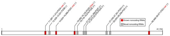
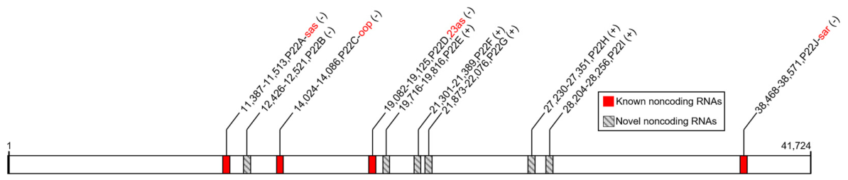
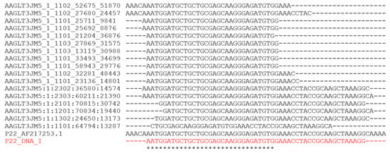
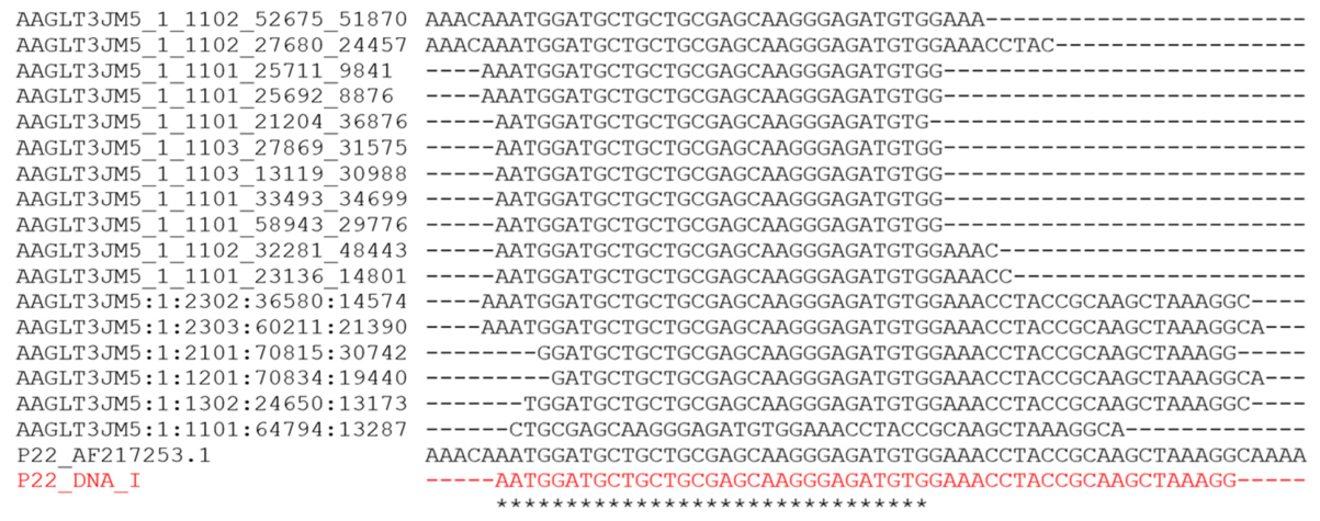
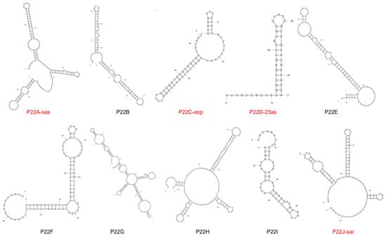
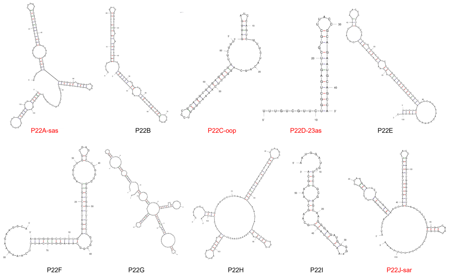
All reads were also independently aligned to the P22 genome. We found that the majority of reads perfectly aligning to the P22 genome identically aligned to a 46 nt sequence 100% conserved in the Salmonella tRNAthr genomic locus. That said, while these reads aligned to both genomes, over 99% of these reads additionally aligned to a flanking sequence specific to the Salmonella genome, confirming that they correspond to Salmonella transcripts. In addition to these tRNA-aligning reads, large numbers of reads perfectly aligning to the P22 genome, but not the Salmonella genome, were also identified, and critically, these reads only occurred in P22-infected isolate datasets. A total of 99.8% of these reads aligned to ten unique positions in P22, accounting for less than 3% of the total genomic sequence. As such, these positions were considered putative P22 sncRNA loci collectively expressing ten sncRNAs appreciably expressed from the P22 genome during infection (Figure 3, Table 2). Importantly, we found that four of these loci correspond to the only four P22 sncRNAs reported to date: sas [16], oop [17,18], 23as [19], and sar [20]. That said, we found each of the six remaining hitherto unreported putative sncRNAs well-supported by significant numbers of independent reads (see Figure 4 for example P22I alignments) and validated the identities of two of the putative sncRNAs by employing a series of specific internal and flanking primers for RT-PCR (Supplemental Scheme S2). Notably, one of the previously characterized sncRNAs we found to be appreciably expressed from the P22 genome during infection, sar, has been described in a number of distinct bacteriophage genomes [21], and as such, we elected to examine whether the novel sncRNAs identified in this work might be similarly conserved. Importantly, we found each of these novel sncRNAs identically maintained in a number of additional bacteriophage genomes (Supplemental Table S1), and furthermore, we found notable similarities between the secondary structures of each of these novel sncRNAs and the characteristic hairpin-heavy structure of sar sncRNA [21,22] (Figure 5).
Figure 3.
SncRNAs transcribed from P22 genome during phage infection. Scale schematic of P22 genome (1–41,724 basepairs) with specific sncRNAs represented by shaded rectangles. SncRNA names assigned in this study (P22A-J arbitrarily assigned by position), genomic positions, strands (+ or -), and previously reported names are indicated. Previously reported sncRNA positions and names are shown in red.
Table 2.
SncRNAs expressed from P22 genome. sncRNA, arbitrary name based on position in P22 genome. If applicable, names of previously reported sncRNAs are included following arbitrary designation assigned by this study. P22 nt Start, start position of RNA in P22 genome. P22 nt End, end position of RNA in P22 genome; P22 strand, strand (− or +) that sncRNA is expressed from; RNA Sequence, sequence that corresponds to denoted start and stop positions in P22 genome. Sal (Y/N) indicates whether or not sequence is also present in Salmonella genome. RPM, reads per million. O−, RNA reads isolated from OMVs secreted from Salmonella cells that were not infected with P22; C−, RNA reads isolated from Salmonella cells not infected with P22; O+, RNA reads isolated from OMVs secreted from Salmonella cells infected with P22; C+, RNA reads isolated from Salmonella cells infected with P22.
Figure 4.
P22I-defining reads. Alignment of P22 genome AF217253.1 (middle), P22I sncRNA (red), and select O+ reads (top) used to define P22I. P22_DNA_I indicates final sncRNA consensus sequence call (as included in Table 1). Final sncRNA calls required each position to be included in ≥80% of full set of aligning reads. “*” signifies nucleotide alignment.
Figure 5.
P22 sncRNA secondary structures. The most thermodynamically stable secondary structures predicted by MFold [21]. The names of previously reported sncRNAs are indicated in red.
Perhaps most notably, we find P22 sncRNAs present in both the cytosol and OMVs. That said, whereas Salmonella sRNAs are slightly more abundant than P22 sncRNAs in the cytosol (15,188 RPM to 12,022 RPM), P22 sncRNA abundance is more than double that of Salmonella sRNAs in OMVs (6757 and 3091 RPM, respectively) potentially indicating the preferential loading of P22 sncRNAs into Salmonella OMVs (Figure 2).
2.4. P22 sncRNA Putative Targets
Each of the four previously reported P22 sRNAs occur antisense to (embedded within and on the opposite strand of) protein-coding genes. In contrast to these antisense sRNAs, the six hitherto uncharacterized P22 sRNAs are each located on the same strand and just upstream (average 21 nt) of a protein-coding gene. As such, all ten P22 sncRNAs can be placed into one of two categories: (1) sncRNAs with 3′ ends located just upstream of a protein-coding gene on the same strand and (2) sncRNAs overlapping and antisense to a protein-coding gene. Six sncRNAs fall into Group 1 as follows: The 3′ end of P22B is 16 nt from the start codon of Gene 24 [2,23]. The 3′ end of P22E is 48 nt from the start codon of Gene 13 [24,25]. The 3′ end of P22F is 6 nt from the start codon of rha. The 3′ end of P22G is 16 nt from the start codon of ORF 25. The 3′ end of P22H is 17 nt from the start codon of Gene 1 (portal protein) [26]. And the 3′ end of P22I is 23 nt from the start codon of Gene 5 (coat/capsid protein) [27]. Interestingly, each of these genes is found sandwiched between two protein-coding genes located on the same strand as the sncRNA. In light of this, these sncRNAs could conceivably function analogously to the sncRNA IsrK encoded by the Gifsy-1 prophage of Salmonella. The 3′ end of IsrK RNA is located only 29 nt from the start codon of a downstream ORF, and the production of the upstream toxic AntQ protein is directly regulated by an interaction between IsrK sncRNA and the downstream ORF [28]. The remaining four sncRNAs fall into Group 2, and each of these has been previously reported. These include: P22A-sas, which is antisense to SieB [16]; P22C-oop, which is antisense to C1 [17,18]; P22D-23as, which is antisense to Gene 23 [19]; and P22J-sar, which is antisense to Ant [20] (Table 3).
Table 3.
P22 sncRNA putative targets. sncRNA, arbitrary name based on the position in the P22 genome. Putative target/function, presumed targets based on being either antisense to or just upstream of a P22 protein-coding gene. The known functions of characterized P22 sncRNAs and/or functions of homologous protein targets in other bacteriophages are included if available.
3. Discussion
OMVs have been associated with a number of different cellular functions such as intercellular communication (including delivery of encapsulated RNAs) [8,9,10,11] and resistance to phage (e.g., Salmonella OMVs significantly inhibit P22 bacteriophage infection [12]). As bacterial sRNAs also participate in bacteriophage defense [13] and are specifically delivered to and enriched in OMVs via Hfq [14], we recently elected to examine the effects of P22 infection on Salmonella cytosolic and OMV sRNA abundance.
We found that P22 infection triggers a global reduction in sRNAs (with Salmonella sRNA expression levels averaging only 15.6% those observed in noninfected cells) coupled with a reciprocal 72.7% global increase in Salmonella tRNA expression levels. In addition to this, these analyses also identified ten sncRNAs (four previously annotated and six unreported) appreciably expressed from the P22 genome during infection (Figure 3, Table 2), and much to our surprise, we found that these bacteriophage-encoded sRNAs actively secreted within host OMVs.
3.1. Salmonella sRNAs
We find that 166 sRNAs are appreciably expressed (>25 RPM) in the cytosol of noninfected cells and find that the expression levels of nearly all of these (151 of 166) decreased to less than half their normal levels during P22 infection. Conversely, we find that the cytosolic expression of only one appreciably expressed sRNA, FinP, increased by ≥50% during P22 infection, and strikingly, we find that the expression of this sRNA (known to be directly involved in regulating plasmid transfer [28,29]) is actually increased by over 500% during infection (Table 1). Furthermore, whereas the OMV levels of most Salmonella sncRNAs are normally ~1/10 that found in the cytosol [30], we find that P22 infection triggers active OMV encapsulation and secretion of a subset of sRNAs. As specific examples, unlike most sRNAs, the OMV abundances of GcvB and RprA are roughly equal to their cytosolic levels, suggesting the differential loading of and active enrichment in these specific sRNAs within OMVs (Table 1). It is tempting to speculate that P22-driven OMV enrichment in these sRNAs and the subsequent delivery of these to recipients could potentially be involved in facilitating P22 infections in neighboring cells.
3.2. Salmonella tRNAs
In stark contrast to sRNAs, P22 infection resulted in an approximate doubling of tRNA abundance in both the cytosol and OMVs (Figure 2, Table 1). The cytosolic expression levels of all tRNAs were increased by at least 50% during P22 infection. That said, while most tRNA OMV abundances were also increased during P22 infection, the expression levels of tRNAAsp, tRNAMet, and tRNAPro were markedly depleted in OMVs (Table 1). Interestingly, we found that roughly 50% of RNAs derived from tRNA loci actually correspond to tRNA fragments (tRFs) in cytosolic isolates and roughly 70% in OMV isolates. That said, while we found the overall percentages of reads corresponding to tRNA fragments to be largely unaffected by P22 infection, we did find that the abundances of a number of specific fragments were markedly changed, suggesting that a more comprehensive evaluation focused specifically on tRNA excisions is likely warranted. Similarly to our current study, in 2015, Ghosal et al. found that the majority of RNA contained within Escherichia coli OMVs has a length < 60 nt with an enrichment between 15 and 40 nt and that E. coli OMV RNA sequencing reads primarily correspond to tRNA fragments (tRFs) [31]. More recently, Koeppen et al. [11] likewise found that tRF levels were significantly higher in RNA isolated from Pseudomonas aeruginosa OMVs than in intracellular RNA, suggesting that tRFs are actively enriched in OMVs prior to secretion. Further, of note, despite tRNAs and tRFs typically constituting only ~15% and 2% of normal human intracellular small RNA populations respectively, much like bacterial OMVs, human exosomes have now been reported by several groups as being similarly enriched with tRFs (typically representing >20% of RNA compositions) [32,33,34,35,36]. Despite this, how tRFs are generated and the functional roles of the majority of tRFs remain unclear, although tRFs have been suggested to have initially arisen as a part of an ancient viral defense [37]. Supporting this concept, human endogenous retrovirus transposition rates can be limited by host tRF expression [38], and specific tRFs are induced by viral infections (e.g., by RSV [39,40]). In addition, increasing titers of HIV have been shown to trigger the production of lysine tRFs that inhibit HIV, and T cells infected with human T-cell leukemia virus type 1 (HTLV-1) similarly upregulate the production of proline tRFs targeting this virus [41]. Together, these findings suggest that tRFs may constitute an uncharacterized component of the innate antiviral response. That said, despite there being several published reports now independently describing (1) roles for specific tRFs in facilitating/inhibiting viral replication [39,40,41,42,43], (2) the marked prevalence of tRFs within secreted vesicles (both exosomes and OMVs) [11,33,34,35,36], and (3) the ability of OMVs to inhibit the phage infection of several bacterial species (e.g., E. coli [44], Prochlorococcus marinus [45], Shigella flexneri [46], Salmonella enterica [12], and Vibrio cholerae [47]), there have been no examinations of the effects of vesicle-secreted tRFs on viral/phage infections reported to date.
3.3. P22 sncRNAs
A number of sncRNAs expressed from various phages have been found to regulate specific gene expressions in their respective hosts [48,49,50]. The most notable Salmonella P22 sRNAs documented so far include oop RNA, a cis-encoded transcriptional repressor whose overexpression likely favors lytic development over lysogenization [17,51], and sar asRNA, a trans-encoded transcriptional repressor located in the intergenic region of the ant gene which is also involved in deciding between lytic development or establishing lysogeny [20]. Interestingly, our analyses suggest that at least five of the ten P22 sncRNAs identified in this work are likely involved in regulating the establishment or maintenance of the lysogenic state (Table 3). That said, extensive research detailing the components necessary for successful bacteriophage infections have described many factors contributing to the interactions between bacteriophages and their host bacteria [5]. While the majority of these efforts have been dedicated to exploring protein interactions, our study represents one of the first aimed at comprehensively identifying the phage sncRNAs expressed during these interactions [48,49,50], and to our knowledge, it is the first to ever examine whether phage sncRNAs are actively secreted in bacterial host OMVs. Notably, we find that phage sncRNAs are not simply confined to the cytosol. Instead, our data indicates that all ten of the P22 sncRNAs identified in this work are actively transported into OMVs and secreted into the extracellular space, potentially indicating a co-opting of bacterial OMVs by infecting phages. Notably, the encapsulation of viral sncRNAs and their transfer to neighboring cells to facilitate infection are well documented for a number of human viruses [52,53]. Regardless of whether or not OMV-encapsulated P22 sncRNAs similarly facilitate P22 infections in neighboring cells, the realization that bacteriophage sncRNAs are being secreted in host OMVs during infection reveals a hitherto overlooked dimension of the interactions between bacteriophages and their hosts.
In summary, the work presented here shows that P22 infection triggers a global reduction in Salmonella sRNAs coupled with a reciprocal global increase in Salmonella tRNA expression levels. Further, we find that P22 infection triggers active OMV encapsulation and secretion of: (1) a subset of Salmonella sRNAs, (2) all Salmonella tRNAs including one highly complementary to the P22 genome, and (3) ten distinct sncRNAs expressed from P22 itself. That said, while the initial realization that bacteriophage sncRNAs are being loaded into OMVs (and presumably delivered to neighboring cells) is exciting, the elucidation of specific targets and functions for the majority of these sncRNAs remains wholly unresolved and will ultimately require future studies employing experimental methodologies capable of directly characterizing RNA–target interactions.
4. Materials and Methods
Strain and growth conditions. Salmonella enterica serovar Typhimurium strain LT2 (ATCC 19585) was cultured and maintained on LB agar (1% w/v tryptone, 1% w/v NaCl, 0.5% w/v yeast extract, 1.5% w/v agar) and incubated overnight at 37 °C. For the preparation of the cell suspension, a loop full bacterial agar culture was inoculated in LB broth (1% w/v tryptone, 1% w/v NaCl, 0.5% w/v yeast extract) and incubated overnight at 37 °C with shaking. For the P22 virus, the lysate was prepared by inoculating 50 µL of Salmonella enterica subsp. enterica serovar Typhimurium bacteriophage P22 (ATCC 19585-B1) in a log-phase attained 1:100 diluted overnight culture of Salmonella enterica subsp. enterica serovar Typhimurium LT2 (ATCC 19585). Log-phase cultures were used for all infection experiments to ensure efficient and reproducible P22 infection under conditions that support active phage replication. The culture was grown until it became clear, signifying the lysis of host bacterial cells. The culture was filtered and then centrifuged for 10 min at 3000 rpm. The lysate was then transferred into a sterile vial containing 500 µL of chloroform and stored at 4 °C until further use. The PFU/mL of the freshly prepared lysate was counted using a standard Plaque-forming assay protocol.
Infecting Salmonella with P22. Log-phase Salmonella cultures containing 5.0 × 10⁸ CFU/mL were infected with 100 µL of P22 lysate with a titer of 1.1 × 10⁹ PFU/mL, corresponding to a multiplicity of infection (MOI) of 0.225, then incubated with shaking at 220 rpm for 1 h at 37 °C. Subsequent centrifugation at 5000× g for 20 min facilitated the separation of viral particles and cellular components. The resulting supernatant, containing extracellular components, was carefully collected for outer membrane vesicle (OMV) isolation, while the cell suspension containing pellet was stored at −80 °C for later RNA isolation. A control experiment using 50 mL of Salmonella log-phase culture without the addition of P22 virus served as baseline.
OMV isolation, visualization, and RNA extraction. OMVs were isolated from the supernatant using the ExoBacteria OMV isolation kit (cat# EXOBAC100A-1, System Biosciences, Palo Alto, CA, USA) in accordance with the manufacturer’s guidelines. RNA was isolated from OMVs and cell pellets using TRIzol (cat# 15596016, Thermo Fisher Scientific, Carlsbad, CA, USA). OMV and cellular RNAs were quantified and assessed using a Qubit 4 Fluorometer (cat# Q33238, Life Technologies Holdings Pte Ltd., Thermo Fisher Scientific, Singapore) and NanoDrop (cat# 840281500,Thermo Fisher Scientific, Waltham, MA, USA) respectively. To confirm the isolation of OMVs, a PMX 220 ZetaView TWIN Laser Nanoparticle tracking analyzer (Particle Metrix GmbH, Bavaria, Germany) was employed to image isolates at 100–200 nm. Samples were diluted 1:10 in ExoBacteria OMV isolation kit elution buffer, and elution buffer alone was run as baseline.
RNA sequencing and analysis. RNA samples isolated on three separate occasions from Salmonella cells infected with P22 (C+), OMVs secreted by Salmonella infected with P22 (O+), and control samples from noninfected Salmonella C− and O− were sent to the High Throughput Sequencing Facility, University of North Carolina, Chapel Hill, where small RNA-seq using an Illumina NextSeq genomic sequencer was performed (single ends, 101 nt, 15M reads). Reads were analyzed using standard Borchert Laboratory protocols [54,55,56,57,58,59,60,61,62]. In brief, raw RNA seq reads were first assessed for quality using FastQC [63]. Adapter sequence (small RNA adapter: 5′-TGGAATTCTCGGGTGCCAAGG-3′), low-quality bases (minimum Phred score 20) were removed using Cutadapt [64], and any reads shorter than 20 nt post-trimming were discarded. FastQC [63] quality analysis was repeated post-trimming to confirm adapter removal and length distributions. The resulting FASTQ sequence files were converted to FASTA format, then reads aligned against the P22 genome (AF217253.1) and LT2 genome (NC_003197.2) with masked ribosomal loci using Standalone BLASTn 2.15.0+ [65]. BLAST parameters were required to be ungapped, have an e value ≤ 0.000001, and have 100% identity across a full alignment of no less than 20 nt. The longest alignment between each read and the genome was identified and all other alignments discarded. Less than 0.02% of reads best mapped to ≥two genomic locations. As each of these multimapping events were found to correspond to specific tRNAs identically represented in the genome, these loci were collectively considered and counted as single alignments. Gene counts were determined by summing the number of aligning reads at each genomic position then averaging the number of aligning reads at each position within Ensembl-defined gene annotations [66].
RT-PCR analyses. RNA samples isolated from Salmonella cells infected with P22 (C+), OMVs secreted by Salmonella infected with P22 (O+), and control samples from noninfected Salmonella C− and O− were used to generate cDNA using Applied Biosystems High-Capacity cDNA Reverse Transcription Kit (cat# 4374967, Thermo Fisher Scientific Baltic UAB, Vilnius, Lithuania). Primer sequences are listed in Supplementary Scheme S2. PCRs were performed with 1 μL (10 μM) forward primer, 1 μL (10 μM) reverse primer, 4 μL MgCl2 (25 mM), 1 μL DNTP (10 mM each), 5 μL 10× Taq buffer, 1 μL Taq polymerase (1 U/μL) (cat# FEREP0404, Thermo Fisher Scientific Baltics UAB, Vilnius, Lithuania), and 10 ng cDNA brought to 50 μL with DNA-grade H2O (cat# AM9922, Thermo Fisher Scientific, Carlsbad, CA, USA). The thermal cycling parameters for these reactions were an initial denaturation at 95 °C for 1 min, followed by 30 cycles of 30 s at 95 °C, 30 s at 58 °C, and 30 s at 72 °C, then a final extension step at 72 °C for 2 min. PCR products were then run on a 1% agarose TBE gel for 1 h at 90 V and stained with EtBr (cat# BP102-5, Fisher Scientific Company, Fair Lawn, NJ, USA).
5. Conclusions
In conclusion, the study presented here shows that P22 bacteriophage infection of its host Salmonella enterica causes a reduction in host sRNAs while increasing tRNA abundance simultaneously. In addition, the infection promotes selective encapsulation and presumably, the secretion of distinct RNA molecules via OMV including host sRNAs, tRNAs and ten bacteriophage-derived small non coding RNA (sncRNAs) to the neighboring cells. These findings shed light on the complex nature of OMV mediated communication and warrant further research into the regulatory roles of the secreted RNA species in phage-host dynamics.
Supplementary Materials
The following supporting information can be downloaded at https://www.mdpi.com/article/10.3390/ncrna12040021/s1, Supplemental Table S1: P22 sncRNAs identically maintained in other bacteriophage genomes. sncRNA, arbitrary name based on position in P22 genome. “Bacteriophage genomes containing the full length, identical sequence” lists bacteriophage genomes bearing 100% identity to indicated P22 RNAs as determined using NCBI Virus [67]. IDs correspond to phage published in NCBI Reference Sequence Database (RefSeq) [68]. Individual phage IDs are separated by commas, and primary host specificity is indicated and separated by semicolons. Supplemental Scheme S1: ZetaView of OMV isolates. (A) OMV from O− sample (left) and from O+ sample (right). (B) OMV from O− sample (left) and from O+ sample (right) following Proteinase K (PK) treatment. (C) Buffer control (left) and 125 Thermo Scientific 3000 Series Nanosphere Size Standard (right). Supplemental Scheme S2: RT-PCR validation of P22 sncRNAs. RT-PCR performed on RNA isolates from P22-infected (C+) and -noninfected (C-) Salmonella. (Top left) Sequences of putative P22 sncRNAs identified by sequence read alignments are shown to right of their genomic position and strand. (Bottom left) Putative P22 sncRNA sequences are highlighted within their local genomic sequences along with color-coded primers employed for RT-PCR. F and R primers were designed within called sncRNA. Fout and Rout primers were designed in sequences flanking called sncRNA. (Right) PCR amplicons visualized after separation on 1.0% agarose gels stained with EtBr. (A) P22F. (B) P22H.
Author Contributions
S.N. was responsible for formal analysis, investigation, visualization, writing—original draft, writing—review and editing and supervision. H.A.K., L.A.H.-G., A.M.M., E.S.A., A.I.H., O.G.C., D.M.K., F.O.P., O.R.R., M.A.S., B.N.S., I.M.S., E.M.H., J.D.D., S.S.P., R.H.P. and M.R.C. were responsible for formal analysis, investigation, visualization, and writing—review and editing. G.M.B. was responsible for conceptualization, funding acquisition, methodology, visualization, writing—original draft, writing—review and editing and supervision. S.N. and G.M.B. had full access to all of the data in this study and take responsibility for the integrity of the data and accuracy of the data analysis. All authors have read and agreed to the published version of the manuscript.
Funding
Funding was provided in part by NSF 2219900 (GMB) awarded by the Systems and Synthetic Biology program in the Division of Molecular and Cellular Biosciences. The project was also funded in part by NSF grant 2243532 (GMB) awarded by the Division of Integrative Organismal Systems.
Institutional Review Board Statement
Not applicable. This study did not involve human participants, human tissue samples, or identifiable private information. Therefore, Institutional Review Board approval was not required.
Informed Consent Statement
Not applicable. This study did not involve human participants, and no informed consent was required. All analyses were performed using sequencing data and did not include any identifiable human subject information.
Data Availability Statement
All small RNA-sequencing datasets generated were deposited in the NCBI SRA repository. Two sets of replicates are available under Bioproject PRJNA1307193 (SRR35017900, SRR35017901, SRR35017902, SRR35017903, SRR35996074, SRR35996075, SRR35996076, SRR35996077), and a third set of replicates is available under Bioproject PRJNA1141709 (SRR30031405, SRR30031404, SRR30031403, SRR30031402). All other relevant data (e.g., alignment files) are available from the authors upon request.
Acknowledgments
We thank the University of South Alabama College of Medicine Department of Pharmacology for ongoing support.
Conflicts of Interest
The authors declare no conflicts of interest.
References
- Fabrega, A.; Vila, J. Salmonella enterica serovar Typhimurium skills to succeed in the host: Virulence and regulation. Clin. Microbiol. Rev. 2013, 26, 308–341. [Google Scholar] [CrossRef] [Scilit] [PubMed]
- Vander Byl, C.; Kropinski, A.M. Sequence of the genome of Salmonella bacteriophage P22. J. Bacteriol. 2000, 182, 6472–6481. [Google Scholar] [CrossRef] [Scilit] [PubMed]
- Neal, B.L.; Brown, P.K.; Reeves, P.R. Use of Salmonella phage P22 for transduction in Escherichia coli. J. Bacteriol. 1993, 175, 7115–7118. [Google Scholar] [CrossRef] [Scilit] [PubMed]
- Fillol-Salom, A.; Bacigalupe, R.; Humphrey, S.; Chiang, Y.N.; Chen, J.; Penadés, J.R. Lateral transduction is inherent to the life cycle of the archetypical Salmonella phage P22. Nat. Commun. 2021, 12, 6510. [Google Scholar] [CrossRef] [Scilit] [PubMed]
- Brussow, H.; Canchaya, C.; Hardt, W.D. Phages and the evolution of bacterial pathogens: From genomic rearrangements to lysogenic conversion. Microbiol. Mol. Biol. Rev. 2004, 68, 560–602. [Google Scholar] [CrossRef] [Scilit] [PubMed]
- Beveridge, T.J. Structures of gram-negative cell walls and their derived membrane vesicles. J. Bacteriol. 1999, 181, 4725–4733. [Google Scholar] [CrossRef] [Scilit] [PubMed]
- Reimer, S.L.; Beniac, D.R.; Hiebert, S.L.; Booth, T.F.; Chong, P.M.; Westmacott, G.R.; Zhanel, G.G.; Bay, D.C. Comparative Analysis of Outer Membrane Vesicle Isolation Methods with an Escherichia coli tolA Mutant Reveals a Hypervesiculating Phenotype with Outer-Inner Membrane Vesicle Content. Front. Microbiol. 2021, 12, 628801. [Google Scholar] [CrossRef] [Scilit] [PubMed]
- Liu, X.; Xiao, J.; Wang, S.; Zhou, J.; Qin, J.; Jia, Z.; Wang, Y.; Wang, Z.; Zhang, Y.; Hao, H. Research Progress on Bacterial Membrane Vesicles and Antibiotic Resistance. Int. J. Mol. Sci. 2022, 23, 11553. [Google Scholar] [CrossRef] [Scilit] [PubMed]
- Kim, S.W.; Park, S.B.; Im, S.P. Outer membrane vesicles from beta-lactam-resistant Escherichia coli enable the survival of beta-lactam-susceptible E. coli in the presence of beta-lactam antibiotics. Sci. Rep. 2018, 8, 5402. [Google Scholar] [CrossRef] [Scilit] [PubMed]
- Bonnington, K.E.; Kuehn, M.J. Protein selection and export via outer membrane vesicles. Biochim. Biophys. Acta 2014, 1843, 1612–1619. [Google Scholar] [CrossRef] [Scilit] [PubMed]
- Koeppen, K.; Hampton, T.H.; Jarek, M.; Scharfe, M.; Gerber, S.A.; Mielcarz, D.W.; Demers, E.G.; Dolben, E.L.; Hammond, J.H.; Hogan, D.A.; et al. A Novel Mechanism of Host-Pathogen Interaction through sRNA in Bacterial Outer Membrane Vesicles. PLoS Pathog. 2016, 12, e1005672. [Google Scholar] [CrossRef] [Scilit] [PubMed]
- Stephan, M.S.; Broeker, N.K.; Saragliadis, A.; Roos, N.; Linke, D.; Barbirz, S. In vitro Analysis of O-Antigen-Specific Bacteriophage P22 Inactivation by Salmonella Outer Membrane Vesicles. Front. Microbiol. 2020, 11, 510638. [Google Scholar] [CrossRef] [Scilit] [PubMed]
- Sprenger, M.; Siemers, M.; Krautwurst, S.; Papenfort, K. Small RNAs direct attack and defense mechanisms in a quorum sensing phage and its host. Cell Host Microbe 2024, 32, 727–738.e6. [Google Scholar] [CrossRef] [Scilit] [PubMed]
- Velez, M.; Arluison, V. Does the Hfq Protein Contribute to RNA Cargo Translocation into Bacterial Outer Membrane Vesicles? Pathogens 2025, 14, 399. [Google Scholar] [CrossRef] [Scilit] [PubMed]
- Hulo, C.; de Castro, E.; Masson, P.; Bougueleret, L.; Bairoch, A.; Xenarios, I.; Le Mercier, P. ViralZone: A knowledge resource to understand virus diversity. Nucleic Acids Res. 2011, 39, D576–D582. [Google Scholar] [CrossRef] [Scilit] [PubMed]
- Ranade, K.; Poteete, A.R. A switch in translation mediated by an antisense RNA. Genes Dev. 1993, 7, 1498–1507. [Google Scholar] [CrossRef] [Scilit] [PubMed]
- Backhaus, H.; Petri, J.B. Sequence analysis of a region from the early right operon in phage P22 including the replication genes 18 and 12. Gene 1984, 32, 289–303. [Google Scholar] [CrossRef] [Scilit]
- Mondal, A.; Chattopadhyaya, R.; Datta, A.B.; Parrack, P. Crystallization and X-ray analysis of the transcription-activator protein C1 of bacteriophage P22 in complex with the PRE promoter element. Acta Crystallogr. F Struct. Biol. Commun. 2015, 71, 1286–1291. [Google Scholar] [CrossRef] [Scilit] [PubMed]
- Ho, Y.S.; Pfarr, D.; Strickler, J.; Rosenberg, M. Characterization of the transcription activator protein C1 of bacteriophage P22. J. Biol. Chem. 1992, 267, 14388–14397. [Google Scholar] [CrossRef] [Scilit] [PubMed]
- Liao, S.M.; Wu, T.H.; Chiang, C.H.; Susskind, M.M.; McClure, W.R. Control of gene expression in bacteriophage P22 by a small antisense RNA. I. Characterization in vitro of the Psar promoter and the sar RNA transcript. Genes Dev. 1987, 1, 197–203. [Google Scholar] [CrossRef] [Scilit] [PubMed]
- Schaefer, K.L.; McClure, W.R. Antisense RNA control of gene expression in bacteriophage P22. I. Structures of sar RNA and its target, ant mRNA. RNA 1997, 3, 141–156. [Google Scholar] [PubMed]
- Zuker, M. Mfold web server for nucleic acid folding and hybridization prediction. Nucleic Acids Res. 2003, 31, 3406–3415. [Google Scholar] [CrossRef] [Scilit] [PubMed]
- Wilson, H.R.; Yu, D.; Peters, H.K., 3rd; Zhou, J.G.; Court, D.L. The global regulator RNase III modulates translation repression by the transcription elongation factor N. EMBO J. 2002, 21, 4154–4161. [Google Scholar] [CrossRef] [Scilit] [PubMed]
- Nam, K.; Blasi, U.; Zagotta, M.T.; Young, R. Conservation of a dual-start motif in P22 lysis gene regulation. J. Bacteriol. 1990, 172, 204–211. [Google Scholar] [CrossRef] [Scilit] [PubMed]
- Blasi, U.; Chang, C.Y.; Zagotta, M.T.; Nam, K.B.; Young, R. The lethal lambda S gene encodes its own inhibitor. EMBO J. 1990, 9, 981–989. [Google Scholar] [CrossRef] [Scilit] [PubMed]
- Bazinet, C.; Benbasat, J.; King, J.; Carazo, J.M.; Carrascosa, J.L. Purification and organization of the gene 1 portal protein required for phage P22 DNA packaging. Biochemistry 1988, 27, 1849–1856. [Google Scholar] [CrossRef] [Scilit] [PubMed]
- Bazinet, C.; King, J. Initiation of P22 procapsid assembly in vivo. J. Mol. Biol. 1988, 202, 77–86. [Google Scholar] [CrossRef] [Scilit] [PubMed]
- Yang, J.; Wu, R.; Xia, Q.; Yu, J.; Yi, L.X.; Huang, Y.; Deng, M.; He, W.Y.; Bai, Y.; Lv, L.; et al. The evolution of infectious transmission promotes the persistence of mcr-1 plasmids. mBio 2023, 14, e0044223. [Google Scholar] [CrossRef] [Scilit] [PubMed]
- Getino, M.; de la Cruz, F. Natural and Artificial Strategies To Control the Conjugative Transmission of Plasmids. Microbiol. Spectr. 2018, 6, 1–25. [Google Scholar] [CrossRef] [Scilit] [PubMed]
- Malabirade, A.; Habier, J.; Heintz-Buschart, A.; May, P.; Godet, J.; Halder, R.; Etheridge, A.; Galas, D.; Wilmes, P.; Fritz, J.V. The RNA Complement of Outer Membrane Vesicles From Salmonella enterica Serovar Typhimurium Under Distinct Culture Conditions. Front. Microbiol. 2018, 9, 2015. [Google Scholar] [CrossRef] [Scilit] [PubMed]
- Ghosal, A.; Upadhyaya, B.B.; Fritz, J.V.; Heintz-Buschart, A.; Desai, M.S.; Yusuf, D.; Huang, D.; Baumuratov, A.; Wang, K.; Galas, D.; et al. The extracellular RNA complement of Escherichia coli. MicrobiologyOpen 2015, 4, 252–266. [Google Scholar] [CrossRef] [Scilit] [PubMed]
- Li, F.; Kaczor-Urbanowicz, K.E.; Sun, J.; Majem, B.; Lo, H.C.; Kim, Y.; Koyano, K.; Rao, S.L.; Kang, S.Y.; Kim, S.M.; et al. Characterization of Human Salivary Extracellular RNA by Next-generation Sequencing. Clin. Chem. 2018, 64, 1085–1095. [Google Scholar] [CrossRef] [Scilit] [PubMed]
- Danielson, K.M.; Rubio, R.; Abderazzaq, F.; Das, S.; Wang, Y.E. High Throughput Sequencing of Extracellular RNA from Human Plasma. PLoS ONE 2017, 12, e0164644. [Google Scholar] [CrossRef] [Scilit] [PubMed]
- Lässer, C.; Shelke, G.V.; Yeri, A.; Kim, D.K.; Crescitelli, R.; Raimondo, S.; Sjöstrand, M.; Gho, Y.S.; Van Keuren Jensen, K.; Lötvall, J. Two distinct extracellular RNA signatures released by a single cell type identified by microarray and next-generation sequencing. RNA Biol. 2017, 14, 58–72. [Google Scholar] [CrossRef] [Scilit] [PubMed]
- Baglio, S.R.; Rooijers, K.; Koppers-Lalic, D.; Verweij, F.J.; Pérez Lanzón, M.; Zini, N.; Naaijkens, B.; Perut, F.; Niessen, H.W.; Baldini, N.; et al. Human bone marrow- and adipose-mesenchymal stem cells secrete exosomes enriched in distinctive miRNA and tRNA species. Stem Cell Res. Ther. 2015, 6, 127. [Google Scholar] [CrossRef] [Scilit] [PubMed]
- Bellingham, S.A.; Coleman, B.M.; Hill, A.F. Small RNA deep sequencing reveals a distinct miRNA signature released in exosomes from prion-infected neuronal cells. Nucleic Acids Res. 2012, 40, 10937–10949. [Google Scholar] [CrossRef] [Scilit] [PubMed]
- Schorn, A.J.; Martienssen, R. Tie-break: Host and retrotransposons play tRNA. Trends Cell Biol. 2018, 28, 793–806. [Google Scholar] [CrossRef] [Scilit] [PubMed]
- Schorn, A.J.; Gutbrod, M.J.; LeBlanc, C.; Martienssen, R. LTR-Retrotransposon Control by tRNA-Derived Small RNAs. Cell 2017, 170, 61–71.e11. [Google Scholar] [CrossRef] [Scilit] [PubMed]
- Deng, J.; Ptashkin, R.N.; Chen, Y.; Cheng, Z.; Liu, G.; Phan, T.; Deng, X.; Zhou, J.; Lee, I.; Lee, Y.S.; et al. Respiratory Syncytial Virus Utilizes a tRNA Fragment to Suppress Antiviral Responses Through a Novel Targeting Mechanism. Mol. Ther. J. Am. Soc. Gene Ther. 2015, 23, 1622–1629. [Google Scholar] [CrossRef] [Scilit] [PubMed]
- Zhou, J.; Liu, S.; Chen, Y.; Fu, Y.; Silver, A.J.; Hill, M.S.; Lee, I.; Lee, Y.S.; Bao, X. Identification of two novel functional tRNA-derived fragments induced in response to respiratory syncytial virus infection. J. Gen. Virol. 2017, 98, 1600–1610. [Google Scholar] [CrossRef] [Scilit] [PubMed]
- Ruggero, K.; Guffanti, A.; Corradin, A.; Sharma, V.K.; De Bellis, G.; Corti, G.; Grassi, A.; Zanovello, P.; Bronte, V.; Ciminale, V.; et al. Small noncoding RNAs in cells transformed by human T-cell leukemia virus type 1: A role for a tRNA fragment as a primer for reverse transcriptase. J. Virol. 2014, 88, 3612–3622. [Google Scholar] [CrossRef] [Scilit] [PubMed]
- Wang, Q.; Lee, I.; Ren, J.; Ajay, S.S.; Lee, Y.S.; Bao, X. Identification and functional characterization of tRNA-derived RNA fragments (tRFs) in respiratory syncytial virus infection. Mol. Ther. J. Am. Soc. Gene Ther. 2013, 21, 368–379. [Google Scholar] [CrossRef] [Scilit] [PubMed]
- Selitsky, S.R.; Baran-Gale, J.; Honda, M.; Yamane, D.; Masaki, T.; Fannin, E.E.; Guerra, B.; Shirasaki, T.; Shimakami, T.; Kaneko, S.; et al. Small tRNA-derived RNAs are increased and more abundant than microRNAs in chronic hepatitis B and C. Sci. Rep. 2015, 5, 7675. [Google Scholar] [CrossRef] [Scilit] [PubMed]
- Manning, A.J.; Kuehn, M.J. Contribution of bacterial outer membrane vesicles to innate bacterial defense. BMC Microbiol. 2011, 11, 258. [Google Scholar] [CrossRef] [Scilit] [PubMed]
- Biller, S.J.; Schubotz, F.; Roggensack, S.E.; Thompson, A.W.; Summons, R.E.; Chisholm, S.W. Bacterial vesicles in marine ecosystems. Science 2014, 343, 183–186. [Google Scholar] [CrossRef] [Scilit] [PubMed]
- Parent, K.N.; Erb, M.L.; Cardone, G.; Nguyen, K.; Gilcrease, E.B.; Porcek, N.B.; Pogliano, J.; Baker, T.S.; Casjens, S.R. OmpA and OmpC are critical host factors for bacteriophage Sf6 entry in Shigella. Mol. Microbiol. 2014, 92, 47–60. [Google Scholar] [CrossRef] [Scilit] [PubMed]
- Reyes-Robles, T.; Dillard, R.S.; Cairns, L.S.; Silva-Valenzuela, C.A.; Housman, M.; Ali, A.; Wright, E.R.; Camilli, A. Vibrio cholerae outer membrane vesicles inhibit bacteriophage infection. J. Bacteriol. 2018, 200, 1–9. [Google Scholar] [CrossRef] [Scilit] [PubMed]
- Hershko-Shalev, T.; Odenheimer-Bergman, A.; Elgrably-Weiss, M.; Ben-Zvi, T.; Govindarajan, S.; Seri, H.; Papenfort, K.; Vogel, J.; Altuvia, S. Gifsy-1 Prophage IsrK with Dual Function as Small and Messenger RNA Modulates Vital Bacterial Machineries. PLoS Genet. 2016, 12, e1005975, Correction in PLoS Genet. 2017, 13, e1006725. [Google Scholar] [CrossRef] [Scilit] [PubMed]
- Bloch, S.; Lewandowska, N.; Wegrzyn, G.; Nejman-Falenczyk, B. Bacteriophages as sources of small non-coding RNA molecules. Plasmid 2021, 113, 102527. [Google Scholar] [CrossRef] [Scilit] [PubMed]
- Lewandowska, N.; Bloch, S.; Łukasiak, A.; Wesołowski, W.; Węgrzyn, G.; Nejman-Faleńczyk, B. The Role of Bacteriophage-Derived Small RNA Molecules in Bacterial and Phage Interactions. Viruses 2025, 17, 834. [Google Scholar] [CrossRef] [Scilit] [PubMed]
- Krinke, L.; Wulff, D.L. OOP RNA, produced from multicopy plasmids, inhibits lambda cII gene expression through an RNase III-dependent mechanism. Genes Dev. 1987, 1, 1005–1013. [Google Scholar] [CrossRef] [Scilit] [PubMed]
- Pegtel, D.M.; Cosmopoulos, K.; Thorley-Lawson, D.A.; van Eijndhoven, M.A.; Hopmans, E.S.; Lindenberg, J.L.; de Gruijl, T.D.; Würdinger, T.; Middeldorp, J.M. Functional delivery of viral miRNAs via exosomes. Proc. Natl. Acad. Sci. USA 2010, 107, 6328–6333. [Google Scholar] [CrossRef] [Scilit] [PubMed]
- Kalamvoki, M.; Du, T.; Roizman, B. Cells infected with herpes simplex virus 1 export to uninfected cells exosomes containing STING, viral mRNAs, and microRNAs. Proc. Natl. Acad. Sci. USA 2014, 111, E4991–E4996. [Google Scholar] [CrossRef] [Scilit] [PubMed]
- Amin, S.V.; Roberts, J.T.; Patterson, D.G.; Coley, A.B.; Allred, J.A.; Denner, J.M.; Johnson, J.P.; Mullen, G.E.; O’Neal, T.K.; Smith, J.T.; et al. Novel small RNA (sRNA) landscape of the starvation-stress response transcriptome of Salmonella enterica serovar typhimurium. RNA Biol. 2016, 13, 331–342. [Google Scholar] [CrossRef] [Scilit] [PubMed]
- Barnhill, E.C.; Crucello, A.; Houserova, D.; King, V.M.; Amin, S.V.; Roberts, J.T.; Zambrano, M.E.; DeMeis, J.D.; Dahmer, D.J.; Ijaz, Z.; et al. Characterization of novel small RNAs (sRNAs) contributing to the desiccation response of Salmonella enterica serovar Typhimurium. RNA Biol. 2019, 16, 1643–1657. [Google Scholar] [CrossRef] [Scilit] [PubMed]
- Houserova, D.; Dahmer, D.J.; Amin, S.V.; King, V.M.; Barnhill, E.C.; Zambrano, M.E.; Dean, M.A.; Crucello, A.; Aria, K.M.; Spector, M.P.; et al. Characterization of 475 novel, putative small RNAs (sRNAs) in carbon-starved salmonella enterica serovar typhimurium. Antibiotics 2021, 10, 305. [Google Scholar] [CrossRef] [Scilit] [PubMed]
- Coley, A.B.; DeMeis, J.D.; Chaudhary, N.Y.; Borchert, G.M. Small Nucleolar Derived RNAs as Regulators of Human Cancer. Biomedicines 2022, 10, 1819. [Google Scholar] [CrossRef] [Scilit] [PubMed]
- Coley, A.B.; Stahly, A.N.; Kasukurthi, M.V.; Barchie, A.A.; Hutcheson, S.B.; Houserova, D.; Huang, Y.; Watters, B.C.; King, V.M.; Dean, M.A.; et al. MicroRNA-like snoRNA-Derived RNAs (sdRNAs) Promote Castration-Resistant Prostate Cancer. Cells 2022, 11, 1302. [Google Scholar] [CrossRef] [Scilit] [PubMed]
- Godang, N.L.; DeMeis, J.D.; Houserova, D.; Chaudhary, N.Y.; Salter, C.J.; Xi, Y.; McDonald, O.G.; Borchert, G.M. Global Switch from DICER-dependent MicroRNA to DICER-independent SnoRNA-derived RNA Biogenesis in Malignancy. MicroPubl. Biol. 2023, 2023, 1–6. [Google Scholar]
- Naaz, S.; Sakib, N.; Houserova, D.; Badve, R.; Crucello, A.; Borchert, G.M. Characterization of a novel sRNA contributing to biofilm formation in Salmonella enterica serovar Typhimurium. MicroPubl. Biol. 2023, 2023, 1–6. [Google Scholar] [CrossRef] [Scilit] [PubMed]
- Delcher, H.A.; DeMeis, J.D.; Ghobar, N.; Godang, N.L.; Knight, S.L.; Alqudah, S.Y.; Nguyen, K.N.; Watters, B.C.; Borchert, G.M. SARS-Cov-2 small viral RNA suppresses gene expression via complementary binding to mRNA 3′ UTR. MicroPubl. Biol. 2024, 2024, 1–6. [Google Scholar] [CrossRef] [Scilit] [PubMed]
- Olliff, N.S.; Hunt, M.A.; Paudel, S.S.; Nguyen, K.N.; Delcher, H.A.; DeMeis, J.D.; Roberts, J.T.; Fouty, B.W.; Audia, J.P.; Kim, J.H.; et al. Human YRNA 4 (HY4) plasma levels are a prognostic indicator of SARS-CoV-2 infection clinical severity. MicroPubl. Biol. 2023, 2023, 1–4. [Google Scholar] [CrossRef] [Scilit] [PubMed]
- Brown, J.; Pirrung, M.; McCue, L.A. FQC Dashboard: Integrates FastQC results into a web-based, interactive, and extensible FASTQ quality control tool. Bioinformatics 2017, 33, 3137–3139. [Google Scholar] [CrossRef] [Scilit] [PubMed]
- Chen, C.; Khaleel, S.S.; Huang, H.; Wu, C.H. Software for pre-processing Illumina next-generation sequencing short read sequences. Source Code Biol. Med. 2014, 9, 8. [Google Scholar] [CrossRef] [Scilit] [PubMed]
- Altschul, S.F.; Gish, W.; Miller, W.; Myers, E.W.; Lipman, D.J. Basic local alignment search tool. J. Mol. Biol. 1990, 215, 403–410. [Google Scholar] [CrossRef] [PubMed]
- Cunningham, F.; Amode, M.R.; Barrell, D.; Beal, K.; Billis, K.; Brent, S.; Carvalho-Silva, D.; Clapham, P.; Coates, G.; Fitzgerald, S.; et al. Ensembl 2015. Nucleic Acids Res. 2015, 43, D662–D669. [Google Scholar] [CrossRef] [Scilit] [PubMed]
- Rojas Labra, O.; Martinez-Garcia, C.M.; Santoyo-Rivera, N.; Montiel-Garcia, D.; Reddy, V.S. Viral Diseases Explorer: A webtool to identify viral disease information derived from multiple LLMs. Bioinformatics 2025, 41, btaf613. [Google Scholar] [CrossRef] [Scilit] [PubMed]
- Maglott, D.R.; Katz, K.S.; Sicotte, H.; Pruitt, K.D. NCBI’s LocusLink and RefSeq. Nucleic Acids Res. 2000, 28, 126–128. [Google Scholar] [CrossRef] [Scilit] [PubMed]

Disclaimer/Publisher’s Note: The statements, opinions and data contained in all publications are solely those of the individual author(s) and contributor(s) and not of MDPI and/or the editor(s). MDPI and/or the editor(s) disclaim responsibility for any injury to people or property resulting from any ideas, methods, instructions or products referred to in the content. |
© 2026 by the authors. Licensee MDPI, Basel, Switzerland. This article is an open access article distributed under the terms and conditions of the Creative Commons Attribution (CC BY) license.