


Abstract
This study aimed to manufacture and characterize highly porous dressings based on gellan gum (GG) and sodium alginate (Alg) hydrogels modified with zinc oxide (ZnO) and bacitracin (BAC) intended for infected and exuding wounds. ZnO nanoparticles (ZnO(n)) were 26 ± 4 nm in size according to atomic force microscopy (AFM), while the size of the microparticles (ZnO(m)) was 1.02 ± 0.01 µm according to laser diffraction measurements. Their relative surface areas were 39.16 m2/g and 4.56 m2/g, respectively. Microbiological studies showed that ZnO(n) exhibited antibacterial activity in contact with the Gram+ Staphylococcus aureus; thus, they were selected for embedding in a hydrogel matrix. Four types of composite hydrogel samples were manufactured: GG/Alg, GG/Alg+ZnO, GG/Alg+BAC, and GG/Alg+ZnO+BAC, which were subjected to freeze drying. The water absorption of all materials exceeded 4000%, showing excellent liquid absorbability. Burst release of BAC was found at a level of 90% in the first 2 h. In vitro cytotoxicity studies on L929 fibroblasts did not show a toxic effect of extracts from the GG/Alg and GG/Alg+BAC samples, contrary to samples supplemented with ZnO(n). In microbiological studies, the enhanced antibacterial effect of ZnO(n) and BAC was observed in contact with Staphylococcus aureus and Staphylococcus epidermidis strains. Therefore, GG/Alg+BAC+ZnO is the most promising dressing system for the treatment of infected and exuding wounds.
1. Introduction
Polysaccharide-based hydrogels considered for the treatment of wounds, burns, and skin infections are most often obtained from cellulose, chitosan, alginates, and starch [1]. Their advantage is maintaining a moist environment that promotes healing, relieves pain, and reduces the risk of infection. For heavily exuding wounds, dressings capable of absorbing large amounts of fluid are required. These dressings can be produced by freeze drying polysaccharide hydrogels [2]. The resulting porous, spongy microstructure allows for the absorption of exudate, thus cleaning the wound [3]. Particularly promising could be the application of gellan gum (GG), an anionic polysaccharide produced by the bacterium Sphingomonas elodea, widely used in food science, tissue engineering and drug delivery [4,5], alone or in combination with other polysaccharides.
The addition of antibiotics, such as bacitracin (BAC), effectively combats the pathogens present in the wound bed [6]. BAC is a polypeptide antibiotic that is routinely used in wound care for its effectiveness against Gram+ bacteria, particularly Staphylococcus aureus and Streptococcus pyogenes. BAC demonstrates antibacterial activity and minimal systemic absorption, making it suitable for localized application in the form of ointments [7]. It can also be incorporated into dressings to prevent infection in minor cuts and burns [8]. For example, dressings based on bacterial cellulose with the addition of BAC have been proposed in the literature [4,9]. BAC can also be combined with other antibiotics (e.g., amoxicillin) in dressings to increase the antibacterial effect [10]. As a result, BAC remains a common component in over-the-counter and clinical wound dressings, valued for its cost effectiveness and ease of use [8]. However, the growing problem of antimicrobial resistance (AMR), resulting from the excessive use of antibiotics, including topical ones, requires the search for solutions that reduce their dosage while maintaining effectiveness [11]. One such solution is the use of additional modifiers with antimicrobial activity, such as ZnO particles. ZnO primarily targets Gram+ bacteria, damaging their cell wall, generating reactive oxygen species, and releasing Zn2+ ions, which disrupt the metabolism of bacterial cells [12].
In this study, we hypothesized that the combination of ZnO particles with BAC in the hydrogel-based dressings can act synergistically, allowing a lower effective antibiotic dose to be administered, thus reducing the concerns of AMR. We considered the application of ZnO particles with two size ranges, micrometric and nanometric, and found that only the latter have antimicrobial activity, particularly against Gram+ bacteria. To this end, we developed highly porous polysaccharide dressings based on GG and sodium alginate (Aln) supplemented with BAC and/or ZnO(n), intended for the treatment of infected and heavily exuding wounds. We analyzed the structure of the obtained materials, and characterized the water absorbability, BAC release, cytotoxicity with L929 fibroblasts, and antimicrobial properties with Staphylococcus aureus, Staphylococcus epidermidis, and Streptococcus pyogenes. To our knowledge, this dressing system has not been reported in the literature so far.
2. Results and Discussion
The first part of the present study aimed to characterize nano- and micrometric ZnO, i.e., ZnO(n) and ZnO(m), in an attempt to determine which one has the best properties for infected wound management. In the second part of the work, GG/Alg matrices enriched with selected ZnO(n) and/or BAC were manufactured, and their physicochemical, biological, and bactericidal properties were characterized.
2.1. Morphology, Size and Surface Area of ZnO(n) and ZnO(m)
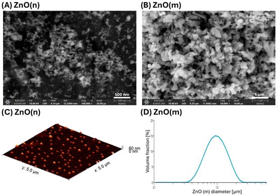
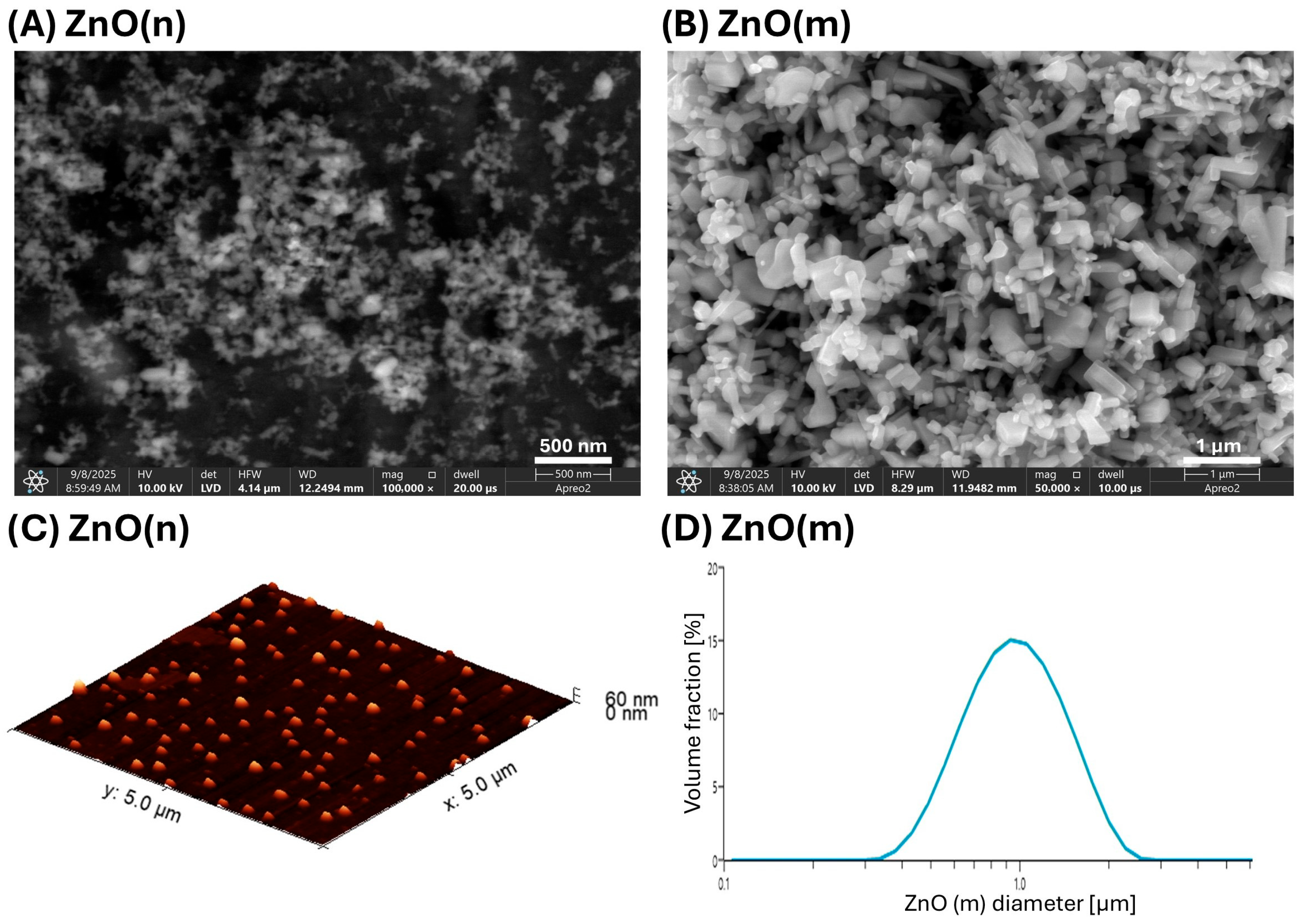
Figure 1A,C show images of ZnO(n) acquired by using scanning electron microscopy (SEM) and atomic force microscopy (AFM), respectively, while Table 1 shows their size measured from AFM pictures and using the Brunauer–Emmett–Teller (BET) relative surface area. ZnO(n) are 26 ± 4 nm in size and have a surface area of 39.16 m2/g. It is coherent with work by Mohandas et al. [13], who reported that ZnO particles in the range of 70–120 nm are optimal for infected wound dressings. Considering that particle size significantly affects specific surface area, ion release, and antibacterial activity, independent particle size and BET analysis were essential for reliable material characterization. Figure 1B shows SEM images of ZnO(m), while Figure 1D shows their size distribution obtained from laser diffraction measurement. ZnO(m) are much larger than ZnO(n), 1.02 ± 0.01 µm, and have a much lower relative surface area of 4.56 m2/g. The high surface area of ZnO(n) may positively impact the antibacterial activity of the particles, as a large specific surface area can generate more reactive oxygen species (ROS), damaging bacterial cell membranes [14]. It has also been reported that the nanometric dimensions of ZnO also improve its solubility in water and the release of Zn2+ ions, which can disrupt the continuity of the bacterial cell membrane [15], making ZnO(n) more effective for infected wound management.
Figure 1.
Images of ZnO(n) (A) and ZnO(m) (B) obtained using scanning electron microscopy at magnification 100,000× and 50,000×, respectively, acquired using LVD—low-vacuum detector; image of ZnO(n) obtained using atomic force microscopy in contact mode, 3D projection (C); and volume-based particle size distribution of ZnO(m) obtained from laser diffraction measurements (D).
Table 1.
Average diameters and BET surface area of ZnO(n) and ZnO(m) particles.
2.2. Antimicrobial Activity of ZnO(n) and ZnO(m)
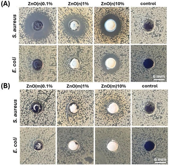
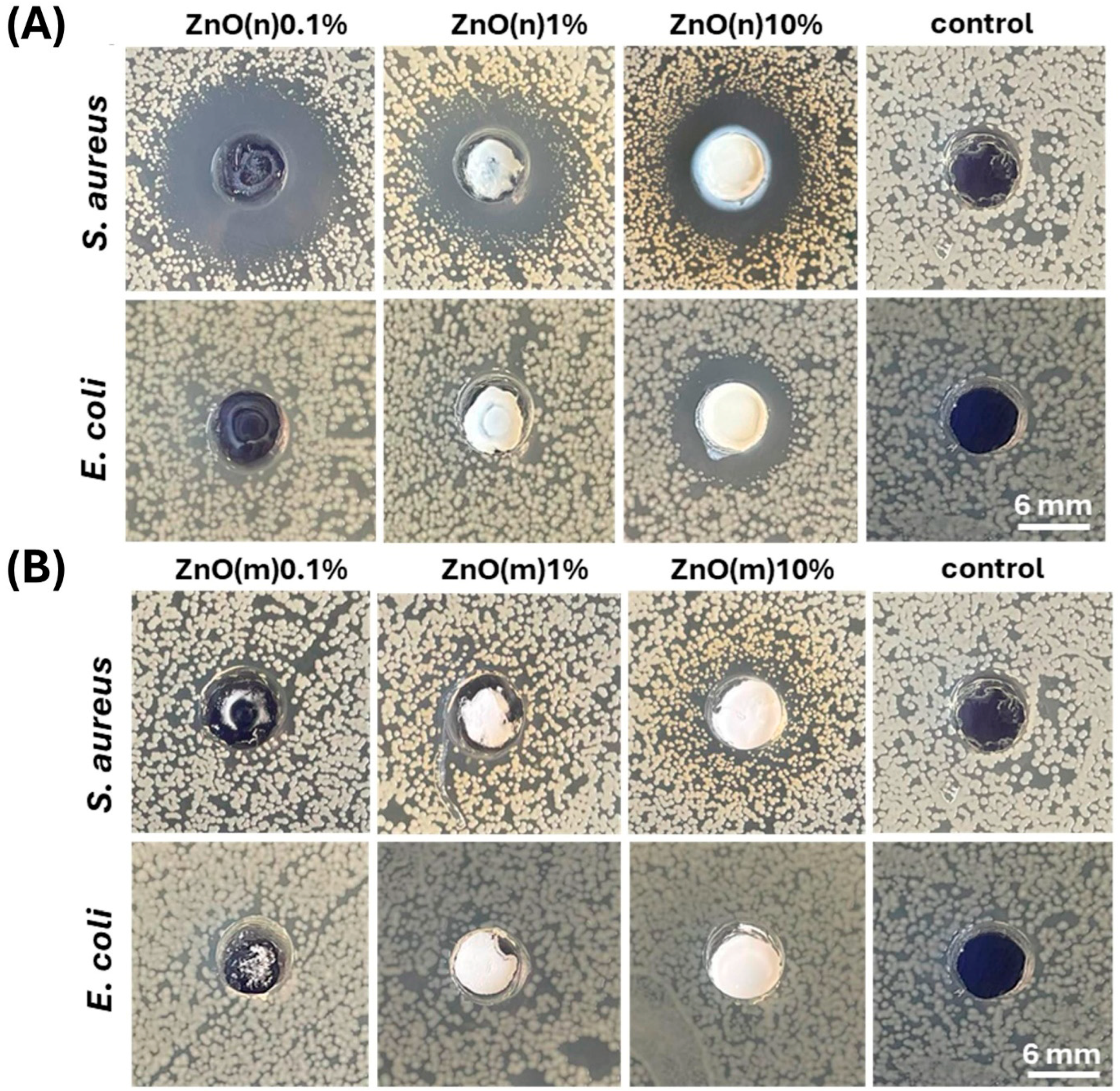
Figure 2 shows the zones of inhibition (ZOIs) of the S. aureus and Escherichia coli strains for suspensions of ZnO(n) and ZnO(m). The results show that ZnO(m) did not produce a visible antibacterial effect in contact with both strains. The ZnO(n) suspensions showed an evident antibacterial effect at all concentrations for the S. aureus strain (ZOI: 11–16 mm), and a visible inhibition zone for the E. coli strain at a 10% concentration of ZnO(n) (ZOI: 8 mm). The micrometric size of the particles results in a lower solubility of ZnO in water than for the nanoparticles; therefore, the release of Zn2+ ions is relatively low. The size of ZnO(m), above 1 µm (Figure 1D, Table 1), also prevents them from penetrating bacterial cells. ZnO(n), due to their small size, sediment slower, allowing for diffusion through the agar. Studies by Prasanna et al. show that ZnO nanoparticles around 30 nm in diameter generate 10 times more OH· and 20 times more H2O2 than microparticles around 2 µm in diameter [16]. So, we can expect that nanoscale size and high surface area of ZnO(n) allow them to generate higher amounts of ROS, which presumably damage bacterial cell walls more effectively. The higher toxicity of nanoparticles towards S. aureus bacteria compared to that of E. coli is probably due to differences in the structure of these bacteria’s cells. S. aureus (Gram+) bacteria have a thicker cell wall than E. coli (Gram-), but they are more porous, making particle penetration easier. S. aureus lacks an outer lipid membrane, and the presence of anionic groups on the surface of the cell wall attracts positively charged Zn2+ cations [17]. As a result of the antibacterial activity of ZnO(n), this type of particle was used in manufacturing composite samples with GG/Alg samples.
Figure 2.
Zones of inhibition (ZOIs) for suspensions of ZnO nanoparticles, ZnO(n) (A) and microparticles, ZnO(m) (B) at concentration of 0.1%, 1% and 10% on S. aureus and E. coli strains. Dulbecco’s phosphate-buffered saline, without calcium and magnesium ions (DPBS), acted as control.
2.3. Gross Morphology and Microstructure of GG/Alg Composites
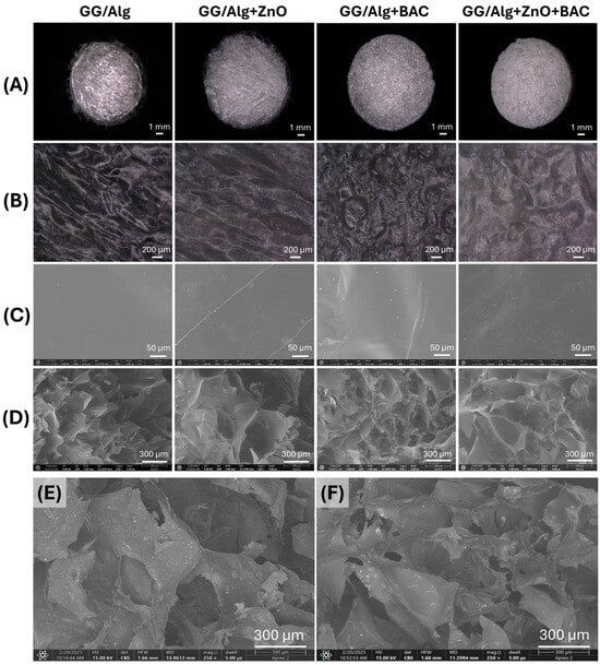
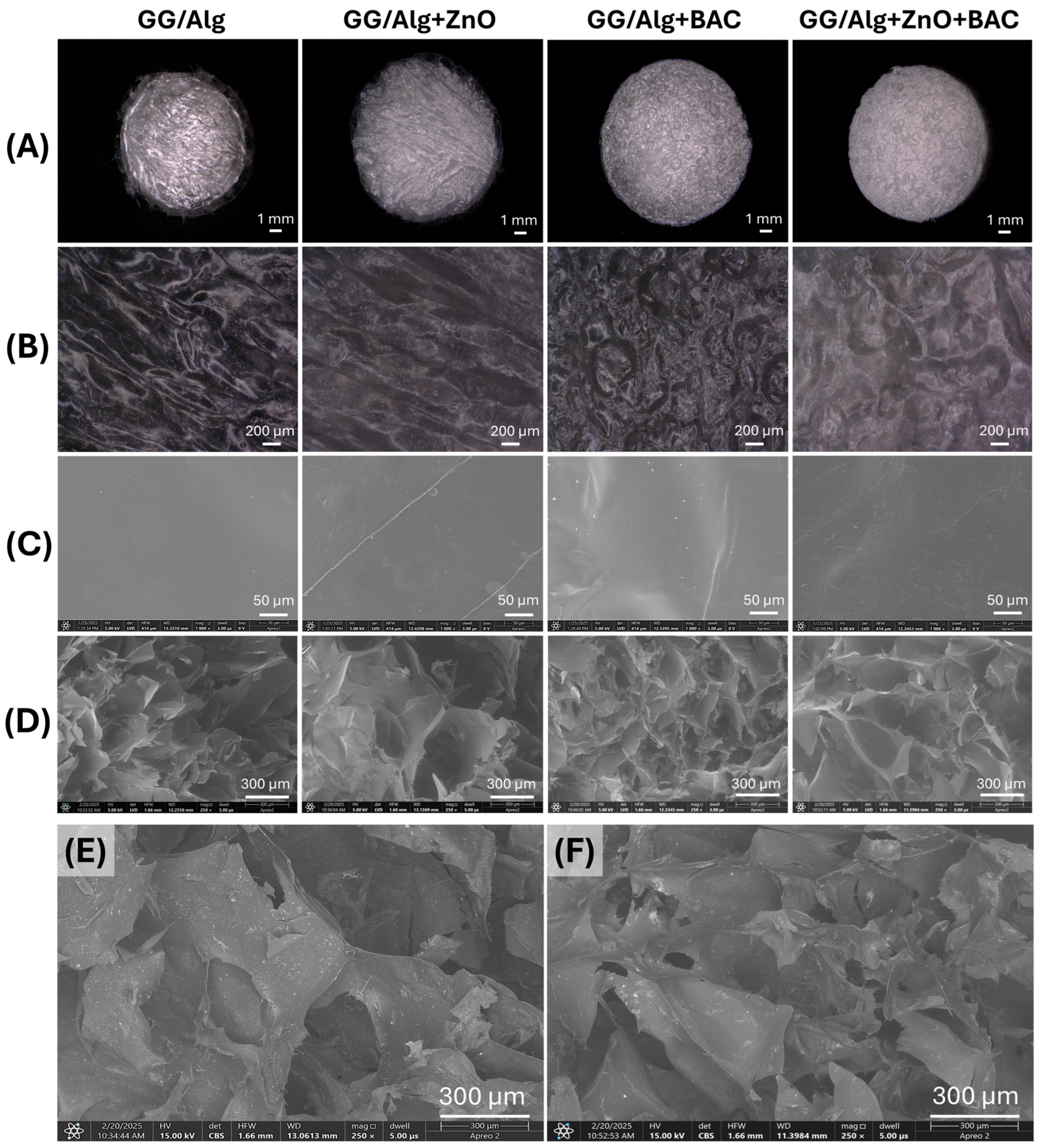
Figure 3, panels A and B present optical microscope images of the lyophilized composite samples at magnifications of 20× and 100×, respectively. The surface is rough but nonporous. At 1000× magnification under scanning electron microscopy (SEM), the surface of the samples is relatively smooth (Figure 3, panel C). Figure 3, panel D shows SEM images of the cross section of the samples, which is sponge-like with a very high porosity and a pore size in the range of 300–600 µm. The images clearly depict the sample’s morphology and are acquired using the low-vacuum detector (LVD). These images reveal the three-dimensional microstructure of the samples. Figure 3E,F show SEM images of cross sections of the GG/Alg+ZnO and GG/Alg+ZnO+BAC samples, respectively, acquired using a concentring backscatter (CBS) detector. It is sensitive to differences in atomic number, which allows for chemical contrast—areas of the sample differing in chemical composition are imaged with different contrast. Therefore, despite the less visible topographic details than with the LVD, this detector allowed us to image the ZnO(n) distribution in the sample. Zinc is the heaviest element present in the samples (with an atomic mass of 65.38 u), so bright areas in the CBS detector images indicate the presence of ZnO(n). A homogeneous distribution of zinc oxide is visible within the samples.
Figure 3.
Images of GG/Alg, GG/Alg+ZnO, GG/Alg+BAC and GG/Alg+ZnO+BAC samples obtained using optical digital microscopy: panel (A)—magnification 20×, panel (B)—magnification 100×; images obtained using scanning electron microscopy: panel (C)—surface, magnification 1000×; panel (D)—cross section, magnification 250×; acquired using LVD—low-vacuum detector; and pictures of: (E)—GG/Alg+ZnO and (F)—GG/Alg+ZnO+BAC—cross section, magnification 250×; acquired using CBS—concentring backscatter detector.
2.4. Water Absorption and Bacitracin (BAC) Release
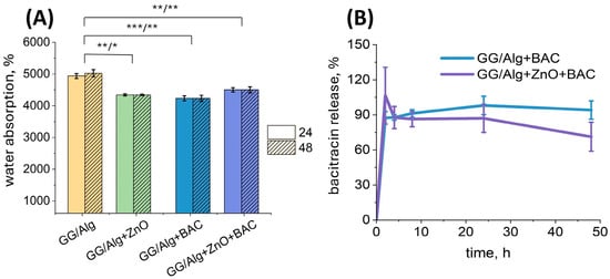
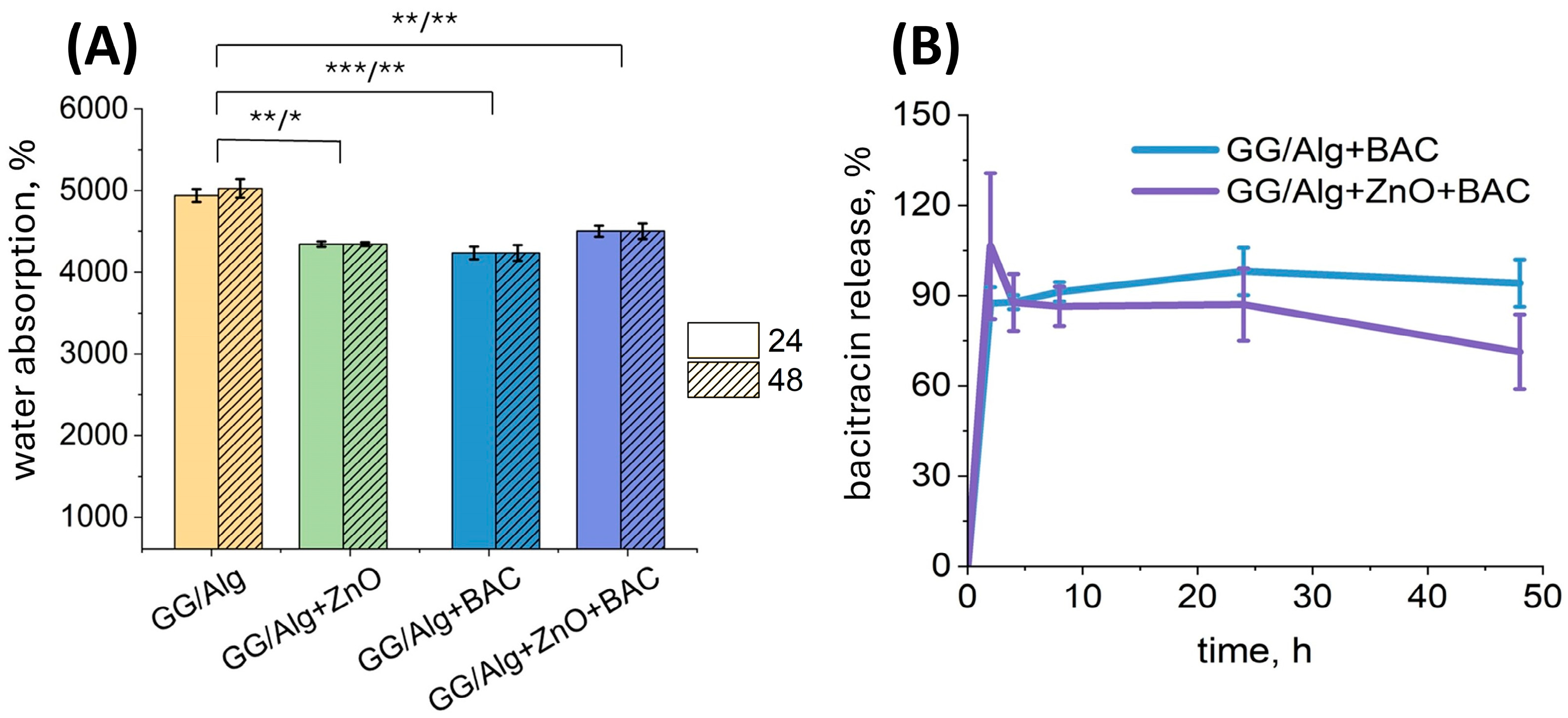
The water absorption of all the samples tested exceeded 4000%, indicating the ability of the composite materials to absorb liquids weighing more than 40 times the dry weight of the sample (Figure 4A). A swelling ratio of 4000% corresponds to a water content exceeding approximately 97% in the fully hydrated state, consistent with highly absorbent alginate-based wound dressings dedicated to bacteria-infected, high exuding wounds, where swelling ratios of up to 5000% have been described by others [18]. There is no significant difference between water absorption values after 24 and 48 h, so the sample absorbs water mainly within the first 24 h, with no increase in water absorption observed after this time. The GG/Alg sample exhibited significantly higher water absorption compared to all other samples, likely due to the absence of modifiers that are released into the liquid environment during the test.
Figure 4.
Water absorption rate after 24 h and 48 h of incubation of GG/Alg, GG/Alg+ZnO, GG/Alg+BAC and GG/Alg+ZnO+BAC samples in MilliQ water (A) and bacitracin release from GG/Alg+BAC and GG/Alg+ZnO+BAC over 48 h in MilliQ water (B). Average from n = 3 samples ± SD (standard deviation). Statistically significant differences at * p < 0.05, ** p < 0.01 and *** p < 0.001.
BAC release studies indicate that most drug (90%) is released from samples within the first 2 h (Figure 4B). The fast release of BAC from lyophilized samples is most likely due to their high porosity, which provides a large surface area in contact with water, making it easier for the drug to penetrate the sample and be washed out. Burst release of the drug from samples may be beneficial in the early phase of wound healing to prevent the development of infection, as well as in the event of sudden onset of pain or inflammation to alleviate symptoms quickly. Moreover, the rapid release of the active substance reduces the risk of antimicrobial resistance due to the absence of extended exposure to subinhibitory doses. However, burst release may pose risks, as the rapid release of a large amount of drug over a short period can lead to a toxic effect [19]. It should be emphasized that the results obtained here are a proof of concept, confirming the functionality of the developed dressing prototype. Under in vivo conditions, this process is likely to be more controlled and slower. This is because, during exudate absorption, the dressing gradually swells, leading to a delayed release of the active substance, which is correlated with the amount of exudate absorbed. The clinical performance of such systems depends on balancing rapid initial antibacterial action with controlled subsequent release. Hydrogel wound dressings with adjusted crosslinking have been shown to influence drug release behavior and antibacterial performance, indicating that modulation of network density and bonding can provide fine control over release kinetics [20]. Furthermore, recent studies highlight that mitigating burst release while preserving sustained delivery remains very challenging in the treatment of infected wounds [21].
2.5. In Vitro Cytotoxicity
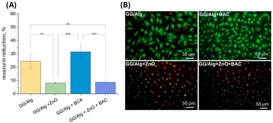
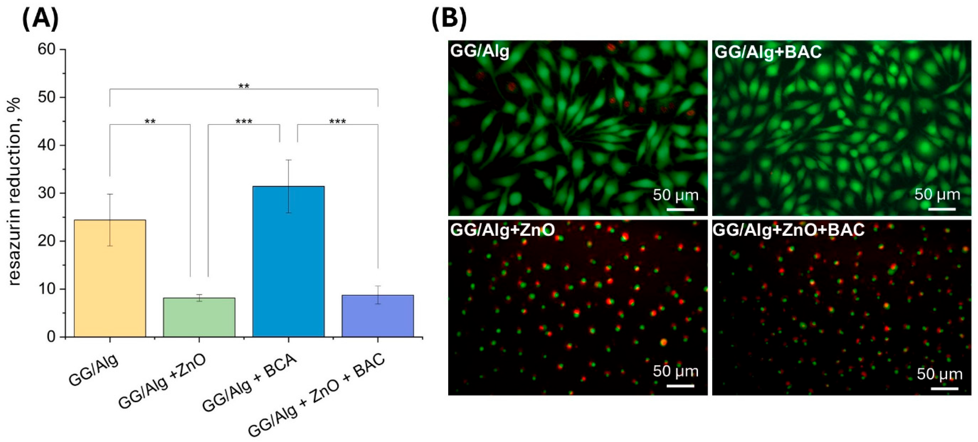
The results of the AlamarBlue test (Figure 5A), which show the metabolic activity of the L929 cells cultured in the extracts, demonstrate a high resazurin reduction of 24.4 ± 5.4% for the GG/Alg control sample and 31.4 ± 5.4% for the GG/Alg+BAC sample. After live–dead fluorescence staining (Figure 5B), cells for the latter samples are mainly stained green, are spindle-shaped, and well spread, indicating that they are alive and that these samples are not cytotoxic. On the other hand, cells cultured in extracts of samples containing ZnO(n) show a reduced viability of 8.2 ± 0.7% for GG/Alg+ZnO and 8.7 ± 1.9% for GG/Alg+ZnO+BAC. After live–dead staining, the green-stained cells are not flattened, but are round and less adherent to the substrate. They are also stained red, which means that they are dying. The biological activity of ZnO(n) is dose-dependent and has been associated with mechanisms including reactive oxygen species (ROS) generation and Zn2+ ion release, both of which contribute to oxidative stress and cytotoxic effects in mammalian cells [22]. However, it must be kept in mind that these conditions of cell culture in extracts are very harsh, because of the very fast release of leachable ingredients in the extracts. Under clinical conditions, the release of cytotoxic moieties from the dressing will be slower and correlated with the absorption of the wound exudate.
Figure 5.
Viability (resazurin reduction AlamarBlue test) (A) and live/dead images (fluorescence staining with calcein AM and PI; live cells are stained green while dead cells are stained red) (B) of L929 fibroblasts cultured for 24 h in contact with 1% extracts from GG/Alg, GG/Alg+ZnO, GG/Alg+BAC and GG/Alg+ZnO+BAC samples. Average from n = 3 samples ± SD (standard deviation). Statistically significant differences at ** p < 0.01 and *** p < 0.001.
2.6. Antimicrobial Activity
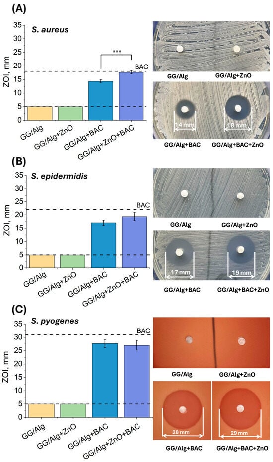
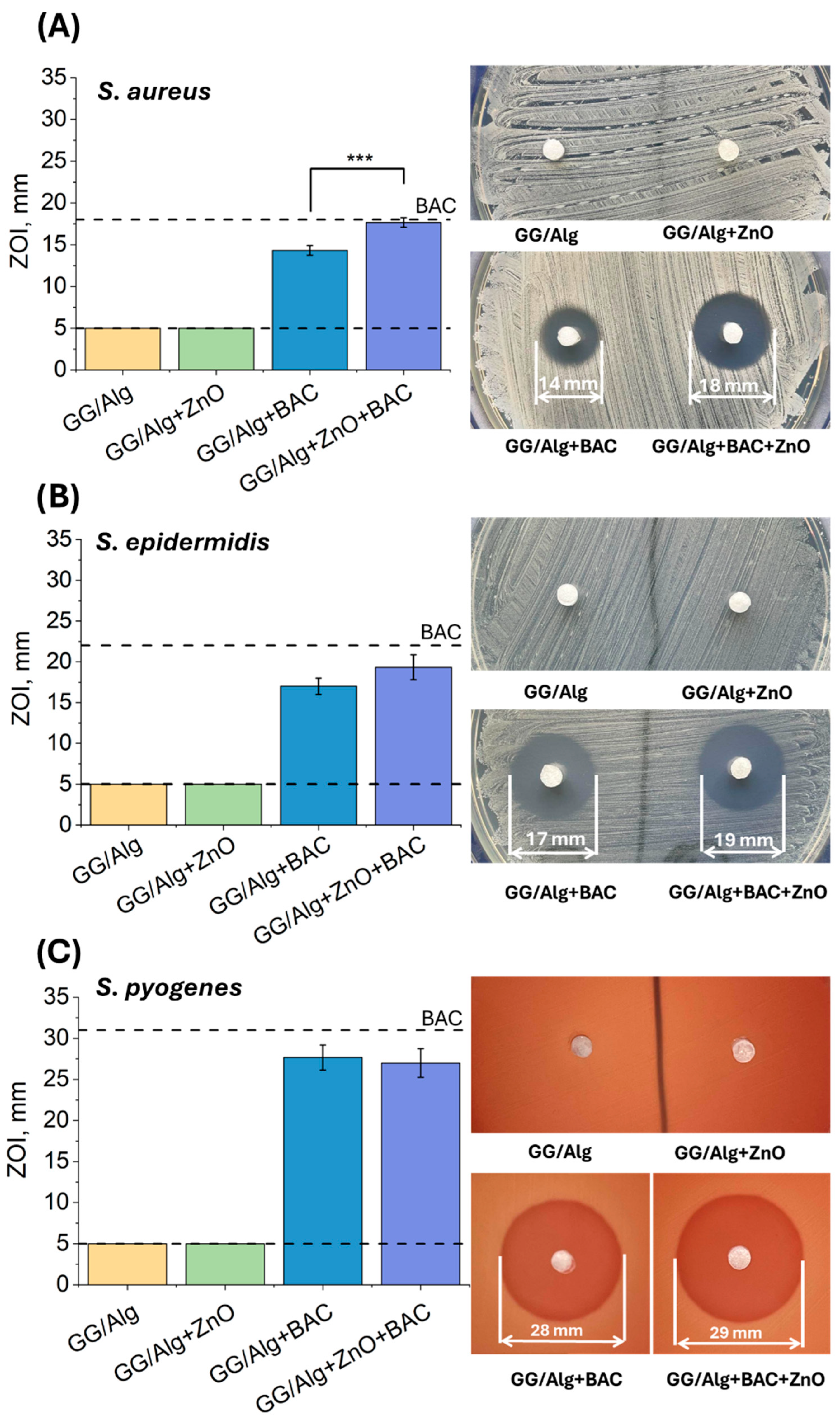
The antibacterial properties of the tested samples were assessed against three Gram+ bacteria strains: S. aureus, S. epidermidis, and S. pyogenes (Figure 6). For the S. aureus strain (Figure 6A), no antimicrobial activity was observed in the control sample (GG/Alg) or the sample containing ZnO(n) (GG/Alg+ZnO). However, the BAC-containing samples demonstrated significant antibacterial activity: The zones of inhibition (ZOIs) for GG/Alg+BAC were 14.3 ± 0.6 mm, while for GG/Alg+ZnO+BAC they was greater, reaching 17.7 ± 0.6 mm. The larger diameter of the ZOIs for the sample with both modifiers suggests a potential synergistic effect of ZnO(n) and BAC. A similar effect was observed against the S. epidermidis strain (Figure 6B): Samples without antibiotics did not show antimicrobial activity. At the same time, samples with BAC have a ZOI of 17.0 ± 1.0 mm for GG/Alg+BAC and 19.3 ± 1.5 mm for GG/Alg+ZnO+BAC. In this case, too, the presence of ZnO(n) enhanced the effect of BAC.
Figure 6.
Growth inhibition zones of S. aureus (A), S. epidermidis (B), and S. pyogenes (C) strains in contact with GG/Alg, GG/Alg+ZnO, GG/Alg+BAC and GG/Alg+ZnO+BAC samples. Average from n = 3 samples ± SD (standard deviation). Statistically significant difference at *** p < 0.001. Lower dashed line shows the diameter of the well, while upper dashed line indicates the inhibition zones for the control sample, i.e., a paper disk impregnated with 10 units of BAC.
For the S. pyogenes strain (Figure 6C), ZnO(n) did not demonstrate antimicrobial activity, but the antibiotic itself was very effective—the zone diameter for the GG/Alg+BAC sample was 27.7 ± 1.5 mm, and for GG/Alg+ZnO+BAC, it was 27.0 ± 1.7 mm. The undetected antimicrobial activity of the composite samples containing only ZnO(n) may be due to the limited diffusion of the nanoparticles in the agar medium or the low concentration in the test environment. In comparison, ZnO(n) suspensions (see Figure 2A) demonstrated antimicrobial activity against S. aureus, suggesting significant differences in the mode of action of ZnO(n) in suspension and incorporated into a hydrogel matrix. Although the available literature lacks reports of synergy between ZnO and BAC, there are studies describing the interaction of ZnO with other antibiotics, such as ciprofloxacin and ampicillin (against S. aureus and E. coli) [23], meropenem (Pseudomonas aeruginosa) [24], and colistin (P. aeruginosa) [25]. In the case of BAC, the synergistic effect with ZnO may result from the formation of complexes with Zn2+ ions, which enhance the antimicrobial effect by inhibiting the growth of Gram+ bacteria and inducing cell lysis [26].
3. Conclusions
Antibiotic resistance is a serious issue in modern medicine. One strategy to reduce the amount of antibiotics in medical devices involves the use of antibacterial modifiers that enhance the antimicrobial properties of the material. Advanced dressings designed for infected wounds are often enriched with antibacterial agents such as antibiotics or metal oxide nanoparticles. Microbiological tests confirmed that ZnO(n), unlike ZnO(m), exhibited antibacterial activity—particularly against Gram+ S. aureus.
A hydrogel composite based on GG and Alg, and enriched with BAC and ZnO(n), was proposed as a potential wound dressing. Microscopic analysis revealed that the samples were highly porous. Scanning electron microscopy (SEM) with a CBS detector confirmed the homogeneous distribution of ZnO(n) in GG/Alg+ZnO+BAC dressings. Swelling tests showed that the lyophilized samples could absorb over 4000% of their own weight in water, indicating a strong potential for the absorption of wound exudate. A burst release of BAC—approximately 90% within the first two hours—suggests that these dressings could be effective in the early stages of wound healing. In vitro cytotoxicity tests showed that ZnO(n) induced cytotoxic effects on fibroblasts. However, cytotoxicity studies performed on extracts do not mimic clinical situations. In these studies, whole samples were placed in water, which resulted in the rapid release of active ingredients into the extract. In clinical use, a dressing placed on a wound with exudate would release antibacterial agents at a slower rate, which could contribute to a reduction in cytotoxicity.
Tests of lyophilized samples against S. aureus and S. epidermidis revealed an enhanced effect of ZnO(n) on the antibacterial activity of bacitracin in the case of the GG/Alg+ZnO+BAC sample. Interestingly, GG/Alg+ZnO alone did not exhibit any antibacterial activity. Based on these results, the GG/Alg+ZnO+BAC sample demonstrated the most promising antibacterial properties, especially against S. aureus and S. epidermidis, which cause the most common wound infections. The results obtained allowed us to assess the antibacterial effectiveness of individual modifiers and show the preliminary evidence of cooperative or additive antibacterial interaction between ZnO and BAC, which may provide the basis for the further development of antibacterial dressings. However, due to the cytotoxicity of the extract to fibroblasts, further optimization is necessary to adjust the concentration of ZnO(n) to ensure the enhanced antibacterial effect while minimizing toxicity. It is also suggested to evaluate obtained samples in more relevant in vitro and in vivo models, resembling clinical conditions of infected and exuding wounds.
4. Materials and Methods
4.1. Materials
Micrometric zinc oxide (ZnO(m)) was provided by ZM Silesia SA, Oława, Poland. Nanometric zinc oxide (ZnO(n)), sodium dodecyl sulfate (SDS), poly(vinyl alcohol) (PVA, Mowiol 4-88, MW 31 kDa), gellan gum (GG, GelzanTM, MW 200–300 kDa), sodium alginate from brown algae (Alg), CaCl2·6H2O, bacitracin A (BAC), bicinchoninic acid, copper (II) sulfate pentahydrate (CuSO4·5H2O), phosphate-buffered saline (PBS), AlamarBlue reagent, calcein AM and propidium iodide (PI) came from Sigma-Aldrich, St. Louis, MO, USA. Ethanol was provided by POCH (Gliwice, Poland). Dulbecco’s modified Eagle’s medium (DMEM), fetal bovine serum (FBS), penicillin/streptomycin and Dulbecco’s phosphate-buffered saline, without calcium and magnesium ions (DPBS) were from PAN-Biotech, Aidenbach, Germany. Muller–Hilton agar and Columbia agar with 5% sheep blood were from Becton Dickinson, Franklin Lakes, NJ, USA. L929 fibroblasts came from the European Collection of Cell Cultures, UK. Bacteria strains Staphylococcus aureus ATCC® 25923™, Escherichia coli ATCC 25922™, Staphylococcus epidermidis ATCC® 700296™ and Streptococcus pyogenes ATCC® 12384™ were from American Type Culture Collection, Manassas, VA, USA.
4.2. Atomic Force Microscopy (AFM)
For measuring ZnO(n) size using an AFM, a suspension of 0.02% w/v ZnO(n) was prepared in MilliQ water. The suspension was ultrasonicated for 15 min. One drop of the prepared suspension was placed on the coverslip and placed in a 37 °C incubator until the water evaporated. The prepared sample was placed under an AFM (Explorer, Thermomicroscopes, Sunnyvale, CA, USA) and observed in contact mode. The resulting image was processed, and the ZnO(n) diameters were measured using the Gwyddion AFM image visualization program (Version 3.1).
4.3. Laser Diffraction Measurements
The diameter of ZnO(m) was measured using a laser diffraction technique (MasterSizer 3000E; Malvern Instruments; Hydro SV, Worcestershire, UK). A 0.1% w/v suspension of ZnO(m) was prepared in MilliQ water of 0.25% w/v SDS and 0.2% w/v PVA and placed in an ultrasonic bath for 30 min to minimize particle agglomeration before the measurements. Measurements were made at an obscuration level of 5–10% with a stirrer speed of 500 rpm. The measurement was performed in triplicate. The results were reported as volume-based particle size distribution and volume-weighted mean diameter.
4.4. Surface Area
The surface area of ZnO(n) and ZnO(m) was determined using the BET method with an adsorption analyzer (ASAP 2020 Plus, Malvern Panalytical, Norcross, GA, USA). The samples were firstly degassed at 120 °C for 24 h before the analysis and the N2 adsorption was at −196 °C.
4.5. Preparation of Composites with ZnO and BAC
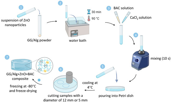
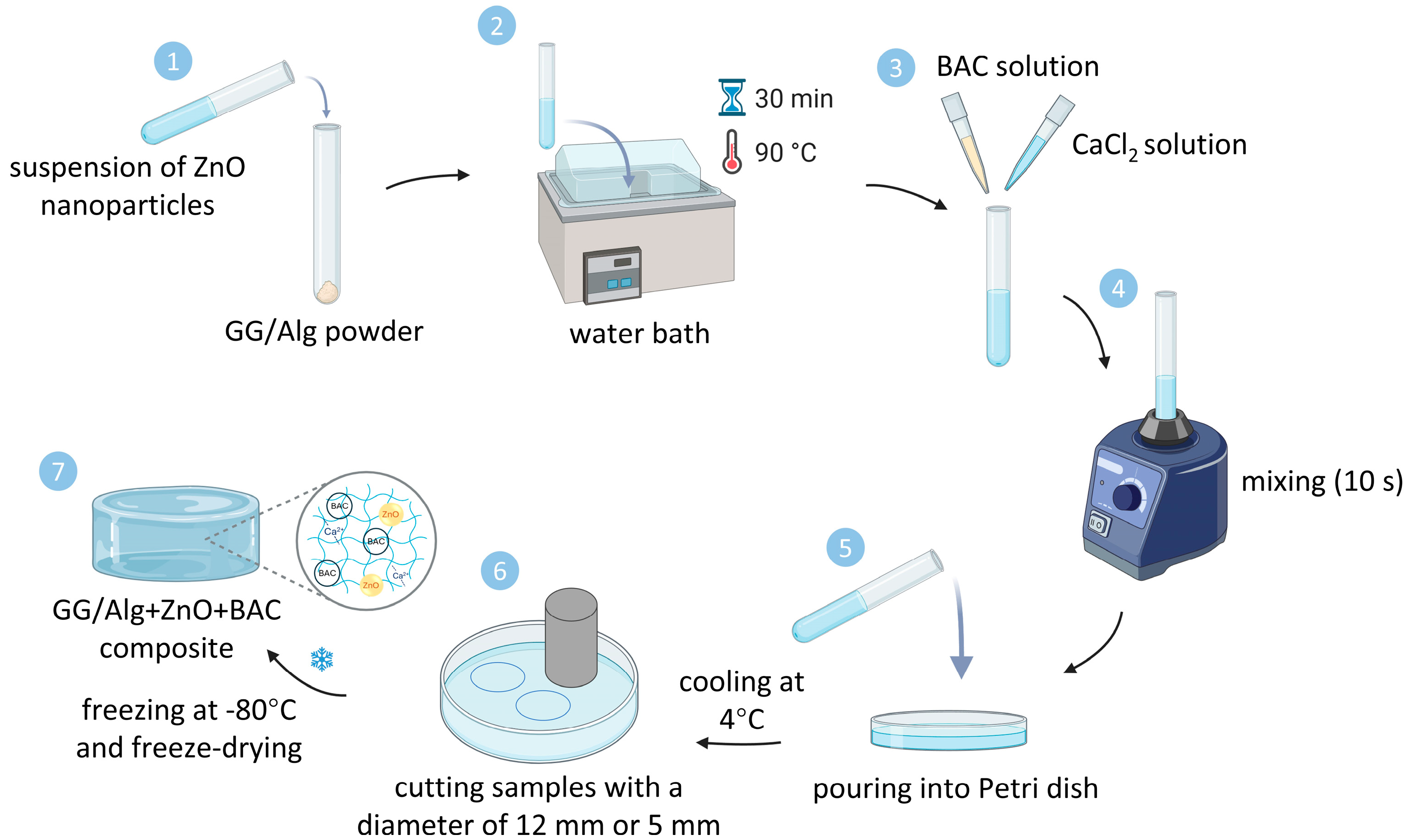
GG/Alg samples were prepared by dissolving in 8 mL of MilliQ water GG and Alg powders in a weight ratio of 10:1 for 30 min in a water bath at 90 °C. Before this step, ZnO(n) was suspended in MilliQ water and vortexed for 10 min for the samples with ZnO. The temperature was then lowered to 70 °C. A solution of BAC in 1 mL of MilliQ water was added for the samples with BAC. Then, 1 mL of 0.6% CaCl2·6H2O solution heated to 70 °C was added and vortexed for 10 s. After that, the samples were poured into a 9 cm diameter Petri dish with spacers (4 mm high) and covered with a glass slide. The composition of the manufactured samples without and with modifiers is presented in Table 2, while the manufacturing method is shown in Figure 7. For the experiments, the samples with a diameter of 12 mm were cut, frozen, and freeze-dried (Alpha 1–2, Martin Christ, Osterode am Harz, Germany). Only for microbiological tests, the diameter of the samples was 5 mm.
Table 2.
Composition of hydrogel samples: amount of ingredients per 10 mL MilliQ water.
Figure 7.
Schematic representation of preparation of GG/Alg+ZnO+BAC samples.
4.6. Optical Microscopy
The freeze-dried samples were observed under an optical digital microscope (Keyence VHX 7000, Mechelen, Belgium) at magnifications of 20× and 100×.
4.7. Scanning Electron Microscopy (SEM)
The ZnO particles were observed using SEM. Before SEM observations, ZnO(n) and ZnO(m) were suspended in ethanol and placed in an ultrasonic bath for 15 min. Then 20 µL of each suspension was placed on a metal holder and left to dry at 37 °C. The samples were observed under a SEM (ThermoFisher Scientific Apreo 2, Waltham, MA, USA) at 10 kV and a magnification of 100,000× and 50,000× using a low-vacuum detector (LVD).
For SEM imaging of the composites, two samples of each type were prepared: one for surface imaging and one for cross-sectional imaging. Samples were carbon-sputtered. For external surfaces, observations were performed at 1000×, while cross-sectional imaging was performed at 250× using LVD. In the case of cross sections of samples containing ZnO (GG/Alg+ZnO, GG/Alg+BAC+ZnO), imaging was also performed using a concentring backscatter (CBS) detector.
4.8. Swelling Capacity
The lyophilized samples (n = 3 of each type) were weighed and then immersed in 2 mL of MilliQ water each. After 24 h and 48 h, the samples were collected, and carefully wiped with a wet Kimtech tissue (Kimwipes, Kimberly-Clark, Dallas, TX, USA) to remove excess water, but avoiding removing water from the pores of the sample.
The swelling of the samples was calculated from Formula (1):
where —mass of the wet sample, —mass of the dry sample.
4.9. Bacitracin (BAC) Release Studies
The release of BAC was examined from lyophilized GG/Alg+BAC and GG/Alg+ZnO+BAC samples. The samples were weighed and then placed in Falcon tubes with 5 mL of PBS solution. The tubes were placed in an incubator at 37 °C. After 2, 4, 8, 24, and 48 h, 0.5 mL of the solution was withdrawn from the tubes into Eppendorf tubes for BAC determinations, each time topping the collected fluid with PBS. BAC was determined using the bicinchionic acid assay (BCA). To determine the BAC concentration, a calibration curve was prepared. BAC solutions of the following concentrations in PBS were prepared: 750, 500, 400, 300, 200, 100, 50, 20, and 0 µg/mL. Subsequently, a BCA reagent was prepared by mixing BCA with a 4% CuSO4·5H2O solution at a 50:1 volume ratio. A total of 50 µL of each calibration solution and 50 µL of test samples containing released BAC were pipetted in triplicate into a transparent 96-well plate. Next, 100 µL of the BCA reagent was then added to each well. The absorbance of each sample was measured after 15 min of incubation using a FLUOstar Omega (BMG Labtech, Ortenberg, Germany) reader at 562 nm. On the basis of the absorbance data, a calibration curve was prepared, and the BAC concentration released over time was determined.
4.10. In Vitro Cytotoxicity
In vitro cytotoxicity was evaluated using L929 fibroblasts cultured in DMEM supplemented with 10% FBS and a 1% mixture of penicillin and streptomycin. Cells were cultured at 37 °C in a 5% CO2 atmosphere. A total of 200 µL of cell suspension (50,000 cells/mL) was added to each well in the 96-well plate (10,000 cells per well). The cells were then left to adhere to the bottom of the wells. Simultaneously, 1% extracts of lyophilized samples were prepared by incubation of 20 mg of samples in 2 mL of DMEM for 24 h. Before extraction, the samples were irradiated under UV light (15 min on each side). After 24 h, the medium was aspirated and replaced with extracts. Cells were incubated with 1% w/v extracts of the tested materials, while control cells were cultured in standard culture medium without extracts (n = 3). The AlamarBlue assay was used to assess cell viability. After 24 h, the extracts in each well were replaced with 150 µL of a 5% AlamarBlue reagent solution in DMEM. After 3 h of incubation, 100 μL aliquots were transferred to the wells of a 96-well black plate and the fluorescence intensity was measured at λex = 530 nm and λem = 590 nm using the FluoroSTAR Omega reader. The following Formula (2) was used to determine the percentage of resazurin reduction:
where —fluorescence of the sample, —fluorescence of the AlamarBlue solution after 3 h incubation without cells, —fluorescence of the AlamarBlue reagent reduced completely in an autoclave.
Live/dead analysis of the cells was performed by replacing the extracts with 100 µL of staining solution containing 0.1% calcein AM and 0.1% propidium iodide (PI) prepared in DPBS. After incubation for 20 min at 37 °C, cells were observed under a fluorescence microscope (Axiovert 40 CFL, Carl Zeiss, Oberkochen, Germany).
4.11. Antimicrobial Activity
For microbiological studies of ZnO(n) and ZnO(m), suspensions were prepared in DPBS at concentrations of 0.1%, 1%, and 10% w/v. Antibacterial activity was tested on S. aureus and E. coli strains. Bacterial suspensions with a density of 0.5 McFarland were prepared and spread on a Muller–Hilton agar plate (Becton Dickinson, Franklin Lakes, NJ, USA) using a disposable swab. The wells were cut on the agar using a 5 mm sterile punch. A total of 100 µL of ZnO suspensions were added to the wells (three replicates for each concentration); 100 µL of DPBS solution was used as a control. The dishes were placed at 4 °C for 4 h to allow initial diffusion of antibacterial moieties in the agar. The dishes were then incubated in a 37 °C incubator for 18 h. After this time, the diameters of the resulting ZOIs were measured.
For microbiological testing of the lyophilized hydrogels, samples measuring 5 mm in diameter were used. Suspensions of three bacterial strains were prepared: S. aureus, S. epidermidis, and S. pyogenes in a DPBS solution with a density of 0.5 McFarland. Muller–Hilton agar was used for S. aureus and S. epidermidis strains, and Columbia agar with 5% sheep blood was used for S. pyogenes strains. Bacteria were transferred to the agar medium using a disposable swab. One control sample (a 5 mm diameter well with DPBS solution) and a 6 mm diameter diagnostic paper disk impregnated with 10 units of BAC were placed on the prepared agar plates, along with samples of GG/Alg, GG/Alg+ZnO, GG/Alg+BAC, and GG/Alg+ZnO+BAC (n = 3). The plates with samples were left in the refrigerator for 4 h and then placed in an incubator at 37 °C for 24 h. After 24 h, the diameters of the ZOIs were measured.
4.12. Statistics
Statistical analysis was performed using one-way analysis of variance (one-way ANOVA) followed by Tuckey’s post hoc test using OriginLab2023 software. Differences were considered significant when p < 0.05. The results are presented as mean ± standard deviation (SD). Statistically significant differences * p < 0.05, ** p < 0.01 and *** p < 0.001 are shown.
Author Contributions
Conceptualization, E.P. and K.R.-K.; methodology, A.M., A.K. and D.O.; software, A.M. and A.K.; validation, E.P., K.R.-K. and M.B.-W.; formal analysis, E.P. and K.R.-K.; investigation, A.M. and A.K.; resources, E.P.; data curation, A.M. and A.K.; writing—original draft preparation, A.M. and A.K.; writing—review and editing, E.P. and K.R.-K.; visualization, A.M. and A.K.; supervision, E.P.; project administration, E.P.; funding acquisition, E.P. All authors have read and agreed to the published version of the manuscript.
Funding
This research was funded by the Ministry of Science and Higher Education, Poland from the Program “Excellence Initiative—Research University” and from the subsidy (no. 16.16.160.557) for the AGH University of Krakow.
Institutional Review Board Statement
Not applicable.
Informed Consent Statement
Not applicable.
Data Availability Statement
The raw data supporting the conclusions of this article will be made available by the authors on request.
Conflicts of Interest
The authors declare no conflicts of interest.
References
- Zhu, T.; Mao, J.; Cheng, Y.; Liu, H.; Lv, L.; Ge, M.; Li, S.; Huang, J.; Chen, Z.; Li, H. Recent Progress of Polysaccharide-Based Hydrogel Interfaces for Wound Healing and Tissue Engineering. Adv. Mater. Interfaces 2019, 6, 1900761. [Google Scholar] [CrossRef] [Scilit]
- Odziomek, K.; Drabczyk, A.K.; Kościelniak, P.; Konieczny, P.; Barczewski, M.; Bialik-Wąs, K. The Role of Freeze-Drying as a Multifunctional Process in Improving the Properties of Hydrogels for Medical Use. Pharmaceuticals 2024, 17, 1512. [Google Scholar] [CrossRef] [Scilit]
- Wei, X.; Ding, S.; Liu, S.; Yang, K.; Cai, J.; Li, F.; Wang, C.; Lin, S.; Tian, F. Polysaccharides-modified chitosan as improved and rapid hemostasis foam sponges. Carbohydr. Polym. 2021, 264, 118028. [Google Scholar] [CrossRef] [Scilit]
- Giavasis, I.; Harvey, L.M.; McNeil, B. Gellan gum. Crit. Rev. Biotechnol. 2000, 20, 177–211. [Google Scholar] [CrossRef] [Scilit] [PubMed]
- Milivojevic, M.; Pajic-Lijakovic, I.; Bugarski, B.; Nayak, A.K.; Hasnain, M.S. Gellan gum in drug delivery applications. In Natural Polysaccharides in Drug Delivery and Biomedical Applications; Academic Press: Cambridge, MA, USA, 2019; pp. 145–186. [Google Scholar] [CrossRef] [Scilit]
- Dire, D.J.; Coppola, M.; Dwyer, D.A.; Lorettec, J.J.; Karr, J.L. Prospective evaluation of topical antibiotics for preventing infections in uncomplicated soft-tissue wounds repaired in the ED. Acad. Emerg. Med. 1995, 2, 4–10. [Google Scholar] [CrossRef] [Scilit] [PubMed]
- Nguyen, R.; Khanna, N.R.; Safadi, A.O.; Patel, P.; Sun, Y. Bacitracin Topical. In StatPearls; StatPearls Publishing: Treasure Island, FL, USA, 2025. [Google Scholar]
- Langford, J.H.; Artemi, P.; Benrimoj, S.I. Topical antimicrobial prophylaxis in minor wounds. Ann. Pharmacother. 1997, 31, 559–563. [Google Scholar] [CrossRef] [Scilit]
- Ao, H.; Duan, K.; Zhang, D.; Xun, X.; Feng, F.; Liu, H.; Li, Y.; Wan, Y. Biofunctionalized bacterial cellulose grafted with bacitracin for wound healing. J. Mater. Sci. 2024, 59, 10987–10997. [Google Scholar] [CrossRef] [Scilit]
- Lemnaru, G.-M.; Truşcă, R.D.; Ilie, C.-I.; Țiplea, R.E.; Ficai, D.; Oprea, O.; Stoica-Guzun, A.; Ficai, A.; Dițu, L.-M. Antibacterial Activity of Bacterial Cellulose Loaded with Bacitracin and Amoxicillin: In Vitro Studies. Molecules 2020, 25, 4069. [Google Scholar] [CrossRef] [Scilit]
- Murray, C.J.L. Antimicrobial Resistance Collaborators. Global burden of bacterial antimicrobial resistance in 2019: A systematic analysis. Lancet 2022, 399, 629–655. [Google Scholar] [CrossRef] [Scilit]
- Janaki, A.C.; Sailatha, E.; Gunasekaran, S. Synthesis, characteristics and antimicrobial activity of ZnO nanoparticles. Spectrochim. Acta Part A Mol. Biomol. Spectrosc. 2015, 144, 17–22. [Google Scholar] [CrossRef] [Scilit] [PubMed]
- Mohandas, A.; Kumar, S.; Raja, B.; Lakshmanan, V.K.; Jayakumar, R. Exploration of alginate hydrogel/nano zinc oxide composite bandages for infected wounds. Int. J. Nanomed. 2015, 10, 53–66. [Google Scholar] [CrossRef] [Scilit]
- Raghupathi, K.R.; Koodali, R.T.; Manna, A.C. Size-dependent bacterial growth inhibition and mechanism of antibacterial activity of zinc oxide nanoparticles. Langmuir 2011, 27, 4020–4028. [Google Scholar] [CrossRef] [Scilit]
- Padmavathy, N.; Vijayaraghavan, R. Enhanced bioactivity of ZnO nanoparticles-an antimicrobial study. Sci. Technol. Adv. Mater. 2008, 9, 035004. [Google Scholar] [CrossRef] [Scilit]
- Lakshmi Prasanna, V.; Vijayaraghavan, R. Insight into the Mechanism of Antibacterial Activity of ZnO: Surface Defects Mediated Reactive Oxygen Species Even in the Dark. Langmuir 2015, 31, 9155–9162. [Google Scholar] [CrossRef] [Scilit] [PubMed]
- Franco, D.; Calabrese, G.; Guglielmino, S.P.P.; Conoci, S. Metal-Based Nanoparticles: Antibacterial Mechanisms and Biomedical Application. Microorganisms 2022, 10, 1778. [Google Scholar] [CrossRef] [Scilit]
- Ngece, K.; Aderibigbe, B.A.; Ndinteh, D.T.; Fonkui, Y.T.; Kumar, P. Alginate-gum acacia based sponges as potential wound dressings for exuding and bleeding wounds. Int. J. Biol. Macromol. 2021, 172, 350–359. [Google Scholar] [CrossRef] [Scilit] [PubMed]
- Huang, X.; Brazel, C.S. On the importance and mechanisms of burst release in matrix-controlled drug delivery systems. J. Control. Release 2001, 73, 121–136. [Google Scholar] [CrossRef] [Scilit]
- Jia, B.; Li, G.; Cao, E.; Jinlong, L.; Zhao, X.; Huang, H. Recent progress of antibacterial hydrogels in wound dressings. Mater. Today Bio 2023, 19, 100582. [Google Scholar] [CrossRef] [Scilit]
- Zattarin, E.; Sotra, Z.; Wiman, E.; Bas, Y.; Rakar, J.; Berglund, L.; Starkenberg, A.; Bjork, E.M.; Khalaf, H.; Oksman, K.; et al. Controlled release of antimicrobial peptides from nanocellulose wound dressings for treatment of wound infections. Mater. Today Bio 2025, 32, 101756. [Google Scholar] [CrossRef] [Scilit]
- Sirelkhatim, A.; Mahmud, S.; Seeni, A.; Kaus, N.H.M.; Ann, L.C.; Bakhori, S.K.M.; Hasan, H.; Mohamad, D. Review on Zinc Oxide Nanoparticles: Antibacterial Activity and Toxicity Mechanism. Nano-Micro Lett. 2025, 7, 219–242. [Google Scholar] [CrossRef] [Scilit] [PubMed]
- Sharma, N.; Jandaikc, S.; Kumar, S. Synergistic activity of doped zinc oxide nanoparticles with antibiotics: Ciprofloxacin, ampicillin, fluconazole and amphotericin B against pathogenic microorganisms. An. Acad. Bras. Ciências 2016, 88, 1689–1698. [Google Scholar] [CrossRef] [Scilit] [PubMed]
- El-Telbany, M.; Mohamed, A.A.; Yahya, G.; Abdelghafar, A.; Abdel-Halim, M.S.; Saber, S.; Alfaleh, M.A.; Mohamed, A.H.; Abdelrahman, F.; Fathey, H.A.; et al. Combination of Meropenem and Zinc Oxide Nanoparticles; Antimicrobial Synergism, Exaggerated Antibiofilm Activity, and Efficient Therapeutic Strategy against Bacterial Keratitis. Antibiotics 2022, 11, 137. [Google Scholar] [CrossRef] [Scilit] [PubMed]
- Fadwa, A.O.; Alkoblan, D.K.; Mateen, A.; Albarag, A.M. Synergistic effects of zinc oxide nanoparticles and various antibiotics combination against Pseudomonas aeruginosa clinically isolated bacterial strains. Saudi J. Biol. Sci. 2021, 28, 928–935. [Google Scholar] [CrossRef] [Scilit]
- Ming, L.J.; Epperson, J.D. Metal binding and structure-activity relationship of the metalloantibiotic peptide bacitracin. J. Inorg. Biochem. 2002, 91, 46–58. [Google Scholar] [CrossRef] [Scilit] [PubMed]

Disclaimer/Publisher’s Note: The statements, opinions and data contained in all publications are solely those of the individual author(s) and contributor(s) and not of MDPI and/or the editor(s). MDPI and/or the editor(s) disclaim responsibility for any injury to people or property resulting from any ideas, methods, instructions or products referred to in the content. |
© 2026 by the authors. Licensee MDPI, Basel, Switzerland. This article is an open access article distributed under the terms and conditions of the Creative Commons Attribution (CC BY) license.