


Abstract
Wound dressings capable of conforming to irregular wound surfaces, withstanding mechanical deformation, and visually monitoring local microenvironmental changes are highly desirable for infected wound management. Herein, a versatile composite hydrogel (PFPU/VCM/BTB) based on the selected PFPU3 formulation was developed for the visual monitoring and treatment of infected wounds. The hydrogel was constructed from aldehyde-functionalized Pluronic F-127 (PF127-(CHO)2) micelles and a multifunctional PMUA copolymer containing primary amine groups, phosphorylcholine units, and 2-ureido-4[1H]-pyrimidinone (UPy) motifs. Reversible imine bonds and UPy-mediated quadruple hydrogen bonds generated the dynamic network, whereas bromothymol blue (BTB) and vancomycin (VCM) supplied pH-responsive visual detection and antibacterial activity, respectively. The resulting hydrogel exhibited porous morphology, solid-like rheological behavior, self-healing capability, tissue adhesion, and injectability. It displayed visible color changes within the pH range of 5.8–7.4 and effectively reduced Staphylococcus aureus (S. aureus) viability. In addition, the hydrogel showed good cytocompatibility and hemocompatibility and promoted fibroblast migration. In a full-thickness infected skin wound model, the hydrogel treatment reduced residual wound area, enhanced epidermal regeneration, and promoted collagen deposition by day 12. This multifunctional hydrogel integrates visual monitoring and localizes antibacterial functions within a single dressing platform, providing a potential approach for infected wound management.
1. Introduction
Skin wounds are complex and highly dynamic biological interfaces involving tissue deformation, exudation, inflammatory responses, and microbial colonization [1,2]. Once bacterial infection develops, persistent microbial burden can delay re-epithelialization, prolong inflammatory responses, and disrupt extracellular matrix remodeling, thereby increasing the risk of chronic or difficult-to-heal wounds [3,4,5]. Accordingly, wound management has gradually shifted from passive tissue coverage toward active therapeutic intervention [6,7]. To prevent irreversible tissue damage caused by severe infection, an ideal wound dressing should combine effective physical protection and antibacterial activity with the ability to monitor wound conditions and deliver localized therapeutic intervention.
During bacterial colonization and tissue repair, the wound microenvironment undergoes a series of dynamic physicochemical changes, including variations in pH, temperature, reactive oxygen species, glucose levels, etc. [8,9]. Among these parameters, pH is particularly suitable for visual monitoring because it can be readily detected through simple colorimetric approaches. Bacterial metabolism, inflammatory exudation, and alterations in local tissue conditions could induce dynamic shifts in wound pH [10,11]. Converting such changes directly into visible color signals may therefore provide an opportunity for in situ and real-time feedback of wound status. In recent years, pH-responsive wound dressings based on colorimetric dyes, photonic structures, and electronic sensors have been widely investigated [12,13,14]. Nevertheless, many existing systems still suffer from limitations, including insufficient adaptation to irregular tissue surfaces, lack of antibacterial functionality, and complicated fabrication procedures. More importantly, sensing platforms that only monitor local changes without providing therapeutic functions have limited practical value in the treatment of infected wounds [15]. Therefore, developing a dressing platform capable of adapting to dynamic wound interfaces while integrating visual detection of infection-associated pH variations with localized antibacterial therapy remains highly desirable.
Hydrogels, characterized by their high-water content, soft three-dimensional networks, and favorable adaptability to tissue interfaces, provide an attractive material basis for addressing these challenges [16,17]. To function effectively on mechanically active wound surfaces, the interfacial and mechanical properties of hydrogels are as important as their biological activity. Dynamic covalent bonds and reversible supramolecular interactions [18] have attracted considerable attention due to their ability to dissipate mechanical stress, reconstruct damaged networks, and support injectable or conformal applications. Specifically, imine bonds formed between aldehyde and primary amine groups provide reversible covalent cross-linking [19], whereas 2-ureido-4[1H]-pyrimidinone (UPy) motifs can establish reversible quadruple hydrogen bonding interactions [20]. Combining these two dynamic interactions is expected to preserve network integrity while endowing hydrogels with injectability, self-healing capability, and adaptability to irregular tissue surfaces. Meanwhile, phosphorylcholine-containing polymers can generate highly hydrated surfaces and are commonly utilized to enhance resistance against nonspecific interfacial fouling [21]. In addition, bromothymol blue (BTB), which exhibits distinct color variations within the near-neutral pH range, is suitable for the visual monitoring of pH changes associated with the Staphylococcus aureus (S. aureus) infection models [22]. Vancomycin (VCM), a well-established glycopeptide antibiotic with antibacterial activity against Gram-positive bacteria, can be incorporated into hydrogel matrices for localized antibacterial therapy [23].
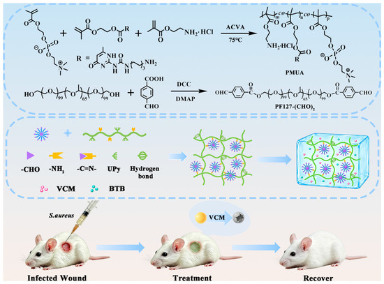
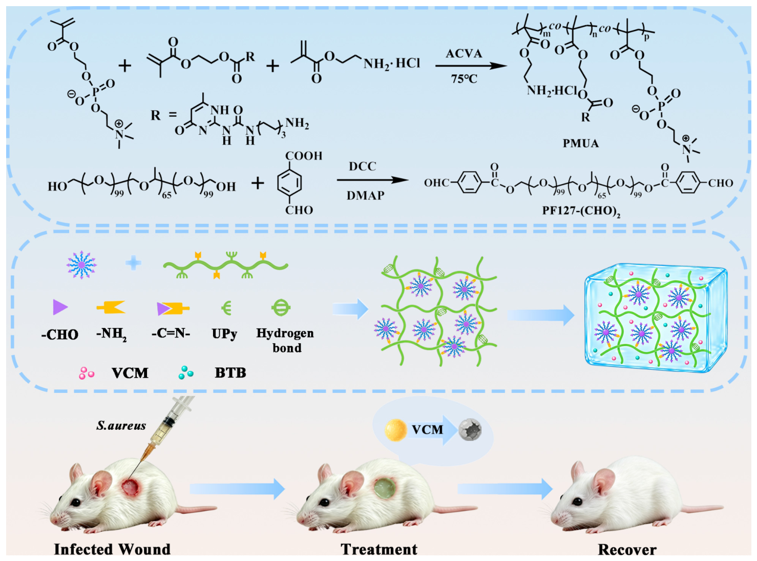
In this study, we designed a multifunctional PFPU/VCM/BTB hydrogel composed of micelles of Pluronic F-127 functionalized with aldehyde groups at both termini (PF127-(CHO)2) and a PMUA copolymer containing primary amines, MPC-derived phosphorylcholine moieties, and UPy units (Scheme 1). The hydrogel network was constructed through dynamic imine bonds and UPy-mediated quadruple hydrogen bonding, while BTB and VCM imparted visible pH-responsive colorimetric behavior and antibacterial activity, respectively. The physicochemical properties of the hydrogel, including chemical structure, rheological behavior, self-healing capability, and injectability, as well as its biological performances, including pH-responsive visualization, antibacterial activity, biosafety, and fibroblast migration ability, were systematically evaluated. The objective of this study is to integrate mechanically adaptive wound coverage, visual monitoring of infection-associated microenvironmental changes, and localized antibacterial therapy within a single hydrogel dressing, offering a feasible approach toward the development of smart hydrogel-based wound dressings.
Scheme 1.
Schematic illustration of PMUA and PF127-(CHO)2 polymer precursor synthesis, the self-assembly and gelation of the dual-dynamic PFPU/VCM/BTB hydrogel, and its application as a therapeutic dressing for the visual monitoring and treatment of S. aureus-infected wounds.
2. Results and Discussion
2.1. Synthesis and Characterization of PMUA and PF127-(CHO)2
The PFPU hydrogels were rationally designed as a dual-crosslinked dynamic network formulated from aldehyde-functionalized PF127 (PF127-(CHO)2) and a multifunctional copolymer, PMUA. To synthesize the crosslinker, the terminal hydroxyl groups of the PF127 triblock copolymer were modified via esterification with 4-carboxybenzaldehyde, yielding a telechelic PF127-(CHO)2 macromer end-capped with reactive aldehyde groups. Meanwhile, the PMUA copolymer was synthesized via free radical copolymerization of MPC, UPyHEMA, and AEMH monomers, and polymer chains containing primary amino groups and 2-ureido-4[1H]-pyrimidinone (UPy) motifs. Upon mixing under neutral conditions, dynamic Schiff base linkages were formed between the aldehyde groups of PF127-(CHO)2 and the amino groups of PMUA. Intermolecular quadruple hydrogen bonding was formed through the UPy motifs. Therefore, the PFPU hydrogel network is stabilized by the synergistic combination of dynamic covalent imine bonds and non-covalent quadruple hydrogen bonds. This dual-cross-linking strategy is expected to endow the hydrogel with self-healing properties, injectability, and dynamic adaptability, which are desirable features for the in situ monitoring and detection of wound infections.
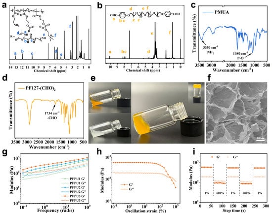
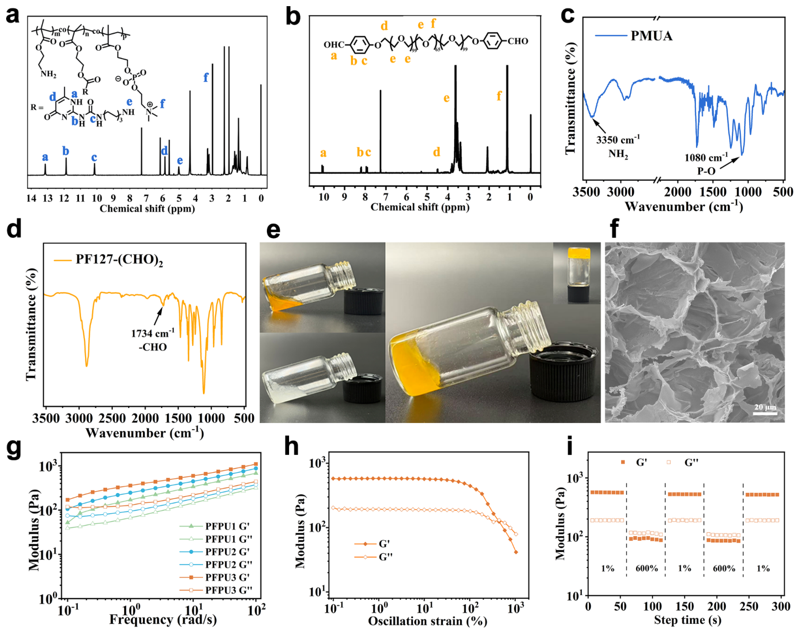
The successful synthesis and chemical structures of the PMUA and PF127-(CHO)2 were confirmed by 1H NMR and FTIR spectroscopy, as shown in Figure 1a–d. The peak at δ 2.97 ppm in the 1H NMR spectrum of PMUA was assigned to methylene protons (–N+(CH3)2–CH2–) of quaternary ammonium groups, verifying the incorporation of MPC units [24,25]. Additionally, characteristic proton peaks derived from UPyHEMA and AEMH were also detected at their corresponding chemical shifts, further supporting the copolymerization of the three monomers. In the FTIR spectrum of PMUA, a broad and strong absorption band at 3350 cm−1 was attributed to N-H stretching vibrations associated with amino groups from AEMH [26], and the absorption at 1080 cm−1 corresponded to P=O and P-O-C stretching vibrations from the MPC units [24], which validated the successful synthesis of the PMUA copolymer. For PF127-(CHO)2, the emergence of a typical aldehyde proton signal at δ 10.08 ppm in the 1H NMR spectrum, together with the carbonyl stretching band at 1734 cm−1 in the FTIR spectrum, confirmed the successful aldehyde functionalization of PF127 [27,28,29]. Taken together, these spectroscopic features support successful functionalization of the two polymer precursors required for subsequent dynamic network formation.
Figure 1.
Chemical characterization and rheological properties of the hydrogel network. (a,b) 1H NMR spectra of (a) the synthesized PMUA copolymer and (b) the aldehyde-functionalized PF127-(CHO)2. (c,d) FTI-R spectra of (c) PMUA and (d) PF127-(CHO)2. (e) Macroscopic photographs of the PFPU hydrogel upon mixing of the precursors. (f) SEM micrograph of the lyophilized PFPU3 hydrogel (scale bar = 20 μm). (g) Dynamic rheological frequency sweeps comparing the storage modulus (G′) and loss modulus (G″) of the PFPU1, PFPU2, and PFPU3 formulations. (h) Strain-amplitude sweep (0.1% to 1000% strain at 10 rad/s) of the optimized PFPU3 hydrogel formulation. (i) Alternating step-strain recovery cycles (alternating between 1% strain and 600% strain) of the PFPU3 hydrogel.
2.2. Characterization of Hydrogels
Hydrogels with in situ monitoring capabilities are highly desirable for intelligent wound management [30]. As illustrated in Figure 1e, the PFPU hydrogel was fabricated via a facile mixing of precursor solutions PMUA and PF127-(CHO)2. The gelation mechanism is primarily driven by dynamic Schiff base linkages formed between the amino and aldehyde groups, together with the non-covalent quadruple hydrogen bonding. The combination of dynamic covalent and supramolecular interactions is expected to provide the PFPU hydrogels with mechanical integrity while enabling reversible network reconstruction. The mechanical properties of the PFPU hydrogels with different formulations were first evaluated through rheological measurements. Frequency sweep analysis showed that the mechanical strength varied with hydrogel composition (Figure 1g). At a frequency of 10 rad/s and a constant strain of 1%, the storage moduli (G′) of PFPU1, PFPU2, and PFPU3 were 338.605, 447.566, and 596.572 Pa, respectively. PFPU3 exhibited the highest storage modulus among the three tested formulations and was therefore selected for subsequent experiments. Unless otherwise specified, PFPU denotes the unloaded PFPU3 hydrogel in the subsequent sections, whereas PFPU/VCM/BTB denotes the composite hydrogel prepared using the PFPU3 formulation with VCM and BTB incorporated before gelation. As shown in Figure 1h, strain amplitude sweeps demonstrated a distinct crossover point (G′ = G″), indicating the critical yield strain at which the hydrogel network ruptures and transitions from an elastic solid to a viscous fluid state. Thereafter, alternating step strain sweeps were conducted to verify the rheological self-healing behavior of the PFPU3 hydrogel. As shown in Figure 1i, at a low strain of 1%, G′ remained stable and higher than G″, indicating that the hydrogel network structure is intact. Upon applying a high strain (γ = 600%), G′ sharply dropped below G″, reflecting network dissociation. Notably, when the strain was returned to 1%, both moduli rapidly recovered to their initial values, indicating the excellent self-healing capability of the PFPU3 hydrogel.
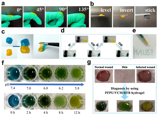
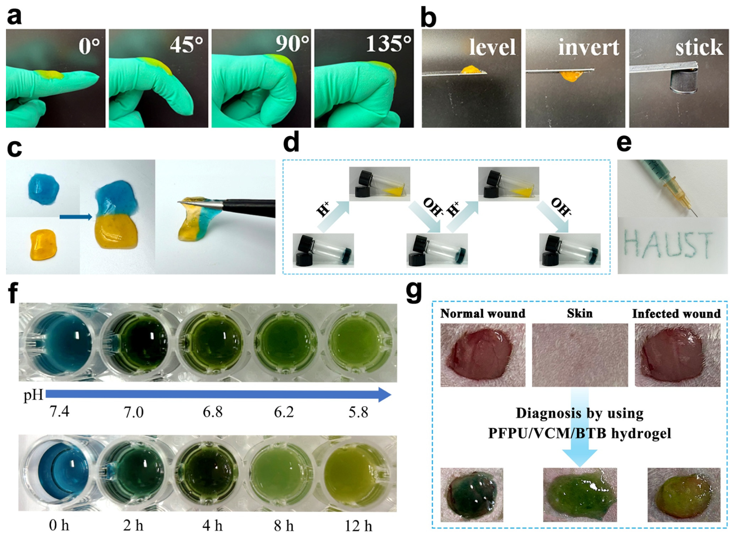
In addition to its rheological properties, the PFPU3 hydrogel exhibited desirable shape adaptability and adhesion properties (Figure 2a,b), self-repairing abilities (Figure 2c), pH responsiveness (Figure 2d), and injectability (Figure 2e). As shown in Figure 2a, the hydrogel PFPU3 firmly adhered to the surface of a finger joint, accommodating dynamic bending deformations at 0°, 45°, 90°, and 135° without apparent detachment. It also adhered to diverse substrates, including metal and plastic surfaces, without external assistance. In addition, the injectability of the PFPU3 hydrogel was confirmed by continuously extruding it through a syringe to write the letters “HAUST” without clogging (Figure 2e). These characteristics facilitate maintaining contact with the dynamic and irregular wound surfaces. Moreover, macroscopic self-healing was visually validated by bringing two differently colored hydrogel pieces (dyed with Orange G and Methylene Blue) into contact at room temperature (Figure 2c). They spontaneously fused into an integral hydrogel without any external intervention. The self-healing behavior is attributable to the dynamic and reversible Schiff base bonds and hydrogen bonds within the hydrogel network. The microstructure of the freeze-dried PFPU3 hydrogel was subsequently examined by SEM (Figure 1f). As shown in the Supporting Information (Figure S1), the swelling ratio of the PFPU/VCM/BTB hydrogel increased progressively and leveled off at approximately 8 h, indicating that the hydrogel can rapidly absorb wound exudate while maintaining its structural integrity. The hydrogel exhibits a highly porous and interconnected network structure. Such morphological features may facilitate the exchange of nutrients and oxygen, uptake of wound exudate, and cellular infiltration, which are beneficial for wound repair. Collectively, these versatile properties indicate the potential of the PFPU3 hydrogel as a wound monitoring dressing in wound management.
Figure 2.
Macroscopic performance, shape adaptability, injectability, and pH-sensing characteristics. (a) Photographs of the PFPU3 hydrogel. (b) Visual demonstration of the adhesive properties of the hydrogel on various material substrates. (c) Macroscopic self-healing property of two severed hydrogel blocks. (d) Macroscopic demonstration of the pH-responsive structural dissociation and subsequent reconstruction of the hydrogel network. (e) Injectability demonstration of the PFPU3 hydrogel through a 21G syringe needle. (f) Colorimetric transition profiles of the PFPU/VCM/BTB hydrogel across a pH range (pH 7.4 to 5.8), and real-time color evolution of the hydrogel during a 0–12 h S. aureus bacterial culture experiment. (g) In situ diagnostic colorimetric response of the PFPU/VCM/BTB hydrogel.
2.3. pH-Responsive Visualization of the PFPU/VCM/BTB Hydrogels
Real-time, in situ visual monitoring of wound microenvironments is crucial for the diagnosis and timely intervention of bacterial infections. Infection-associated bacterial metabolism and inflammatory exudation may alter the local wound pH, thereby providing microenvironmental signals for visual detection [31]. Leveraging this pathophysiological feature, BTB, a pH-indicating dye, was incorporated into the hydrogel network. As illustrated in Figure 2f, the PFPU/VCM/BTB hydrogel exhibits noticeable color changes within the pH range of 5.8 to 7.4, which closely matches the pH shift from an acidic infected microenvironment to a normal physiological state. This detection capability was further validated in vitro by the growth of bacterial biofilms, where the color of the PFPU/VCM/BTB hydrogel changed significantly at the 8 h time point. In addition, an S. aureus-infected wound model was established to evaluate the in situ detection performance of the PFPU/VCM/BTB hydrogel (Figure 2g). The PFPU/VCM/BTB hydrogel could effectively differentiate infected wounds from normal wounds through in situ colorimetric changes. When applied to the infected wounds, the hydrogel rapidly transitioned from its initial dark green state to a pale yellow color, driven by the acidic exudate of the infection, while the hydrogel on normal, uninfected wounds retained its original color. These results demonstrate that the incorporation of BTB endows the PFPU/VCM/BTB hydrogels with pH-responsive characteristics, enabling the in situ visual detection of bacteria-infected wounds via naked-eye visible color changes.
2.4. Antibacterial Activity of PFPU/VCM/BTB Hydrogels
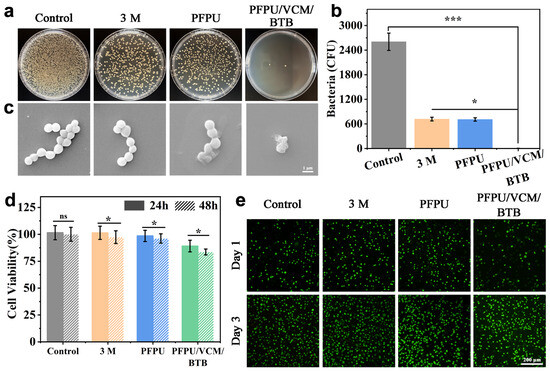
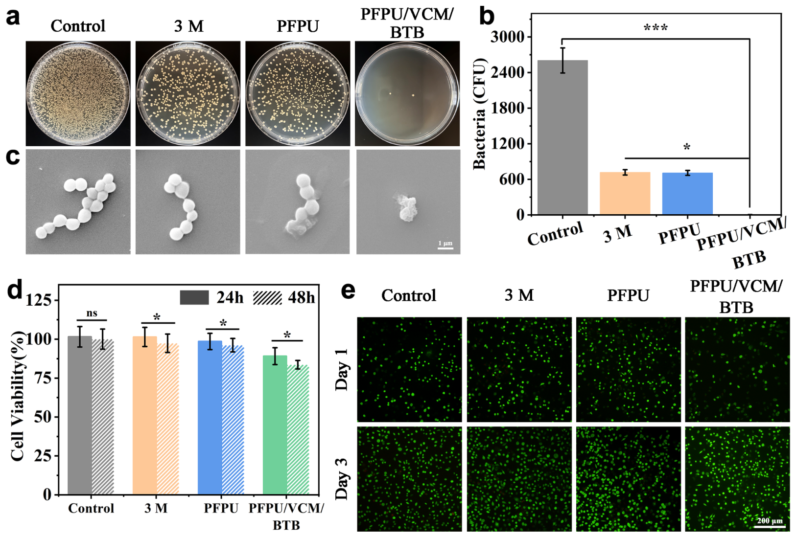
Open wounds, particularly under compromised physiological conditions such as hyperglycemia, are highly susceptible to pathogenic bacterial infections, and endowing wound dressings with inherent antibacterial activity is essential for promoting efficient wound healing [32]. The in vitro antibacterial efficacy of the PFPU/VCM/BTB hydrogels was evaluated against S. aureus using a standard plate colony counting assay. As shown in Figure 3a, after 24 h grew at 37 °C, abundant bacterial colonies were observed in the PBS control group. The commercial 3M dressing and the PFPU hydrogel exhibited only limited bactericidal effects, with colony numbers comparable to those of the control group. The mild antibacterial capacity of the PFPU hydrogel may be attributed to the MPC component in the hydrogel network, which is known to form a strong hydration layer that suppresses bacterial adhesion rather than directly killing bacteria. In contrast, the PFPU/VCM/BTB hydrogels displayed potent antibacterial activity, as evidenced by the lowest colony count among all groups. Quantitative analysis (Figure 3b) further confirmed the potent bactericidal activity of the PFPU/VCM/BTB hydrogels. Furthermore, SEM was employed to examine the morphological alterations of S. aureus following hydrogel treatment (Figure 3c). Compared with the intact bacterial morphology observed in the control groups, bacteria treated with PFPU/VCM/BTB hydrogel displayed severe structural damage and membrane disruption, consistent with the colony counting results. Together, these findings indicate that the incorporation of VCM endows the PFPU/VCM/BTB hydrogel with excellent antibacterial efficacy against S. aureus, supporting its potential application as a multifunctional antibacterial dressing for visual detection and management of infected wounds.
Figure 3.
In vitro antibacterial efficacy and cytocompatibility evaluations. (a) Representative digital images of agar plate cultures. (b) Quantitative evaluation of viable S. aureus colonies (CFU) on the hydrogel. (c) SEM micrographs of S. aureus bacteria after the different treatments. (d) Cell viability after incubation with extracts of different hydrogels for 24 h and 48 h. (e) Live/dead staining fluorescence micrographs of L929 cells on days 1 and 3 of co-culture. (* p < 0.05 and *** p < 0.001, “ns” means no significant differences).
The antibacterial spectrum of the VCM should be considered when interpreting these findings. VCM primarily targets susceptible Gram-positive bacteria and does not provide coverage against Gram-negative pathogens such as Pseudomonas aeruginosa. Both the in vitro antibacterial evaluation and the infected wound model in this study were based on S. aureus; therefore, efficacy against Gram-negative bacteria or polymicrobial wound infections has not been established.
2.5. Biocompatibility and Cell Migration
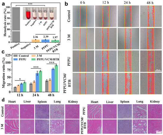
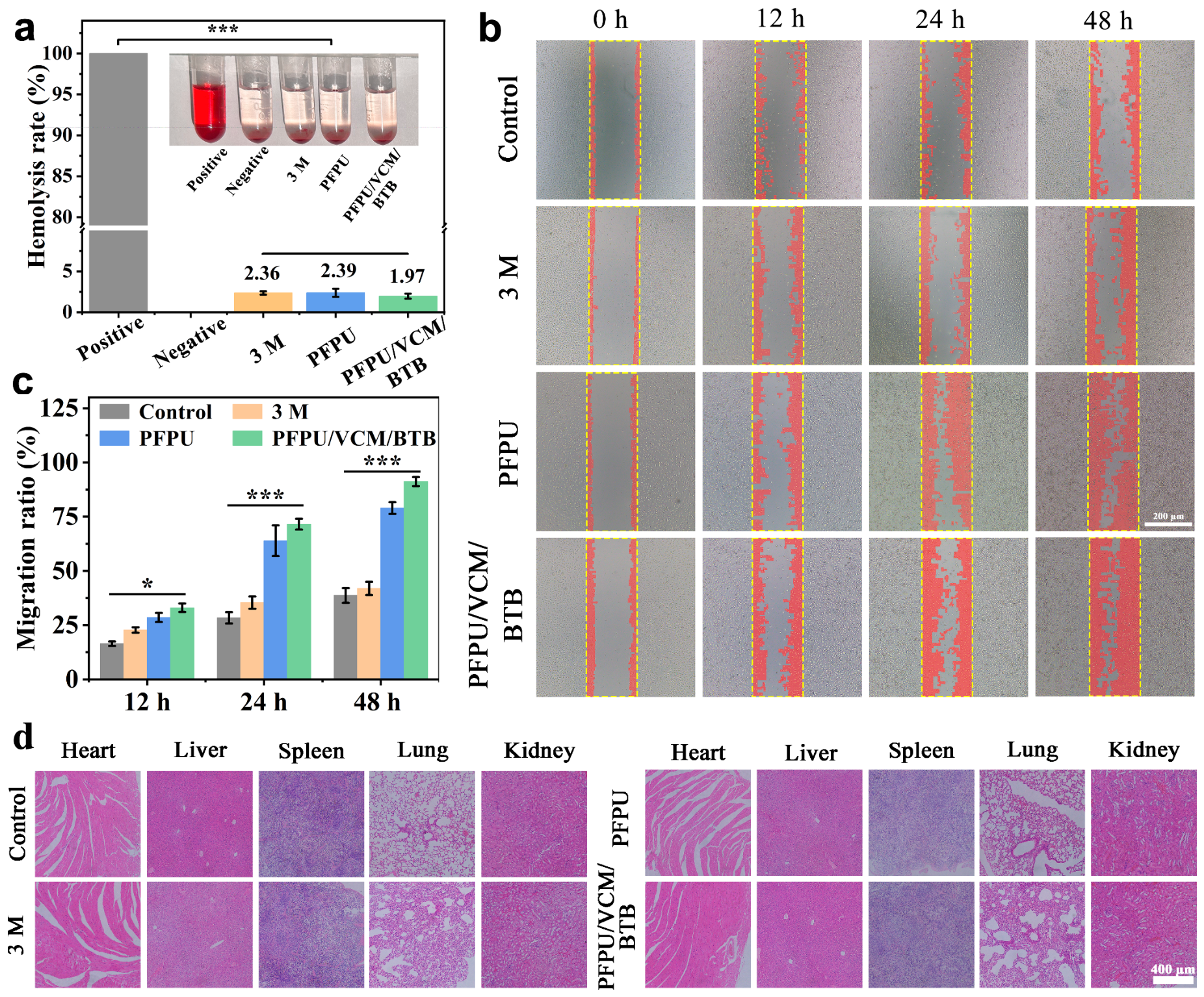
A comprehensive biosafety evaluation is essential for clinical wound dressing applications. In this study, the in vitro cytocompatibility of PFPU/VCM/BTB hydrogel with L929 fibroblasts was confirmed via MTT and Live/Dead assays. Quantitative MTT results (Figure 3d) revealed that L929 cells maintained over 81% viability after 48 h of incubation with the PFPU/VCM/BTB hydrogel extract, demonstrating that the hydrogel exhibits negligible cytotoxicity and does not affect cellular proliferation. This was further corroborated by Live/Dead fluorescence imaging (Figure 3e), which displayed predominantly viable cells (green fluorescence) with negligible dead cells (red fluorescence) after 1 day and 3 days of co-incubation, showing no observable difference from the control group. Additionally, excellent hemocompatibility is vital for wound dressings, as they directly interface with blood exudates during the angiogenesis phase of tissue repair. The hemolysis assay (Figure 4a) revealed a bright red supernatant in the positive control group, while the supernatants in the 3M, PFPU, and PFPU/VCM/BTB groups remained clear and colorless. Specifically, the OD value of the PFPU/VCM/BTB hydrogel extract was 0.184, corresponding to a minimal hemolysis rate well below the 5% safety threshold. Overall, the PFPU/VCM/BTB hydrogel exhibits favorable cytocompatibility and hemocompatibility.
Figure 4.
In vitro hemocompatibility, scratch-wound migration, and systemic in vivo safety. (a) Hemolytic rate (%) of the different dressing formulations. (b) Optical micrographs of the scratch-wound assay when treated with the different dressing extracts. The yellow dashed lines indicate the initial scratch boundaries, and red overlays indicate areas occupied by cells migrating into the initial scratch region. (c) Quantitative cell migration rates (%) calculated from the scratch areas at 0, 12, 24, and 48 h. (d) Representative H&E stained histopathological sections of major organs harvested from mice on day 12 post-treatment. (* p < 0.05 and *** p < 0.001).
Cell migration is a critical step in tissue remodeling and efficient wound closure. Therefore, an in vitro scratch wound healing assay was performed to evaluate the effect of the PFPU/VCM/BTB hydrogel on fibroblast migration. As shown in Figure 4b, the PFPU/VCM/BTB hydrogel significantly accelerated L929 cell migration, resulting in nearly complete closure of the scratched gap within 48 h. Quantitative analysis (Figure 4c) further verified that the migration rate of the PFPU/VCM/BTB group reached 93.61%, which was substantially higher than the 39.41% measured in the control group. These results indicate that the PFPU/VCM/BTB hydrogel possesses excellent pro-migration capabilities, which are highly advantageous for accelerating wound healing.
To eliminate concerns regarding potential systemic side effects arising from the localized administration of the PFPU/VCM/BTB hydrogel, the in vivo biosafety characterization was conducted. Mice were euthanized on day 12 post-surgery, and major organs (heart, liver, spleen, lungs, and kidneys) were harvested for histological evaluation via H&E staining. As shown in Figure 4d, no pathological damage was observed in the PFPU/VCM/BTB group compared to the PBS control. The tissue structures of the organs were intact with normal cell morphology and regularly arranged specific structures. No obvious abnormalities or inflammatory responses were observed. Moreover, the mice exhibited steady body weight increases within normal physiological ranges (Figure 5e) during the treatment period, confirming the absence of toxicity or metabolic interference. Collectively, the multifunctional PFPU/VCM/BTB hydrogel satisfies the biosafety requirements for wound management, integrating excellent cytocompatibility, hemocompatibility, pro-migratory properties, and systemic biosafety.
Figure 5.
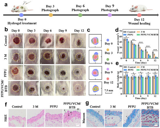
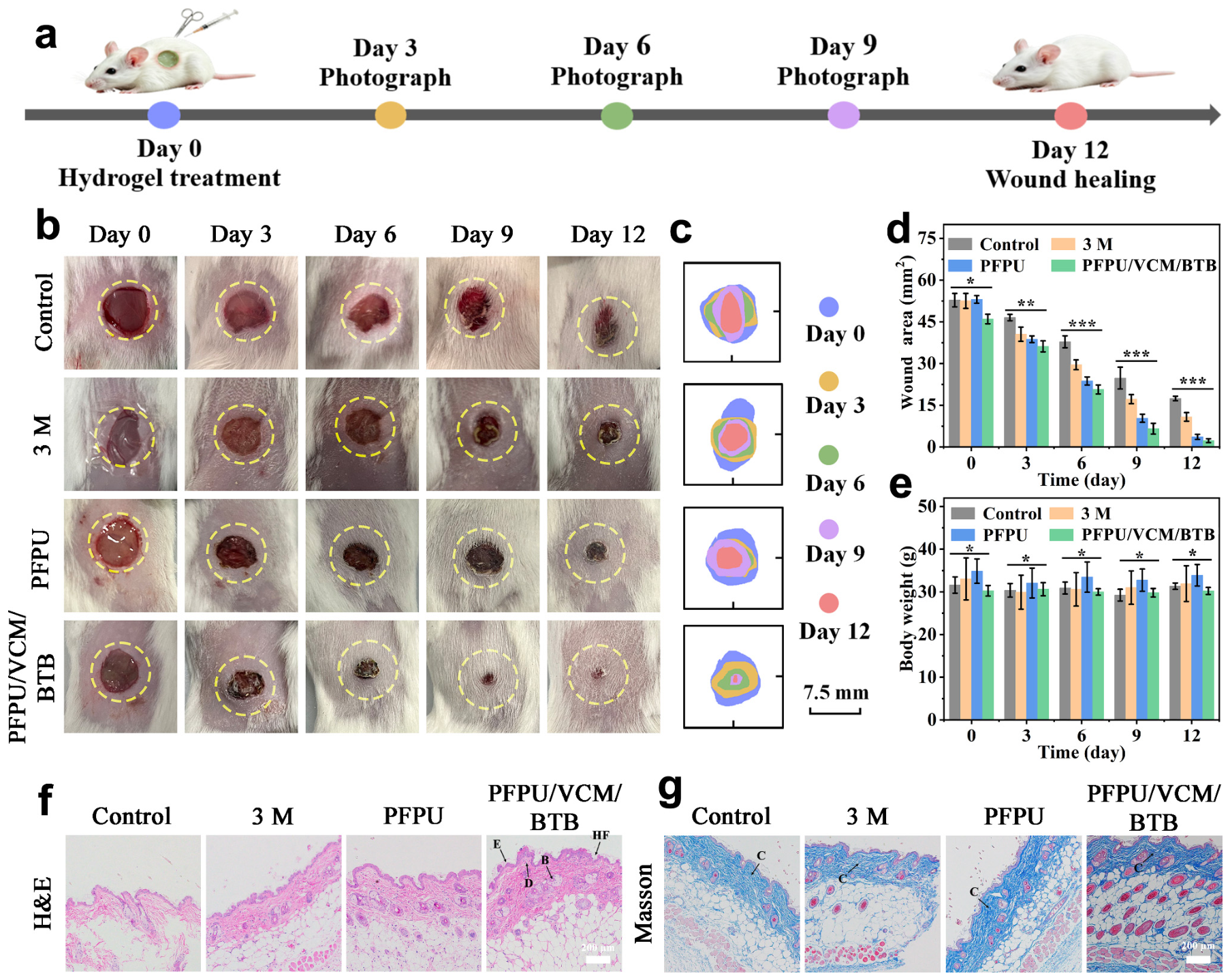
In vivo wound healing efficacy in an acute S. aureus-infected full-thickness wound model. (a) Schematic diagram of the experimental protocol and establishment of the acute S. aureus-infected full-thickness skin defect mouse model. (b) Representative digital photographs of the wound sites on mice treated with PBS control, commercial 3M dressing, PFPU hydrogel, and PFPU/VCM/BTB hydrogel on days 0, 3, 6, 9, and 12 post-infection. Yellow dashed circles indicate the initial wound locations and approximate boundaries. (c) Digitally processed wound contraction trajectory outlines over the 12-day treatment period. (d) Quantitative analysis of wound areas (mm2) on days 0, 3, 6, 9, and 12 post-treatment in the PFPU/VCM/BTB group. (e) Body weight variations in the mice throughout the 12-day therapy. (f,g) Histopathological evaluations of the regenerated wound skin tissue harvested on day 12 via (f) H&E staining, and (g) Masson’s trichrome staining in the PFPU/VCM/BTB group (Arrows indicate the epidermis (E), dermis (D), neovascularization (B) and hair follicles (HF) in the H&E-stained sections (f), and blue-stained collagen regions (C) in the Masson’s trichrome-stained sections (g)). (* p < 0.05, ** p < 0.01, and *** p < 0.001).
2.6. In Vivo Wound Healing Evaluation of PFPU/VCM/BTB Hydrogels
To further evaluate the in vivo therapeutic efficacy of the PFPU/VCM/BTB hydrogels, a full-thickness skin defect model infected with S. aureus was established on the mice. After 24 h of infection, the wounds were treated with PBS, a commercial 3M dressing, the PFPU hydrogel, and the PFPU/VCM/BTB hydrogel, respectively. Wound healing progression was recorded photographically on days 0, 3, 6, 9 and 12 (Figure 5b), and the corresponding wound areas were quantified. Figure 5c shows the overlaid wound-healing trajectory images processed by ImageJ software (version 1.54g). Generally, both the 3M dressing and the PFPU hydrogel promoted early wound contraction but exhibited slow healing rates, failing to achieve complete closure of the infected wounds. In contrast, the PFPU/VCM/BTB group showed rapid wound shrinkage on day 3. As shown in Figure 5b–d, by day 6, the wound area in the PFPU/VCM/BTB group was sharply reduced with a regular morphology, whereas the remaining wound areas in the PBS control, 3M, and PFPU groups were 39.95 mm2, 31.32 mm2, and 23.74 mm2, respectively. By day 12, the PFPU/VCM/BTB group achieved nearly full wound closure with only 2.95 mm2 wound area. Conversely, the PBS control and 3M groups retained large unhealed wounds, and the PFPU group exhibited limited healing capacity compared to the PFPU/VCM/BTB group. This indicates that the PFPU/VCM/BTB hydrogel effectively delivers VCM to eradicate bacterial infection, thereby accelerating the healing of infected wounds. Furthermore, mice were euthanized on day 12 post-treatment, and the wound tissues were collected for histological analysis. H&E and Masson’s trichrome staining were performed to evaluate the histological alterations during the wound healing. As shown in Figure 5f, although neo-epidermis was formed in all groups, the PFPU/VCM/BTB group exhibited a more intact neo-epidermal structure. Meanwhile, abundant mature hair follicles within the skin tissue were detected in the PFPU/VCM/BTB group, demonstrating the superior wound repair capacity. Masson’s trichrome staining further validated these findings (Figure 5g), revealing that the PFPU/VCM/BTB group exhibited more collagen deposition on day 12 compared to the other groups. Together, the H&E and Masson staining results confirm that the PFPU/VCM/BTB hydrogels can accelerate the healing of infected wounds by promoting epidermal reconstruction, stimulating dermal tissue regeneration, and enhancing collagen deposition.
3. Conclusions
In summary, we developed a multifunctional PFPU/VCM/BTB hydrogel based on the PFPU3 as a dynamic wound dressing for the monitoring and treatment of infected wounds. The hydrogel network was formed through the synergistic interactions of reversible Schiff base bonds formed between PF127-(CHO)2 and PMUA, together with UPy-mediated quadruple hydrogen bonding interactions. Benefiting from this dual dynamic cross-linking structure, the hydrogel possessed excellent injectability, self-healing capability, structural adaptability, and tissue adhesion, enabling effective coverage of irregular wound surfaces. The incorporation of BTB endowed the hydrogel with visible color changes within the pH range of 5.8–7.4, providing a direct indication of infection-associated acidic wound environments, while VCM contributed effective antibacterial activity against S. aureus. Furthermore, the PFPU/VCM/BTB hydrogel exhibited good biosafety and promoted wound repair in an infected mouse wound model. Overall, this study indicates that integrating a dual-dynamic hydrogel network, a pH-responsive visualization module, and local antibacterial activity constitutes an effective strategy for infected wound management. The PFPU/VCM/BTB hydrogel therefore shows considerable potential as a smart wound dressing capable of monitoring infection-related microenvironmental changes and promoting wound repair.
4. Experimental Section
4.1. Materials
4,4’-Azobis (4-cyanovaleric acid) (ACVA) was obtained from Shanghai Chuangsai Technology Co., Ltd. (Shanghai, China). 2-Hydroxyethyl methacrylate (HEMA), 2-aminoethyl methacrylate hydrochloride (AEMH), Pluronic F-127(PF127), 2-methacryloyloxyethyl phosphorylcholine (MPC) and vancomycin (VCM) were purchased from Aladdin Biochemical Technology Co., Ltd. (Shanghai, China). 4-Dimethylaminopyridine (DMAP) and N, N’-dicyclohexylcarbodiimide (DCC) were obtained from Shandong West Asia Chemical Industry Co., Ltd. (Linyi, China). 4-Carboxybenzaldehyde, thiazolyl blue tetrazolium bromide (MTT) and Calcein-AM/PI double staining kit were supplied by Shanghai McLean Biochemical Technology Co., Ltd. (Shanghai, China). Bromothymol blue (BTB) was purchased from Shanghai TCI Development Co., Ltd. (Shanghai, China). MEM medium (containing 10% fetal bovine serum and 1% penicillin/streptomycin) was obtained from Wuhan Procell Life Science & Technology Co., Ltd. (Wuhan, China). All chemical reagents were used as received without further purification unless otherwise stated.
4.2. Synthesis of PMUA and Preparation of PF127-(CHO)2 Micelles
The UPyHEMA monomer was synthesized according to our previous work [33]. PMUA was prepared via free-radical polymerization. Briefly, MPC (147.6 mg, 0.5 mmol), UPyHEMA (21.2 mg, 0.05 mmol) and AEMH (74.5 mg, 0.45 mmol) were completely dissolved in a mixture of DMF and methanol, followed by the addition of ACVA (1.4 mg, 0.005 mmol) as the initiator. The reaction system was purged with nitrogen for 30 min to remove dissolved oxygen and then polymerized at 75 °C for 24 h. The product solution was dialyzed for 3 days and freeze-dried to yield the PMUA copolymer.
PF127 functionalized with aldehyde groups at both termini (PF127-(CHO)2) was synthesized according to a previously reported procedure [27,28]. Firstly, PF127 (3.9 g, 0.3 mmol), DMAP (0.021 g, 0.18 mmol), 4-carboxybenzaldehyde (0.27 g, 1.8 mmol) and DCC (0.372 g, 1.8 mmol) were completely dissolved in 50 mL of anhydrous tetrahydrofuran, and the reaction mixture was stirred at room temperature (RT) for 48 h. The crude product was subjected to filtration, precipitation, washing and centrifugation, and then dried under vacuum to obtain PF127-(CHO)2. Afterwards, PF127-(CHO)2 was dissolved in deionized water under magnetic stirring for 6 h and allowed to stand at RT for more than 12 h to enable micellar self-assembly. PF127-(CHO)2 micelle particles were obtained after lyophilization.
4.3. Preparation of PFPU and PFPU/VCM/BTB Hydrogels
For hydrogel preparation, PF127-(CHO)2 was dissolved in 0.01 M phosphate-buffered saline (PBS) at precursor concentrations of 25, 30, or 35 wt%, while PMUA was dissolved in PBS at 8 wt%. PFPU1, PFPU2, and PFPU3 hydrogels were obtained by mixing the PF127-(CHO)2 and PMUA solutions at a 1:1 volume ratio (See Supporting Information Table S1 for the detailed composition). Gelation occurred rapidly after mixing through reactions between aldehyde groups on PF127-(CHO)2 and primary amines on PMUA, together with reversible supramolecular association of UPy motifs. To prepare the PFPU/VCM/BTB hydrogels, 0.05% BTB and 50 μg/mL VCM were dispersed into PF127-(CHO)2 solution prior to gelation. The mixture was then combined with the 8 wt% PMUA to form the PFPU/VCM/BTB hydrogels. The VCM concentration was determined based on the preliminary screening results (Figure S2).
4.4. Structural, Morphological, Rheological, and Macroscopic Characterization
The chemical structures of PMUA copolymer and PF127-(CHO)2 were characterized via 1H NMR and FT-IR. The internal porous microstructure of the lyophilized hydrogels was observed using scanning electron microscopy (Flex SEM 1000 II, Hitachi High-Tech Corporation, Tokyo, Japan). The gelation process was recorded by photography. To evaluate injectability, the PFPU/VCM/BTB hydrogel was loaded into a syringe equipped with a 21-gauge (21G) needle and subjected to uniform extrusion tests. The macroscopic self-healing capacity was assessed by physically attaching two separate cut hydrogel blocks; the self-healing process was recorded. The pH-responsive behavior of the PFPU/VCM/BTB hydrogel was investigated by adjusting the pH environments using 0.1 M HCl and 0.1 M NaOH solutions, with the dynamic structural dissociation and reconstruction of the hydrogel network recorded visually. Moreover, the adhesion performance of the hydrogel was assessed via contact adhesion tests, and photographs were taken to document the interfacial contact state.
Rheological measurements were performed on a Discovery HR-2 rheometer (TA Instruments, New Castle, DE, USA). Frequency sweeps were first conducted from 0.1 to 100 rad/s at 1.0% strain and 25 °C to determine the storage modulus (G′) and loss modulus (G″). Subsequently, strain sweep tests from 0.1% to 1000% were conducted at 10 rad/s for the PFPU/VCM/BTB hydrogel to determine the critical breaking strain, defined as the crossover point of G′ and G″. Furthermore, alternating step-strain cycles between 1% and 600% strain were applied to assess the rheological self-healing performance of the hydrogel.
4.5. pH Response and Wound-State Visualization
The pH-responsive color changes in the PFPU/VCM/BTB hydrogel were further investigated. 9 mm filter paper disks were soaked in PBS buffers at pH 5.8, 6.2, 6.8, 7.0, and 7.4 and placed in 96-well plates. Then, PFPU/VCM/BTB hydrogel (200 μL) was added to each well and incubated at 37 °C for 30 min, and the color changes in the hydrogel were visually observed and recorded. Meanwhile, to assess bacteria-induced color changes, bacteria were cultured in 96-well plates for different culture durations (0, 2, 4, 8, and 12 h) using a similar procedure. After complete removal of the culture medium, PFPU/VCM/BTB hydrogel was added for incubation at 37 °C, and the dynamic color variation in the hydrogel was monitored in real time. For in situ wound diagnosis, full-thickness dorsal wounds on mice (normal skin group, simple wound group, and S. aureus-infected wound group) were covered with the PFPU/VCM/BTB hydrogels. The color variation in the hydrogels at the wound sites was recorded to assess their monitoring capability for the acidic microenvironment of infected wounds.
4.6. Antibacterial Efficacy Assessment
The in vitro antibacterial activity of the PFPU/VCM/BTB hydrogels was evaluated against Gram-positive S. aureus. The revived bacteria were inoculated into Luria–Bertani (LB) medium and cultured at 37 °C for 24 h to reach the logarithmic growth phase. The bacteria were diluted with sterile PBS to obtain a bacterial suspension (1 × 10⁷ CFU/mL). Subsequently, the 3M dressing, PFPU hydrogel and PFPU/VCM/BTB hydrogel samples were separately immersed in bacterial suspension for 24 h. Antibacterial efficacy was assessed by plate colony counting, and colony morphology was photographed. Bacterial morphological changes were further observed by scanning electron microscopy (SEM). Samples were fixed in 2.5% glutaraldehyde at 4 °C overnight. After washing three times with PBS, the samples were sequentially dehydrated in ethanol solutions (30%, 50%, 70%, 90%, and 100%) for 15 min at each concentration. The treated bacterial suspension was then dropped onto silicon wafers, and bacterial morphology was observed by SEM.
4.7. Cytocompatibility and Hemocompatibility
The cytotoxicity of the PFPU/VCM/BTB hydrogel was evaluated using the mouse fibroblast cells (L929) via the MTT assay. First, sterilized 3M dressings, PFPU hydrogels, and PFPU/VCM/BTB hydrogels were separately immersed in MEM medium (containing 10% fetal bovine serum and 1% penicillin/streptomycin) for 24 h to prepare the extracts. L929 cells (8 × 10³ cells per well) were seeded in a 96-well plate, followed by 24 h incubation at 37 °C with 5% CO2 to allow cell adhesion. Thereafter, the original medium was replaced with the corresponding extract for further incubation at 37 °C, and untreated cells were used as the control group. After 24 and 48 h of incubation, the medium was removed, and the MTT reagent (5 mg/mL) was added to each well for an additional 4 h of incubation. The supernatant was removed, and 150 μL isopropanol was added to dissolve formazan crystals. The optical density (OD) was measured at 570 nm using a microplate reader, and cell viability was calculated using the following equation:
Furthermore, the effect of PFPU/VCM/BTB hydrogels on cell proliferation was assessed using the Calcein-AM/PI double staining. Cells were stained at 37 °C for 30 min, and live/dead cell images were captured via a fluorescence microscopy (IX73, Olympus, Hachioji, Tokyo, Japan). Hemolysis assays were performed to evaluate the hemocompatibility of PFPU and PFPU/VCM/BTB hydrogels. Fresh erythrocytes were collected from 6-week-old mice and diluted with PBS to prepare 4% (v/v) erythrocyte suspension. The 3M dressing, PFPU hydrogel and PFPU/VCM/BTB hydrogel were separately incubated with the prepared erythrocyte suspension and incubated at 37 °C for 4 h. PBS buffer was used as the negative control without hemolysis, and a 0.1% Triton X-100 solution was served as the positive control for complete hemolysis. Five experimental groups were established: (1) Triton X-100 solution, (2) PBS buffer, (3) 3M dressing group, (4) PFPU hydrogel, (5) PFPU/VCM/BTB hydrogel. All samples were centrifuged at 2000 rpm for 10 min. Photographs of hemolysis samples in each group were recorded. The supernatants were collected to measure the absorbance at 540 nm. The hemolysis rate (%) was calculated using the following formula:
4.8. Cell Migration Assay
A scratch wound assay was performed to evaluate the effect of PFPU/VCM/BTB hydrogel on cell migration. L929 cells (5 × 10⁵ cells per well) were seeded in culture plates and incubated at 37 °C for 24 h. A uniform scratch was created in the cell monolayer using a sterile 10 μL pipette tip and detached cells were removed by rinsing with PBS. The original medium was then replaced with extracts from different groups. Cell images were captured at 0 h, 12 h, 24 h and 48 h after scratching. The scratch area was analyzed using ImageJ software, and the migration rate was calculated as follows:
where A0 represents the initial scratch area measured at 0 h, and At represents the remaining scratch area at the predetermined time point.
4.9. In Vivo Full-Thickness Wound Healing Experiments
The in vivo animal experiments in this study were approved by the Animal Ethical Committee of Henan University of Science and Technology (Approval No. HAUST-026-M03160109; Approval Date: 16 March 2026). Six to eight-week-old male BALB/c mice were used to establish an infected full-thickness wound model. After anesthesia, a full-thickness skin wound with a diameter of 8 mm was created on each mouse, then inoculated with S. aureus suspension (50 μL of 2 × 10⁷ CFU/mL). A total of 20 mice were randomly divided into four groups (n = 5): (1) PBS control group, (2) 3M treatment group, (3) PFPU treatment group, and (4) PFPU/VCM/BTB treatment group. Wound photographs were taken on days 0, 3, 6, 9 and 12 and wound areas were measured to assess wound closure.
On day 12, all mice were euthanized, and the wound tissues with surrounding healthy skin edges were collected and fixed in 4% paraformaldehyde for 24 h. The tissue samples were embedded in paraffin and sectioned into 4 μm-thick slices. Histological alterations were evaluated by the hematoxylin–eosin (H&E) and Masson’s trichrome staining. In addition, to assess systemic biocompatibility in vivo of PFPU/VCM/BTB hydrogels, major organs including heart, liver, spleen, lung and kidney were collected from mice euthanized on day 12 and subjected to H&E staining for pathological analysis. All histological sections were examined using an optical microscope (IX73, Olympus, Hachioji, Tokyo, Japan).
4.10. Statistical Analysis
Quantitative data are presented as mean ± standard deviation (SD). Origin 2024 software was used for data processing and plotting. Statistical comparisons were performed using one-way analysis of variance (ANOVA). Differences with p < 0.05 were regarded as statistically significant (* p < 0.05, ** p < 0.01, *** p < 0.001).
Supplementary Materials
The following supporting information can be downloaded at https://www.mdpi.com/article/10.3390/gels12110999/s1, Table S1: Formulation of hydrogel; Figure S1: Swelling characteristics of hydrogel; Figure S2: Bacterial culture images of the hydrogels with different VCM concentrations.
Author Contributions
Conceptualization, L.Z. and R.N.; methodology, W.W.; validation, L.Z. and J.L.; formal analysis, Q.C., Z.L. and M.G.; investigation, Q.C., Z.L. and M.G.; data curation, Q.C. and J.C.; writing-original draft preparation, Q.C. and Z.L.; writing-review and editing, R.N.; visualization, Q.C.; supervision, J.L.; project administration, W.W.; funding acquisition, Q.C., J.L., L.Z. All authors have read and agreed to the published version of the manuscript.
Funding
This work was supported by the National Natural Science Foundation of China (Grant Nos. 22305069 and 22306055), the Joint Fund of Henan Province Science and Technology R&D Program (235200810008), and Heluo Youth Talent Support Project (Grant No. 2026HLTJ32).
Institutional Review Board Statement
The animal study protocol was approved by the Animal Ethical Committee of Henan University of Science and Technology (Approval No. HAUST-026-M03160109; Approval Date: 16 March 2026).
Informed Consent Statement
Not applicable.
Data Availability Statement
The data presented in this study is available on request from the corresponding author.
Acknowledgments
We thank the staff of the shared instrumentation facilities at Henan University of Science and Technology for their technical assistance.
Conflicts of Interest
The authors declare no conflict of interest.
References
- Peña, O.A.; Martin, P. Cellular and molecular mechanisms of skin wound healing. Nat. Rev. Mol. Cell Biol. 2024, 25, 599–616. [Google Scholar] [CrossRef] [Scilit]
- Tottoli, E.M.; Dorati, R.; Genta, I.; Chiesa, E.; Pisani, S.; Conti, B. Skin Wound Healing Process and New Emerging Technologies for Skin Wound Care and Regeneration. Pharmaceutics 2020, 12, 735. [Google Scholar] [CrossRef] [Scilit]
- Wang, W.; Gui, H.; Wei, X.; Liu, H.; Wang, X. Cuproptosis to Cuproptosis-Like: Therapeutic Strategies for Bacterial Infection. Adv. Mater. 2025, 37, e2506119. [Google Scholar] [CrossRef] [Scilit]
- Lin, S.; Nie, Z.; Xing, Y.; Wang, R.; Yu, F. SERS detection and antibacterial treatment of bacterial infections. Coord. Chem. Rev. 2026, 546, 217079. [Google Scholar] [CrossRef] [Scilit]
- Zhao, F.; Su, Y.; Wang, J.; Romanova, S.; DiMaio, D.J.; Xie, J.; Zhao, S. A Highly Efficacious Electrical Biofilm Treatment System for Combating Chronic Wound Bacterial Infections. Adv. Mater. 2022, 35, e2208069. [Google Scholar] [CrossRef] [Scilit]
- Farahani, M.; Shafiee, A. Wound Healing: From Passive to Smart Dressings. Adv. Healthc. Mater. 2021, 10, 2100477. [Google Scholar] [CrossRef] [Scilit]
- Joshi, S.; Maan, M.; Barman, P.; Sharely, I.; Verma, K.; Preet, S.; Saini, A. Advances in biomaterials for wound care management: Insights from recent developments. Adv. Colloid Interface Sci. 2025, 343, 103563. [Google Scholar] [CrossRef] [Scilit]
- Li, H.; Li, B.; Lv, D.; Li, W.; Lu, Y.; Luo, G. Biomaterials releasing drug responsively to promote wound healing via regulation of pathological microenvironment. Adv. Drug Deliv. Rev. 2023, 196, 114778. [Google Scholar] [CrossRef] [Scilit]
- Alves, P.M.; Barrias, C.C.; Gomes, P.; Martins, M.C.L. Smart biomaterial-based systems for intrinsic stimuli-responsive chronic wound management. Mater. Today Chem. 2021, 22, 100623. [Google Scholar] [CrossRef] [Scilit]
- Haller, H.L.; Sander, F.; Popp, D.; Rapp, M.; Hartmann, B.; Demircan, M.; Nischwitz, S.P.; Kamolz, L.P. Oxygen, pH, Lactate, and Metabolism—How Old Knowledge and New Insights Might Be Combined for New Wound Treatment. Medicina 2021, 57, 1190. [Google Scholar] [CrossRef] [Scilit]
- Aranson, I.S. Bacterial active matter. Rep. Prog. Phys. 2022, 85, 076601. [Google Scholar] [CrossRef] [Scilit]
- Wang, L.; Zhou, M.; Xu, T.; Zhang, X. Multifunctional hydrogel as wound dressing for intelligent wound monitoring. Chem. Eng. J. 2022, 433, 134625. [Google Scholar] [CrossRef] [Scilit]
- Pang, Q.; Yang, F.; Jiang, Z.; Wu, K.; Hou, R.; Zhu, Y. Smart wound dressing for advanced wound management: Real-time monitoring and on-demand treatment. Mater. Des. 2023, 229, 111917. [Google Scholar] [CrossRef] [Scilit]
- Han, Z.; Yuan, M.; Liu, L.; Zhang, K.; Zhao, B.; He, B.; Liang, Y.; Li, F. PH-Responsive wound dressings: Advances and prospects. Nanoscale Horiz. 2023, 8, 422–440. [Google Scholar] [CrossRef] [Scilit]
- Horta-Velázquez, A.; Mota-Morales, J.D.; Morales-Narváez, E. Next-generation of smart dressings: Integrating multiplexed sensors and theranostic functions. Int. J. Biol. Macromol. 2024, 254, 127737. [Google Scholar] [CrossRef] [Scilit]
- Norahan, M.H.; Pedroza-González, S.C.; Sánchez-Salazar, M.G.; Álvarez, M.M.; Trujillo de Santiago, G. Structural and biological engineering of 3D hydrogels for wound healing. Bioact. Mater. 2023, 24, 197–235. [Google Scholar] [CrossRef] [Scilit]
- Duan, S.; Hua, M.; Zhang, C.W.; Hong, W.; Yan, Y.; Jazzar, A.; Chen, C.; Shi, P.; Si, M.; Wu, D.; et al. Noncovalent Aggregation for Diverse Properties in Hydrogels: A Comprehensive Review. Chem. Rev. 2025, 125, 7918–7964. [Google Scholar] [CrossRef] [Scilit]
- Pizzi, A.; Terraneo, G.; Lo Iacono, C.; Beccaria, R.; Dhaka, A.; Resnati, G. Taxonomy of Chemical Bondings: Opportunities and Challenges. Angew. Chem. Int. Ed. 2025, 64, e202506525. [Google Scholar] [CrossRef] [Scilit]
- Xu, Y.; Li, Y.; Chen, Q.; Fu, L.; Tao, L.; Wei, Y. Injectable and Self-Healing Chitosan Hydrogel Based on Imine Bonds: Design and Therapeutic Applications. Int. J. Mol. Sci. 2018, 19, 2198. [Google Scholar] [CrossRef] [Scilit]
- Gu, Y.; Wang, S.; Liu, Y.; Yang, Z.; Li, W.; Lu, J.; Xu, J.; Gu, G.; Liu, Y.; Zhao, Z.; et al. Quadruple Hydrogen-Bonds Engineering for Intrinsically Stretchable and Healable Semiconducting Polymers. Angew. Chem. Int. Ed. 2026, 65, e9060680. [Google Scholar] [CrossRef] [Scilit]
- Li, Q.; Wen, C.; Yang, J.; Zhou, X.; Zhu, Y.; Zheng, J.; Cheng, G.; Bai, J.; Xu, T.; Ji, J.; et al. Zwitterionic Biomaterials. Chem. Rev. 2022, 122, 17073–17154. [Google Scholar] [CrossRef] [Scilit]
- Wang, H.; Zhou, S.; Guo, L.; Wang, Y.; Feng, L. Intelligent Hybrid Hydrogels for Rapid In Situ Detection and Photothermal Therapy of Bacterial Infection. ACS Appl. Mater. Interfaces 2020, 12, 39685–39694. [Google Scholar] [CrossRef] [Scilit]
- Lei, E.; Tao, H.; Jiao, S.; Yang, A.; Zhou, Y.; Wang, M.; Wen, K.; Wang, Y.; Chen, Z.; Chen, X.; et al. Potentiation of Vancomycin: Creating Cooperative Membrane Lysis through a “Derivatization-for-Sensitization” Approach. J. Am. Chem. Soc. 2022, 144, 10622–10639. [Google Scholar] [CrossRef] [Scilit]
- Feliciano, A.J.; Soares, E.; Bosman, A.W.; van Blitterswijk, C.; Moroni, L.; LaPointe, V.L.S.; Baker, M.B. Complementary Supramolecular Functionalization Enhances Antifouling Surfaces: A Ureidopyrimidinone-Functionalized Phosphorylcholine Polymer. ACS Biomater. Sci. Eng. 2023, 9, 4619–4631. [Google Scholar] [CrossRef] [Scilit]
- Zhang, Y.; Yi, M.; Bao, Y.; Zhang, S. Fabrication of micelles from poly(butylene succinate) and poly(2-methacryloyloxyethyl phosphorylcholine) copolymers as a potential drug carrier. Polym. Int. 2017, 67, 155–165. [Google Scholar] [CrossRef] [Scilit]
- Oktay, B.; Kayaman-Apohan, N.; Süleymanoğlu, M.; Erdem-Kuruca, S. Zwitterionic phosphorylcholine grafted chitosan nanofiber: Preparation, characterization and in-vitro cell adhesion behavior. Mater. Sci. Eng. C 2017, 73, 569–578. [Google Scholar] [CrossRef] [Scilit]
- Wei, Q.; Jin, Z.; Zhang, W.; Zhao, Y.; Wang, Y.; Wei, Y.; He, X.; Ma, G.; Guo, Y.; Jiang, Y.; et al. Honokiol@PF127 crosslinked hyaluronate-based hydrogel for promoting wound healing by regulating macrophage polarization. Carbohydr. Polym. 2023, 303, 120469. [Google Scholar] [CrossRef] [Scilit]
- Qu, J.; Zhao, X.; Liang, Y.; Zhang, T.; Ma, P.X.; Guo, B. Antibacterial adhesive injectable hydrogels with rapid self-healing, extensibility and compressibility as wound dressing for joints skin wound healing. Biomaterials 2018, 183, 185–199. [Google Scholar] [CrossRef] [Scilit]
- Liu, W.; Long, L.; Wang, Z.; He, S.; Han, Y.; Yang, L.; Hu, C.; Wang, Y. A Whole-Course-Repair System Based on Stimulus-Responsive Multifunctional Hydrogels for Myocardial Tissue Regeneration. Small Methods 2024, 8, e2400121. [Google Scholar] [CrossRef] [Scilit]
- Yang, J.; He, Y.; Li, Z.; Yang, X.; Gao, Y.; Chen, M.; Zheng, Y.; Mao, S.; Shi, X. Intelligent wound dressing for simultaneous in situ detection and elimination of pathogenic bacteria. Acta Biomater. 2024, 174, 177–190. [Google Scholar] [CrossRef] [Scilit]
- Liu, J.; Elias, J.; Wang, X.; Tian, J.; Dong, P.-T.; Cao, H.; Cen, L.; Zahedul Islam Nizami, M.; He, X.; Sun, J. Exploiting Bacterial Metabolism for Targeted Antimicrobial Release from Smart Nanocarriers. ACS Appl. Mater. Interfaces 2026, 18, 42261–42272. [Google Scholar] [CrossRef] [Scilit]
- Huang, F.; Lu, X.; Yang, Y.; Yang, Y.; Li, Y.; Kuai, L.; Li, B.; Dong, H.; Shi, J. Microenvironment-Based Diabetic Foot Ulcer Nanomedicine. Adv. Sci. 2022, 10, 2203308. [Google Scholar] [CrossRef] [Scilit]
- Cheng, Q.; Ding, S.; Zheng, Y.; Wu, M.; Peng, Y.-Y.; Diaz-Dussan, D.; Shi, Z.; Liu, Y.; Zeng, H.; Cui, Z.; et al. Dual Cross-Linked Hydrogels with Injectable, Self-Healing, and Antibacterial Properties Based on the Chemical and Physical Cross-Linking. Biomacromolecules 2021, 22, 1685–1694. [Google Scholar] [CrossRef] [Scilit]
Disclaimer/Publisher’s Note: The statements, opinions and data contained in all publications are solely those of the individual author(s) and contributor(s) and not of MDPI and/or the editor(s). MDPI and/or the editor(s) disclaim responsibility for any injury to people or property resulting from any ideas, methods, instructions or products referred to in the content. |
© 2026 by the authors. Licensee MDPI, Basel, Switzerland. This article is an open access article distributed under the terms and conditions of the Creative Commons Attribution (CC BY) license.