


Abstract
Severe oral mucositis requires local treatments that relieve pain, limit microbial proliferation, and support mucosal repair while minimizing exposure to potentially harmful excipients, particularly in pediatric patients. This study aimed to develop thermosensitive mucoadhesive gels co-loaded with lidocaine hydrochloride (2% w/v) and chlorhexidine digluconate (0.2% w/v) for localized buccal therapy. Poloxamer 407 was combined with hyaluronic acid sodium salt, xanthan gum, Carbopol 934, or hydroxypropylmethylcellulose (HPMC). Formulations containing lidocaine alone or both drugs were compared to assess the influence of chlorhexidine addition. Following preliminary screening based on pH and gelation properties, selected formulations were characterized for technological, mucoadhesive, biopharmaceutical, stability, and antibacterial properties. Poloxamer 407 concentration was the main factor governing gel performance, whereas the mucoadhesive polymer primarily affected specific technological attributes. Chlorhexidine incorporation did not compromise overall gel performance, preserving antibacterial activity. The gels exhibited suitable gelation, spreadability, mucoadhesion, residence time, and release properties with negligible membrane permeation. The dual-drug formulation containing 20% poloxamer 407 and 0.50% HPMC emerged as the most promising one, combining appropriate mechanical properties, drug release, antibacterial efficacy, and storage stability. Overall, these findings support dual-drug thermosensitive mucoadhesive gels as a feasible strategy for the local management of severe oral mucositis, especially in pediatrics.
1. Introduction
Chemotherapy and radiotherapy treatments represent the best hope for cure in pediatric patients affected by neoplastic diseases [1,2,3]. However, these therapeutic interventions cause numerous and significant side effects, among which severe oral mucositis (SOM) should not be underestimated. Oral mucositis is an inflammatory condition associated with damage to the mucosal protective barrier that, clinically, can range from mucosal erythema to painful ulcerations, potentially leading to malnutrition, secondary bacterial, viral or fungal infections and increased medical costs due to prolonged hospitalizations [4,5,6,7].
The severity of oral mucositis is commonly assessed using standardized grading systems, such as the World Health Organization (WHO) oral mucositis grading scale. This scale is straightforward to use in routine clinical practice and combines subjective symptoms, objective clinical findings, and the patient’s ability to eat to provide an overall assessment of oral mucositis severity: Grade 0, no oral mucositis; Grade 1, oral soreness and erythema; Grade 2, oral ulcers, but able to eat solid food; Grade 3, oral ulcers requiring a liquid diet because of mucositis; and Grade 4, oral ulcers, alimentation not possible because of mucositis [8].
The incidence of SOM is influenced by numerous factors, including the type of cancer, the chemotherapy and/or radiotherapy regimen employed, and the relative dose of drug administered [1,5,9]. Indeed, patients affected by hematological malignancies and head and neck tumors are at greater risk of developing SOM [3,7]. In the case of metastatic solid tumors and stem cell transplantation, the incidence of SOM increases, owing to the need for more aggressive high-dose chemotherapeutic regimens [5,10,11].
The oral mucosa is highly susceptible to chemotherapy-induced damage, resulting in atrophy, thinning and ulceration of the mucosal epithelium, bacterial and viral infections (primarily caused by Staphylococcus aureus, Candida albicans, and Herpes simplex virus), xerostomia and difficulty or severe inability to eat, drink, or swallow [10,12,13,14]. The disruption of the mucosal barrier, together with chemotherapy-induced immunosuppression, markedly increases susceptibility to secondary microbial infections and further exacerbates tissue damage [15]. Consequently, the compromised oral environment promotes microbial colonization, delays mucosal healing and contributes to the progression and persistence of oral lesions [16,17].
Particularly in pediatric patients, SOM can cause significant disability by making oral nutrition nearly impossible (requiring nasogastric tube feeding), complicating the administration of potent analgesics, prolonging hospitalization with a consequent increase in healthcare costs and, most importantly, severely compromising the patient’s quality of life [9,18,19,20]. Furthermore, SOM may cause temporary or prolonged interruptions in oncological treatment, potentially compromising treatment efficacy and reducing the likelihood of successful tumor eradication [9]. Children and adolescents are particularly susceptible to chemotherapy-induced SOM owing to the high proliferative rate of pediatric malignancies. Indeed, approximately 40% of patients undergoing chemotherapeutic regimens manifest side effects at the oral cavity level and this figure rises to as high as 90% in children under 12 years of age [2].
Despite representing a major clinical challenge, to date SOM remains insufficiently investigated, with no established gold standard available for its prevention and accurate management. Particularly at the pediatric level, therapeutic protocols for patients affected by severe mucositis are limited to proper oral hygiene and the use of mouthwashes based on anesthetic and/or bacteriostatic/anti-inflammatory agents for the control of lesion-associated pain. Management is particularly challenging in children younger than 6 years since the difficulty in performing oral rinses often requires application of the mouthwash solution to the mucosa using a soft cloth, rendering the treatment even less effective [1,19]. Moreover, these conventional formulations have a very limited residence time in the mouth, being quickly removed by the salivary flow and clearance, with the consequent short duration of action and need for frequent administration.
The limitations of current therapeutic approaches highlight the need for alternative local drug delivery systems capable of improving treatment efficacy and patient compliance. Among these, thermosensitive mucoadhesive in situ gels represent a particularly promising strategy for the topical management of SOM and other buccal diseases [21,22,23]. In situ gels provide a highly versatile formulation with a large surface area for application, allowing rapid drug release and prolonged contact, allowing sustained local drug release, in virtue of their ability to establish a prolonged contact with the oral mucosa through mucoadhesion, and forming at the same time a sort of protective barrier [24,25]. Additionally, thermosensitive systems facilitate administration by enabling the formulation to be applied as a liquid, thus easily reaching even remote or hidden sites of the mouth, and to rapidly gel at the site of application, thereby improving retention on the damaged mucosa. These properties make thermosensitive mucoadhesive hydrogels designed for local buccal delivery superior to mucoadhesive tablets or films in terms of greater patient compliance and comfort (with no unpleasant mouthfeel sensations), easier application, and full adaptation to the irregular/damaged surfaces of the oral mucosa, without risks of accidental swallowing or expulsion. A further disadvantage of mucoadhesive tablets or films is their fixed size, with the consequent difficulty, in the case of pediatric patients, of suitably adapting the dose to be administered to the patient’s weight.
Micro- and nano-based drug delivery systems, including liposomes, nanofibers and polymeric nanoparticles, have also been recently investigated as innovative approaches for improving the treatment of SOM [26]. However, despite their emerging potential, problems related to their more complex and expensive preparation methods, challenges in large-scale production and reproducibility, and/or limited stability currently strongly restrict their actual applicability in clinical practice.
Despite the growing interest in thermosensitive and mucoadhesive hydrogels for oral drug delivery [24,25,27,28], only a limited number of formulations have been specifically designed for the local management of SOM [22,23,29,30,31] and none specifically intended for pediatric patients.
Based on these considerations, the primary aim of the present work was to develop and characterize a novel thermosensitive mucoadhesive gel for the local management of SOM in pediatric patients. The formulation was designed to provide local pain relief, help prevent oral microbial infections, and promote regeneration of the damaged oral mucosa [10,13]. As a peculiarity of this study, special efforts were made to develop formulations free of additional stabilizers and other non-essential excipients, thereby minimizing exposure to potentially harmful substances in pediatric patients. Another important aspect taken into consideration was paying particular attention to designing a simple and reproducible preparation method, with the perspective of facilitating future extemporaneous compounding in hospital pharmacies and the rapid dispensing of the formulation to hospitalized patients, also improving their compliance.
Thermosensitive gels were formulated using Poloxamer 407, selected as a thermosensitive polymer [24,32,33,34] in combination with different mucoadhesive polymers including sodium hyaluronate, xanthan gum, Carbopol 934, and HPMC [35,36,37,38]. A novel dual-action formulation was obtained by co-loading Lidocaine HCl (2% w/v) and chlorhexidine digluconate (0.2% w/v) to provide local analgesic activity and antimicrobial activity, respectively. Some different kinds of mucoadhesive hydrogels for buccal applications, loaded with lidocaine [39,40,41,42,43] or chlorhexidine [40,41,44,45] have been previously investigated, as summarized in Table 1. However, none of these formulations have been specifically designed for the treatment of oral mucositis.
Table 1.
Examples of mucoadhesive and/or thermosensitive hydrogels loaded with lidocaine or chlorhexidine for the treatment of buccal diseases.
Moreover, to the best of our knowledge, no previous studies have investigated the simultaneous use of these two drugs in the development of any type of buccal dosage forms, including in situ hydrogels, intended for the treatment of oral mucosa diseases, and particularly of pediatric SOM. Furthermore, although chlorhexidine has been widely used in oral formulations, its influence on the physicochemical and technological performance of thermosensitive mucoadhesive gels has received very limited attention [41,44].
Therefore, to investigate the influence of chlorhexidine on formulation performance, gels containing lidocaine alone were compared with those containing both lidocaine and chlorhexidine. Following preliminary selection based on pH, gelation temperature and gelation time, the most promising formulations were comprehensively characterized in terms of spreadability, syringeability, mucoadhesion, deformability, residence time, hydration, and erosion properties, in vitro drug release, membrane permeation, stability, and antimicrobial activity against Staphylococcus aureus.
2. Results and Discussion
2.1. Preformulation Studies
Gelation Temperature and Time
Thermosensitive mucoadhesive gels were prepared using increasing concentrations of the thermosensitive polymer P407 (15–20–25% w/v) in combination with increasing concentrations of different mucoadhesive polymers (0.25 and 0.5% w/v). The concentration range for the thermosensitive polymer was selected based on preliminary studies that showed that formulations with higher P407 amounts (30% w/v) were already gelled at room temperature. Among the mucoadhesive polymers, HA and xanthan gum were chosen as natural polymers, whereas Carbopol®934 and HPMC were selected as synthetic and semisynthetic ones, respectively. The Tgel of each binary system was determined by the simple tube-inverting method [47,48] in the range 20–40 °C. tgel was recorded at 37 ± 0.5 °C for those formulations showing an ideal Tgel (Tsol-gel ≥ 28 °C).
Formulations containing 15% w/v P407 did not undergo gelation, regardless of the type of the additional polymer, and were therefore excluded from further investigation, as they were considered unsuitable for administration as liquid thermosensitive formulations. The gelation properties of the formulations containing 20% and 25% P407, containing different mucoadhesive polymers at different concentrations, both unloaded and loaded with LID alone or in combination with CHX, are summarized in Table 2.
Table 2.
Gelation temperature (Tgel) and gelation time (tgel) of thermosensitive mucoadhesive formulations containing 20% or 25% P407 combined with different types and concentrations of mucoadhesive polymers, before and after incorporation of lidocaine (LID) or chlorhexidine (CHX) alone or in combination (see Table 8 for the gel composition).
This preliminary screening allowed us to identify formulations with gelation properties compatible with the intended application and thus suitable to be loaded with the drugs. A Tgel ≥ 28 °C and a gelation time shorter than 1 min were considered optimal for the intended application. Conversely, formulations that were already gelled at room temperature or showed excessively long gelation times at 37 °C were excluded. However, empty formulations exhibiting gelation times up to 90 min were also selected for subsequent drug loading, since preliminary observations suggested that drug incorporation promoted gelation, leading to gelation times within the target range. These formulations were therefore included in the subsequent investigation to evaluate the influence of LID and CHX incorporation on gelation temperature and time.
For the empty formulations, both gelation temperature and time were strongly influenced by the thermosensitive polymer concentration as well as by the type and concentration of the mucoadhesive polymer. In general, increasing the P407 concentration from 20% to 25% w/v decreased the gelation temperature due to enhanced micellar packing and stronger interactions between polymer chains.
Among xanthan gum-based formulations, systems containing 0.25% w/v xanthan gum exhibited gelation temperatures ≥ 30 °C, resulting in relatively long gelation times at 37 °C (about 2 min). Conversely, increasing the xanthan gum concentration to 0.5% w/v led to formulations that were already gelled at room temperature, irrespective of the P407 concentration. This behavior can be attributed to the higher viscosity and denser polymeric network formed at higher xanthan gum concentrations, which strongly reduced molecular mobility and promoted gel formation even at lower temperatures. A similar behavior was observed for Carbopol® 934-based formulations, due to its high swelling capacity and pronounced viscosity-enhancing properties at higher concentrations.
The effect of HPMC was strongly dependent on the poloxamer concentration and on the overall polymeric network density. In formulations containing 20% w/v P407, the increase in HPMC concentration from 0.25% to 0.5% w/v resulted in a modest increase in gelation temperature, with only a slight effect on gelation time. In contrast, in formulations containing 25% w/v P407, the addition of 0.5% w/v HPMC promoted gel formation already at room temperature, indicating that at higher poloxamer concentrations the combined effect of polymer entanglement and micellar interactions becomes sufficient to induce premature gelation.
Among the investigated polymers, HA showed the most favorable balance between gelation temperature and gelation time, particularly at 0.25% w/v, in the presence of 20% P407, providing a suitable gelation temperature (29–30 °C) and the shortest gelation time (55 s) while avoiding premature gel formation at room temperature.
All empty formulations exhibited a pH close to neutrality (7.0–7.5) when measured in the liquid state at room temperature.
Based on this preliminary screening, only selected HA- and HPMC-containing formulations were selected for drug loading. The influence of drug incorporation on gelation behavior was then investigated.
The presence of LID in the gels led to a marked decrease in both gelation temperature and time. This behavior may be attributed to the amphiphilic nature of this drug, which may promote micellar packing and enhance dehydration of the polymer chains, facilitating the sol–gel transition at lower temperatures.
The subsequent addition of CHX produced a slight increase in both gelation temperature and time. This behavior may be attributed to the highly hydrophilic and cationic nature of CHX, which tends to partition into the aqueous phase, modifying the interactions between water and poloxamer, likely by hindering the aggregation and packing, and requiring higher temperatures to achieve gel network formation. Nevertheless, all selected formulations fulfilled the predefined acceptance criteria for buccal administration.
LID incorporation slightly reduced the pH to values around 6.0, whereas the subsequent addition of CHX did not further modify the pH, which remained within the same range and was considered suitable for buccal application.
Based on these results, the formulations containing 20% w/v P407 combined with 0.25% w/v HA, 0.25% w/v or 0.5% w/v HPMC and formulations containing 25% w/v P407 combined with 0.25% w/v HA were selected for further characterization.
2.2. Characterization of the Selected Formulations
Spreadability and mucoadhesion both contribute to the performance of buccal gels during administration and residence at the application site, and therefore their determination is of great importance for their proper characterization. The results of both tests are presented in Table 3.
Table 3.
Spreadability and mucoadhesive properties of the developed gels in the unloaded state and after loading with lidocaine (LID), chlorhexidine (CHX), or their combination (LID + CHX). Spreadability is expressed as firmness, work of shear, and stickiness, while mucoadhesion is expressed as maximum adhesive force, work of adhesion, and debonding distance. Data are presented as mean ± SD (n = 3).
Spreadability analysis was performed to evaluate the ease of application of the thermosensitive gels onto the oral mucosa. Results are expressed in terms of firmness, work of shear and stickiness. Lower firmness and work of shear values are generally associated with improved spreadability and, consequently, easier application, which is a desirable feature for topical administration on painful oral mucosal lesions. The presence of LID gave rise to some improvement in gel spreadability, as indicated by the reduction in work of shear and particularly in firmness values, especially in formulations containing 25% w/v P407 and 0.25% w/v HA.
Compared with the corresponding LID-loaded formulations, the LID/CHX-loaded formulations showed analogous results for the spreadability-related parameters, indicating that the addition of the antimicrobial agent did not substantially modify the application properties of the gels. The only exception was the formulation containing 25% w/v P407 and 0.25% w/v HA, in which the combined incorporation of LID and CHX increased firmness, work of shear, and absolute magnitude of stickiness, suggesting the formation of a more structured polymeric network. To gain further insight and determine whether it resulted from the combined presence of the two drugs or could instead be attributed to CHX alone, the corresponding formulation containing CHX as the sole active ingredient was also prepared and evaluated.
The study revealed that formulations containing CHX alone showed textural properties similar to those of the corresponding unloaded gels, indicating that the effect could arise from the combination of both drugs with a specific P407 and HA concentration ratio.
Overall, these findings indicate that spreadability was primarily governed by the polymeric composition of the formulation. From a practical perspective, this limited effect is advantageous, since the antimicrobial agent can be included without compromising the ease of gel application onto painful oral lesions.
Although ease of application is an important prerequisite, effective buccal formulations must also establish sufficient adhesion to the mucosal surface to ensure prolonged in situ residence.
Mucoadhesion was then evaluated in terms of adhesiveness, work of adhesion, and debonding distance. Adhesiveness reflects the maximum force required to detach the formulation from the mucosal surface, whereas the work of adhesion represents the total energy required for detachment. Debonding distance reflects the extent to which the formulation can be stretched before complete separation from the mucosa-mimetic membrane.
The incorporation of LID as well as of the LID/CHX combination had only a limited influence on the mucoadhesive properties of most thermosensitive gels, indicating that the presence of the drugs did not adversely affect gel adhesion to the mucosal surface. Only the formulation containing 20% P407 and 0.25% HA, in which LID and CHX were incorporated simultaneously, exhibited a markedly higher adhesiveness and a greater debonding distance than those observed for the corresponding single drug-loaded formulations. The study revealed that formulations containing CHX alone showed textural properties comparable to those of the corresponding unloaded gels, indicating that the effect could be attributable to the specific combination of both drugs with P407 and HA.
Overall, the results indicate that the effect of CHX presence on mucoadhesion depended on the polymeric composition of the formulation. Importantly, CHX incorporation did not compromise the mucoadhesive performance of the gels and, in the HA-based formulation, even improved their adhesive behavior.
Drug incorporation had only a minimal effect on syringeability. In particular, CHX incorporation did not significantly modify either the stiction force (1.5–3.1 N) or the plateau force (<1.6 N) compared with the corresponding LID-loaded formulations, indicating comparable extrusion behavior among the developed gels. These findings indicate that CHX presence did not adversely affect the extrusion behavior of the formulations before gelation. The developed gels could be readily dispensed, allowing accurate placement of the formulation onto the target lesion prior to in situ gelation.
The deformation behavior of the thermosensitive gels was evaluated under static and dynamic conditions, the latter performed to simulate the mechanical stresses generated by tongue movements after gel application. The static deformation test was performed on all selected formulations, whereas the tongue-assisted deformation test was carried out only on LID- and LID/CHX-loaded gels, representing the final therapeutic formulations. For a direct comparison between testing conditions, only formulations evaluated by both methods are reported in Table 4.
Table 4.
Static and dynamic (tongue-assisted) deformation of LID (L) and LID/CHX (LC)-loaded gels after gelation at 37 °C (see Table 8 for formulation composition).
Under static conditions, all formulations exhibited high dimensional stability after gelation at 37 °C. Most gels retained their original shape, showing no measurable displacement from the initial deposition area, while only formulations P20-HPMC025-L and P20-HPMC025-LC exhibited a slight deformation (1.5 mm). These findings indicate the rapid formation of a sufficiently stable gel network capable of maintaining the formulation at the site of deposition under static conditions. In contrast, all formulations tested under the tongue-assisted conditions showed substantially greater deformation, with values ranging from 7.0 to 12.3 mm. These results indicate that the mechanical stresses generated by tongue movements play a major role in gel deformation and displacement after application. Such mechanical action may reduce the amount of formulation retained at the application site and potentially shorten its local residence time. Therefore, the tongue-assisted deformation test provides a more physiologically relevant assessment of gel behavior following oral administration than conventional static evaluation alone. This aspect may be particularly relevant in pediatric applications, where tongue movements could increase the displacement of a formulation perceived as a foreign body, potentially affecting its local retention. Nevertheless, this effect may be less pronounced in severe oral mucositis, in which pain and ulceration can markedly restrict tongue mobility.
Also, the clinical relevance of residence time deserves particular consideration in the context of pediatric oral mucositis, in which intense pain, reduced oral intake, dehydration, impaired nutritional status, and reduced quality of life make effective local treatment particularly important [49].
Nevertheless, to the best of our knowledge, no clinically validated minimum or optimal residence time has been established for thermosensitive formulations intended for this condition. Prolonged residence may increase the duration of contact between the formulation and the affected mucosa, sustain local drug availability, and potentially reduce the need for frequent reapplication.
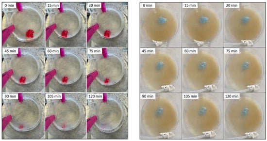
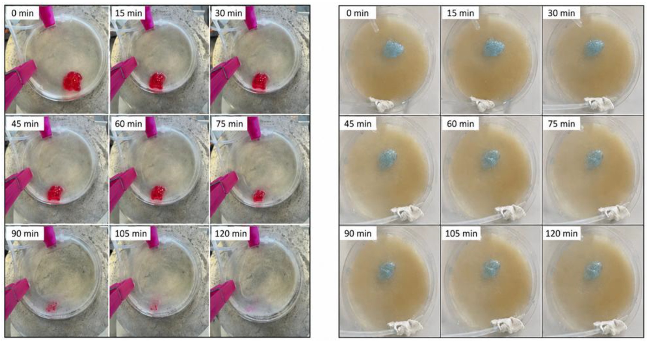
The in situ residence time study revealed formulation-dependent residence times under simulated oral conditions (Figure 1). P20-HPMC025-L and P20-HPMC050-L exhibited a progressive reduction in gel diameter, indicating gradual erosion of the gel matrix. In contrast, P25-HA025-L showed better preservation of its initial structure throughout the experiment, although its dimensions progressively decreased, suggesting greater resistance to erosion under the tested conditions.
Figure 1.
Representative photographs illustrating the residence time behavior of formulations P20-HPMC050-L (left) and P20-HPMC050-LC (right) under simulated oral conditions (see Table 8 for formulation composition). Images were acquired every 15 min over a period of 120 min.
The corresponding LID/CHX (LC)-loaded formulations also showed different behaviors. Formulations P20-HPMC025-LC and P20-HPMC050-LC remained clearly detectable after 120 min, ensuring adequate contact time for LID and CHX exposure at the ulcerated site. Conversely, P25-HA025-LC underwent more extensive erosion and was almost completely removed at the end of the test. Thus, CHX incorporation did not produce a uniform effect on the residence behavior, which appeared to be dependent on the overall polymeric composition of each formulation. Overall, all gels exhibited residence times not less than one hour, while differing in their erosion profiles.
Hydration and erosion studies were restricted to formulations containing 20% P407 only, in order to evaluate the influence of the mucoadhesive polymer and of LID and CHX incorporation while maintaining a constant thermosensitive polymer concentration. This approach reduced the potentially confounding influence of variations in P407 concentration and allowed a more direct comparison among the selected formulations.
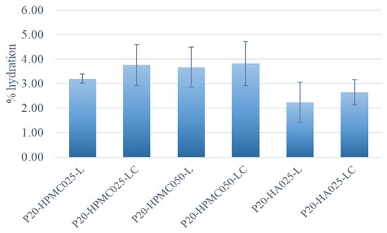
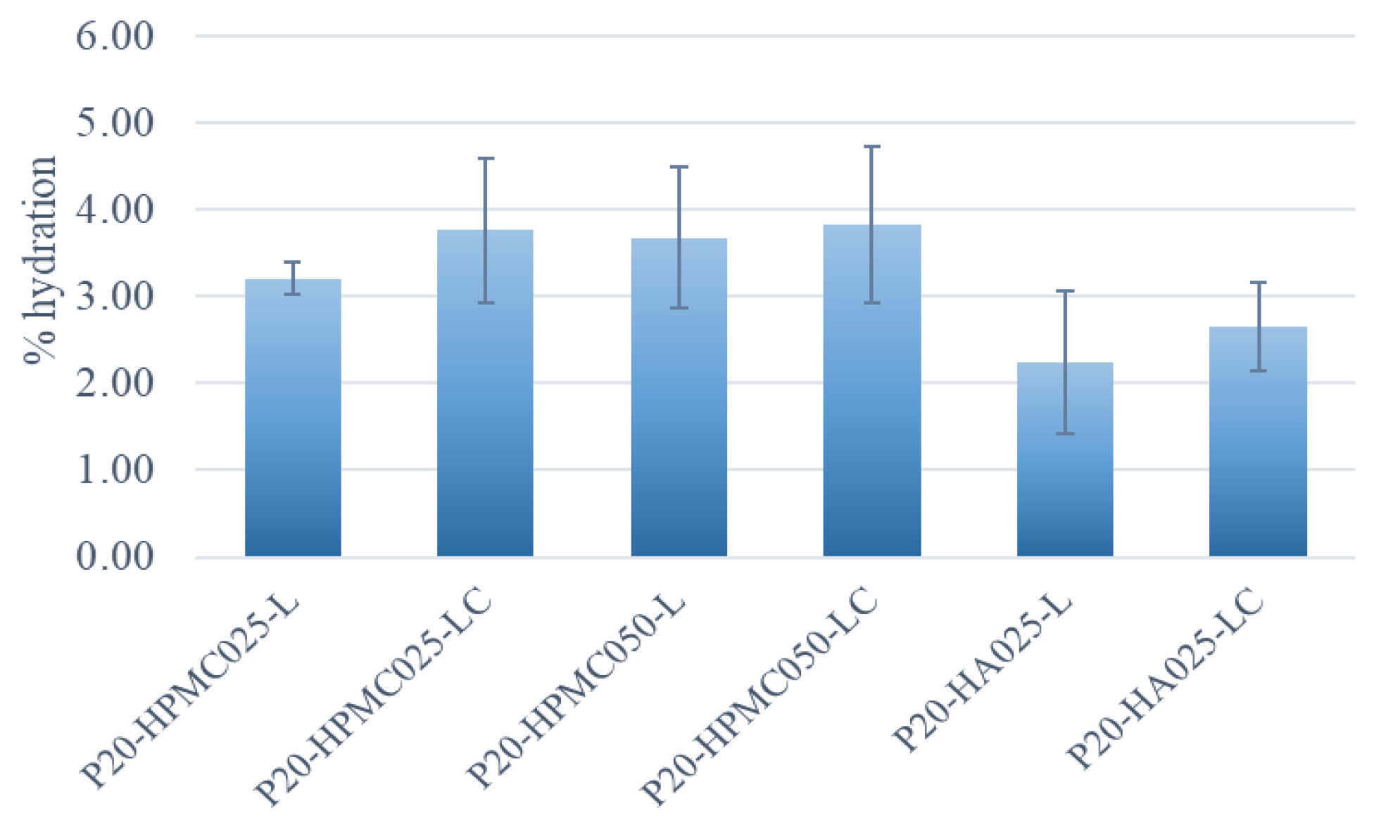
The hydration test demonstrated that all systems promoted moderate hydration of the mucin-agar gel, with % hydration values ranging from 2.24 to 3.83% (Figure 2). Formulations containing CHX exhibited slightly higher hydration values than the corresponding LID-loaded gels. However, the differences were modest, indicating that CHX incorporation had only a limited influence on the hydration behavior of the developed systems. Overall, all formulations maintained a comparable hydration profile, suggesting their ability to provide a moderately hydrated environment at the gel–mucosa interface, which may contribute to patient comfort during the treatment of oral mucosal lesions.
Figure 2.
Hydration degree of the mucin-agar gel mimicking the buccal mucosa after 2 h of contact with the developed thermosensitive gels (see Table 8 for formulation composition). Data are expressed as the percentage increase in membrane hydration after incubation at 37 °C and are reported as mean ± SD (n = 3).
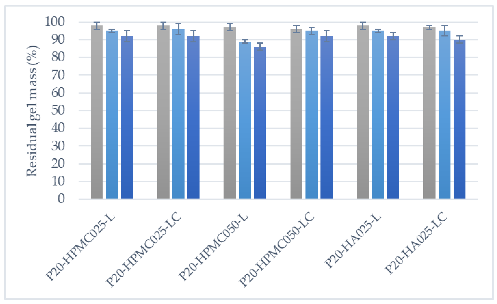
Among the formulations containing 20% P 407, the erosion study demonstrated a gradual weight loss for all formulations, reaching approximately 8–14% after 120 min under simulated salivary conditions, indicating progressive erosion of the gel matrix (see Figure 3). These findings suggest that matrix erosion may contribute to drug release, although release studies are required to establish the relative contribution of matrix erosion and diffusion to the overall release mechanism.
Figure 3.
Erosion profiles of the selected gel formulations, expressed as residual gel mass (%) after 30 (gray), 60 (light blue) and 120 min (blue) (see Table 8 for formulation composition). Data are expressed as mean ± SD (n = 3).
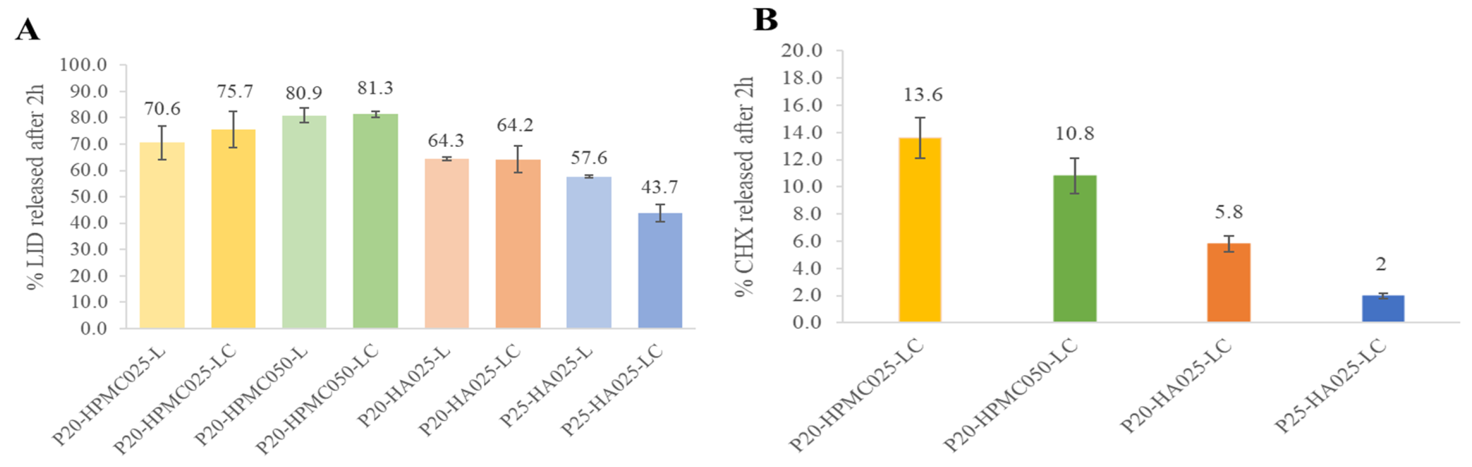
The release studies highlighted that LID release was strongly influenced by the formulation composition, whereas CHX exhibited a slower and more limited release profile (Figure 4). The presence of CHX did not significantly affect LID release from the 20% P407 formulations, irrespective of the mucoadhesive polymer employed. On the contrary, in the 25% P407 formulation, CHX incorporation resulted in a marked reduction in LID release. This finding suggests that CHX may have modified drug–matrix interactions or micellar organization, thereby reducing LID mobility within the gel. In fact this effect is more pronounced in the more densely packed micellar network formed at 25% w/v P407.
Figure 4.
Percentage of LID (A) and CHX (B) released from the selected thermosensitive gel formulations after 120 min (see Table 8 for formulation composition). Data are presented as mean ± SD (n = 3). The time-dependent release profiles of LID and CHX are reported in the Supplementary Material as Figure S1.
Increasing the HPMC concentration from 0.25% to 0.5% w/v produced a limited increase in LID release, either in the presence or absence of CHX, indicating that, within the investigated range, HPMC concentration had only a minor contribution to the release behavior. Conversely, the type of mucoadhesive polymer affected drug release in the dual-loaded systems. Formulations containing HA showed slightly lower LID release than the corresponding HPMC-based gels. These differences may be attributed to differences in the physicochemical properties and network organization of the mucoadhesive polymers. HPMC is a non-ionic cellulose derivative, whereas sodium hyaluronate is an anionic polysaccharide whose carboxylate groups may electrostatically interact with the cationic drugs, potentially affecting their mobility and promoting drug retention within the polymeric matrix [50,51].
The role of the mucoadhesive polymer should also be considered in combination with P407, which provides the thermoresponsive gelation mechanism. Therefore, the observed performance likely results from the combined effects of polymer composition, gel structure, and drug–matrix interactions, rather than reflecting an intrinsic superiority of HPMC in all formulations.
Overall, increasing the P407 concentration from 20% to 25% w/v reduced LID release, an effect that became more pronounced in the presence of CHX. These findings further support the role of the thermosensitive polymer in controlling drug diffusion through the gel network. CHX release was considerably lower than that of LID, reaching only approximately 12% after 120 min, consistent with its stronger retention within the polymeric matrix.
The experimental data of LID release profiles were fitted according to different kinetic models (Korsmeyer-Peppas, zero-order, first-order, Higuchi and Hixson–Crowell) in order to obtain insight into the mechanism of its release from the hydrogel formulations. The results are reported in Table 5 in terms of correlation coefficient (R2) values, as an index of the goodness of fit.
Table 5.
Values of correlation coefficients (R2) and release exponent (n) obtained with the different kinetic models for the various hydrogel formulations.
The values for the Korsmeyer-Peppas release exponent n, indicative of the release mechanism, are also presented.
The Korsmeyer–Peppas model provided the best fit for the drug release profiles from all the examined formulations, as indicated by the highest R2 values, although first-order and Higuchi fits gave rather similar R2 values for several formulations. No differences in the results were observed as a consequence of CHX addition. The Korsmeyer–Peppas diffusional exponent ranged from 0.41 to 0.49 across the various formulations, indicative in all cases of a Fickian diffusion mechanism (n < 0.5). However, the higher n values observed in the case of the hydrogels containing HPMC seem to suggest a tendency of such formulations towards an anomalous transport, where the swelling of the polymer also plays a role. This different behavior, compared to the hydrogels with HA, could be explained by the higher hydration ability shown by HPMC-containing formulations.
These values are broadly consistent with an important contribution of diffusion to the drug release [52,53], although the exponent alone does not allow a definitive assignment of the release mechanism.
Permeation studies showed negligible permeation of both LID and CHX across the artificial membrane for all formulations. It must nevertheless be taken into account that this assay provides only a preliminary assessment of drug permeation. In fact, the artificial lipophilic membrane model used, previously developed by an experimental design methodology, has actually demonstrated some predictive power of drug buccal absorption in the case of intact mucosa, being then useful for an initial fast screening and evaluation [54]. However, in the case of inflamed/damaged mucosa, such as that targeted by this study, further studies will be necessary to more reliably assess drug permeation by using more physiologically relevant models, such as ex vivo buccal mucosa, reconstructed oral epithelium, or inflamed mucosal models.
Based on the overall physicochemical, mechanical and mucoadhesive properties as well as on drug release results, one representative formulation was selected for each mucoadhesive polymer. Since it has been proved that CHX incorporation did not adversely influence the overall performance of the developed gels, while providing an additional antimicrobial effect, only the dual-loaded formulations were considered for further investigation.
Among the HPMC-based gels, formulation P20-HPMC050-LC was selected because it provided the most balanced overall performance, particularly in terms of drug release profile, whereas formulation P20-HA025-LC was selected among the HA-based formulations, owing to its superior mechanical properties, while exhibiting satisfactory performance in the remaining characterization tests.
The antibacterial activity of the two selected formulations was then evaluated using the agar diffusion assay. As can be observed in Table 6, both formulations (P20-HPMC050-LC and P20-HA025-LC) produced wide inhibition zones after 24 h, confirming that CHX retained its antibacterial activity after incorporation into the thermosensitive mucoadhesive gels. Comparable inhibition zones were observed for both formulations, indicating that the type of mucoadhesive polymer did not adversely affect the antibacterial efficacy of CHX.
Table 6.
Diameters of growth inhibition zones obtained for the tested formulations against Staphylococcus aureus (see Table 8 for formulation composition). Data are expressed as mean ± standard deviation (n = 4).
Finally, the stability of the selected formulations was evaluated after 1 and 2 months of storage (see Table 7). Formulation P20-HPMC050-LC maintained its physicochemical characteristics throughout the study, with no significant changes in pH, gelation temperature, or gelation time, demonstrating good stability. In contrast, formulation P20-HA025-LC showed limited long-term stability, with the appearance of visible precipitates after 2 months, indicating a loss of physical stability during storage despite the absence of significant changes in the measured physicochemical parameters. These findings identify formulation P20-HPMC050-LCas the most promising candidate for further pharmaceutical development.
Table 7.
Stability assessment of the selected gel formulations at time 0 and after 1 and 2 months of storage by pH, gelation temperature (Tgel), and gelation time (tgel) monitoring (see Table 8 for formulation composition).
3. Conclusions
Thermosensitive mucoadhesive gels co-loaded with LID and CHX were successfully developed for local buccal drug delivery. The obtained results demonstrated that the concentration of P407 was the main factor governing gel performance, whereas the type and concentration of the mucoadhesive polymer mainly influenced specific technological properties.
Importantly, CHX incorporation did not compromise the overall technological performance of the developed gels, preserving their physicochemical, mechanical, mucoadhesive, hydration, and in situ residence characteristics while maintaining antibacterial efficacy. Its influence on LID release was limited to the formulation containing 25% P407, where the denser gel network further restricted drug diffusion.
The developed gels exhibited suitable gelation characteristics, spreadability, mucoadhesion, in situ residence time, and local retention, together with negligible drug permeation across the lipophilic artificial membrane, supporting their suitability for localized buccal therapy. Moreover, the tongue-assisted deformation test provided a more physiologically relevant evaluation of the gel behavior than conventional static testing, highlighting the impact of tongue movements on formulation displacement after administration. This approach may represent a useful complementary tool for the characterization of buccal semisolid formulations.
Among the investigated systems, formulation P20-HPMC050-LC emerged as the most promising candidate, combining appropriate mechanical properties, controlled drug release, preserved antibacterial efficacy, and good physicochemical stability during storage. Overall, these findings demonstrate that the simultaneous incorporation of LID and CHX into thermosensitive mucoadhesive gels represents a feasible strategy for the local management of painful oral lesions.
4. Materials and Methods
4.1. Materials
Lidocaine hydrochloride (LID) was kindly donated by Meyer Children’s Hospital (Florence, Italy). Chlorhexidine digluconate aqueous solution (20% w/v) (CHX), low molecular weight (800–1600 kDa) hyaluronic acid sodium salt (HA), hydroxypropyl methylcellulose (HPMC), orthophosphoric acid, mucin (Type II, porcine stomach), L-α-phosphatidylcholine and cholesterol were purchased from Sigma-Aldrich (Milan, Italy). Lutrol® F127 (Poloxamer 407, P407) was supplied by BASF (Ludwigshafen, Germany), Carbopol® 934 (CP) by Lubrizol Advanced Materials, Inc. (Cleveland, OH, USA), and xanthan gum (XG) by ACEF (Milan, Italy). Sodium phosphate dibasic dodecahydrate and sodium chloride were obtained from VWR Chemicals (VWR International S.r.l., Milan, Italy), potassium phosphate monobasic from Fluka Analytical (Milan, Italy), Tryptic soy agar from Oxoid (Thermo Fisher Scientific, Birmingham, UK), and n-octanol from Merck (Darmstadt, Germany). Simulated saliva was prepared by dissolving Na2HPO4 (2.38 g/L), KH2PO4 (0.19 g/L), and NaCl (8.00 g/L) in deionized water; the pH was adjusted to 6.75 with H3PO4. Type II purified water was obtained using an Elix® Essential water purification system (Millipore, Bedford, MA, USA), which was used for the preparation of all solutions. Staphylococcus aureus DSM 346 was used for antimicrobial studies.
4.2. Methods
4.2.1. Preparation of Thermosensitive Mucoadhesive Gels
LID was dissolved in a 25 mL flask containing 20 mL of ultrapure water, under magnetic stirring at room temperature. Once fully dissolved, the mucoadhesive polymer (HA, xanthan gum, Carbopol 934, or HPMC) was added. The flask was then placed in an ice bath, and the thermosensitive polymer (P407) was added gradually in small portions, under stirring. In the case of dual-drug formulations, after complete dissolution of the thermosensitive polymer, the flask was removed from the ice bath, and an appropriate volume of a 20% w/v CHX stock solution was added, corresponding to a nominal CHX concentration of 0.20% w/v. For comparison purposes, gels loaded with only CHX were also prepared according to the same procedure, without the initial addition of LID, and used as additional controls for selected tests.
The gels were left to stand for 24 h at room temperature to allow proper hydration of the thermosensitive polymer and formation of the gel network.
Blank gels, and loaded gels containing CHX alone (0.2%), LID alone (2%), and dual-drug gels (containing both APIs) were formulated using different types and concentrations (0.25–0.5% w/v) of mucoadhesive polymers, combined with varying concentrations of the thermosensitive polymer (P407; 15–20–25–30% w/v), as summarized in Table 8.
Table 8.
Composition of unloaded and drug-loaded thermosensitive gel formulations containing different concentrations of P407 combined with different types and concentrations of mucoadhesive polymers.
4.2.2. Gelation Temperature
Gelation temperature (Tgel) was determined by the simple tube-inverting method [23,47,48]. Briefly, 2.5 mL of each binary system consisting of thermosensitive polymer and mucoadhesive polymer was transferred to a test tube and immersed in a thermostatically controlled water bath. The temperature was increased from 20° to 40 °C at a heating rate of 2.5 °C/min. The gel formation was determined as a non-flowing gel when the tube was inverted. Formulations were considered suitable when Tgel ≥ 28 °C.
4.2.3. Gelation Time
Gelation time (tgel) was recorded for those formulations with a Tgel ≥ 28 °C both in the absence and in the presence of the drugs. 5 mL of formulations were placed into a 25 mL beaker and immersed in a thermostatically controlled water bath at 37 ± 0.5 °C under magnetic stirring [55]. The time required to stop the bar was considered as the complete gelation time. A tgel of less than 1 min was considered ideal for the intended application.
4.2.4. pH Measurements
The pH was measured with a Crison 20 pH meter (Crison Instruments, Barcelona, Spain) which was calibrated before use with standard buffer solutions.
4.2.5. Spreadability
Gel spreadability was determined using a TA.XT Plus Texture Analyser (Stable Micro Systems Ltd., Godalming, UK) equipped with the appropriate Spreadability Rig (HDP/SR) with a 90° male cone probe. The instrumental parameters were set as follows: compression test mode, with a pre-test speed of 1.00 mm/s, a trigger force of 10.0 g, and a trigger distance of 2.0 mm. Formulations that met the acceptance criteria in terms of Tgel, tgel, and pH were selected for analysis. Measurements were performed in triplicate (n = 3) at room temperature. Force–time curves were recorded and used to determine firmness (g), work of shear (g·s), and stickiness (g).
4.2.6. Mucoadhesion
Mucoadhesion was assessed using a TA.XT Plus Texture Analyser (Stable Micro Systems Ltd., Godalming, UK) equipped with the Gel Mucoadhesion Probe (A/MUC). The instrumental settings were as follows: tension test mode with a test speed of 0.30 mm/s, a trigger force of 5.1 g, and a trigger distance of 2.0 mm. The test was performed at 37 °C, using a heating plate on which a Petri dish containing a 2 mm layer of a gel of porcine mucin (2% w/v)–agar (1.5% w/v), used as a model of mucoadhesive tissue [56,57] was placed. A 2.5 mL sample was then applied onto the membrane, and gelation was allowed to occur before starting the analysis. Formulations that met the acceptance criteria in terms of Tgel, tgel and pH were tested in triplicate (n = 3). Measurements were performed in triplicate (n = 3), and adhesiveness was determined from the resulting force–time profiles.
4.2.7. Deformation Tests
Gel deformation was evaluated using two different experimental methods: (1) a static spreading test on a thermostated Petri dish and (2) a TA.XT Plus texture analyzer (Stable Micro Systems Ltd., Godalming, UK) equipped with a P/40 probe fitted with an artificial silicone tongue.
Static deformation test: 2.5 mL of gel was deposited within a 1 cm diameter circle on a Petri dish thermostated at 37 °C using a heating plate, allowing the gel to undergo gelation. Without applying any additional force, the displacement of the gel from its initial position was evaluated after 5 min by using millimeter graph paper placed underneath the Petri dish. Formulations that met the acceptance criteria in terms of Tgel, tgel,, and pH were tested in triplicate (n = 3).
Texture analyser tongue-assisted deformation test: The analytical method developed for the mucoadhesion test was adapted by replacing the original probe with a P/40 probe fitted with an artificial silicone tongue model [58].
To improve gel visualization, a blue dye was incorporated into the formulation after 24 h of hydration of the polymeric network. The test was performed at 37 °C, using a heating plate supporting a Petri dish containing a 2 mm-thick mucin-agar gel [56,57]. A 2.5 mL sample of the formulation was then applied onto the membrane within a 1 cm diameter circle, and allowed to gel before analysis. The displacement of the gel from its initial position under the external mechanical stress was evaluated. Formulations that met the acceptance criteria in terms of Tgel, tgel,, and pH were tested in triplicate (n = 3).
4.2.8. Syringeability
Syringeability was determined using a TA.XT Plus Texture Analyser (Stable Micro Systems, Ltd., Godalming, UK) equipped with a 1 mL syringe fixed to the instrument’s vertical support and connected to a cylindrical probe (P/0.5), used to actuate the syringe plunger. The instrumental settings were as follows: compression test mode with a test speed of 0.5 mm/s, a distance of 25 mm, and a hold time of 30 s. Gel samples (500 µL) were automatically loaded into the syringe and allowed to equilibrate for 30 s prior to extrusion. Extrusion was carried out through a 18G needle into a dedicated collection container.
The force–time profile obtained during extrusion was analyzed to determine the stiction force, corresponding to the maximum force required to initiate gel extrusion, and the plateau force, representing the force required to maintain continuous extrusion.
The gels were tested at room temperature in quadruplicate (n = 4).
4.2.9. Gel Hydration Test
For each formulation, three perforated-tip syringes were prepared by placing 1 mL of a mucin–agar gel [56,57] into each syringe. The syringes were left to stand for 24 h to allow complete membrane gelation. The initial weight (Wi) of each syringe containing the membrane was then recorded. Subsequently, 1 mL of the formulation was carefully layered onto the membrane, and the syringes were incubated in a climatic chamber at 37 °C and 50 ± 5% RH for 2 h. After incubation, the syringes were weighed again to obtain the final weight (Wf). The degree of hydration was calculated as the percentage increase in the sample weight relative to its initial weight.
All measurements were performed in triplicate (n = 3).
4.2.10. Gel Erosion Test
For the gel erosion test, three independent samples (500 μL) of gel were placed in Eppendorf tubes. The samples were incubated in a climatic chamber at 37 °C to allow complete gelation. After gelation, all individual samples were weighed to determine the initial gel weight (W0). Subsequently, 500 μL of simulated saliva was added. After each timepoint (15, 30, 60, and 120 min), the Eppendorf tubes were removed from the climatic chamber, the simulated saliva was carefully removed by pipetting, the residual gel was weighed again (Wt), and the simulated saliva was replaced. The percentage of gel erosion was calculated as the percentage of sample weight loss relative to its initial weight.
The test was carried out in triplicate (n = 3).
4.2.11. Residence Time
2.5 mL of gel was deposited onto a Petri dish containing a 2 mm-thick mucin-agar gel, covering a circular area 1 cm in diameter [56,57]. To improve gel visualization, a blue dye was incorporated into the formulation after polymer hydration. The Petri dish was placed onto a heating plate (37 °C) tilted at a 45-degree angle, over which simulated saliva was dripped at a flow rate of 1.5 mL/min using a peristaltic pump. The test was carried out in triplicate (n = 3) for a total duration of 2 h. The presence of residual gel on the artificial mucoadhesive tissue was visually assessed every 15 min until complete disappearance of the gel or the end of the 2 h test period.
4.2.12. HPLC Assay of LID and CHX
The simultaneous quantitative determination of LID and CHX was performed using a Hitachi Chromaster (VWR) HPLC system. Chromatographic separation was achieved on a Phenomenex Luna® 5 μm C18(2), 100 Å column (150 × 4.6 mm; Phenomenex, Torrance, CA, USA) at 40 °C. The mobile phase consisted of 0.1% (w/v) formic acid in acetonitrile (mobile phase A) and 22 mM ammonium acetate buffer (pH 3.5) (mobile phase B), and it was pumped at a flow rate of 1.0 mL/min. The gradient program was as follows: 20% A (0–2 min); linear increase to 70% A (2–10 min); return to 20% A (10–15 min). Detection was performed at a wavelength of 254 nm using a diode array detector (DAD). The retention times were 4.2 min for LID and 8 min for CHX.
Standard calibration curves were prepared over the concentration range of 0–20 mg/mL for LID and of 0–0.2 mg/mL for CHX. The analytical method was validated in terms of linearity (R2 = 0.9997 and 0.9998, respectively), accuracy and precision, according to the ICH Q2 (R2). The limits of detection (LOD) and quantification (LOQ) were 0.03 mg/mL and 0.1 mg/mL for LID, and 0.003 mg/mL and 0.01 mg/mL for CHX, respectively.
4.2.13. In Vitro Drug Release Studies
In vitro release studies were performed by the dialysis method [59,60].
To mimic in vivo conditions, 1.5 mL of gel was placed inside a dialysis membrane (flat width, 33 mm; MWCO, 12–14 kDa, Spectra/Por Spectrum, Canada), previously soaked in simulated saliva for 12 h according to Cirri et al., 2026 [61]. The membrane was then immersed in 40 mL of simulated saliva. The system was maintained at 37 °C in a thermostatically controlled water bath under magnetic stirring at 300 rpm.
Aliquots (3 mL) were withdrawn at predetermined time intervals over a period of 2 h. After each sampling, an equal volume of simulated saliva preheated to 37 °C was added to maintain a constant release volume.
LID and CHX concentrations in the collected samples were quantified using the validated HPLC method described above. Cumulative drug release was calculated after correction for the progressive dilution after each sampling. All experiments were performed in triplicate.
Drug release data were analyzed according to the Korsmeyer–Peppas, zero-order, first-order, Higuchi, and Hixson–Crowell kinetic models.
4.2.14. Permeation Studies
In vitro permeation studies were performed in triplicate using Franz diffusion cells (Vidrafoc, Barcelona, Spain). The donor and receptor compartments were separated by a cellulose acetate membrane (diameter, 13 mm, pore size, 0.45 µm, effective diffusion area 1.32 cm2), on which 2.5 mL of gel were uniformly applied. Before use, the membrane was soaked in a lipid mixture appositely optimized in a previous study to mimic the lipophilic properties of the buccal mucosa and allow a preliminary evaluation of buccal drug absorption [54]. The receptor chamber was filled with a phosphate buffer (pH 7.4) at 37 °C. Samples (0.5 mL) were withdrawn at predetermined time intervals over 2 h, replaced with an equal volume of fresh phosphate buffer, and analyzed by HPLC. The cumulative amount of drug permeated into the receptor compartment was calculated after correction for the progressive dilution. All experiments were performed in triplicate.
4.2.15. Stability Studies
Stability studies of the selected gel formulations were performed in triplicate on samples stored at room temperature for 2 months. Samples were evaluated immediately after preparation and after 1 and 2 months of storage. At each time point, Tgel, tgel and pH were determined using the methods described above. Physical stability was assessed by visual inspection for changes in clarity or color and for the presence of precipitates. Samples were also examined for visible mold growth.
4.2.16. Inhibition Zone Assay
The antibacterial activity of the selected thermosensitive gels was evaluated against Staphylococcus aureus DSM 346 using an agar diffusion assay. S. aureus was selected as the single test organism for this preliminary assay because the oral cavity is an established colonization site for S. aureus [62] and increased oral colonization has been proposed as potentially relevant to oral mucositis during radiotherapy [63]. The untreated S. aureus culture was included as a positive growth control.
TSA (Tryptic Soy Agar) plates were inoculated with 100 µL of a bacterial suspension containing 1.5 × 10⁶ CFU/mL and allowed to dry before depositing 3 µL of each selected gel formulation at the center of the agar surface. After incubation at 37 °C for 24 h, the diameters of the inhibition zones were measured using ImageJv.1.54 software. All experiments were performed in quadruplicate.
4.2.17. Use of Artificial Intelligence Tools
The graphical abstract was created by the authors with the assistance of ChatGPT Edu (GPT-6.1; OpenAI, San Francisco, CA, USA). The AI-assisted graphical elements were subsequently reviewed, edited, and assembled by the authors, who verified the scientific accuracy of the final content.
Supplementary Materials
The following supporting information can be downloaded at: https://www.mdpi.com/article/10.3390/gels12110993/s1, Figure S1: titleTime-dependent release profiles of LID (A) and CHX (B) from the selected thermosensitive gel formulations.
Author Contributions
Conceptualization, M.C. and R.M.; Methodology, R.M., S.F., N.M. and L.G.; Validation, L.G.; Formal analysis, R.M., E.P., S.F. and L.G.; Investigation, M.C., R.M., E.P., S.F. and L.G.; Data curation, R.M., E.P., S.F. and L.G.; Writing—original draft, M.C., R.M. and E.P.; Writing—review and editing, M.C., E.P., N.M., F.M. and P.A.M.; Supervision, M.C. and P.A.M.; Project administration, M.C. All authors have read and agreed to the published version of the manuscript.
Funding
This research received no external funding.
Institutional Review Board Statement
Not applicable.
Informed Consent Statement
Not applicable.
Data Availability Statement
The original contributions presented in this study are included in the article. Further inquiries can be directed to the corresponding author.
Acknowledgments
The financial support provided by the MUR—Dipartimenti di Eccellenza 2023–2027 (DICUS 2.0) to the Department of Chemistry ‘Ugo Schiff’ of the University of Florence is acknowledged. During the preparation of this manuscript, the author(s) used ChatGPT Edu (GPT-6.1; OpenAI, San Francisco, CA, USA) for the purpose of generating the graphical abstract. The authors have reviewed and edited the output and take full responsibility for the content of this publication.
Conflicts of Interest
The authors declare no conflicts of interest.
References
- Cheng, K.K.F.; Molassiotis, A.; Chang, A.M.; Wai, W.C.; Cheung, S.S. Evaluation of an Oral Care Protocol Intervention in the Prevention of Chemotherapy-Induced Oral Mucositis in Paediatric Cancer Patients. Eur. J. Cancer 2001, 37, 2056–2063. [Google Scholar] [CrossRef] [Scilit] [PubMed]
- Damascena, L.C.L.; De Lucena, N.N.N.; Ribeiro, I.L.A.; Pereira, T.L.; Lima-Filho, L.M.A.; Valença, A.M.G. Severe Oral Mucositis in Pediatric Cancer Patients: Survival Analysis and Predictive Factors. Int. J. Environ. Res. Public Health 2020, 17, 1235. [Google Scholar] [CrossRef] [Scilit] [PubMed]
- Docimo, R.; Anastasio, M.D.; Bensi, C. Chemotherapy-Induced Oral Mucositis in Children and Adolescents: A Systematic Review. Eur. Arch. Paediatr. Dent. 2022, 23, 501–511. [Google Scholar] [CrossRef] [Scilit] [PubMed]
- Gholizadeh, N.; Sheykhbahaei, N.; Sadrzadeh-Afshar, M.-S. New Treatment Approaches of Oral Mucositis: A Review of Literature. Adv. Hum. Biol. 2016, 6, 66. [Google Scholar] [CrossRef] [Scilit]
- Blijlevens, N.; Donnelly, J.; De Pauw, B. Mucosal Barrier Injury: Biology, Pathology, Clinical Counterparts and Consequences of Intensive Treatment for Haematological Malignancy: An Overview. Bone Marrow Transplant. 2000, 25, 1269–1278. [Google Scholar] [CrossRef] [Scilit] [PubMed]
- Tomlinson, D.; Judd, P.; Hendershot, E.; Maloney, A.-M.; Sung, L. Measurement of Oral Mucositis in Children: A Review of the Literature. Support. Care Cancer 2007, 15, 1251–1258. [Google Scholar] [CrossRef] [Scilit] [PubMed]
- Colella, G.; Boschetti, C.E.; Vitagliano, R.; Colella, C.; Jiao, L.; King-Smith, N.; Li, C.; Nuoh Lau, Y.; Lai, Z.; Mohammed, A.I.; et al. Interventions for the Prevention of Oral Mucositis in Patients Receiving Cancer Treatment: Evidence from Randomised Controlled Trials. Curr. Oncol. 2023, 30, 967–980. [Google Scholar] [CrossRef] [Scilit] [PubMed]
- Bell, A.; Kasi, A. Oral Mucositis. In StatPearls; StatPearls Publishing: Treasure Island, FL, USA, 2023. [Google Scholar]
- Allen, G.; Logan, R.; Revesz, T.; Keefe, D.; Gue, S. The Prevalence and Investigation of Risk Factors of Oral Mucositis in a Pediatric Oncology Inpatient Population; a Prospective Study. J. Pediatr. Hematol./Oncol. 2018, 40, 15–21. [Google Scholar] [CrossRef] [Scilit] [PubMed]
- Pulito, C.; Cristaudo, A.; Porta, C.L.; Zapperi, S.; Blandino, G.; Morrone, A.; Strano, S. Oral Mucositis: The Hidden Side of Cancer Therapy. J. Exp. Clin. Cancer Res. 2020, 39, 210. [Google Scholar] [CrossRef] [Scilit] [PubMed]
- Villa, A.; Sonis, S.T. Pharmacotherapy for the Management of Cancer Regimen-Related Oral Mucositis. Expert Opin. Pharmacother. 2016, 17, 1801–1807. [Google Scholar] [CrossRef] [Scilit] [PubMed]
- Sonis, S.T.; Elting, L.S.; Keefe, D.; Peterson, D.E.; Schubert, M.; Hauer-Jensen, M.; Bekele, B.N.; Raber-Durlacher, J.; Donnelly, J.P.; Rubenstein, E.B. Perspectives on Cancer Therapy-Induced Mucosal Injury: Pathogenesis, Measurement, Epidemiology, and Consequences for Patients. Cancer 2004, 100, 1995–2025. [Google Scholar] [CrossRef] [Scilit] [PubMed]
- Scully, C.; Sonis, S.; Diz, P. Oral Mucositis. Oral Dis. 2006, 12, 229–241. [Google Scholar] [CrossRef] [Scilit] [PubMed]
- De Carvalho E Silva, R.M.; Mendes, F.M.; Degasperi, G.R.; Pinheiro, S.L. Photobiomodulation for the Management of Xerostomia and Oral Mucositis in Patients with Cancer: A Randomized Clinical Trial. Lasers Med. Sci. 2023, 38, 101. [Google Scholar] [CrossRef] [Scilit] [PubMed]
- Şenel, S. An Overview of Physical, Microbiological and Immune Barriers of Oral Mucosa. Int. J. Mol. Sci. 2021, 22, 7821. [Google Scholar] [CrossRef] [Scilit] [PubMed]
- Kusiak, A.; Jereczek-Fossa, B.A.; Cichońska, D.; Alterio, D. Oncological-Therapy Related Oral Mucositis as an Interdisciplinary Problem—Literature Review. Int. J. Environ. Res. Public Health 2020, 17, 2464. [Google Scholar] [CrossRef] [Scilit] [PubMed]
- Kalimuthu, S.; Leung, Y.Y.; Neelakantan, P. Oral Mucositis in Cancer Therapy: A Review of the Clinical Landscape and the Emerging Role of Microbiome-Host Interactions. Crit. Rev. Oncol./Hematol. 2026, 222, 105282. [Google Scholar] [CrossRef] [Scilit] [PubMed]
- Fadda, G.; Campus, G.; Lugliè, P. Risk Factors for Oral Mucositis in Paediatric Oncology Patients Receiving Alkylant Chemotherapy. BMC Oral Health 2006, 6, 13. [Google Scholar] [CrossRef] [Scilit] [PubMed]
- Miller, M.M.; Donald, D.V.; Hagemann, T.M. Prevention and Treatment of Oral Mucositis in Children with Cancer. J. Pediatr. Pharmacol. Ther. 2012, 17, 340–350. [Google Scholar] [CrossRef] [Scilit] [PubMed]
- Andriakopoulou, C.S.; Yapijakis, C.; Koutelekos, I.; Perdikaris, P. Prevention and Treatment of Oral Mucositis in Pediatric Patients: Systematic Review and Meta-Analysis of Randomized Controlled Trials. In Vivo 2024, 38, 1016–1029. [Google Scholar] [CrossRef] [Scilit] [PubMed]
- Alqahtani, A.S. Topical Drug Delivery in Oral Mucosal Diseases: Challenges, Carriers, and Innovations: A Comprehensive Review. Biomed. Pharmacol. J. 2025, 18, 1835–1848. [Google Scholar] [CrossRef] [Scilit]
- Pagano, C.; Giovagnoli, S.; Perioli, L.; Tiralti, M.C.; Ricci, M. Development and Characterization of Mucoadhesive-Thermoresponsive Gels for the Treatment of Oral Mucosa Diseases. Eur. J. Pharm. Sci. 2020, 142, 105125. [Google Scholar] [CrossRef] [Scilit] [PubMed]
- Li, T.; Bao, Q.; Shen, J.; Lalla, R.V.; Burgess, D.J. Mucoadhesive in Situ Forming Gel for Oral Mucositis Pain Control. Int. J. Pharm. 2020, 580, 119238. [Google Scholar] [CrossRef] [Scilit] [PubMed]
- Chen, Y.; Lee, J.-H.; Meng, M.; Cui, N.; Dai, C.-Y.; Jia, Q.; Lee, E.-S.; Jiang, H.-B. An Overview on Thermosensitive Oral Gel Based on Poloxamer 407. Materials 2021, 14, 4522. [Google Scholar] [CrossRef] [Scilit] [PubMed]
- Okur, N.Ü.; Bülbül, E.Ö.; Yağcılar, A.P.; Siafaka, P.I. Current Status of Mucoadhesive Gel Systems for Buccal Drug Delivery. Curr. Pharm. Des. 2021, 27, 2015–2025. [Google Scholar] [CrossRef] [Scilit] [PubMed]
- Maia, L.Â.S.; Siqueira, T.T.A.; Bezerra, C.A.A.S.; Farias, J.H.P.D.; Oliveira, E.E. Micro and Nano Drug Delivery Systems for the Treatment of Oral Mucositis: A Review. Pharmaceutics 2025, 17, 1025. [Google Scholar] [CrossRef] [Scilit] [PubMed]
- Mfoafo, K.; Omidi, Y.; Omidian, H. Thermoresponsive Mucoadhesive Hybrid Gels in Advanced Drug Delivery Systems. Int. J. Pharm. 2023, 636, 122799. [Google Scholar] [CrossRef] [Scilit] [PubMed]
- Wei, Y.; Zheng, L.; Xie, X.; Yang, X.; Liao, J. Recent Advances in Stimuli Responsive Hydrogels for Oral Disease Treatment. Mater. Des. 2024, 240, 112817. [Google Scholar] [CrossRef] [Scilit]
- Rezazadeh, M.; Jafari, N.; Akbari, V.; Amirian, M.; Tabbakhian, M.; Minaiyan, M.; Rostami, M. A Mucoadhesive Thermosensitive Hydrogel Containing Erythropoietin as a Potential Treatment in Oral Mucositis: In Vitro and in Vivo Studies. Drug Deliv. Transl. Res. 2018, 8, 1226–1237. [Google Scholar] [CrossRef] [Scilit] [PubMed]
- Ongun, M.; Tunçel, E.; Kodan, E.; Tuğcu DemїRöz, F.N.; Tirnaksiz, F.F. Development and Characterization of Mucoadhesive-Thermosensitive Buccal Gel Containing Metronidazole for the Treatment of Oral Mucositis. Ank. Univ. Eczaci. Fak. Derg. 2020, 44, 517–539. [Google Scholar] [CrossRef] [Scilit]
- Shao, W.; Chen, R.; Lin, G.; Ran, K.; Zhang, Y.; Yang, J.; Pan, H.; Shangguan, J.; Zhao, Y.; Xu, H. In Situ Mucoadhesive Hydrogel Capturing Tripeptide KPV: The Anti-Inflammatory, Antibacterial and Repairing Effect on Chemotherapy-Induced Oral Mucositis. Biomater. Sci. 2022, 10, 227–242. [Google Scholar] [CrossRef] [Scilit] [PubMed]
- Dumortier, G.; Grossiord, J.L.; Agnely, F.; Chaumeil, J.C. A Review of Poloxamer 407 Pharmaceutical and Pharmacological Characteristics. Pharm. Res. 2006, 23, 2709–2728. [Google Scholar] [CrossRef] [Scilit] [PubMed]
- Giuliano, E.; Paolino, D.; Fresta, M.; Cosco, D. Mucosal Applications of Poloxamer 407-Based Hydrogels: An Overview. Pharmaceutics 2018, 10, 159. [Google Scholar] [CrossRef] [Scilit] [PubMed]
- Brambilla, E.; Locarno, S.; Gallo, S.; Orsini, F.; Pini, C.; Farronato, M.; Thomaz, D.V.; Lenardi, C.; Piazzoni, M.; Tartaglia, G. Poloxamer-Based Hydrogel as Drug Delivery System: How Polymeric Excipients Influence the Chemical-Physical Properties. Polymers 2022, 14, 3624. [Google Scholar] [CrossRef] [Scilit] [PubMed]
- Laffleur, F. Mucoadhesive Polymers for Buccal Drug Delivery. Drug Dev. Ind. Pharm. 2014, 40, 591–598. [Google Scholar] [CrossRef] [Scilit] [PubMed]
- Mašková, E.; Kubová, K.; Raimi-Abraham, B.T.; Vllasaliu, D.; Vohlídalová, E.; Turánek, J.; Mašek, J. Hypromellose—A Traditional Pharmaceutical Excipient with Modern Applications in Oral and Oromucosal Drug Delivery. J. Control. Release 2020, 324, 695–727. [Google Scholar] [CrossRef] [Scilit] [PubMed]
- Graça, M.F.P.; Miguel, S.P.; Cabral, C.S.D.; Correia, I.J. Hyaluronic Acid—Based Wound Dressings: A Review. Carbohydr. Polym. 2020, 241, 116364. [Google Scholar] [CrossRef] [Scilit] [PubMed]
- Sharma, M.; Sheeba, F.R.; Yadav, R.P.; Patel, A.K.; Kumar, Y. Mucoadhesive Polymers for Buccal Drug Delivery System: An Overview. Asian J. Pharm. Res. Dev. 2021, 9, 57–64. [Google Scholar] [CrossRef] [Scilit]
- Pignatello, R.; Basile, L.; Puglisi, G. Chitosan Glutamate Hydrogels with Local Anesthetic Activity for Buccal Application. Drug Deliv. 2009, 16, 176–181. [Google Scholar] [CrossRef] [Scilit] [PubMed]
- Fiorillo, L. Chlorhexidine Gel Use in the Oral District: A Systematic Review. Gels 2019, 5, 31. [Google Scholar] [CrossRef] [Scilit] [PubMed]
- Fini, A.; Bergamante, V.; Ceschel, G.C. Mucoadhesive Gels Designed for the Controlled Release of Chlorhexidine in the Oral Cavity. Pharmaceutics 2011, 3, 665–679. [Google Scholar] [CrossRef] [Scilit] [PubMed]
- Bender, L.; Boostrom, H.M.; Varricchio, C.; Zuanon, M.; Celiksoy, V.; Sloan, A.; Cowpe, J.; Heard, C.M. A Novel Dual Action Monolithic Thermosetting Hydrogel Loaded with Lidocaine and Metronidazole as a Potential Treatment for Alveolar Osteitis. Eur. J. Pharm. Biopharm. 2020, 149, 85–94. [Google Scholar] [CrossRef] [Scilit] [PubMed]
- Arpa, M.D.; Biltekin Kaleli, S.N. Thermosensitive Sprayable Lidocaine–Allantoin Hydrogel: Optimization and In Vitro Evaluation for Wound Healing. Pharmaceutics 2025, 17, 1607. [Google Scholar] [CrossRef] [Scilit] [PubMed]
- Şenel, S.; İkinci, G.; Kaş, S.; Yousefi-Rad, A.; Sargon, M.F.; Hıncal, A.A. Chitosan Films and Hydrogels of Chlorhexidine Gluconate for Oral Mucosal Delivery. Int. J. Pharm. 2000, 193, 197–203. [Google Scholar] [CrossRef] [Scilit] [PubMed]
- Wannachaiyasi, S.; Phaechamud, T. Development of Chlorhexidine Thermosensitive Gels as a Mouth Antiseptic. J. Met. Mater. Miner. 2010, 20, 165–168. [Google Scholar]
- Supachawaroj, N.; Hanpramukkun, N.; Kerdmanee, K.; Limsitthichaikoon, S. Fabrication and Evaluation of Lidocaine-Loaded Thermoresponsive Organogels for Enhanced Pain Management in Dry Socket Wounds. J. Drug Deliv. Sci. Technol. 2025, 103, 106423. [Google Scholar] [CrossRef] [Scilit]
- Jung, Y.; Park, W.; Park, H.; Lee, D.-K.; Na, K. Thermo-Sensitive Injectable Hydrogel Based on the Physical Mixing of Hyaluronic Acid and Pluronic F-127 for Sustained NSAID Delivery. Carbohydr. Polym. 2017, 156, 403–408. [Google Scholar] [CrossRef] [Scilit] [PubMed]
- Sheshala, R.; Quah, S.Y.; Tan, G.C.; Meka, V.S.; Jnanendrappa, N.; Sahu, P.S. Investigation on Solution-to-Gel Characteristic of Thermosensitive and Mucoadhesive Biopolymers for the Development of Moxifloxacin-Loaded Sustained Release Periodontal in Situ Gels. Drug Deliv. Transl. Res. 2019, 9, 434–443. [Google Scholar] [CrossRef] [Scilit] [PubMed]
- Attinà, G.; Romano, A.; Maurizi, P.; D’Amuri, S.; Mastrangelo, S.; Capozza, M.A.; Triarico, S.; Ruggiero, A. Management of Oral Mucositis in Children With Malignant Solid Tumors. Front. Oncol. 2021, 11, 599243. [Google Scholar] [CrossRef] [Scilit] [PubMed]
- Dong, Q.; Zhong, X.; Zhang, Y.; Bao, B.; Liu, L.; Bao, H.; Bao, C.; Cheng, X.; Zhu, L.; Lin, Q. Hyaluronic Acid-Based Antibacterial Hydrogels Constructed by a Hybrid Crosslinking Strategy for Pacemaker Pocket Infection Prevention. Carbohydr. Polym. 2020, 245, 116525. [Google Scholar] [CrossRef] [Scilit] [PubMed]
- Wójcik-Pastuszka, D.; Stawicka, K.; Dryś, A.; Musiał, W. Influence of HA on Release Process of Anionic and Cationic API Incorporated into Hydrophilic Gel. Int. J. Mol. Sci. 2023, 24, 5606. [Google Scholar] [CrossRef] [Scilit] [PubMed]
- Ruan, X.; Hu, J.; Lu, L.; Wang, Y.; Tang, C.; Liu, F.; Gao, X.; Zhang, L.; Wu, H.; Huang, X.; et al. Poloxamer 407/188 Binary Thermosensitive Gel as a Moxidectin Delivery System: In Vitro Release and In Vivo Evaluation. Molecules 2022, 27, 3063. [Google Scholar] [CrossRef] [Scilit] [PubMed]
- Langer, R.; Peppas, N. Chemical and Physical Structure of Polymers as Carriers for Controlled Release of Bioactive Agents: A Review. J. Macromol. Sci. Part C 1983, 23, 61–126. [Google Scholar] [CrossRef] [Scilit]
- Mura, P.; Orlandini, S.; Cirri, M.; Maestrelli, F.; Mennini, N.; Casella, G.; Furlanetto, S. A Preliminary Study for the Development and Optimization by Experimental Design of an in Vitro Method for Prediction of Drug Buccal Absorption. Int. J. Pharm. 2018, 547, 530–536. [Google Scholar] [CrossRef] [Scilit] [PubMed]
- Arbelaez-Camargo, D.; Suñé-Negre, J.M.; Roig-Carreras, M.; García-Montoya, E.; Pérez-Lozano, P.; Miñarro-Carmona, M.; Ticó-Grau, J.R. Preformulation and Characterization of a Lidocaine Hydrochloride and Dexamethasone Sodium Phosphate Thermo-Reversible and Bioadhesive Long-Acting Gel for Intraperitoneal Administration. Int. J. Pharm. 2016, 498, 142–152. [Google Scholar] [CrossRef] [Scilit] [PubMed]
- Jug, M.; Maestrelli, F.; Mura, P. Native and Polymeric β-Cyclodextrins in Performance Improvement of Chitosan Films Aimed for Buccal Delivery of Poorly Soluble Drugs. J. Incl. Phenom. Macrocycl. Chem. 2012, 74, 87–97. [Google Scholar] [CrossRef] [Scilit]
- Mura, P.; Cirri, M.; Mennini, N.; Casella, G.; Maestrelli, F. Polymeric Mucoadhesive Tablets for Topical or Systemic Buccal Delivery of Clonazepam: Effect of Cyclodextrin Complexation. Carbohydr. Polym. 2016, 152, 755–763. [Google Scholar] [CrossRef] [Scilit] [PubMed]
- Srivastava, R.; Mantelet, M.; Saint-Eve, A.; Gennisson, J.-L.; Restagno, F.; Souchon, I.; Mathieu, V. Ultrasound Monitoring of a Deformable Tongue-Food Gel System during Uniaxial Compression–an in Vitro Study. Innov. Food Sci. Emerg. Technol. 2021, 70, 102695. [Google Scholar] [CrossRef] [Scilit]
- Maestrelli, F.; González-Rodríguez, M.L.; Rabasco, A.M.; Mura, P. Preparation and Characterisation of Liposomes Encapsulating Ketoprofen–Cyclodextrin Complexes for Transdermal Drug Delivery. Int. J. Pharm. 2005, 298, 55–67. [Google Scholar] [CrossRef] [Scilit] [PubMed]
- Cirri, M.; Bragagni, M.; Mennini, N.; Mura, P. Development of a New Delivery System Consisting in “Drug—In Cyclodextrin—In Nanostructured Lipid Carriers” for Ketoprofen Topical Delivery. Eur. J. Pharm. Biopharm. 2012, 80, 46–53. [Google Scholar] [CrossRef] [Scilit] [PubMed]
- Cirri, M.; Fiani, S.; Maestrelli, F.; Mennini, N.; Salvatici, M.C.; Mura, P. Development and Characterization of Orally Disintegrating Films of Nebivolol as Complex with Sulfobutylether-β-Cyclodextrin. J. Drug Deliv. Sci. Technol. 2026, 117, 108043. [Google Scholar] [CrossRef] [Scilit]
- Campos, J.; Pires, M.F.; Sousa, M.; Campos, C.; Da Costa, C.F.F.A.; Sampaio-Maia, B. Unveiling the Relevance of the Oral Cavity as a Staphylococcus Aureus Colonization Site and Potential Source of Antimicrobial Resistance. Pathogens 2023, 12, 765. [Google Scholar] [CrossRef] [Scilit] [PubMed]
- Liao, Z.; Xiong, X.; Zhao, L.; Zhang, Z.; Guan, C.; Zhang, L.; Zhong, F.; Rao, J.; Wang, X.; Xiao, Y.; et al. Bacterial Decolonization With Mupirocin Ointment for Acute Radiation Oral Mucositis Prevention: A Phase 3 Randomized Clinical Trial. JAMA Oncol. 2025, 11, 1141. [Google Scholar] [CrossRef] [Scilit] [PubMed]

Disclaimer/Publisher’s Note: The statements, opinions and data contained in all publications are solely those of the individual author(s) and contributor(s) and not of MDPI and/or the editor(s). MDPI and/or the editor(s) disclaim responsibility for any injury to people or property resulting from any ideas, methods, instructions or products referred to in the content. |
© 2026 by the authors. Licensee MDPI, Basel, Switzerland. This article is an open access article distributed under the terms and conditions of the Creative Commons Attribution (CC BY) license.