


Abstract
Wearable sweat sensors are promising tools for non-invasive biomarker monitoring, but their development is hindered by the lack of standardized testing platforms and human sweat variability. Although hydrogels with high swelling capacity are attractive for sweat collection, poor post-swelling mechanical resistance often limits their application. To overcome this limitation, this study presents a mechanically robust, highly swellable hydrogel featuring surface microneedles (MN) that enhance sweat absorption kinetics through capillary fluid dispersion without microstructural collapse. The microneedle patch incorporates a PDMS-molded hydrophilic copolymer network with PEG-β-cyclodextrin-based pseudopolyrotaxanes as mobile crosslinking junctions to promote stress dissipation and structural integrity. Additionally, a benchtop sweat simulator was developed to deliver synthetic sweat with tunable, reproducible flow rates, compositions, and temporal profiles through a programmable interface. Using colorimetric pH monitoring as a proof-of-concept, the hydrogel demonstrated excellent performance across simulated biological variability, highlighting the vital role of material design in accelerating wearable biosensor translation.
1. Introduction
Sweat has emerged as an attractive biological fluid for non-invasive diagnosis because it can be collected continuously and painlessly [1]. Produced by eccrine sweat glands and readily accessible at the skin surface, sweat contains a wide range of biomarkers, including electrolytes, metabolites, hormones, proteins, drugs, and disease-related molecules that can provide valuable information about physiological status and health conditions. The growing development of wearable sensing platforms has further increased interest in sweat analysis [2,3]. Nevertheless, important challenges must be addressed to ensure reliable diagnostic performance, particularly in selecting the appropriate material for the sensing interface [4].
Hydrogel-based platforms have emerged as a versatile class of skin-interfaced bioelectronics for autonomous, non-invasive sweat sampling and biosensing [5,6,7]. Because of their high water-retention capacity, intrinsic 3D porous polymer network, soft tissue-like elasticity, and excellent biocompatibility (if adequate monomers are chosen), hydrogels function both as efficient interfaces for passive eccrine sweat collection and as protective reservoirs that minimize biofluid evaporation and surface contamination [8,9]. Also, they can be integrated with microfluidic channels and/or colorimetric, fluorescent or electrochemical transducers for in situ detection of key biomolecules, such as lactate, glucose, electrolytes, hormones, metabolites, and other disease-related analytes [6,8]. Hydrogels for sweat monitoring are commonly fabricated from highly hydrophilic and biocompatible polymers, whose physicochemical properties can be tailored via crosslinking density, porosity, swelling behavior, mechanical flexibility, and surface chemistry to facilitate reliable sweat collection in wearable monitoring platforms [3,10,11,12].
Although the overall swelling capacity and bulk water uptake of a hydrogel are governed by its internal volumetric network rather than its surface topography, surface microstructuring can fundamentally alter liquid behavior at the interface. Fabricating the hydrogel with surface microstructures—such as microneedles (MNs)—modifies interfacial wettability and contact angle dynamics while introducing targeted capillary microchannels. These microstructural geometries continuously guide, disperse, and drain sweat by leveraging capillary action, fluid ‘self-splitting’, and ‘self-suction’ effects. By preventing droplet accumulation at the interface and maintaining stable solid–solid contact, this surface engineering enhances sweat harvesting without compromising the intrinsic bulk absorption properties of the polymer matrix [13,14,15].
Conventional hydrogels applied to microstructures such as microneedles face important mechanical limitations due to fixed chemical crosslinking, which concentrates stress in shorter polymer chains and renders high-aspect-ratio features prone to collapse, bending, or fracture during demolding, hydration, or mechanical loading [16,17,18]. To address these challenges in wearable sweat-sampling devices, we hypothesized that topological slide-ring networks based on polyrotaxanes could provide a compelling rationale. In these networks, macrocyclic crosslinkers (such as cyclodextrin) slide freely along polymer backbones, acting as dynamic pulleys that homogeneously redistribute internal stresses under compression or skin penetration. This ‘pulley effect’ provides superior fracture energy and toughness compared to traditional hydrogels, preserving high geometric fidelity during demolding and fluid uptake. Additionally, slide-ring hydrogels exhibit rapid elastic recovery with minimal hysteresis and plastic deformation, ensuring cyclic fatigue resistance to repeated body movements, bending, and shear [19,20,21].
Hydrogels offer versatile platforms for sweat pH monitoring, providing a non-invasive approach for tracking physiological and disease-related changes as well as wound healing. Alterations in sweat pH have been reported in association with conditions including diabetes, renal disorders, and cystic fibrosis [22]. Continuous monitoring enables real-time assessment of physiological states during exercise and daily activities, supporting early disease diagnosis and personalized medicine. Monitoring approaches can be broadly classified into systems that transduce pH changes through alterations in the hydrogel network itself and systems that incorporate pH-sensitive indicator dyes [23]. In network-responsive hydrogels, variations in hydronium ion concentration induce reversible protonation or deprotonation of ionizable groups within the polymer backbone, thereby modifying the fixed charge density and osmotic pressure of the matrix. These changes alter hydrogel swelling, volume, or mechanical properties, which can be monitored through optical, electrical, or mechanical transduction mechanisms [24,25,26]. In contrast, dye-loaded hydrogels provide a simpler and more direct sensing strategy, in which pH-dependent color changes are generated immediately within the hydrated matrix. They can be interpreted visually or quantified using basic imaging devices such as smartphone cameras [27,28]. The porous hydrogel network promotes rapid diffusion of hydronium ions while retaining the indicator molecules, minimizing dye leaching and biofouling during prolonged sweat exposure.
The development of hydrogel-based wearables also requires reliable testing platforms; however, human trials can pose significant challenges during early-stage validation because sweat generation is inherently variable and difficult to control. In wearable sensing studies, inter-individual physiology, thermoregulation, environmental factors, and body movement can substantially affect sweat production, hiding the true performance of the material under investigation [2,4]. To overcome the limitations of human testing, a variety of in vitro platforms have been developed to evaluate wearable sweat-sensing materials or skin adhesives. Simple methods rely on immersion experiments or syringe-driven delivery of artificial sweat, thereby providing controlled sweat composition and flow rates for studying hydrogel swelling, fluid uptake, and analyte transport [29]. However, these approaches do not adequately reproduce the distributed secretion pattern and skin-material interactions associated with physiological sweating. Several groups have developed artificial skin and perspiration simulators as preclinical platforms for evaluating wearable sensors, adhesives, and skin-interfacing materials [30,31,32]. Still, most of the current systems remain limited by high fabrication costs, variable micro-pore densities and geometries, or static fluidic designs. Therefore, a key challenge is engineering testbeds with skin-mimicking surfaces and multi-pore sweat release, as these systems provide a realistic yet reproducible environment for assessing hydrogel performance and optimizing wearable devices prior to human testing.
This work presents a two-fold advancement in the field of wearable biofluid sampling. First, it reports the first-time fabrication of hydrogel microneedles utilizing a topological slide-ring architecture, where the stress-dissipating mechanism of slidable cross-links preserves high-aspect-ratio surface microstructures during extensive volumetric swelling. Second, to bridge the gap between material synthesis and complex biological testing, a simple and accessible in-house microfluidic sweating simulator is proposed. Similarly to custom analytical platforms that streamline early-stage research, this user-friendly benchtop simulator enables rapid, reproducible screening and optimization of microneedle formulations under controlled fluidic conditions without requiring human trials or animal skin models during the initial material selection phase.
2. Results and Discussion
2.1. Materials Synthesis and Characterization
2.1.1. Material Optimization
Surface engineering of hydrogels into 3D MN arrays introduces directional microchannels that exploit capillary action and fluid self-splitting to overcome interfacial transport barriers [6,9]. However, preserving high-aspect-ratio MNs under continuous liquid absorption requires exceptional mechanical toughness to prevent tip fracture or swelling-induced deformation [8].
Thus, we designed a superabsorbent hydrogel MN patch to collect a sufficient volume of sweat rapidly. Most of the reported hydrogel-based MNs with high sorption capacity and swelling rate lose mechanical consistency upon swelling [33]. While this property can be advantageous for applications such as drug delivery, it becomes a significant limitation when the MN patch is intended for sweat collection or interstitial fluid (ISF) sampling, where structural stability after swelling is essential.
Here, we hypothesized that incorporating mobile crosslinking junctions into the polymer would help dissipate mechanical stress, thereby providing more robust materials. For that, we used the concept of slide-ring polymers proposed by Ito [19]. Unlike standard networks with fixed chemical crosslinking that concentrate stress on shorter polymer chains, slide-ring hydrogels feature macrocyclic cyclodextrin rings threaded along polymer backbones that slide freely as molecular pulleys under external loading [20,34]. This unique “pulley effect” homogenously redistributes internal stress throughout the entire network [21,35]. These enhanced mechanical properties ensure that high-aspect-ratio microneedles maintain their geometric fidelity during demolding, skin penetration (stratum corneum insertion), and extensive volumetric swelling without suffering tip snapping or microstructural collapse [20,36].
Thus, two materials were prepared. The first one consisted of an optimized mixture of poly(methyl vinyl ether-co-maleic anhydride) (PVM/MA, or Gantrez S-97), polyacrylic acid (PAA), and polyethylene glycol (PEG). The crosslinking reaction was based on esterification between the carboxylic acids in PVM/MA and PAA and the hydroxyl groups of PEG. For the mixture optimization, several PEG ratios were assayed (7–10% w/w), and two bases, triethylamine (TEA) and sodium carbonate, were tested. Due to the mixture’s high viscosity, it was difficult to remove the bubbles generated during polymerization. Thus, sonication, vacuum and centrifugation were used independently or in combination for bubble clearance; the best results were obtained by sonicating for 30 min, followed by centrifugation cycles, with TEA as the base. The optimized mixture contained, by weight, 20% PVM/MA, 5% PAA, and 10% PEG.
The second formulation was prepared using the same base composition but incorporating 6.2 g of the PEG–β-CD stock solution in place of 6.2 g of distilled water, as described in Section 4.1. This formulation is referred to as the formulation containing PEG–β-CD-based pseudopolyrotaxanes. To verify inclusion complex formation (threading) between β-CD and PEG, 2D NOESY NMR spectra were recorded in D2O for both the threaded pseudopolyrotaxane—prepared by mixing PEG and β-CD, sonicating for 30 min, and heating at 40 °C for 24 h—and a freshly prepared non-threaded physical mixture of PEG and β-CD. For the pseudopolyrotaxane sample, distinct 2D NOESY cross-peaks were observed between the methylene protons (–CH2–) of PEG and the inner cavity protons (H3 and H5) of β-CD (Figure S1). In contrast, no intermolecular correlation peaks were detected in the non-threaded control mixture, confirming successful threading of the cyclodextrin rings onto the PEG chains. As both PEG chains and β-CDs contain hydroxyl moieties, the anchoring to PVM/MA and PAA can occur through both, giving rise to fixed junctions for the PEG and freely movable tethering for the β-CDs (Scheme 1).
Scheme 1.
Scheme illustrating the formation of PEG–β-CD inclusion complexes and the subsequent principal reactions leading to hydrogel synthesis using poly(methyl vinyl ether-co-maleic anhydride) (PVM/MA), polyethylene glycol (PEG), polyacrylic acid (PAA), and triethylamine (TEA).
For both hydrogel formulations, thermal cross-linking via esterification was conducted at 60 °C for 48 h, following prior sample drying at 40 °C, which consistently yielded bubble-free polymer networks. The formation of covalent ester linkages upon curing was confirmed by ATR-FTIR spectroscopy, as evidenced by the appearance of a characteristic high-wavenumber shoulder in the carbonyl stretching band across both formulations (Figure S2). During thermal curing, ester bonds are established between the carboxylic acid groups of the synthetic polymers (PVM/MA and PAA) and the terminal hydroxyl groups of the PEG chains, effectively anchoring the polymer backbones into the 3D network. This mechanism proceeds in both the conventional (non-slide-ring) and pseudopolyrotaxane-containing hydrogels. In the latter, these covalently bound network junctions act as permanent macromolecular ‘stoppers’ that physically prevent the cyclodextrin rings from unthreading or leaching out during extensive volumetric swelling in aqueous media. Simultaneously, esterification also takes place at the hydroxyl groups situated along the rims of the cyclodextrin macrocycles, creating dynamic inter-chain mobile cross-links within the slide-ring network. To evaluate the efficiency of the thermal cross-linking process and verify the retention of the network components, gel fraction analysis was carried out. Following thermal curing, hydrogel samples were immersed in deionized water for 24 h to wash out any unreacted precursors or unattached cyclodextrin molecules, followed by freeze-drying. The slide-ring hydrogel exhibited a high gel fraction of 95% (72.9 mg initial dry weight vs. 69.1 mg washed dry weight), which was comparable to that of the conventional hydrogel (96%, 71.2 mg vs. 68.4 mg). This near-quantitative insoluble gel yield demonstrates that the thermal esterification reaction effectively anchors both the polymer backbones and the cyclodextrin macrocycles into the 3D network, preventing cyclodextrin leaching or dethreading during extensive aqueous exposure.
2.1.2. Microneedle Molding
Once the hydrogel precursor formulation was optimized, the microneedle arrays were fabricated using commercial polydimethylsiloxane (PDMS) molds (Figure 1). The precursor mixture was dispensed into the mold cavities and subsequently subjected to thermal polymerization. To achieve complete filling of the microcavities and overcome surface-tension-induced air trapping, several filling strategies were systematically evaluated, combining air compression, vacuum, and centrifugation. The optimal protocol that consistently yielded defect-free, fully formed hydrogel microneedles involved sequential additions of small aliquots of the reaction mixture, each followed by a centrifugation step to drive the solution into the high-aspect-ratio tips, and a brief intermediate drying step at 40 °C before final curing.
Figure 1.
(A) Hydrogel microneedle after demolding from PDMS. (B) Optical microscope image showing the correct formation of microneedles through PDMS molding without (left, black) and with (right, red) slide-ring junctions. Geometry used: 10 × 10 microneedle cavities.
2.1.3. Swelling Studies
Hydrogel microneedles were characterized for their swelling behavior. For this, after synthesis, they were incubated in water for 24 h, then lyophilized and weighed. The hydrogels were immersed in DI water again, and the weight change was monitored over time until saturation (Figure S3). Also, their morphology was inspected by optical microscopy after each hydration, freeze-drying, and rehydration step (Figure S4).
Although the hydrogel without mobile junctions reached a higher maximum swelling capacity (Figure 2A), both hydrogels showed similar behavior at short times. The generated hydrogels could uptake up to 30 times their dry weight in water within the first 5 min. Moreover, the slide-ring material provided better reproducibility and slightly improved kinetics at short times (Figure 2C). The image analysis showed that in both cases, the needle shapes were retained after swelling. Once swelled, the mechanical resistance of the slide-ring material was significantly greater than that of the material without mobile junctions. In contrast, the hydrogel without slide-rings became fragile and cracked easily (Figure 2B).
Figure 2.
Morphological expansion, swelling kinetics, and post-swelling mechanical stability of hydrogel microneedle patches. (A) Optical photographs illustrating the dimension change from the dry state (8 mm) to the swollen equilibrium for conventional hydrogel without slide-rings (HG w/o slide-ring, 37.3 mm) and topological hydrogel with slide-rings (HG with slide-ring, 26.7 mm). (B) Qualitative mechanical evaluation of fully hydrated patches: hydrogels without slide-rings are brittle (left, black arrow) upon handling compared to the higher stretchability, and structural preservation of the hydrogel with slide-rings (right, red arrow). (C) Swelling degree (%) with time (0–20 min); the hydrogel with slide-rings (red squares) shows quicker initial absorption within the first 5 min, while the hydrogel without slide-rings (black squares) reaches a higher swelling degree at equilibrium. Data are presented as mean ± SD. Replicates: 3. Geometry used: 10 × 10 microneedle cavities.
This difference in maximum swelling capacity can be attributed to the higher cross-linking density in the slide-ring hydrogel, as ester linkages are formed not only at the PEG chain terminals but also with the hydroxyl groups located along the cyclodextrin macrocycles. Despite this denser network, the slidable nature of the cyclodextrin junctions introduces a ‘molecular pulley’ effect that allows the PEG chains to glide freely during fluid ingress. This structural mobility facilitates rapid initial water diffusion—yielding improved short-time absorption kinetics—and homogenously redistributes localized mechanical stress throughout the matrix. Consequently, while the additional cross-linking via cyclodextrins moderately restricts ultimate volumetric expansion at equilibrium, it imparts exceptional mechanical toughness that prevents brittle cracking upon swelling and handling. For wearable biofluid sampling, where rapid sweat harvesting within minutes and structural preservation during skin application are paramount, trading a portion of ultimate equilibrium capacity for enhanced absorption speed and mechanical robustness represents an optimal design.
2.1.4. SEM Analysis
Hydrogel permeability depends on the network mesh size and porosity, which are driven by polymer composition, crosslinking density, and swelling behavior.
Thus, changes in hydrogel composition can significantly affect the diffusion of sweat fluids and dissolved biomarkers through the sensing matrix. Thus, SEM analyses were conducted to assess the porosity of the materials (Figure 3). The two hydrogels showed clearly different pore-size distributions, which are expected to influence transport and mechanical behavior in different ways.
Figure 3.
(A) Pore size distribution and (B) SEM images of microneedles of hydrogels with (red) and without (black) slide-rings. Hydrogels with mobile junctions exhibit a narrower pore size distribution and a smaller average pore size, while better preserving the microneedle geometry after lyophilization for SEM analysis. The SEM images show the hydrogel without slide-rings (left; magnification: 73×; scale bar: 100 µm) and the hydrogel with slide-rings (right; magnification: 83×; scale bar: 100 µm). Geometry used: 10 × 10 microneedle cavities.
For the hydrogel MN without slide-ring, the SEM micrograph revealed a broader, unimodal pore-size distribution ranging from 2 to 28 µm, with an equivalent pore diameter of 9.51 µm (median; IQR 5.38–14.3 µm). Most of the pores fell within 4–16 µm, and the porous structure was heterogeneous. For the hydrogel MN with slide-ring, SEM analysis revealed a more homogeneous porous structure; 90% of the pores ranged from 2 to 10 μm in size. The median equivalent pore diameter was 6.43 μm (IQR: 4.51–7.74 μm). Thus, this hydrogel exhibited a relatively fine pore structure suggesting a more homogeneous network arrangement.
The broader distribution in the first hydrogel is consistent with greater structural heterogeneity, whereas the narrower distribution in the second hydrogel points to a more niform and compact network. Pore size values were derived from SEM images of freeze-dried hydrogels and may therefore differ from those in the hydrated state [37]. Nevertheless, because all samples were processed under identical freezing and lyophilization conditions, the measurements allow relative comparison across formulations.
2.1.5. Mechanical Tests
To evaluate the mechanical performance and insertion efficiency of the microneedle arrays, an in vitro penetration test was conducted using a Parafilm M stack according to the protocol described by Larrañeta et al. [38]. Although Parafilm M® cannot fully reproduce the complexity and mechanical properties of human skin, it is widely accepted as a surrogate membrane for microneedle insertion studies and has been shown to provide insertion depths comparable to those obtained in excised porcine skin. Accordingly, the quantitative determination of insertion depth and hole creation efficiency was evaluated by assessing the number of perforated layers under different applied forces (30, 60, and 90 N). (Figure 4A) Both systems (with and without slide-ring) perforated up to the 4th Parafilm M layer at 30 N, achieving 100% hole-creation efficiency across the array up to the 3rd layer (Figure 4B). Penetration to the 3rd–4th layer corresponds to a depth of ~390–520 µm (130 ± 10 µm/layer), representing ~49–65% of the microneedle height (800 µm). At 60 N and 90 N, perforation increased to 5 and 7 layers, respectively, with no differences between systems. These higher values reflect Parafilm stack compression rather than actual insertion depth. The insertion depth can be estimated from the number of perforated Parafilm® layers, following the methodology proposed by Larrañeta et al. [38].
Figure 4.
Insertion depth and mechanical evaluation using a Parafilm M stack model. (A) Perforated Parafilm M layers at forces of 30, 60, and 90 N for microneedle arrays with and without slide-ring polymer. Values at 30 N correspond to an insertion depth of ~390–520 µm (~49–65% of the total 800 µm needle height), whereas higher values at 60 and 90 N reflect stack compression. (B) Optical micrograph confirming 100% hole-creation efficiency across the array up to the 3rd Parafilm M layer at 30 N for hydrogel microneedles path without (black) and with (red) slide-ring. (C) Representative tensile stress–strain curves of fully swollen hydrogel microneedle patches with (red line) and without (black line) slide-ring junctions (replicates: 3). Testing was performed on hydrated rectangular specimens cut from swollen patches (conventional hydrogel: width = 25.5 mm, thickness = 3.08 mm, initial cross-sectional area = 79 mm2; slide-ring hydrogel: width = 23.14 mm, thickness = 3.03 mm, initial cross-sectional area = 70.0 mm2). Replicates: 3. Geometry used: 10 × 10 microneedle cavities for (B) and 15 × 15 for (C).
Furthermore, no structural failure or fracture occurred at 60 N, well above the average manual application force (~32 N) [38]. Importantly, the penetration depth achieved at 30 N surpasses the stratum corneum (10–20 µm) and viable epidermis (50–100 µm) [39], reaching the papillary dermis (100–200 µm) where dermal sweat ducts reside [39,40]. Although Parafilm M slightly underestimates penetration depth relative to skin (<10%) [38], these findings confirm that the microneedle array can reliably breach the skin barrier for sweat sampling, pending further in vivo validation.
To comparatively evaluate the mechanical integrity of the hydrogel matrices in their functional, fully hydrated state, monotonic uniaxial tensile tests were conducted on swollen microneedle patches (Figure 4C). It should be noted that while monotonic tensile testing does not quantify dynamic viscoelastic phenomena such as cyclic elastic recovery or stress relaxation, it provides a direct comparative measure of ultimate tensile strength and deformation capacity. The conventional hydrogel without slide-rings exhibited brittle fracture under low strain 79 mm2 cross-sectional area). In contrast, the topological slide-ring network (70 mm2 cross-sectional area) demonstrated significantly enhanced structural preservation and stretchability without catastrophic cracking, confirming that the dynamic supramolecular architecture effectively reinforces the hydrated matrix against mechanical failure during handling.
2.1.6. Fluorescence Absorption Assays
Additional absorption assays were carried out using an antibody labeled with a fluorescence tag (antiBSA-Ax647). Thus, the microneedle patches of both compositions were inserted into a Parafilm sheet so that the needles were in contact with a solution containing the labeled antibody (Figure 5A). The fluorescence of the patches was recorded at 10, 20, and 30 min. After this, the patches were dried at 37 °C for 30 min, and their fluorescence was recorded again. Total fluorescence was quantified from fluorescence images, and the structural integrity of the microneedles was assessed by visual inspection. As shown in Figure 5B, both hydrogels exhibited high fluorescence intensity, indicating efficient penetration of biomolecules into the microneedle matrices. The hydrogel microneedle patch without slide-rings had higher fluorescence intensity at long incubation times, which correlates with its higher swelling capacity (Section 2.1.3). However, visual inspection of the microneedles revealed structural collapse upon peeling off from the Parafilm substrate. On the contrary, the slide-ring hydrogel patches successfully preserved their original microneedle morphology (Figure 5C), demonstrating that topological crosslinking was key to maintaining structural stability during mechanical handling.
Figure 5.
(A) Schematic illustration of the absorption assay. Representation showing the hydrogel microneedle patch penetrating a Parafilm membrane layer, with MN tips in contact with the fluorescence-labeled antibody solution. (B) Quantitative fluorescence intensity (a.u.) recorded over time (10, 20, 30 min, and after an additional 30 min drying period) for microneedle hydrogels with (red) and without (black) slide-rings. Higher cumulative fluorescence is observed in the non-slide-ring hydrogel, which correlates with its greater volumetric swelling capacity. Statistical comparisons were performed using Student’s t-test (n = 3); * indicates p < 0.05 and ns indicates no significant difference. (C) Representative fluorescence microscopy images comparing post-testing microneedle morphology. The topological slide-ring hydrogel patch preserves a highly uniform, intact microneedle grid array (top, red frame). In contrast, the conventional hydrogel exhibits severe structural collapse and irregular folding upon peeling (bottom, grey frame). Replicates: 3. Geometry used: 10 × 10 microneedle cavities.
2.2. Fabrication and Characterization of the Sweating Simulator
A custom simulator capable of delivering artificial sweat through skin-mimicking outlet pores was fabricated for in vitro evaluation of hydrogel-based wearable sensors (Figure 6A,B). The sweating simulator consisted of a programmable syringe pump connected via flexible tubing to a microporous PDMS chamber containing 42 micro-outlets, mounted on a temperature-controlled heating platform. The pore density of the PDMS chip, fabricated using soft lithography, was 53.5 pores cm−2, which falls within the physiological range reported for human skin. This value closely matches the sweat pore densities typically found in the back, chest, and abdomen (50–100 pores cm−2) and slightly exceeds those reported for the forearm and thigh (30–50 pores cm−2).
Figure 6.
(A) Schematic of the human sweating process. (B) Sweating simulator device and polydimethylsiloxane (PDMS) fluidic chip. (C) Optical microscopy images of the master (microneedles). (D) Optical microscopy images of the resulting PDMS microchannel network. Instrument: Microscope Olympus SZ61.
Optical micrographs captured during critical prototyping steps confirmed high feature fidelity and structural integrity. Following soft-lithography casting and thermal curing, the fabricated PDMS channels exhibited a depth of 1900 µm and a radius of 120 µm, with a fabrication variation of 12% among replicates (Figure 6C,D). Relative to the mold dimensions, the measured shrinkage was below 10%, consistent with a thermal curing contraction. Surface inspection verified that the chamber remained free of structural defects, including microcracks, critical trapped air bubbles, and edge tearing along the channel boundaries. The sharp and well-defined channel edges are essential for maintaining consistent hydraulic resistance and ensuring reproducible fluid transport during artificial sweat delivery.
The following experiments focused on evaluating fluid transport through the device via volumetric flow rate tracking and transmission stereomicroscopy. Driven by a programmable syringe pump, the solutions were delivered under controlled hydrostatic pressure and distributed evenly throughout the chamber. The device maintained robust interface sealing without channel collapse, leakage, or detectable cross-sectional deformation during operation. Fluid discharge occurred as discrete droplets, with local variations in droplet formation arising from minor fabrication differences and the intrinsic hydrophobicity of PDMS (Figure S5). The overall fluid delivery remained stable and reproducible, with no evidence of bulk pooling or preferential macroscopic flow paths. This flow behavior resembles physiological sweating, where eccrine gland activation is spatially heterogeneous and sweat forms discrete droplets across the hydrophobic skin lipid barrier.
The drip rate of the simulator was compared with conventional needle-based dispensing configurations, which comprised flexible tubing coupled directly to needle outlets. Large-bore needles (26G) generated droplets of approximately 7 µL, governed by surface-tension detachment thresholds at the tip boundary and resulting in low dripping frequencies. These values diverged significantly from human eccrine sweat emissions, which occur on the nanoliter (nL) scale per pore. Reducing the needle gauge (34G) decreased individual droplet volumes to approximately 3 µL, thereby increasing detachment frequency at a constant volumetric flow rate. This behavior aligns with the modified Tate model, which predicts an increase in detached droplet volume with larger needle tip radii. However, fluid delivery remained spatially localized depending on the substrate contact angle, and thus failed to achieve homogeneous surface coverage. In contrast, the 42-pin microneedle array distributed the incoming flow through multiple parallel micro-outlets, reducing the volume released at each outlet while increasing the density of secretion sites across the surface. This microporous architecture minimized localized fluid accumulation, distributed hydraulic pressure more evenly, and more closely replicated the spatial secretion pattern of eccrine sweat glands during active perspiration.
These results highlight the importance of developing physiologically relevant sweat simulation platforms. Compared with conventional syringe-based dispensing approaches [29], which rely on point-source liquid delivery, the proposed system integrates controlled fluid transport within a microstructured chamber that more closely resembles the distributed secretion of human skin. Unlike multilayer sweat simulators that require laser ablation, membrane integration, or complex bonding procedures [31,41], the microneedle-templated PDMS chamber can be fabricated through a simple, cost-effective process. Furthermore, whereas several reported devices incorporate rigid components or complex three-dimensional architectures [32,42], the monolithic PDMS design provides skin-like mechanical compliance and excellent optical transparency, enabling direct visualization of fluid transport while minimizing risks of interfacial delamination. Fluid delivery can be tuned across the full physiological perspiration spectrum, from 0.01 to 10 µL min−1 cm−2, as well as elevated flow regimes designed for accelerated operational testing. Its low cost, ease of use, rapid operation, and widespread accessibility make it a practical and effective approach for in vitro assays. These characteristics make the platform particularly well-suited for evaluating hydrogel-based sweat-sampling and sensing materials.
2.3. Sweat Sampling Testing of Superabsorbent Hydrogel Patch
The development of hydrogel-based wearable patches involved three main steps: (i) selection and integration of the novel hydrogel as the sweat-absorbing material, (ii) optimization of the patch enclosure geometry to regulate sweat collection and distribution, and (iii) evaluation of sample-capture performance under simulated sweating conditions. The collected sweat mass was quantified at multiple time intervals to compare design configurations and identify the most efficient architecture for sweat sampling.
The initial experiments focused on comparing absorbent materials, as their properties govern capillary wicking, fluid-holding capacity, and sample recovery efficiency. The evaluated materials included the novel hydrogel, cotton balls, cellulose matrices, polyurethane foam, and cotton swabs, which represent the principal absorbent media used in clinical and research applications for regional sweat collection [43]. These materials encompass distinct fluid transport mechanisms, ranging from capillary wicking in fibrous cotton and cellulose structures to capillary suction in open-cell polyurethane foams and swelling-driven absorption in crosslinked hydrogels. Experiments conducted using the sweating simulator revealed marked differences in sweat collection performance (Figure 7A). In contrast to some conventional absorbents, which exhibited rapid initial wicking followed by early saturation, hydrogel matrices maintained efficient sweat uptake over extended collection periods (Figure 7B). These observations are consistent with the capillary penetration dynamics described by the Lucas-Washburn model, whereby pore size, surface wettability, and swelling-induced changes in tortuosity influence both the driving capillary pressure and resistance to flow [44]. Accordingly, absorption kinetics and saturation capacity varied significantly among the tested materials as a function of their intrinsic porosity, wettability, and network architecture. Overall, the hydrogel demonstrated favorable absorption kinetics and sustained collection capacity, supporting its use as an integrated sweat-sampling interface for subsequent bioanalytical analysis [45,46].
Figure 7.
Patch design: (A) Image of the tested hydrogel (open configuration) on the sweating simulator. (B) Sweat absorption capability depending on the composition of the absorptive material (initial mass: 100 mg). (C) Effect of patch type on sampling efficiency up to 90 min. For long-duration sweat collection, a closed patch of hydrogel is expected to provide substantially greater capacity per unit mass than other fibrous materials. Pairwise Student’s t-test (n = 6): **** p < 0.0001. (D) Sweat collection efficiency of the wearable patch, evaluated in the benchtop perspiration simulator under variable physiological flow rates (25 min duration). Replicates: 3. Geometry used: 15 × 15 microneedle cavities.
The next factor investigated was the enclosure architecture, specifically the inclusion of a top layer. Encapsulation of the sensing hydrogel was achieved using a medical-grade, semi-permeable, and highly elastic polyurethane film. The degree of patch sealing directly influenced environmental protection, internal humidity, and evaporative losses during continuous sweat collection (Figure 7C). In the open configuration, direct exposure to ambient air led to substantial evaporation, resulting in a capture efficiency of only 46 ± 12%. The semi-open configuration provided partial shielding from the external environment while still permitting vapor exchange, increasing capture efficiency to 62 ± 6%. In contrast, the fully enclosed architecture minimized evaporative losses and reduced peripheral liquid accumulation outside the absorbent matrix, achieving the highest capture efficiency (85 ± 4%). These observations are consistent with the role of enclosure design in controlling sweat retention and transport within wearable collection systems. The remaining unrecovered volume may be attributed to interfacial fluid retention at the skin-patch boundary, dead volume at the gel-pore interface, and minor edge diffusion losses.
The performance of the wearable patch was further evaluated under a range of artificial sweat flow rates representative of different sweating conditions, from low perspiration levels at rest to elevated sweat production during moderate and intense physical activity. Under all tested conditions, the device maintained stable operation and efficient fluid collection (Figure 7D). As expected, the volume of sweat collected increased progressively with increasing flow rate, demonstrating the capability of the hydrogel-based patch to accommodate varying sweating intensities while preserving effective sampling. Overall, the proposed design exhibited good performance, supporting its suitability for reliable sweat sampling and downstream bioanalytical measurements [46,47].
2.4. Sweat pH Monitoring: Comparison of Hydrogel Compositions
The developed hydrogel was fabricated as a thin-film wearable patch for simultaneous sweat sampling and pH determination. The sensing functionality was achieved by incorporating pH-sensitive indicator dyes into the hydrogel network, allowing direct colorimetric measurement of sweat pH. The selected dye formulation was a mixture of methyl red, bromothymol blue, and phenol red, chosen for their complementary and overlapping pKa values. Its composition was optimized to produce color transitions from pH 4.5 to 8.5, thereby enabling distinct RGB-based colorimetric discrimination (Figure S6). Therefore, dye loading was performed via immersion to functionalize the hydrogel matrix, yielding a uniform indicator distribution. Hydrogel pre-neutralization established fixed matrix charges to promote rapid fluid uptake while adjusting the initial protonation equilibrium of the entrapped indicator dye. This protocol provided a rapid preparation of a homogeneous pH-responsive hydrogel without compromising the structural integrity or baseline crosslinking density of the matrix. Image histogram analysis demonstrated uniform coloration and reproducible dye distribution, with no significant differences in pixel intensity among replicates. The storage stability of the fabricated patches at 4 °C was evaluated (Figure S7). Following an initial homogenization phase, both the physical dimensions and color of the dye-loaded hydrogel remained constant up to a 48 h storage period. In addition, dye leaching during operation was not observed, even at flow rates exceeding physiological sweating conditions.
The swelling kinetics and dimensional growth of the hydrogel patches were evaluated under simulated sweat exposure to assess the effect of composition on structural expansion. The formulation containing the slide-ring exhibited faster dimensional growth over time than the hydrogel without slide-ring, indicating enhanced sweat uptake and accelerated swelling kinetics (Figure 8A). The indicator dyes entrapped within the hydrogel matrix underwent rapid protonation and deprotonation upon contact with the incoming fluid, producing stable color changes within 15–20 min after the onset of sweat secretion. The colorimetric response of the hydrogels was assessed by monitoring changes in red-channel intensity during artificial sweat sampling (Figure 8B). Notably, the slide-ring hydrogel exhibited a faster variation in red-channel intensity than the conventional formulation, indicating more rapid sweat penetration and accelerated equilibration of the pH-responsive dyes within the hydrogel network. The influence of sweat pH on hydrogel swelling was further investigated by calculating the swelling kinetic slope from the dimensional growth profiles (Figure 8C). A linear relationship was observed between swelling rate and sweat pH for both formulations; however, the slide-ring hydrogel exhibited a steeper slope, indicating greater sensitivity to pH variations and a stronger swelling response across the studied pH range. This behavior is consistent with the presence of mobile junctions. As the pH increases, the carboxylic acid groups in the polymer become progressively deprotonated, resulting in electrostatic repulsion between the negatively charged carboxylate groups. The presence of mobile junctions facilitates a faster and greater separation between these charges compared with the covalently crosslinked hydrogel, thereby enabling greater network expansion. For both hydrogel formulations, red-channel intensity decreased progressively as pH increased, demonstrating the expected pH-dependent optical behavior of the indicator system. The slide-ring and conventional hydrogels exhibited nearly parallel response profiles, indicating similar pH sensitivity. This behavior is expected because both formulations utilize the same dyes, whose acid-base equilibria govern the optical response of the materials. Nonetheless, it is worth highlighting that the slide-ring formulation reached the equilibrium signal more rapidly owing to its enhanced swelling and mass-transport properties (Figure 8D).
Figure 8.
Comparative evaluation of conventional and slide-ring hydrogels during simulated sweat sampling: (A) Swelling-induced dimensional growth, quantified as the ratio of the hydrogel width at a given time point to its initial width. (B) Temporal evolution of colorimetric response (red-channel intensity), using artificial sweat at pH 8.5. (C) Effect of sweat pH on swelling kinetics. (D) Relationship between final red-channel intensity and sweat pH. Replicates: 3. Geometry used: 15 × 15 microneedle cavities.
The distinct optical response to pH changes is directly governed by the interplay between electrostatic swelling kinetics and indicator dye concentration within the hydrogel matrix. Upon exposure to basic pH, deprotonation of the carboxylic acid groups (–COOH to –COO−) induces strong electrostatic repulsion. In the slide-ring hydrogel, the mobile cyclodextrin junctions facilitate rapid chain sliding, enabling a steeper and faster volumetric expansion compared to the fixed-network conventional hydrogel. This rapid early-stage swelling directly influences the optical signal (red-channel intensity) via a transient dye-dilution effect: at short contact times, the accelerated water uptake of the slide-ring material leads to a higher rate of volume expansion, temporarily lowering the volumetric concentration of the embedded dye relative to the conventional hydrogel (Figure 8D). However, after 30 min of incubation (Figure 8B), the color intensities of both formulations converge. This equilibration occurs as the conventional hydrogel reaches its higher equilibrium swelling limit, ultimately balancing the internal dye concentration across both network architectures.
2.5. Sweat pH Monitoring: Application to Clinical Diagnosis
To validate the diagnostic performance for sweat pH determination, prototype patches incorporating the new hydrogels were evaluated using the developed skin-inspired sweating simulator. Artificial sweat samples representing different physiological conditions were secreted through the simulator and collected by the wearable patch. The studied pH range aligned with the clinically relevant interval reported for human sweat. Upon exposure to the sweating simulator, the pH-indicator-loaded hydrogel exhibited progressive dimensional expansion due to sweat absorption and swelling of the polymer network (Figures S7 and S8).
As the pH of the artificial sweat varied, the hydrogel patch exhibited distinct color transitions, enabling direct visual monitoring of sweat acidity (Figure 9A). Specifically, the patch exhibited a color transition from light green at pH 6.0–7.0 to yellow-orange at pH 4.5–5.5 and blue at pH values above 7.5. Thus, sweat pH was directly estimated from the final hydrogel color based on its correlation with the acidity of the collected sample. Visual observation of the hydrogel color or quantitative analysis of the red-channel intensity enabled rapid discrimination between physiological and pathological conditions associated with altered sweat acidity. These findings establish the basis for a pH-sensitive wearable that provides diagnostic information via a simple colorimetric readout (Figure 9B). While a slightly acidic pH (4.5–5.5) is generally associated with healthy skin barrier function and normal hydration, shifts toward neutral or alkaline values may indicate dehydration, increased sweating during intense physical activity, or conditions characterized by altered sweat chemistry, including cystic fibrosis.
Figure 9.
(A) Colorimetric response of the wearable device (red-channel intensity) upon exposure to artificial human sweat samples with different pH: 5.5 (I), 6.5 (II), 7.5 (III), and 8.5 (IV). Experiments were performed using a benchtop sweat simulator: rate 5 µL min−1, sampling time 90 min. (B) Clinical decision guide based on the response of the sweat pH-sensing hydrogel. pH indicator: mixture of indicators. Sample: Human artificial sweat (BioChemaZone, Leduc, AB, Canada). (C) Preliminary visual demonstration of the wearable patch prototype attached to a human forearm, illustrating skin conformability and physical appearance. Geometry used: 15 × 15 microneedle cavities.
2.6. Translational On-Body Evaluation and Operational Considerations
This section addresses the translation of the technology from controlled benchtop validation to practical on-body use and identifies its current limitations.
To complement the in vitro findings, the hydrogel patch was evaluated on human volunteers (Figure 9C). Preliminary in vivo testing showed that the patch conformed to the contours of the skin and withstood folding, bending, twisting, and stretching without loss of integrity or dye leaking. According to user comfort questionnaires, volunteers found the patch comfortable to wear during routine daily activities, and it remained securely attached throughout use. Standardized skin irritation assessments were not performed in this study and should be included in future work. Nevertheless, preliminary observations did not reveal any adverse skin reactions, which is consistent with the use of a film that is already clinically validated.
Several operational parameters were considered to standardize the protocol and to adapt the patch to the skin interface. (i) Stability: the dye-loaded hydrogel remained stable throughout the storage and testing periods studied, but longer-term studies are needed. (ii) Readout: reproducible quantitative smartphone-based readouts were achieved under laboratory conditions; for field use, a standardized optical acquisition protocol is still required to minimize variability from ambient illumination and camera orientation. (iii) Sampling time: as the patch provides a discrete, end-point readout, in vivo sampling time must be tailored to the clinical application. The current slide-ring prototype required approximately 1.5 h of moderate-intensity activity on the forearm to complete the color transition. Hydrogel dimensions and sampling duration can therefore be adjusted to the target anatomical location, its sweat rate, and the exercise intensity. (iv) Environmental and inter-individual variability: the response may be affected by ambient temperature and humidity, motion artifacts, and differences in sweat composition between individuals, so future work should evaluate the patch under environmental extremes and across diverse demographic cohorts.
3. Conclusions
A superabsorbent microneedle hydrogel was successfully developed by incorporating PEG–β-CD pseudopolyrotaxane motifs as mobile crosslinking junctions within a PVM/MA-PAA-PEG copolymer network. The inclusion of these slide-ring motifs promoted efficient stress dissipation, significantly enhancing the structural homogeneity and post-swelling mechanical robustness of the network while preserving a high swelling capacity (>4000%).
The resulting hydrogel arrays exhibited robust mechanical integrity, enabling successful insertion into skin-mimicking models and maintaining their original geometry upon swelling. In parallel, a programmable PDMS-based sweating simulator was engineered to deliver synthetic sweat with highly controlled and reproducible flow rates, temporal profiles, and compositions via multiple micro-outlets, effectively mimicking human biological variability.
The integrated system demonstrated rapid and efficient sweat collection across physiological flow conditions. As proof of concept, the hydrogel patch was loaded with pH-sensitive dyes for visual pH monitoring, which exhibited rapid and distinct chromatic responses. Overall, this work highlights the potential of stress-dissipating microneedle hydrogels for biofluid sampling and provides a standardized, versatile benchtop platform to accelerate the translation and validation of wearable biosensors.
4. Materials and Methods
PVM/MA polymer (Gantrez™ s-97 BF powder) was purchased from Ashland Global (Wilmington, DE, USA), poly(ethylene glycol) Mw 8000 g/mol, (PEG), poly(acrylic acid) (PAA), triethylamine (TEA), β-Cyclodextrin (β-CD) were purchased from Sigma-Aldrich (Madrid, Spain). Anti-BSA was labeled with Alexa Fluor 647 following the supplier’s instructions to produce anti-BSA-Alexa 647. PDMS microneedle molds with 10 × 10 and 15 × 15 microneedle cavities were purchased from Micropoint Technologies Pte Ltd. (Singapore). The microneedle cavities had a base width of 200 µm, with heights of 800 µm and 600 µm for the 10 × 10 and 15 × 15 configurations, respectively. For simulator fabrication, polydimethylsiloxane (PDMS) Sylgard 184 and curing agent were obtained from Dow Corning (Wiesbaden, Germany). Microneedles (42-pin Meetcare model) were obtained from Shenzhen Ihome Technology Co. (Shenzhen, China) and a protective film patch from TattooMed (Stuttgart, Germany).
Commercial artificial human sweat (Biochemazone (Leduc, AB, Canada), Cat. No. BZ320-0126A; stabilized formulation, pH 6.25), colorimetric pH indicator was prepared with bromothymol blue (0.04%), methyl red (0.5%), and phenol red (0.5%) at a 3.5:2.3:0.7 (v/v/v) ratio. The buffers used were sodium citrate buffer (50 mM citric acid monohydrate; pH 5.5), MES buffer (50 mM 2-(N-morpholino) ethanesulfonic acid; pH 6.5), phosphate-buffered saline (PBS; 137 mM NaCl, 2.7 mM KCl, 10 mM Na2HPO4, 1.8 mM KH2PO4; pH 7.5), and Tris-HCl buffer (50 mM Tris base; pH 8.5).
Infrared spectra were acquired with a Tensor 27 in ATR-FTIR mode (Brucker, Spain); 2D-NOESY NMR spectra were acquired with a Brucker 400 (Brucker, Madrid, Spain).
4.1. Hydrogel Synthesis and Characterization
Hydrogel synthesis was carried out by adapting the protocol from [48]. First, a hydrogel precursor mixture was prepared by dissolving Gantrez™ s-97 (2 g), poly(ethylene glycol) (1 g), poly(acrylic acid) (0.5 g), and triethylamine (0.3 g). All components were weighed individually. Distilled water was added to bring the total mass of the formulation to 10 g, and the mixture was homogenized. The resulting mixture was sonicated for 30 min while maintained in an ice bath and subsequently centrifuged twice for 15 min at 12,800 rpm and 15 °C in 2 mL Eppendorf tubes to remove entrapped air bubbles.
The PEG–β-CD-based pseudopolyrotaxanes used to prepare the pseudopolyrotaxane-containing formulation were prepared by combining β-cyclodextrin (β-CD, 800 mg) and PEG (200 mg, molecular weight 8 kDa) with 10 mL of distilled water. The mixture was sonicated for 30 min at room temperature and subsequently stirred at 40 °C for 24 h.
Two hydrogel formulations were prepared: a control formulation without pseudopolyrotaxanes and a formulation containing PEG–β-CD-based pseudopolyrotaxanes. For the control formulation, distilled water was added to reach a final mass of 10 g. For the pseudopolyrotaxane-containing formulation, 6.2 g of the PEG–β-CD pseudopolyrotaxane mixture was added, replacing 6.2 g of distilled water, while maintaining a final formulation mass of 10 g.
4.1.1. Microneedle Mold Filling and Fabrication
The hydrogel formulations were transferred to the corresponding molds for microneedle fabrication. Approximately 250 µL of the hydrogel formulation was used for each mold and introduced using a stepwise filling procedure. Initially, 100 µL of the formulation was added to each mold and centrifuged at 2500× g for 5 min at 20 °C using a plate centrifuge. The plates were then rotated and centrifuged again under the same conditions to promote uniform filling of the microstructured cavities.
The molds were then placed in an oven at 40 °C for 1 h. Additional aliquots of approximately 50 µL were then added, followed by the same centrifugation and drying procedure. The addition, centrifugation, and drying steps were repeated until the microstructured cavities were completely filled. Approximately 250 µL of formulation was used per mold in total, although some material was lost during the centrifugation steps. Finally, the filled molds were transferred to an oven at 60 °C and maintained at this temperature for 48 h to obtain the polymerized MN arrays.
4.1.2. Swelling Assays
The swelling behavior of the hydrogels was evaluated by immersing lyophilized samples in 20 mL of distilled water at room temperature. At predetermined time intervals, the samples were removed from the medium and weighed to monitor water uptake over a total period of 24 h. Before each measurement, excess surface water was gently removed with filter paper to minimize the contribution of unabsorbed water to the measured mass. The swelling degree () was calculated according to Equation (1), where is the weight of the lyophilized hydrogel, and is the weight of the hydrogel after immersion in water for a given time .
4.1.3. Scanning Electron Microscopy (SEM) Analysis
Hydrogel samples were characterized by field-emission scanning electron microscopy (SEM) using a ZEISS ULTRA 55 microscope (Oberkochen, Germany). Prior to imaging, the hydrogel samples were fully swollen in distilled water and subsequently frozen at −20 °C. The frozen samples were then freeze-dried for 12 h using a Telstar Lyoquest freeze dryer (Terrassa, Spain). The resulting dry aerogel samples were coated with an approximately 15 nm-thick gold (Au) layer using a BAL-TEC SCD 005 sputter coater (Leica Microsystems, Mannheim, Germany) prior to SEM analysis.
Pore-size analysis was performed from the SEM micrographs using ImageJ software 1.54g. Each micrograph was individually calibrated using the scale bar provided in the corresponding image. Pore regions were identified by image thresholding, and particle analysis was subsequently performed after excluding objects intersecting the image boundaries. Pore area was determined in µm2, and the equivalent circular diameter (ECD, µm) was calculated from Equation (2), where A is the pore area in µm2. Pore-size distributions were expressed as relative frequency within predefined pore-diameter intervals. A minimum pore area of 5 µm2 was used for particle analysis to minimize the contribution of small objects not corresponding to the pores of interest.
4.1.4. Absorption Capability Assessment by Fluorescence
The absorption capacity of the MN arrays was evaluated using two experimental conditions. Distilled water was used as a control, while an anti-BSA-Alexa Fluor 647 solution was used as a fluorescent marker at a concentration of 25 mg L−1. After demolding, the MN arrays were positioned through a layer of Parafilm, exposing only the tips to the test solution. The MN arrays were immersed either in distilled water or in the anti-BSA-Alexa Fluor 647 solution, depending on the experimental condition. Images were acquired after 10, 20, and 30 min of immersion to quantify the fluorescence signal and evaluate the absorption of the fluorescent marker. After the final measurement, the MN arrays were dried at 37 °C for 30 min, and images were acquired again to evaluate the fluorescence remaining in the MN tips after drying. Fluorescence was measured using a custom-built surface fluorescence reader equipped with a CCD camera ( = 633 nm, = 670 nm) and quantified using GenePix Pro 6.0 software [49]. All experiments were performed in triplicate.
4.1.5. Mechanical Properties Assays
The piercing ability and mechanical properties of the dry MN arrays were assessed using a Mecmesin MultiTest 1-V universal testing machine (Mecmesin, West Sussex, UK) equipped with a compression fixture. Each array was placed between the two compression plates together with ten layers of Parafilm, which served as a model membrane for the piercing test [38]. The applied load was progressively increased until failure of the MN structures was observed, and the highest load reached before failure was recorded as the breaking load. The MN arrays and the different Parafilm layers were examined after each loading step with a transmission stereomicroscope with an attached LC20 camera from Evident Scientific (Tokyo, Japan) to determine the extent of penetration and any visible damage to the microneedles. Holes were identified as transparent perforations in the film and the hole creation efficiency (%) was calculated as the number of successfully formed holes divided by the total number of microneedles, multiplied by 100. Three independent materials were tested for each hydrogel formulation, using dry samples throughout the experiments.
The tensile properties of the hydrogel microneedle arrays were evaluated after swelling in distilled water overnight. Before testing, the dimensions of the hydrated specimens were measured, and the initial cross-sectional area was calculated as the product of the specimen width and the thickness of the hydrogel base. For the hydrogel without slide-rings, the mean specimen width, base thickness and cross-sectional area were 25.5 ± 1.5 mm, 3.08 ± 0.09 mm, and 79 ± 7 mm2, respectively. For the hydrogel with slide-rings, the corresponding values were 23.14 ± 0.19 mm, 3.03 ± 0.04 mm and 70.0 ± 1.2 mm2. The initial gauge length was set to 15 mm. The swollen samples were carefully positioned between the grips of a tensile testing machine to minimize sample slippage during testing, and a layer of gauze was placed between the samples and the grips. Tensile tests were performed at a constant cross-head speed of 50 mm min−1 until sample failure. During the test, stress and strain were recorded using the instrument software.
4.2. Sweating Chamber Simulator Fabrication
A skin-inspired microfluidic platform that mimics physiological sweat secretion was fabricated by soft lithography for the in vitro evaluation of wearable sensing patches.
4.2.1. Description of Mold
The complete mold assembly comprised a custom modular frame, a fluidic connector, a silicone layer, and the 42-pin microneedle array. The modular mold frame was constructed using acrylonitrile butadiene styrene (ABS) interlocking bricks (Lego-type building components). To enable controlled fluid feeding into the chamber, a threaded female Luer-lock connector was integrated at the base of the modular mold. Finally, to control artificial skin thickness and pore geometry, the 42-pin microneedle array was passed through a polypropylene piece (featuring a hole matching the needle head) down to the needle base and then pierced into the 1.0 mm thick silicone base sheet of the mold. This dual-layer template maintained vertical needle alignment and established a precise height cavity (Figure S5A).
4.2.2. Fabrication of PDMS Sweating Chamber
The polymer base and curing agent were mixed thoroughly at a standard 10:1 mass ratio and degassed under vacuum for 30 min to remove trapped microbubbles. The mold assembly was positioned inside a Petri dish (50 mm diameter, 9 mm depth) and the PDMS prepolymer mixture was poured. The system was thermally cured in an oven at 60 °C for at least 2 h. After curing, the mold was disassembled. Subsequently, a second PDMS casting step was performed to encapsulate the reservoir base and seal the chamber following the same protocol (Figure S5B).
4.2.3. Microfluidic Pumping System and Flow Delivery
Controlled fluidic perfusion into the artificial sweating chamber was driven by a high-precision infusion syringe pump (PHD 2000, Harvard Apparatus, Holliston, MA, USA) and a 50 mL Luer-Lock polypropylene syringe (Nipro Corp., Osaka, Japan) filled with the artificial sweat solution. A fluidic connection was established using microfluidic tubing and micro-fittings (inner diameter < 0.25 mm).
4.3. Application. pH Sensing
4.3.1. Fabrication of pH Sensing Patch
The pH-responsive sensor was prepared by incorporating a pH indicator mixture into the tested hydrogels. For dye loading, the hydrogels were immersed for at least 2 h in a solution containing 2 M NaOH (1.5 µL mg−1 hydrogel) and the pH indicator mixture (1 µL mg−1 hydrogel). The sensing patch was prepared by combining the resulting hydrogel and a protective polyurethane top-cover film. Quality control was performed to ensure a flexible, conformable patch capable of seamless skin adhesion via the low flexural rigidity of the polyurethane cover film.
4.3.2. Dispensation of Sweat Samples via Skin-Inspired Microfluidic Platform
Standardized buffers (calibrators) and commercial artificial sweat were used to test the pH-sensitive patch. The reagent mimics sweat from healthy individuals, with a pH of 6.25 and a standardized aqueous matrix composed of key physiological constituents, including sodium, potassium, chloride, lactic acid, and urea. To obtain sweat at other clinically representative pH values, buffer solutions were added to artificial sweat, including citrate (pH 5.5), MES (pH 6.5), phosphate-buffered saline (pH 7.5), and Tris-HCl (pH 8.5). Sweat samples were delivered to the patch via the simulator chamber using a syringe pump operating at flow rates from 0 to 25 µL min−1. The patch was maintained at normal physiological body temperature (37 °C) via an underlying compact Peltier module.
4.3.3. Smartphone-Based Image Acquisition and Image Processing
Optical detection of the color variation in the sensing hydrogel was performed using a commercial smartphone equipped with a high-resolution 50 MP camera. Images were recorded under controlled lighting at a working distance of 15 cm, following protocols established in [50,51].
Quantitative image analysis was conducted using ImageJ software (National Institutes of Health). For each captured image, a circular Region of Interest (ROI) was defined and centered precisely over the active swelling hydrogel sensor zone. The mean absolute color intensity values for the Red (R), Green (G), and Blue (B) channels were extracted from the ROI to quantify the chromatic shifts associated with each tested pH solution.
4.4. Statistical Analysis and AI Usage
All experimental measurements were performed in at least triplicate (n ≥ 3), with results presented as mean ± standard deviation (SD). For SEM pore-size distribution analysis, non-parametric metrics are reported as median and interquartile range (IQR).
Statistical comparisons between experimental groups were performed using two-tailed paired or unpaired Student’s t-tests, one-way analysis of variance (ANOVA), and Levene’s test with a 95% confidence level (Statgraphics Centurion). Data processing, image histogram quantification, and the generation of statistical graphs were carried out using ImageJ, Origin (OriginLab Corp., Northampton, MA, USA), and GraphPad Prism (GraphPad Software, Boston, MA, USA).
During the preparation of this manuscript, the authors used generative AI tools (Chat GPT, Copilot) for language editing, English polishing, and grammar checking. The authors used ChatGPT for the purposes of creating part of the images to compose the Graphical Abstract. After using these tools/services, the authors comprehensively reviewed and revised the content as needed and take full responsibility for the content of the published article.
Supplementary Materials
The following supporting information can be downloaded at: https://www.mdpi.com/article/10.3390/gels12110973/s1, Figure S1: NOESY spectrum of complex inclusion β-CD-PEG pseudorotaxane in D2O; Figure S2: ATR-FTIR spectra; Figure S3: Swelling degree as a function of time of hydrogel films; Figure S4: Optical microscope image of hydrogel microneedle patches; Figure S5: Fabrication and performance of sweating chamber; Figure S6: Molecular structures of indicators. Visible absorbance spectra as a function of pH. Peak absorbance intensity as a function of pH and RGB channel intensities as a function of solution pH; Figure S7: Representative photographs of the hydrogel at different stages of patch preparation and operation; Figure S8: Time-lapse images of hydrogel swelling and color evolution.
Author Contributions
Conceptualization, L.A.T.-G. and M.-J.B.; methodology, M.R.-S. and P.C.; validation, M.R.-S., P.C. and M.I.L.; formal analysis, L.A.T.-G., M.-J.B. and M.I.L.; investigation, M.R.-S. and P.C.; resources, L.A.T.-G. and M.-J.B.; data curation, M.R.-S., P.C. and M.I.L.; writing—original draft preparation, M.R.-S., P.C., M.I.L., L.A.T.-G. and M.-J.B.; writing—review and editing, Á.M., L.A.T.-G. and M.-J.B.; visualization, Á.M., L.A.T.-G. and M.-J.B.; supervision, Á.M., L.A.T.-G. and M.-J.B.; project administration, L.A.T.-G. and M.-J.B.; funding acquisition, L.A.T.-G. and M.-J.B.; M.R.-S. and P.C. contributed equally to this work. All authors have read and agreed to the published version of the manuscript.
Funding
This research was funded by Project WEAROPSENS (PID2022-140653OB-I00, MICIU/AEI and ERDF/EU), LUXBIOSENS (PID2025-168825OB-I00, MICIU/AEI and ERDF/EU), PROMETEO 2024-CIPROM/2023/18 (GVA). M.R. gratefully acknowledges the PhD grant from the UPV. P.C. acknowledges the PID2022-140653OB-I00.
Institutional Review Board Statement
The study was approved by the Ethics Committee of the Universitat Politècnica de València (UPV) and conducted in accordance with institutional ethical guidelines. Approval Code: HYDROGEL-BASED WEARABLE OPTICAL BIOSENSORS (WEAROPSENS) P02_27-05-2025. Approval Date: 7 July 2025.
Informed Consent Statement
Informed consent was obtained from all subjects involved in the study.
Data Availability Statement
Data is contained within the article.
Acknowledgments
The authors gratefully acknowledge the Drug Bioavailability Optimization Group (DRUGBIOP) at the Department of Pharmacy, Pharmaceutical Technology and Parasitology, Faculty of Pharmacy and Food Sciences, University of Valencia, for their valuable support throughout the experimental work. The authors particularly appreciate their assistance and guidance during the microneedle molding process, as well as access to their laboratory facilities and equipment.
Conflicts of Interest
The authors declare no conflicts of interest.
References
- Ursem, R.F.R.; Steijlen, A.; Parrilla, M.; Bastemeijer, J.; Bossche, A.; De Wael, K.J.L.C. Worth Your Sweat: Wearable Microfluidic Flow Rate Sensors for Meaningful Sweat Analytics. Lab Chip 2025, 25, 1296–1315. [Google Scholar] [CrossRef] [Scilit]
- Heikenfeld, J.; Jajack, A.; Rogers, J.; Gutruf, P.; Tian, L.; Pan, T.; Kim, J. Wearable Sensors: Modalities, Challenges, and Prospects. Lab Chip 2018, 18, 217–248. [Google Scholar] [CrossRef] [Scilit]
- Gao, F.; Liu, C.; Zhang, L.; Liu, T.; Wang, Z.; Song, Z.; Xue, N. Wearable and Flexible Electrochemical Sensors for Sweat Analysis: A Review. Microsyst. Nanoeng. 2023, 9, 1. [Google Scholar] [CrossRef] [Scilit]
- Childs, A.; Mayol, B.; Lasalde-Ramírez, J.A.; Song, Y.; Sempionatto, J.R.; Gao, W. Diving into Sweat: Advances, Challenges, and Future Directions in Wearable Sweat Sensing. ACS Nano 2024, 18, 24605. [Google Scholar] [CrossRef] [Scilit]
- Kuruvinashetti, K.; Komeili, A.; Sanati Nezhad, A. Autonomous Wearable Sensing Enabled by Capillary Microfluidics: A Review. Lab Chip 2025, 25, 3879–3920. [Google Scholar] [CrossRef] [Scilit]
- Jo, S.; Sung, D.; Kim, S.; Koo, J. A Review of Wearable Biosensors for Sweat Analysis. Biomed. Eng. Lett. 2021, 11, 117–129. [Google Scholar] [CrossRef] [Scilit]
- Lu, L.; Sun, Q.; Lin, Z.; Xu, W.; Li, X.; Wang, T.; Liu, A. Sweat-Sensing Patches with Integrated Hydrogel Interface for Resting Sweat Collection and Multi-Information Detection. Biosensors 2025, 15, 342. [Google Scholar] [CrossRef] [Scilit]
- Wang, L.; Xu, T.; Zhang, X. Multifunctional Conductive Hydrogel-Based Flexible Wearable Sensors. TrAC Trends Anal. Chem. 2021, 134, 116130. [Google Scholar] [CrossRef] [Scilit]
- Yu, H.; Sun, J. Sweat Detection Theory and Fluid Driven Methods: A Review. Nanotechnol. Precis. Eng. 2020, 3, 126–140. [Google Scholar] [CrossRef] [Scilit]
- Davis, N.; Heikenfeld, J.; Milla, C.; Javey, A. The Challenges and Promise of Sweat Sensing. Nat. Biotechnol. 2024, 42, 860–871. [Google Scholar] [CrossRef] [Scilit]
- Nie, Z.; Qu, X.; He, M.; Zhen, S.; Peng, K.; Chen, Y. Hydrogels for Emerging Wearable Sweat Monitoring Devices. Intell. Sports Health 2025, 1, 201–209. [Google Scholar] [CrossRef] [Scilit]
- Bandodkar, A.J.; Gutruf, P.; Choi, J.; Lee, K.H.; Sekine, Y.; Reeder, J.T.; Jeang, W.J.; Aranyosi, A.J.; Lee, S.P.; Model, J.B.; et al. Battery-Free, Skin-Interfaced Microfluidic/Electronic Systems for Simultaneous Electrochemical, Colorimetric, and Volumetric Analysis of Sweat. Sci. Adv. 2019, 5, eaav3294. [Google Scholar] [CrossRef] [Scilit]
- Cui, L.; Yao, Y.; Yim, E.K.F. The Effects of Surface Topography Modification on Hydrogel Properties. APL Bioeng. 2021, 5, 031509. [Google Scholar] [CrossRef] [Scilit]
- Díaz-Marín, C.D.; Zhang, L.; Lu, Z.; Alshrah, M.; Grossman, J.C.; Wang, E.N. Kinetics of Sorption in Hygroscopic Hydrogels. Nano Lett. 2022, 22, 1100–1107. [Google Scholar] [CrossRef] [Scilit]
- Chen, Y.; Li, L.; Tang, W.; Wang, Y.; Luo, X.; You, H. Bioinspired Microstructured Conductive Adhesive Hydrogel with Electronic–Ionic Dual Networks for Robust Health Monitoring on Dry/Wet Skin. ACS Appl. Electron. Mater. 2026, 8, 282–292. [Google Scholar] [CrossRef] [Scilit]
- Cao, J.; Wu, B.; Yuan, P.; Liu, Y.; Hu, C. Advances in Research of Hydrogel Microneedle-Based Delivery Systems for Disease Treatment. Pharmaceutics 2024, 16, 1571. [Google Scholar] [CrossRef] [Scilit]
- Aroche, A.F.; Nissan, H.E.; Daniele, M.A. Hydrogel-Forming Microneedles and Applications in Interstitial Fluid Diagnostic Devices. Adv. Healthc. Mater. 2025, 14, 2401782. [Google Scholar] [CrossRef] [Scilit]
- Shriky, B.; Babenko, M.; Whiteside, B.R. Dissolving and Swelling Hydrogel-Based Microneedles: An Overview of Their Materials, Fabrication, Characterization Methods, and Challenges. Gels 2023, 9, 806. [Google Scholar] [CrossRef] [Scilit]
- Okumura, Y.; Ito, K. The Polyrotaxane Gel: A Topological Gel by Figure-of-Eight Cross-Links. Adv. Mater. 2001, 13, 485–487. [Google Scholar] [CrossRef] [Scilit]
- Ito, K. Novel Cross-Linking Concept of Polymer Network: Synthesis, Structure, and Properties of Slide-Ring Gels with Freely Movable Junctions. Polym. J. 2007, 39, 489–499. [Google Scholar] [CrossRef] [Scilit]
- Wang, S.; Chen, Y.; Sun, Y.; Qin, Y.; Zhang, H.; Yu, X.; Liu, Y. Stretchable Slide-Ring Supramolecular Hydrogel for Flexible Electronic Devices. Commun. Mater. 2022, 3, 2. [Google Scholar] [CrossRef] [Scilit]
- Li, S.; Sun, C.; Gao, M.; Ma, H.; Xu, L.; Li, X. Development and Electrochemical Performance of a PANI-PA-PVA Hydrogel-Based Flexible PH Fiber Sensor for Real-Time Sweat Monitoring. Gels 2025, 11, 853. [Google Scholar] [CrossRef] [Scilit]
- Xu, Z.; Qiao, X.; Tao, R.; Li, Y.; Zhao, S.; Cai, Y.; Luo, X. A Wearable Sensor Based on Multifunctional Conductive Hydrogel for Simultaneous Accurate PH and Tyrosine Monitoring in Sweat. Biosens. Bioelectron. 2023, 234, 115360. [Google Scholar] [CrossRef] [Scilit]
- Scarpa, E.; Mastronardi, V.M.; Guido, F.; Algieri, L.; Qualtieri, A.; Fiammengo, R.; Rizzi, F.; De Vittorio, M. Wearable Piezoelectric Mass Sensor Based on PH Sensitive Hydrogels for Sweat PH Monitoring. Sci. Rep. 2020, 10, 10854. [Google Scholar] [CrossRef] [Scilit]
- Alqurashi, Y.; Elsherif, M.; Hendi, A.; Essa, K.; Butt, H. Optical Hydrogel Detector for PH Measurements. Biosensors 2022, 12, 40. [Google Scholar] [CrossRef] [Scilit]
- Chenani, H.; Razaghi, Z.; Saeidi, M.; Aghaii, A.H.; Rastkhiz, M.A.; Orouji, M.; Ekrami, A.; Simchi, A. A Stretchable, Adhesive, and Wearable Hydrogel-Based Patches Based on a Bilayer PVA Composite for Online Monitoring of Sweat by Artificial Intelligence-Assisted Smartphones. Talanta 2025, 287, 127640. [Google Scholar] [CrossRef] [Scilit]
- Caldara, M.; Colleoni, C.; Guido, E.; Re, V.; Rosace, G. Optical Monitoring of Sweat PH by a Textile Fabric Wearable Sensor Based on Covalently Bonded Litmus-3-Glycidoxypropyltrimethoxysilane Coating. Sens. Actuators B Chem. 2016, 222, 213–220. [Google Scholar] [CrossRef] [Scilit]
- Ha, J.-H.; Jeong, Y.; Ahn, J.; Hwang, S.; Jeon, S.; Kim, D.; Ko, J.; Kang, B.; Jung, Y.; Choi, J.; et al. Correction: Ha et al. A Wearable Colorimetric Sweat PH Sensor-Based Smart Textile for Health State Diagnosis. Mater. Horiz. 2023, 10, 4163–4171. [Google Scholar] [CrossRef] [Scilit]
- Eiler, J.; Hansen, D.; Bingöl, B.; Hansen, K.; Heikenfeld, J.; Thormann, E. In Vitro Evaluation of Skin Adhesives during Perspiration. Int. J. Adhes. Adhes. 2020, 99, 102574. [Google Scholar] [CrossRef] [Scilit]
- Brueck, A.; Bates, K.; Wood, T.; House, W.; Martinez, Z.; Peters, S.; Root, B.; Yelamarthi, K.; Kaya, T. An Artificial Sweating System for Sweat Sensor Testing Applications. Electronics 2019, 8, 606. [Google Scholar] [CrossRef] [Scilit]
- Moonen, E.J.; Ul Islam, T.; van Kemenade, S.; Pelssers, E.; Heikenfeld, J.; den Toonder, J.M. A Versatile Artificial Skin Platform for Sweat Sensor Development. Lab Chip 2023, 23, 2268–2275. [Google Scholar] [CrossRef] [Scilit]
- Yang, C.; Rodríguez-Villarreal, Á.I.; López-Martínez, J.A.; Farré-Lladós, J.; Rabost-García, G.; Casals-Terré, J. Mimicking Human Perspiration: A Layered Microfluidic Skin Phantom with Tunable Hydrodynamics. Adv. Mater. Interfaces 2026, 13, e70545. [Google Scholar] [CrossRef] [Scilit]
- Alves, W.A.; Rojas, J.E.U.; Castro-Kochi, A.C.H.; Kochi, L.T.; Reis, A.C.D.L.V.; Esteves, F.A.N.; Ferreira, P.S.; de Castro, F.L.; Otoni, R.C.; Barreto, J.B.; et al. Polymeric Microneedles for Biomedical Applications: Innovations in Transdermal Drug Delivery and Biosensing Technologies. Biomed. Mater. Devices 2025, 4, 1325–1362. [Google Scholar] [CrossRef] [Scilit]
- Murakami, T.; Schmidt, B.V.K.J.; Brown, H.R.; Hawker, C.J. Structural Versatility in Slide-Ring Gels: Influence of Co-Threaded Cyclodextrin Spacers. J. Polym. Sci. A Polym. Chem. 2017, 55, 1156–1165. [Google Scholar] [CrossRef] [Scilit]
- Bin Imran, A.; Esaki, K.; Gotoh, H.; Seki, T.; Ito, K.; Sakai, Y.; Takeoka, Y. Extremely Stretchable Thermosensitive Hydrogels by Introducing Slide-Ring Polyrotaxane Cross-Linkers and Ionic Groups into the Polymer Network. Nat. Commun. 2014, 5, 5124. [Google Scholar] [CrossRef] [Scilit]
- Feng, L.; Jia, S.S.; Chen, Y.; Liu, Y. Highly Elastic Slide-Ring Hydrogel with Good Recovery as Stretchable Supercapacitor. Chem.–A Eur. J. 2020, 26, 14080–14084. [Google Scholar] [CrossRef] [Scilit]
- Franco, A.; Van Durme, B.; Van Vlierberghe, S.; Dupont-Gillain, C. Misleading Pore Size Measurements in Gelatin and Alginate Hydrogels Revealed by Confocal Microscopy. Tissue Eng. Part C Methods 2024, 30, 307–313. [Google Scholar] [CrossRef] [Scilit]
- Larrañeta, E.; Moore, J.; Vicente-Pérez, E.M.; González-Vázquez, P.; Lutton, R.; Woolfson, A.D.; Donnelly, R.F. A Proposed Model Membrane and Test Method for Microneedle Insertion Studies. Int. J. Pharm. 2014, 472, 65–73. [Google Scholar] [CrossRef] [Scilit]
- Bolognia, J.L.; Jorizzo, J.L.; Rapini, R.P.; Bolognia, J.L.; Jorizzo, J.L.; Rapini, R.P. Eczematous Dermatoses. In Dermatoloji, 8th ed.; Saricaoglu, H., Başkan, E.B., Eds.; Nobel Tıp Kitabevleri: İstanbul, Turkey, 2012; pp. 135–157. [Google Scholar]
- Ita, K. Transdermal Delivery of Drugs with Microneedles—Potential and Challenges. Pharmaceutics 2015, 7, 90–105. [Google Scholar] [CrossRef] [Scilit]
- Sutterby, E.; Thurgood, P.; Baratchi, S.; Khoshmanesh, K.; Pirogova, E. Microfluidic Skin-on-a-chip Models: Toward Biomimetic Artificial Skin. Small 2020, 16, 2002515. [Google Scholar] [CrossRef] [Scilit]
- Peng, Y.; Zhou, J.; Yang, Y.; Lai, J.C.; Ye, Y.; Cui, Y. An Integrated 3D Hydrophilicity/Hydrophobicity Design for Artificial Sweating Skin (I-TRANS) Mimicking Human Body Perspiration. Adv. Mater. 2022, 34, 2204168. [Google Scholar] [CrossRef] [Scilit]
- Wu, C.H.; Ma, H.J.H.; Baessler, P.; Balanay, R.K.; Ray, T.R. Skin-Interfaced Microfluidic Systems with Spatially Engineered 3D Fluidics for Sweat Capture and Analysis. Sci. Adv. 2023, 9, eadg4272. [Google Scholar] [CrossRef] [Scilit]
- Li, G.; Wen, D. Wearable Biochemical Sensors for Human Health Monitoring: Sensing Materials and Manufacturing Technologies. J. Mater. Chem. B 2020, 8, 3423–3436. [Google Scholar] [CrossRef] [Scilit]
- Xu, J.; Fang, Y.; Chen, J. Wearable Biosensors for Non-Invasive Sweat Diagnostics. Biosensors 2021, 11, 245. [Google Scholar] [CrossRef] [Scilit]
- Nyein, H.Y.Y.; Tai, L.-C.; Ngo, Q.P.; Chao, M.; Zhang, G.B.; Gao, W.; Bariya, M.; Bullock, J.; Kim, H.; Fahad, H.M. A Wearable Microfluidic Sensing Patch for Dynamic Sweat Secretion Analysis. ACS Sens. 2018, 3, 944–952. [Google Scholar] [CrossRef] [Scilit]
- Min, J.; Tu, J.; Xu, C.; Lukas, H.; Shin, S.; Yang, Y.; Solomon, S.A.; Mukasa, D.; Gao, W. Skin-Interfaced Wearable Sweat Sensors for Precision Medicine. Chem. Rev. 2023, 123, 5049. [Google Scholar] [CrossRef] [Scilit]
- Laszlo, E.; De Crescenzo, G.; Nieto-Argüello, A.; Banquy, X.; Brambilla, D. Superswelling Microneedle Arrays for Dermal Interstitial Fluid (Prote)Omics. Adv. Funct. Mater. 2021, 31, 2106061. [Google Scholar] [CrossRef] [Scilit]
- Mira, D.; Llorente, R.; Morais, S.; Puchades, R.; Maquieira, A.; Marti, J. High-Throughput Screening of Surface-Enhanced Fluorescence on Industrial Standard Digital Recording Media. In Proceedings of the Optically Based Biological and Chemical Sensing for Defence; SPIE: Bellingham, WA, USA, 2004; Volume 5617, p. 364. [Google Scholar]
- Tortajada-Genaro, L.A.; Yamanaka, E.S.; Maquieira, Á. Consumer Electronics Devices for DNA Genotyping Based on Loop-Mediated Isothermal Amplification and Array Hybridisation. Talanta 2019, 198, 424–431. [Google Scholar] [CrossRef] [Scilit]
- Quintero-Campos, P.; Salvador-Clavell, R.; Martín, B.; Fouz, B.; Amaro, C.; Tortajada-Genaro, L.A.; Maquieira, Á. Environmental Monitoring of a Climate Change Indicator (Vibrio vulnificus) in Coastal Wetland Water Samples Based on Field-Deployable Detection. Sci. Total Environ. 2025, 986, 179791. [Google Scholar] [CrossRef] [Scilit]
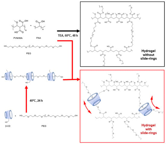
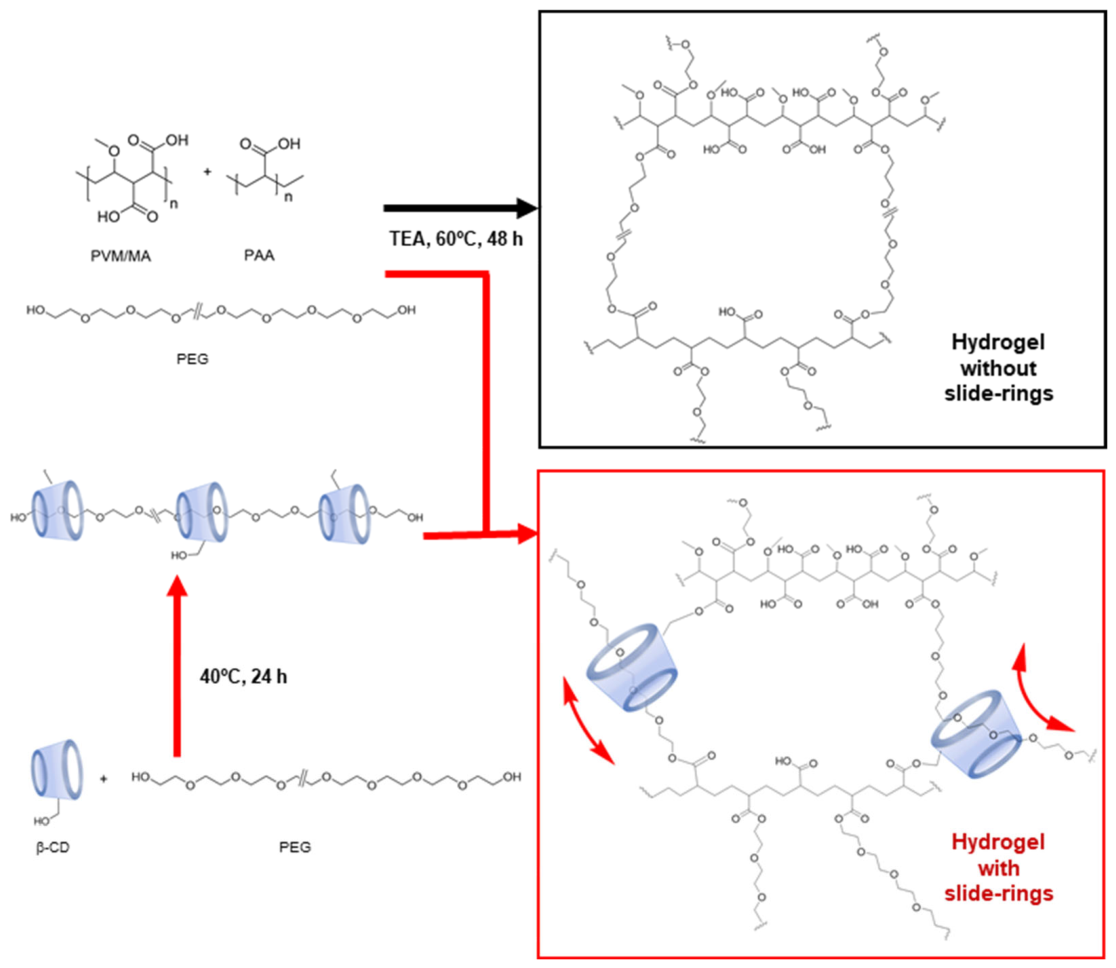
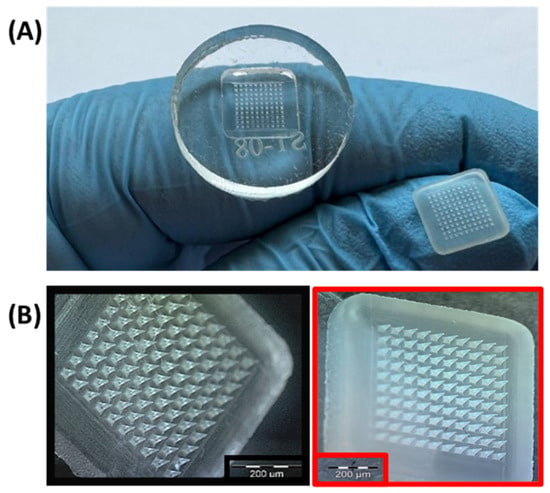
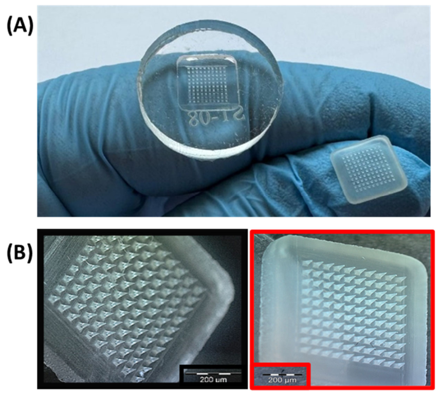
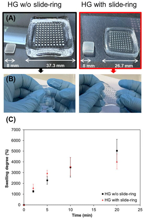
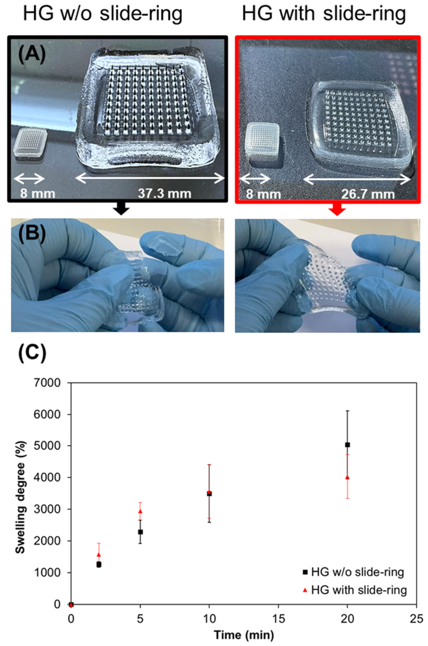
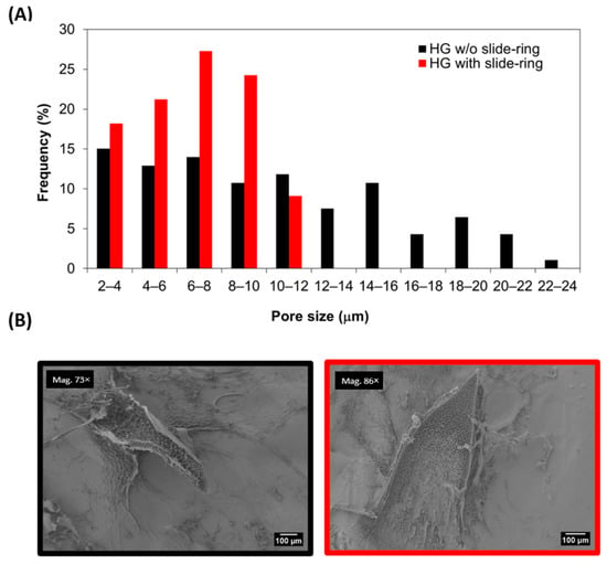
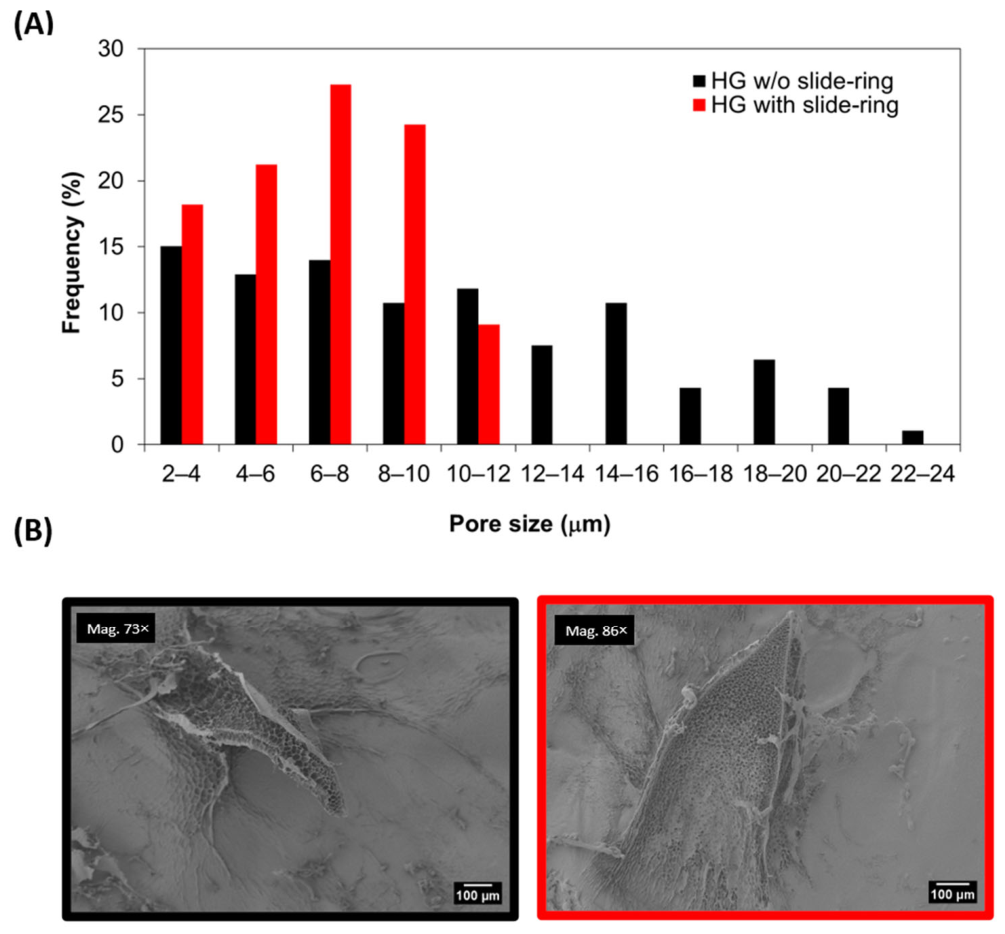
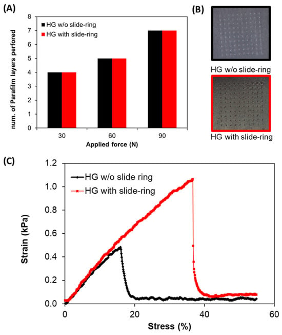
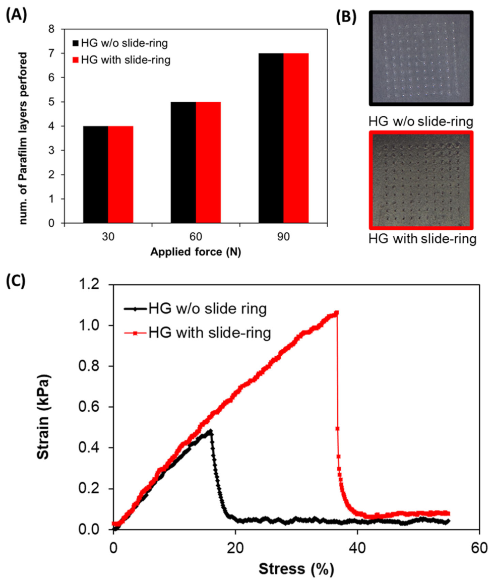
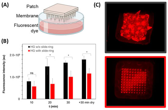
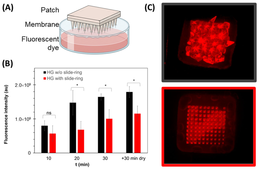
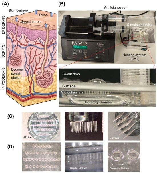
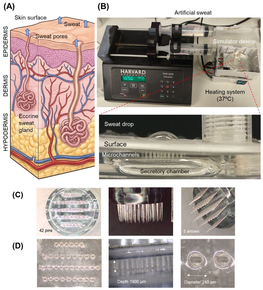
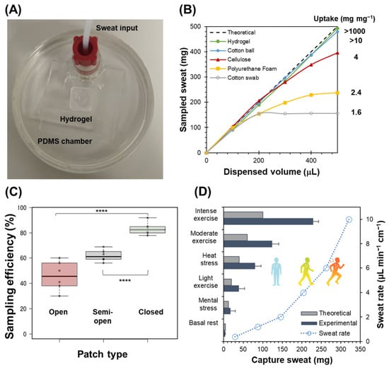
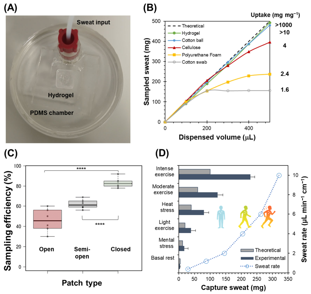
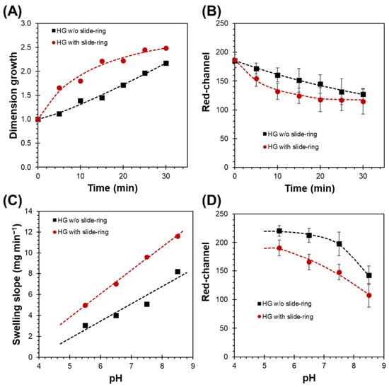
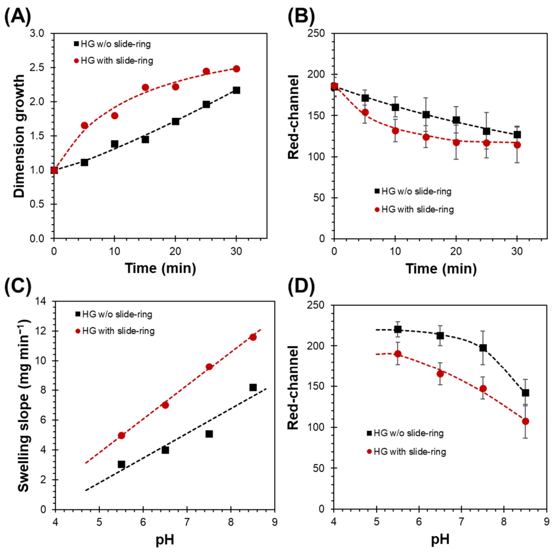
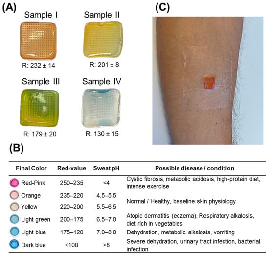
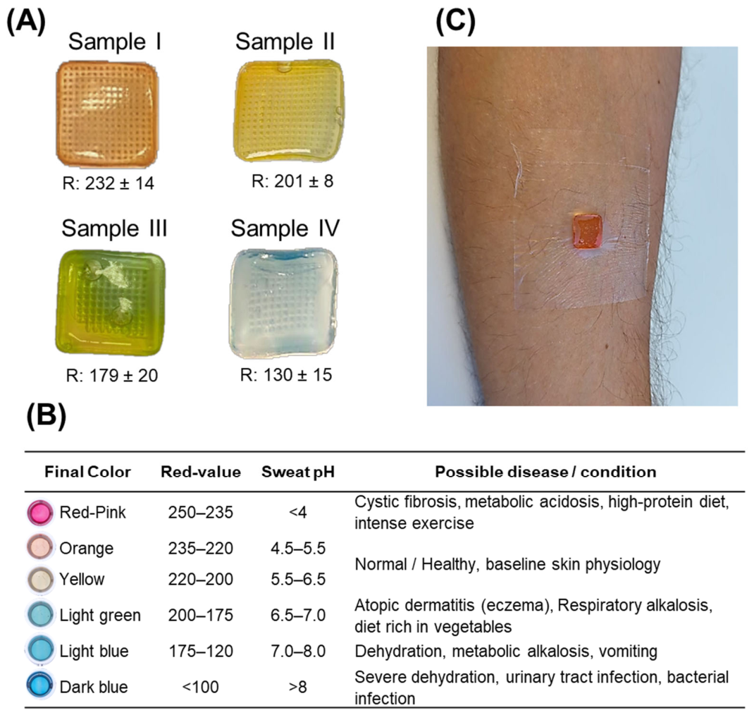
Disclaimer/Publisher’s Note: The statements, opinions and data contained in all publications are solely those of the individual author(s) and contributor(s) and not of MDPI and/or the editor(s). MDPI and/or the editor(s) disclaim responsibility for any injury to people or property resulting from any ideas, methods, instructions or products referred to in the content. |
© 2026 by the authors. Licensee MDPI, Basel, Switzerland. This article is an open access article distributed under the terms and conditions of the Creative Commons Attribution (CC BY) license.