


Abstract
3D bioprinting has evolved from developing simple cellular constructs to fabricating intricate, physiologically relevant tissue models. However, translation is bottlenecked by high equipment costs and processing trade-offs. This narrative review evaluates cost-effective engineering strategies over the past five years designed to enhance printing fidelity independently of material-chemistry innovations. Our observation reveals that thermal stabilization (2–60 °C) and static mixing elements yield resolutions down to 15 µm from cell-friendly 1 mm nozzles, while automated nozzle arrays accelerate spheroid processing 10 times. In-flow microfluidic gelation and self-healing granular baths eliminate architectural collapse, successfully preserving blood-vessel lumens (~50 µm). Additionally, newly developed procedural strategies significantly optimize output: nozzle-free painting and delayed-mixing droplet collision bypass clogging limits to handle ultra-dense networks, while vat-free projection methods reduce bioink waste to under 1%. These affordable modifications consistently preserve high post-printing cell viability (>90%) across complex, heterogeneous configurations. Ultimately, we conclude that the future of biofabrication relies on a transition toward closed-loop, AI-driven parameter optimization and the systematic integration of bioprinting platforms with active bioreactors to guide early structural constructs into functional, mature tissues.
1. Introduction
3D printing, or additive manufacturing (AM), is the process of fabricating a 3D object through computer-aided design and the automated addition of material [1]. Bioprinting is a set of AM technologies that aims to create parts that mimic natural tissue and organ functionalities by combining living and non-living materials in a specific three-dimensional (3D) spatial organization structure. As in traditional 3D printing or AM, the target is achieved by using computer-aided design (CAD) that represents the fundamental configuration of the target tissue or organ. Bioengineered structures are used in various fields, including regenerative medicine, tissue engineering, reconstructive surgery, drug discovery, pharmacokinetics, and basic cell biology research [2,3]. A typical bioprinter is a robotic automated machine that creates biological tissue structures using layering of biomaterials and living cells [4]. To accomplish this goal, bioprinters use bioinks, which are soft biomaterials loaded with living cells and manipulated according to specific protocols to create biological constructs [5].
Several 3D printing technologies have been adapted for bioprinting, with three main approaches dominating biomedical applications: inkjet-based, extrusion-based, and light-based bioprinting [6]. Inkjet bioprinting technology uses thermal or piezoelectric actuation to drop biomaterials in layers. This technique offers high printing speed, high resolution, and compatibility with a wide range of biomaterials [7]. Extrusion-based bioprinting uses mechanical or pneumatic forces to continuously extrude cell-laden biomaterials through a nozzle. Its key advantages include the absence of heat during printing and the ability to process highly viscous bioinks, making it one of the most widely used techniques in bioprinting [8]. Light-based bioprinting uses ultraviolet, laser, or visible light patterns to crosslink objects with great spatial resolution by projecting them into a photosensitive hydrogel precursor [9]. All three technologies are used in tissue engineering and regenerative medicine, but extrusion-based and inkjet-based approaches are still the most frequent for bioprinting.
Some critical design requirements for biomimetic 3D tissue constructs include 3D anatomically relevant structures, a biomimetic microenvironment, and vascularization. Bioprinting aims to recreate native tissues by integrating advanced CAD with increasingly sophisticated biomaterials to precisely control tissue architecture and microenvironmental cues [10]. Since tissue shape and size are tightly linked to physiological function and integration within the body, accurate anatomical replication is essential [11], yet it remains challenging for large 3D constructs due to limitations in mechanical stability and structural integrity [12]. Beyond macroscopic geometry, successful tissue regeneration requires a biomimetic microenvironment that provides appropriate biochemical and mechanical signals to guide cellular behavior [13]. Current bioinks, such as those based on decellularized extracellular matrix, provide significant biological cues, but they are unable to accurately replicate the tissue-specific composition and spatial organization of the native extracellular matrix [14]. Moreover, the fabrication of functional vascular networks remains a major bottleneck, as cell-driven vascularization strategies are slow and inefficient for supporting thick, complex tissues [15].
Over the years, bioprinting research has advanced from demonstrating basic cell-laden constructs to engineering anatomically and functionally sophisticated tissues [16]. Therefore, an extensive amount of review literature has emerged; however, these existing works mainly focus on bioink development, including material-chemistry innovations, such as the formulation of novel photopolymers, decellularized matrix functionalization, and complex chemical crosslinking kinetics [17,18,19]. While valuable, this chemical-centric paradigm often neglects the critical role of processing mechanics and requires specialized, high-cost proprietary formulations. This leaves a significant gap in the literature regarding how straightforward, affordable mechanical interventions can independently maximize fidelity on accessible printing setups. Furthermore, commercial robotic bioprinters remain prohibitively expensive for many research laboratories, which severely restricts entry and hinders collaborative breakthroughs across resource-limited environments [20].
To address these gaps, the primary objective of this review is to comprehensively evaluate how engineering-driven, cost-effective hardware adjustments and procedural modifications can isolate printing resolution from cellular damage independently of bioink chemical innovations. To establish a clear boundary for our analysis, we define “cost-effective” innovations based on three specific criteria: first, the hardware modifications must rely on open-source designs or inexpensive, off-the-shelf components that can be retrofitted onto standard desktop 3D printers; second, the strategy must bypass the need for proprietary, high-cost industrial bioprinting platforms; and third, the method must improve operational efficiency, either by drastically minimizing bioink and cellular waste during processing or by eliminating slow, labor-intensive workflows such as sacrificial support bath removal. Specifically, we analyze recent advancements covering the past five years across advanced nozzle architecture, microfluidic integration, yield-stress support media, and alternative cross-scale deposition modalities. To guide this review, Figure 1 provides a comprehensive framework that maps out how these mechanical interventions directly resolve fundamental processing limitations. As illustrated in Figure 1, the purpose of this review is to demonstrate how addressing core challenges, such as shear-induced cell damage, poor vascularization, and geometric instability, via target-specific engineering solutions directly leads to crucial functional outcomes like perfusable channel creation and high cell viability (>90%). By clarifying these technical mechanisms, this review serves as an engineering roadmap to bridge the gap between affordable proof-of-concept print modifications and organized, long-term biological tissue maturation within hybrid bioreactor environments.
Figure 1.
Bioprinting main challenges, recent engineering solutions, desired functional outcome, and future perspective for bioprinting.
To ensure a rigorous and balanced analysis, a systematic literature search was conducted across the PubMed, Web of Science, and Google Scholar databases, covering articles published over the past five years. The search strings utilized a combination of key terms, including (“bioprinting” OR “3D bioprinting”) AND (“modification” OR “innovation” OR “accessory” OR “add-on” OR “retrofit”) AND (“printhead” OR “nozzle” OR “microneedle” OR “coaxial” OR “multi-material” OR “platform” OR “stage” OR “substrate” OR “crosslinking” OR “in situ crosslinking” OR “sacrificial printing” OR “embedded printing” OR “support bath” OR “temperature control” OR “perfusion”) AND (“structure” OR “shape fidelity” OR “resolution” OR “mechanical property” OR “complexity” OR “vascularization” OR “channel”) AND (“cost-effective” OR “open-source” OR “retrofitting” OR “high-throughput”). Titles and abstracts were screened according to strict inclusion and exclusion criteria. To remain aligned with the core scope of this review, articles focusing primarily on novel biomaterial synthesis, chemical crosslinking formulations, or proprietary high-end industrial systems were excluded. Prioritization was given to peer-reviewed studies that demonstrated reproducible hardware modifications, multi-material processing advancements, or procedural efficiencies that drastically minimized cellular waste, directly preserving cell viability (>90%) on accessible desktop printing setups.
2. Nozzle/Printhead Innovations
Advanced printheads are the foundation of modern 3D printing that employs material extrusion, providing unprecedented control over shape, material properties, and composition. These systems can be classified into three types. Shape-modulation printheads, which include fixed, adaptive, and multi-output nozzles, dynamically control the geometry of the extruded filament to optimize resolution, speed, and surface quality [21]. Adaptive nozzles can change size and shape during printing. Property-modulation printheads use kinematic (e.g., rotation, vibration) or stimulation (e.g., thermal, magnetic, acoustic, optical) mechanisms to tune material properties in real time, such as fiber alignment or phase change induction. Ultimately, multimaterial processing printheads incorporate switching, mixing, and coextrusion capabilities, facilitating seamless material transitions, in situ blending to produce functional gradients, and concurrent extrusion of multiple materials within a single filament for sub-nozzle resolution. Hybrid printheads that combine different functions are also pushing the field forward. They make it possible to build complex structures for new uses, such as soft robotics, mechanical metamaterials, and biological structures [21]. This section of our review paper examines the most recent advancements in printhead designs that can improve bioprinting procedures.
2.1. Rheological and Thermal Considerations for Micro Extrusion
Multifunctional extrusion printheads are considered the most important component of direct ink writing (DIW) 3D printers, as they can overcome traditional additive manufacturing (AM) and materials processing limitations [21]. Multi-output (multinozzle) printheads, for instance, solve the resolution-speed trade-off [22], while core–shell nozzles allow the extrusion of materials that are difficult to print, like liquid metals, which combine high surface tension and low viscosity [23], or in bioprinting, they can create multimaterial hierarchical structures and hollow structures using sacrificial core materials [24]. The printability of a material is determined by its rheological properties, which should exhibit shear-thinning and yield-stress behavior to facilitate extrusion and shape retention after deposition [25].
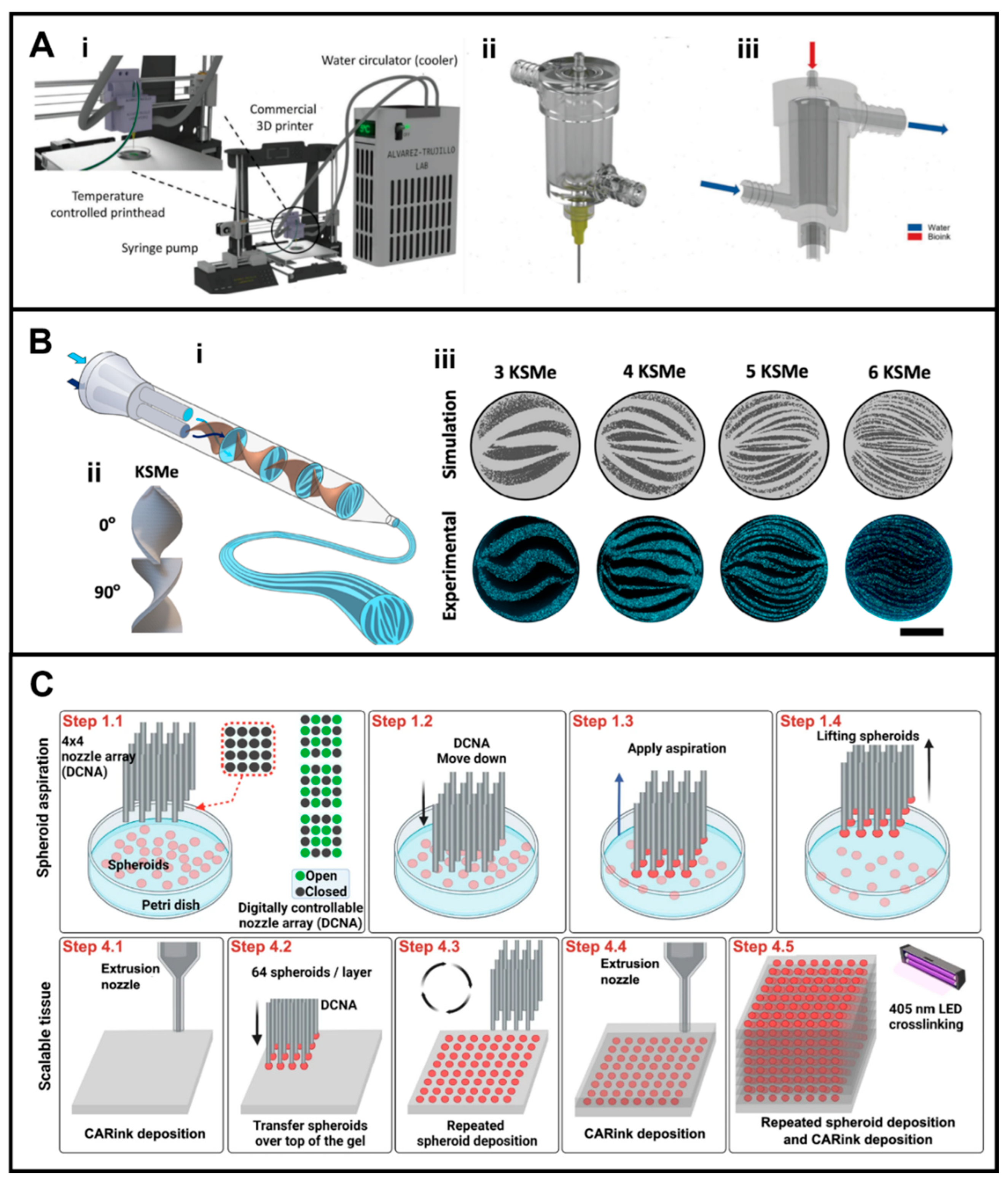
To achieve optimal printability for bioinks, it is necessary to carefully examine their rheology, cross-linking properties, and specific printing parameters to guarantee the precision of the final features [26]. As most materials are not intrinsically printable, their rheological properties are frequently modified with additives, such as nanoclays for low-viscosity inks [27] or plasticizers and foaming agents for high-viscosity formulations [28,29], while employing precise thermal regulation to maintain stability [21].Not all the biofabrication labs are capable of affording commercial bioprinters for their research; in addition, not all the commercial bioprinters have the capability to handle the delicate, temperature-sensitive bioinks needed to print living cells. These bioinks require specific thermal conditions to maintain optimal viscosity and printability. Sanz-Garcia et al. developed a low-cost temperature-controlled printhead for microextrusion-based bioprinting (MEBB) that could be integrated into commercially available desktop 3D printers (Figure 2A) [30]. This modular printhead was made to work with syringes of different sizes and to keep the printing temperature between 2 °C and 60 °C. The central component of the temperature control system is an aluminum block that has been specially machined to hold the syringe barrel. Aluminum was chosen for its superior thermal conductivity, ensuring consistent heat distribution. By putting Peltier modules (thermoelectric devices) in direct contact with the aluminum block, active thermal management was possible. These solid-state devices work by using an electric current to drive heat from one side to the other through dissimilar semiconductor junctions, creating a hot and cold side, and if the current is reversed, the heat flow and the hot/cold sides are also reversed. This setup allows accurate and steady control of the temperature of the bioink inside the syringe. This feature is crucial for keeping the thermosensitive hydrogels’ rheological properties during the extrusion process. Three separate low-cost 3D printers successfully installed the printhead, demonstrating its adaptability and versatility. The system achieved resolutions comparable to high-end bioprinters when tested using calibration models printed with poloxamer 407, a synthetic hydrogel selected for its reliable, temperature-responsive qualities and minimal swelling to test printer performance precisely. The shown resolutions included consistently printing multilayer lattice structures with pore size errors as low as 39 µm, making fine, straight filaments with widths that could be adjusted from 600 µm to 210 µm by changing the print speed, and making stable vertical pillars that could be up to 7.5 mm tall. Importantly, cell-laden constructs printed with gelatin-alginate bioinks at optimized temperatures (20 °C) showed high cell viability of more than 90% after 24 h, attributed to reduced shear stress and maintained bioink rheology during extrusion [30,31]. In another study, Garciamendez et al. designed an affordable custom 3D-printed printhead with an internal water jacket that gentleness was confirmed through the use of living C2C12 cells, which demonstrated a post-printing viability of around 90% and successfully proliferated over a 7-day culture, thereby validating the system’s efficacy for practical cellular applications [32].
2.2. Multimaterial Deposition and Coaxial Nozzle Configurations
Native tissues generally display anisotropic hierarchical structures and various components and cell types, enabling them to execute numerous biological functions. Building biomimetic hierarchical structures with various functions using 3D bioprinting technologies with a single material is difficult or impossible due to these intricate configurations [33]. For the production of patient-specific 3D scaffolds, several studies have shown the distinct advantages of 3D printing technology over traditional manufacturing methods, such as the scalability, high-precision introduction of multiscale pore features, and flexible formation of heterogenous/gradient architectures [33]. Bioprinting of multimaterial structures has progressed with different extrusion-based strategies. Using a multimaterial reservoir connected to a single nozzle enables quick switching between different bioinks from different reservoirs using one printhead. This makes it feasible to fabricate complex heterogeneous constructs [34]. An alternative method that effectively prevents cross-contamination and increases printing throughput is the multi-nozzle strategy surrounds the bioink cartridge, which can be used with a commercial 3D printer. They were able to precisely control the bioink’s temperature by attaching this jacket to an external circulator. This enabled them to maintain the bioink’s fluidity for extrusion and its viscosity for shape maintenance. This mechanism enabled the printing of high-fidelity, multi-layered 3D structures with GelMA, which could not be done at room temperature. Cooling GelMA to 5–15 °C through the printhead enhanced its viscosity during printing, facilitating the formation of high-fidelity lines approximately 522 µm thick and intricate 3D structures. In contrast, printing without temperature control resulted in poorly defined, low-quality constructs. The bioprinter’s, which uses arrays of nozzles to deposit different materials either simultaneously or alternately. However, it necessitates careful control to ensure smooth interfacial integration between materials [35]. Another well-known strategy is coaxial nozzles, which form core–shell structures like vascular tubes by enabling the simultaneous extrusion of different materials in concentric layers. They work especially well with bioinks that quickly crosslink when they come into contact, which makes them perfect for creating hollow, perfusable tissue models [36]. Recently, researchers have achieved a controlled multimaterial structure with adjustable resolution using the printhead design as a mixer, in addition to the mentioned strategies. Trujillo-de Santiago et al. developed a high-resolution bioprinting strategy by employing a chaotic printhead based on a Kenics Static Mixer (KSM) to address the significant compromise between extrusion nozzle dimensions and cellular viability (Figure 2B) [37]. Their innovative solution was a printhead containing a series of static, helical elements that consistently split, stretch, and fold two incoming bioink streams as they flow through it. When chaotically mixed bioinks are introduced into the crosslinking solution bath, a single extruded filament is created with a complex internal architecture consisting of dozens of alternating, ultra-thin layers, achieving a resolution as fine as 15 µm from a wide, cell-friendly 1 mm nozzle. The result is a resolution that normally requires a nozzle ten times smaller, which would be lethal to cells [38]. Unlike traditional high-resolution printing, which employs narrow nozzles and exerts detrimental shear stress on cells, this chaotic printing method maintained cell viability and functionality, evidenced by the successful development of aligned, multinucleated myotubes from C2C12 cells after seven days in culture. The delicate and effective layering was accomplished as the resolution was determined by the iterative fluid deformation within the printhead, rather than by forceful extrusion through a microscale orifice, thus separating structural complexity from cellular damage. In another study, Wei et al. attempted to optimize the Kenics static mixer printhead, specifically how to mix high-viscosity bioinks thoroughly right before extrusion without putting the embedded living cells through deadly amounts of shear stress inside the printhead [39]. The authors designed an optimized printhead system including a static mixer with a specific helical blade geometry (Type A) with a specific height-to-diameter ratio, (k = 1.2) which maximizes mixing efficiency (>66% mixing rate) by creating gentle, controlled swirls that minimize sudden flow changes and thus shear stress. Furthermore, they found that a conical needle is more appropriate than a cylindrical one, as its tapered geometry ensures a smooth, gradual acceleration of the bioink, preventing the sudden, damaging pressure spikes found in cylindrical designs. Cell viability was considerably increased when printing using the optimized system, demonstrating how this integrated approach directly improved biological outcomes. For example, cell survival and metabolic activity (as determined by live/dead staining, ALP, and MTT assays) were significantly better with a 0.51 mm nozzle at 2 mm/s than with configurations with smaller diameters or faster speeds. Cell integrity and function are maintained throughout the extrusion process by the optimized geometry’s control over the flow path, which reduces the crushing shear forces on cells [39].
2.3. High-Throughput Spheroid Fabrication and Array Handling
One of the challenges in mimicking native tissues in bioprinting is to provide high cell densities as they exist in native tissues. Several limitations persist in conventional bioprinting, such as shear stress leading to a lack of efficient viable cell densities [40], or the necessity of using low-cell-concentration bioinks [41], and these limitations hinder the success of developing functional tissues. In fact, to achieve printability, the majority of hydrogel bioinks necessitate a balance between cell density and polymer density, which frequently leads to less-than-ideal conditions for the intended application [42]. To address these issues, the scaffold-free bioprinting method uses cell aggregates to create tissue without the use of scaffold support, which causes cells to generate their own extracellular matrix (ECM) [43]. Tissue spheroids are cellular aggregates that have been utilized in tissue fabrication due to their advantages, including native-like cell density and the ability to secrete significant amounts of ECM components while maintaining efficient cell–cell communication in a 3D native-like microenvironment, making them promising candidates for this approach [44]. To achieve native-like tissue physiology, spheroid density and spatial arrangement are essential because they provide the high cell concentration and reliable arrangement required for successful tissue formation [45]. However, issues like low throughput [46], scalability, efficiency, and lack of control over the exact number and placement [40], can still affect spheroid bioprinting [47]. To achieve scalable spheroid bioprinting, Kim et al. introduced HITS-Bio (High-throughput Integrated Tissue Fabrication System for Bioprinting) by developing a multi-nozzle printhead known as the Digitally Controlled Nozzle Array (DCMA), which functions as a microscopic stamp, enabling the simultaneous transfer of multiple spheroids onto a gel substrate rather than sequentially (Figure 2C) [42]. The DCMA is characterized as a tiny, high-tech stamp with 16 separate, controllable suction tips. Instead of using one single nozzle to pick up spheroids sequentially, this multi-nozzle array could be lowered into a dish containing hundreds of spheroids. Based on a digital design, specific suction tips are turned on to pick up the desired spheroids, lifting all 16 at the same time and placing them simultaneously onto a gel substrate. The strategy was the layer-by-layer deposition of ink to form a gel substrate and spheroid deposition to create 3D structures in a high-throughput manner. This approach made a significant difference, achieving a bioprinting speed ten times faster than the existing high-precision technique [48], while maintaining excellent cell viability (>90%). The authors were able to print a substantial 1 cm3 cartilage construct comprising nearly 600 spheroids in under 40 min, a process that would have required several hours using a single-nozzle technique. In vivo analysis indicated that this technique allowed for the rapid repair of large bone defects in rats. The group treated with a high density of bioprinted spheroids demonstrated a bone coverage of ~91% in just 3 weeks and ~96% in 6 weeks, significantly outperforming the control groups with empty defects or bioink alone. The parallelized, simultaneous spheroid handling is the fundamental mechanism behind this achievement. The system can quickly pick up a predefined pattern of spheroids from a reservoir and deposit them all at once into their precise positions on the gel by using a digitally controlled array of nozzles, each of which can be turned on or off. This feature significantly reduces the overall fabrication time and enables scalable tissue fabrication [42].
2.4. Resolution Limits Driven by In-Nozzle Cellular Dynamics
Although a printhead can control parameters of bioprinting, such as resolution, the dynamism and mechanical behavior of cells inside the nozzle/printhead can affect extruders with high resolution. In a study, Fathi et al. tried to develop a novel dispenser with a picoliter resolution for cell deposition [49]. They developed a high-resolution micro-extrusion system called “Picodis” that can deposit cells with picoliter-level accuracy. Their innovative solution was a microcapillary dispensing system that used a piezoelectric actuator to drive a plunger inside a finely pulled glass microcapillary tip. Newtonian liquids, like coloring ink, could be dispensed with a precision of approximately 3.6 pL per step, enabling the generation of consistent, isolated droplets due to the exceptional mechanical control provided by this mechanism. However, when dispensing 3T3 fibroblast suspensions or pellets, this high mechanical precision was lost by cellular dynamics: rapid sedimentation (~10 µm/s) and aggregation within the narrow tip led to clogging and the unpredictable, turbulent ejection of cells. In contrast to the controlled ink flow, cell deposition showed significant cell count variability, especially with smaller tip diameters. It was demonstrated that cellular dynamics, like in-nozzle aggregation and sedimentation, regulate the movements of the cells in the culture medium. Consequently, laminar fluid flow within a tip does not solely trigger the cells’ flow paths. The study’s results indicate that the extrusion resolution of cell-laden bioinks, in the absence of dispersing agents such as hydrogels or surfactants, is not predominantly affected by the mechanical resolution control of a dispenser. This suggests that biological behavior, rather than printer mechanics, may be the limiting factor for deposition fidelity for carrier-free bioinks. Although the high-precision control extruder setup provides some control over the cell deposition process, it still needs to be improved for applications that demand greater microscale precision. For instance, modifying the cell loading strategy could potentially optimize the advantages of the high-resolution extrusion platform [49].
The printhead, or nozzle, is the last modulatory component of a bioprinter that can control the fidelity and functionality of the printed construct. The printhead directly affects the bioink’s architectural and biological performance by controlling the diameter of the extrusion, the spatial resolution, the temperature, and the flow of the material. Recent advancements indicate that the future of advanced bioprinting relies on the development of intelligent, computer-aided printhead systems capable of dynamically regulating the dispensing rate, bioink state, and the sequential or simultaneous deposition of multiple materials. To make these systems work, engineers must overcome significant challenges, such as achieving precise, real-time regulation of the bioink state and dispensing while maintaining complete biocompatibility. This requires protecting cells from harmful stresses, such as thermal, shear, and hydrodynamic ones, as well as resolving issues like cell sedimentation to guarantee a uniform dispersion that enables reliable high-resolution dispensing. It is anticipated that next-generation printheads will be more multifunctional, incorporating on-nozzle mixing units, in situ casting molds, dedicated reservoirs for various bioinks, or microfluidic modules that allow for real-time composition or rheology tuning during printing. Ultimately, the printhead will be a single, adaptable unit for more coordinated and effective processes. When combined with AI-driven parameter optimization and high-throughput data collection, these systems could independently adjust printing parameters like resolution, extrusion pressure, mixing efficiency, and multimaterial coordination according to bioink rheology, cell density, cell type, or tissue-specific needs. According to this paradigm, next-generation bioprinters will use printheads tailored to specific applications, such as simple dual-nozzle configurations for layered tissues like skin, ultra-high-resolution microfluidic printheads for tissues with complex hierarchical architecture, like neural or hepatic tissue, and specialized multimaterial printheads for constructs with various cell populations, like cardiac or osteochondral tissues. Moreover, the geometry and internal architecture of the printhead can be consciously designed to promote favorable microstructural characteristics during deposition. Nozzles that facilitate fiber alignment, micro-indentations, gradient porosity, or regulated shear-induced patterning serve as effective instruments to impart mechanical and architectural signals that promote tissue maturation. Innovations in printhead design are pivotal for enhancing bioprinting precision, biological efficacy, and the production capabilities of next-generation engineered tissues.
Figure 2.
Nozzle printhead innovations. (A) The bioprinter system schematic. (i) The entire system, including the tubing, water cooler, syringe pump, modified 3D printer, and integrated printhead. The extruder mount with the bioink and water tubing attached to the printhead is depicted in close-up. (ii) A Luer lock connector that permits the exchange of needles of any size and a cooling printhead with a water jacket to cool the extruded bioink. (iii) Diagram of the encapsulating circulating water compartment in the printhead jacket and the bioink counterflow inside the bioink chamber. Red arrow represents bioink, blue - water. Reprinted and adapted with the permission from Garciamendez-Mijares et al., 2023 [32]. (B) Kenics static mixer (KSM) extrusion printhead. (i) The printhead includes a lid with inlet ports, a pipe that houses the inner KSM elements (KSMe), and an outlet tip. Arrows represents different components of the bioink. (ii) The KSMe rotate at 90◦ and acts as flow dividers, splitting flow streams in two at each element. (iii) Inner structures formed at the cross-section of chaotically printed fibers containing 3, 4, 5, and 6 KSM elements (KSMe). Scale bar: 500 µm. Reprinted and adapted with the permission from Trujillo-de Santiago, Alvarez, 2024 [37]. (C) Schematic of the HITS-Bio process. A spheroid loaded with DCNA that iteratively activated or disabled aspiration based on a user-defined design. Scalable cartilage tissue bioprinted in 1 cm3 using CARink and about 600 spheroids. Reprinted and adapted with the permission from Kim et al., 2024 [42].
3. Microfluidics-Assisted Bioprinting
Microfluidics is a revolutionary discipline and developing technology that accurately controls fluid dynamics and behavior at micro-, nano-, and pico-scales, using channels with dimension sizes ranging from tens to hundreds of micrometers [50]. Microfluidics was initially employed in chemical analysis; however, it is currently applied across various domains, including lab-on-a-chip, organ-on-a-chip, biological assays, sensors, and bioprinting [51]. Microfluidic systems are an appealing tool to incorporate into microextrusion bioprinting technology because they can control overflow and mix cells with bioinks in a controlled way [52]. This control has led microextrusion bioprinting to be more intricate, creating concepts of “printhead-on-a-chip” [53] or “lab-on-a-tip” [54]. A new bioprinting method based on microfluidics has emerged. This method makes it easier to adjust the structural and compositional characteristics of tissue constructs while they are being printed by combining microfluidic systems with conventional extrusion-based bioprinting. Microfluidic fabrication techniques can control small quantities of molecules, cells, and liquids. A range of control devices, such as sensors and valves, can also be used to reinforce these systems [52]. Fluid bioink flows through microchannels in microfluidics-assisted 3D bioprinting, enabling precisely controlled flow, switching, and component mixing. Additionally, microfluidic bioprinting is linked to less shear stress throughout the procedure. The sheath flow encircling the laminar core is responsible for this. Furthermore, it is possible to effectively control the direction, dimensions, and morphology of generated objects. Microfluidic processing can enhance the printing process’s resolution when used in conjunction with extrusion bioprinting, surpassing the current microextrusion resolution of approximately 50 µm [52,55]. Microfluidic-based bioprinting has been the subject of excellent review papers recently [51,52,56]. This section of the paper examines current research on cutting-edge microfluidic-assisted bioprinting.
3.1. Microfluidic Mixers for Fluid Homogeneity and Viscosity Alignment
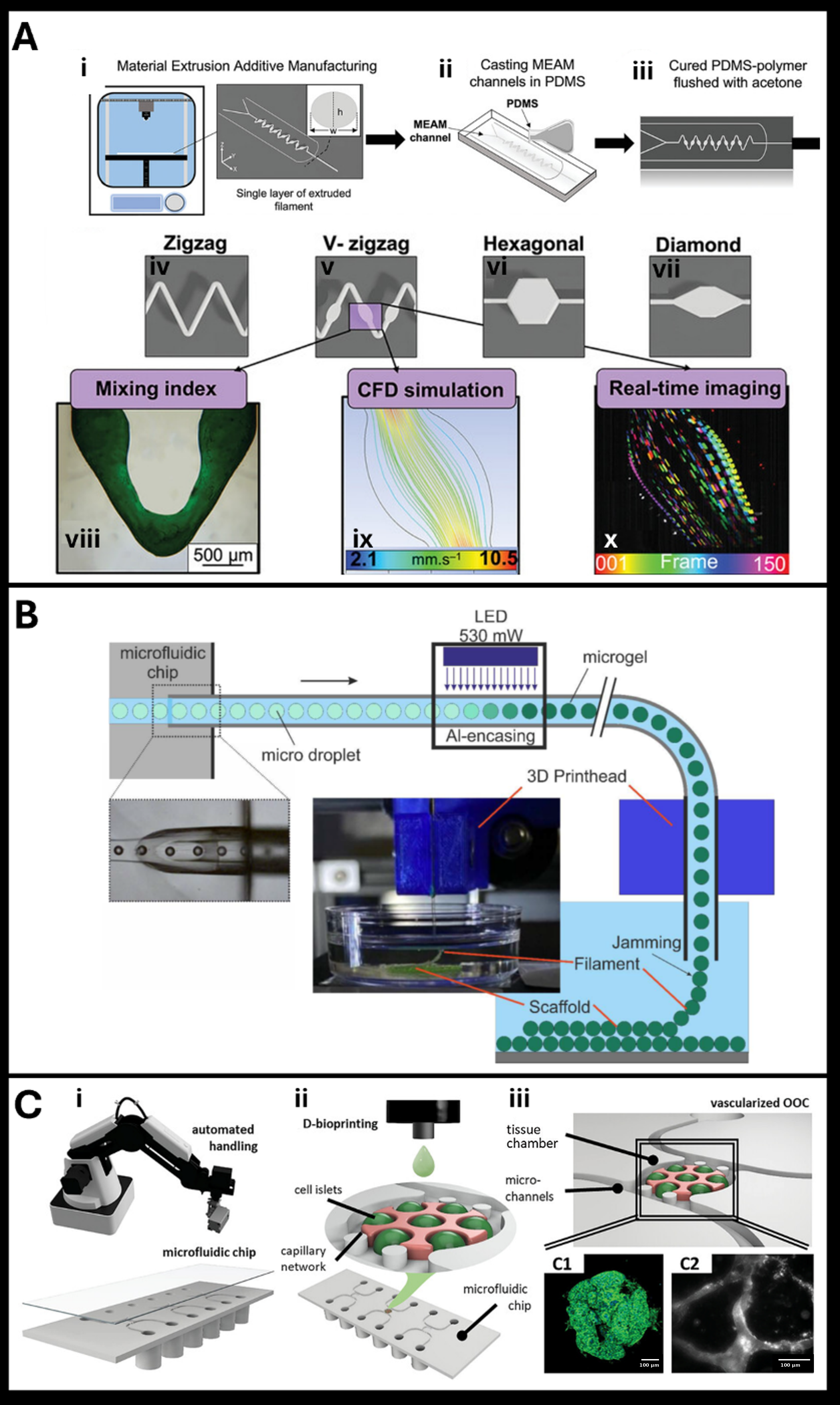
One of the purposes of using microfluidics in bioprinting is to mix solutions such as bioinks and crosslinkers to fabricate either compartmentalized or efficiently crosslinked hydrogels for the final bioprinted construct. However, challenges remain, such as the ability to mix fluids with varying viscosities and compositions, as well as affordability and versatility. This challenge, in fact exists in that traditional passive mixers, which rely on slow diffusion, cannot rapidly and uniformly mix bioinks (such as alginate and crosslinkers) at high, practical flow rates (100–1000 µL min−1) without compromising cell viability. Moetazedian et al. designed an agile manufacturing pipeline that used Material Extrusion Additive Manufacturing (MEAM) with a pivotal Continuously Varied Extrusion (CONVEX) design approach (Figure 3A) [57]. CONVEX allows for the creation of complex, seamless geometries with actively varied channel widths within a single printed feature, which was previously impossible. They created four passive mixer configurations using CONVEX: zigzag, V-zigzag, hexagonal, and diamond. The variable-width zigzag (V-zigzag) was found to be the most efficient. The design intentionally included alternating narrow and wide segments to slow the fluid in the expanded areas and extend the diffusion time without causing turbulence. This design enabled complete mixing in just 15 mm across an expansive range of flow rates (1–1000 µL min−1), surpassing many lithographic mixers as well as other MEAM-printed ones. The V-zigzag achieved a near-perfect mixing index (≈0.99) within a remarkably short 15 mm channel across all flow rates. The standard zigzag, hexagonal, and diamond mixers, on the other hand, did not work as well at higher flow rates. Their mixing indexes dropped to 0.6–0.8, and they needed more than 40 mm to mix as well at low flow rates. The device could handle fluids with varying viscosities, create uniform droplets, and, by using a microfluidic flow-focusing module, produce helical, wavy, and straight core–shell fibers by co-extruding 2 wt.% sodium alginate (the shell) with 2 wt.% CaCl2 in 30 wt.% Pluronic (the core). These fibers had diameters and lumen sizes that were about the same as human arterioles (~50 µm), demonstrating the system’s biomimetic capability. The bioprinting validity of the nozzle also demonstrated that, despite directional changes imposed by the V-zigzag geometry, shear-induced cell damage remained minimal. Even through a narrow 100 µm nozzle, the extrusion of 10 wt.% GelMA containing SaOS-2 cells resulted in exceptionally high cell viability (>86%) [57].
3.2. Rapid Laminar Flow Switching and Multi-Inlet Manifolds
Creating multimaterial structures in a controlled laminar flow regime without turbulent mixing is one of the potential applications of microfluidics for bioprinting. Over the past ten years, various studies have demonstrated the potential for developing microfluidic devices to produce multimaterial components. However, these studies have mostly focused on individual elements such as microfibers or microparticles. Building the intended 3D complex structures out of multicompartmental single units, which are typically gathered during the solution phase, is difficult with this microfluidics technology. Moreover, continuous extrusion techniques, like bioprinting systems, are more efficient than microfluidic devices at continuously building larger, multilayered structures with a high concentration of viscous, cell-rich biomaterials while preserving high cellular viability [51]. Hassan et al. designed a novel microfluidic single-nozzle printhead (the M3 platform) and an implantable, gelatin-based colloidal gel (GCG) as a support bath [58]. The M3 system operates as an advanced mixing manifold. It comprises seven distinct input channels—six positioned in a circular formation and one from above—that converge into a singular outlet nozzle. Most importantly, a computer-controlled pneumatic valve that can open and close in milliseconds (up to 10 Hz) controls each channel. This eliminates the need to physically align multiple printheads and enables the quick and accurate switching of up to seven different bioinks. Laminar flow is the fundamental fluidic principle in this case; the bioinks flow alongside one another inside the nozzle without chaotic mixing, allowing for the production of intricate “voxelated” fibers with core–shell, donut, or side-by-side configurations in a single extrusion. In vitro, a bioprinted liver-like construct with HepG2 cells self-organized into functional spheroids after 14 days. They also printed a skeletal muscle mimic with a C2C12 cell core and an HUVEC shell. After being cultured, the muscle fibers were aligned and showed heavy myosin and early vascularization. In an in vivo rat model, a spiral structure printed within the GCG was explanted after 7 days, demonstrating exceptional structural retention of its 3D configuration. The fast-switching nozzle mechanism facilitated architectural complexity, while the GCG support bath ensured printing stability and post-implantation bio-integration, collectively establishing a platform for constructing implantable living tissues that preserve their form and function [58].
3.3. Actuator-Driven Displacement and Active Dynamic Flow Valves
Precise fluid and cell flow control, which are essential for structural features and shear stress control, are among the challenges of microfluidic-assisted bioprinting. Mani et al. designed a printhead to facilitate the printing of multiple materials through a single nozzle, ensuring cell viability and enabling precise control over the quantity of each material printed at any moment [59]. They engineered a microfluidic nozzle head with three distinct inlets: a central inlet for a cell-laden bio-ink, accompanied by two lateral inlets for a scaffold hydrogel. The main innovation was integrating Shape Memory Alloy (SMA) actuators, which function as tiny, thermally activated switches. In contrast to conventional printer head designs, like piezoelectric printer heads [60], microvalves driven by shape memory alloy (SMA) require a low-cost, low-voltage power source that is appropriate for high-frequency applications. In the designed microfluidic system, when electrically heated, these SMA coils contract predictably to deform the PDMS walls of the side hydrogel channels, effectively pinching them to reduce hydrogel flow. This provides high-precision displacement control, thus enabling gentle, tunable flow restriction ideal for sensitive bioinks. This action dynamically forces a greater proportion of the central cell stream to exit the common nozzle. This mechanism allows for instantaneous adjustments to the composition of the printed structure. The experiment showed that when the duty cycle was 90%, the wall moved 220.30 ± 5.34 μm, which changed the hydrogel flow width from 80 μm to 42 μm. This indicated that the material composition could be controlled very precisely. Moreover, cell viability tests using rat glial C6 cells showed significant proliferation over 24 h, confirming the biocompatibility of the printed hydrogel scaffolds [59].
3.4. On-Chip Microgel Generation and Continuous Direct 3D Printing
Common challenges arise when using typical hydrogel precursors, which are in a lightly precrosslinked state and need to be cross-linked after printing to ensure the fidelity of the constructs. These challenges include cell sedimentation, mixing inhomogeneities, and a compromise between biocompatible formulation and printing, as well as the resulting shape fidelity, considering that cells are subjected to shear forces within the flowing formulation [61]. Microgel-based bioinks represent a significant advancement in bioprinting because they inherently protect encapsulated cells from the damaging shear forces of printing and allow precise control over the cellular microenvironment [62]. However, the reliance on microfluidic production, typically a slow, isolated process that creates a bottleneck, has prevented them from reaching their full potential. As a potential solution for this, Reineke et al. developed an integrated system that seamlessly combines microgel production and 3D printing into one continuous process (Figure 3B) [61]. They designed a microfluidic chip that leverages a “double 3D-flow-focusing” geometry. This design delicately created a central stream of cells and polymer precursor through the chip, surrounding it with focusing streams to position the cells in the channel’s center where shear forces are lowest, thereby conserving viability during encapsulation into uniform droplets. These cell-laden droplets were then instantly solidified into microgels via in-flow UV cross-linking before being sent straight into a 3D printhead. In the next step, the microgels were automatically jammed together into a stable filament, which was immediately deposited to build a stable 3D scaffold. This integrated strategy successfully skipped traditional, multi-step bioink preparation. This system was tested by encapsulating yeast cells and human HEK293T cells, with analyses confirming that the number of cells per microgel followed a predictable Poisson distribution and that the process maintained high cell viability. Furthermore, the final constructs exhibited an inherent microporosity, which allowed for effective nutrient perfusion, a significant advantage over solid hydrogels. The successful printing mechanism is attributed to the soft, adhesive characteristics of the microgels; when jammed, the polymer chains at their surfaces rapidly interpenetrate and entangle, creating the necessary cohesion to produce a stable, printable filament upon extrusion, thus facilitating the fabrication of intricate, perfusable tissue constructs [61].
3.5. Template-Free Coaxial Fabrication of Biomimetic Tubular Networks
One of the obstacles in tissue engineering has been the creation of tubular structures to mimic tubular-shaped tissues and organs, such as trachea, blood vessels, and esophageal tissue, because of their intricate hierarchical macro- and micro-structures that contain various cell types and ECM [63]. Complex multi-layered architecture for artificial functional tubular tissues has been attempted using various methods. However, due to technical constraints, these methods not only fail to produce a tubular structure with a target-specific mechanical property, but they also limit shape freedom [63]. In bioprinting, constructing tubular tissue structures is challenging due to the difficulty of handling native extracellular matrix (ECM) proteins, such as collagen, which gel slowly and provide poor structural support during printing; this severely limits their use in creating perfusable, cell-laden channels without sacrificial templates. Gao et al. developed a microfluidic-enabled coaxial extrusion strategy that effectively integrates flow shaping with protected in situ gelation [64]. They designed a multilayer PDMS-based printhead that generates a stable triple-layer core-annular flow, where the ECM solution is focused between inner and outer buffer flows. This printhead is coupled with a cylindrical confinement tube that provides sufficient relaxation time for slow pH- or temperature-induced gelation to occur, enabling continuous, template-free formation of thin-walled, cell-compatible tubular structures. To achieve tunable tube dimensions (inner diameter from approximately 500 µm), wall thickness, and even heterogeneous composition in axial, circumferential, or radial directions, the innovation resides in the precise microfluidic control over flow rates and channel architecture. In addition to demonstrating device-level functions like ECM-based valves and pumps, this method successfully created functional, perfusable collagen tubes that could support confluent endothelial and epithelial barriers and enabled tissue-level functions like albumin secretion and inflammatory response. This approach achieved superior biomimicry, mechanical compliance, and biological functionality in comparison to traditional methods, which require high-concentration bioinks or sacrificial templates. This makes it appropriate for advanced organ-on-a-chip applications, like simulating airway collapse and reopening in lung injury studies. Moreover, the bioprinting was implemented with Huh7 cell-laden collagen, which maintained high cell viability and sustained albumin secretion over 14 days. The compliant tubes also made it possible to create confluent human endothelial and bronchial epithelial barriers on their lumens. These barriers showed important physiological functions, such as a cytokine-induced inflammatory response and high expression of the junctional protein ZO-1. The successful formation of these functional tissues is attributed to the cell-compatible gelation conditions within the confinement and the native, soft mechanical properties of the pure ECM tube walls, which closely mimic the in vivo basement membrane [64].
In another coaxial microfluidic strategy, Soliman et al. could use diffusion-based microfluidics to advance bioprinting by controlling both macroscale architecture and microscale mechanical heterogeneity within cell-laden constructs [65]. The authors created a two-step procedure to overcome the difficulty of reproducing native “shape-to-function” relationships. Initially, a bioink comprising hydrogel precursors and cells is dispersed in a continuous oil phase within the microfluidics tubing, forming into specific geometries, such as spheres or filaments, inside a microfluidic T-junction. Then, crucially, a second T-connector adds an aqueous “outer phase” that contains the crosslinking co-initiator sodium persulfate (SPS). By controlling this diffusion and simultaneously polymerizing the hydrogel with UV light, the authors created hydrogels with radial stiffness gradients—stiffer on the outside and softer in the center—as well as hollow hydrogels. Higher SPS concentrations resulted in stronger stiffness gradients in tests comparing hydrogels made with varying concentrations in the outer phase. The basic idea is that SPS is needed for the crosslinking reaction. By controlling how it diffuses from the outside in, it creates a concentration gradient that makes the core less crosslinked and the outside more crosslinked, which makes the material stiffer. Robust cell culture results validated the biological efficacy: encapsulated mesenchymal stromal cells (MSCs) showed nuclear translocation of the mechanosensor YAP, confirming their active response to the engineered microenvironment, and distinct morphologies, elongating in the soft core regions while remaining rounded in the stiff peripheries. Additionally, it was demonstrated that these stiffness gradients control cadherin expression and cellular aggregation in a breast cancer model, affecting phenotypic plasticity. This method goes beyond the potential of traditional bioprinting by using diffusion kinetics as a design tool. It provides a high-throughput platform for creating biomimetic tissues with complex physico-mechanical cues that are crucial for directing complex cell behavior [65].
3.6. Automated Platforms for Organ-on-Chip (OOC) Matrix Integration
Another approach that combines microfluidic technology and bioprinting aims to overcome the challenges associated with organ-on-chip (OOC) technology by utilizing bioprinting techniques and principles. Organs-on-chips are microfluidic devices that can replicate the essential structures and functions of living organisms by using bioengineered tissues or portions of natural tissues or organs [66]. One of the primary bottlenecks in OOC production is low throughput, high manual intervention, and the difficulty of creating complex, vascularized tissue models. Fritschen et al. created an integrated automated platform by combining a specially designed microfluidic chip with a customized drop-on-demand (DoD) bioprinting process and robotic handling (Figure 3C) [67]. The authors created two fluidic channels to simulate the body’s arterial and venous flow systems, with a round central chamber in the middle to house the vascularized bioprinted tissue model. A bioprinter can access the top of the main chamber through an opening that is sealed by a lid after printing. To print the structure inside the OOC chamber, the bioprinting innovation was the custom design of two separate, temperature-controlled printheads, which were required to handle the two bioinks’ incompatible gelation mechanisms. The authors used a dual-printhead system, with one heated to 37 °C to keep the agarose-based liver cell (HepG2) bioink liquid for printing and the other actively cooled to 10 °C to prevent the fibrinogen-based vasculogenesis bioink (containing HUVECs and HDFs) from prematurely forming a fibrin clot. This targeted thermal management enabled the sequential printing of well-defined parenchymal cell islands, followed by immediate encapsulation within the vascular matrix, all in one automated process. By making it possible to quickly (within 40 s) and automatically create a tissue model, this approach made the difference. Over the course of 14 days in culture, a functional, self-assembled microvascular network could develop around the liver cell islands. When compared to controls without the surrounding vascular network, the tests revealed that the liver cells not only survived but also multiplied rapidly and formed three-dimensional spheroid structures. Additionally, the presence of the network improved the liver cells’ functions, such as albumin secretion [67].
In another study, Pun et al. introduced a hybrid fabrication strategy called GlioFlow3D, which combines extrusion bioprinting to create living tissue inside a rigid, transparent microfluidic scaffold made by stereolithography (SLA) 3D printing to create a more accurate and accessible microphysiological system (MPS) for studying glioblastoma (GBM) [68]. This strategy avoids the main drawbacks of traditional materials like PDMS, which absorbs small-molecule drugs and skews test results [69]. While classical material for microfluidic systems, PDMS silicone—absorbs small molecules like the chemotherapy drug temozolomide (TMZ), the new approach retains over 95% of the initial drug concentration compared to less than 5% in PDMS devices. The developed structure of microfluidic channels recreated the key features of the tumor microenvironment, such as blood vessels. Additionally, co-cultivation of the glioblastoma cell line adjacent to the engineered blood vessels augmented the CD133 marker and established a TMZ-resistant native-like perivascular niche. This study presents an innovative approach to fabricating vascularized MPSs, which is very important for real glioblastoma studying. Also, controlled fluid dynamics and 3D architecture of the bioprinted hydrogel guided the cancer cells to self-organize into specialized zones, effectively recapitulating the spatial heterogeneity and resistance mechanisms of GBM [68].
Figure 3.
Microfluidic-assisted bioprinting. (A) A summary of the new production process for MEAM-enabled microfluidic devices. (i) creating a single layer of ABS filament with a specified cross-sectional area by employing intricate passive mixer designs and the CONVEX design method. (ii) To be cured, the MEAM microchannels were incorporated into a PDMS matrix. (iii) The ABS was dissolved by flushing the cured PDMS microfluidic devices with acetone. (iv–vii) All four passive mixer designs might be modular units that enable flexibility and scalability while maintaining high mixing properties based on (viii) experimental, (ix) computational fluid dynamics (CFD), and (x) real-time imaging. Reprinted and adapted with the permission from Moetazedian et al., 2023 [57]. (B) Diagram of the microfluidic/3D printing device’s LED photo-crosslinking and 3D printing section. In a straight segment with almost the same inner channel and inner microtubing diameter, the droplet train is moved from the chip microchannel into a microtubing. To create the microgels, the microtubing goes through an LED section further downstream for quick photo cross-linking. In order to prevent stray light from prematurely cross-linking polymer precursor in upstream channel sections, this part is encased in aluminum. The microgels are then packed into a filament that is 3D-printed into the scaffold when the microtubing is included into a 3D printer. Reprinted with the permission from Reineke et al., 2021 [61]. (C) An illustration of the methods involved in creating vascularized OOCs automatically. Custom-designed microfluidic chips (i) are automatically handled by a robotic device during the pre- and post-process of 3D bioprinting (ii). A microvascular network forms during the culture period as printed islets of HepG2 cells multiply and form agglomerates inside the tissue chamber of the microfluidic chip (iii). Reprinted and adapted with the permission from Fritschen et al., 2024 [67].
Microfluidics-assisted bioprinting provides a powerful yet practical engineering route to overcome key limitations of conventional extrusion-based bioprinting by enabling precise spatiotemporal control over small volumes of hydrogel precursors, crosslinkers, and cell-laden bioinks under well-defined laminar flow conditions. Microfluidic integration minimizes shear-induced cellular damage while enabling the fabrication of multimaterial, multicompartmental, and hierarchically organized constructs, such as core–shell, coaxial, hollow, or stiffness-graded architectures, within a single extrusion process through controlled co-flow, rapid fluid switching, and tunable mixing. These capabilities enable localized control of biochemical composition, mechanical properties, and cellular distribution, all of which are necessary for replicating native tissue heterogeneity and function. Importantly, many demonstrated systems rely on relatively straightforward modifications at the printhead or chip level, preserving compatibility with existing extrusion bioprinters and supporting cost-effective implementation. Beyond construct-level advantages, microfluidics-assisted bioprinting offers a promising pathway toward scalable and automated organ-on-chip fabrication by enabling reproducible, high-throughput assembly of vascularized and multi-tissue models with precise microenvironmental control. Continued efforts toward simplifying device designs, improving robustness against viscosity mismatches and clogging, and standardizing modular microfluidic printheads will be essential to translate these sophisticated yet accessible engineering solutions into widely adoptable bioprinting platforms.
4. Embedded/Suspension Bioprinting
Over the years, technical innovations in bioprinting have expanded beyond the deposition process and the incorporation of bioinks into more complex designs. Due to the inherent physical characteristics of bioinks, which are usually based on soft hydrogel matrices and have a tendency to deform or collapse when printed, bioprinting complex multilayer constructs is challenging [70]. Gravity’s unavoidable effects on hydrogels at the air-water interface make it difficult to print complicated structures and maintain their fidelity [71]. To solve these problems, some techniques have emerged recently that use physical media to supply the embedded bioinks with external support during bioprinting. This has made it possible to design and produce 3D structures that are more like the biological structures of the human body. Support bath-assisted systems enable the printing of freeform structures with smaller features, greater resolution, and fewer geometric constraints [72]. Wu et al. initially described an omnidirectional printing technique for creating a microvascular network in 2011 [73]. They demonstrated the enormous potential of the support bath by using Pluronic F-127 diacrylate as a support bath to physically support the structures, enabling truly freeform fabrication. A new era of 3D embedded printing began three years later with the consecutive publication of additional studies on this printing technique [74]. This section of the paper reviews the most recent advancements in embedded bioprinting.
4.1. Removal-Free Support Baths and Intermolecular Network Kinetics
One of the main limitations of embedded bioprinting is the need to remove the support bath, which disrupts delicate structures like vasculature networks. Another drawback is the limitation of choosing biocompatible materials for the suspension bath. Li et al. developed a free removal suspension bath containing a mixture of starch and gelatin that could be a permanent, biocompatible, and high-fidelity support matrix [75]. They suggested a “removal-free” suspension bath known as a 4S (simple, storable, stable, scalable) starch hydrogel, which was created from a straightforward starch and gelatin mixture. This innovation made a difference by successfully fabricating complex, hierarchical tissue models with embedded vascular networks without the need for bath removal. It was discussed that the specific molecular interaction between starch and gelatin could inhibit the starch’s intrinsic tendency to recrystallize to a solid gel (a process known as retrogradation), thus maintaining the bath in an optimally soft, self-healing state suitable for printing. The phenomenon is due to hydrogen bonding between the hydroxyl groups of starch molecules and the amino groups of gelatin chains [76]. This bonding effectively prevents starch molecules from reassociating after heating, leading to a dynamic, shear-thinning material rather than a rigid network. This indicates that while the hydrogel firmly encapsulates the printed bioink when at rest, it temporarily liquefies when subjected to the shear stress of the moving printing nozzle, enabling smooth extrusion and accurate placement of intricate structures. Initially, when employed as a tissue matrix that is permanent and “removal-free” (Embedded 3D printing), after evacuating a sacrificial Pluronic F-127 ink, the results showed that using fine rice starch granules (8.1 µm) resulted in superior fabrication of vascular channels with a remarkably smooth inner surface roughness (roughness of 2.5 µm), a significant improvement compared to larger granule baths (e.g., buckwheat starch, 26.2 µm). These smooth channels, when seeded with endothelial cells (HUVECs), after 7 days supported the formation of a confluent, biologically active endothelial monolayer. In another phase of this study, when this suspension bath was used as a sacrificial support (suspension bath-assisted 3D printing), the starch-containing bath was tested to host various bioinks, such as alginate and GelMA, to fabricate freestanding, hollow vascular conduits that were preserved intact after the bath was liquefied and washed away. Furthermore, in both scenarios, the starch environment demonstrated biocompatibility, supporting high cell viability (>90%), the proliferation and osteogenic differentiation of bone cells (MC3T3-E1), and the sophisticated development of human brain organoids from iPSCs [75].
4.2. Coaxial In-Bath Printing of Functional Vascular Models
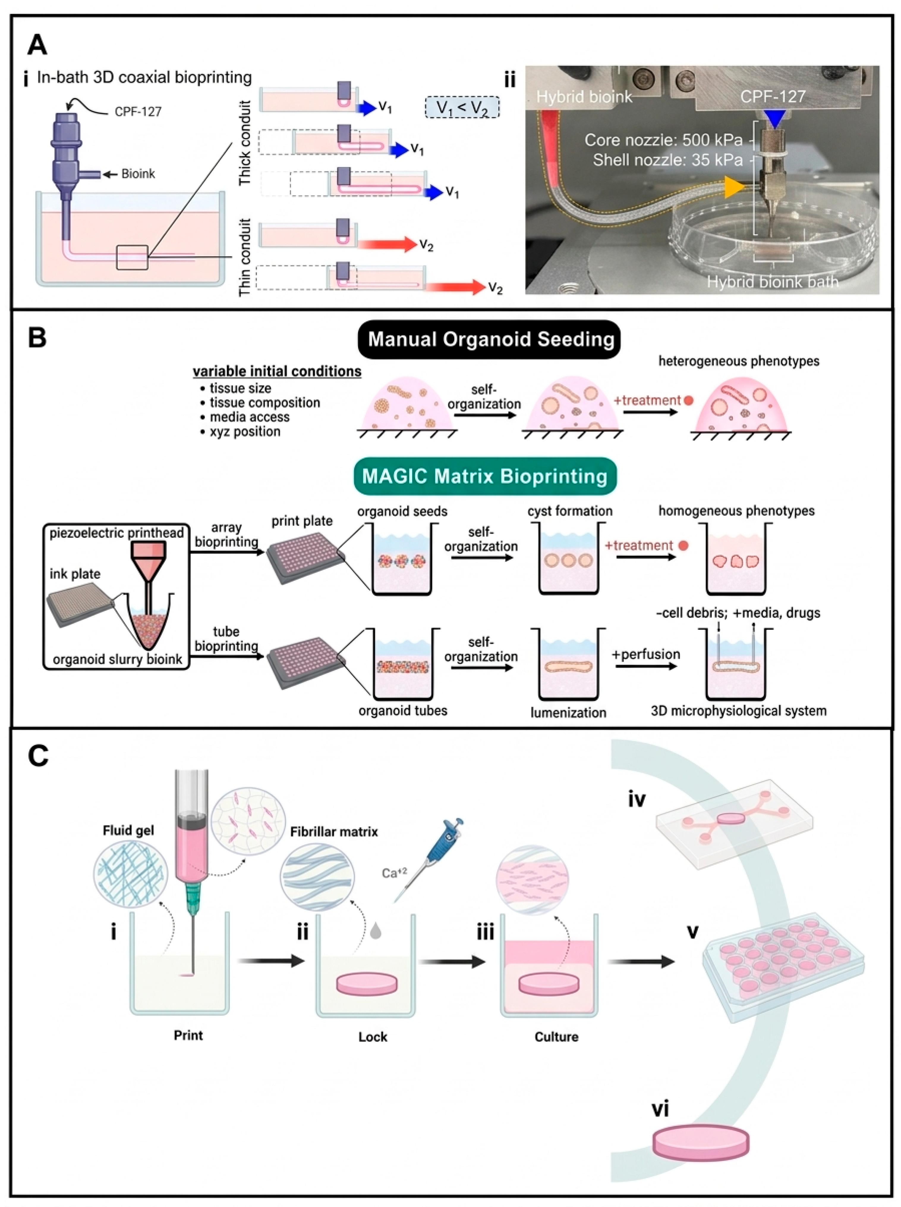
3D bioprinting is improving to enable researchers to fabricate more intricate, physiologically relevant in vitro models, such as disease models [77]. By combining embedded bioprinting and a coaxial printhead, Park et al. developed an in vitro model of stenotic brain blood vessels to study shear-responsive endothelial function as a key factor in diseases like atherosclerosis (Figure 4A) [78]. Their hypothesis was to overcome limitations such as limited scalability in geometries in conventional systems and replication of key pathophysiological characteristics of cerebrovascular diseases in vitro. To achieve this, they devised a plan that used a new hybrid bioink and embedded 3D-coaxial bioprinting. The solution involved directly printing hollow, tubular structures with a special coaxial printhead that functions like two syringes nested inside one another. The core nozzle extruded a temporary support material, and the shell nozzle extruded cell-laden bioink around it, forming the vessel wall. To address the structural instability and shrinkage common in natural bioinks, the authors created a mechanically enhanced hybrid bioink by supplementing a vascular decellularized extracellular matrix (VdECM) with collagen to resist cell-mediated contraction and alginate to promote rapid ionic crosslinking. This hybrid bioink (1V2C1A: 1% VdECM, 2% collagen, 1% alginate) was systematically tested against control groups of lower-concentration VdECM-only bioinks (1V, 2V) and a collagen-added VdECM without alginate (1V2C). The results showed a stark difference: while the control groups shrank a lot and collapsed when printed into tubes, the hybrid bioink kept its shape and made stable, perfusable vascular conduits. The use of an embedded supporting bath, which was made of the same hybrid bioink, was the key mechanism that enabled this precise fabrication, resulting in a self-supporting yield-stress environment. This bath was critical because its shear-thinning and immediate self-recovery properties provided transient, localized support to the delicate tubular structure as it was extruded from the coaxial nozzle, holding the geometry in place until the alginate in the filament was permanently crosslinked by calcium from the core fluid, preventing collapse and ensuring high structural fidelity when replicating complex stenotic geometries. This approach enabled the rapid and precise fabrication of perfusable vessels with controllable, narrow diameters (250–500 μm) to mimic stenosis in less than 5 min. The ingenious combination of the sturdy bio-ink and the coaxial printer design made it possible to print tiny, living blood vessels that could be narrowed in specific places to mimic disease. Upon fabrication, they were able to circulate fluid through these conduits and illustrate that the constricted segments induced turbulent flow, which exerted stress on the endothelial cells and effectively elicited an inflammatory response, recapitulating the conditions observed in actual human pathologies [78].
4.3. Granular Matrix Support for Homogeneous Organoid Self-Organization
Creating organoids, which are one of the most promising approaches in tissue engineering, faces a significant limitation due to the lack of reproducibility in homogeneous organoid fabrication. This heterogeneity may arise from various factors, including adjacent interfaces that induce morphological variability [79]. An innovative 3D bioprinting study used a combination of a novel support material called “MAGIC matrix” and a custom-designed piezoelectric printhead to control the production of homogeneous organoid phenotypes (Figure 4B) [80]. The MAGIC matrix support bath was created as a composite material by combining a slurry of cell-sized, soft alginate microgels with liquid Matrigel. This particular composition directly influences its essential yield-stress characteristic. During printing at 4 °C, the tightly packed alginate microgels form a yield-stress network, which means they behave like a solid and temporarily liquefy only where the printer needle moves, which allows the printed structure to be held perfectly in place. Moreover, when cultured at 37 °C, the Matrigel component cross-links to create a soft, nurturing environment that provides critical biological signals for morphogenesis. This specific composition is critical for producing homogeneous organoids. The MAGIC matrix effectively eliminates heterogeneity by ensuring that each organoid develops from the same starting point in terms of cell number, shape, and surrounding matrix. This controlled guidance canalizes the cells’ innate self-organization, resulting in a highly uniform array of organoids that mature at the same rate and have the same complex structure. In addition to the MAGIC matrix, a piezoelectric printhead was simultaneously developed to handle tiny, precise volumes of extremely dense cell slurries. It works by using a ceramic actuator that expands or contracts in response to an electrical voltage to directly suck in (aspirate) and push out (extrude) cell ink, reducing waste and providing exceptional control. This combined strategy made a significant difference by enabling the formation of organoid “seeds” with precisely controlled initial size, shape, and position. When tested, these bioprinted intestinal organoid arrays demonstrated significantly higher homogeneity in size and structure than manually seeded ones [80]. In another study, Bakht et al. developed a nanoscale fibrillar ECM-mimicking support bath with an innovative “print-then-lock” strategy (Figure 4C) [81]. The novel “print-then-lock” strategy made use of a support bath containing plant-derived cellulose nanocrystals (CNC). Initially, they developed a fluid gel from CNC, functioning as a semi-solid capable of temporarily maintaining the shape of intricate, 3D-bioprinted constructs composed of diverse low-viscosity bioinks. Subsequent to printing, calcium solution was introduced, triggering the CNC particles to self-assemble and permanently secure the printed structure within a stable, transparent, fibrillar hydrogel matrix. This method made it much easier to make high-resolution, complex structures like miniaturized kidneys and perfusable channels. The printing resolution was as precise as 37 micrometers, which is much superior to the 900-micrometer resolution that a standard agarose support bath can get. The authors illustrated that the fabricated CNC matrix closely resembled the native ECM, facilitating optimal cell viability and the diffusion of biomolecules to sustain long-term culture. The underlying mechanism for this success is CNC’s unique tunable self-assembly: calcium ions shield the negative charges on the CNC particles, allowing them to stick together side by side via hydrogen bonding to form a strong, biomimetic nanofibrillar network [82]. This mechanism serves two functions: a shear-thinning fluid for high-fidelity printing and a stable, ECM-like scaffold for long-term tissue maturation, all in one simple, automatable, and cost-effective process [81].
4.4. Multi-Component Yield-Stress and Self-Healing Fluid Formulations
Another composite support bath was developed by Afagh et al. to overcome limitations such as the difficulty of printing with low-viscosity bioinks, poor shape fidelity for overhanging features, and potential cell damage during the printing and harvesting processes [83]. The novel support bath was composed by mixing calcium ions (CaCl2) with two other materials: thermoresponsive Pluronic F127 (PF) and thixotropic Laponite nanoclay (RDS). This strategy was effective due to its integration of the distinct characteristics of each component. The Pluronic gel creates a stable matrix at body temperature, while the Laponite nanoclay gives the bath a shear-thinning and self-healing quality. Comparative tests of various formulations indicated that a particular formulation (10% PF, 3% Laponite, 0.5% CaCl2) exhibited superior rheological properties, characterized by a high storage modulus for strength and exceptional recovery. This was superior to control groups that used only Pluronic or Laponite, which had lower mechanical strength or slower recovery. This is because a network forms between the Pluronic polymer chains and charged Laponite nanodiscs, with help from calcium ions [84]. This network gave the strength needed to print detailed shapes like hollow tubes and blood vessel networks using a low-viscosity, cell-friendly bioink (3% alginate). Furthermore, the calcium ions in the bath crosslinked the alginate bioink during deposition, solidifying the structure from within the support. As a result, the authors successfully printed complex structures with high shape fidelity and demonstrated high cell viability (more than 80%), as the gentle, low-pressure extrusion process within this supportive and removable bath reduced shear stress on the encapsulated cells [83].
4.5. Process Mechanics: Shearing Protocols and Particle Size Resolution
Besides compositional innovations in the supporting bath, there have been other strategies to create a superior support bath for bioprinting. Granular hydrogels, out of all the varieties, have been widely used in embedding printing because of their self-recovery, shear-thinning, and simple production processes. Granular baths demonstrate yield-stress characteristics and swift self-recovery, functioning as solid-like substances at rest while flowing around the moving nozzle and rapidly reconsolidating. Altering the microgel size and density can readily modify their rheological properties without altering the chemistry. Furthermore, gentle, cell-compatible conditions generally promote the elimination of granular support baths post-printing [72]. Recently, Senior et al. developed an agarose support bath with an innovative strategy by stirring the liquid agarose as it cooled and gelled, which broke it into a dense suspension of tiny, sticky microgels [85]. This shearing action physically inhibits the agarose polymers from establishing a singular, continuous solid gel network. The underlying mechanism of this support bath is based on the unique microstructure of agarose fluid gels, which is formed by applying shear force during gelation. As the hot agarose solution cools, the polymer chains naturally form a network. However, constant stirring physically shears the forming network, resulting in a dense suspension of microscopic gelled particles rather than a single solid block. Moreover, this process leaves reactive, un-gelled polymer chains on the surface of these particles. These sticky chains allow the microparticles to instantly re-bond through weak, physical interactions when shear from the print nozzle passes, similar to Velcro. This rapid, reversible interparticle bonding drives the bath’s solid-like behavior at rest, which holds the bioink, enables liquid-like flow under shear for nozzle movement, and facilitates immediate self-healing to keep the printed structure in place. The authors successfully printed high-resolution, intricate structures, such as a skin model with multiple cell layers and a delicate, branching carotid artery, using low-viscosity bioinks that would typically collapse, achieving filament diameters as fine as 323 µm [85].
Suspension or embedded bioprinting has shown enormous promise compared to traditional bioprinting. These systems enhance the bioprinting of multimaterial and multicellular structures. Furthermore, as the crosslinking process occurs post-bioprinting rather than layer-by-layer as in traditional methods, an additional benefit of bioprinting in support baths is the potential to accelerate the production of cellularized constructs, thereby facilitating the scaling of the bioprinting process and decreasing both printing durations and expenses [72]. However, further in-depth study is still needed. Researchers should study the synergy of the bioinks and the supporting bath, both compositionally and in terms of mechanical properties, to better optimize the resolution and fidelity of the final structure [74]. Moreover, parameters involved in the printing results, specifically the resolution, can vary based on the type of support bath. For example, in a bulk hydrogel bath, rheological properties are the key factor. An appropriate bath should exhibit a suitable yield stress and swift thixotropic recovery time, typically within a few seconds [86]. The rheological properties can be modified by altering variables such as polymer concentration, filler content, and intermolecular interactions. Furthermore, when performing high-precision 3D printing, consideration must be given to the shrinkage/swelling of the printed structure in a bath and the deformation brought on by cell migration in cell-laden printing. In a microgel bath, the particle size and distribution significantly influence printing resolution [74,87]. A reduced particle size and consistent size distribution enhance resolution. Furthermore, aggregation or swelling of gels in the bath should be avoided during the printing process to ensure a consistent printing outcome.
Figure 4.
Recent innovations in embedded/suspension bioprinting. (A) Optimizing printing parameters to produce stenotic vascular structure. (i) A schematic illustration of how a change in print stage velocity affects tube diameter control. (ii) Brain blood vessel development using an in-bath 3D-coaxial bioprinting system. Reprinted and adapted with the permission from Park et al., 2025 [78]. (B) MAGIC matrix bioprinting makes it possible to create perfusable 3D microphysiological systems and high-throughput organoid arrays with enhanced statistical power. Due to variations in the initial tissue size, content, and microenvironment, traditional manual organoid seeding techniques result in heterogeneity in organoid growth and morphogenesis. Bioprinting platforms enable the quick creation of repeatable organoid arrays or freeform 3D microphysiological systems by adjusting for beginning variables like cell quantity, medium access, and organoid spacing. Reprinted and adapted with the permission from Graham et al., 2025 [80]. (C) The production of bioprinted microphysiological systems integrated in a bioinspired hierarchical fibrillar matrix using the print-then-lock method. (i) Support fluid gels made of colloidal suspensions of CNC with specific rheology are utilized to create 3D structures with any shape. (ii) After adding calcium ions to induce CNC self-assembly, the printed constructions are bound within a fibrillar matrix. (iii) Bioprinted structures incorporated in the fibrillar matrix are developed for bioassays and in vitro maturation. This platform makes it feasible to use it as (iv) tissue and organ-on-chip microfluidic device housing support materials; (v) on-plate 3D bioprinted models for medium- to high-throughput assay replicates; (vi) transient templating bioreactors for in vitro maturation of 3D bioprinted tissue constructs that can be harvested following bio-orthogonal digestion of the CNC matrix. Reprinted with the permission from Bakht et al., 2021 [81].
5. Other Innovative Approaches
Besides the above-mentioned innovative approaches, in this section, we are exploring other engineering approaches and ideas to improve the efficacy and quality of current bioprinting technology.
5.1. Nozzle-Free Painting Architecture and Layer-by-Layer Transference
To address the chronic trade-offs between printing resolution, bioink viscosity, and cell survival that limit conventional inkjet and microextrusion platforms when processing high-density cell suspensions, Chikae et al. developed a nozzle-free microscopic biofabrication framework combining an automated painting needle method with a layer-by-layer (LbL) cell-accumulation technique (Figure 5A) [88]. The primary innovation bypasses nozzle-clogging constraints by attaching high-viscosity bioinks directly onto a painting needle tip, transferring the material to a substrate via high-resolution vertical mechanical strokes across an exceptionally wide viscosity range of 1 to 1 × 10⁵ mPa·s. To fabricate functional tissue, a first bioink composed of hyaluronic acid (HA) thickener, thrombin, and LbL-nanofilm-coated human-induced pluripotent stem cell-derived cardiomyocytes (hiPSC-CMs) co-cultured with fibroblasts is painted onto the substrate, which is immediately followed by the addition of a secondary fibrinogen-based ink. This localized interaction drives rapid dome-shaped fibrin gel crosslinking, while the subsequent outward diffusion of the HA thickener safely precipitates the encapsulated cells into an ultra-dense network. Biological evaluations verified that this nozzle-free painting mechanism avoids mechanical cell damage, maintaining high cell viability (92.8% post-printing and 97.5% after 3 days) [PDF: Page 3]. Ultimately, this process yielded tightly stacked, 7-layer cardiac tissue chips (~60 µm thick, 1.1 mm wide) with a high cellular density (3.5 × 10⁸ cells/cm3) mimicking native myocardium, exhibiting authentic sarcomeric F-actin and cTnT cross-striations and sustained, synchronous contraction at 50–60 beats per minute [88].
5.2. Delayed “Print-Then-Mix” Multi-Jet Droplet Collision Dynamics
To overcome the challenges of conventional “mixing-then-printing” methods, which frequently result in inks solidifying or becoming unprintable prior to deposition, Christensen et al. developed a novel strategy based on a “printing-then-mixing” method at voxel resolution (Figure 5B) [89]. This method enables the fabrication of biological structures from reactive materials and the creation of structures with defined compositional gradients. In order to achieve this, the authors developed and used the “intersecting jets” method, in which two distinct printheads are angled to fire droplets of various reactive substances (such as cells in buffer and a hydrogel precursor, or alginate and calcium chloride), causing the droplets to collide and mix precisely at the deposition point in the process. This provided a controllable process for creating heterogeneous structures with designed shapes. A major step forward was the successful direct-in-air printing of 3D structures from materials like alginate and calcium chloride, which had been a problem before. To demonstrate cytocompatibility, the authors printed cell-laden constructs using PuraMatrix hydrogel containing neural stem cells (NSCs) and sodium alginate hydrogel laden with 3T3 fibroblasts. The NSC-laden constructs exhibited high cell viability, with cells spreading and showing increased metabolic activity over three days in culture. Similarly, for alginate structures printed in air with embedded fibroblasts, cell viability remained above 90%, a level comparable to traditional inkjet bioprinting [90]. Collagen-based bioinks with hydroxyapatite (HAP) nanoparticles were also printed with controlled HAP frequencies for each layer. The dispersion of nanoparticles inside the construct structure was used as a heterogenous marker. The underlying mechanism of this strategy is the delayed mixing of reactive components. By avoiding pre-mixing within the printhead, it prevents clogging and preserves the printability and cytocompatibility of each ink until they coalesce on the substrate [89].
5.3. Dot-Extrusion Microbead Deposition and Thermal Maintenance
To create heterogeneous tumor constructs, it is essential for a bioprinting system to manage multiple cellular bioinks in a spatial arrangement to replicate the intricate physiological tumor microenvironment [91]. Wei et al. created a 3D bioprinting strategy for heterogeneous tumor tissue modeling [92]. The dot-extrusion printing (DEP) technique eliminates the need for extra crosslinking steps and prevents droplet spreading or fusion problems that are typical in traditional bioprinting by enabling the one-step creation of cell-laden GelMA hydrogel beads (HBs) in a semi-gelled state. GelMA HBs were fabricated utilizing a one-step printing technique in which cooled bioink is extruded through intermittent pneumatic pressure. Droplets formed from surface tension upon contacting the substrate, detaching when extrusion and surface tension forces surpassed capillary force. Maintaining a precise distance of 300 μm between the nozzle and the substrate ensures that beads are made consistently. Temperature is essential and tailored for each concentration (e.g., 14–16 °C for 10% GelMA) to preserve an appropriate gel state for efficient printing. GelMA HBs loaded with human breast cancer cells (MCF-7) and normal human dermal fibroblasts (NHDFs) provided a biomimetic ECM microenvironment and facilitated the growth of both cell types, proving to be an excellent building block for 3D construct development. The developed printing strategy allowed for the creation of spatially defined constructs, such as juxtapositional or overlapping microcapsules, while maintaining high cell viability (>95%). Moreover, the bioprinted heterogeneous constructs demonstrated enhanced cell proliferation compared to control monocultural constructs, forming larger spheroids [92].
5.4. Vat-Free Light Projection and Meniscus-Driven Surface Forces
Digital light projection (DLP)-based bioprinting methods can print 3D architecture at high resolutions, speeds, and overall fidelity [93]. In this method, a liquid crystal display or a digital micromirror device (DMD) is used to make digital masks that spatially pattern and project light onto a vat or reservoir filled with liquid photosensitive resin bioink. This method makes it possible to print 3D structures layer by layer [94]. However, using a vat in DLP bioprinting has several problems. For example, it requires filling a whole tank with expensive bioink, which leads to a lot of waste and high costs, as well as issues like contamination and cell settling during long printing processes [60]. To address this, Kunwar et al. devised a novel “vat-free, droplet bioprinting” method. Instead of a large tank, they used a single, small droplet of bioink (slightly larger than the final object’s volume) to fill a tiny fabrication window between a specialized oxygen-permeable window and the printing stage [95]. The main part of the mechanism was a very precise “up-and-down” motion of the stage. The stage would move down after each layer of the structure had been solidified by light. The process would bring fresh, uncured bioink from the droplet into the window to make the next layer. This smart design used surface forces and a meniscus to pull the bioink up quickly so that one droplet could keep providing it without the need for a reservoir. This strategy worked effectively. It reduced the amount of bioink waste to less than 1% and kept cells from settling by constantly mixing the bioink. Because of this, it was able to print complicated 3D structures with high resolution, with or without cells. Tests on model cells (C3H/10T1/2) showed that this method kept cell viability high (over 90%) and allowed for even cell distribution and spreading. This procedure was superior to vat-based systems, which can lower cell viability if bioink is exposed for a long time [95].
5.5. Cross-Scale Electro-Manipulations and Modulable Photopolymerization
Zhao et al., in another study using DLP bioprinting, implemented an innovative strategy to create tissue-mimicking heterogeneous microstructures with specific multicellular densities [96]. To achieve true biomimicry, the authors devised a clever “cross-scale” method that functions as a multi-tool, performing different tasks at varying sizes (Figure 5C). First, they used a digital microfluidic (DMF) chip to manipulate entire droplets of various cell-laden hydrogels (such as PEGDA and GelMA) at the millimeter scale. This chip functions similarly to a programmable tile floor in that each tile (electrode) can be electrified. Using electrowetting (EWOD), a voltage applied to an electrode attracts a hydrogel droplet to that specific location, allowing for precise exchange, splitting, and merging of different bioinks to create macroscopic composite droplets. Second, at the micrometer scale, they worked within individual hydrogel droplets. Using a different electrical phenomenon known as dielectrophoresis (DEP), they applied voltage to create micro-electric fields that gently pushed and pulled cells or microparticles into specific, pre-programmed patterns and densities within the liquid hydrogel, which is impossible with standard nozzle-based bioprinting. Finally, they used a projection-based system (DLP) to secure the carefully arranged structures. Instead of solidifying the entire droplet at once, they used dynamic UV light patterns, similar to a microscopic video projector, to polymerize only specific regions of the droplet. This “modulable encapsulation” transforms complex cellular patterns and composite hydrogel geometries into a stable, solid microstructure. This approach was important because it allowed for the creation of microstructures with precise cell density gradients and patterns, Young’s modulus, and controlled variations in ECM components (like PEGDA and GelMA). This was not possible with earlier techniques, which led to random cell encapsulation. The biological evaluation demonstrated the success of this approach: the constructed microstructures supported long-term culture with high cell viability (more than 90%), and tests comparing control groups revealed that the density and ordered spatial distribution of cocultured hepatocytes (HepG2) and fibroblasts (NIH3T3) had a significant effect on their proliferation and functional output. A hepatic lobule model with an ordered arrangement of HepG2 cells secreted significantly more urea (** p < 0.01) compared to a model where cells were cocultured without a defined pattern [96].
Figure 5.
Other innovations in bioprinting. (A) Schematic of the layer-by-layer (LbL) cell coating process and the fabrication of an LbL-3D heart model via painting needle delivery. Workflow detailing the complete biofabrication sequence of the LbL-3D heart. The initial LbL-encapsulated cellular bioink is patterned onto a receiving substrate using a specialized painting needle. Reprinted and adapted with the permission from Chikae et al., 2019 [88]. (B) Intersecting jets-based inkjet bioprinting approach, (i) Printing schematic; (ii) Printing sequence demonstrating the intersecting jets method. Key components are displayed, and process factors are indicated. Reprinted and adapted with the permission from Christensen et al., 2017 [89]. (C) Bioprinting method with composite cross manipulation. (i) Composite printing system schematic diagram. (ii) A personalized digital mask. (iii) A hydrogel microstructure filled with biomimetic cells. CCD stands for charge-coupled device, PC for personal computer, BE for beam expansion optics, and PO for projection optics. Reprinted and adapted with the permission from Zhao et al., 2024 [96].
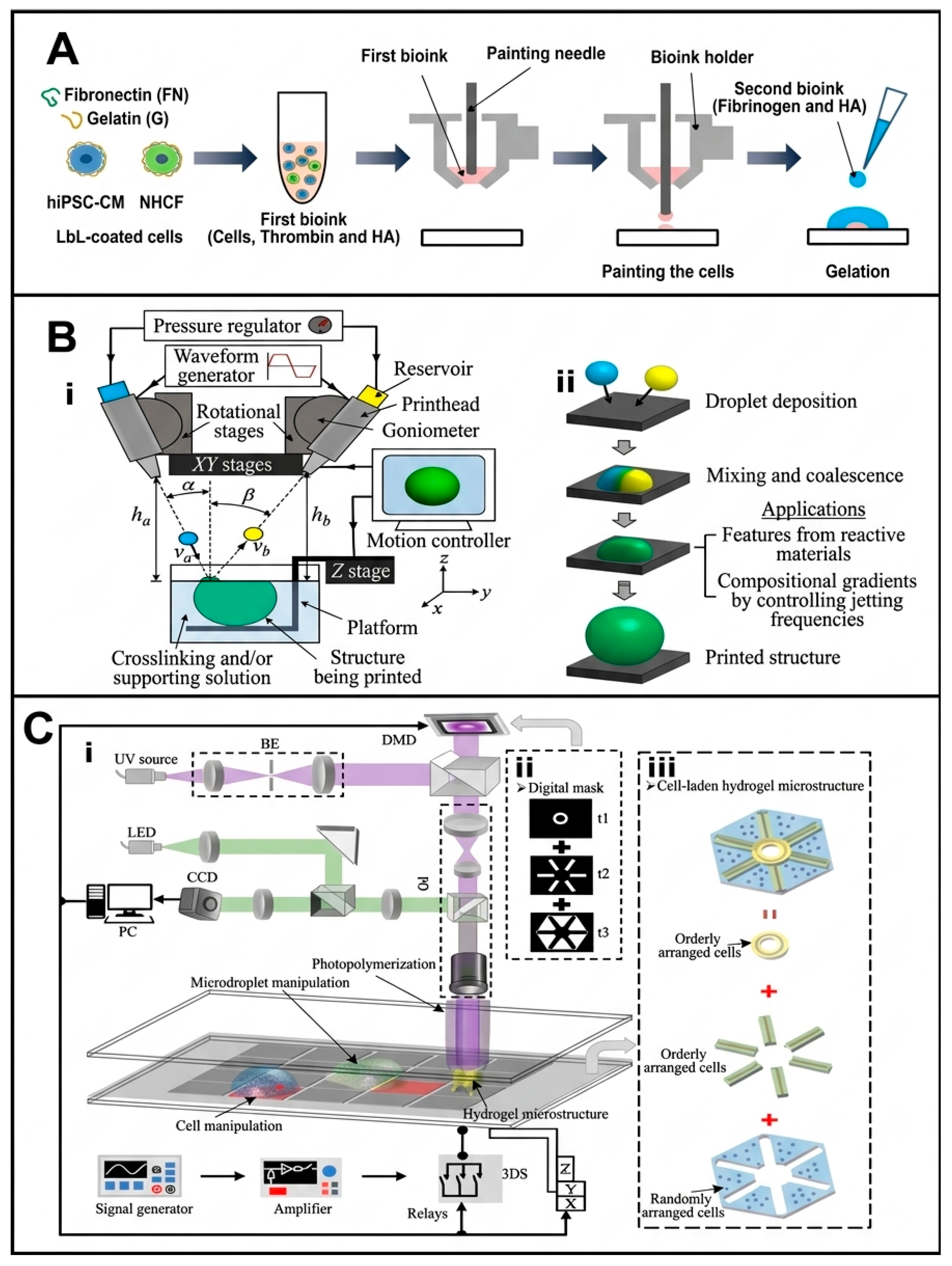
6. Future Perspective
Bioprinting has advanced significantly over the last ten years, primarily due to improvements in the formulation of bioink, the addition of bioactive agents, and a better comprehension of cellular behavior in three-dimensional environments. However, many of the remaining challenges in the field are not just biological; they are also caused by the limitations of the printing process itself. Parameters such as extrusion pressure, nozzle geometry, printhead architecture, deposition patterns, resolution limits, multimaterial switching, and structural fidelity critically influence the final construct. Future progress will thus be dependent not only on more sophisticated bioinks but also on the intelligent refinement of the mechanical and procedural aspects of bioprinting. As discussed throughout this review, simple and inexpensive engineering changes, such as optimized nozzle designs, modular printheads, microfluidic integration, and embedded printing strategies, can significantly improve resolution, heterogeneity, and structural complexity without requiring prohibitively expensive systems. These advancements have taken the field from simple proof-of-concept demonstrations to the development of more physiologically relevant tissue models. The next big challenge, however, is no longer just printing a structure; it is about guiding that structure to become a functional tissue. Tackling this challenge will require the field to deal with new obstacles, shifting from simple step-by-step improvements to a complete approach that combines engineering ideas with new computer tools and, most importantly, a better understanding of how living tissues develop.
The first bottleneck is the transition from generic to specialized fabrication. Rather than developing universal printers for all tissue types, the field must question whether such a device is even desirable. Highly specialized, tissue-specific fabrication units are likely to emerge in the future, where biology adapts to engineering and fundamentally constrains it. In such platforms, the structural and technical architecture of the printer would be co-designed with the bioink’s biological composition. For instance, neurons, which are sensitive to shear, may need low-pressure extrusion systems with nozzles that are optimized for tapering. On the other hand, articular cartilage, which forms gradients, may benefit from integrated microfluidic mixing modules. However, the critical step is not just building these modules but defining the precise biological parameters that guide their design. The shift towards precision biofabrication will be prompted not solely by engineering customization but by a stringent, quantitative comprehension of the biological necessities they are required to fulfill.
For the upcoming generation of bioprinters, the engineering changes covered in this review offer a comprehensive toolkit (Table 1). The next step will be the intelligent, rather than accidental, integration of these separate innovations into hybrid platforms. For instance, a microfluidic printhead could be integrated with a support bath system to print complex, hollow organoids. But managing the extreme complexity of such a system, balancing multiple materials, crosslinking kinetics, and deposition patterns in real time, will quickly outpace human intuition. This is where artificial intelligence (AI) becomes more than just an enhancement but a requirement. However, better data is more important than better algorithms when it comes to AI. The field currently lacks large-scale, standardized, and richly annotated datasets for training predictive models [97]. AI-assisted platforms could analyze datasets generated by printing experiments to predict optimal parameter combinations, but this would necessitate a community-wide shift toward open data and standardized reporting of printing parameters, bioink properties, and biological results [97,98]. Real-time feedback from embedded sensors could allow closed-loop control systems to dynamically adjust printing parameters, but only if we can define the biologically relevant setpoints for those controls. Furthermore, AI can integrate data from material science and biology to predict long-term construct behavior, but its predictions will only be as reliable as our understanding of the complex, dynamic interactions between cells and their printed matrix. This coming together will eventually create special bioprinting machines, like a “vascular bioprinter,” that uses a special nozzle and an AI feedback system, mainly because the need for proper blood flow sets a clear engineering target.
Table 1.
Highlight of in-lab innovations for developing bioprinting.
Perhaps the most significant conceptual shift—and the most difficult to implement—is the integration of “specialized” printing and maturation onto a single platform, resulting in a technological merger of the bioprinter and the bioreactor. This is a direct response to the fundamental conflict between printability and functional maturation. Printable hydrogels must be sufficiently soft and shear-thinning to maintain cell viability during extrusion, but soft matrices frequently lack the mechanical robustness required for long-term tissue function. Instead of attempting to print fully mature tissues directly, future systems must adopt an integrated bioprinter–bioreactor paradigm. In this model, the “bioprinter” deposits a structurally permissive and cell-friendly construct, which then remains within a controlled maturation chamber where its properties are progressively refined. In our perspective, this “womb-like” device must function as an active regulatory ecosystem that fundamentally expands beyond the passive capabilities of a traditional, static incubator. We envision an integrated bioprinter-bioreactor platform where embedded multi-modal sensors and automated feedback loops continuously track dynamic parameters, such as microvascular oxygen tension, localized pH, cellular metabolic activity, and mechanical strain, enabling autonomous, real-time adaptive adjustments to the maturation microenvironment. These data-driven responses could start processes that happen over time, like secondary crosslinking, enzymatic matrix reinforcement, or gradual stiffening mechanisms that make the material more stable while preserving early cell viability.
However, the critical challenge here is that we are still learning the “language” of development. We can imagine programmable biochemical patterning and physical stimulation, like using electrical fields on heart tissues or cyclic loading on bones, but we do not know the exact sequence and orchestration yet. What is the optimal sequence of growth factor delivery? How do we combine mechanical and biochemical cues without inducing conflicting cellular responses? The integrated bioprinter–bioreactor platform is not just an engineering problem; it is a tool for fundamental discovery, a way to systematically probe and analyze the complex process of tissue formation. For example, if sensors detected hypoxia within the core of a large construct, the system could automatically initiate localized release of angiogenic factors. But this requires a pre-existing, validated model of how, when, and where to deliver those factors, a model we largely lack today.
This complexity also applies to the dynamic control of multicellular constructs. Many functional tissues depend on co-culture systems that are carefully balanced. Future bioprinting platforms must therefore include strategies for temporal and spatial adaptation of cellular composition, allowing the sequential introduction of additional cell populations, controlled release of bioactive molecules, or targeted reinforcement of specific regions. In order to have this level of control, the bioprinting process needs to be in sync in a number of ways. Specifically, bioink formulations need to be co-engineered as responsive materials that interact with the bioreactor environment in addition to being printable. For instance, shear-thinning polymers and embedded nanoparticles that react to external magnetic fields could be combined in a bioink intended for a vascularized bone construct to provide noninvasive post-printing reinforcement. However, the critical question remains: can we design these materials to respond in ways that are both spatially precise and biologically significant?
Looking forward, the evolution of bioprinting may shift away from a singular, universal “bioprinter–bioreactor system” toward a decentralized landscape of specialized, customizable biofabrication platforms customized to the unique mechanical and biological requirements of different tissue types. By systematically pairing cost-effective engineering innovations with parallel advancements in biomaterial chemistry, cellular engineering, and data-driven computational optimization, the field could gradually move closer to generating reproducible, scalable, and clinically relevant functional constructs. In this perspective, the future of bioprinting may depend not on isolated technological breakthroughs but on intelligent, system-level integration guided by a humble understanding of how much we still need to learn about the fundamental biology of tissue formation. The real breakthrough will occur when our engineering platforms are advanced enough to ask the appropriate biological questions.
Author Contributions
Conceptualization, M.H.N. and A.S.; writing—original draft preparation, M.H.N.; writing—review and editing, P.K., F.D.A.d.S.P., P.B., visualization, P.K.; supervision, P.T.; project administration, P.B.; funding acquisition, A.S., P.B. and P.T. All authors have read and agreed to the published version of the manuscript.
Funding
This research was funded by Russian Science Foundation, grant number 26-75-10148 (in part of bioprinting innovations for microfluidics-assisted bioprinting) and Russian Science Foundation, grant number 24-45-10012 (in part of other hydrogel-based bioprinting approaches, dependent on gels parameters such as viscosity, opacity, temperature-dependent behavior, etc.).
Data Availability Statement
No new data were created or analyzed in this study. Data sharing is not applicable to this article.
Acknowledgments
During the preparation of this manuscript, the authors used ChatGPT (OpenAI, GPT-5.6 Sol) for the purposes of creating graphical abstract. The authors have reviewed and edited the output and take full responsibility for the content of this publication.
Conflicts of Interest
The authors declare no conflicts of interest.
References
- Mladenovska, T.; Choong, P.F.; Wallace, G.G.; O’Connell, C.D. The Regulatory Challenge of 3D Bioprinting. Regen. Med. 2023, 18, 659–674. [Google Scholar] [CrossRef] [Scilit] [PubMed]
- Murphy, S.V.; Atala, A. 3D Bioprinting of Tissues and Organs. Nat. Biotechnol. 2014, 32, 773–785. [Google Scholar] [CrossRef] [Scilit] [PubMed]
- Ganguly, S.; Wulff, D.; Phan, C.-M.; Jones, L.W.; Tang, X.S. Injectable and 3D Extrusion Printable Hydrophilic Silicone-Based Hydrogels for Controlled Ocular Delivery of Ophthalmic Drugs. ACS Appl. Bio Mater. 2024, 7, 6286–6296. [Google Scholar] [CrossRef] [Scilit] [PubMed]
- Mironov, V.; Kasyanov, V.; Markwald, R.R. Organ Printing: From Bioprinter to Organ Biofabrication Line. Curr. Opin. Biotechnol. 2011, 22, 667–673. [Google Scholar] [CrossRef] [Scilit] [PubMed]
- Santoni, S.; Gugliandolo, S.G.; Sponchioni, M.; Moscatelli, D.; Colosimo, B.M. 3D Bioprinting: Current Status and Trends—A Guide to the Literature and Industrial Practice. Bio-Des. Manuf. 2022, 5, 14–42. [Google Scholar] [CrossRef] [Scilit]
- Tao, H.; Zhang, X.; Yuan, X.; Lian, Z.; Xu, J.; Shan, D.; Guo, B. 3D Bioprinting Technology in Tissue Engineering Printing Method and Material Selection. BioNanoScience 2025, 15, 199. [Google Scholar] [CrossRef] [Scilit]
- DeMel, D.C.; Wagner, G.A.; Maresca, J.A.; Geibel, J.P. Application of a 3D Bioprinter: Jet Technology For ‘Biopatch’ Development Using Cells on Hydrogel Supports. BioTechniques 2024, 76, 52–62. [Google Scholar] [CrossRef] [Scilit] [PubMed]
- Jergitsch, M.; Mateos-Timoneda, M.A. 3D Extrusion Bioprinting: Rational Bioink Design and Advanced Fabrication Techniques. Trends Biotechnol. 2026, 44, 378–388. [Google Scholar] [CrossRef] [Scilit] [PubMed]
- Levato, R.; Dudaryeva, O.; Garciamendez-Mijares, C.E.; Kirkpatrick, B.E.; Rizzo, R.; Schimelman, J.; Anseth, K.S.; Chen, S.; Zenobi-Wong, M.; Zhang, Y.S. Light-Based Vat-Polymerization Bioprinting. Nat. Rev. Methods Primers 2023, 3, 47. [Google Scholar] [CrossRef] [Scilit] [PubMed]
- Ng, W.L.; Vyas, C.; Huang, B.; Yeong, W.Y.; Bartolo, P. Advanced Bioprinting Strategies for Fabrication of Biomimetic Tissues and Organs. Int. J. Extrem. Manuf. 2025, 7, 062006. [Google Scholar] [CrossRef] [Scilit]
- Lecuit, T.; Le Goff, L. Orchestrating Size and Shape during Morphogenesis. Nature 2007, 450, 189–192. [Google Scholar] [CrossRef] [Scilit] [PubMed]
- Murphy, S.V.; De Coppi, P.; Atala, A. Opportunities and Challenges of Translational 3D Bioprinting. Nat. Biomed. Eng. 2020, 4, 370–380. [Google Scholar] [CrossRef] [Scilit] [PubMed]
- Norahan, M.H.; Pedroza-González, S.C.; Sánchez-Salazar, M.G.; Álvarez, M.M.; Trujillo de Santiago, G. Structural and Biological Engineering of 3D Hydrogels for Wound Healing. Bioact. Mater. 2023, 24, 197–235. [Google Scholar] [CrossRef] [Scilit] [PubMed]
- Wang, H.; Yu, H.; Zhou, X.; Zhang, J.; Zhou, H.; Hao, H.; Ding, L.; Li, H.; Gu, Y.; Ma, J.; et al. An Overview of Extracellular Matrix-Based Bioinks for 3D Bioprinting. Front. Bioeng. Biotechnol. 2022, 10, 905438. [Google Scholar] [CrossRef] [Scilit] [PubMed]
- O’Connor, C.; Brady, E.; Zheng, Y.; Moore, E.; Stevens, K.R. Engineering the Multiscale Complexity of Vascular Networks. Nat. Rev. Mater. 2022, 7, 702–716. [Google Scholar] [CrossRef] [Scilit] [PubMed]
- Fang, Y.; Guo, Y.; Liu, T.; Xu, R.; Mao, S.; Mo, X.; Zhang, T.; Ouyang, L.; Xiong, Z.; Sun, W. Advances in 3D Bioprinting. Chin. J. Mech. Eng. Addit. Manuf. Front. 2022, 1, 100011. [Google Scholar] [CrossRef] [Scilit]
- Mobaraki, M.; Ghaffari, M.; Yazdanpanah, A.; Luo, Y.; Mills, D.K. Bioinks and Bioprinting: A Focused Review. Bioprinting 2020, 18, e00080. [Google Scholar] [CrossRef] [Scilit]
- Decante, G.; Costa, J.B.; Silva-Correia, J.; Collins, M.N.; Reis, R.L.; Oliveira, J.M. Engineering Bioinks for 3D Bioprinting. Biofabrication 2021, 13, 032001. [Google Scholar] [CrossRef] [Scilit] [PubMed]
- Debnath, S.; Agrawal, A.; Jain, N.; Chatterjee, K.; Player, D.J. Collagen as a Bio-Ink for 3D Printing: A Critical Review. J. Mater. Chem. B 2025, 13, 1890–1919. [Google Scholar] [CrossRef] [Scilit] [PubMed]
- Tashman, J.W.; Shiwarski, D.J.; Feinberg, A.W. Development of a High-Performance Open-Source 3D Bioprinter. Sci. Rep. 2022, 12, 22652. [Google Scholar] [CrossRef] [Scilit] [PubMed]
- Brown, N.C.; Ames, D.C.; Mueller, J. Multimaterial Extrusion 3D Printing Printheads. Nat. Rev. Mater. 2025, 10, 807–825. [Google Scholar] [CrossRef] [Scilit]
- Skylar-Scott, M.A.; Mueller, J.; Visser, C.W.; Lewis, J.A. Voxelated Soft Matter via Multimaterial Multinozzle 3D Printing. Nature 2019, 575, 330–335. [Google Scholar] [CrossRef] [Scilit] [PubMed]
- Ladd, C.; So, J.-H.; Muth, J.; Dickey, M.D. 3D Printing of Free Standing Liquid Metal Microstructures. Adv. Mater. 2013, 25, 5081–5085. [Google Scholar] [CrossRef] [Scilit] [PubMed]
- Mistry, P.; Aied, A.; Alexander, M.; Shakesheff, K.; Bennett, A.; Yang, J. Bioprinting Using Mechanically Robust Core–Shell Cell-Laden Hydrogel Strands. Macromol. Biosci. 2017, 17, 1600472. [Google Scholar] [CrossRef] [Scilit] [PubMed]
- Rau, D.A.; Williams, C.B.; Bortner, M.J. Rheology and Printability: A Survey of Critical Relationships for Direct Ink Write Materials Design. Prog. Mater. Sci. 2023, 140, 101188. [Google Scholar] [CrossRef] [Scilit]
- Yuan, Z.; Bai, X.; Li, S.; Fu, Y.; Wan, Z.; Guo, X.; Zhai, M.; Yi, J.; Liu, Y.; Zhou, Y.; et al. Multimaterial and Multidimensional Bioprinting in Regenerative Medicine: Advances, Limitations, and Future Directions. Adv. Healthc. Mater. 2025, 14, 2500475. [Google Scholar] [CrossRef] [Scilit] [PubMed]
- Saadi, M.A.S.R.; Maguire, A.; Pottackal, N.T.; Thakur, M.S.H.; Ikram, M.M.; Hart, A.J.; Ajayan, P.M.; Rahman, M.M. Direct Ink Writing: A 3D Printing Technology for Diverse Materials. Adv. Mater. 2022, 34, 2108855. [Google Scholar] [CrossRef] [Scilit] [PubMed]
- Nie, J.; Li, M.; Liu, W.; Li, W.; Xing, Z. The Role of Plasticizer in Optimizing the Rheological Behavior of Ceramic Pastes Intended for Stereolithography-Based Additive Manufacturing. J. Eur. Ceram. Soc. 2021, 41, 646–654. [Google Scholar] [CrossRef] [Scilit]
- Muth, J.T.; Dixon, P.G.; Woish, L.; Gibson, L.J.; Lewis, J.A. Architected Cellular Ceramics with Tailored Stiffness via Direct Foam Writing. Proc. Natl. Acad. Sci. USA 2017, 114, 1832–1837. [Google Scholar] [CrossRef] [Scilit] [PubMed]
- Sanz-Garcia, A.; Sodupe-Ortega, E.; Pernía-Espinoza, A.; Shimizu, T.; Escobedo-Lucea, C. A Versatile Open-Source Printhead for Low-Cost 3D Microextrusion-Based Bioprinting. Polymers 2020, 12, 2346. [Google Scholar] [CrossRef] [Scilit] [PubMed]
- Blaeser, A.; Duarte Campos, D.F.; Puster, U.; Richtering, W.; Stevens, M.M.; Fischer, H. Controlling Shear Stress in 3D Bioprinting Is a Key Factor to Balance Printing Resolution and Stem Cell Integrity. Adv. Healthc. Mater. 2016, 5, 326–333. [Google Scholar] [CrossRef] [Scilit] [PubMed]
- Garciamendez-Mijares, C.E.; Guerra-Alvarez, G.E.; Sánchez-Salazar1, M.G.; Garcia Rubio, A.A.; García Martínez, G.; Mertgen, A.S.; Ceballos-González, C.; Bolívar Monsalve, E.; Zhang, Y.S.; Trujillo de Santiago, G.; et al. Development of an Affordable Extrusion 3D Bioprinter Equipped with a Temperature-Controlled Printhead. Int. J. Bioprinting 2023, 9, 0244. [Google Scholar] [CrossRef] [Scilit]
- Chen, A.; Wang, W.; Mao, Z.; He, Y.; Chen, S.; Liu, G.; Su, J.; Feng, P.; Shi, Y.; Yan, C.; et al. Multimaterial 3D and 4D Bioprinting of Heterogenous Constructs for Tissue Engineering. Adv. Mater. 2024, 36, 2307686. [Google Scholar] [CrossRef] [Scilit] [PubMed]
- Liu, W.; Zhang, Y.S.; Heinrich, M.A.; De Ferrari, F.; Jang, H.L.; Bakht, S.M.; Alvarez, M.M.; Yang, J.; Li, Y.-C.; Trujillo-de Santiago, G.; et al. Rapid Continuous Multimaterial Extrusion Bioprinting. Adv. Mater. 2017, 29, 1604630. [Google Scholar] [CrossRef] [Scilit] [PubMed]
- Yang, K.; Gao, C.; Tian, Y.; Zhu, Z.; She, J.; Qiao, W.; Shi, J.; Zhang, D.; Wang, J.; Xu, C.; et al. Multi-Nozzle Bioprinting of Biomimetic Tissue Models with Mechanical Reinforcement and Bioactive Matrix Remodeling. Biofabrication 2025, 18, 015019. [Google Scholar] [CrossRef] [Scilit] [PubMed]
- Sun, J.; Gong, Y.; Xu, M.; Chen, H.; Shao, H.; Zhou, R. Coaxial 3D Bioprinting Process Research and Performance Tests on Vascular Scaffolds. Micromachines 2024, 15, 463. [Google Scholar] [CrossRef] [Scilit] [PubMed]
- Trujillo-de Santiago, G.; Alvarez, M.M. Together but Not Scrambled: A Perspective on Chaotic Printing/Bioprinting. Aggregate 2024, 5, e548. [Google Scholar] [CrossRef] [Scilit]
- Samandari, M.; Alipanah, F.; Majidzadeh-A, K.; Alvarez, M.M.; Trujillo-de Santiago, G.; Tamayol, A. Controlling Cellular Organization in Bioprinting through Designed 3D Microcompartmentalization. Appl. Phys. Rev. 2021, 8, 021404. [Google Scholar] [CrossRef] [Scilit] [PubMed]
- Wei, Q.; An, Y.; Zhao, X.; Li, M.; Zhang, J.; Cui, N. Optimal Design of Multi-Biomaterials Mixed Extrusion Nozzle for 3D Bioprinting Considering Cell Activity. Virtual Phys. Prototyp. 2025, 20, e2438897. [Google Scholar] [CrossRef] [Scilit]
- Satpathy, A.; Datta, P.; Wu, Y.; Ayan, B.; Bayram, E.; Ozbolat, I.T. Developments with 3D Bioprinting for Novel Drug Discovery. Expert. Opin. Drug Discov. 2018, 13, 1115–1129. [Google Scholar] [CrossRef] [Scilit] [PubMed]
- Zheng, Z.; Eglin, D.; Alini, M.; Richards, G.R.; Qin, L.; Lai, Y. Visible Light-Induced 3D Bioprinting Technologies and Corresponding Bioink Materials for Tissue Engineering: A Review. Engineering 2021, 7, 966–978. [Google Scholar] [CrossRef] [Scilit]
- Kim, M.H.; Singh, Y.P.; Celik, N.; Yeo, M.; Rizk, E.; Hayes, D.J.; Ozbolat, I.T. High-Throughput Bioprinting of Spheroids for Scalable Tissue Fabrication. Nat. Commun. 2024, 15, 10083. [Google Scholar] [CrossRef] [Scilit] [PubMed]
- Ozbolat, I.T. Scaffold-Based or Scaffold-Free Bioprinting: Competing or Complementing Approaches? J. Nanotechnol. Eng. Med. 2015, 6, 024701. [Google Scholar] [CrossRef] [Scilit]
- Banerjee, D.; Singh, Y.P.; Datta, P.; Ozbolat, V.; O’Donnell, A.; Yeo, M.; Ozbolat, I.T. Strategies for 3D Bioprinting of Spheroids: A Comprehensive Review. Biomaterials 2022, 291, 121881. [Google Scholar] [CrossRef] [Scilit] [PubMed]
- Kim, S.; Byun, H.; Lee, S.; Kim, E.; Lee, G.M.; Huh, S.J.; Joo, J.; Shin, H. Spatially Arranged Encapsulation of Stem Cell Spheroids within Hydrogels for the Regulation of Spheroid Fusion and Cell Migration. Acta Biomater. 2022, 142, 60–72. [Google Scholar] [CrossRef] [Scilit] [PubMed]
- Aguilar, I.N.; Smith, L.J.; Olivos, D.J.; Chu, T.-M.G.; Kacena, M.A.; Wagner, D.R. Scaffold-Free Bioprinting of Mesenchymal Stem Cells with the Regenova Printer: Optimization of Printing Parameters. Bioprinting 2019, 15, e00048. [Google Scholar] [CrossRef] [Scilit] [PubMed]
- Utama, R.H.; Atapattu, L.; O’Mahony, A.P.; Fife, C.M.; Baek, J.; Allard, T.; O’Mahony, K.J.; Ribeiro, J.C.C.; Gaus, K.; Kavallaris, M.; et al. A 3D Bioprinter Specifically Designed for the High-Throughput Production of Matrix-Embedded Multicellular Spheroids. iScience 2020, 23, 101621. [Google Scholar] [CrossRef] [Scilit] [PubMed]
- Ayan, B.; Heo, D.N.; Zhang, Z.; Dey, M.; Povilianskas, A.; Drapaca, C.; Ozbolat, I.T. Aspiration-Assisted Bioprinting for Precise Positioning of Biologics. Sci. Adv. 2020, 6, eaaw5111. [Google Scholar] [CrossRef] [Scilit] [PubMed]
- Fathi, S.; Lei, I.M.; Cao, Y.; Huang, Y.Y.S. Microcapillary Cell Extrusion Deposition with Picolitre Dispensing Resolution. Bio-Des. Manuf. 2023, 6, 1–11. [Google Scholar] [CrossRef] [Scilit] [PubMed]
- Hajam, M.I.; Khan, M.M. Microfluidics: A Concise Review of the History, Principles, Design, Applications, and Future Outlook. Biomater. Sci. 2024, 12, 218–251. [Google Scholar] [CrossRef] [Scilit] [PubMed]
- Chand, R.; Kamei, K.; Vijayavenkataraman, S. Advances in Microfluidic Bioprinting for Multi-Material Multi-Cellular Tissue Constructs. Cell Ther. Eng. Connect. 2025, 1, 1. [Google Scholar] [CrossRef] [Scilit]
- Davoodi, E.; Sarikhani, E.; Montazerian, H.; Ahadian, S.; Costantini, M.; Swieszkowski, W.; Willerth, S.M.; Walus, K.; Mofidfar, M.; Toyserkani, E.; et al. Extrusion and Microfluidic-Based Bioprinting to Fabricate Biomimetic Tissues and Organs. Adv. Mater. Technol. 2020, 5, 1901044. [Google Scholar] [CrossRef] [Scilit] [PubMed]
- Zhang, P.; Liu, C.; Modavi, C.; Abate, A.; Chen, H. Printhead on a Chip: Empowering Droplet-Based Bioprinting with Microfluidics. Trends Biotechnol. 2024, 42, 353–368. [Google Scholar] [CrossRef] [Scilit] [PubMed]
- Serex, L.; Bertsch, A.; Renaud, P. Microfluidics: A New Layer of Control for Extrusion-Based 3D Printing. Micromachines 2018, 9, 86. [Google Scholar] [CrossRef] [Scilit] [PubMed]
- Holland, I. Extrusion Bioprinting: Meeting the Promise of Human Tissue Biofabrication? Prog. Biomed. Eng. 2025, 7, 023001. [Google Scholar] [CrossRef] [Scilit] [PubMed]
- Jha, A.; Abinaya, R.; Ojha, R.; Paul, M.K.; Sivakumar, M.B.; Rao, S.R.; Santra, T.S. Microfluidics, 3D Bioprinted Scaffold and Organ-on-Chip with Biosensing Activated Therapeutic Strategies for Diabetic Wound Healing. Front. Bioeng. Biotechnol. 2026, 14, 1865992. [Google Scholar] [CrossRef] [Scilit] [PubMed]
- Moetazedian, A.; Candeo, A.; Liu, S.; Hughes, A.; Nasrollahi, V.; Saadat, M.; Bassi, A.; Grover, L.M.; Cox, L.R.; Poologasundarampillai, G. Versatile Microfluidics for Biofabrication Platforms Enabled by an Agile and Inexpensive Fabrication Pipeline. Adv. Health Mater. 2023, 12, e2300636. [Google Scholar] [CrossRef] [Scilit] [PubMed]
- Hassan, S.; Gomez-Reyes, E.; Enciso-Martinez, E.; Shi, K.; Campos, J.G.; Soria, O.Y.P.; Luna-Cerón, E.; Lee, M.C.; Garcia-Reyes, I.; Steakelum, J.; et al. Tunable and Compartmentalized Multimaterial Bioprinting for Complex Living Tissue Constructs. ACS Appl. Mater. Interfaces 2022, 14, 51602–51618. [Google Scholar] [CrossRef] [Scilit] [PubMed]
- Mani, K.; Lin, W.-C.; Wang, C.-F.; Panigrahi, B.; Wu, Y.-J.; Wu, C.-L.; Chen, C.-Y. A Multi-Inlet Microfluidic Nozzle Head with Shape Memory Alloy-Based Switching for Biomaterial Printing with Precise Flow Control. BioChip J. 2020, 14, 340–348. [Google Scholar] [CrossRef] [Scilit]
- Ng, W.L.; Shkolnikov, V. Optimizing Cell Deposition for Inkjet-Based Bioprinting. Int. J. Bioprinting 2024, 10, 2135. [Google Scholar] [CrossRef] [Scilit]
- Reineke, B.; Paulus, I.; Hazur, J.; Vollmer, M.; Tamgüney, G.; Hauschild, S.; Boccacini, A.R.; Groll, J.; Förster, S. On-Chip Fabrication and In-Flow 3D-Printing of Cell-Laden Microgel Constructs: From Chip to Scaffold Materials in One Integral Process. ChemRxiv 2021. [Google Scholar] [CrossRef] [Scilit]
- Highley, C.B.; Song, K.H.; Daly, A.C.; Burdick, J.A. Jammed Microgel Inks for 3D Printing Applications. Adv. Sci. 2019, 6, 1801076. [Google Scholar] [CrossRef] [Scilit] [PubMed]
- Jeong, H.-J.; Nam, H.; Jang, J.; Lee, S.-J. 3D Bioprinting Strategies for the Regeneration of Functional Tubular Tissues and Organs. Bioengineering 2020, 7, 32. [Google Scholar] [CrossRef] [Scilit] [PubMed]
- 3D Bioprinting of Protein-Based Tubular Structures for Functional Tissue Models—ProQuest. Available online: https://www.proquest.com/openview/9503e40ef17774c2a4605eb395c5caf1/1?pq-origsite=gscholar&cbl=18750&diss=y (accessed on 9 September 2026).
- Soliman, B.G.; Chin, I.L.; Li, Y.; Ishii, M.; Ho, M.H.; Doan, V.K.; Cox, T.R.; Wang, P.Y.; Lindberg, G.C.J.; Zhang, Y.S.; et al. Droplet-Based Microfluidics for Engineering Shape-Controlled Hydrogels with Stiffness Gradient. Biofabrication 2024, 16, 045026. [Google Scholar] [CrossRef] [Scilit] [PubMed]
- Chliara, M.A.; Elezoglou, S.; Zergioti, I. Bioprinting on Organ-on-Chip: Development and Applications. Biosensors 2022, 12, 1135. [Google Scholar] [CrossRef] [Scilit] [PubMed]
- Fritschen, A.; Lindner, N.; Scholpp, S.; Richthof, P.; Dietz, J.; Linke, P.; Guttenberg, Z.; Blaeser, A. High-Scale 3D-Bioprinting Platform for the Automated Production of Vascularized Organs-on-a-Chip. Adv. Health Mater. 2024, 13, e2304028. [Google Scholar] [CrossRef] [Scilit] [PubMed]
- Pun, S.; Prakash, A.; Demaree, D.; Krummel, D.P.; Sciumè, G.; Sengupta, S.; Barrile, R. Rapid Biofabrication of an Advanced Microphysiological System Mimicking Phenotypical Heterogeneity and Drug Resistance in Glioblastoma. Adv. Health Mater. 2024, 13, e2401876. [Google Scholar] [CrossRef] [Scilit] [PubMed]
- van Meer, B.J.; de Vries, H.; Firth, K.S.A.; van Weerd, J.; Tertoolen, L.G.J.; Karperien, H.B.J.; Jonkheijm, P.; Denning, C.; IJzerman, A.P.; Mummery, C.L. Small Molecule Absorption by PDMS in the Context of Drug Response Bioassays. Biochem. Biophys. Res. Commun. 2017, 482, 323–328. [Google Scholar] [CrossRef] [Scilit] [PubMed]
- Andreotti, B.; Bäumchen, O.; Boulogne, F.; Daniels, K.E.; Dufresne, E.R.; Perrin, H.; Salez, T.; Snoeijer, J.H.; Style, R.W. Solid Capillarity: When and How Does Surface Tension Deform Soft Solids? Soft Matter 2016, 12, 2993–2996. [Google Scholar] [CrossRef] [Scilit] [PubMed]
- M’Barki, A.; Bocquet, L.; Stevenson, A. Linking Rheology and Printability for Dense and Strong Ceramics by Direct Ink Writing. Sci. Rep. 2017, 7, 6017. [Google Scholar] [CrossRef] [Scilit] [PubMed]
- Öztürk-Öncel, M.Ö.; Leal-Martínez, B.H.; Monteiro, R.F.; Gomes, M.E.; Domingues, R.M.A. A Dive into the Bath: Embedded 3D Bioprinting of Freeform in Vitro Models. Biomater. Sci. 2023, 11, 5462–5473. [Google Scholar] [CrossRef] [Scilit] [PubMed]
- Wu, W.; DeConinck, A.; Lewis, J.A. Omnidirectional Printing of 3D Microvascular Networks. Adv. Mater. 2011, 23, H178–H183. [Google Scholar] [CrossRef] [Scilit] [PubMed]
- Zhou, K.; Sun, Y.; Yang, J.; Mao, H.; Gu, Z. Hydrogels for 3D Embedded Bioprinting: A Focused Review on Bioinks and Support Baths. J. Mater. Chem. B 2022, 10, 1897–1907. [Google Scholar] [CrossRef] [Scilit] [PubMed]
- Li, S.; Li, J.; Xu, J.; Shen, Y.; Shang, X.; Li, H.; Wang, J.; Liu, Y.; Qiang, L.; Qiao, Z.; et al. Removal-Free and Multicellular Suspension Bath-Based 3D Bioprinting. Adv. Mater. 2024, 36, e2406891. [Google Scholar] [CrossRef] [Scilit] [PubMed]
- Wang, W.; Wang, K.; Xiao, J.; Liu, Y.; Zhao, Y.; Liu, A. Performance of High Amylose Starch-Composited Gelatin Films Influenced by Gelatinization and Concentration. Int. J. Biol. Macromol. 2017, 94, 258–265. [Google Scholar] [CrossRef] [Scilit] [PubMed]
- Juraski, A.C.; Sharma, S.; Sparanese, S.; da Silva, V.A.; Wong, J.; Laksman, Z.; Flannigan, R.; Rohani, L.; Willerth, S.M. 3D Bioprinting for Organ and Organoid Models and Disease Modeling. Expert. Opin. Drug Discov. 2023, 18, 1043–1059. [Google Scholar] [CrossRef] [Scilit] [PubMed]
- Wonbin, P.; Choi, M.; Lee, J.-S.; Ahn, M.; Han, W.; Gao, G.; Kim, B. Embedded 3D-Coaxial Bioprinting of Stenotic Brain Vessels with a Mechanically Enhanced Extracellular Matrix Bioink for Investigating Hemodynamic Force-Induced Endothelial Responses. Adv. Funct. Mater. 2025, 35, e04276. [Google Scholar] [CrossRef] [Scilit]
- Srivastava, V.; Hu, J.L.; Garbe, J.C.; Veytsman, B.; Shalabi, S.F.; Yllanes, D.; Thomson, M.; LaBarge, M.A.; Huber, G.; Gartner, Z.J. Configurational Entropy Is an Intrinsic Driver of Tissue Structural Heterogeneity. BioRxiv 2023. [Google Scholar] [CrossRef] [Scilit] [PubMed]
- Graham, A.J.; Khoo, M.W.L.; Srivastava, V.; Viragova, S.; Kim, H.; Parekh, K.; Hennick, K.M.; Bird, M.; Goldhammer, N.; Yu, J.Z.; et al. Stress Relaxing Granular Bioprinting Materials Enable Complex and Uniform Organoid Self-Organization. Nat. Mater. 2026, 25, 1239–1251. [Google Scholar] [CrossRef] [Scilit] [PubMed]
- Bakht, S.M.; Gomez-Florit, M.; Lamers, T.; Reis, R.L.; Domingues, R.; Gomes, M. 3D Bioprinting of Miniaturized Tissues Embedded in Self-Assembled Nanoparticle-Based Fibrillar Platforms. Adv. Funct. Mater. 2021, 31, 2104245. [Google Scholar] [CrossRef] [Scilit]
- Shafiei-Sabet, S.; Hamad, W.Y.; Hatzikiriakos, S.G. Rheology of Nanocrystalline Cellulose Aqueous Suspensions. Langmuir 2012, 28, 17124–17133. [Google Scholar] [CrossRef] [Scilit] [PubMed]
- Afghah, F.; Altunbek, M.; Dikyol, C.; Koc, B. Preparation and Characterization of Nanoclay-Hydrogel Composite Support-Bath for Bioprinting of Complex Structures. Sci. Rep. 2020, 10, 5257. [Google Scholar] [CrossRef] [Scilit] [PubMed]
- Yang, K.; Liu, Z.; Wang, J.; Yu, W. Stress Bifurcation in Large Amplitude Oscillatory Shear of Yield Stress Fluids. J. Rheol. 2018, 62, 89–106. [Google Scholar] [CrossRef] [Scilit]
- Senior, J.J.; Moakes, R.J.A.; Cooke, M.E.; Moxon, S.R.; Smith, A.M.; Grover, L.M. Agarose Fluid Gels Formed by Shear Processing During Gelation for Suspended 3D Bioprinting. J. Vis. Exp. 2023, 195, e64458. [Google Scholar] [CrossRef] [Scilit] [PubMed]
- Wu, Q.; Song, K.; Zhang, D.; Ren, B.; Sole-Gras, M.; Huang, Y.; Yin, J. Embedded Extrusion Printing in Yield-Stress-Fluid Baths. Matter 2022, 5, 3775–3806. [Google Scholar] [CrossRef] [Scilit]
- O’Bryan, C.S.; Bhattacharjee, T.; Niemi, S.R.; Balachandar, S.; Baldwin, N.; Ellison, S.T.; Taylor, C.R.; Sawyer, W.G.; Angelini, T.E. Three-Dimensional Printing with Sacrificial Materials for Soft Matter Manufacturing. MRS Bull. 2017, 42, 571–577. [Google Scholar] [CrossRef] [Scilit]
- Chikae, S.; Kubota, A.; Nakamura, H.; Oda, A.; Yamanaka, A.; Akagi, T.; Akashi, M. Three-Dimensional Bioprinting Human Cardiac Tissue Chips of Using a Painting Needle Method. Biotechnol. Bioeng. 2019, 116, 3136–3142. [Google Scholar] [CrossRef] [Scilit] [PubMed]
- Christensen, K.; Compaan, A.; Chai, W.; Xia, G.; Huang, Y. In Situ Printing-Then-Mixing for Biological Structure Fabrication Using Intersecting Jets. ACS Biomater. Sci. Eng. 2017, 3, 3687–3694. [Google Scholar] [CrossRef] [Scilit] [PubMed]
- Li, X.; Liu, B.; Pei, B.; Chen, J.; Zhou, D.; Peng, J.; Zhang, X.; Jia, W.; Xu, T. Inkjet Bioprinting of Biomaterials. Chem. Rev. 2020, 120, 10793–10833. [Google Scholar] [CrossRef] [Scilit] [PubMed]
- Gudapati, H.; Dey, M.; Ozbolat, I. A Comprehensive Review on Droplet-Based Bioprinting: Past, Present and Future. Biomaterials 2016, 102, 20–42. [Google Scholar] [CrossRef] [Scilit] [PubMed]
- Wei, X.; Huang, B.; Chen, K.; Fan, Z.; Wang, L.; Xu, M. Dot Extrusion Bioprinting of Spatially Controlled Heterogenous Tumor Models. Mater. Des. 2022, 223, 111152. [Google Scholar] [CrossRef] [Scilit]
- Li, H.; Dai, J.; Wang, Z.; Zheng, H.; Li, W.; Wang, M.; Cheng, F. Digital Light Processing (DLP)-Based (Bio)Printing Strategies for Tissue Modeling and Regeneration. Aggregate 2023, 4, e270. [Google Scholar] [CrossRef] [Scilit]
- Li, W.; Wang, M.; Ma, H.; Chapa-Villarreal, F.A.; Lobo, A.O.; Zhang, Y.S. Stereolithography Apparatus and Digital Light Processing-Based 3D Bioprinting for Tissue Fabrication. iScience 2023, 26, 106039. [Google Scholar] [CrossRef] [Scilit] [PubMed]
- Kunwar, P.; Aryal, U.; Poudel, A.; Fougnier, D.; Geffert, Z.J.; Xie, R.; Li, Z.; Soman, P. Droplet Bioprinting of Acellular and Cell-Laden Structures at High-Resolutions. Biofabrication 2024, 16, 035019. [Google Scholar] [CrossRef] [Scilit] [PubMed]
- Zhao, Y.; Dong, X.; Li, Y.; Cui, J.; Shi, Q.; Huang, H.-W.; Huang, Q.; Wang, H. Integrated Cross-Scale Manipulation and Modulable Encapsulation of Cell-Laden Hydrogel for Constructing Tissue-Mimicking Microstructures. Research 2024, 7, 0414. [Google Scholar] [CrossRef] [Scilit] [PubMed]
- Lee, J.-S.; Cha, J.-H.; Kim, B.S. Artificial Intelligence in 3D Bioprinting and Biofabrication: A Validation-Stringency Assessment. Biofabrication 2026, 18, 032003. [Google Scholar] [CrossRef] [Scilit] [PubMed]
- Lalonde, J.N.; Circi, D.; Marrone, B.L.; Zauscher, S.; Brinson, L.C. Challenges and Vision for Standardization of Biopolymer Data Sets for Machine Learning. Biomacromolecules 2026, 27, 4059–4080. [Google Scholar] [CrossRef] [Scilit] [PubMed]
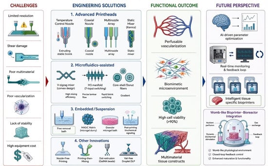
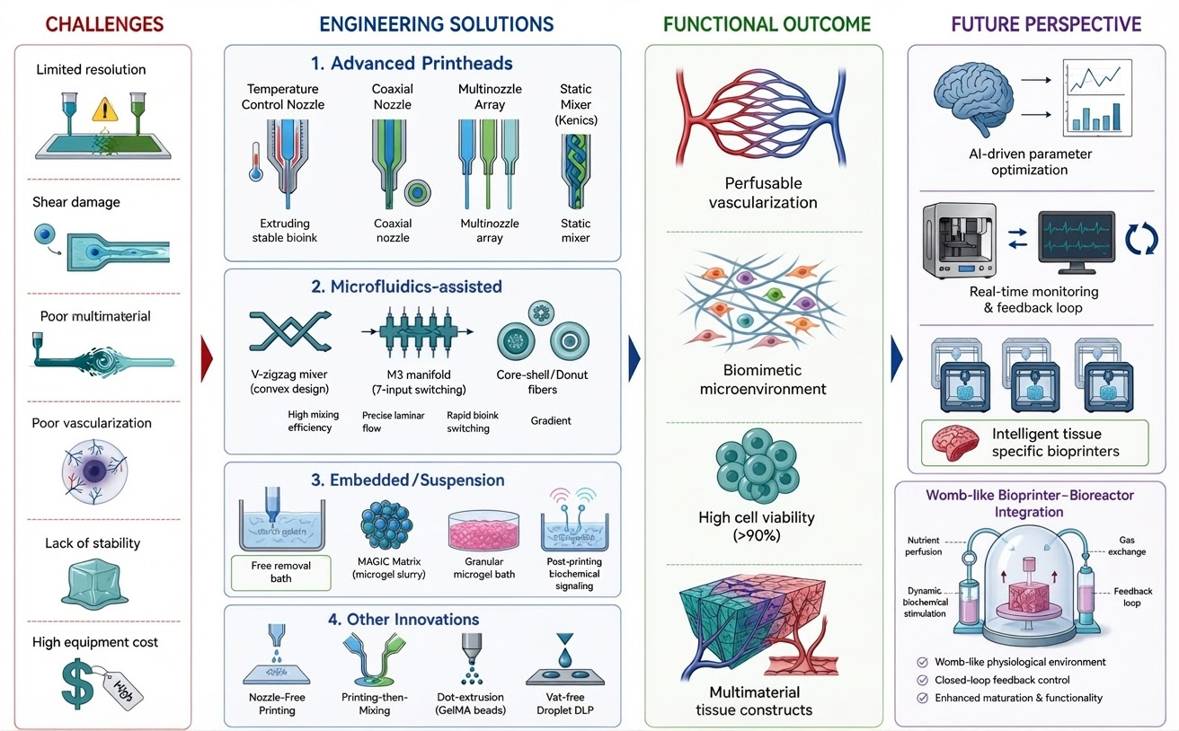

Disclaimer/Publisher’s Note: The statements, opinions and data contained in all publications are solely those of the individual author(s) and contributor(s) and not of MDPI and/or the editor(s). MDPI and/or the editor(s) disclaim responsibility for any injury to people or property resulting from any ideas, methods, instructions or products referred to in the content. |
© 2026 by the authors. Licensee MDPI, Basel, Switzerland. This article is an open access article distributed under the terms and conditions of the Creative Commons Attribution (CC BY) license.