


Abstract
Leptosphaeriaceae, a diverse family in the order Pleosporales, is remarkable for its scleroplectenchymatous or plectenchymatous peridium cells. Four Leptosphaeriaceae species were discovered and studied during the investigation of saprobic fungi from plant substrates in China. Novel taxa were defined using multiloci phylogenetic analyses and are supported by morphology. Based on maximum likelihood (ML) and Bayesian inference (BI) analyses, these isolates represent three novel taxa and one new record within Leptosphaeriaceae. A new genus, Angularia, is introduced to accommodate Angularia xanthoceratis, with a synopsis chart for 15 genera in Leptosphaeriaceae. This study also revealed a new species, Plenodomus changchunensis, and a new record of Alternariaster centaureae-diffusae. These species add to the increasing number of fungi known from China.
1. Introduction
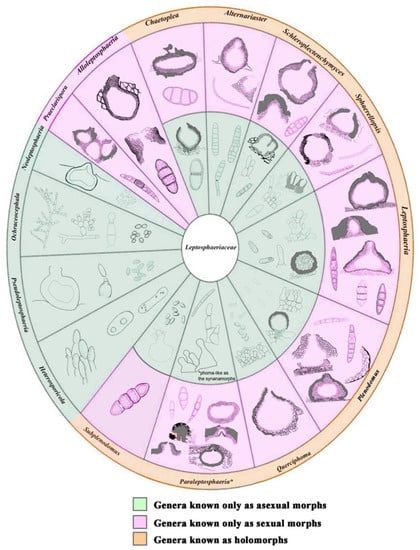
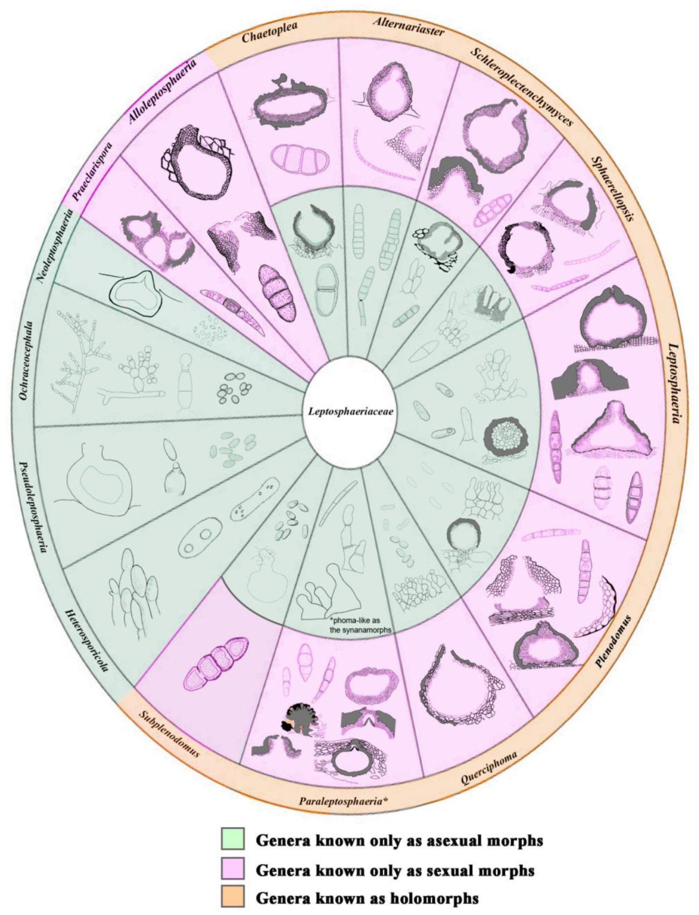
Leptosphaeriaceae is an important group of fungi in the order Pleosporales [1,2,3,4,5,6]. Leptosphaeriaceae was segregated from Pleosporaceae by Barr (1987) and was typified by Leptosphaeria Ces. & De Not. [1,2,3]. This family is characterized by conical or globose ascomata, scleroplectenchymatous or plectenchymatous peridium cells, cylindrical to oblong pedicellate asci, and septate reddish-brown or yellowish-brown ascospores (Figure 1) [2,4,7,8,9,10,11,12,13,14]. Although Leptosphaeriaceae is similar to Phaeosphaeriaceae, the peridium structure is morphologically distinguishable [15]. Most Leptosphaeriaceae species occur abundantly on dicotyledons, and the asexual morph can be coelomycetous (coniothyrium-like or phoma-like) or hyphomycetous [12,16,17]. Members of Leptosphaeriaceae are saprobes, hemibiotrophs, and pathogens [18,19,20,21,22]. Five genera Curreya, Didymolepta, Heptamaeria, Leptosphaeria, and Ophiobolus were previously included in the family [1]. Hyde et al. [2] accepted Heterosporicola, Leptosphaeria, Neophaeosphaeria, Paraleptosphaeria, Plenodomus, and Subplenodomus in the family by integrating molecular data. Simmons [23] introduced Alternariaster to accommodate Alternariaster helianthi (=Alternaria helianthi) as the first hyphomycetous record for Leptosphaeriaceae. Trakunyingcharoen et al. [24] subsequently introduced Sphaerellopsis from Dianthus caryophyllus and Vachellia karroo. The family was revised based on morphological characteristics and phylogenetic evidence, and ten genera were accepted [4]. Several other genera have also been added to Leptosphaeriaceae, such as Heterosporicola, Ochraceocephala, Querciphoma, Sclerenchymomyces, and Praeclarispora [8,12,13,14].
Figure 1.
Morphology of ascomata, conidiomata, ascospores, and conidiogenous cells; and conidia of 15 genera in Leptosphaeriaceae. Asterisk (*) indicates the genera with synanamorphs asexual characters.
Preuss (1851) introduced Plenodomus, which was typified by P. rabenhorstii [25]. The Plenodomus species belong to Leptosphaeriaceae and are one of the members with phoma-like taxa [2,5,17]. The type material of P. rabenhorstii was lost, and therefore P. lingam (Tode) Hohn. (Sexual morph: Leptosphaeria maculans (Desm.) Ces. & De Not.) was replaced as the type species of Plenodomus [26]. Phoma-like taxa were previously classified into nine sections including Plenodomus based on morphological characteristics [27,28]. de Gruyter et al. [29] determined that the Plenodomus section was distinct from Phoma sensu stricto based on phylogenetic analyses and classified Phoma under Didymellaceae. The Plenodomus species are the causal agents of diverse diseases on different plants throughout the world [30,31]. Plenodomus species are also isolated as saprobes on dead branches and stems of plants [17].
Alternariaster was introduced by Simmons [23] to accommodate Alternaria helianthi, a causal agent of leaf spots of Helianthus annuus (sunflower) worldwide [23,32,33]. This genus was segregated from Alternaria based on different conidial morphology. Alves et al. [8] confirmed that Alternariaster is a member of Leptosp haeriraceae and is distinct from Alternaria (Pleosporaceae). Four species have been reported in Alternariaster, including A. bidentis [16], A. centaureae-diffusae [4], A. helianthi [23], and A. trigonosporus [2]. Alternariaster helianthi has been reported worldwide as a pathogen of leaf spots on sunflowers, and Alternariaster bidentis was reported only from Brazil, whereas Alternariaster centaureae-diffusae and Alternariaster trigonosporus were reported from Russia [2,4]. This genus has been associated with Bidens sulphurea, Centaurea diffusa, Cirsium sp., and Helianthus annuus [2,4,16,23].
In this study, we introduce one new genus (Angularia), two new species (Angularia xanthoceratis and Plenodomus changchunensis), and one new record of Alternariaster centaureae-diffusae collected from China. The species were compared morphologically with other Leptosphaeriaceae species. Phylogenetic analyses were performed to confirm the taxonomic position based on maximum likelihood and Bayesian inference of combined LSU, SSU, ITS, and tub2 datasets.
2. Materials and Methods
2.1. Sample Collection and Isolation
The dried stems of Xanthoceras sorbifolium Bunge, Poaceae, and Clematis L. were collected from Changchun, Jilin Province and Kunming, Yunnan Province, China. The samples were preserved in plastic bags with labels describing location, date, host, and collection details. Pure fungal colonies were obtained using single spore isolation [34]. Germinating spores were transferred aseptically to potato dextrose agar (PDA), and the cultures were incubated at 25 °C. The specimens and pure cultures were deposited in the Herbarium of Mycology, Jilin Agricultural University (HMJAU), Changchun, China and International Cooperation Research Center of China for New Germplasm Breeding of Edible Mushrooms Culture Collection (CCMJ), respectively. The new taxa were registered in Mycobank [35].
2.2. Morphological Observation
Ascomata and conidiomata characteristics of the hosts were observed using a Zeiss Stemi 2000C stereomicroscope equipped with a Leica DFC450C digital camera (Leica, Wetzlar, Germany). Hand sections of the ascomata were carried out, and the sections were mounted on a slide with a drop of distilled water. Morphological characteristics were observed and photographed using a Zeiss AX10 light microscope equipped with an Axiocam 506 digital camera. Microscopic measurements were carried out using the ZEN 3.4 (blue edition) program (ZEISS, Jena, Germany). Adobe Photoshop CC2020 (Adobe Systems, San Jose, CA, USA) was used to process the images.
2.3. DNA Extraction, PCR Amplification and Sequencing
DNA was extracted from pure culture using a NuClean PlantGen DNA Kit (CWBIO, China) following the manufacturer’s instructions. Polymerase chain reaction (PCR) was used for the amplification of the large subunit (LSU), small subunit (SSU), internal transcribed spacer regions (ITS), β-tubulin (tub2), and the RNA polymerase II second largest subunit (rpb2). The LSU gene was amplified with the primers LROR and LR5 [36]; the SSU gene was amplified with the primers NS1 and NS4 [37]; the nuclear ITS was amplified with the primers ITS5 and ITS4 [37]; the tub2 gene was amplified with primers T1 and Bt2b [38]; and the rpb2 gene was amplified with primers RPB2-5f2 and fRPB2-7cr [39]. The amplification reactions were performed using 20 μL PCR mixtures containing 9 μL sterilized water, 10 μL of 2 × Es Taq MasterMix (Dye), 0.3 μL (10 μM) of forward and reverse primers, and 0.4 μL (200 ng/μL) of DNA template. The PCR conditions for LSU, SSU, ITS, and tub2 were as follows: 94 °C for 5 min, then 35 cycles of denaturation at 94 °C for 30 s, annealing at 53 °C for 45 s, elongation at 72 °C for 90 s, and a final extension at 72 °C for 10 min. All the PCR products were visualized on 1% agarose gels stained with standard DNA dye.
2.4. Phylogenetic Analysis
The sequence data were assembled using BioEdit v.7.2.5 [40] The closest matches for the new strains were obtained by using BLASTn searches (accessed on 13 December 2021, http://www.blast.ncbi.nlm.nih.gov/), and reference sequence data were downloaded from recent publications (Table 1) [41,42]. Didymella exigua (CBS 183.55) and D. rumicicola (CBS 683.79) were selected as the outgroup taxa. The sequences were aligned by using MAFTT version 7 (accessed on 7 March 2022, mafft.cbrc.jp/alignment/server) [43], and ambiguous nucleotides were manually adjusted by visual examination in AliView where necessary [44]. Leading or trailing gaps beyond the primer binding site were trimmed from the alignments prior to phylogenetic analyses, and the alignment gaps were treated as missing data.
Table 1.
Taxa and GenBank accession numbers used in the phylogenetic analyses. The extypes are shown in bold, and newly generated sequences are shown in blue.
Phylogenetic analyses of individual and multiloci phylogenetic analyses (ITS, LSU, SSU, and tub2) were performed to determine the phylogenetic placement of the isolated taxa. Maximum likelihood analysis was performed using RAxML-HPC2 on XSEDE on the CIPRES web portal (accessed on 7 March 2022, http://www.phylo.org/portal2/) [45,46,47]. The GTR + GAMMA model of nucleotide evolution was used for the datasets, and RAxML rapid bootstrapping of 1000 replicates was performed. The best-fit evolutionary models for individual and combined datasets were estimated under the Akaike Information Criterion (AIC) using jModeltest 2.1.10 on the CIPRES web portal for posterior probability [48]. The GTR model was the best model for all the datasets. Bayesian inference analyses were performed using MrBayes v. 3.2.6 on the CIPRES web portal [49]. Simultaneous Markov chains were run for seven million generations, and trees were sampled every 100th generations.
FigTree v. 1.4 [50] was used to visualize phylogenetic trees. The phylogram was edited by using Adobe Illustrator CS v. 6. All newly generated sequences were deposited in GenBank. All the alignments and trees were deposited in TreeBASE (Submission ID: 29394 and 29395).
3. Results
3.1. Phylogenetic Analyses
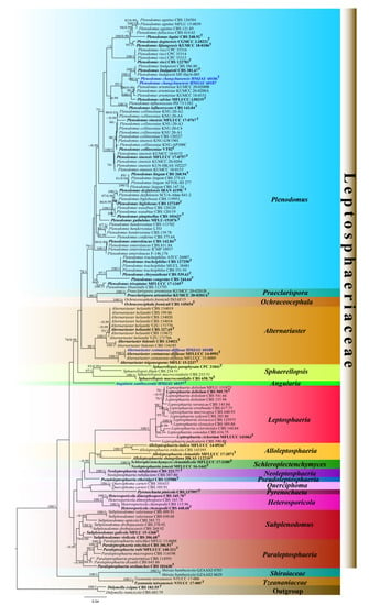
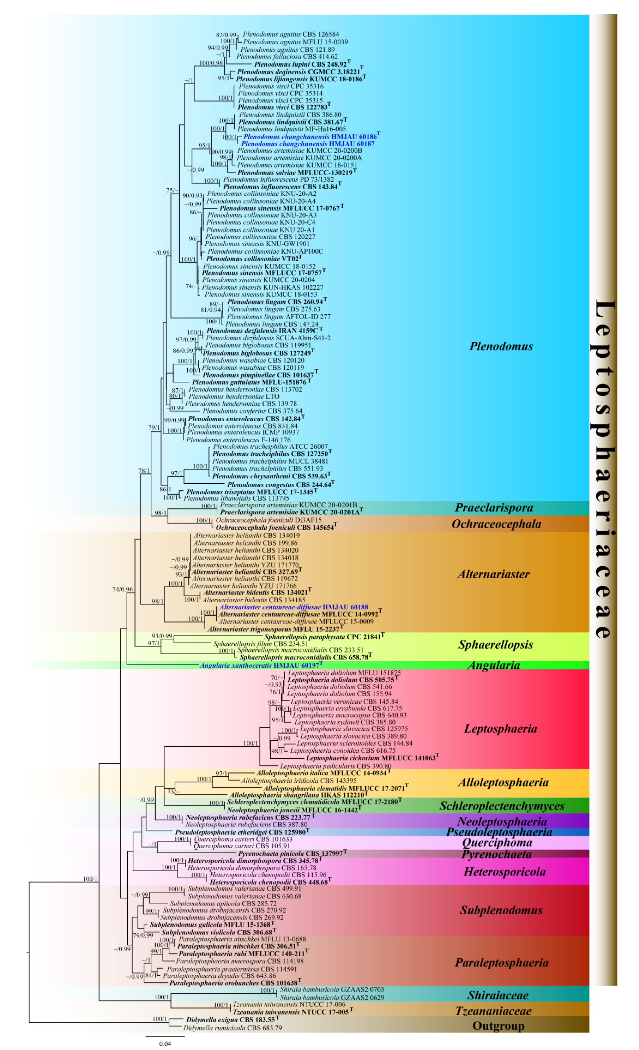
The combined LSU, SSU, ITS, and tub2 datasets comprised 138 strains, including our newly sequenced strains. Multiloci data were concatenated, which comprised 2958 characteristics, including gaps (ITS: 1–643, LSU: 644–1509, SSU: 1510–2573, and tub2: 2574–2970). The RAxML analysis yielded a best scoring tree (Figure 2) with a final ML optimization likelihood value of −19828.46. The matrix had 928 distinct alignment patterns, with 39.78% undetermined characteristics or gaps. Estimated base frequencies were as follows: A = 0.240304, C = 0.229231, G = 0.271334, and T = 0.259131; substitution rates AC = 1.321448, AG = 2.815733, AT = 1.680962, CG = 0.694608, CT = 5.562821, and GT = 1.000000; proportion of invariable sites I = 0.704486; and gamma distribution shape parameter α = 0.555544. Phylogenetic trees generated from the Bayesian and maximum likelihood analyses had similar topologies (Figure 2 and Figure S1). However, in the Bayesian analysis, Alloleptosphaeria shangrilana did not cluster within the Alloleptosphaeria clade, but was sister to the Schleroplectenchymyces species with low support (0.72 BPP). The MLBP values (left) and BPP values (right) are provided near each node (Figure 2). For the Bayesian analysis, a total of 10,338 trees were sampled after the 20% burn-in with a stop value of 0.009971.
Figure 2.
The best scoring RAxML tree of Leptosphaeriaceae based on a concatenated ITS, LSU, SSU, and tub2 datasets. The tree is rooted with Didymella exigua (CBS 183.55) and D. rumicicola (CBS 683.79). RAxML bootstrap support values ≥70% (ML, left) and Bayesian posterior probabilities ≥0.90 (BPP, right) are shown near the nodes. The new isolates are in blue. The type strains are in bold and marked with T.
Leptosphaeriaceae was strongly supported in the maximum likelihood and Bayesian analyses (100% ML/1.00 BPP). Within Leptosphaeriaceae, Heterosporicola, Leptosphaeria, Neoleptosphaeria, Ochraceocephala, Praeclarispora, Querciphoma, and Schleroplectenchymyces strongly supported clades (100% ML/1.00 BPP) were formed. Alternariaster (98% ML/1.00 BPP) and Sphaerellopsis (97% ML/1.00 BPP) formed strongly supported clades, while Alloleptosphaeria and Plenodomus were only moderately supported in the maximum likelihood analyses (73% ML and 79% ML, respectively). The newly introduced genus formed an independent lineage basal to Sphaerellopsis with 35% ML/0.81 BPP support. A new genus Angularia is therefore introduced within Leptosphaeriaceae. The newly generated taxa Plenodomus changchunensis (HMJAU 60186 and HMJAU 60187) clustered with Plenodomus lindquistii with 100% ML/1.00 BPP support, while the strain HMJAU 60188 formed a strongly supported clade with Alternariaster centaureae-diffusae taxa (Figure 2).
3.2. Taxonomy
Angularia R. Xu, Phukhams. & Y. Li, gen. nov.
MycoBank Number: 843307.
Etymology: referring to the angular peridium of the type species.
Description:Saprobic on decaying wood or herbaceous plant material in terrestrial habitats. Sexual morph: Undetermined. Asexual morph: Conidiomata pycnidial, solitary, sometimes aggregated, uniloculate, immersed in host substrate, dark brown to brown, globose, coriaceous. Ostioles absent. Conidiomatal wall thick-walled, multilayered, scleroplectenchymatous cells thick at base, composed of textura angularis, lined with a thick hyaline layer bearing conidiogenous cells. Conidiophores reduced to conidiogenous cells. Conidiogenous cells enteroblastic, phialidic, determinate, discrete, subcylindrical to truncate, smooth-walled, hyaline, arising from the inner layers of conidiomata. Conidia fusiform, truncate at both ends, aseptate, hyaline, smooth.
Type species: Angulariaxanthoceratis R. Xu, Phukhams. & Y. Li.
Notes: Angularia is introduced for a strongly supported lineage comprising Angularia xanthoceratis (1.00 BPP, Figure 2). Angularia formed a distinct lineage to Alternariaster, Ochraceocephala, Plenodomus, Praeclarispora and Sphaerellopsis based on multiloci phylogenetic analyses. For individual loci, Angularia formed a sister clade distinct from Heterosporicola (ITS) and formed a sister clade distinct from Pseudoleptosphaeria_etheridgei (LSU). Leptosphaeriaceae species are remarkable for having superficial to semi-immersed, shiny ascomata or conidiomata, with thick, multilayers of scleroplectenchymatous or pseudoparenchymatous tissue types [4]. The fungus has semi-immersed to immersed conidiomata, black, with a multilayer scleroplectenchymatous-type tissue (Figure 3). Angularia is similar to Plenodomus and Alternariaster in having peridium with scleroplectenchymatous cells [4]. Angularia is also similar to Plenodomus and Sphaerellopsis in having textura angularis cells in the conidiomatal wall [4,24]. However, Angularia and Ochraceocephala differ substantially in morphology. Ochraceocephala has long and branched conidiophores, and the branching is commonly irregularly verticillate, while the conidiophores of Angularia are reduced to conidiogenous cells. Ochraceocephala has hyaline to yellowish, mostly sand to olive yellow, and mostly globose to subglobose conidia, while Angularia has hyaline and fusiform conidia; the conidia are smaller than in our new genus (4.8 vs. 18.7 × 3.6 vs. 5.4 μm).
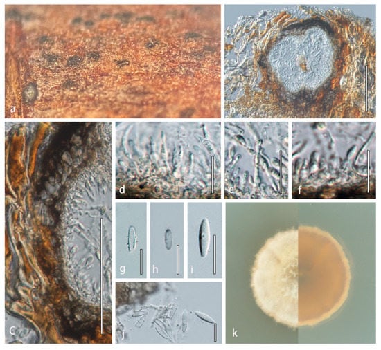
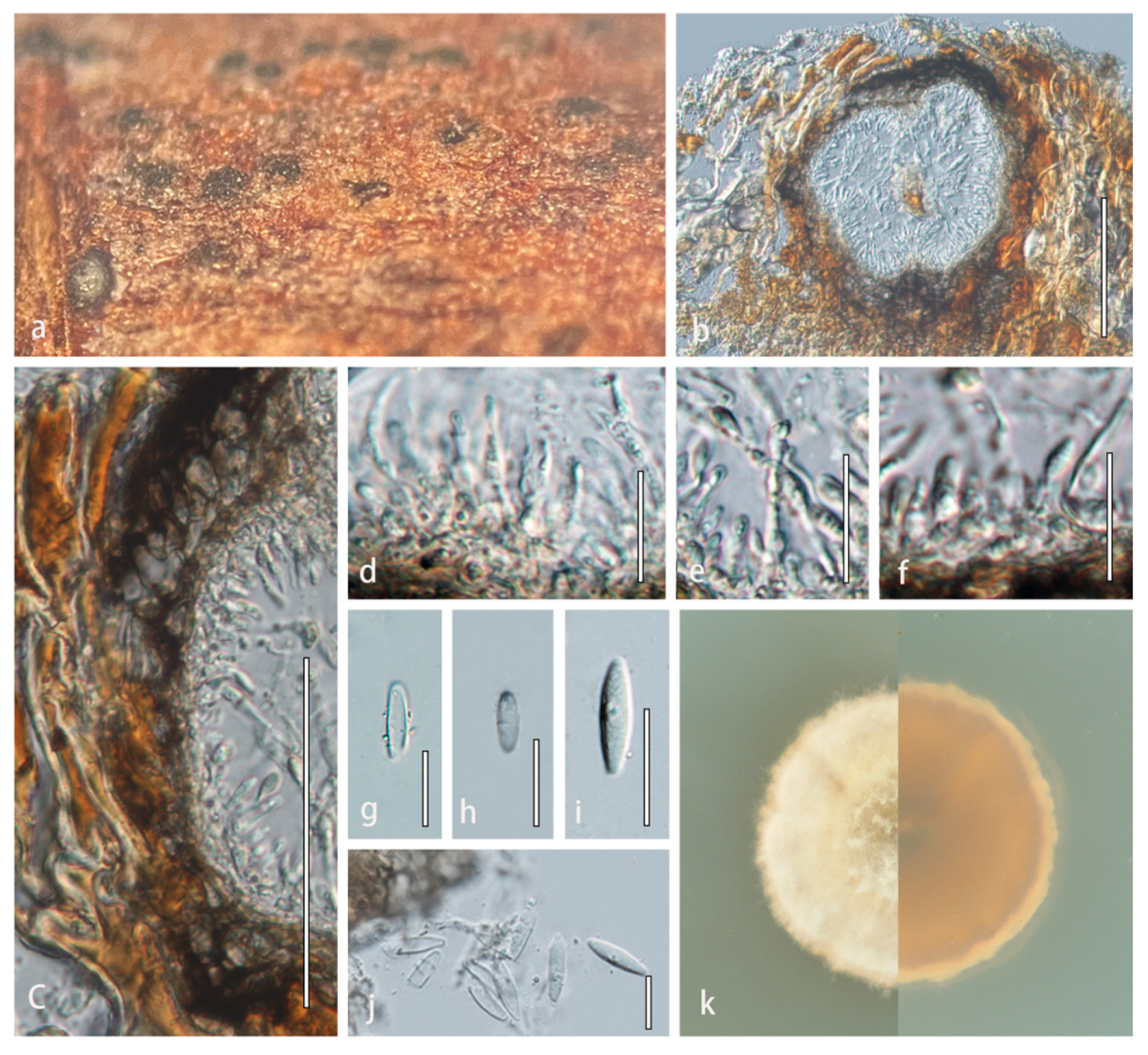
Figure 3.
Angularia xanthoceratis (HMJAU 60197, holotype). (a) Appearance of conidiomata on host substrate. (b) Vertical section of conidioma. (c) Section of conidioma wall. (d–f) Conidiogenous cells and conidia. (g–j) Conidia. (k) Culture characteristics on PDA after two weeks at 25 °C. Scale bars: (b) = 100 µm; (c) = 50 µm; and (d–j) = 20 µm.
MycoBank Number: 843308.
Etymology: referring to the host genus, Xanthoceras.
Holotype: HMJAU 60197.
Description: Saprobic on dead stems of Xanthoceras sorbifolium. Sexual morph: Undetermined. Asexual morph: Conidiomata 180–220 × 195–224 μm ( = 200 × 210 μm, n = 5), pycnidial, solitary, aggregated, uniloculate, immersed in host substrate, globose, thick-walled, subcoriaceous to coriaceous at the outer layers, dark brown to brown, without distinct ostioles. Ostioles absent. Conidiomatal wall 20–46 μm wide, thick, multilayered, scleroplectenchymatous cells, outer layer composed of 6–8 layers of dark brown to brown cells of textura angularis, lined with a thick hyaline layer bearing conidiogenous cells. Conidiophores reduced to conidiogenous cells. Conidiogenous cells 7.8–20.8 × 1.7–3.5 μm ( = 14.3 × 2.6 μm, n = 20), enteroblastic, phialidic, determinate, discrete, subcylindrical to truncate, smooth-walled, hyaline, arising from the inner layers of conidiomata. Conidia 13–24.5 × 4–7 μm ( = 18.7 × 5.4 μm, n = 30), fusiform, truncate at both ends, aseptate, hyaline, smooth-walled.
Culture characteristics: Colonies on PDA reaching 20 mm in diameter after 2 weeks at 25 °C. Cultures from above, dome-shaped in the center, milky white radiating outward, dense, round, creeping hyphae; reverse dark at the center, light orange radiating outward.
Material examined: CHINA, Jilin Province, Changchun, on dead stem of Xanthoceras sorbifolium (Sapindaceae), 15 September 2021, Rong Xu, HMJAU 60197 (holotype); extype living culture, CCMJ5013.
GenBank accession numbers: LSU = OM295682, SSU = OM295681, ITS = OM295683, and tub2 = OM304358
Notes: Angularia xanthoceratis is distinct from the closely related Sphaerellopsis species in conidial characteristics (Figure 3). Angularia xanthoceratis has fusiform, smooth-walled, hyaline, aseptate conidia, which are truncate at both ends, while Sphaerellopsis has fusoid-ellipsoidal, occasionally Y-shaped or digitate, subcylindrical to ellipsoid or globose, pale brown, 0–1(−3)-euseptate conidia [24]. In a BLASTn search, the LSU sequence of Angularia xanthoceratis was 99.55% similar to Leptosphaeria etheridgei (CBS 125980) with 96% query cover which translates to 95.6% similarity. The ITS region was 97.44% similar to Leptosphaeria sp. (Ct-BC63) with 82% query cover which translates to 79.9% similarity. A pairwise comparison of the ITS region revealed 119 bases pair differences (18.39%) between A. xanthoceratis and Sphaerellopsis macroconidialis, while the tub2 region was 98 bases pair different (24.62%).
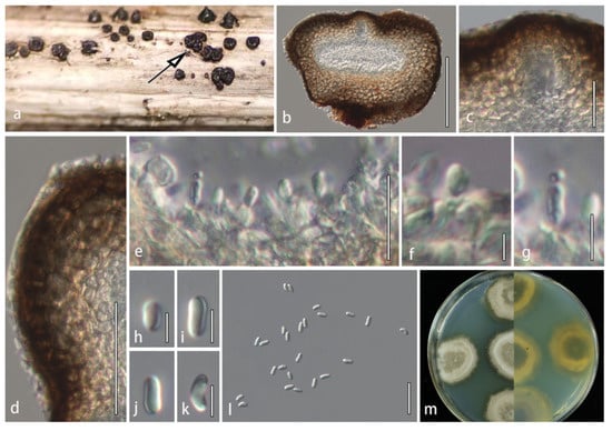
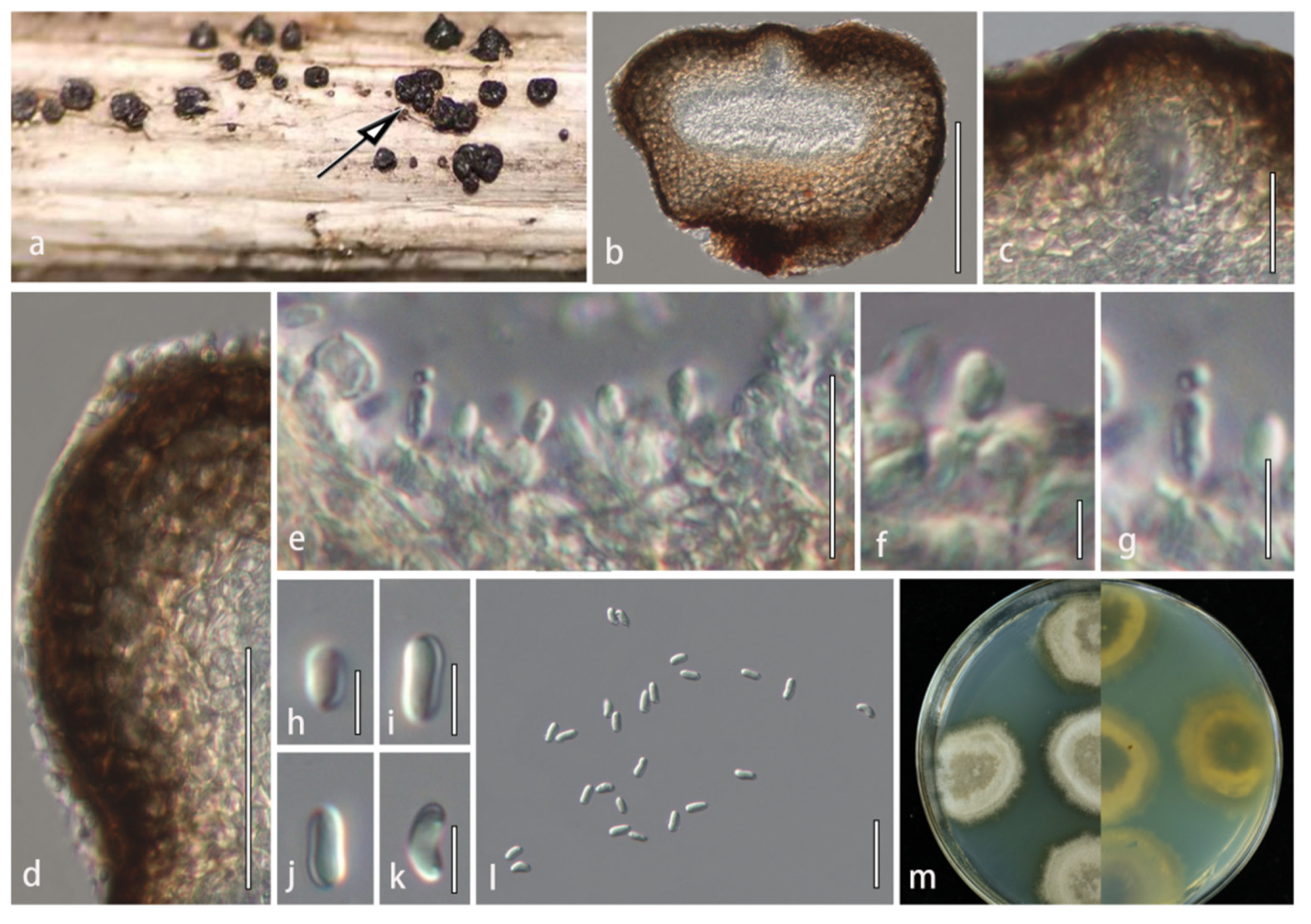
Figure 4.
Plenodomus changchunensis (HMJAU 60186, holotype). (a) Appearance of conidiomata on host substrate; black arrow indicates the conidiomata of P. changchunensis on the host. (b) Vertical section of conidioma. (c) Ostiolar canal. (d) Section of conidioma wall. (e–g) Conidiogenous cells and conidia. (h–l) Conidia. (m) Culture characteristics on PDA after three weeks at 25 °C. Scale bars: (b) = 100 µm; (c,e,l) = 20 µm; (d) = 50 µm; and (f–k) = 5 µm.
MycoBank Number: 843304
Holotype: HMJAU 60186
Etymology: referring to Changchun city where this fungus was collected.
Description: Saprobic on dead stems of Poaceae. Sexual morph: Undetermined. Asexual morph: Conidiomata 163–192 × 193–245 μm (x = 175 × 207 μm, n = 5), pycnidial, solitary or in groups of 2–5, erumpent, aggregated, globose to subglobose, depression in the middle, thick-walled, subcoriaceous to coriaceous at the outer layers, dark brown to black, ostiolate. Ostioles 20–45 μm, central, papillate, ovoid, filled with short periphyses. Conidiomatal wall 24–48 μm wide, thick, multilayered, outer layer composed of 8–10 layers of dark brown to brown cells of textura angularis, lined with a thick hyaline layer bearing conidiogenous cells. Conidiophores reduced to conidiogenous cells. Conidiogenous cells 2.8–5.8 × 1.5–2.8 μm (x = 4.1 × 2 μm, n = 30), enteroblastic, phialidic, determinate, smooth-walled, hyaline. Conidia 5–7.6 × 2–3.4 μm (x = 6.2 × 2.7 μm, n = 50), oblong or oval, slightly curved toward the ends, rounded ends, aseptate, hyaline, smooth-walled.
Culture characteristics: Colonies on PDA reaching 30 mm diam. after 3 weeks at 25 °C. Cultures from above, gray in the center, milky white radiating outward, dense, circular, creeping hyphae, grayish-green at the margins; reverse dark at the center, milky white radiating outward. Yellow pigmentation diffused into the media.
Material examined: CHINA. Jilin Province: Changchun, on dead twigs of Poaceae sp., 20 May 2021, C. Phukhamsakda, HMJAU 60186 (holotype); extype living culture, CCMJ5011; HMJAU 60187 (isotype), ex-isotype living culture, CCMJ5012.
GenBank accession numbers: LSU = OL897174, SSU = OL984031, ITS = OL996123, and tub2 = OM009247
Notes: Plenodomus changchunensis (CCMJ5011 and CCMJ5012) formed a sister clade distinct from Plenodomus lindquistii with 99% ML/1.00 BPP support based on phylogenetic analysis of the concatenated ITS, LSU, SSU, and tub2 datasets (Figure 2). Plenodomus changchunensis is similar to P. lindquistii in the size of conidia [51]. This species can be distinguished from P. lindquistii (CBS 381.67) by 34 nucleotides in the ITS region (34/643 in the ITS region and 0/866 in the LSU region). In the BLASTn search, the closest match to the LSU and ITS sequences of P. changchunensis were 100% and 89.57% similar to Leptosphaeria sp. (PHY-30) and P. lindquistii (MCN535002) with 95% query cover which translates to a 95% and 85.1% similarity, respectively. Plenodomus changchunensis was found associated with a grass near the water resources in temperate regions. Therefore, this fungus is introduced as a novel species.
Alternariaster centaureae-diffusae R.H. Perera, Bulgakov, Ariyawansa & K.D. Hyde, in Fungal Diversity, 74: 32 (2015), new host record and new geological record (Figure 5)
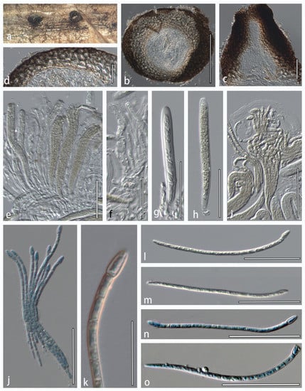
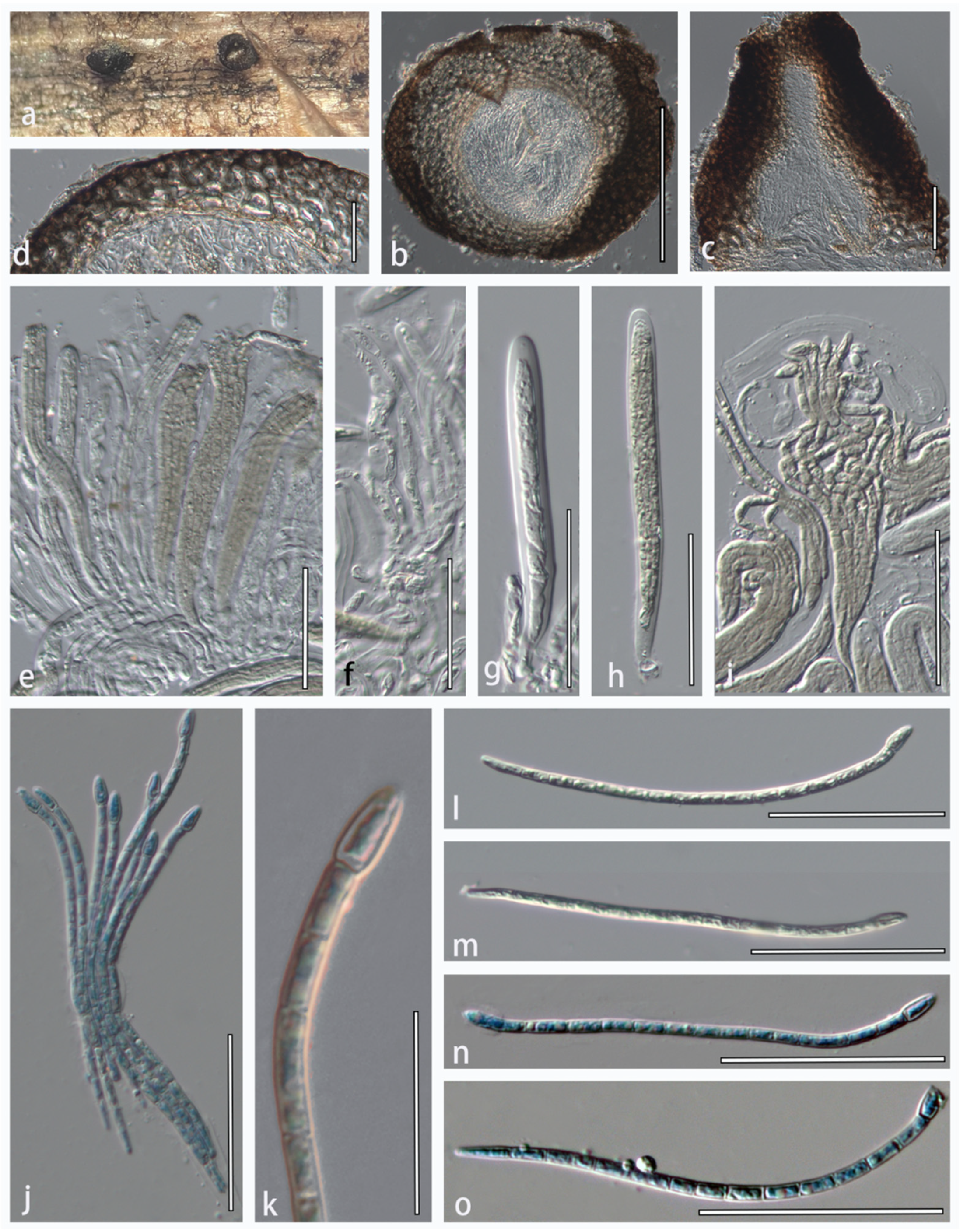
Figure 5.
Alternariaster centaureae-diffusae (HMJAU 60188). (a) Appearance of ascomata on host substrate. (b) Vertical section of ascoma. (c) Ostiole with periphyses. (d) Close-up of peridium. (e,g,h) Immature and mature asci. (f) Pseudoparaphyses. (i,j) Fissitunicate asci. (k) Top part of ascospore. (l–o) Ascospores. (j,n,o) Ascospores were stained in cotton blue. Scale bars: (b) = 200 μm; (c,d,f–j,l–o) = 50 μm; (e) = 100 μm; and (k) = 20 μm.
Index Fungorum Identifier: IF551462
Description: Saprobic on dried stems of Clematis sp. Sexual morph: Ascomata 170–360 × 146–290 μm diam., solitary or in groups of 2–10, erumpent, semi-immersed or nearly superficial, uniloculate, globose to subglobose, coriaceous, black, ostiolate. Ostiole papillate, black, filled with periphyses. Periphyses aseptate, with a blunt apex, hyaline. Peridium 40–75 μm wide (x = 57.5 μm, n = 10), comprising thick-walled cells of textura globularis, inner layer composed of flattened cells of textura angularis, 5–10 rows of scleroplectenchymatous cells, outer layer thick, black. Hamathecium 2.5–3.8 μm wide, dense, distinctly septate, branched, cellular pseudoparaphyses, hyaline, embedded in a gelatinous matrix. Asci 110–140 × 10–14 μm (x = 125 × 12 μm, n = 20), 8-spored, bitunicate, fissitunicate, cylindrical to cylindric-subclavate, with a short bulbous pedicel, rounded at the apex. Ascospores 80–138 × 2.3–4.3 μm (x = 109 × 3.3 μm, n = 40), fasciculate, filiform, 14–16-septate, constricted at the apical septum, apical cell swollen, conical, yellowish-brown, smooth-walled, with a mucilaginous cap. Asexual morph: Undetermined.
Material examined: CHINA, Yunnan Province, dead aerial branch of Clematis spp., 24 April 2021, (HMJAU 60188).
Host associations: Centaurea diffusa, Clematis spp. ([4] and this study).
GenBank accession numbers: LSU = OL897175, SSU = OL891810, ITS = OL996125, and tub2 = OL898721
Notes: Alternariaster centaureae-diffusae was originally described from the dead stems of Centaurea diffusa Lam. in Russia [4]. The new isolate (HMJAU 60188) has similar morphology to the type strain of A. centaureae-diffusae (MFLU 15–1521) in having fasciculate, filiform, constricted at the apical septum, conical, yellowish-brown ascospores with swollen apical cell [4]. A pairwise comparison of the sequences of the new isolate (HMJAU 60188) with the type species of A. centaureae-diffusae revealed minor differences. The new isolate clustered in the same clade as the type strain of A. centaureae-diffusae (Figure 2). Therefore, we report A. centaureae-diffusae on Clematis spp. as a new host and new geological record.
4. Discussion
Molecular biology has helped to elucidate the phylogenetic relationships among members of Dothideomycetes, particularly among several phoma-like taxa [13,52]. Multi-loci analyses based on LSU, SSU, ITS, tub2, rpb2, and tef-1 sequences have been widely used to define species boundaries in Leptosphaeriaceae and other families of Dothideomycetes [13,52,53]. We carried out phylogenetic analyses with a concatenated dataset of five loci (ITS, LSU, SSU, tub2, and rbp2) for Leptosphaeriaceae members. The final alignment included 138 strains representing 132 ingroup taxa and six outgroup strains. However, the Plenodomus species were polyphyletic and mixed with Alternariaster, Ochraceocephala, and Praeclarispora taxa. It is often encouraged to use additional taxon-specific secondary barcode loci to delineate taxa. We therefore compared the phylogenetic informativeness of tub2 (52 sequences translated to 37.7%) and rpb2 (46 sequences translated to 33.3%) sequences of Leptosphaeriaceae. Our study shows that the polyphyletic topology of the Plenodumus group is due to the rpb2 gene (Figures S2–S4). This could be due to a lack of rpb2 barcodes in several related taxa, but the rpb2 gene can be useful for delineation at the genus level [12,41]. In contrast, using the tub2 gene provides a better resolution at the species level within the genera (Figure 2). Therefore, we performed phylogenetic analyses of Leptosphaeriaceae species with a concatenated dataset of ITS, LSU, SSU, and tub2 loci. Three new species of Leptosphaeriaceae were revealed from China based on multilocus phylogeny combined with morphology.
The phylogeny from our analyses is similar to several previous studies [4,12,13]. The Leptosphaeriaceae taxa clustered in fifteen clades based on the ITS, LSU, SSU, and tub2 datasets. A novel genus Angularia is also introduced in Leptosphaeriaceae to accommodate a new species, A. xanthoceratis. Conidial characteristics are the primary morphological characteristics that distinguish Angularia from the allied genus Sphaerellopsis (Figure 1). Plenodomus formed a separate clade, sister to Ochraceocephala, and revealed a novel species P. changchunensis with strong support. Many new genera have been introduced in Leptosphaeriaceae [2,4,8,12,13,14,23], which indicates that this family has a high degree of fungal diversity and distribution.
Plenodomus lingam was chosen to be the representative type species of Plenodomus over P. rabenhorstii Preuss [14,54]. There are 36 epithets listed under Plenodomus in Species Fungorum (2022) and 107 epithets in MycoBank. The host specificity of Plenodomus has not yet been clarified as species have been recorded from various plant families (Asteraceae, Fabaceae, Lamiaceae, and Liliaceae) [9]. In our study, P. changchunensis was found on Poaceae, which suggests that the Leptosphaeriaceae species are widely associated with many types of substrates. Members of Plenodomus appear to be cosmopolitan, as they have been recorded in both temperate and tropical countries (China, Greece, France, Japan, Netherlands, Peru, and Spain) [55].
Alternariaster centaureae-diffusae has been isolated from Centaurea diffusa Lam. (Asteraceae) in Shakhty city, Rostov region, Russia [4]. In this study, it was isolated from Clematis spp. (Ranunculaceae) in Kunming, Yunnan province, China. Therefore, our study extended the host range of A. centaureae-diffusae even though the environment of the two cities is different (temperate and subtropical). Therefore, we speculate that this species could be found in different environments and hosts [56].
Fungal diversity and taxonomy are constantly changing, necessitating a continuous assessment [57,58,59]. It is especially significant where taxa are described from genera that usually accommodate pathogens [60,61]. For example, Plenodomus and Alternariaster are the causal agents of blackleg disease and leaf spots of Helianthus annuus (sunflower) worldwide [31,32,62,63]. The discovery of novel species in a pathogenic genus could also indicate the discovery of emerging pathogens that can cause damage to economically important crops [64,65]. The formation of new fungi species has been reported to be intricately linked to their evolutionary relationships and ecological roles [20]. These phenomena can also occur when species are associated with different hosts and environments, as in the case of A. centaureae-diffusae in this study. The presence of the Alternariaster and Plenodomus species in different substrates reflects their ecological importance. Further studies focusing on fungal diversity from different niches are needed to understand the relationships between these organisms in ecosystems.
Supplementary Materials
The following are available online at https://www.mdpi.com/article/10.3390/jof8050416/s1, Figure S1: Phylogram generated from Bayesian inference analysis based on combined ITS, LSU, SSU, and tub2 sequence data. Figure S2: Phylogram generated from maximum likelihood analysis based on combined ITS, LSU, SSU, tub2, and rpb2 sequence data. Figure S3: Phylogram generated from maximum likelihood analysis based on combined ITS, LSU, SSU, and rpb2 sequence data. Figure S4: Phylogram generated from maximum likelihood analysis using rpb2 sequence data. Figure S5: Phylogram generated from maximum likelihood analysis using tub2 sequence data.
Author Contributions
Conceptualization, Y.L. and C.P.; Writing—original draft and formal analysis, R.X.; Data curation, R.X. and W.S.; Investigation, R.X. and C.P.; Methodology, R.X., W.S. and S.T. (Shangqing Tian); Supervision, K.D.H., Y.L. and C.P.; Writing—review and editing, R.X., Y.L., C.S.B., S.T. (Saowaluck Tibpromma) and C.P.; funding acquisition, Y.L. and C.P. All authors have read and agreed to the published version of the manuscript.
Funding
This research was funded by the National Natural Science Foundation of China (no. 32100007) and the Program of Creation and Utilization of Germplasm of Mushroom Crop of “111” Project (no. D17014).
Institutional Review Board Statement
Not applicable.
Informed Consent Statement
Not applicable.
Data Availability Statement
All sequences generated in this study were submitted to GenBank. The accession number for the rpb2 gene for the new taxon Plenodomus changchunensis (HMJAU 60187) is OL944508.
Acknowledgments
Xu Rong and Chayanard Phukhamsakda (Postdoctoral number 271007) would like to thank Jilin Agricultural University, National Natural Science Foundation of China (NSFC) for granting a Youth Science Fund Project (number 32100007) and the Program of Creation and Utilization of Germplasm of Mushroom Crop of “111” Project (No. D17014). The authors would like to thank Shaun Pennycook from Manaaki Whenua, Landcare Research, New Zealand.
Conflicts of Interest
The authors declare no conflict of interest.
References
- Barr, M.E. New taxa and combinations in the loculoascomycetes. Mycotaxon 1987, 29, 501–505. [Google Scholar]
- Hyde, K.D.; Jones, E.B.G.; Liu, J.K.; Ariyawansa, H.A.; Boehm, E.; Boonmee, S.; Braun, U.; Chomnunti, P.; Crous, P.W.; Dai, D.Q.; et al. Families of Dothideomycetes. Fungal Divers. 2013, 63, 1–313. [Google Scholar] [CrossRef] [Scilit]
- Hyde, K.D.; Hongsanan, S.; Jeewon, R.; Bhat, D.J.; McKenzie, E.H.C.; Jones, E.B.G.; Phookamsak, R.; Ariyawansa, H.A.; Boonmee, S.; Zhao, Q.; et al. Fungal diversity notes 367–490: Taxonomic and phylogenetic contributions to fungal taxa. Fungal Divers. 2016, 80, 1–270. [Google Scholar] [CrossRef] [Scilit]
- Ariyawansa, H.A.; Phukhamsakda, C.; Thambugala, K.M.; Bulgakov, T.S.; Wanasinghe, D.N.; Perera, R.H.; Mapook, A.; Camporesi, E.; Kang, J.C.; Jones, E.B.G.; et al. Revision and phylogeny of Leptosphaeriaceae. Fungal Divers. 2015, 74, 19–51. [Google Scholar] [CrossRef] [Scilit]
- Wijayawardene, N.N.; Crous, P.W.; Kirk, P.M.; Hawksworth, D.L.; Boonmee, S.; Braun, U.; Chomnunti, P.; Dai, D.Q.; D’souza, M.J.; Diederich, P.; et al. Naming and outline of Dothideomycetes–2014 including proposals for the protection or suppression of generic names. Fungal Divers. 2014, 69, 1–55. [Google Scholar] [CrossRef] [Scilit]
- Zhang, Y.; Crous, P.W.; Schoch, C.L.; Hyde, K.D. Pleosporales. Fungal Divers. 2012, 52, 1–225. [Google Scholar] [CrossRef] [Scilit]
- Barr, M.E. Some dictyosporous genera and species of Pleosporales in North America. Mem. New York Bot. Gard. 1990, 62, 1–92. [Google Scholar]
- Crous, P.W.; Groenewald, J.Z. The genera of Fungi–G 4: Camarosporium and Dothiora. IMA Fungus 2017, 8, 131–152. [Google Scholar] [CrossRef] [Scilit]
- Phookamsak, R.; Hyde, K.D.; Jeewon, R.; Bhat, D.J.; Jones, E.B.G.; Maharachchikumbura, S.S.N.; Raspé, O.; Karunarathna, S.C.; Wanasinghe, D.N.; Hongsanan, S.; et al. Fungal diversity notes 929–1035: Taxonomic and phylogenetic contributions on genera and species of fungi. Fungal Divers. 2019, 95, 1–273. [Google Scholar] [CrossRef] [Scilit]
- Phookamsak, R.; Liu, J.K.; McKenzie, E.H.C.; Manamgoda, D.S.; Ariyawansa, H.; Thambugala, K.M.; Dai, D.Q.; Camporesi, E.; Chukeatirote, E.; Wijayawardene, N.N.; et al. Revision of Phaeosphaeriaceae. Fungal Divers. 2014, 68, 159–238. [Google Scholar] [CrossRef] [Scilit]
- Piątek, M.; Rodriguez-Flakus, P.; Domic, A.; Palabral-Aguilera, A.N.; Gómez, M.I.; Flakus, A. Phylogenetic placement of Leptosphaeria polylepidis, a pathogen of Andean endemic Polylepis tarapacana, and its newly dis-covered mycoparasite Sajamaea mycophila gen. et sp. nov. Mycol. Prog. 2020, 19, 1–14. [Google Scholar] [CrossRef] [Scilit]
- Aiello, D.; Vitale, A.; Polizzi, G.; Voglmayr, H. Ochraceocephala foeniculi gen. et sp. nov., a new pathogen causing crown rot of fennel in Italy. MycoKeys 2020, 66, 1–22. [Google Scholar] [CrossRef] [Scilit]
- Phukhamsakda, C.; McKenzie, E.H.C.; Phillips, A.J.L.; Jones, E.B.G.; Jayarama Bhat, D.; Stadler, M.; Bhunjun, C.S.; Wanasinghe, D.N.; Thongbai, B.; Camporesi, E.; et al. Microfungi associated with Clematis (Ranunculaceae) with an integrated approach to delimiting species boundaries. Fungal Divers. 2020, 102, 1–203. [Google Scholar] [CrossRef] [Scilit]
- Doilom, M.; Hyde, K.D.; Dong, W.; Liao, C.F.; Suwannarach, N.; Lumyong, S. The plant family Asteraceae is a cache for novel fungal diversity: Novel species and genera with remarkable ascospores in Leptosphaeriaceae. Front. Microbiol. 2021, 12, 660261. [Google Scholar] [CrossRef] [Scilit]
- Câmara, M.P.S.; Palm, M.E.; van Berkum, P.; O’Neill, N.R. Molecular phylogeny of Leptosphaeria and Phaeosphaeria. Mycologia 2002, 94, 630–640. [Google Scholar] [CrossRef]
- Alves, J.L.; Woudenberg, J.H.C.; Duarte, L.L.; Crous, P.W.; Barreto, R.W. Reappraisal of the genus Alternariaster (Dothideomycetes). Persoonia 2013, 31, 77. [Google Scholar] [CrossRef] [Scilit]
- de Gruyter, J.; Woudenberg, J.H.C.; Aveskamp, M.M.; Verkley, G.J.M.; Groenewald, J.Z.; Crous, P.W. Redisposition of phoma-like anamorphs in Pleosporales. Stud. Mycol. 2013, 75, 1–36. [Google Scholar] [CrossRef] [Scilit]
- Dayarathne, M.C.; Phookamsak, R.; Ariyawansa, H.A.; Jones, E.B.G.; Camporesi, E.; Hyde, K.D. Phylogenetic and morphological appraisal of Leptosphaeria italica sp. nov. (Leptosphaeriaceae, Pleosporales) from Italy. Mycosphere 2015, 6, 634–642. [Google Scholar] [CrossRef] [Scilit]
- Tennakoon, D.S.; Phookamsak, R.; Wanasinghe, D.N.; Yang, J.B.; Lumyong, S.; Hyde, K.D. Morphological and phylogenetic insights resolve Plenodomus sinensis (Leptosphaeriaceae) as a new species. Phytotaxa 2017, 324, 73–82. [Google Scholar] [CrossRef] [Scilit]
- Hyde, K.D.; de Silva, N.; Jeewon, R.; Bhat, D.J.; Phookamsak, R.; Doilom, M.; Boonmee, S.; Jayawardena, R.S.; Maharachchikumbura, S.S.N.; Senanayake, I.C.; et al. AJOM new records and collections of fungi: 1–100. Asian J. Mycol. 2020, 3, 22–294. [Google Scholar] [CrossRef] [Scilit]
- Jones, E.B.G.; Suetrong, S.; Sakayaroj, J.; Bahkali, A.H.; Abdel-Wahab, M.A.; Boekhout, T.; Pang, K.L. Classification of marine Ascomycota, Basidiomycota, Blastocladiomycota and Chytridiomycota. Fungal Divers. 2015, 73, 1–72. [Google Scholar] [CrossRef] [Scilit]
- Liu, J.K.; Hyde, K.D.; Jones, E.B.G.; Ariyawansa, H.A.; Bhat, J.D.; Boonmee, S.; Maharachchikumbura, S.S.N.; McKenzie, E.H.C.; Phookamsak, R.; Phukhamsakda, C.; et al. Fungal Diversity Notes 1–110: Taxonomic and phylogenetic contributions to fungal species. Fungal Divers. 2015, 72, 1–197. [Google Scholar] [CrossRef] [Scilit]
- Simmons, E.G. Alternaria: An identification manual. In Biodiversity No 6; CBS Fungal Diversity Centre: Utrecht, The Netherlands, 2007. [Google Scholar]
- Trakunyingcharoen, T.; Lombard, L.; Groenewald, J.Z.; Cheewangkoon, R.; To-anun, C.; Alfenas, A.C.; Crous, P.W. Mycoparasitic species of Sphaerellopsis, and allied lichenicolous and other genera. IMA Fungus 2014, 5, 391–414. [Google Scholar] [CrossRef] [Scilit]
- Preuss, C.G.T. Ubersicht untersuchter pilze, besonders aus der umgegend Hoyerswerda. In Linnaea 1851, 24, 99–153. [Google Scholar]
- Torres, M.S.; Bergen, M.; Singh, S.; Bischoff, J.; Sullivan, R.F.; White, J.F. Plenodomus morganjonesii sp. nov. and a discussion of the genus Plenodomus. Mycotaxon 2005, 93, 333–344. [Google Scholar]
- Boerema, G.H.; de Gruyter, J.; Noordeloos, M.E. Contributions towards a monograph of Phoma (Coelomycetes)—IV. Section: Taxa with large sized conidial dimorphs, in vivo sometimes as Stagonosporopsis synanamorphs. Persoonia 1997, 16, 335–371. [Google Scholar]
- Boerema, G.H.; de Gruyter, J.; Noordeloos, M.E.; Hamers, M.E.C. Phoma Identification Manual: Differentiation of Specific and Infra-Specific Taxa in Culture; CABI Publishing: Wallingford, UK, 2004. [Google Scholar]
- de Gruyter, J.; Aveskamp, M.M.; Woudenberg, J.H.C.; Verkley, G.J.M.; Groenewald, J.Z.; Crous, P.W. Molecular phylogeny of Phoma and allied anamorph genera: Towards a reclassification of the Phoma complex. Mycol. Res. 2009, 113, 508–519. [Google Scholar] [CrossRef] [Scilit] [PubMed]
- Marin-Felix, Y.; Groenewald, J.Z.; Cai, L.; Chen, Q.; Marincowitz, S.; Barnes, I.; Bensch, K.; Braun, U.; Camporesi, E.; Damm, U.; et al. Genera of phytopathogenic fungi: GOPHY 1. Stud. Mycol. 2017, 86, 99–216. [Google Scholar] [CrossRef] [Scilit] [PubMed]
- Zhao, P.; Crous, P.W.; Hou, L.W.; Duan, W.J.; Cai, L.; Ma, Z.Y.; Liu, F. Fungi of quarantine concern for China I: Dothideomycetes. Persoonia 2021, 47, 45–105. [Google Scholar] [CrossRef] [Scilit]
- Alcorn, J.L.; Pont, W. Alternaria helianthi on sunflower. Australas. Plant Path. 1972, 1, 30. [Google Scholar] [CrossRef] [Scilit]
- Ribeiro, I.J.O.; Filho, O.M.; Soave, J.; Corvellini, G.D.S. Ocorrência de Alternaria Helianthi (Hansf.) Tubaki et Nishihara sobre girassol (Heljanthus annuus L.). Bragantia 1974, 33, 81–85. [Google Scholar] [CrossRef] [Scilit]
- Senanayake, I.C.; Rathnayaka, A.R.; Marasinghe, D.S.; Calabon, M.S.; Gentekaki, E.; Lee, H.B.; Hurdeal, V.G.; Pem, D.; Dissanayake, L.S.; Wijesinghe, S.N.; et al. Morphological approaches in studying fungi: Collection, examination, isolation, sporulation and preservation. Mycosphere 2020, 11, 2678–2754. [Google Scholar] [CrossRef] [Scilit]
- Crous, P.W.; Gams, W.; Stalpers, J.A.; Robert, V.; Stegehuis, G. MycoBank: An online initiative to launch mycology into the 21st century. Stud. Mycol. 2004, 50, 19–22. [Google Scholar]
- Vilgalys, R.; Hester, M. Rapid genetic identification and mapping of enzymatically amplified ribosomal DNA from several Cryptococcus species. J. Bacteriol. 1990, 172, 4238–4246. [Google Scholar] [CrossRef] [Scilit]
- White, T.J.; Bruns, T.D.; Lee, S.B.; Taylor, J.W. Amplification and direct sequencing of fungal ribosomal RNA genes for phylogenetics. PCR Protoc. Guid. Methods Appl. 1990, 18, 315–322. [Google Scholar]
- O’Donnell, K.; Cigelnik, E. Two divergent intragenomic rDNA ITS2 types within a monophyletic lineage of the fungus Fusarium are nonorthologous. Mol. Phylogenet. Evol. 1997, 7, 103–116. [Google Scholar] [CrossRef] [Scilit] [PubMed]
- Liu, A.; Hall, W.J. Unbiased estimation following a group sequential test. Biometrika 1999, 86, 71–78. [Google Scholar] [CrossRef] [Scilit]
- Hall, T.A. BioEdit: A user-friendly biological sequence alignment editor and analysis program for windows 95/98/NT. Nucleic Acids Symp. Ser. 1999, 41, 95–98. [Google Scholar]
- Thiyagaraja, V.; Wanasinghe, D.N.; Karunarathna, S.C.; Tennakoon, D.S.; Hyde, K.D.; To-anun, C.; Cheewangkoon, R. Alloleptosphaeria shangrilana sp. nov. and first report of the genus (Leptosphaeriaceae, Dothideomycetes) from China. Phytotaxa 2021, 491, 12–22. [Google Scholar] [CrossRef] [Scilit]
- Safi, A.; Mehrabi-Koushki, M.; Farokhinejad, R. Plenodomus dezfulensis sp. nov. causing leaf spot of Rapeseed in Iran. Phytotaxa 2021, 523, 141–154. [Google Scholar] [CrossRef] [Scilit]
- Katoh, K.; Standley, D.M. MAFFT multiple sequence alignment software version 7: Improvements in performance and usability. Mol. Biol. Evol. 2013, 30, 772–780. [Google Scholar] [CrossRef] [Scilit]
- Larsson, A. AliView: A fast and lightweight alignment viewer and editor for large datasets. Bioinformatics 2014, 30, 3276–3278. [Google Scholar] [CrossRef] [Scilit]
- Stamatakis, A. RAxML-VI-HPC: Maximum likelihood-based phylogenetic analyses with thousands of taxa and mixed models. Bioinformatics 2006, 22, 2688–2690. [Google Scholar] [CrossRef] [Scilit]
- Stamatakis, A.; Hoover, P.; Rougemont, J. A rapid bootstrap algorithm for the RAxML web servers. Syst. Biol. 2008, 57, 758–771. [Google Scholar] [CrossRef] [Scilit]
- Stamatakis, A. RAxML version 8: A tool for phylogenetic analysis and post-analysis of large phylogenies. Bioinformatics 2014, 30, 1312–1313. [Google Scholar] [CrossRef] [Scilit] [PubMed]
- Nylander, J.A.A. MrModeltest 2.0; Program Distributed by the Author; Uppsala University: Uppsala, Sweden, 2004. [Google Scholar]
- Ronquist, F.; Huelsenbeck, J.P. MrBayes 3: Bayesian phylogenetic inference under mixed models. Bioinformatics 2003, 19, 1572–1574. [Google Scholar] [CrossRef] [Scilit] [PubMed]
- Rambaut, A.; Drummond, A. FigTree: Tree Figure Drawing Tool; Version 1.2.2; Institute of Evolutionary Biology, University of Edinburgh: Edinburgh, UK, 2008. [Google Scholar]
- Duan, W.-J.; Li, L.; Mo, S.-M.; Duan, L.-J.; Cai, L. Identification of the quarantine fungus Plenodomus lindquistii from the sunflower seeds imported from Kazakhstan. J. Plant Protect. 2015, 42, 795–800. [Google Scholar]
- Hongsanan, S.; Hyde, K.D.; Phookamsak, R.; Wanasinghe, D.N.; Mckenzie, E.; Sarma, V.V.; Boonmee, S.; Lücking, R.; Pem, D.; Bhat, D.J.; et al. Refined families of Dothideomycetes: Dothideomycetidae and Pleosporomycetidae. Mycosphere 2020, 11, 1553–2107. [Google Scholar] [CrossRef] [Scilit]
- Bhunjun, C.S.; Phukhamsakda, C.; Jeewon, R.; Promputtha, I.; Hyde, K.D. Integrating different lines of evidence to establish a novel Ascomycete genus and family (Anastomitrabeculia, Anastomitrabeculiaceae) in Pleosporales. J. Fungi 2021, 7, 94. [Google Scholar] [CrossRef] [Scilit] [PubMed]
- Kirk, P.M.; Cannon, P.F.; Minter, D.W.; Staplers, J.A. Dictionary of the Fungi, 10th ed.; CABI Bioscience: Wallingford, UK, 2008. [Google Scholar]
- Farr, D.F.; Rossman, A.Y. Fungal Databases, U.S. National Fungus Collections, ARS, USDA. 2021. Available online: https://nt.ars-grin.gov/fungaldatabases (accessed on 15 January 2022).
- Chaiwan, N.; Gomdola, D.; Wang, S.; Monkai, J.; Tibpromma, S.; Doilom, M.; Wanasinghe, D.N.; Mortimer, P.E.; Lumyong, S.; Hyde, K.D. https://gmsmicrofungi.org: An online database providing updated information of microfungi in the Greater Mekong Subregion. Mycosphere 2021, 12, 1513–1526. [Google Scholar] [CrossRef] [Scilit]
- Bhunjun, C.S.; Niskanen, T.; Suwannarach, N.; Wannathes, N.; Chen, Y.J.; McKenzie, E.H.C.; Maharachchikumbura, S.S.N.; Buyck, B.; Zhao, C.L.; Fan, Y.G.; et al. The numbers of fungi: Are the most speciose genera truly diverse? Fungal Divers. 2022. [Google Scholar] [CrossRef] [Scilit]
- Phukhamsakda, C.; Nilsson, R.H.; Bhunjun, C.S.; Gomes de Farias, A.R.; Sun, Y.R.; Wijesinghe, S.N.; Raza, M.; Bao, D.F.; Lu, L.; Tibpromma, S.; et al. The numbers of fungi; contributions from traditional taxonomic studies and challenges of metabarcoding. Fungal Divers. 2022; in press.
- Wijayawardene, N.N.; Hyde, K.D.; Al-Ani, L.K.T.; Tedersoo, L.; Haelewaters, D.; Rajeshkumar, K.C.; Zhao, R.L.; Aptroot, A.; Leontyev, D.V.; Saxena, R.K.; et al. Outline of Fungi and fungus-like taxa. Mycosphere 2020, 11, 1060–1456. [Google Scholar] [CrossRef] [Scilit]
- Bhunjun, C.S.; Phukhamsakda, C.; Jayawardena, R.S.; Jeewon, R.; Promputtha, I.; Hyde, K.D. Investigating species boundaries in Colletotrichum. Fungal Divers. 2021, 107, 107–127. [Google Scholar] [CrossRef] [Scilit]
- Bhunjun, C.S.; Dong, Y.; Jayawardena, R.S.; Jeewon, R.; Phukhamsakda, C.; Bundhun, D.; Hyde, K.D.; Sheng, J. A polyphasic approach to delineate species in Bipolaris. Fungal Divers. 2020, 102, 225–256. [Google Scholar] [CrossRef] [Scilit]
- Fitt, B.D.L.; Brun, H.; Barbetti, M.J.; Rimmer, S.R. World-wide importance of Phoma stem canker (Leptosphaeria maculans and L. biglobosa) on oilseed rape (Brassica napus). Eur. J. Plant Pathol. 2006, 114, 3–15. [Google Scholar] [CrossRef] [Scilit]
- Jayawardena, R.S.; Hyde, K.D.; Jeewon, R.; Ghobad-Nejhad, M.; Wanasinghe, D.N.; Liu, N.G.; Phillips, A.J.L.; Oliveira-Filho, J.R.C.; da Silva, G.A.; Gibertoni, T.B.; et al. One stop shop II: Taxonomic update with molecular phylogeny for important phytopathogenic genera: 26–50 (2019). Fungal Divers. 2019, 94, 41–129. [Google Scholar] [CrossRef] [Scilit]
- Jayawardena, R.S.; Hyde, K.D.; McKenzie, E.H.C.; Jeewon, R.; Phillips, A.J.L.; Perera, R.H.; de Silva, N.I.; Maharachchikumbura, S.S.N.; Samarakoon, M.C.; Ekanayake, A.H.; et al. One stop shop III: Taxonomic update with molecular phylogeny for important phytopathogenic genera: 51–75 (2019). Fungal Divers. 2019, 98, 77–160. [Google Scholar] [CrossRef] [Scilit]
- Zhang, Y.; Jeewon, R.; Fournier, J.; Hyde, K.D. Multi-gene phylogeny and morphotaxonomy of Amniculicola lignicola: Novel freshwater fungus from France and its relationships to the Pleosporales. Mycol. Res. 2008, 112, 1186–1194. [Google Scholar] [CrossRef] [Scilit]

Publisher’s Note: MDPI stays neutral with regard to jurisdictional claims in published maps and institutional affiliations. |
© 2022 by the authors. Licensee MDPI, Basel, Switzerland. This article is an open access article distributed under the terms and conditions of the Creative Commons Attribution (CC BY) license (https://creativecommons.org/licenses/by/4.0/).