


Abstract
Postharvest soft rot is a major constraint in kiwifruit production, causing substantial economic losses and raising food safety concerns. Therefore, the development of sustainable biocontrol alternatives to conventional chemical treatments is urgently needed. In this study, thirty-seven fungi were isolated from healthy kiwifruit tissues and rhizosphere soil and screened for their biocontrol potential. Three highly effective antagonistic strains were ultimately identified: Mucor circinelloides N2-1-1, Aspergillus niger Pb-2-2, and Trichoderma hamatum Nd-1-1. In vitro antagonism assays showed that A. niger Pb-2-2 exhibited the strongest and broadest-spectrum inhibitory activity against four postharvest pathogens, namely Fusarium sp., Alternaria sp., Botryosphaeriaceae sp., and Phomopsis sp. In contrast, in vivo protection assays demonstrated that T. hamatum Nd-1-1 almost provided the highest control efficacy against all four pathogens in kiwifruit fruit. Fermentation broth protection experiments further revealed that A. niger Pb-2-2 produced stable and broad-spectrum antimicrobial metabolites. Although M. circinelloides N2-1-1 showed the weakest antagonistic activity among the three fungi, this is the first report indicating that M. circinelloides has antagonistic activity against phytopathogenic fungi. Physiological analyses indicated that all three antagonistic fungi alleviated the inhibitory effects of pathogens on superoxide dismutase, catalase, and ascorbate peroxidase activities, helped maintain reactive oxygen species homeostasis, and activated the phenylpropanoid pathway, thereby enhancing disease resistance in kiwifruit. These findings provide a theoretical basis and promising microbial resources for the biological control of postharvest soft rot in kiwifruit.
1. Introduction
Kiwifruit (Actinidia chinensis), a prominent species of the Actinidiaceae family within the order Ericales, is not only native to China but has also emerged as a globally economically important crop, celebrated for its superior nutritional profile and proven benefits for digestive, immune, and metabolic health [1,2]. However, the sustainable expansion of the kiwifruit industry is currently facing a critical bottleneck of fungal soft rot. This disease has evolved into a persistent and devastating epidemic across China’s major production regions, with incidence rates escalating to 40% to 50% [3]. As a primary limiting factor, soft rot causes precipitous economic losses and threatens the long-term viability of the sector.
The etiology of this disease is complex; Fusarium sp., Alternaria sp., Phomopsis sp., and Botryosphaeriaceae sp. have been identified as the predominant pioneer pathogens [4,5,6,7], often co-occurring with other agents such as Lasiodiplodia theobromae, Neofusicoccum parvum, Penicillium expansum, and Botrytis cinerea [8,9,10]. Currently, disease management remains heavily reliant on synthetic fungicides. Agents such as copper succinate, Amobam, hexaconazole, and trifloxystrobin-tebuconazole combinations are widely deployed [11,12,13]. Notably, the combination of calcium and azoxystrobin has shown efficacy, achieving control rates of up to 67.24% under optimal conditions. Concurrently, alternative strategies involving gaseous signaling molecules like hydrogen sulfide (H2S) and natural antifungal agents like natamycin have demonstrated potential in inducing systemic resistance or exerting direct antifungal activity [14,15]. Despite these interventions, the long-term, intensive application of chemical fungicides has precipitated severe adverse consequences, including the emergence of pathogen resistance and the disruption of the orchard micro-ecological balance. This has triggered a vicious cycle of disease recurrence and chemical escalation, exacerbating environmental contamination and raising alarming food safety concerns regarding residue accumulation. In response to the growing global imperative for ecological safety and food quality, there is an urgent need to transition toward environmentally sound and economically viable biological control strategies.
Recent progress in biological control has focused largely on bacteria and yeasts. For instance, the fermentation broth of the endophytic bacterium Bacillus amyloliquefaciens M9 and the cell-free supernatant of Bacillus subtilis BS-1 have shown efficacy in inhibiting Botryosphaeria dothidea and maintaining fruit quality [16,17]. Similarly, the yeast Meyerozyma guilliermondii 39 has proven effective as a postharvest agent by boosting host defense enzymes [18]. Although recent studies have demonstrated that postharvest biocontrol can suppress kiwifruit decay through direct antibiosis, nutrient and space competition, volatile or diffusible antifungal metabolites, and induction of host defense responses, the existing literature remains fragmented in several important respects. Most investigations have focused on a single bacterial or yeast antagonist and one dominant pathogen, particularly Botryosphaeria dothidea, Alternaria alternata, Botrytis cinerea, or Diaporthe sp., rather than addressing the pathogen complex associated with kiwifruit soft rot [15,19,20,21]. In addition, fungal antagonists remain substantially less explored than bacterial and yeast biocontrol agents. The available studies on the relationships among in vitro antagonism, in vivo fruit protection, fermentation-broth-mediated inhibition, and host defense responses within the same experimental framework remain insufficiently integrated.
Therefore, the present study aims to investigate antagonistic fungi isolated from kiwifruit tissues and rhizosphere soil, and to evaluate their biocontrol potential against four major pathogen groups associated with postharvest soft rot, including Fusarium sp., Alternaria sp., Botryosphaeriaceae sp., and Phomopsis sp. By integrating broad-spectrum antifungal assessment with analyses of antagonistic mechanisms and host-resistance induction, this study seeks to bridge the gap between the discovery of individual biocontrol strains and the development of a mechanistically supported, broad-spectrum biological control strategy for kiwifruit soft rot.
2. Materials and Methods
2.1. Isolation and Purification of Antagonistic Fungi from Kiwifruit Tissues and Rhizosphere Soil
Samples (leaves, bark and rhizosphere soil) were collected from organic orchards in Guizhou Province, China (no pesticides for >3 years). Plant tissues were surface-sterilized (1% NaOCl for 10 min, 75% ethanol for 30 s, sterile water rinse ×4), transferred to PDA (Hope-Bio, Qingdao, Shandong, China) plates, and incubated at 28 °C for 7 days [22]. Soil fungi were isolated using a dilution method [23]: 10 g soil in 90 mL water, shaken (180 rpm, 1 h), diluted (10−3–10−6 g/mL), plated on PDA (100 µL), incubated (28 °C, 7 days). Colonies were purified via monosporic isolation.
2.2. Screening for Antagonistic Fungi as Biocontrol Agents Through Dual-Culture Assays
The dual-culture assay [24] was used to screen antagonistic fungi. Four pathogenic fungal strains responsible for kiwifruit soft rot, namely Fusarium sp., Alternaria sp., Phomopsis sp. and Botryosphaeriaceae sp., were sourced from the culture collection of the authors’ laboratory, where they had been previously isolated and preserved. Pathogen mycelial plugs (5 mm) were placed on PDA plates with antagonistic fungal plugs inoculated 2 cm away. Controls contained only pathogens. All treatments were triplicated and incubated at 28 ± 2 °C for 5 days.
2.3. Determination of the Inhibition of Antagonistic Fungi on the Pathogenic Fungi In Vivo
Kiwifruits (Actinidia chinensis cv. Hongyang) at commercial maturity, weighing 80–100 g and free from mechanical damage, visible lesions, and surface defects, were selected and surface-disinfected with 1% (v/v) sodium hypochlorite for 10 min, followed by 75% (v/v) ethanol for 30 s, and rinsed three times with sterile distilled water. Uniform wounds (3 mm diameter × 3 mm depth) were created on the equatorial surface of each fruit using a sterile cork borer. For the treatment group, each wound was first inoculated with 10 μL of antagonist spore suspension (1 × 10⁷ spores/mL), whereas control wounds received no antagonist treatment. After incubation at ambient temperature for 2 h, 10 μL of pathogen spore suspension (1 × 10⁷ spores/mL) was applied to all wounds. Inoculated fruits were placed in plastic boxes and incubated at 28 ± 1 °C under 80–85% relative humidity. Each treatment consisted of three biological replicates with six fruits per replicate (18 fruits per treatment), and pathogen-only inoculated fruits served as the negative control. Lesion diameters were measured on days 3, 5, and 7 post-inoculation [25].
2.4. Antifungal Activity of Fermentation Broths Against Pathogenic Fungi
Fermentation broths from antagonistic fungi cultured in Sabouraud Dextrose Broth (SDB; Shanghai Bio-way technology Co., Ltd., Shanghai, China; 5 g/L tryptone, 5 g/L animal tissue digest,20 g/L glucose, pH 5.4–5.8), Potato Dextrose Broth (PDB; Shanghai Bio-way technology Co., Ltd., Shanghai, China; 5 g/L potato infusion powder, 20 g/L glucose, 1 g/L chloramphenicol, pH 5.8–6.2) and Martin-Modified Medium (MMM; Shanghai Bio-way technology Co., Ltd., Shanghai, China; 5 g/L peptone, 2 g/L yeast extract powder, 20 g/L glucose, 1 g/L K2HPO4, 0.5 g/L MgSO4, pH 6.2~6.6). For fermentation, 100 mL of each medium was dispensed into 250 mL Erlenmeyer flasks. A 5 mm diameter mycelial plug of purified antagonistic fungus was inoculated into each flask. The flasks were incubated in a rotary shaker at 180 rpm and 28 °C for 7 days. After fermentation, the crude culture liquid was first vacuum-filtered with a Buchner funnel to remove fungal mycelia and spores. The resulting filtrate was further sterilized using 0.22 μm membrane filters (Millipore, Burlington, MA, USA, GSWP02500) to obtain sterile fermentation broths. Kiwifruits (Actinidia chinensis cv. Hongyang) at commercial maturity, weighing 80–100 g and free from mechanical damage, visible lesions, and surface defects, were surface-disinfected with 0.1% (v/v) NaOCl for 1 min, followed by 75% (v/v) ethanol for 2–3 min, and rinsed with sterile distilled water. Uniform wounds (3 mm diameter × 3 mm depth) were created on the equatorial surface of each fruit. Each wound was first treated with 10 μL of sterile fermentation broth, while the control received an equal volume of sterile water. After 2 h at ambient temperature, 10 μL of pathogen spore suspension (1 × 10⁷ spores/mL) was applied to all wounds. Inoculated fruits were incubated at 28 °C under 90–95% relative humidity. Each treatment consisted of three biological replicates, each containing six fruits (18 fruits per treatment). Disease incidence and lesion diameters were recorded 7 days post-inoculation [26].
2.5. Enzyme Activities
Kiwifruit samples for enzyme assays were prepared following the spore suspension inoculation procedure described above, with three groups established for each assay: a blank control consisting of healthy, unwounded fruit without any inoculation, a pathogen-only control in which wounds were inoculated solely with the pathogen spore suspension, and a co-inoculation treatment in which wounds received the antagonist spore suspension first, followed by the pathogen after 2 h, exactly as in the in vivo inhibition assay. At each sampling time point (days 3, 5, and 7 post-inoculation), fruit tissues surrounding the wound site were excised, immediately snap-frozen in liquid nitrogen, and stored at −80 °C until analysis. For enzyme extraction, frozen tissue was homogenized on ice in a pre-chilled mortar with the appropriate extraction buffer and centrifuged at 12,000× g for 30 min at 4 °C. The supernatant was collected for spectrophotometric assays. Superoxide dismutase (SOD) was extracted from 2.0 g of tissue in 5 mL of ice-cold buffer (0.1 mM EDTA, 1 mM ascorbic acid, 2% PVPP, all reagents were purchased from Shanghai Macklin Biochemical Co., Ltd., Shanghai, China), with one unit defined as the amount of enzyme inhibiting the photochemical reduction in NBT by 50% per minute per gram of fresh weight [27]. Ascorbate peroxidase (APX) was extracted from 1.0 g of tissue in 5 mL of the same buffer, and one unit was defined as a decrease of 0.01 in absorbance at 290 nm per minute per gram of fresh weight [28]. Catalase (CAT) was extracted from 3.0 g of tissue in 5 mL of ice-cold buffer (5 mM DTT, 5% PVP, all reagents were purchased from Shanghai Macklin Biochemical Co., Ltd., Shanghai, China), with one unit corresponding to a decrease of 0.01 in absorbance at 240 nm per minute per gram of fresh weight [29]. Phenylalanine ammonia-lyase (PAL) was extracted from 4.0 g of tissue in 5 mL of ice-cold buffer (40 g/L PVP, 2 mM EDTA, 5 mM β-mercaptoethanol, Shanghai Yuanye Bio-Technology Co., Ltd., Shanghai, China), and one unit was defined as an increase of 0.01 in absorbance at 290 nm per hour per gram of fresh weight [30]. All enzyme activities were expressed as U/g fresh weight. Reactions were conducted at 25 °C in a total volume of 3 mL using 1 cm path-length cuvettes, with uniform illumination maintained for the SOD NBT photoreduction assay and continuous absorbance recording at the respective wavelengths for CAT, APX, and PAL to monitor activity changes.
2.6. Identification of Antagonistic Fungi from Kiwifruit Through Polyphasic Analysis
Mycelial plugs (5 × 5 mm) were inoculated on PDA plates, incubated at 28 °C for 5–7 days to observe colony characteristics. After 7–14 days of incubation, mycelia and spores were examined microscopically. Genomic DNA was extracted using the Rapid Fungi Genomic DNA Isolation Kit (Sangon Biotech, Shanghai, China). PCR amplification targeted the ITS region and specific primers (Table 1) using a 25 µL reaction mix: 1 µL DNA, 0.5 µL each primer, 12.5 µL Taq PCR Master Mix, and ddH2O. Cycling: 95 °C for 5 min; 35 cycles of 95 °C/30 s, 50–55 °C/30 s, 72 °C/1 min; 72 °C/5 min. PCR products were commercially sequenced, and the obtained sequences were subjected to homology searches usingthe BLASTN algorithm via the online NCBI BLAST server (https://blast.ncbi.nlm.nih.gov, accessed on 17 June 2026) with default parameters against the non-redundant (nr) nucleotide database. Reference sequences showing more than 90% similarity to the obtained sequences were selected for phylogenetic analysis. Sequence alignment was performed using MEGA 11. Different gene loci were concatenated in PhyloSuite, and a Bayesian phylogenetic tree was reconstructed using MrBayes based on the combined multilocus dataset [31]. The concatenation order was calmodulin + ITS for strain Pb-2-2, actin + ITS for strain Nd-1-1, and CFS + mcm7 + ITS for strain N2-1-1.
Table 1.
Primers and their sequences for the identification of endophytic fungal species.
2.7. Statistical Analysis
All statistical analyses were performed to evaluate the significance of the observed effects. The lesion diameter and enzyme activity data were analyzed with GraphPad Prism (version 9.5, GraphPad Software, San Diego, CA, USA) using a two-way ANOVA, factoring in treatment and time, followed by Tukey’s multiple range test at p < 0.05.
3. Results
3.1. Isolation and Screening of Kiwifruit Soft Rot Antagonistic Fungi
Dual-culture assays showed that, among the 37 fungal isolates obtained from kiwifruit, N2-1-1, Nd-1-1, and Pb-2-2 displayed the strongest and most consistent antagonistic activity against the four soft rot pathogens tested, including Fusarium sp., Alternaria sp., Botryosphaeriaceae sp., and Phomopsis sp. (Table 2).
Table 2.
Inhibition Rates of Antagonistic Fungi Against Kiwifruit Soft Rot Pathogens through Dual-Culture Plate Assay.
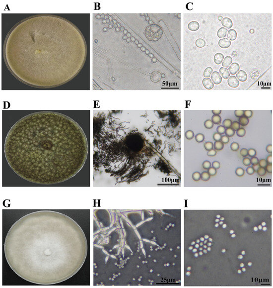
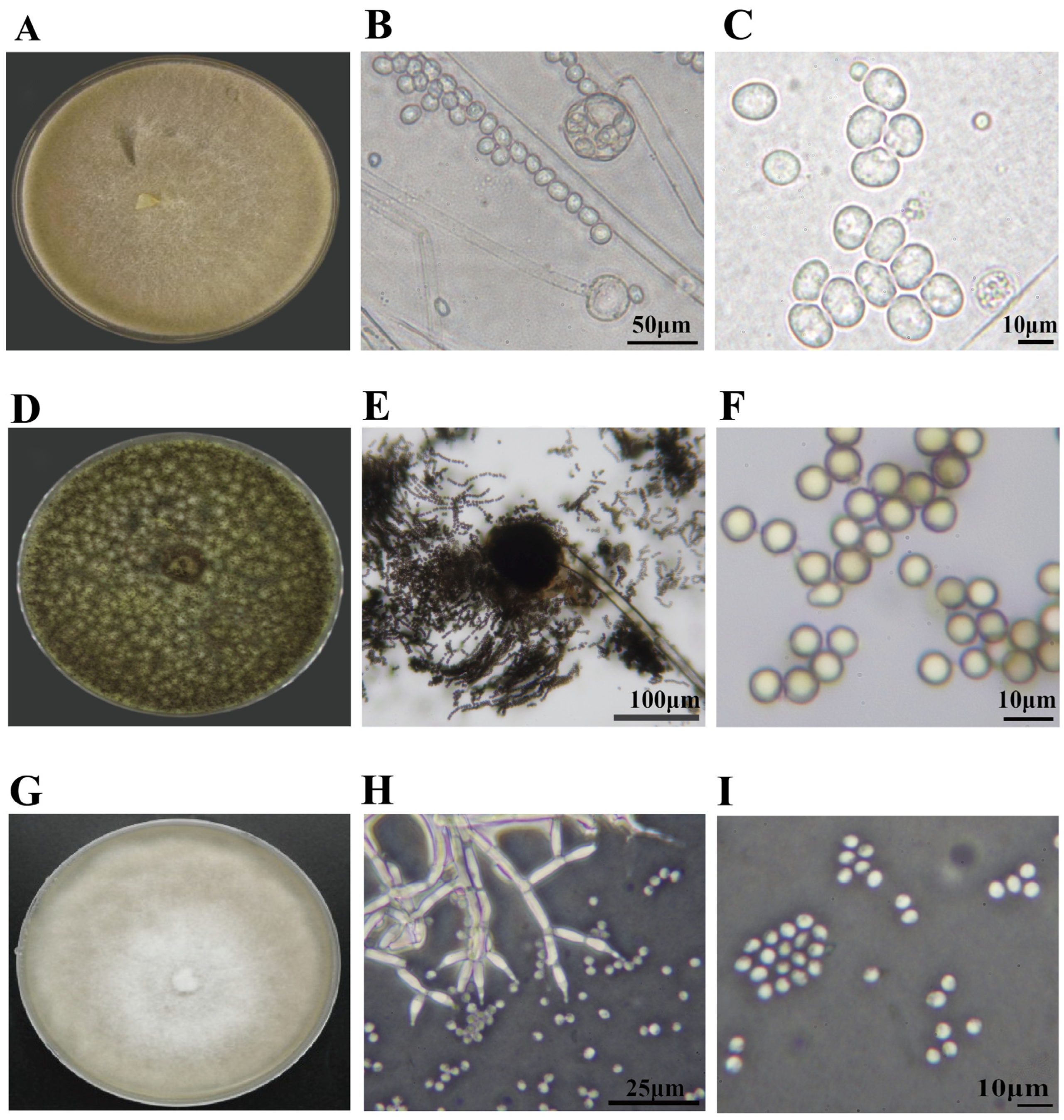
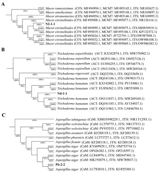
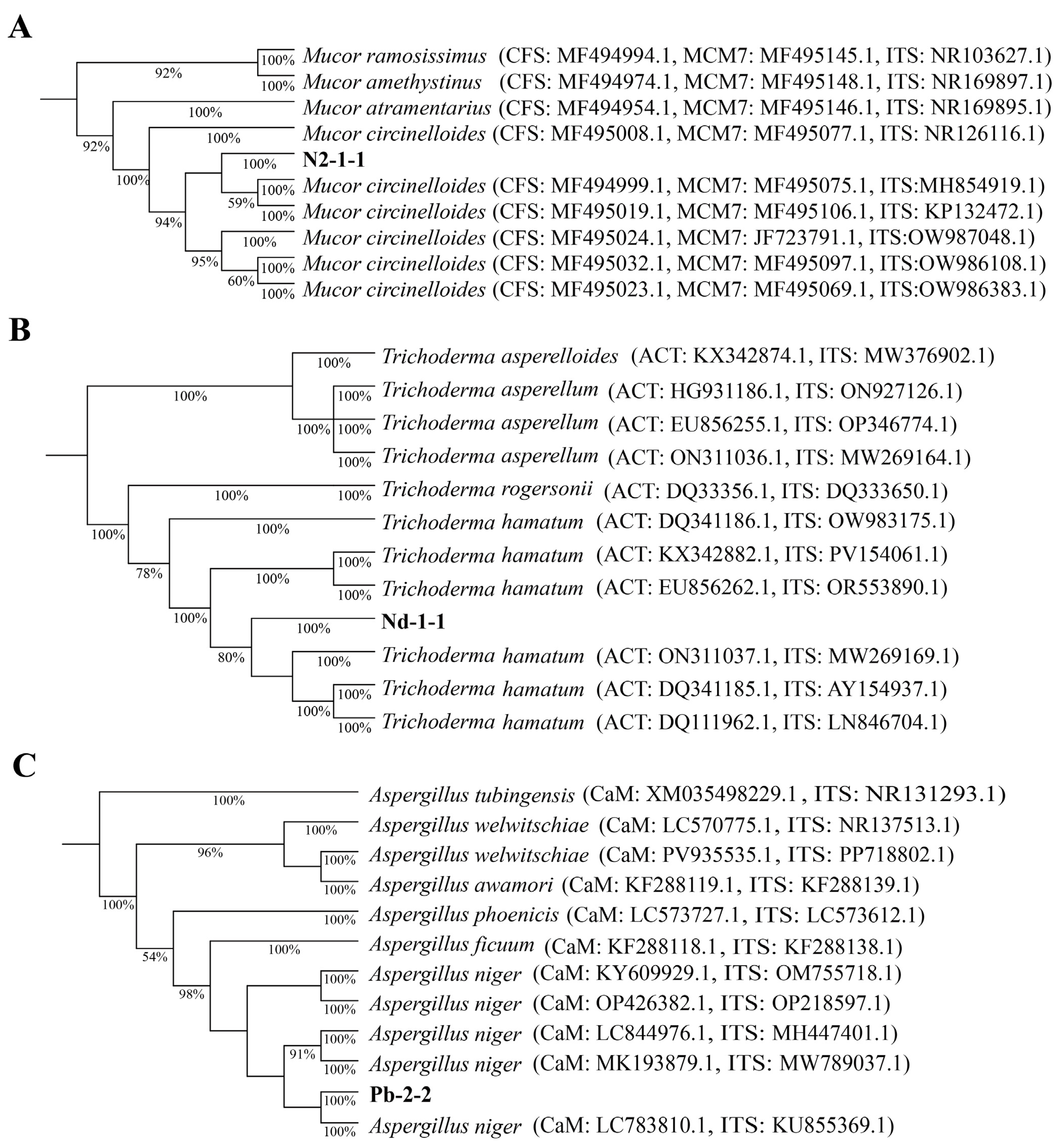
The three antagonistic fungi were identified through a combination of morphological and phylogenetic analyses. Strain N2-1-1 was identified as Mucor circinelloides, exhibiting cottony colonies with aseptate hyphae and spherical to ellipsoidal sporangiospores [37] (Figure 1A–C), and was confirmed based on rDNA-ITS (PX396488), CFS (PV976146), and mcm7 (PV976147) gene sequences (Figure 2A). Strain Pb-2-2 was identified as Aspergillus niger, with grayish green to yellowish green colonies and spherical to subglobose conidia arranged in chains [38] (Figure 1D–F), and this was supported by rDNA-ITS (PX396490) and calmodulin (PV976151) gene sequences (Figure 2B). Strain Nd-1-1 was identified as Trichoderma hamatum, showing white to green cottony colonies and producing phialides with spherical to subglobose conidia [39] (Figure 1G–I), and was determined based on rDNA-ITS (PX396489) and actin (PV976149) gene sequences (Figure 2C).
Figure 1.
Morphological characteristics of the three antagonistic fungal isolates. Note: (A–C) Strain N2-1-1, showing a cottony colony, aseptate hyphae, and spherical to ellipsoidal sporangiospores. (D–F) Strain Pb-2-2, showing grayish-green to yellowish-green colonies and spherical to subglobose conidia arranged in chains. (G–I) Strain Nd-1-1, showing white cottony colonies and phialides producing spherical to subglobose conidia. Scale bars: (B) = 50 μm, (C) = 10 μm, (E) = 100 μm, (F) = 10 μm, (H) = 25 μm, and (I) = 10 μm.
Figure 2.
Phylogenetic identification of the three antagonistic fungal isolates based on multilocus sequence analysis. Note: (A) Strain N2-1-1, inferred from concatenated CFS, mcm7 and rDNA-ITS gene sequences. (B) Strain Nd-1-1, inferred from concatenated actin and rDNA-ITS gene sequences. (C) Strain Pb-2-2, inferred from concatenated calmodulin and rDNA-ITS gene sequences. Bootstrap support values from 1000 replicates are shown at the nodes; only values >50% are shown. Accession numbers are provided after strain names.
3.2. Effects of Spore Suspensions of Antagonistic Fungi on Pathogenic Fungi of Kiwifruit Soft Rot In Vivo
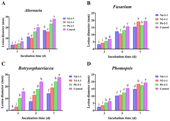
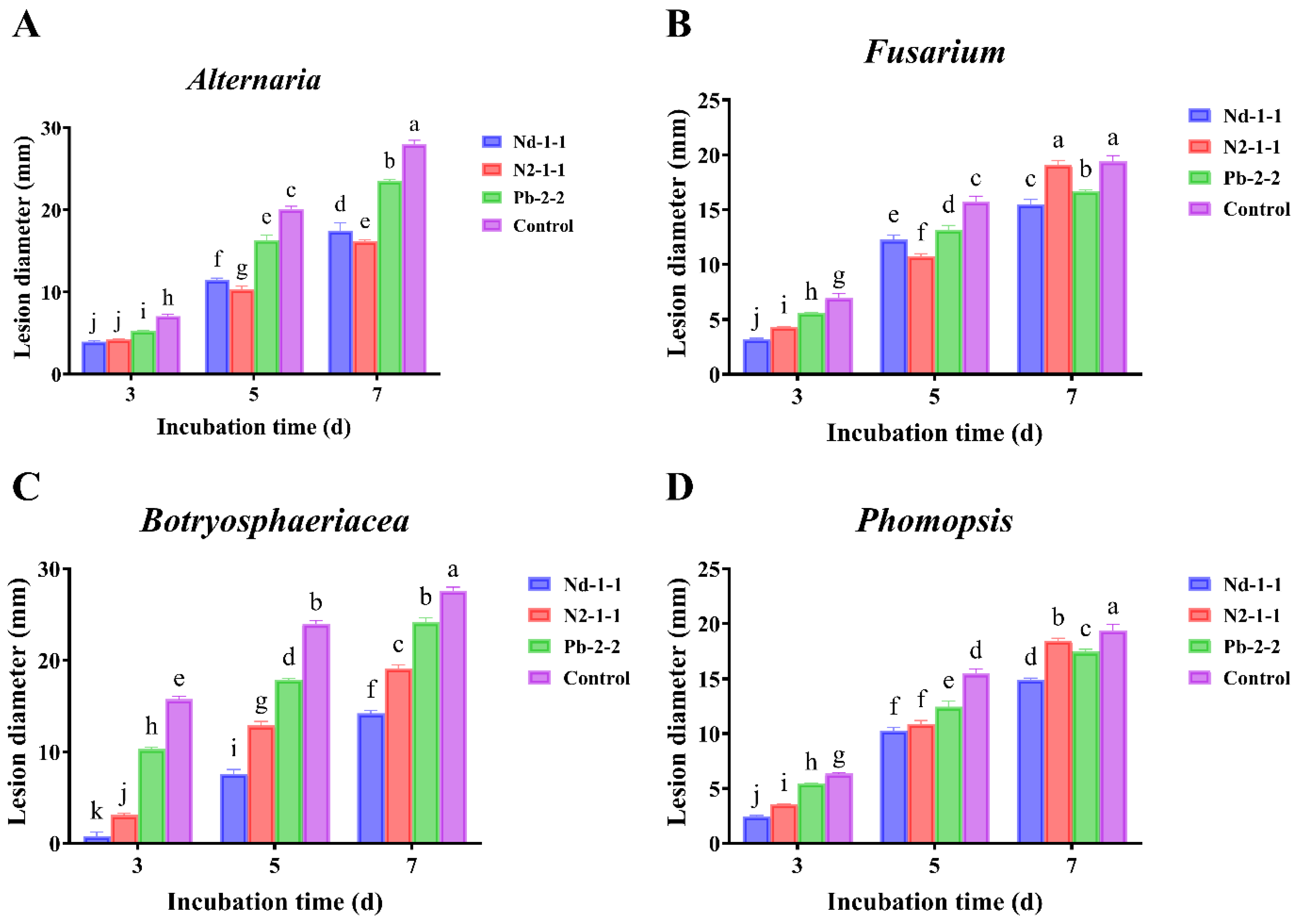
In the spore suspension inoculation assay, the three antagonistic strains exhibited distinct temporal patterns of disease suppression across the four pathogens. Against Alternaria sp. (Figure 3A), M. circinelloides N2-1-1 consistently restricted lesion expansion from day 3 onward, outperforming T. hamatum Nd-1-1 by day 7, while A. niger Pb-2-2, despite an exceptionally stable and reproducible inhibition, allowed a late-stage surge in lesion development that markedly diminished its overall efficacy. In contrast, T. hamatum Nd-1-1 established early dominance against the remaining three pathogens and sustained the strongest suppression through the experimental period. For Fusarium sp. (Figure 3B), T. hamatum Nd-1-1 maintained lesions at a notably lower level than the other strains and the control, with A. niger Pb-2-2 displaying a moderate, tightly clustered response that contrasted with the progressive weakening observed in M. circinelloides N2-1-1. Against Botryosphaeriaceae sp. (Figure 3C), T. hamatum Nd-1-1 exerted the most powerful and sustained control, virtually preventing lesion progression, whereas M. circinelloides N2-1-1 and A. niger Pb-2-2 proved progressively less effective, with the latter showing only marginal reduction relative to the control. A similar hierarchy was evident for Phomopsis sp. (Figure 3D), where T. hamatum Nd-1-1 again limited lesion size most effectively, while the other two strains converged toward the control level over time. Notably, although A. niger Pb-2-2 was rarely the most suppressive strain, it consistently produced the least variable lesion diameters across replicates, confirming a uniquely stable antagonistic phenotype.
Figure 3.
Inhibitory effects of spore suspensions of three antagonistic fungi on lesion development caused by four soft rot pathogens in kiwifruit. Note: Lesion diameters on kiwifruit caused by (A) Alternaria, (B) Phomopsis, (C) Fusarium, and (D) Botryosphaeriaceae were determined after treatment with spore suspensions of three antagonistic fungi (Nd-1-1, N2-1-1, Pb-2-2) at 3, 5 and 7 days post-inoculation in vivo. All data are presented as mean ± standard deviation (SD, n ≥ 3). Different lowercase letters above the bars indicate significant differences among treatments at the same incubation time, as determined by two-way ANOVA followed by Tukey’s multiple range test (p < 0.05) (Table A1 and Table A4).
3.3. Effects of Fermentation Broth on Pathogenic Fungi of Kiwifruit Soft Rot
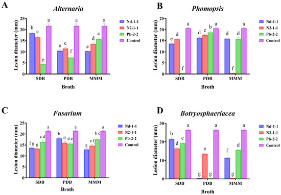
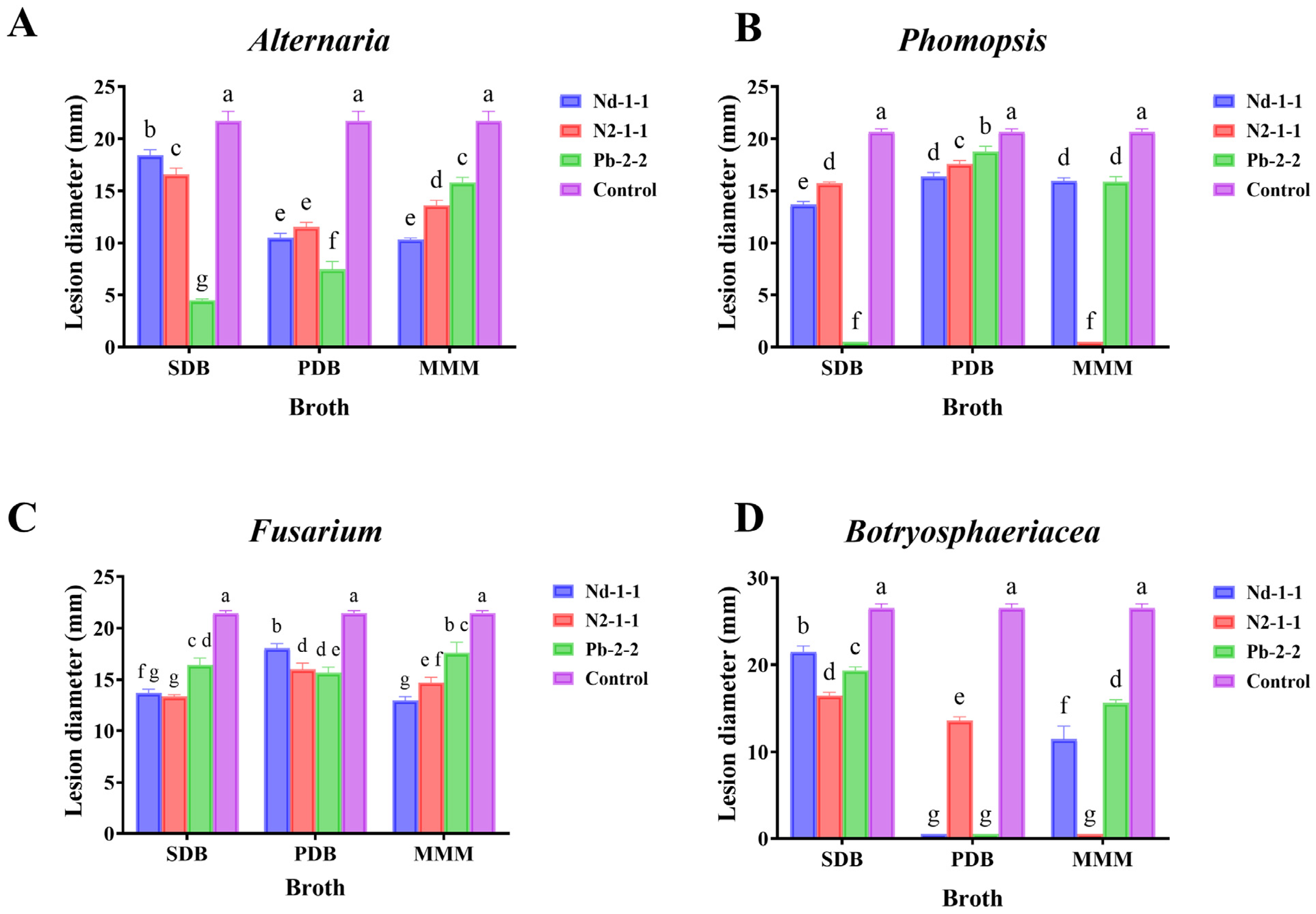
Fermentation assays confirmed that all three antagonistic fungi secreted antimicrobial metabolites capable of reducing lesion diameter relative to the control, yet the extent of suppression varied dramatically with strain, culture medium, and pathogen identity. Complete arrest of lesion development was restricted to specific combinations. The SDB filtrate of A. niger Pb-2-2 and the MMM filtrate of M. circinelloides N2-1-1 abolished disease caused by Phomopsis sp. (Figure 4B), while against Botryosphaeriaceae sp. (Figure 4D), such thorough inhibition additionally required the PDB filtrate of T. hamatum Nd-1-1. In most other pairings, the filtrates afforded only partial protection, and in a subset of cases the reduction was marginal, most conspicuously for T. hamatum Nd-1-1 in SDB against Botryosphaeriaceae sp., where lesions remained large despite treatment. The impact of the culture medium was especially evident for T. hamatum Nd-1-1; its PDB filtrate could eliminate Botryosphaeriaceae lesions entirely, whereas its SDB filtrate was among the least effective treatments across the whole dataset (Figure 4D). Among the four pathogens, Fusarium sp. (Figure 4C) proved consistently the least responsive; no treatment achieved lesion clearance, and even the strongest filtrates only moderately slowed expansion, leaving lesion sizes well above those recorded for other pathogens. Conversely, Alternaria sp. (Figure 4A) was highly susceptible to A. niger Pb-2-2 filtrates, particularly in SDB, which nearly prevented symptom development, and the same strain in PDB also exerted strong suppression. T. hamatum Nd-1-1 and M. circinelloides N2-1-1 provided moderate control against Alternaria, with their PDB and MMM filtrates outperforming other media. For Phomopsis sp., the exceptionally low lesion diameters achieved by A. niger Pb-2-2 in SDB and M. circinelloides N2-1-1 in MMM contrasted with the more modest effects of T. hamatum Nd-1-1, whose best performance occurred in SDB.
Figure 4.
Inhibitory effects of fermentation broth from three antagonistic fungi cultured in different media on lesion expansion caused by four soft rot pathogens in kiwifruit. Note: Lesion diameters on kiwifruit caused by (A) Alternaria, (B) Phomopsis, (C) Fusarium, and (D) Botryosphaeriaceae were determined after treatment with fermentation broth of three antagonistic fungi (Nd-1-1, N2-1-1, Pb-2-2) grown in three different media: SDB (Sabouraud Dextrose Broth), PDB (Potato Dextrose Broth), and MMM (Minimal Medium). All data are presented as mean ± standard deviation (SD, n ≥ 3). Different lowercase letters above the bars indicate significant differences among treatments within the same medium, as determined by two-way ANOVA followed by Tukey’s multiple range test (p < 0.05) (Table A2 and Table A5).
3.4. Effects of Antagonistic Fungi on the Resistance of Kiwifruit Fruits
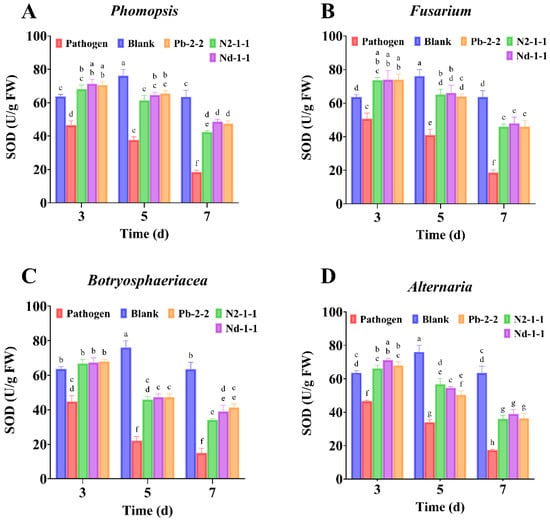
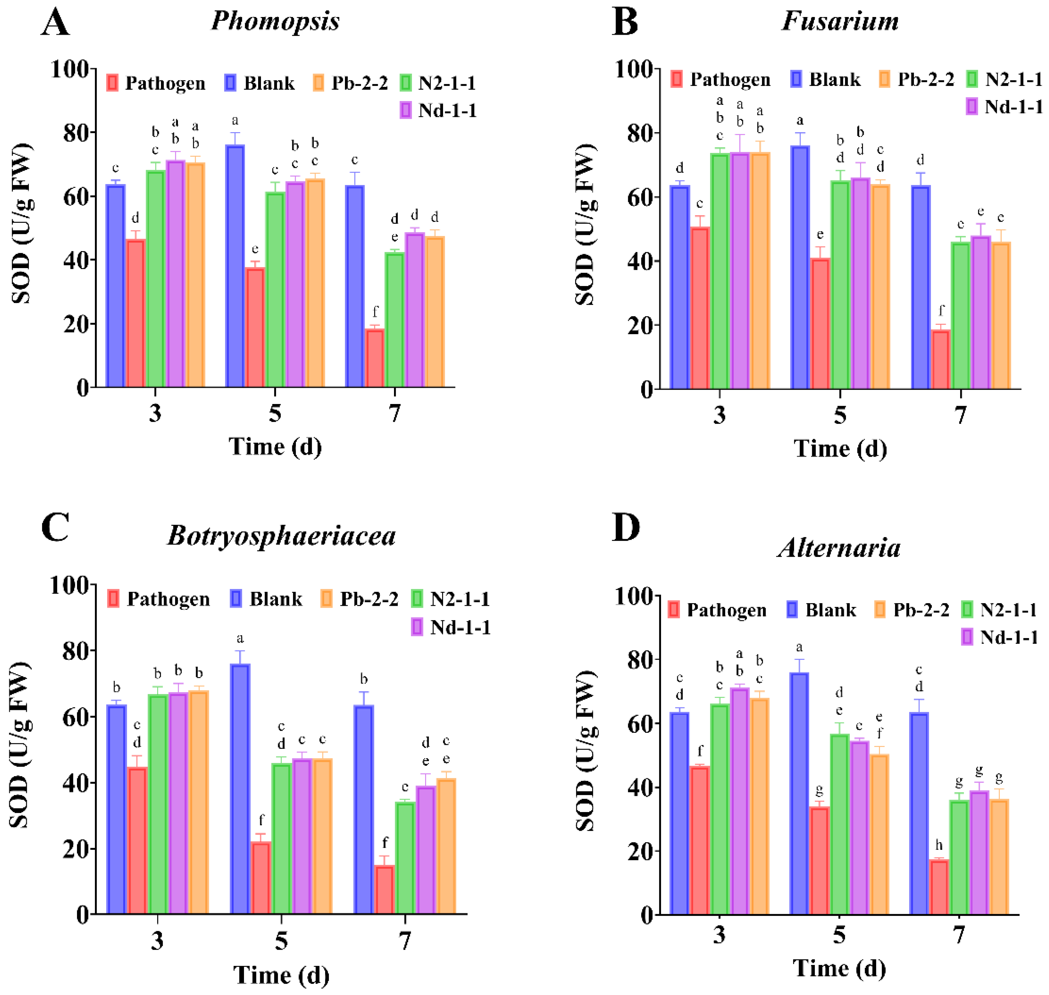
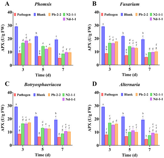
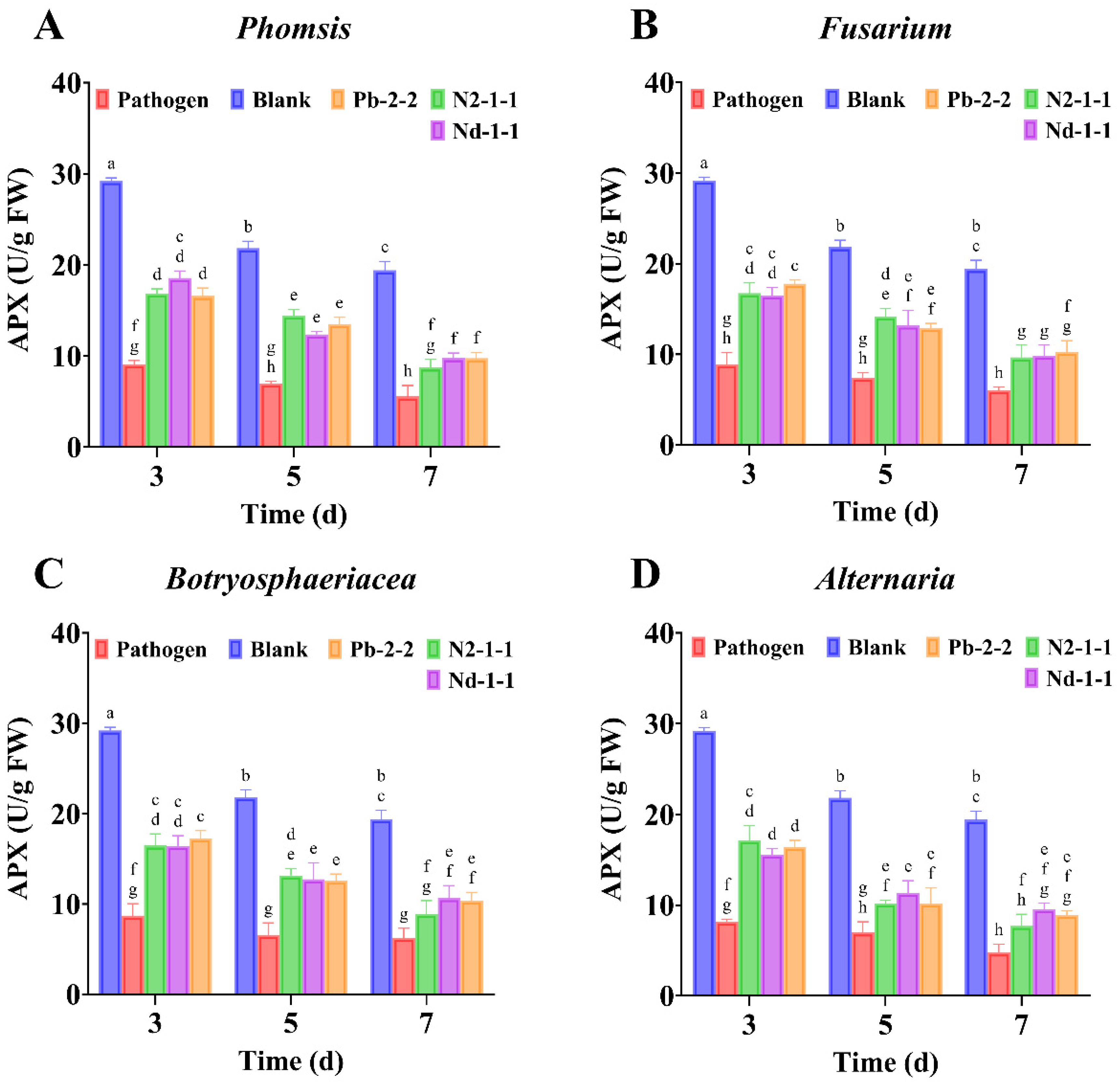
To evaluate how biocontrol fungi modulate kiwifruit defenses, SOD, CAT, APX and PAL activities were monitored following pathogen inoculation alone and antagonist–pathogen co-inoculation. Statistical analyses indicated highly significant effects of treatment, time and their interaction on all enzymes assayed (Table A3, Table A6, Table A7, Table A8 and Table A9). Relative to the blank group, pathogen inoculation alone significantly reduced SOD (Figure 5) and APX (Figure 6) activities, indicating compromised antioxidant capacity and perturbed ROS homeostasis under infection pressure, whereas co-inoculation effectively maintained SOD and APX activities, suggesting mitigation of oxidative stress and improved maintenance of ROS balance.
Figure 5.
Effects of three antagonistic fungi on SOD enzyme activity in kiwifruit fruits inoculated with four soft rot pathogens. Note: SOD enzyme activity (U/g FW) was measured in kiwifruit fruits after inoculation with different pathogens. The pathogens were: (A) Phomopsis, (B) Fusarium, (C) Botryosphaeriaceae, and (D) Alternaria. Treatments included a blank control (Blank) and three antagonistic fungi (Pb-2-2, N2-1-1, Nd-1-1). All data are presented as mean ± standard deviation (SD, n ≥ 3). Different lowercase letters above the bars indicate significant differences among treatments within the same pathogen, as determined by two-way ANOVA followed by Tukey’s multiple range test (p < 0.05).
Figure 6.
Effects of three antagonistic fungi on APX enzyme activity in kiwifruit fruits inoculated with four soft rot pathogens. Note: APX enzyme activity (U/g FW) was measured in kiwifruit fruits after inoculation with different pathogens. The pathogens were: (A) Phomopsis, (B) Fusarium, (C) Botryosphaeriaceae, and (D) Alternaria. Treatments included a blank control (Blank) and three antagonistic fungi (Pb-2-2, N2-1-1, Nd-1-1). All data are presented as mean ± standard deviation (SD, n ≥ 3). Different lowercase letters above the bars indicate significant differences among treatments within the same pathogen, as determined by two-way ANOVA followed by Tukey’s multiple range test (p < 0.05).
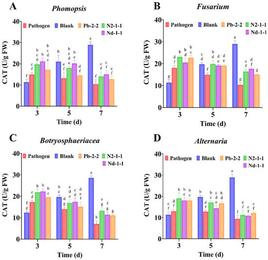
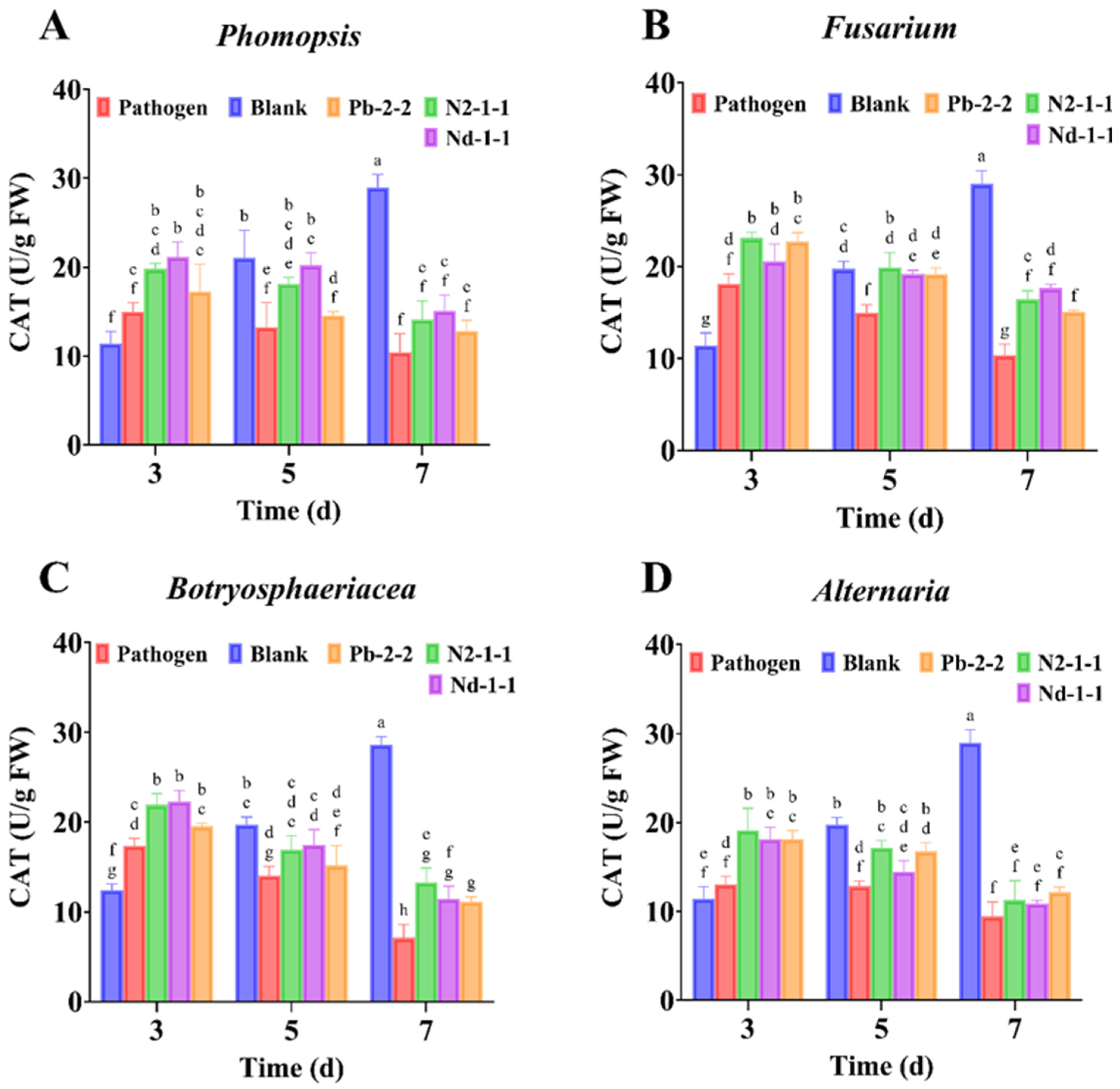
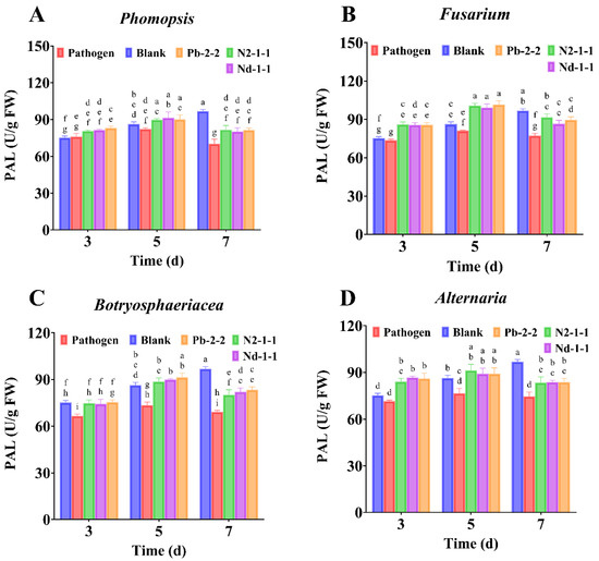
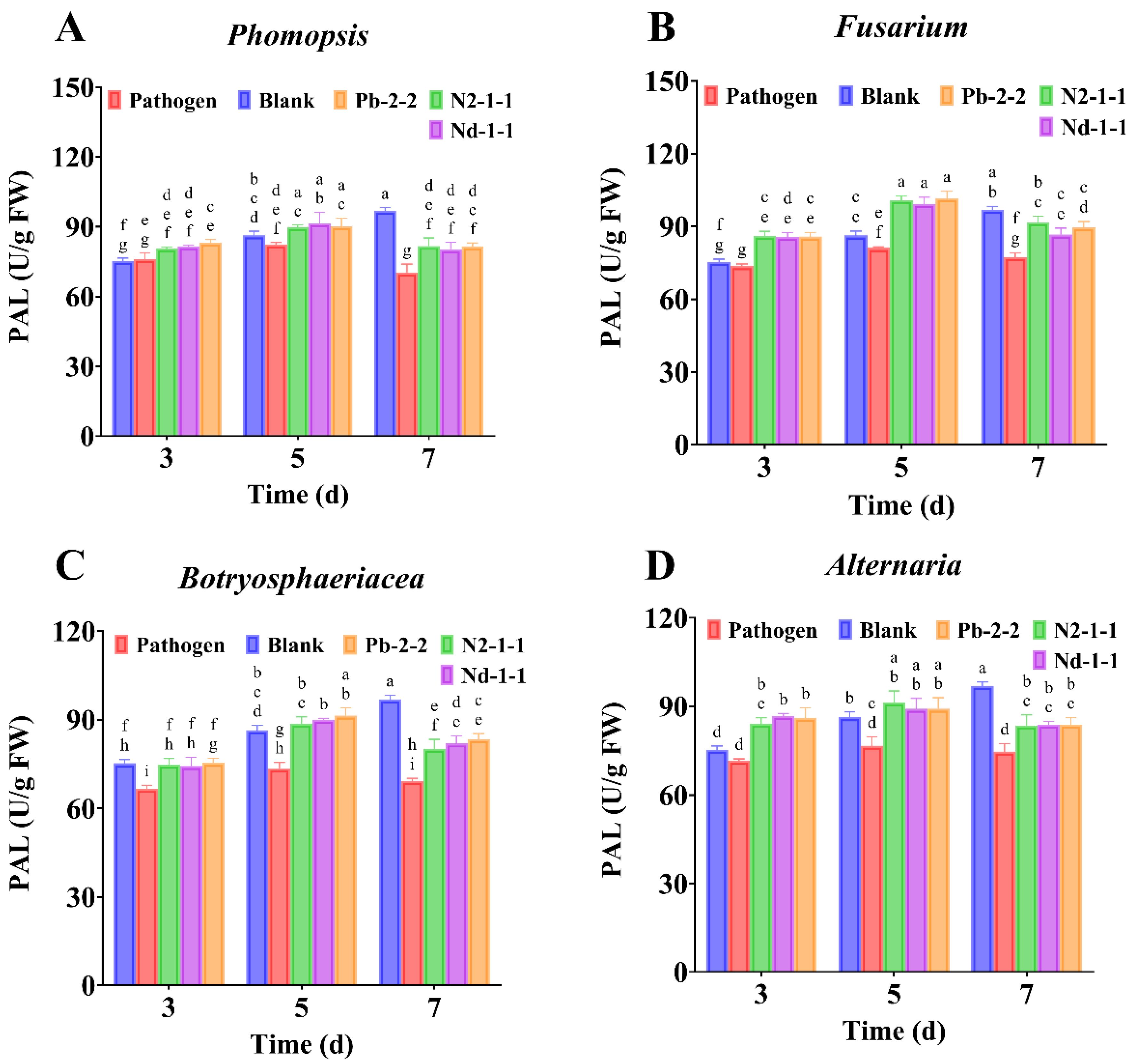
CAT (Figure 7) exhibited distinct temporal patterns between non-inoculated and inoculated fruit: CAT increased steadily in the blank group, whereas it increased initially and then declined continuously under pathogen-only inoculation, becoming significantly lower than the blank group by day 7. Under co-inoculation, CAT followed a similar trajectory to the pathogen-only treatment but was significantly higher at day 3, indicating a transient enhancement of H2O2-scavenging capacity early during infection (Figure 7). For phenylpropanoid metabolism, PAL (Figure 8) responses were pathogen-dependent and stage-dependent: Botryosphaeriaceae sp. and Alternatia sp. reduced PAL at the early stage (day 3), while Fusarium sp. and Phomopsis sp. decreased PAL mainly at middle and late stages (days 5–7); under co-inoculation, PAL generally increased initially and then declined (Figure 7). Notably, no significant differences were detected among the three antagonistic fungal strains (T. hamatum Nd-1-1, M. circinelloides N2-1-1, and A. niger Pb-1-1) in their capacity to maintain the activities of SOD, APX, and PAL. Although inter-strain variation was observed in the magnitude of early-stage (day 3) CAT activity enhancement, the overall temporal trends in modulating the four defense enzyme activities were highly consistent across all three antagonists. This finding suggests that these distinct biocontrol agents may elicit convergent host defense outputs and/or that the fruit defense response has reached a plateau-like saturation level. Although co-inoculation generally maintained host defense-related enzyme activities, these parameters did not clearly discriminate among the three antagonists, suggesting that differences in in vivo efficacy may depend more on ecological establishment and direct antagonistic traits than on final enzyme levels alone.
Figure 7.
Effects of three antagonistic fungi on CAT enzyme activity in kiwifruit fruits inoculated with four soft rot pathogens. Note: CAT enzyme activity (U/g FW) was measured in kiwifruit fruits after inoculation with different pathogens. Note: (A) Phomopsis, (B) Fusarium, (C) Botryosphaeriaceae, and (D) Alternaria. Treatments included a blank control (Blank) and three antagonistic fungi (Pb-2-2, N2-1-1, Nd-1-1). All data are presented as mean ± standard deviation (SD, n ≥ 3). Different lowercase letters above the bars indicate significant differences among treatments within the same pathogen, as determined by two-way ANOVA followed by Tukey’s multiple range test (p < 0.05).
Figure 8.
Effects of three antagonistic fungi on PAL enzyme activity in kiwifruit fruits inoculated with four soft rot pathogens. Note: PAL enzyme activity (U/g FW) was measured in kiwifruit fruits after inoculation with different pathogens. The pathogens were: (A) Phomopsis, (B) Fusarium, (C) Botryosphaeriaceae, and (D) Alternaria. Treatments included a blank control (Blank) and three antagonistic fungi (Pb-2-2, N2-1-1, Nd-1-1). All data are presented as mean ± standard deviation (SD, n ≥ 3). Different lowercase letters above the bars indicate significant differences among treatments within the same pathogen, as determined by two-way ANOVA followed by Tukey’s multiple range test (p < 0.05).
4. Discussion
Postharvest soft rot poses a serious and persistent threat to the extensive kiwifruit (Actinidia chinensis) industry in China, inflicting considerable economic damage. A promising alternative for control lies in the diverse native microorganisms that colonize kiwifruit surfaces, which serve as a valuable source of antagonistic microbes [40]. These naturally occurring yeasts, bacteria, and filamentous fungi exhibit inherent competitive advantages against pathogens in the postharvest environment, positioning them as safe and effective biocontrol agents [41]. This potential prompted the present study to isolate antagonistic fungi from kiwifruit, screen their biocontrol activity against soft rot pathogens, and preliminarily characterize their modes of action.
A total of 37 fungal isolates were obtained from kiwifruit leaves, bark, and rhizosphere soil, and their antagonistic activities against four major groups of soft-rot-associated pathogens, namely Fusarium sp., Alternaria sp., Botryosphaeriaceae sp., and Phomopsis sp., were evaluated. Among these isolates, Mucor circinelloides N2-1-1, Aspergillus niger Pb-2-2, and Trichoderma hamatum Nd-1-1 showed strong biocontrol potential. (Table 3).
Table 3.
Inhibition rates (%) of antagonistic strains against four phytopathogenic fungi under three biocontrol evaluation systems.
Fungi from the genus Mucor are more frequently reported as pathogens [42,43] than as biocontrol agents, and their potential for biological control remains largely unexplored. M. hiemalis [44] was previously reported as an antagonist against inflorescence brown rot of date palm caused by Thielaviopsis paradoxa, where its protective effect was attributed primarily to competitive colonization and spatial exclusion on floral tissues, without evidence of phytopathogenicity. Similarly, M. moelleri [45] was further shown to possess both antagonistic activity toward plant pathogens and plant growth-promoting properties, highlighting the functional versatility of this genus. In addition, Mucor moelleri AA1 inhibited Athelia rolfsii and Colletotrichum spp. in dual-culture and liquid co-culture assays, reduced disease development in tomato pot experiments, and promoted plant growth. Volatile compounds, β-1,3-glucanase, protease, and catalase produced by this strain may jointly contribute to its antagonistic and growth-promoting effects [45]. Cell-free culture supernatant of Mucor lusitanicus inhibited Fusarium oxysporum, F. solani, and Alternaria solani and exerted antifungal effects in soil and tomato fruit. The Rfs enzyme involved in rhizoferrin biosynthesis affected the antifungal activity of the culture supernatant [46]. Mucor circinelloides N2-1-1 showed strong inhibitory activity in dual-culture assays, and its fermentation filtrate strongly inhibited Botryosphaeriaceae and Phomopsis; the present study expands the range of Mucor species with potential biocontrol activity.
Aspergillus niger can inhibit plant-pathogenic fungi through multiple mechanisms, including rapid growth and niche occupation, competition for nutrients and space, mycoparasitism, extracellular hydrolytic enzymes, and diffusible or volatile antifungal metabolites [47,48,49]. Previous studies have shown that A. niger hyphae can attach to, coil around, form penetration structures in, and damage Fusarium hyphae. In addition, the extracellular chitinase produced by A. niger LOCK 62 strongly inhibited Fusarium culmorum and F. solani, but showed little or no activity against F. oxysporum, Alternaria alternata, and Botrytis cinerea [50,51]. A. niger Pb-2-2 exhibited strong and relatively consistent inhibition against all four pathogen groups tested in the dual-culture assay. This suggests that inhibition by A. niger Pb-2-2 in the dual-culture system may have been mainly driven by rapid growth, niche occupation, and competition for nutrients and space. Compared with the dual-culture assay, the protective effect of the A. niger Pb-2-2 spore suspension in kiwifruit was substantially reduced, possibly because of limitations in spore germination and colonization of fruit tissues. Moreover, A. niger Pb-2-2 fermentation filtrate provided better protection against all four pathogens in inoculated kiwifruit, and its inhibition of lesion expansion remained strongly pathogen-dependent, with greater effects against Alternaria, Phomopsis, and Botryosphaeriaceae than against Fusarium. This result is consistent with previous reports showing selective inhibition of different pathogens by extracellular chitinase from A. niger LOCK 62 [50].
Trichoderma sp. is a kind of classical biocontrol fungus, and numerous studies have reported its role in the control of field diseases and postharvest fungal diseases of fruits and vegetables. Trichoderma sp. is generally considered to antagonize plant-pathogenic fungi through rapid growth and spatial occupation, nutrient competition, mycoparasitism, extracellular hydrolytic enzymes, and antimicrobial metabolites. For example, Trichoderma Tha739 can grow alongside and coil around Colletotrichum hyphae, resulting in hyphal deformation [52]. Strain T88 can penetrate and damage the hyphal cells of apple ring-rot pathogens through mycoparasitism, causing cytoplasmic thinning and structural damage [53]. In tomato fruit, Trichoderma can also cover, coil around, and degrade the sporangiophores and sporangia of Rhizopus [54].
Consistent with these findings, T. hamatum Nd-1-1 showed strong inhibitory activity in the dual-culture assay. Its inhibition rates against Fusarium sp., Alternaria sp., Botryosphaeriaceae sp., and Phomopsis sp. were 78.23%, 78.37%, 78.61%, and 60.10%, respectively. The weaker inhibition of Phomopsis suggests that this pathogen may be relatively less sensitive to the mycoparasitism, cell-wall-degrading enzymes, or other antagonistic factors produced by T. hamatum Nd-1-1.
Similar to A. niger Pb-2-2 and M. circinelloides N2-1-1, the direct protective effect of the T. hamatum Nd-1-1 spore suspension was substantially reduced in fruit. However, the Nd-1-1 spore suspension showed the highest inhibition among the three strains against Fusarium sp., Botryosphaeriaceae sp., and Phomopsis sp., and the second-highest activity against Alternaria sp. Thus, although the in vivo efficacy of T. hamatum Nd-1-1 spore suspension was lower than its activity in the dual-culture assay, its overall efficacy as a living spore preparation remained higher than that of A. niger Pb-2-2 and M. circinelloides N2-1-1. It suggests that T. hamatum Nd-1-1 may be better adapted to the kiwifruit environment and may have stronger spore germination, wound colonization, or local competitive ability than the other two strains.
Previous studies have shown that Trichoderma sp. can produce chitinase, β-1,3-glucanase, protease, and several potentially active compounds after recognizing pathogen cell walls, including 6-pentyl-α-pyrone, harzianic acid, isoharzianic acid, viridiofungin A, harzianopyridone, harzianolide, and peptaibols [55,56,57]. Synergistic interactions may occur between cell-wall-degrading enzymes and these active compounds. In the present study, T. hamatum Nd-1-1 fermentation filtrate inhibited Fusarium sp., Alternaria sp., Botryosphaeriaceae sp., and Phomopsis sp. by 45.73%, 65.56%, 98.11%, and 33.80%, respectively. Further studies could compare the antifungal activities of the original filtrate, pH-neutralized filtrate, heat-treated filtrate, and protease-treated filtrate, together with fractionation, metabolomic analysis, and extracellular enzyme assays, to identify the active components in T. hamatum Nd-1-1 fermentation filtrate.
SOD catalyzes the dismutation of superoxide radicals (O2•−) into oxygen and hydrogen peroxide (H2O2) [58]. APX has a high affinity for H2O2 and mainly removes low concentrations of H2O2 in chloroplasts and the cytoplasm [59], whereas CAT has a high processing capacity and primarily decomposes high concentrations of H2O2 in peroxisomes [60]. Following pathogen infection, fruit tissues generally produce superoxide anions and hydrogen peroxide, resulting in an early oxidative burst. Moderate levels of ROS can function as defense signals and participate in the activation of MAPK, SA, JA, and ethylene signaling pathways, whereas excessive ROS can cause membrane lipid peroxidation, membrane damage, and oxidative stress in fruit tissues [61]. In the present study, pathogen infection decreased SOD and APX activities in kiwifruit, whereas treatment with the antagonistic fungi partially alleviated these decreases and enhanced CAT activity. These results suggest that M. circinelloides N2-1-1, A. niger Pb-2-2, and T. hamatum Nd-1-1 may coordinate ROS homeostasis by maintaining SOD and APX activities and enhancing CAT responses. This may reduce excessive O2•− accumulation and promote the timely removal of excess H2O2. In this process, an appropriate level of H2O2 may remain available as a defense signal, while excessive H2O2 can be efficiently decomposed by CAT, thereby reducing oxidative damage and maintaining redox homeostasis in fruit cells. Similarly, previous studies have shown that biocontrol microorganisms can enhance antioxidant enzyme systems in infected fruits and help maintain relatively stable ROS levels under pathogen stress [62,63].
PAL is a key rate-limiting enzyme in the phenylpropanoid pathway, and changes in its activity reflect the capacity of fruit tissues to synthesize phenolic compounds, lignin, and other defense-related secondary metabolites. Compared with pathogen-only inoculation, co-inoculation with the antagonistic fungi and pathogens maintained or increased PAL activity during the early stage of infection. At 3–5 days after inoculation, PAL activity generally approached, reached, or even exceeded that of uninoculated fruit. This indicates that the antagonistic fungi delayed pathogen-mediated suppression of phenylpropanoid metabolism and supported early defense responses in kiwifruit. Previous studies have shown that antagonistic fungi or yeasts can promote phenylpropanoid metabolism by increasing PAL activity and enhancing the synthesis of phenolic and other antimicrobial secondary metabolites [64,65].
However, during the late stage of infection, PAL activity in the co-inoculation treatments, although still significantly higher than that in pathogen-only fruit, generally failed to remain at or above the level of healthy controls. It is noteworthy that the effects of the three antagonistic fungi on SOD, CAT, APX, and PAL activities were not statistically different at most sampling times, which does not indicate that the three strains have identical biocontrol mechanisms. Rather, it may suggest that they regulate the fruit defense system in a partially convergent manner, whereas their main differences occur at the levels of direct antagonism, tissue colonization, and metabolite activity.
5. Conclusions
This study isolated, screened, and identified antagonistic fungi associated with kiwifruit and demonstrated that Trichoderma hamatum Nd-1-1, Aspergillus niger Pb-2-2, and Mucor circinelloides N2-1-1 exhibited significant antagonistic activity against the major fungal pathogens of kiwifruit soft rot disease. Among them, A. niger Pb-2-2 showed the strongest and most stable antifungal performance in vitro, whereas T. hamatum Nd-1-1 achieved the greatest disease suppression in vivo. All three antagonistic fungi showed strong and broad-spectrum antifungal activity in the dual-culture assay; however, their spore suspensions were substantially less effective in fruit than in vitro. This indicates that inhibition rates obtained from dual-culture assays cannot fully predict the practical efficacy of antagonistic fungi in fruit, which is also influenced by spore germination, wound colonization, and adaptation to the fruit environment. Fermentation assays further revealed that all three antagonists were capable of secreting antifungal secondary metabolites, although the inhibitory spectrum and stability of these metabolites were markedly influenced by culture medium, fermentation duration, and target pathogen. Moreover, these antagonistic fungi may enhance kiwifruit resistance to soft rot through modulation of host antioxidant defense systems and phenylpropanoid metabolism, as reflected by the maintenance of SOD and APX activities, the transient induction of CAT activity at the early stage of infection, and the upregulation of PAL activity. These responses likely contribute to the alleviation of oxidative damage and the activation of host defense.
Overall, our study identified three antagonistic fungi with biocontrol potential against multiple pathogens associated with kiwifruit soft rot. This is the first report of the biocontrol potential of Mucor circinelloides strain N2-1-1, thereby expanding the current understanding of the potential application of Mucor sp. in biological disease control. However, though reports on Mucor sp. as biocontrol agents remain limited, the potential of Mucor circinelloides N2-1-1 for controlling kiwifruit soft rot requires further investigation. Before practical application can be achieved, further studies are required to identify the active metabolites, optimize fermentation and formulation strategies, and validate efficacy under commercial storage conditions. In addition, comprehensive biosafety assessments, particularly regarding toxigenic potential, allergenicity, and storage safety, are essential to ensure safe and feasible application in sustainable postharvest management of kiwifruit soft rot.
Author Contributions
Conceptualization, J.L. and Y.S.; methodology, Y.S.; software, Y.S. and J.L.; validation, J.L., H.L. and L.Y.; formal analysis, J.L. and H.L.; investigation, J.L.; resources, J.L. and N.J.; data curation, J.L.; writing—original draft preparation, Y.S. and H.L.; writing—review and editing, J.L.; visualization, H.L., L.Y. and J.D.; supervision, J.L.; project administration, R.W.; funding acquisition, R.W. All authors have read and agreed to the published version of the manuscript.
Funding
This research was funded by the Guizhou Key Laboratory of New Quality Processing and Storage of Ecological Specialty Food (Grant No. ZSYS [2025]023), the Guizhou Provincial Department of Science and Technology (Project for Technological Innovation Talent Team of Efficient Utilization for Guizhou’s Characteristic Fruits and Vegetables (Grant No. Qiankehe Talent CXTD [2025]056).
Data Availability Statement
The original contributions presented in this study are included in the article and Appendix A. Further inquiries can be directed to the corresponding author.
Acknowledgments
Authors are very thankful to the College of Food Science and Technology, Guiyang University, and Key Laboratory for Novel Processing and Storage of Ecological Characteristic Foods of Guizhou Province, for providing the necessary facilities and assistance to complete this research work.
Conflicts of Interest
The authors declare no conflicts of interest.
Appendix A
Table A1.
ANOVA table for antibacterial activity of spore suspension.
Table A2.
In vivo Lesion Sizes of Pathogens Treated with Fermentation Broths from Antagonistic Fungi.
Table A3.
ANOVA table for enzyme activity.
Table A4.
Tukey’s multiple comparisons test for antibacterial activity of spore suspension.
Table A5.
Tukey’s multiple comparisons test for antagonistic activity of antagonistic Fungi fermentation broth.
Table A6.
Tukey’s multiple comparisons test for SOD enzyme activity.
Table A7.
Tukey’s multiple comparisons test for CAT enzyme activity.
Table A8.
Tukey’s multiple comparisons test for APX enzyme activity.
Table A9.
Tukey’s multiple comparisons test for PAL enzyme activity.
References
- Huang, H.; Ferguson, A.R. kiwifruit in China. N. Z. J. Crop Hortic. Sci. 2001, 29, 1–14. [Google Scholar] [CrossRef] [Scilit]
- Richardson, D.P.; Ansell, J.; Drummond, L.N. The nutritional and health attributes of kiwifruit: A review. Eur. J. Nutr. 2018, 57, 2659–2676. [Google Scholar] [CrossRef] [Scilit] [PubMed]
- Li, L.; Hui, P.; Lei, D.; Wenyi, L.; Dandan, F.; Caihong, Z. Occurrence rule and comprehends control of kiwifruit rot disease. China Fruits 2020, 40, 1–5. [Google Scholar] [CrossRef]
- Pan, L.; Gao, Z.; Mao, J.; Lin, M.; Zhang, X.; Wang, X.; Chen, Q. First Report of Fusarium fujikuroi Causing Postharvest Rot of Kiwifruit (Actinidia chinensis) in Jiangxi Province, China. Plant Dis. 2024, 108, 2579. [Google Scholar] [CrossRef] [Scilit]
- Jiqing, L.; Wenneng, W.; Ying, L.; Xiuqing, L.; Weijie, L.; Rui, W. Identification of Pathogen Isolation and Pathogenicity Difference of ‘HongYang’ Kiwifruiy Ripe Rot in Liupanshui City, GuiZhou Province. North. Hortic. 2019, 4, 31–38. [Google Scholar] [CrossRef]
- Hawthorne, B.; Rees-George, J.; Samuels, G.J. Fungi associated with leaf spots and post-harvest fruit rots of kiwifruit (Actinidia chinensis) in New Zealand. N. Z. J. Bot. 1982, 20, 143–150. [Google Scholar] [CrossRef] [Scilit]
- Di Marco, S.; Calzarano, F.; Osti, F.; Mazzullo, A. Pathogenicity of fungi associated with a decay of kiwifruit. Australas. Plant Pathol. 2004, 33, 337–342. [Google Scholar] [CrossRef] [Scilit]
- Li, L.; Pan, H.; Chen, M.; Zhang, S.; Zhong, C. Isolation and identification of pathogenic fungi causing postharvest fruit rot of kiwifruit (Actinidia chinensis) in China. J. Phytopathol. 2017, 165, 782–790. [Google Scholar] [CrossRef] [Scilit]
- Manning, M.; Meier, X.; Olsen, T.; Johnston, P. Fungi associated with fruit rots of Actinidia chinensis ‘Hort16A’in New Zealand. N. Z. J. Crop Hortic. Sci. 2003, 31, 315–324. [Google Scholar] [CrossRef] [Scilit]
- McDonald, V.; Lynch, S.; Eskalen, A. First report of Neofusicoccum australe, N. luteum, and N. parvum associated with avocado branch canker in California. Plant Dis. 2009, 93, 967. [Google Scholar] [CrossRef] [Scilit] [PubMed]
- Wang, X.J.; Li, S.Y.; Li, Y.W.; He, R.; Zhu, L.W.; Liu, P. Pathogen identification of kiwifruit soft rot and fungicide screening for control of the disease. J. Plant Prot. 2017, 44, 826–832. [Google Scholar] [CrossRef]
- Wenneng, W.; Qi, Z.; Jiqing, L.; Yaxin, H.; Chenwen, C.; Rui, W.; Sen, C. Identification and Pharmaceutical Screening of Kiwifruit Soft Rot Disease on ‘Guichang’ Gooseberry. North. Hortic. 2018, 16, 47–54. [Google Scholar] [CrossRef]
- Lei, D.; Hui, P.; Caihong, Z.; Qiuling, H.; Yifu, X.; Peng, Z.; Jing, L.; Li, L. Field Verification of Highly Effective Agents for Controlling Kiwifruit Fruit Rot Disease. South China Fruits 2020, 49, 127–129+132. [Google Scholar] [CrossRef]
- Huang, T.; Duan, B.; Zuo, X.; Du, H.; Wang, J.; Cai, Z.; Shen, Y.; Zhang, W.; Chen, J.; Zhu, L. Hydrogen sulfide enhances kiwifruit resistance to soft rot by regulating jasmonic acid signaling pathway. Plant Physiol. Biochem. 2024, 214, 108880. [Google Scholar] [CrossRef] [Scilit] [PubMed]
- Pan, H.; Zhong, C.; Xia, L.; Li, W.; Wang, Z.; Deng, L.; Li, L.; Long, C.-a. Antifungal activity of natamycin against kiwifruit soft rot caused by Botryosphaeria dothidea and potential mechanisms. Sci. Hortic. 2022, 305, 111344. [Google Scholar] [CrossRef] [Scilit]
- Pang, L.; Xia, B.; Liu, X.; Yi, Y.; Jiang, L.; Chen, C.; Li, P.; Zhang, M.; Deng, X.; Wang, R. Improvement of antifungal activity of a culture filtrate of endophytic Bacillus amyloliquefaciens isolated from kiwifruit and its effect on postharvest quality of kiwifruit. J. Food Biochem. 2021, 45, e13551. [Google Scholar] [CrossRef] [Scilit] [PubMed]
- Fan, Y.; Liu, K.; Lu, R.; Gao, J.; Song, W.; Zhu, H.; Tang, X.; Liu, Y.; Miao, M. Cell-free supernatant of Bacillus subtilis reduces kiwifruit rot caused by Botryosphaeria dothidea through inducing oxidative stress in the pathogen. J. Fungi 2023, 9, 127. [Google Scholar] [CrossRef] [Scilit] [PubMed]
- Pan, H.; Zhong, C.; Wang, Z.; Deng, L.; Li, W.; Zhao, J.; Long, C.-a.; Li, L. Biocontrol ability and action mechanism of Meyerozyma guilliermondii 37 on soft rot control of postharvest kiwifruit. Microorganisms 2022, 10, 2143. [Google Scholar] [CrossRef] [Scilit] [PubMed]
- Li, W.; Long, Y.; Mo, F.; Shu, R.; Yin, X.; Wu, X.; Zhang, R.; Zhang, Z.; He, L.; Chen, T.; et al. Antifungal Activity and Biocontrol Mechanism of Fusicolla violacea J-1 against Soft Rot in Kiwifruit Caused by Alternaria alternata. J. Fungi 2021, 7, 937. [Google Scholar] [CrossRef] [Scilit] [PubMed]
- Chen, T.; Zhang, Z.; Li, W.; Chen, J.; Chen, X.; Wang, B.; Ma, J.; Dai, Y.; Ding, H.; Wang, W.; et al. Biocontrol potential of Bacillus subtilis CTXW 7-6-2 against kiwifruit soft rot pathogens revealed by whole-genome sequencing and biochemical characterisation. Front. Microbiol. 2022, 13, 1069109. [Google Scholar] [CrossRef] [Scilit] [PubMed]
- Ren, C.; Liu, Y.; Su, W.; Tian, B. Bacillus velezensis LMY3-5 for the biocontrol of soft rot in kiwifruit: Antifungal action and underlying mechanisms. Front. Microbiol. 2025, 16, 1562366. [Google Scholar] [CrossRef] [Scilit] [PubMed]
- Zhang, S.L.; Wang, B.; Cao, Y.; Ji, Y.P.; Sun, Q.; Shi, X.C.; Herrera-Balandrano, D.D.; Laborda, P. First Report of Fusarium luffae Causing Leaf Blight on Loquat in Eastern China. Plant Dis. 2023, 108, 789. [Google Scholar] [CrossRef] [Scilit]
- Zhang, Y.; Chu, H.; Dong, K.; Yuan, K. First report of Pectobacterium aroidearum causing soft rot on leaf mustard in China. Plant Dis. 2023, 108, 514. [Google Scholar] [CrossRef] [Scilit]
- Shen, S.; Yu, F.; Hao, X.; Chen, J.; Gao, H.; Lai, X. A novel Bacillus sp. with antagonistic activity against a plant pathogen, Fusarium graminearum, and its potential antagonistic mechanism. Lett. Appl. Microbiol. 2023, 76, ovad098. [Google Scholar] [CrossRef] [Scilit] [PubMed]
- Choudhary, B.; Nagpure, A.; Gupta, R.K. Biological control of toxigenic citrus and papaya-rotting fungi by Streptomyces violascens MT7 and its extracellular metabolites. J. Basic Microbiol. 2015, 55, 1343–1356. [Google Scholar] [CrossRef] [Scilit] [PubMed]
- Zhang, X.; Wu, F.; Gu, N.; Yan, X.; Wang, K.; Dhanasekaran, S.; Gu, X.; Zhao, L.; Zhang, H. Postharvest biological control of Rhizopus rot and the mechanisms involved in induced disease resistance of peaches by Pichia membranefaciens. Postharvest Biol. Technol. 2020, 163, 111146. [Google Scholar] [CrossRef] [Scilit]
- Durak, I.; Yurtarslanl, Z.; Canbolat, O.; Akyol, Ö. A methodological approach to superoxide dismutase (SOD) activity assay based on inhibition of nitroblue tetrazolium (NBT) reduction. Clin. Chim. Acta 1993, 214, 103–104. [Google Scholar] [CrossRef] [Scilit] [PubMed]
- Chao, J.; Zhang, S.; Chen, Y.; Tian, W.-M. Cloning, heterologous expression and characterization of ascorbate peroxidase (APX) gene in laticifer cells of rubber tree (Hevea brasiliensis Muell. Arg.). Plant Physiol. Biochem. 2015, 97, 331–338. [Google Scholar] [CrossRef] [Scilit] [PubMed]
- Farman, A.A.; Hadwan, M.H. Simple kinetic method for assessing catalase activity in biological samples. MethodsX 2021, 8, 101434. [Google Scholar] [CrossRef] [Scilit] [PubMed]
- Holländer, H.; Amrhein, N. The estimation of phenylalanine ammonia-lyase (PAL) activity in intact cells of higher plant tissue. Planta 1981, 152, 374–378. [Google Scholar] [CrossRef] [Scilit] [PubMed]
- Xiang, C.Y.; Gao, F.; Jakovlić, I.; Lei, H.P.; Hu, Y.; Zhang, H.; Zou, H.; Wang, G.T.; Zhang, D. Using PhyloSuite for molecular phylogeny and tree-based analyses. Imeta 2023, 2, e87. [Google Scholar] [CrossRef] [Scilit] [PubMed]
- Hershkovitz, M.A.; Lewis, L.A. Deep-level diagnostic value of the rDNA-ITS region. Mol. Biol. Evol. 1996, 13, 1276–1295. [Google Scholar] [CrossRef] [Scilit] [PubMed]
- Wagner, L.; Stielow, J.; de Hoog, G.; Bensch, K.; Schwartze, V.; Voigt, K.; Alastruey-Izquierdo, A.; Kurzai, O.; Walther, G. A new species concept for the clinically relevant Mucor circinelloides complex. Persoonia-Mol. Phylogeny Evol. Fungi 2020, 44, 67–97. [Google Scholar] [CrossRef] [Scilit] [PubMed]
- Schmitt, I.; Crespo, A.; Divakar, P.; Fankhauser, J.; Herman-Sackett, E.; Kalb, K.; Nelsen, M.; Nelson, N.; Rivas-Plata, E.; Shimp, A. New primers for promising single-copy genes in fungal phylogenetics and systematics. Persoonia-Mol. Phylogeny Evol. Fungi 2009, 23, 35–40. [Google Scholar] [CrossRef] [Scilit] [PubMed]
- Rasmussen, C.D.; Means, R.L.; Lu, K.P.; May, G.S.; Means, A.R. Characterization and expression of the unique calmodulin gene of Aspergillus nidulans. J. Biol. Chem. 1990, 265, 13767–13775. [Google Scholar] [CrossRef] [Scilit]
- Chaverri, P.; Branco-Rocha, F.; Jaklitsch, W.; Gazis, R.; Degenkolb, T.; Samuels, G.J. Systematics of the Trichoderma harzianum species complex and the re-identification of commercial biocontrol strains. Mycologia 2015, 107, 558–590. [Google Scholar] [CrossRef] [Scilit] [PubMed]
- Ziaee, A.; Zia, M.; Bayat, M.; Hashemi, J. Identification of Mucorales isolates from soil using morphological and molecular methods. Curr. Med. Mycol. 2016, 2, 13. [Google Scholar] [CrossRef] [Scilit] [PubMed]
- Samson, R.A.; Visagie, C.M.; Houbraken, J.; Hong, S.-B.; Hubka, V.; Klaassen, C.H.; Perrone, G.; Seifert, K.A.; Susca, A.; Tanney, J.B. Phylogeny, identification and nomenclature of the genus Aspergillus. Stud. Mycol. 2014, 78, 141–173. [Google Scholar] [CrossRef] [Scilit] [PubMed]
- Gams, W.; Bissett, J. Morphology and identification of Trichoderma. Trichoderma Gliocladium 2002, 1, 3–34. [Google Scholar] [CrossRef] [Scilit]
- Droby, S.; Wisniewski, M. The fruit microbiome: A new frontier for postharvest biocontrol and postharvest biology. Postharvest Biol. Technol. 2018, 140, 107–112. [Google Scholar] [CrossRef] [Scilit]
- Berg, G.; Rybakova, D.; Fischer, D.; Cernava, T.; Vergès, M.-C.C.; Charles, T.; Chen, X.; Cocolin, L.; Eversole, K.; Corral, G.H. Microbiome definition re-visited: Old concepts and new challenges. Microbiome 2020, 8, 103. [Google Scholar] [CrossRef] [Scilit] [PubMed]
- Saito, S.; Michailides, T.; Xiao, C. Mucor rot—An emerging postharvest disease of mandarin fruit caused by Mucor piriformis and other Mucor spp. Plant Dis. 2016, 100, 1054–1063. [Google Scholar] [CrossRef] [Scilit] [PubMed]
- Zhang, C.; Guo, M.; Kong, Y.; Zhang, J.; Wang, J.; Sun, S.; Li, X.; Zeng, X.; Gong, H.; Fan, X. Antifungal mechanism of phenyllactic acid against Mucor investigated through proteomic analysis. Food Chem. 2024, 452, 139525. [Google Scholar] [CrossRef] [Scilit] [PubMed]
- Ziedan, E.-S.H.; Farrag, E.S.; Sahab, A. First record and preliminary evaluation of Mucor hiemalis as biocontrol agent on inflorescence brown rot incidence of date palm. Arch. Phytopathol. Plant Prot. 2013, 46, 617–626. [Google Scholar] [CrossRef] [Scilit]
- Nartey, L.K.; Pu, Q.; Zhu, W.; Zhang, S.; Li, J.; Yao, Y.; Hu, X. Antagonistic and plant growth promotion effects of Mucor moelleri, a potential biocontrol agent. Microbiol. Res. 2022, 255, 126922. [Google Scholar] [CrossRef] [Scilit] [PubMed]
- Alejandre-Castañeda, V.; Patiño-Medina, J.A.; Guzmán-Pérez, J.B.; Valle-Maldonado, M.I.; Villegas, J.; Solorio-Alvarado, C.R.; Ruiz-Herrera, L.F.; Ortiz-Alvarado, R.; Macías-Sánchez, K.; Ramírez-Díaz, M.I.; et al. Role of the nonribosomal peptide synthetase siderophore enzyme (Rfs) of Mucor lusitanicus in controlling the growth of fungal phytopathogens. Can. J. Microbiol. 2023, 69, 185–198. [Google Scholar] [CrossRef] [Scilit] [PubMed]
- Patibanda, A.; Sen, B. Mechanism of Aspergillus niger van Teigh antagonism towards Fusarium oxysporum Schlect f. sp. melonis Sny. & Hans., muskmelon wilt pathogen. J. Biol. Control 2005, 19, 115–120. [Google Scholar]
- Gangaraj, R.; Kundu, A.; Prakash, G.; Das, A.; Nagaraja, A.; Kamil, D. Profiling of bioactive secondary metabolites from Aspergillus niger against a guava wilt pathogen, Fusarium oxysporum f. sp. psidii. Arch. Microbiol. 2024, 206, 473. [Google Scholar] [CrossRef] [Scilit] [PubMed]
- Gangaraj, R.; Kundu, A.; Rana, V.S.; Das, A.; Chawla, G.; Prakash, G.; Debbarma, R.; Nagaraja, A.; Bainsla, N.K.; Gupta, N.C. Metabolomic profiling and its association with the bio-efficacy of Aspergillus niger strain against Fusarium wilt of guava. Front. Microbiol. 2023, 14, 1142144. [Google Scholar] [CrossRef] [Scilit] [PubMed]
- Brzezinska, M.S.; Jankiewicz, U. Production of Antifungal Chitinase by Aspergillus niger LOCK 62 and Its Potential Role in the Biological Control. Curr. Microbiol. 2012, 65, 666–672. [Google Scholar] [CrossRef] [Scilit] [PubMed]
- Venkatasubbaiah, P.; Safeeulla, K.M. Aspergillus niger for biological control of Rhizoctonia solani on coffee seedlings. Trop. Pest Manag. 1984, 30, 401–406. [Google Scholar] [CrossRef] [Scilit]
- Zhang, H.; Kong, N.; Liu, B.; Yang, Y.; Li, C.; Qi, J.; Ma, Y.; Ji, S.; Liu, Z. Biocontrol potential of Trichoderma harzianum CGMCC20739 (Tha739) against postharvest bitter rot of apples. Microbiol. Res. 2022, 265, 127182. [Google Scholar] [CrossRef] [Scilit] [PubMed]
- Kexiang, G.; Xiaoguang, L.; Yonghong, L.; Tianbo, Z.; Shuliang, W. Potential of Trichoderma harzianum and T. atroviride to Control Botryosphaeria berengeriana f. sp. piricola, the Cause of Apple Ring Rot. J. Phytopathol. 2002, 150, 271–276. [Google Scholar] [CrossRef] [Scilit]
- Mokhtar, H.; Dehimat, A. In vitro and in vivo efficiency of Trichoderma harzianum against Rhizopus soft rot occurred on tomato fruits (Lycopersicon esculentum). Agric. Biol. J. N. Am. 2014, 5, 240–244. [Google Scholar]
- Patra, D.; Dureja, P.; Somayajulu, K.K. Secondary Metabolites from Mycelia Extract of Trichoderma harzianum. Pestic. Res. J. 2003, 15, 151–154. [Google Scholar]
- El-Hasan, A.; Walker, F.; Schöne, J.; Buchenauer, H. Detection of viridiofungin A and other antifungal metabolites excreted by Trichoderma harzianum active against different plant pathogens. Eur. J. Plant Pathol. 2009, 124, 457–470. [Google Scholar] [CrossRef] [Scilit]
- Vinale, F.; Manganiello, G.; Nigro, M.; Mazzei, P.; Piccolo, A.; Pascale, A.; Ruocco, M.; Marra, R.; Lombardi, N.; Lanzuise, S.; et al. A Novel Fungal Metabolite with Beneficial Properties for Agricultural Applications. Molecules 2014, 19, 9760–9772. [Google Scholar] [CrossRef] [Scilit] [PubMed]
- Mhamdi, A.; Van Breusegem, F. Reactive oxygen species in plant development. Development 2018, 145, dev164376. [Google Scholar] [CrossRef] [Scilit] [PubMed]
- Meitha, K.; Pramesti, Y.; Suhandono, S. Reactive oxygen species and antioxidants in postharvest vegetables and fruits. Int. J. Food Sci. 2020, 2020, 8817778. [Google Scholar] [CrossRef] [Scilit] [PubMed]
- Apel, K.; Hirt, H. Reactive oxygen species: Metabolism, oxidative stress, and signal transduction. Annu. Rev. Plant Biol. 2004, 55, 373–399. [Google Scholar] [CrossRef] [Scilit] [PubMed]
- Peng, J.; Zhao, P.; Lin, X.; Njie, A.; Zhang, Q.; Luo, D.; Zhang, Y.; Huber, D.J.; Dong, X. Progress in research on the effects of preharvest inducers on postharvest fruit disease resistance and physiological disorders. Hortic. Environ. Biotechnol. 2025, 66, 393–405. [Google Scholar] [CrossRef] [Scilit]
- Zhou, Y.; Zhao, L.; Chen, Y.; Dhanasekaran, S.; Chen, X.; Zhang, X.; Yang, X.; Wu, M.; Song, Y.; Zhang, H. Study on the control effect and physiological mechanism of Wickerhamomyces anomalus on primary postharvest diseases of peach fruit. Int. J. Food Microbiol. 2024, 413, 110575. [Google Scholar] [CrossRef] [Scilit] [PubMed]
- Nawrocka, J.; Małolepsza, U. Diversity in plant systemic resistance induced by Trichoderma. Biol. Control 2013, 67, 149–156. [Google Scholar] [CrossRef] [Scilit]
- Zhao, L.; Xiao, Y.; Tong, H.; Jiang, S.; Zhou, Y.; Dhanasekaran, S.; Zhang, X.; Zhang, H. Biological control of postharvest soft rot of strawberries by Wickerhamomyces anomalus and the involved mechanisms. Food Microbiol. 2026, 134, 104927. [Google Scholar] [CrossRef] [Scilit] [PubMed]
- Prusky, D.; Romanazzi, G. Induced resistance in fruit and vegetables: A host physiological response limiting postharvest disease development. Annu. Rev. Phytopathol. 2023, 61, 279–300. [Google Scholar] [CrossRef] [Scilit] [PubMed]

Disclaimer/Publisher’s Note: The statements, opinions and data contained in all publications are solely those of the individual author(s) and contributor(s) and not of MDPI and/or the editor(s). MDPI and/or the editor(s) disclaim responsibility for any injury to people or property resulting from any ideas, methods, instructions or products referred to in the content. |
© 2026 by the authors. Licensee MDPI, Basel, Switzerland. This article is an open access article distributed under the terms and conditions of the Creative Commons Attribution (CC BY) license.