


Abstract
Gibellula (Cordycipitaceae, Hypocreales) represents a group of highly specialized obligate fungal pathogens restricted to spider hosts. Species delimitation was conducted using morphological characteristics in combination with multilocus phylogenetic analyses (nrSSU, ITS, nrLSU, tef1-α, rpb1, rpb2), and we recognized nine spider-associated Gibellula species from specimens collected in the Lancang–Mekong biodiversity hotspot (China, Laos, Thailand, and Vietnam). Among them, four are described as new to science: Gibellula longiconidiophora sp. nov., G. mekongensis sp. nov., G. ovorum sp. nov., and G. pseudopilosa sp. nov. The other five species represent new national distributional records: G. yunnanensis (new to Laos), G. pseudopigmentosa (new to Thailand), G. trimorpha (new to Vietnam), G. penicillioides (new to Laos), and G. scorpioides (new to China and Laos). Phylogenetic analyses resolved these taxa into well-supported lineages. Notably, G. ovorum is a rare example of a Gibellula species parasitizing spider egg sacs rather than adult spiders, revealing an unusual substrate shift. Morphological distinctions among the new species include differences in conidiophore length, synnematal development, conidial size, and sporulation patterns. Detailed descriptions, illustrations, and taxonomic comparisons are provided. This study significantly expands the known diversity and geographic distribution of Gibellula in the Lancang–Mekong region and underscores the importance of integrative taxonomy for uncovering hidden diversity in spider-pathogenic fungi.
1. Introduction
Entomopathogenic fungi are key components of terrestrial ecosystems, acting as natural antagonists that regulate arthropod population dynamics. A defining feature of these fungi is their capacity to manipulate host behavior, an adaptive strategy that optimizes fungal propagation, spore dispersal, and horizontal transmission. Many fungal taxa cause infected hosts to die in microhabitats that are physiologically and spatially favorable for fungal colonization and reproduction [1,2,3]. A well-documented example is the manipulation of arthropods to die in an elevated, suspended position on vegetation, which enhances exposure to suitable microclimates and increases encounter rates with susceptible new hosts [1,4,5]. Such behavioral manipulation is widespread among hypocrealean entomopathogens, which infect a diverse range of insect hosts, including ants, wasps, beetles, locusts, flies, cicadas, and lepidopteran larvae [1,3,6,7,8,9].
In stark contrast, fungal pathogens of spiders, despite growing evidence of similar behavioral manipulation [10], remain critically understudied. The rapid overgrowth of somatic mycelia on spider cadavers frequently obliterates diagnostic morphological features and prevents accurate host identification. Consequently, host records are fragmentary and unreliable, host ranges are systematically underestimated, and fundamental knowledge of the ecological interactions between spiders and their fungal parasites remains largely absent [4,11].
The genus Gibellula (Cordycipitaceae, Hypocreales) is the most speciose and cosmopolitan group of obligate spider-pathogenic fungi [12,13]. Members of this genus are morphologically characterized by the production of one to several synnemata bearing Aspergillus-like (rarely Penicillium-like) conidiophores. These structures terminate in swollen vesicles that give rise to metulae, phialides, and unicellular, hyaline conidia [14,15]. Taxonomically, the genus has long been complicated by the separate placement of its sexual morphs in Torrubiella, creating persistent ambiguity between anamorphic and teleomorphic circumscriptions [11,16,17]. Following the implementation of the “one fungus, one name” nomenclatural principle and the widespread application of molecular phylogenetics, Gibellula is now universally accepted as the correct and sole generic name for this monophyletic clade [16,18].
According to Index Fungorum and MycoBank, more than 50 Gibellula species have been described to date. However, fewer than 30 of these have been included in multilocus phylogenetic analyses, and the vast majority of described species lack any molecular data [10,19,20,21,22,23,24,25,26]. As a result, the backbone phylogeny of the genus remains poorly resolved, intrageneric relationships are uncertain, and the true species diversity, particularly in tropical and subtropical regions, is almost certainly grossly underestimated [14,15,19,20,21,22,23,24,25,26]. Although integrative taxonomic approaches combining morphological observation and molecular sequence data have become standard practice in recent studies [20,21,22,23,24], molecular data are still available for only a subset of described species.
The Lancang–Mekong Basin represents a globally significant biodiversity hotspot, extending from the Hengduan Mountains in southwestern China to the lowland tropics of mainland Southeast Asia. Despite its recognized importance for fungal diversity, systematic surveys of spider-pathogenic fungi across this basin have been virtually nonexistent. During extensive field expeditions conducted from 2023 to 2025 across four countries (China, Thailand, Vietnam, and Laos), we recovered a rich assemblage of Gibellula specimens, including several previously undescribed taxa and numerous new national records. These collections provide a unique opportunity to fill critical knowledge gaps regarding the diversity and distribution of spider-associated fungi in Southeast Asia.
Against this background, the present study aims to: (1) formally describe four new species of Gibellula based on comprehensive morphological evidence and robust multilocus phylogenetic inference (nrSSU, ITS, nrLSU, tef-1α, rpb1, rpb2); (2) provide the first phylogenetically informed overview of Gibellula diversity in the Lancang–Mekong Basin, including five newly recorded species across China, Thailand, Vietnam, and Laos; and (3) synthesize an updated distributional summary and discuss the biogeographic implications of these findings, with an emphasis on the underappreciated diversity of spider-pathogenic fungi in Southeast Asia.
2. Materials and Methods
2.1. Specimen Collection and Fungus Isolation
Fungus-infected spider specimens were collected from Pu’er City, Yunnan Province, China; Đắk Lắk Province (Chu Yang Sin National Park), Vietnam; Chiang Mai Province (Maejo University), Thailand; and Oudomxay Province (Nam Kat Yorla Pa Resort), Laos during 2023–2025. Specimens were placed in sterile sampling bags in the field, and information on altitude, geographic coordinates, and habitat type was recorded for each collection site. Fungal isolation was performed using tissue isolation. Infected spider cadavers were first gently rinsed with sterile distilled water to remove surface debris, followed by surface sterilization with 75% ethanol and sterile distilled water. After drying on sterile filter paper, small pieces of fungal tissue were aseptically excised using a sterile scalpel and transferred onto potato dextrose agar (PDA; per liter: 200 g potato infusion, 20 g dextrose, 20 g agar) plates. The plates were incubated at 25 °C and checked periodically for colony development. Emerging colonies were subcultured onto fresh PDA plates to obtain pure isolates. Purified strains were maintained on PDA at 25 °C and preserved as slants at 4 °C for long-term storage. Voucher specimens and their corresponding living cultures were deposited in the Herbarium of Guizhou Medical University (GMB) and the Guizhou Medical University Culture Collection (GMBC).
2.2. Morphological Observations
Morphological features were evaluated based on asexual reproductive structures produced on the host. Observations spanned from macroscopic inspection to detailed microscopic examination using both dissecting and compound microscopes. Macromorphological traits, including the number, color, shape, and length of synnemata, together with the coloration of the mycelial layer covering the host, were recorded using a Nikon SMZ745T stereomicroscope (Tokyo, Japan). Micromorphology was examined by assessing the form and dimensions of vesicles, metulae, phialides, conidial heads, conidia, and conidiophores, as well as the arrangement of conidiophores on the synnematal surface. Observations and image acquisition were carried out using a Nikon ECLIPSE Ni (Nikon, Japan) equipped with a Canon EOS 700D. For detailed observation, phialides and conidia were mounted in lactophenol cotton blue. Measurements were obtained using Tarosoft Image Frame Work v.0.9.7. Cultures grown on potato dextrose agar (PDA) were additionally examined for diagnostic characters, particularly conidia and phialides.
2.3. DNA Extraction, Polymerase Chain Reaction (PCR), and Sequencing
DNA was extracted from both specimens and axenic cultures using the Genomic DNA Purification Kit (Qiagen GmbH, Hilden, Germany) in accordance with the manufacturer’s instructions. The ITS region was amplified with primer pair ITS5/ITS4 [27]. Partial nrSSU and nrLSU regions were generated using primer pairs nrSSU-CoF/nrSSU-CoR [28] and LR5/LR0R [29,30], respectively, while tef-1α was amplified with 983F/2218R [31]. Fragments of rpb1 and rpb2 were obtained using primer pairs RPB1-5′F/RPB1-5′R and RPB2-5′F/RPB2-5′R [13,32,33] (Table 1). PCR amplifications were conducted in 25 μL reaction mixtures consisting of 12.5 μL 2× Taq PCR Master Mix (Tiangen Biotech Co., Ltd., Beijing, China), 1 μL of each primer (10 μM), 1 μL DNA template, and 9.5 μL RNase-free water. PCR products were purified and subsequently sequenced by a commercial provider (Beijing Sinogenomax Co., Ltd., Beijing, China).
Table 1.
The primer information of each gene fragment used for DNA amplification in this study.
2.4. Phylogenetic Analyses
Taxon sampling for phylogenetic analyses was designed to represent the principal evolutionary lineages of Gibellula and closely related genera within Cordycipitaceae, with priority given to taxa for which multilocus sequence data were available. Sequence data for six loci (nrSSU, ITS, nrLSU, tef-1α, rpb1, and rpb2) were retrieved from GenBank, and corresponding taxonomic details and accession numbers are listed in Table 2. Sequence alignments were produced using MAFFT v.7 and MEGA v.7.0.26 [34], followed by manual refinement where required. Alignments of individual loci were then concatenated into a combined dataset in MEGA v.7.0.26. Phylogenetic relationships were reconstructed using both Maximum Likelihood (ML) and Bayesian Inference (BI) approaches. For ML analyses, the GTR + I + G model was applied. ML trees were generated in RAxML v.7.0.3 with branch support estimated from 1000 rapid bootstrap replicates [35]. An additional ML analysis was conducted using IQ-TREE v.2.1.3 [36], in which TIM3 + F + I + G4 was selected as the best-fit model based on the Bayesian Information Criterion (BIC), and node support was evaluated using ultrafast bootstrap approximation. For BI analyses, optimal substitution models were determined with jModelTest v.2.1.4 [37]. The GTR + I + G model was assigned to the nrSSU, ITS, nrLSU, and tef-1α partitions, whereas the GTR + I model was applied to rpb1 and rpb2. Bayesian analyses were performed in MrBayes v.3.2.7a [38] for 5 million generations. Blackwellomyces kaihuaensis (HMAS 285455) and Blackwellomyces lateris (MFLU 18-0663) were selected as outgroup taxa. The resulting phylogenetic trees were visualized and edited using FigTree v.1.4.4 and iTOL v.7 [39].
Table 2.
Relevant species information and GenBank accession numbers for phylogenetic research in this study.
3. Results
3.1. Sequencing and Phylogenetic Analyses
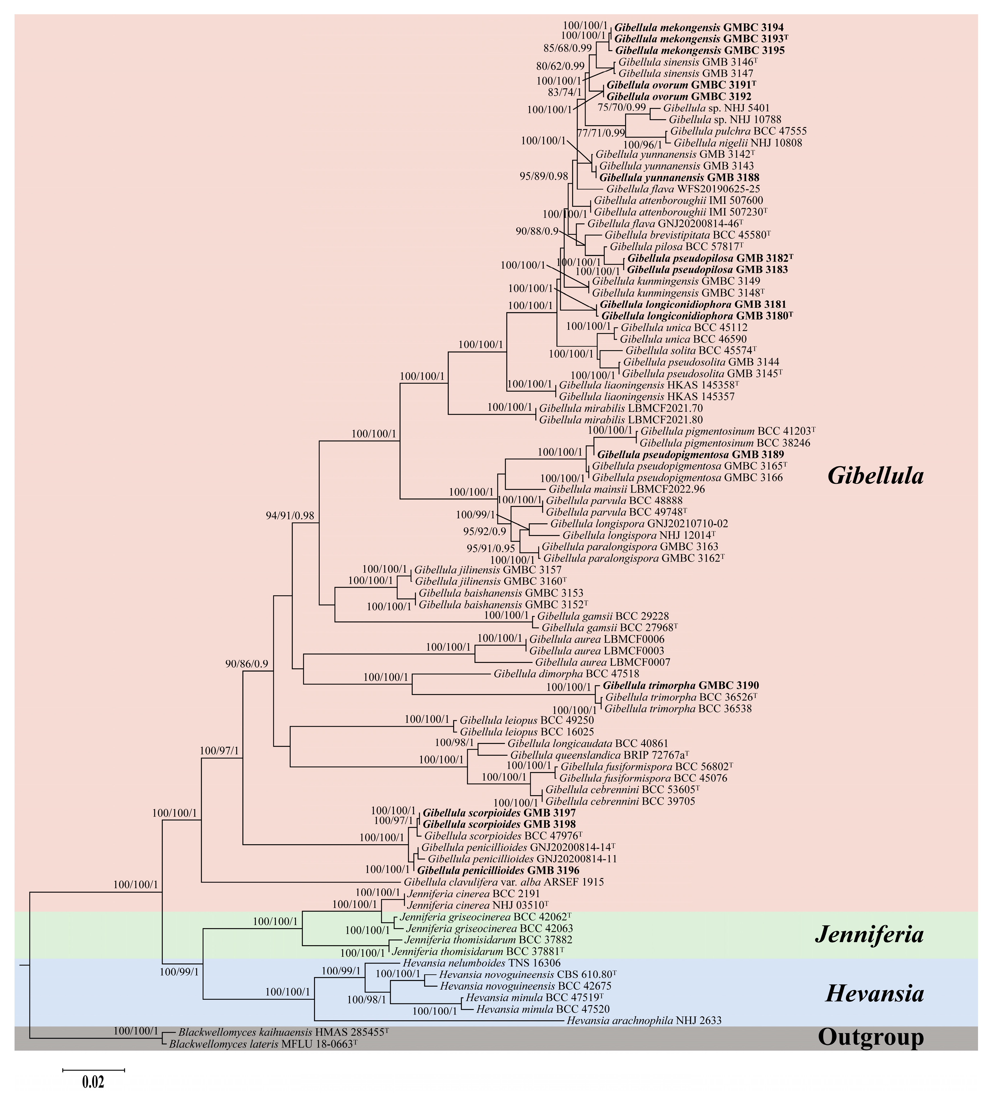
Phylogenetic relationships among Gibellula and its closely related genera were reconstructed based on a concatenated six-locus dataset (nrSSU, ITS, nrLSU, tef-1α, rpb1, and rpb2). The final alignment comprised 5699 base pairs, including nrSSU (1081 bp), ITS (780 bp), nrLSU (946 bp), tef-1α (992 bp), rpb1 (773 bp), and rpb2 (1127 bp). The dataset encompassed 92 fungal taxa, including 78 representatives of Gibellula, six of Hevansia, six of Jenniferia, and two of Blackwellomyces. Phylogenetic reconstructions obtained from Maximum Likelihood (ML) and Bayesian Inference (BI) analyses showed highly consistent topological structures. Node support was evaluated using ultrafast bootstrap values (BSIQ), RAxML bootstrap values (BSRAx), and Bayesian posterior probabilities (PP), and is presented throughout as BSIQ/BSRAx/PP. The analyses provided strong support for the monophyly of Gibellula (100%/100%/1), Hevansia (100%/100%/1), and Jenniferia (100%/100%/1). Hevansia and Jenniferia formed a strongly supported sister group (100%/99%/1), which was subsequently resolved as sister to Gibellula with well-support (100%/100%/1). Collectively, these three genera constituted a well-supported clade nested within Cordycipitaceae (Figure 1).
Within Gibellula, most taxa resolved into well-delimited, highly supported terminal clades, indicating clear species boundaries. Based on integrative morphological and phylogenetic evidence, four new species are described herein. Specimens GMB 3180 and GMB 3181 formed a strongly supported independent lineage closely clustering with G. kunmingensis, G. pseudosolita, G. solita, and G. unica (100%/100%/1). Despite its close phylogenetic affinity to these species, this lineage represents a distinct evolutionary branch with consistent morphological differences and is therefore established as Gibellula longiconidiophora sp. nov. Cultures of Gibellula mekongensis sp. nov. (GMBC 3193, GMBC 3194, GMBC 3195) formed a highly supported monophyletic lineage (100%/100%/1) and clustered with G. sinensis with moderate support (85%/68%/0.99). This subclade was sister to G. ovorum sp. nov. (GMBC 3191, GMBC 3192; 100%/100%/1) with moderate support (80%/62%/0.99), together forming a larger clade. Both novel species can be clearly distinguished from G. sinensis and from each other by molecular sequence divergence and morphological characteristics. Specimens GMB 3182 and GMB 3183 were resolved as a distinct lineage, Gibellula pseudopilosa sp. nov., which is strongly supported as the sister species to G. pilosa (100%/100%/1). Although phylogenetically closely related, the two species can be clearly distinguished by fixed nucleotide differences across these six nuclear loci. This molecular divergence, together with diagnostic morphological traits, robustly supports the species status of G. pseudopilosa.
Figure 1.
Phylogenetic tree of Gibellula and related genera based on a combined six-locus dataset (nrSSU + ITS + nrLSU + tef-1α + rpb1 + rpb2). Branch support values (BSIQ/BSRax/PP) above 75%/60%/0.9 are shown. Ex-type materials are marked with “T”. Bold labels indicate sequences generated in this study.
3.2. Taxonomy
In this study, nine spider-associated Gibellula species were recognized from fungal specimens collected in the Lancang–Mekong Basin. Four taxa are described as novel species, namely Gibellula longiconidiophora sp. nov., G. mekongensis sp. nov., G. ovorum sp. nov., and G. pseudopilosa sp. nov. The other five species represent new national distributional records for the study region: G. yunnanensis (new record for Laos, Oudomxay Province, Nam Kat Yorla Pa Resort; voucher specimen GMB 3188, no culture obtained), G. pseudopigmentosa (new record for Thailand, Chiang Mai Province, Maejo University; voucher specimen GMB 3189, no culture obtained), G. trimorpha (new record for Vietnam, Đắk Lắk Province, Chu Yang Sin National Park; living culture GMBC 3190), G. penicillioides (new record for Laos, Oudomxay Province, Nam Kat Yorla Pa Resort; voucher specimen GMB 3196, no culture obtained), and G. scorpioides (new records for China and Laos, voucher specimens GMB 3197 from Pu’er City, Yunnan Province, China, and GMB 3198 from Oudomxay Province, Nam Kat Yorla Pa Resort, Laos; no cultures obtained). Culture accession numbers are provided for isolates with viable living cultures, while taxa without cultures were identified exclusively based on morphological examination of dried voucher specimens. Comprehensive morphological descriptions, micrographs, and taxonomic comparisons with allied species are provided for the four newly described taxa in the following sections.
Gibellula longiconidiophora Y. Wang & H. Chen, sp. nov.
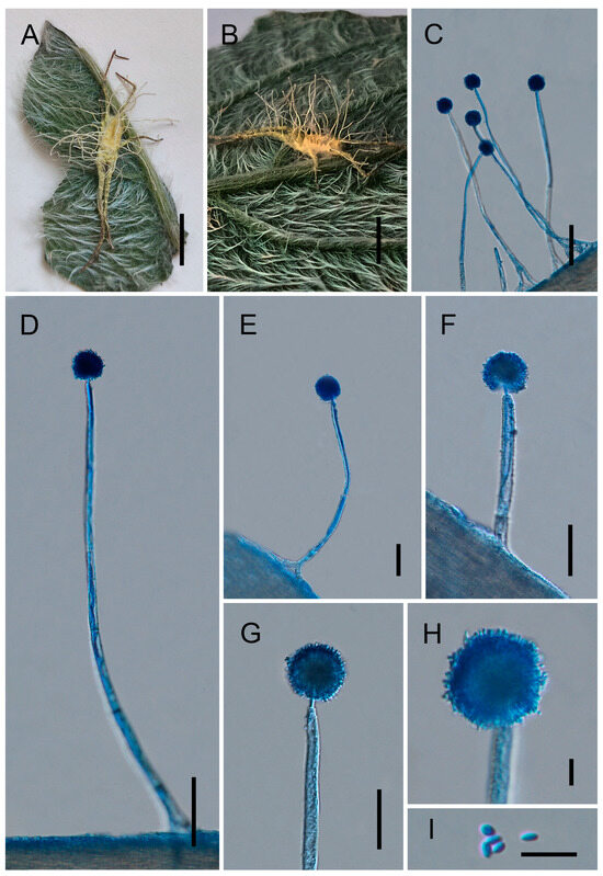
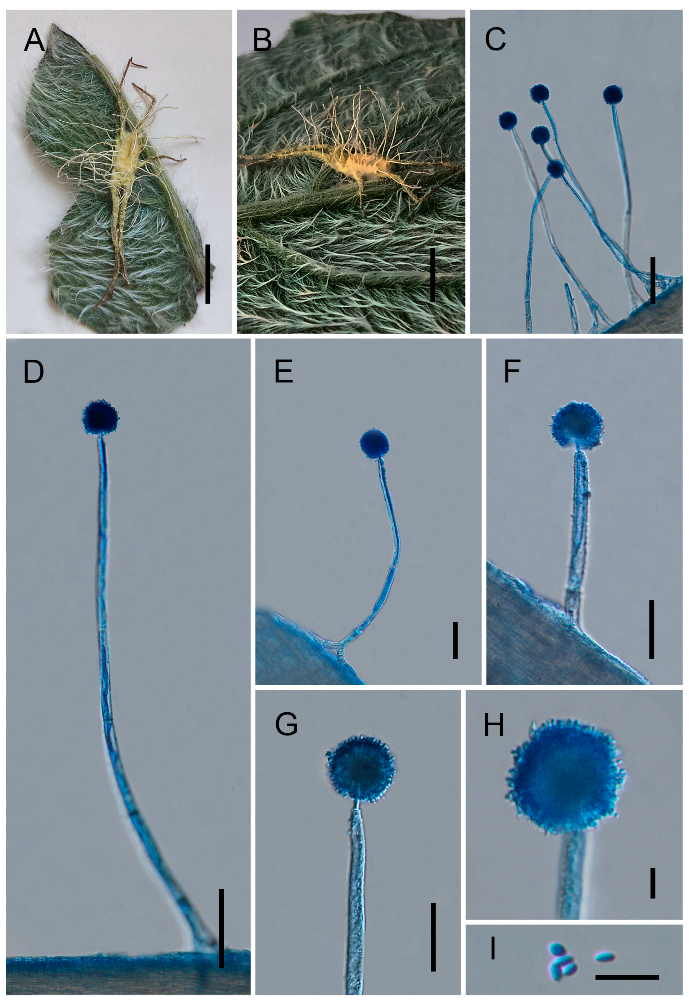
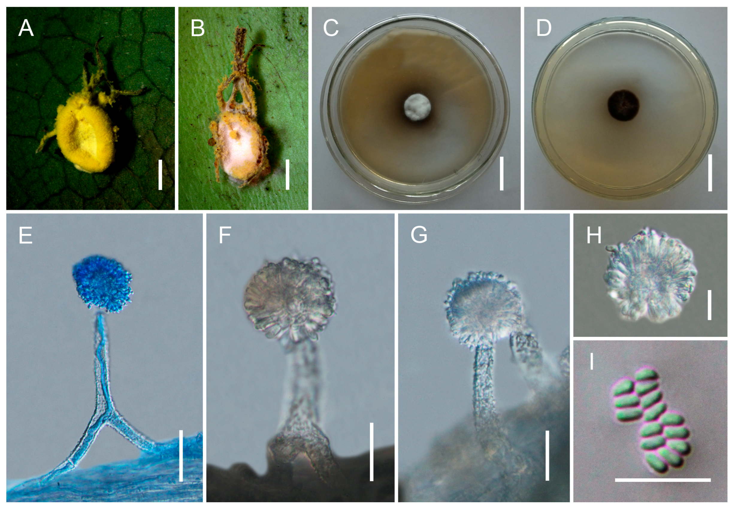
Figure 2.
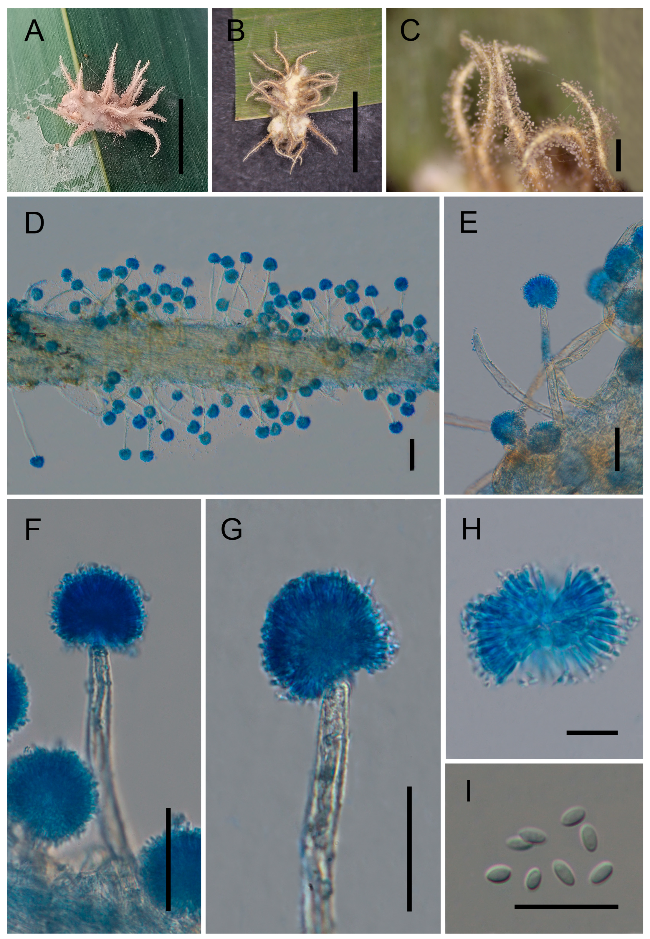
Morphology of Gibellula longiconidiophora. (A,B) Fungus on spider hosts. (C–H) Conidiophores showing conidial heads. (I) Conidia. Scale bars: 5 mm (A,B); 50 μm (C); 40 μm (D–G); 10 μm (H,I).
Mycobank No: 863473
Etymology. The epithet longiconidiophora refers to the long conidiophores produced by this species, from Latin longus, meaning long and conidiophorus, meaning conidium-bearing structure.
Type. China, Yunnan Province, Pu’er City (22.5270° N, 99.8777° E, 1125 m above sea level), on a spider from leaf litter on the forest floor, collected in August 2024 by Yao Wang (holotype: GMB 3180; ex-type living culture: not available).
Description. Teleomorph: Not observed. Anamorph: Mycelium yellow, covering the entire spider body and generally extending onto the legs, occasionally reaching the tarsi. Synnemata yellow, slender, cylindrical, tapering towards the apex, (2–)2.5–7(–9) mm long, formerly ca. 0.3 mm wide. Conidiophores densely aggregated, arising from the outer layer of synnemata and from hyphae attached to the host surface; short and stout, multiseptate, verrucose, (77–)182–297(–310) × (9–)10–14(–15) µm (0119 = 226 × 12 µm, n = 30); gradually shortening towards the synnematal apex, abruptly constricted into a distinct neck and expanding into a vesicle. Vesicles globose to subglobose, (13.5–)15–19(–20) µm ( = 18 µm, n = 30) in diameter, bearing a whorl of metulae. Metulae broadly obovoid, (5.5–)6.5–8(–9) × (2–)3–5(–7) µm ( = 7 × 4 μm, n = 30). Phialides narrowly clavate to cylindrical, (3.5–)5–7(–7.5) × 2.5–3.5 µm ( = 6 × 3 μm, n = 30), each producing a single conidium at the apex. Conidial heads subglobose, composed of vesicles, metulae, and phialides, (15–)17.5–38(–42.5) µm ( = 29 µm, n = 30) in diameter. Conidia ellipsoid to ovoid, occasionally subglobose, (1.5–)2.5–3.8(–4) × 1.2–1.8 µm ( = 3.2 × 1.5 μm, n = 50).
Distribution. Yunnan Provinces, China
Other Material Examined. China, Yunnan Province, Pu’er City (22.5380° N, 100.0047° E, 1615 m above sea level), on a spider from leaf litter on the forest floor, collected in August 2024 by Yao Wang (GMB 3181; living culture: not available).
Notes. Phylogenetically, Gibellula longiconidiophora forms a strongly supported independent lineage closely clustering with G. kunmingensis, G. pseudosolita, G. solita, and G. unica (100%/100%/1; Figure 1). This subclade is nested within a larger well-supported clade that also includes G. attenboroughii, G. brevistipitata, G. flava, G. nigelii, G. pilosa, G. pseudopilosa, G. pulchra, G. sinensis, G. yunnanensis, and other related species. Morphologically, G. longiconidiophora is a spider-parasitizing fungus characterized by yellow mycelia that entirely cover the host surface. The conidial dimensions are similar to those of G. kunmingensis and G. brevistipitata, measuring 2.5–3.8 × 1.2–1.8 μm in G. longiconidiophora, compared with 2–4 × 1.2–2 μm in G. kunmingensis and G. brevistipitata [44,45]. However, G. longiconidiophora can be clearly distinguished from other species of Gibellula by its unusually long conidiophores, measuring 182–297 × 10–14 μm, which are considerably longer than those of G. kunmingensis (52–120 × 14–16 μm) and G. brevistipitata (58–100 × 6–8 μm). The combination of its distinct phylogenetic position and morphological characteristics strongly supports the recognition of G. longiconidiophora as a distinct species.
Gibellula mekongensis Y. Wang & H. Chen, sp. nov.
Figure 3.
Morphology of Gibellula mekongensis. (A,B) Fungus on spider hosts. (C,D) Colonies on PDA (front and reverse). (E–G) Conidiophores showing conidial heads. (H) Conidial head. (I) Conidia. Scale bars: 10 mm (A,B); 20 mm (C,D); 30 µm (E–G); 10 µm (H,I).
Mycobank No: 863474
Etymology. The epithet mekongensis refers to the Mekong River basin, where the specimens were collected.
Type. China, Yunnan Province, Pu’er City (23.7834° N, 100.3243° E, 1223 m above sea level), on a spider attached to the underside of a leaf, collected in August 2023 by Yao Wang (holotype: GMB 3193; ex-type living culture: GMBC 3193).
Description. Conidiophores sparsely arising from the mycelia covering the host, smooth to finely verrucose, (76–)82–102(–112) × (9–)10–16(–18) μm ( = 94 × 14 μm, n = 20). Each conidiophore terminates in a swollen vesicle bearing metulae and phialides, forming globose conidial heads (20–)35–46(–49) μm ( = 41 µm, n = 20) in diameter. Vesicles mostly subglobose. Metulae broadly obovoid, (6–)7–10(–12) × (3–)4–7(–9) μm ( = 8 × 5.5 μm, n = 20). Phialides obovoid to clavate with short necks, (8–)9–13(–15) × 3–6(–7) μm ( = 12 × 5 μm, n = 20). Conidia obovoid, tapering at the apex, (1.8–)2.1–2.5(–2.8) × 1–1.5(–1.8) μm ( = 2.3 × 1.2 μm, n = 30). Granulomanus-type synanamorph not observed.
Culture characteristics. Colonies on PDA grow slowly at 25 °C, reaching 10–12 mm in diameter after 30 days. Mycelium initially white, becoming pale yellow to grayish brown with age. Sporulation not observed in culture.
Distribution. Currently known from Pu’er City, Yunnan Province, China; Đắk Lắk Province, (Chu Yang Sin National Park), Vietnam; and Chiang Mai Province (Maejo University), Thailand.
Other Material Examined. Vietnam, Đắk Lắk Province, Chu Yang Sin National Park (12.4180° N, 108.3530° E; 900 m above sea level), on a spider attached to the underside of a leaf, collected in Jul 2023 by Yao Wang (GMB 3194; living culture: GMBC 3194); Thailand, Chiang Mai Province, Maejo University (18.8958° N, 99.0133° E; 330 m above sea level), on spiders attached to the underside of leaves, collected in August 2023 by Yao Wang (GMB 3195; living culture: GMBC 3195).
Notes. Gibellula mekongensis is phylogenetically placed in a moderately supported clade with G. sinensis (85%/68%/0.99) and forms a highly supported monophyletic lineage (100%/100%/1) together with three isolates (GMBC 3193–3195). It is sister to G. ovorum sp. nov. (described below) with moderate support (80%/62%/0.99). Morphologically, G. mekongensis resembles G. sinensis in conidiophore structure and globose conidial heads, but can be clearly distinguished by its shorter and broader conidiophores (82–102 × 10–16 μm vs. 92–161 × 7.7–8.5 μm) [54] and significantly smaller conidia (2.1–2.5 × 1–1.5 μm vs. 3–4.5 × 1.7–2.3 μm). The bright yellow mycelial mat covering the host in G. mekongensis also differs from the whitish to pale yellow mycelium reported in G. sinensis [54]. These stable morphological differences, in combination with multilocus phylogenetic evidence, support the recognition of G. mekongensis as a distinct species.
Gibellula ovorum Y. Wang & H. Chen, sp. nov.
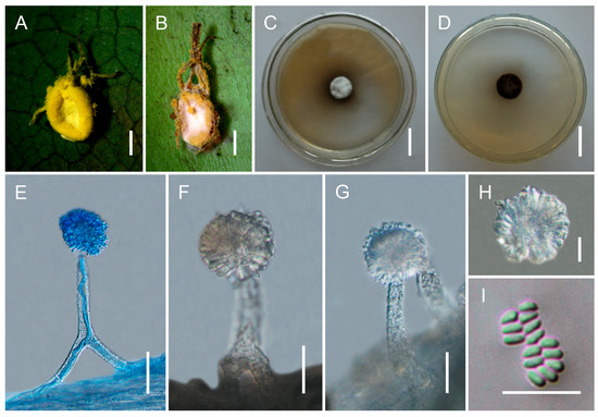
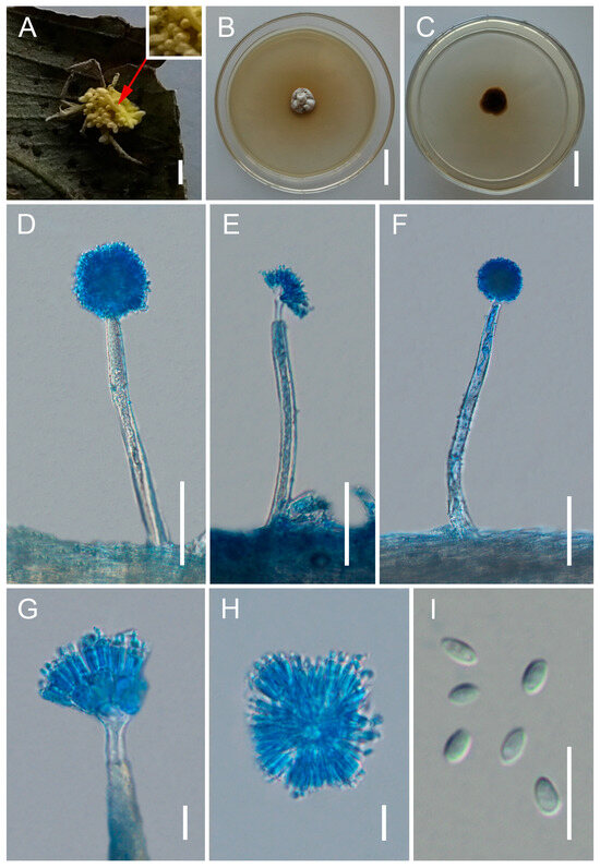
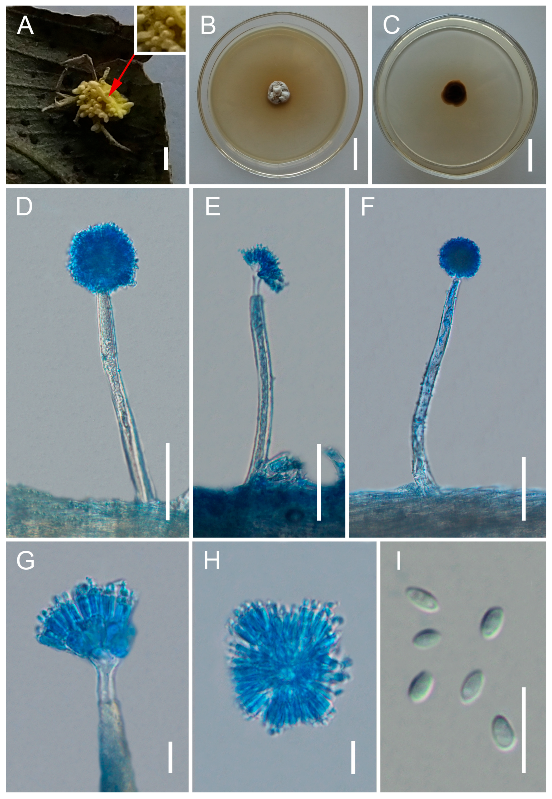
Figure 4.
Morphology of Gibellula ovorum. (A) Fungus on spider egg sacs, covered by a dense yellowish mycelial mat. The host structure is largely obscured due to rapid fungal overgrowth. The arrow indicates the position of the egg (egg sac) beneath the mycelium. Enlarged view of the indicated region showing the emerging egg structure. (B,C) Colonies on PDA (front and reverse). (D–G) Conidiophores showing conidial heads. (H) Conidial head. (I) Conidia. Scale bars: 10 mm (A); 20 mm (B,C); 50 µm (D–F); 10 µm (G–I).
Mycobank No: 863475
Etymology. The epithet ovorum (from Latin ovum, egg, genitive plural ovorum, meaning “of eggs”) refers to the spider egg sacs from which the fungus was collected.
Type. Laos, Oudomxay Province, Nam Kat Yorla Pa Resort (20.8112° N, 102.8545° E, 762 m above sea level), on spider egg sacs attached to the underside of a leaf, collected in August 2023 by Yao Wang (holotype: GMB 3191; ex-type living culture: GMBC 3191).
Description. Teleomorph: Not observed. Anamorph: Conidiophores sparsely arising from the mycelia covering the host, smooth to finely verrucose, (112–)132.5–158.9(–165) × (9–)10–12(–12.5) μm ( = 145.4 × 11.5 μm, n = 20). Each conidiophore terminates in a swollen vesicle bearing metulae and phialides, forming globose conidial heads (25–)28–47(–53) μm ( = 37.5 μm, n = 20) in diameter. Vesicles mostly subglobose. Metulae broadly obovoid, (8.5–)10–18(–24) × (3–)5–8(–9) μm ( = 15 × 6.5 μm, n = 20). Phialides obovoid to clavate with short necks, (7–)9–14(–18) × 2–4 μm ( = 12.4 × 2.5 μm, n = 20). Conidia obovoid, tapering at the apex, (3–)3.5–4.5(–5) × 1.7–2(–2.4) μm ( = 4 × 1.9 μm, n = 30). Granulomanus-type synanamorph not observed.
Culture characteristics. Colonies on PDA grow slowly at 25 °C, reaching 13–15 mm in diameter after 90 days. Mycelium initially white, becoming pale yellow to grayish brown with age. Sporulation not observed in culture.
Distribution. Currently known only from Oudomxay Province, Laos.
Other Material Examined. Laos, Oudomxay Province, Nam Kat Yorla Pa Resort (20.8767° N, 102.8632° E, 744 m above sea level), on spider egg sacs attached to the underside of a leaf, collected in August 2023 by Yao Wang (GMB 3192; living culture: GMBC 3192).
Notes. Gibellula ovorum is phylogenetically sister to the clade comprising G. mekongensis and G. sinensis with moderate support (80%/62%/0.99; Figure 1). Morphologically, G. ovorum can be clearly distinguished from G. mekongensis by its larger conidia (3.5–4.5 × 1.7–2 μm vs. 2.1–2.5 × 1–1.5 μm) and by its unique substrate (spider egg sacs vs. adult spiders). From G. sinensis, G. ovorum differs in having broader conidiophores (10–12 μm vs. 7.7–8.5 μm) [54] and in producing a yellowish mycelial mat that covers the host, in contrast to the whitish to pale yellow mycelium of G. sinensis that bears abundant conidiophores [54]. These morphological differences, together with phylogenetic evidence, support the recognition of G. ovorum as a distinct species.
Gibellula pseudopilosa Y. Wang & H. Chen, sp. nov.
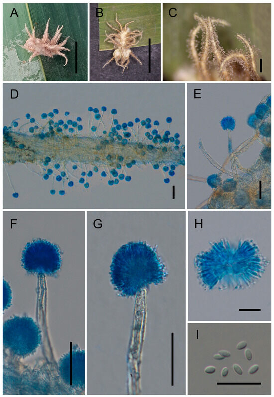
Figure 5.
Morphology of Gibellula pseudopilosa. (A,B) Fungus on spider hosts. (C,D) Detail of synnemata showing conidiophores. (E–G) Conidiophores showing conidial heads. (H) Conidial head. (I) Conidia. Scale bars: 10 mm (A,B); 500 μm (C); 70 μm (D); 40 μm (E–G); 10 μm (H,I).
Mycobank No: 863476
Etymology. The epithet pseudopilosa refers to its morphological resemblance to Gibellula pilosa, with the prefix pseudo-, meaning false.
Type. China, Yunnan Province, Pu’er City (22.5366° N, 100.0353° E, 1322 m above sea level), on a spider from leaf litter on the forest floor, collected in August 2024 by Yao Wang (holotype: GMB 3182; ex-type living culture: not available).
Description. Teleomorph: Not observed. Anamorph: Mycelium whitish-yellow, densely covering the entire spider body and usually extending onto the legs, forming a fluffy, raised mycelial layer. Synnemata yellow, slender, cylindrical, tapering towards the apex. Conidiophores densely aggregated, arising from the outer layer of synnemata and from hyphae attached to the host surface; short and stout, multiseptate, verrucose, (75–)95–131(–194) × (8–)9–12(–14) µm ( = 112 × 10 μm, n = 30); gradually shortening towards the synnematal apex, abruptly constricted into a distinct neck and expanding into a vesicle. Vesicles globose to subglobose, (15–)16–19(–20) µm ( = 17 µm, n = 30) in diameter, bearing a whorl of metulae. Metulae broadly obovoid, (6–)7–9(–11) × (1–)1.3–2 µm ( = 8 × 1.8 μm, n = 30). Phialides narrowly clavate to cylindrical, (4–)6–8(–9) × 2–3.4 µm ( = 6.5 × 2.5 μm, n = 30), each producing a single conidium at the apex. Conidial heads subglobose, composed of vesicles, metulae, and phialides, (28–)30.5–42(–50) µm ( = 38 µm, n = 30) in diameter. Conidia ellipsoid to ovoid, occasionally subglobose, (1.5–)2–3(–3.5) × 1.4–1.7 µm ( = 2.4 × 1.5 μm, n = 50).
Distribution. Yunnan Provinces, China.
Other Material Examined. China, Yunnan Province, Pu’er City (22.3865° N, 99.9385° E, 1478 m above sea level), on a spider from leaf litter on the forest floor, collected in August 2024 by Yao Wang (GMB 3183; living culture: not available).
Notes. Gibellula pseudopilosa is phylogenetically strongly supported as the sister species to G. pilosa (100%/100%/1; Figure 1). Morphologically, both species are spider-parasitizing fungi that produce mycelia completely covering the host surface [45]. However, G. pseudopilosa can be distinguished by its whitish-yellow mycelium, which forms a dense, fluffy, raised layer over the host, whereas G. pilosa has a less dense mycelial covering. Additionally, G. pseudopilosa typically produces multiple synnemata on a single host, while G. pilosa usually bears only two to three synnemata [45]. The two species also differ in conidial size: conidia of G. pseudopilosa measure 2–3 × 1.4–1.7 μm, notably smaller than those of G. pilosa (3–4 × 1.5–2 μm) [45]. These consistent morphological differences, together with phylogenetic evidence, strongly support the recognition of G. pseudopilosa as a distinct species.
4. Discussion
4.1. Summary of the Main Findings
In this study, we describe four new species of Gibellula—G. longiconidiophora, G. mekongensis, G. ovorum, and G. pseudopilosa—from the Lancang–Mekong region, together with five newly recorded species (G. yunnanensis, G. pseudopigmentosa, G. trimorpha, G. penicillioides, and G. scorpioides). These findings substantially expand the known diversity and geographic distribution of Gibellula within Cordycipitaceae. Notably, one of the new species, G. ovorum, represents a rare case of a Gibellula fungus parasitizing spider egg sacs rather than adult spiders, highlighting the previously overlooked ecological plasticity of the genus. The co-occurrence of multiple new taxa and new national records in a single study underscores both the high diversity of spider-pathogenic fungi in this biodiversity hotspot and the persistent sampling bias across many regions of Southeast and East Asia.
4.2. Taxonomic and Phylogenetic Significance
The integration of multilocus phylogenetic analyses (six loci: nrSSU, ITS, nrLSU, tef-1α, rpb1, rpb2) with detailed morphological comparisons provides robust evidence for species delimitation in Gibellula. The newly described species are distributed across several well-supported clades, reflecting both phylogenetic clustering and clear species-level divergence.
Gibellula longiconidiophora is nested within a large clade containing G. pseudosolita, G. solita, G. unica, G. kunmingensis, G. brevistipitata, G. pilosa, and related taxa, but forms a fully supported distinct lineage. Morphologically, it is readily distinguished by its exceptionally elongated conidiophores, a trait that appears evolutionarily labile and taxonomically informative. Similarly, G. pseudopilosa forms a strongly supported sister relationship with G. pilosa and can be consistently distinguished by a combination of subtle but stable features: dense whitish-yellow mycelium forming a raised hyphal layer, production of multiple synnemata on a single host, and smaller conidia. These cases illustrate that closely related species within Gibellula may undergo fine-scale morphological divergence, emphasizing the need for integrative taxonomy. In contrast, G. mekongensis and G. ovorum form a clade together with G. sinensis. They share similar conidiophore structures and globose conidial heads, reflecting close evolutionary relationships. However, they are reliably distinguished by differences in conidiophore dimensions, conidial size, and sporulation patterns on the host surface. Both G. mekongensis and G. ovorum produce a yellow mycelial mat that predominantly covers the host, with sparsely arranged conidiophores—a departure from the more typical, conidiophore-rich sporulation of G. sinensis. Such divergence in reproductive structures may reflect ecological or developmental differentiation and provides useful diagnostic characters. Moreover, G. ovorum is unique among the new species in its exclusive occurrence on spider egg sacs, a substrate preference that has rarely been documented in Gibellula. Overall, the strong concordance between multilocus phylogenetic evidence and stable morphological differentiation supports the recognition of the four new species. This study further demonstrates that the diversity of Gibellula, particularly in the Lancang–Mekong region, remains substantially underestimated, and that integrative taxonomy is essential for resolving species boundaries in this genus.
4.3. Biogeography and Distribution Patterns
The five newly recorded species reveal notable patterns of geographic expansion and distributional discontinuity within Gibellula. Gibellula yunnanensis, previously known only from Yunnan, China [44], is here recorded from Laos; G. pseudopigmentosa, originally described from Laos [54], is now documented in Thailand; G. trimorpha, previously known from Thailand [45], is newly recorded from Vietnam; G. penicillioides, previously known from Anhui Province, China [50], is newly recorded from Laos; and G. scorpioides, originally reported from Thailand [25], is confirmed from both China (Pu’er) and Laos. These cross-regional occurrences demonstrate that many Gibellula species have wider geographic distributions than previously recognized, often spanning multiple countries in Southeast and East Asia.
Such patterns suggest that the apparent geographic restriction reported in earlier studies is likely influenced by limited sampling rather than true distribution boundaries. Two main factors may account for these observations. First, Gibellula species may possess effective dispersal capabilities, including aerial conidial dissemination and potential host-mediated transport, facilitating long-distance movement. Second, the increasing number of new records across different countries highlights a strong sampling bias, particularly in tropical and subtropical regions where spider-associated fungi remain underexplored. Overall, these findings indicate that the Lancang–Mekong region represents a continuous biogeographic unit for spider-pathogenic fungi, rather than discrete, isolated distribution zones.
4.4. Ecological Implications
As obligate pathogens of spiders, Gibellula species may indirectly influence terrestrial food webs by affecting spider abundance and trophic interactions [59,60,61]. Field observations have reported relatively high infection rates of spiders by hypocrealean fungi in humid environments such as forests and agricultural systems [6,12,61]. Under favorable conditions, these fungi could contribute to regulating spider populations, although quantitative data on infection rates, host specificity, and ecological impact remain limited.
Among our findings, G. ovorum stands out as a rare example of a Gibellula species parasitizing spider egg sacs rather than adult spiders. This substrate shift may have profound ecological implications. Spider egg sacs represent a nutrient-rich, defenseless microhabitat that is often aggregated and spatially predictable, potentially offering a competitive advantage to fungi that can exploit this niche. To our knowledge, only a handful of spider-egg-associated fungi have been reported in Hypocreales, and G. ovorum is the first formally described Gibellula from this substrate in the Lancang–Mekong region. This discovery suggests that the ecological diversity of Gibellula may be broader than previously recognized, and that targeted sampling of spider egg sacs could reveal additional hidden diversity.
4.5. Host Association and Evolutionary Implications
Spider-pathogenic fungi in the genus Gibellula exhibit a high degree of host association, which likely plays a central role in their diversification. Previous studies have suggested that host specificity is a key driver of speciation in entomopathogenic fungi, particularly within Cordycipitaceae [13,62]. Although precise host identification remains challenging for many specimens, field observations indicate that Gibellula species often infect specific spider taxa occupying distinct ecological niches. Such host-associated differentiation may promote reproductive isolation and lead to lineage diversification. The discovery of G. ovorum on spider egg sacs adds a novel dimension to this host-associated diversification. Shifts to alternate substrates, such as from adult spiders to egg sacs, may represent a key innovation that reduces competition and facilitates niche partitioning. Comparative genomic or transcriptomic studies between G. ovorum and its close relatives (e.g., G. mekongensis) could uncover genetic adaptations underlying this substrate transition. The expanded distribution patterns revealed in this study provide insight into the relative roles of host distribution and fungal dispersal in shaping Gibellula biogeography. The occurrence of identical or closely related species across multiple countries in the Lancang–Mekong region suggests that dispersal limitation may be less restrictive than previously assumed. Instead, the continuity of suitable habitats and the widespread distribution of spider hosts may facilitate gene flow or repeated colonization events, as observed in other hypocrealean fungi [44,61].
Finally, the integration of morphological and multilocus phylogenetic data highlights the potential presence of cryptic diversity within Gibellula. Morphologically similar taxa may represent genetically distinct lineages, indicating that species richness in this genus is likely underestimated—a pattern consistent with broader trends in fungal systematics [17,63]. Taken together, these findings suggest that the evolution of Gibellula is shaped by a combination of host association, ecological factors, and historical biogeographic processes.
Author Contributions
Conceptualization, Y.W.; methodology, B.T. and H.C.; software, X.Z.; validation, D.-X.T.; formal analysis, B.T. and H.C.; investigation, V.-M.D., C.L. and Y.W.; resources, Y.W.; data curation, X.Z.; writing—original draft preparation, H.C. and B.T.; writing—review and editing, Y.W.; visualization, B.T.; supervision, Y.W.; project administration, Y.W.; funding acquisition, Y.W. and B.T. All authors have read and agreed to the published version of the manuscript.
Funding
This study was jointly supported by the National Natural Science Foundation of China under grant [32460004], and the Science and Technology Planning Project of Guizhou Province ([2024]023).
Institutional Review Board Statement
Not applicable.
Informed Consent Statement
Not applicable.
Data Availability Statement
The DNA sequence data obtained in this study have been deposited in GenBank. The accession numbers can be found in the article (Table 1).
Conflicts of Interest
The authors declare no conflicts of interest.
References
- Andersen, S.B.; Gerritsma, S.; Yusah, K.M.; Mayntz, D.; Hywel-Jones, N.L.; Billen, J.; Boomsma, J.J.; Hughes, D.P. The life of a dead ant: The expression of an adaptive extended phenotype. Am. Nat. 2009, 174, 424–433. [Google Scholar] [CrossRef]
- Hughes, D.P.; Araújo, J.P.M.; Loreto, R.G.; Quevillon, L.; de Bekker, C.; Evans, H.C. From so simple a beginning: The evolution of behavioural manipulation by fungi. Adv. Genet. 2016, 94, 437–469. [Google Scholar]
- Samson, R.A.; Evans, H.C.; Latgé, J.P. Atlas of Entomopathogenic Fungi; Springer: Berlin/Heidelberg, Germany, 1988. [Google Scholar]
- Pontoppidan, M.B.; Himaman, W.; Hywel-Jones, N.L.; Boomsma, J.J.; Hughes, D.P. Graveyards on the move: The spatio-temporal distribution of dead Ophiocordyceps-infected ants. PLoS ONE 2009, 4, e4835. [Google Scholar] [CrossRef] [Scilit]
- Poulin, R. Parasite manipulation of host behaviour: An update and frequently asked questions. Adv. Study Behav. 2010, 41, 151–186. [Google Scholar]
- Samson, R.A.; Evans, H.C. Notes on entomogenous fungi from Ghana. II. The genus Akanthomyces. Acta Bot. Neerl. 1974, 23, 28–35. [Google Scholar] [CrossRef] [Scilit]
- Sánchez-Peña, S.R. Entomopathogenic fungi associated with insects. Southwest. Entomol. 1990, 15, 31–38. [Google Scholar]
- Araújo, J.P.M.; Evans, H.C.; Geiser, D.M.; Mackay, W.P.; Hughes, D.P. Unravelling the diversity behind the Ophiocordyceps unilateralis complex: Three new species of zombie-ant fungi from the Brazilian Amazon. Phytotaxa 2018, 220, 224–238. [Google Scholar] [CrossRef] [Scilit]
- Cooley, J.R.; Marshall, D.C.; Hill, K.B. A specialized fungal parasite (Massospora) manipulates cicada behaviour. Sci. Rep. 2018, 8, 1432. [Google Scholar] [CrossRef] [Scilit]
- Mendes-Pereira, T.; de Araújo, J.P.M.; Mendes, F.; Fonseca, E. Gibellula aurea sp. nov. (Ascomycota, Cordycipitaceae): A new golden spider-devouring fungus from a Brazilian Atlantic Rainforest. Phytotaxa 2022, 573, 85–102. [Google Scholar] [CrossRef] [Scilit]
- Evans, H.C. Fungal pathogens of spiders. In Spider Ecophysiology; Springer: Berlin/Heidelberg, Germany, 2013. [Google Scholar]
- Evans, H.C. Natural control of arthropods with fungi. Mycopathol. Mycol. Appl. 1974, 52, 189–200. [Google Scholar]
- Sung, G.H.; Hywel-Jones, N.L.; Sung, J.M.; Luangsa-Ard, J.J.; Shrestha, B.; Spatafora, J.W. Phylogenetic classification of Cordyceps and the clavicipitaceous fungi. Stud. Mycol. 2007, 57, 5–59. [Google Scholar] [CrossRef] [Scilit]
- Mains, E.B. Entomogenous species of Akanthomyces, Hymenostilbe and Insecticola in north America. Mycologia 1950, 42, 566–589. [Google Scholar] [CrossRef] [Scilit]
- Samson, R.A.; Evans, H.C. New species of Gibellula on spiders (Araneida) from South America. Mycologia 1992, 84, 300–314. [Google Scholar] [CrossRef] [Scilit]
- Kobayasi, Y.; Shimizu, D. Some species of Cordyceps and its allies on spiders. Kew Bull. 1977, 31, 557–566. [Google Scholar] [CrossRef] [Scilit]
- Tzean, S.S.; Hsieh, L.S.; Wu, W.J. Torrubiella dimorpha, a new species of spider parasite from Taiwan. Mycol. Res. 1998, 102, 1350–1354. [Google Scholar] [CrossRef] [Scilit]
- Turland, N.J.; Wiersema, J.H.; Barrie, F.R.; Greuter, W.; Hawksworth, D.L.; Herendeen, P.S.; Smith, G.F. International Code of Nomenclature for Algae, Fungi, and Plants (Shenzhen Code); Koeltz Botanical Books: Koenigstein, Germany, 2018. [Google Scholar]
- Kepler, R.M.; Luangsa-Ard, J.J.; Hywel-Jones, N.L.; Quandt, C.A.; Sung, G.H.; Rehner, S.A.; Aime, M.C.; Henkel, T.W.; Sanjuan, T.; Zare, R.; et al. A phylogenetically-based nomenclature for Cordycipitaceae (Hypocreales). IMA Fungus 2017, 8, 335–353. [Google Scholar] [CrossRef] [Scilit]
- Chen, W.H.; Han, Y.F.; Liang, Z.Q.; Jin, D.C. A new araneogenous fungus in the genus Beauveria from Guizhou, China. Phytotaxa 2017, 302, 57–64. [Google Scholar] [CrossRef] [Scilit]
- Han, Y.F.; Chen, W.H.; Zou, X.; Liang, Z.Q. Gibellula curvispora, a new species of Gibellula. Mycosystema 2013, 32, 777–780. [Google Scholar]
- Han, Y.F.; Liang, J.D.; Liu, A.Y.; Zeng, N.K.; Liang, Z.Q. New species of Gibellula from China. Mycol. Prog. 2013, 12, 567–575. [Google Scholar]
- Kuephadungphan, W.; Macabeo, A.P.G.; Luangsa-Ard, J.J.; Tasanathai, K.; Thanakitpipattana, D.; Phongpaichit, S.; Yuyama, K.; Stadler, M. Studies on the biologically active secondary metabolites of the new spider parasitic fungus Gibellula gamsii. Mycol. Prog. 2019, 18, 135–146. [Google Scholar] [CrossRef] [Scilit]
- Zou, X.; Liu, A.Y.; Liang, Z.Q.; Han, Y.F.; Yang, M. New species of Gibellula from China. Mycosystema 2016, 35, 12–20. [Google Scholar]
- Kuephadungphan, W.; Tasanathai, K.; Petcharad, B.; Khonsanit, A.; Stadler, M.; Luangsa-Ard, J.J. Phylogeny and morphology-based recognition of new species in the spider-parasitic genus Gibellula (Hypocreales, Cordycipitaceae) from Thailand. MycoKeys 2020, 72, 17–42. [Google Scholar] [CrossRef] [Scilit]
- Kuephadungphan, W.; Tasanathai, K.; Thanakitpipattana, D.; Luangsa-Ard, J.J. Phylogeny and taxonomy of Gibellula. Fungal Biol. 2020, 124, 731–748. [Google Scholar]
- White, T.J.; Bruns, T.; Lee, S.; Taylor, J.W. Amplification and direct sequencing of fungal ribosomal RNA genes for phylogenetics. In PCR Protocols: A Guide to Methods and Applications; Academic Press: San Diego, CA, USA, 1990; pp. 315–322. [Google Scholar] [CrossRef] [Scilit]
- Wang, Y.B.; Yu, H.; Dai, Y.D.; Chen, Z.H.; Zeng, W.B.; Yuan, F.; Liang, Z.Q. Polycephalomyces yunnanensis (Hypocreales), a new species of Polycephalomyces parasitizing Ophiocordyceps nutans and stink bugs (hemipteran adults). Phytotaxa 2015, 208, 34–44. [Google Scholar] [CrossRef] [Scilit]
- Vilgalys, R.; Hester, M. Rapid genetic identification and mapping of enzymatically amplified ribosomal DNA from several Cryptococcus species. J. Bacteriol. 1990, 172, 4238–4246. [Google Scholar] [CrossRef] [Scilit]
- Rehner, S.A.; Samuels, G.J. Taxonomy and phylogeny of Gliocladium analysed from nuclear large subunit ribosomal DNA sequences. Mycol. Res. 1994, 98, 625–634. [Google Scholar] [CrossRef] [Scilit]
- Rehner, S.A.; Buckley, E. A Beauveria phylogeny inferred from nuclear ITS and EF1-α sequences: Evidence for cryptic diversification and links to Cordyceps teleomorphs. Mycologia 2005, 97, 84–98. [Google Scholar] [CrossRef] [Scilit]
- Liu, Y.J.; Whelen, S.; Hall, B.D. Phylogenetic relationships among ascomycetes: Evidence from an RNA polymerase II subunit. Mol. Biol. Evol. 1999, 16, 1799–1808. [Google Scholar] [CrossRef] [Scilit] [PubMed]
- Bischoff, J.F.; Rehner, S.A.; Humber, R.A. Metarhizium frigidum sp. nov. A cryptic species of M. anisopliae and a member of the M. flavoviride complex. Mycologia 2006, 98, 737–745. [Google Scholar] [CrossRef]
- Kumar, S.; Stecher, G.; Tamura, K. MEGA7: Molecular evolutionary genetics analysis version 7.0 for bigger datasets. Mol. Biol. Evol. 2016, 33, 1870–1874. [Google Scholar] [CrossRef] [Scilit] [PubMed]
- Edler, D.; Klein, J.; Antonelli, A.; Silvestro, D. Raxml GUI 2.0: A graphical interface and toolkit for phylogenetic analyses using RAxML. Methods Ecol. Evol. 2021, 12, 373–377. [Google Scholar] [CrossRef] [Scilit]
- Minh, B.Q.; Schmidt, H.A.; Chernomor, O.; Schrempf, D.; Woodhams, M.D.; von Haeseler, A.; Lanfear, R. IQ-TREE 2: New models and efficient methods for phylogenetic inference in the genomic era. Mol. Biol. Evol. 2020, 37, 1530–1534. [Google Scholar] [CrossRef] [Scilit]
- Darriba, D.; Taboada, G.L.; Doallo, R.; Posada, D. jModelTest 2: More models, new heuristics and parallel computing. Nat. Methods 2012, 9, 772. [Google Scholar] [CrossRef] [Scilit] [PubMed]
- Ronquist, F.; Teslenko, M.; van der Mark, P.; Ayres, D.L.; Darling, A.; Höhna, S.; Larget, B.; Liu, L.; Suchard, M.A.; Huelsenbeck, J.P. MrBayes 3.2: Efficient Bayesian phylogenetic inference and model choice across a large model space. Syst. Biol. 2012, 61, 539–542. [Google Scholar] [CrossRef] [Scilit] [PubMed]
- Letunic, I.; Bork, P. Interactive Tree of Life (iTOL) v6: Recent updates to the phylogenetic tree display and annotation tool. Nucleic Acids Res. 2024, 52, 78–82. [Google Scholar] [CrossRef] [Scilit]
- Li, Y.; Zhao, X.C.; Wu, L.X.; Wang, Y.; Xu, A.; Lin, W.F. Blackwellomyces kaihuaensis and Metarhizium putuoense (Hypocreales), two new entomogenous fungi from subtropical forests in Zhejiang Province, Eastern China. Forests 2023, 14, 2333. [Google Scholar] [CrossRef] [Scilit]
- Hyde, K.D.; Tennakoon, D.S.; Jeewon, R.D.; Bhat, J.; Maharachchikumbura, S.S.N.; Rossi, W.; Leonardi, M.; Lee, H.B.; Mun, H.Y.; Houbraken, J.; et al. Fungal diversity notes 1036–1150: Taxonomic and phylogenetic contributions on genera and species of fungal taxa. Fungal Divers. 2019, 96, 1–242. [Google Scholar] [CrossRef] [Scilit]
- Alves, J.E.R.; Santos, A.C.S.; Belo Pedroso, S.K.; Ribeiro Melo, R.F.; Tiago, P.V. Untangling a web of spider fungi: Gibellula agroflorestalis (Hypocreales, Ascomycota), a new species of spider parasite from Brazil. J. Invertebr. Pathol. 2025, 209, 108278. [Google Scholar] [CrossRef] [Scilit]
- Evans, H.C.; Fogg, T.; Buddie, A.G.; Yeap, Y.T.; Araújo, J.P.M. The araneopathogenic genus Gibellula (Cordycipitaceae: Hypocreales) in the British Isles, including a new zombie species on orb-weaving cave spiders (Metainae: Tetragnathidae). Fungal Syst. Evol. 2025, 15, 153–178. [Google Scholar] [CrossRef] [Scilit]
- Tu, B.; Chen, H.; Zhang, X.; Guan, Y.-H.; Tang, D.-X.; Li, Q.-R.; Wang, Y. Five New Species of Gibellula (Hypocreales, Cordycipitaceae) from China. J. Fungi 2025, 11, 891. [Google Scholar] [CrossRef] [Scilit]
- Kuephadungphan, W.; Petcharad, B.; Tasanathai, K.; Thanakitpipattana, D.; Kobmoo, N.; Khonsanit, A.; Samson, R.A.; Luangsa-Ard, J.J. Multilocus phylogeny unmasks hidden species within the specialised spider-parasitic fungus, Gibellula (Hypocreales, Cordycipitaceae) in Thailand. Stud. Mycol. 2022, 101, 245–286. [Google Scholar] [CrossRef] [Scilit]
- Chaverri, P.; Bischoff, J.F.; Evans, H.C.; Hodge, K.T. Regiocrella, a new entomopathogenic genus with a pycnidial anamorph and its phylogenetic placement in the Clavicipitaceae. Mycologia 2005, 97, 1225–1237. [Google Scholar] [CrossRef]
- Spatafora, J.W.; Sung, G.H.; Sung, J.M.; Hywel-Jones, N.L.; White, J.F. Phylogenetic evidence for an animal pathogen origin of ergot and the grass endophytes. Mol. Ecol. 2007, 16, 1701–1711. [Google Scholar] [CrossRef] [Scilit] [PubMed]
- Chen, M.J.; Wang, T.; Lin, Y.A.N.; Huang, B.O. Gibellula flava sp. nov. (Cordycipitaceae, Hypocreales), a new pathogen of spider from China. Phytotaxa 2021, 527, 125–133. [Google Scholar] [CrossRef] [Scilit]
- Liu, Z.L.; Wei, D.P.; Chen, J.H.; Wu, W.J.; Liu, Z.H.; Zhang, W.; Chen, H.; Peng, X.C.; Kang, J.C.; Qian, Y.X.; et al. A new spider-pathogenic species Gibellula liaoningensis (Cordycipitaceae) from Liaoning Province, China. Phytotaxa 2025, 702, 48–60. [Google Scholar] [CrossRef] [Scilit]
- Chen, M.; Wang, T.; Lin, Y.; Huang, B. Morphological and molecular analyses reveal two new species of Gibellula (Cordycipitaceae, Hypocreales) from China. MycoKeys 2022, 90, 53–69. [Google Scholar] [CrossRef] [Scilit] [PubMed]
- Johnson, D.; Sung, G.H.; Hywel-Jones, N.L.; Luangsa-Ard, J.J.; Bischoff, J.F.; Kepler, R.M.; Spatafora, J.W. Systematics and evolution of the genus Torrubiella (Hypocreales, Ascomycota). Mycol. Res. 2009, 113, 279–289. [Google Scholar] [CrossRef] [Scilit]
- Mendes-Pereira, T.; de Araújo, J.P.M.; Kloss, T.G.; Costa-Rezende, D.H.; de Carvalho, D.S.; Góes-Neto, A. Disentangling the taxonomy, systematics, and life history of the spider-parasitic fungus Gibellula (Cordycipitaceae, Hypocreales). J. Fungi 2023, 9, 457. [Google Scholar] [CrossRef] [Scilit]
- Helaly, S.E.; Kuephadungphan, W.; Phainuphong, P.; Ibrahim, M.A.A.; Tasanathai, K.; Mongkolsamrit, S.; Luangsa-Ard, J.J.; Phongpaichit, S.; Rukachaisirikul, V.; Stadler, M. Pigmentosins from Gibellula sp. as antibiofilm agents and a new glycosylated asperfuran from Cordyceps javanica. Beilstein J. Org. Chem. 2019, 15, 2968–2981. [Google Scholar] [CrossRef] [Scilit]
- Chang, C.X.; Chen, H.; Loinheuang, C.; Dai, Y.-D.; Wang, Y. Morphological and phylogenetic analyses reveal three new species of Gibellula (Cordycipitaceae, Hypocreales) from spiders. MycoKeys 2026, 127, 135–154. [Google Scholar] [CrossRef] [Scilit]
- Tan, Y.P.; Shivas, R.G. Nomenclatural novelties. Index Aust. Fungi 2023, 10, 1–17. [Google Scholar]
- Kuephadungphan, W.; Phongpaichit, S.; Luangsa-Ard, J.J.; Rukachaisirikul, V. Antimicrobial activity of invertebrate-pathogenic fungi in the genera Akanthomyces and Gibellula. Mycoscience 2014, 55, 127–133. [Google Scholar] [CrossRef] [Scilit]
- Mongkolsamrit, S.; Noisripoom, W.; Tasanathai, K.; Kobmoo, N.; Thanakitpipattana, D.; Khonsanit, A.; Petcharad, B.; Himaman, W. Comprehensive treatise of Hevansia and three new genera Jenniferia, Parahevansia and Polystromomyces on spiders in Cordycipitaceae from Thailand. MycoKeys 2022, 91, 113–149. [Google Scholar] [CrossRef] [Scilit] [PubMed]
- Mongkolsamrit, S.; Noisripoom, W.; Tasanathai, K.; Khonsanit, A.; Thanakitpipattana, D.; Himaman, W.; Kobmoo, N.; Luangsa-Ard, J.J. Molecular phylogeny and morphology reveal cryptic species in Blackwellomyces and Cordyceps (Cordycipitaceae) from Thailand. Mycol. Prog. 2020, 19, 957–983. [Google Scholar] [CrossRef] [Scilit]
- Nyffeler, M.; Birkhofer, K. An estimated 400–800 million tons of prey are annually killed by the global spider community. Sci. Nat. 2017, 104, 30. [Google Scholar] [CrossRef] [Scilit] [PubMed]
- Nyffeler, M.; Sunderland, K.D. Composition, abundance and pest control potential of spider communities in agroecosystems: A comparison of European and US studies. Agric. Ecosyst. Environ. 2003, 95, 579–612. [Google Scholar] [CrossRef] [Scilit]
- Meyling, N.V.; Thorup-Kristensen, K.; Eilenberg, J. Below- and aboveground abundance and distribution of fungal entomopathogens in experimental conventional and organic cropping systems. Biol. Control 2011, 59, 180–186. [Google Scholar] [CrossRef] [Scilit]
- Boomsma, J.J.; Jensen, A.B.; Meyling, N.V.; Eilenberg, J. Evolutionary interaction networks of insect pathogenic fungi. Annu. Rev. Entomol. 2014, 59, 467–485. [Google Scholar] [CrossRef] [Scilit]
- Hawksworth, D.L.; Rossman, A.Y. Where are all the undescribed fungi? Phytopathology 1997, 87, 888–891. [Google Scholar] [CrossRef] [Scilit] [PubMed]

Disclaimer/Publisher’s Note: The statements, opinions and data contained in all publications are solely those of the individual author(s) and contributor(s) and not of MDPI and/or the editor(s). MDPI and/or the editor(s) disclaim responsibility for any injury to people or property resulting from any ideas, methods, instructions or products referred to in the content. |
© 2026 by the authors. Licensee MDPI, Basel, Switzerland. This article is an open access article distributed under the terms and conditions of the Creative Commons Attribution (CC BY) license.