


Abstract
Emericellopsis species from extreme environmental conditions provide a rich source of unique and biologically active secondary metabolites. The paper exhibits a comprehensive genomic analysis including complete genome sequencing, phylogenetic reconstruction, and functional annotation of two Emericellopsis species from highly saline and alkaline coastal soil ecosystems. Comparative genomics has been applied to reveal the genetic evolution, metabolic diversity, and environmental adaptation of the Emericellopsis genus. The genomes of E. alkalina E101 and E. fimetaria p24 have been found to encode various enzymes, including carbohydrate-active enzymes such as endoxylanases, that are useful for many ecological adaptations. The genomes of E. alkalina E101 and E. fimetaria p24 feature numerous biosynthetic gene clusters (BGCs), capable of synthesizing both known and potentially novel secondary metabolites with antimicrobial activity. Some BGCs show similarity to those producing known secondary metabolites, such as leucostatin A/B, clavaric acid, ascochlorin, (-)-mellein, and apicidin, among others. However, the majority of BGCs do not display any known similarities. Thus, comparative genomics offers new insights into the biology, adaptation, and evolutionary history of Emericellopsis fungi and may serve as a highly useful tool within biotechnological applications.
1. Introduction
The genus Emericellopsis (Bionectriaceae, Hypocreales, Ascomycota) was first named by Dutch botanist J.F.H. van Beyma in 1940 to accommodate the species E. terricola and its variety E. terricola var. glabra [1]. Fungi that belong to this genus are found within both terrestrial and marine habitats: in soil and as endophytes of plants, in animal and bird droppings, in sediments of freshwater and salt continental lakes, as epiphytes and endophytes of sea plants, in marine bottom deposits, and in the cavities of the sponges [2,3]. Currently, 31 species are recognized within the genus Emericellopsis, which phylogenetically is divided into two large clades—“marine” and “terrestrial” [2,3]. However, such division does not fully reflect substrate specificity since some species from the “terrestrial” clade occur in marine environments while others from the “marine” clade inhabit terrestrial ecosystems. Previously, a separate “soda” clade with only one species E. alkalina has been proposed adjacent to the “marine” group [4].
The species E. alkalina Bilanenko & Georgieva [4] was first described in 2013 from highly alkaline (pH 10.1) saline soil from the edge of soda lake Tanatar-2, Altai Krai, Russia. This species is widely distributed in coastal soils of saline and soda lakes in the Altai region and Zabaykalye (Russian Federation), later being recorded in sediment samples from saline lakes in Egypt, Red Sea sediments, and deep-sea Atlantic Ocean sediments [4,5,6,7]. The preferred substrates for this species remain debatable. It is an alkaliphilic halotolerant species, with optimal growth occurring at medium pH values ranging between 9.5 and 10.0 [4,8]. E. alkalina produces a complex of bioactive peptaibols known as emericellipsins (EmiA–E), which exhibit potent antifungal activity against pathogenic fungi, including drug-resistant Candida spp. and Aspergillus spp. The maximum production of these compounds occurs under high alkaline conditions, specifically at pH 10–11 [9].
The species E. fimetaria (Persoon) L.W. Hou, L. Cai & Crous was initially described in 1799 by the mycologist Christian Hendrik Persoon as Leotia fimetaria on cattle dung [10], and in 2023, based on phylogenetic reconstruction, this species was placed into the genus Emericellopsis within the “terrestrial” clade [2]. Most records of this species, starting with L. fimetaria, were listed on excrement of various animals (mainly herbivores) and birds worldwide, as noted by Lindau and Seifert, but recent findings include occurrences in other biotopes: marine sediments, sediments of the Tambukan saline lake (North Caucasus, Russia) [11,12,13,14]. It is known that strains of E. fimetaria produce diverse compounds with antimicrobial activity, including peptaibols like antiamoebins and terpenoid-like substances [11,12,15,16,17].
Currently, genomes of three Emericellopsis species have been published: E. atlantica [18], E. cladophorae [19], and E. maritima [20]. An unannotated genome assembly of E. terricola is available in GenBank (Genbank: GCA_027569275.1). The analysis of these genomes aimed at identifying clusters of genes involved in secondary metabolism (BGC) reveals their potential capacity for biosynthesis of diverse nonribosomal peptides and terpenoids, which may exhibit antimicrobial properties.
The aim of this study is whole-genome sequencing and functional annotation of two Emericellopsis species: E. alkalina E101 ex-type strain and E. fimetaria p24 strain, followed by an analysis of their potential as producers of different antimicrobial compounds.
2. Materials and Methods
2.1. Strains
Emericellopsis alkalina E101 strain (=CBS 127350 =VKM-F 4108, ex-type) was isolated from soil of the edge of soda lake Tanatar-2 in 2002 (Altai Region, Russia); soil pH 10.1, soluble salt content 73 g/kg [4]. Emericellopsis fimetaria p24 strain was isolated from sediment samples of the Tambukan saline lake in 2018 (Northern Caucasus, Russia); sample pH range 7.5–8.0, water mineralization 27.3 g/L [14].
All genomes utilized for comparative purposes have been obtained from publicly accessible databases: E. atlantica TS7 (ex-type, from the sponge Stelletta normani, Northern Atlantic Ocean) [18] (Genbank: GCF_019669845.1), E. cladophorae MUM 19.33 (=CMG 25, ex-type, endophyte of Cladophora sp., Atlantic Ocean) [19] (Genbank: GCF_022114955.1), E. maritima CBS 491.71 (ex-type, coastal water from Black Sea) [20] (Mycocosm: Ememar1) and E. terricola NRRL 54109 (weed from root crown crop field, USA) (Genbank: GCA_027569275.1).
Fungal strains were cultivated for 5 days in potato-glucose broth at the temperature of 25 °C and agitation speed of 120 rpm in 250 mL flask. Mycelium has been separated by centrifugation at 4740× g and +4 °C.
2.2. DNA Extraction
High-molecular-weight fungal genomic DNA has been extracted from the mycelium using the SKYHMW Plant Genomic DNA kit (SkyGen, Moskov, Russia) in accordance with the manufacturer’s instructions and used for long-read DNA sequencing (Nanopore). Genomic DNA for short-read sequencing has been obtained using a kit for the extraction and purification of genomic DNA from animal and bacterial cell cultures, animal and plant tissues (DU. Cells, tissues, blood DNA isolation kit, Biolabmix, Novosibirsk, Russia). The concentration of the extracted DNA has been estimated using UV spectrometry on an EzDrop 1000 device (Blue-Ray Biotech, New Taipei City, Taiwan), measuring the optical density of the samples at a wavelength of 260 nm. A DNA sample was considered pure if the ratio of light absorption at wavelengths of 260 nm and 280 nm (A260/280) was nearly equal to 1.8. Accurate DNA concentration determination has been performed using a Qubit 4 Fluorometer (Thermo Fisher Scientific, Waltham, MA, USA) and the Qubit dsDNA HS Assay Kit (Thermo Fisher Scientific, Waltham, MA, USA). DNA samples have been adjusted to 400 ng in a 12 µL volume and to a concentration of 30 ng/µL for subsequent library preparation for nanopore sequencing and sequencing by synthesis, respectively.
2.3. Library Preparation and Sequencing
The ONT (Oxford Nanopore Technologies) library has been prepared without an amplification step using the Native Barcoding kit (SQK-NBD114-24; ONT, Oxford, UK) and the NEBNext ONT Companion Module for end-repair, and ligation (New England Biolabs, Ipswich, MA, USA). Library enrichment with long fragments has been performed using a long fragment buffer (LFB) according to the manufacturer’s protocol. Sequencing has been performed for 72 h on a MinION using R10.4.1 flow cells (FLO-MIN114; ONT, Oxford, UK) and MinKNOW software (v.22.10.7, Oxford Nanopore Technologies, Oxford, UK). Base-calling has been performed using Guppy (v.6.3.8, Oxford Nanopore Technologies, Oxford, UK). The resulting ONT fast5 data has been converted to pod5 format and base-calling has been performed using Dorado Basecaller v.0.8.3 (Oxford Nanopore, UK, URL: https://github.com/nanoporetech/dorado (accessed on 21 September 2025)) in the super-precision mode (SUP), UK quality control of reads in QUAST v.5.3.0 [21].
A DNA sequence library for sequencing by synthesis has been prepared using the Raissol SG_GM Maxi reagent kit (Sesana LLC, Moscow, Russia), a system for the enzymatic preparation of genomic DNA libraries larger than 300 bp using “ShotGun Sequencing” technology. DNA fragmentation levels, library fragment size distribution, and fragment concentration have been analyzed using a Qsep400 analyzer (BiOptic, New Taipei City, Taiwan). Whole-genome sequencing has been performed using a Genoscan 4000 nucleic acid sequencer (Sesana LLC, Moscow, Russia).
Data processing has been carried out using FastQC version 0.23.1 [22], and read quality control has been done in QUAST version 5.3.0 [21]. Hybrid assembly has been conducted using Unicycler (version 0.5.1) in the mode combining long-read assembly with polishing by short reads [23].
2.4. Structural and Functional Annotation
For structural annotation, we employed Augustus [24] using the model species “Fusarium”, which is phylogenetically the most closely related one. After obtaining structural annotations and extracting protein sequences, we analyzed genome completeness using BUSCO (“Benchmarking Universal Single-Copy Orthologs”) in the “protein” mode with the Hypocreales odb12 orthologous gene set [25]. Additionally, we conducted orthology analysis using OrthoFinder version 3.0 [26] to detect putative pseudogenes and incorrect gene models. Functional annotation has been performed using the tools InterProScan version 6.0.0 [27,28] and eggnog-mapper (Emapper version 2) [29]. To identify carbohydrate-active enzymes, the standalone tool run_dbcan (for dbCAN3) has been applied [30,31,32]. Search for sulfatases and peptidases has been carried out against the Sulfatlas 2.3.1 [33] and MEROPS [34] databases using the BLAST tool (v. 2.12.0+) [35]. Transport proteins have been identified by searching sequence databases and HMM profiles in TCDB [36,37] using BLAST [35] and HMM-search [38]. Prediction of signal peptides has been accomplished using SignalP version 6.0 (slow-sequential mode) [39]. Filtering and data analysis have been conducted using R software (v. 4.5.3) [40].
Search for gene clusters associated with secondary metabolism (BGCs) has been carried out using the meta-server FungiSMASH (the fungal version of AntiSMASH) version 8.0.4 (https://fungismash.secondarymetabolites.org/ (accessed on 21 September 2025)) in relaxed strictness mode with additional modules enabled: KnownClusterBlast, MIBiG cluster comparison, Cluster Pfam analysis, ClusterBlast, ActiveSiteFinder, SubClusterBlast, RREFinder, and TIGRFam analysis [41].
2.5. Reconstruction of Phylogeny
Phylogenetic reconstruction has been performed using sequences of internal transcribed spacer regions (ITS1-5.8S-ITS2), β-tubulin (tub2), large subunit ribosomal RNA (LSU), translation elongation factor 1-alpha (tef1a), and second largest subunit of RNA polymerase II (rpb2) from type strains (Supplementary Table S1). Multiple alignment has been executed in MAFFT using the L-INS-i algorithm [42] with subsequent manual adjustments in UGENE [43]. Bayesian inference-based phylogenetic analyses have been carried out in MrBayes [44,45,46] with the most comprehensive model GTR+I+G, independent parameters for each locus, and a common tree topology. Tree visualization has been done using the treedataverse package (URL: https://github.com/YuLab-SMU/treedataverse (accessed on 21 September 2025)) in the R environment [40].
3. Results
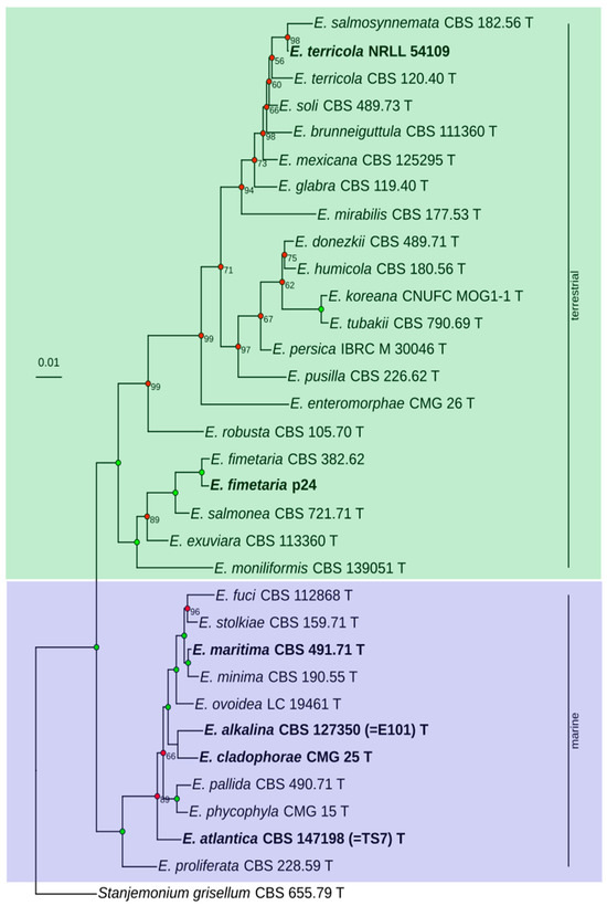
A phylogenetic tree has been reconstructed based on the sequences of ITS1-5.8S-ITS2, LSU, tef1α, rpb2, and tub2 from 29 type strains of the genus Emericellopsis, and isolates E. fimetaria CBS 382.62, E. fimetaria p24, and E. terricola NRRL 54109. As an outgroup, Stanjemonium grisellum CBS 655.79 (ex-type) has been selected. Overall, 139 sequences have been included in the analysis (Supplementary Table S1). The search for the optimal model of nucleotide substitutions have been performed by means of IQTree (the “Model Finder” module). However, among the selected models (TIM2+F+I+G for ITS1-5.8s-ITS2, TN+F+I for LSU, TN+F+G for rpb2, tub2, and GTR+I+G for tef1α), only the model GTR+I+G has been implemented in MrBayes. We remark that this model comprises the models TIM2 and TN as particular cases.
E. alkalina E101 positions itself within the “marine” clade of the genus Emericellopsis, forming a distinct subclade together with E. cladophorae CMG 25 (Figure 1, Supplementary Figure S1). The average nucleotide identity (ANI) between the genomes of E. alkalina E101 and E. cladophorae CMG 25 is approximately 90.04%.
Figure 1.
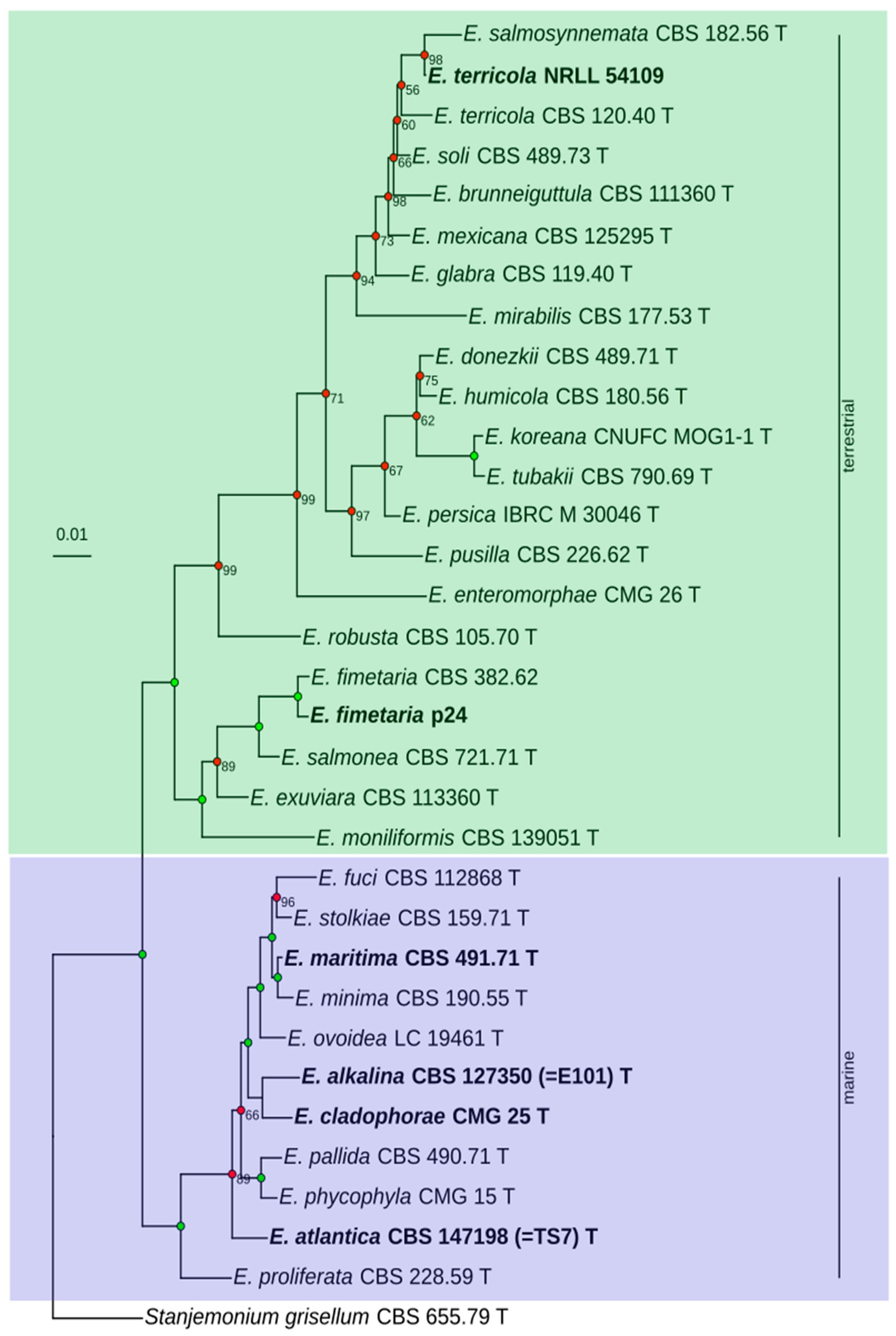
Phylogenetic tree of the Emericellopsis genus. Phylogeny reconstruction for ITS1-5.8s-ITS2, LSU, tef1a, rpb2, and tub2, independent parameters for each partition, GTR+I+G model. Analysis for 10,000,000 generations, burnin 25%. Bayesian posterior probability (BPP) values equal to 100% are not shown and are indicated by green dots; BPP values less than 100% are shown and indicated by red dots. T—ex-type strains. The strains whose genomes are discussed in this study are shown in bold.
E. fimetaria p24 forms a well-supported isolated subclade within the “terrestrial” clade of the genus Emericellopsis, alongside E. fimetaria CBS 382.62, E. salmonea CBS 721.71 (ex-type), E. exuviara CBS 113360 (ex-type), and E. moniliformis CBS 139051 (ex-type).
The strain E. terricola NRRL 54109, for which genomic data is available has also been incorporated into the phylogenetic tree. This strain forms a well-supported subclade with E. salmosynnemata CBS 182.56 (ex-type), whereas the strain E. terricola CBS 120.40 (ex-type) branches externally relative to them. When adding the sequences of E. microspora CBS 380.62 (former type strain of the species now considered synonymous with E. salmosynnemata), it also forms an external group relative to the strains E. terricola NRRL 54109 and E. salmosynnemata CBS 182.56 (ex-type) (Supplementary Figure S1). This may suggest that the species identification of strain NRRL 54109 should be re-evaluated. Hereafter, we will use the species name under which the genome is currently deposited in GenBank.
Genome Analysis
The genome of E. alkalina E101 (ex-type) consists of 25,722,796 base pairs assembled into 89 contigs, with an average contig size of 1,321,188 base pairs and the longest contig reaching 2,875,143 base pairs. On the other hand, the genome of E. fimetaria p24 consists of 24,658,382 base pairs across 43 contigs, averaging 979,341 base pairs per contig, with the longest contig measuring 3,154,750 base pairs. Detailed information can be found in Table 1.
Table 1.
Assembly metrics for E. alkalina E101 and E. fimetaria p24 genomes.
In the genome of E. alkalina E101, 8612 protein-coding sequences have been predicted, with 8467 (98.3%) encoded proteins assigned to orthogroups and 8217 (95.4%) annotated at least to the level of individual domains. Similarly, in the genome of E. fimetaria p24, 8616 protein-coding sequences have been predicted, with 8383 (98.4%) encoded proteins classified into orthogroups and 8205 (95.2%) annotated minimally up to domain level.
For 3897 (47.4% of total annotated) genes in E. alkalina E101 and 3853 (46.9% of total annotated) genes in E. fimetaria p24, identifiers from the KEGG database have been retrieved, providing functional annotations including KOG groups. The details on the number of annotated genes, carbohydrate-active enzymes, peptidases, sulfotransferases, transport systems, and BGCs are presented in Table 2.
Table 2.
Annotation features for E. alkalina E101 and E. fimetaria p24 genomes.
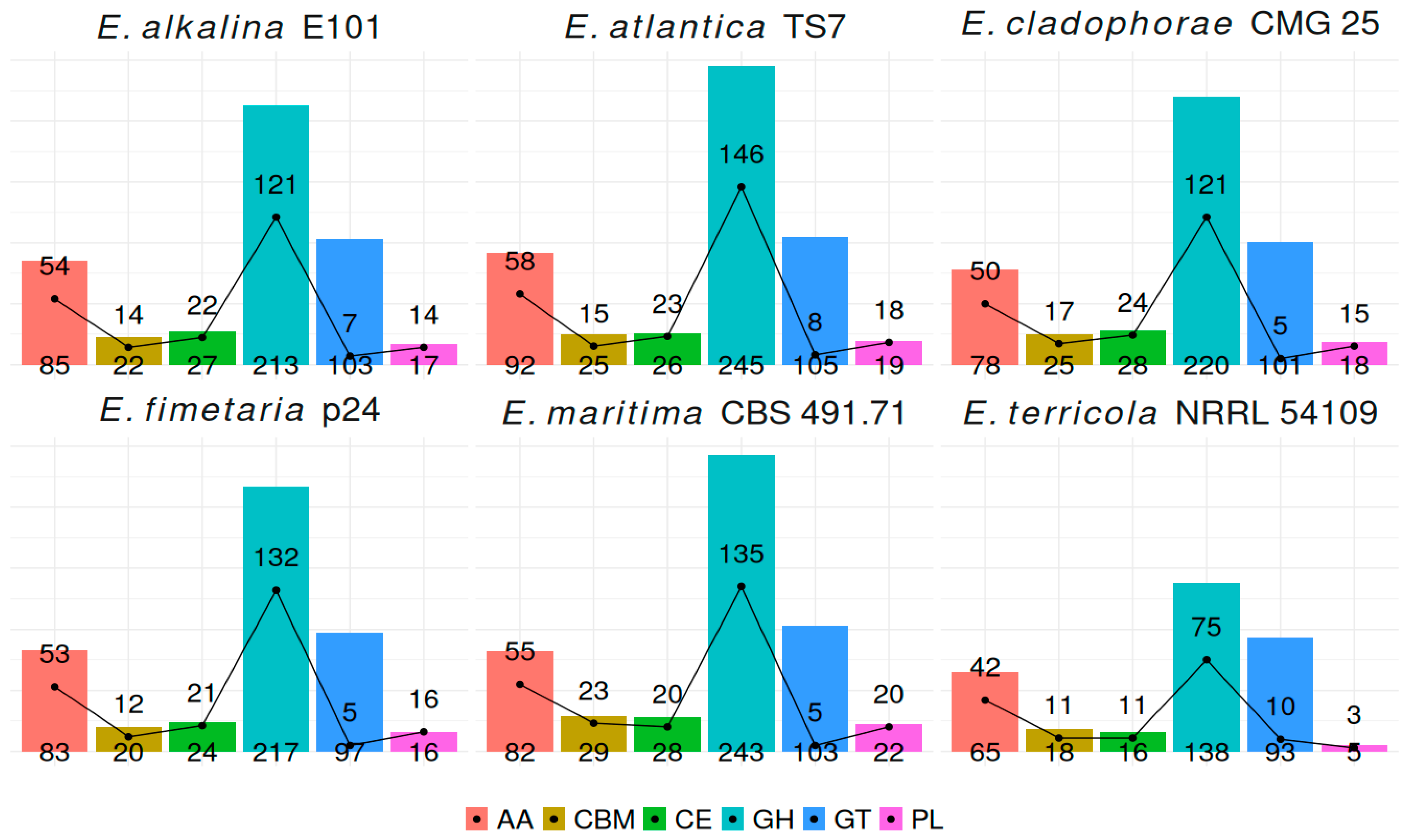
In the genomes of E. alkalina E101 and E. fimetaria p24, 447 and 428 genes encoding CAZymes have been respectively detected. Some transcripts contain multiple modules, typically glycoside hydrolase (GH) and carbohydrate-binding module (CBM). The most abundant CAZymes belong to families: Glycosyl-Hydrolases (GH): Family GH16 (β-1,4 and β-1,3 glucanases), Family GH18 (predominantly chitinases, secreted and intracellular), Family GH43 (β-D-xylosidases, α-L-arabinofuranosidases, and endo-α-L-arabinanases), Family GH3 (β-glucosidases), Family GH5 (mostly cellulases). Auxiliary Activity Enzymes (AA): Family AA7 (chito-oligosaccharide oxidases), Family AA3 (glucose dehydrogenases), Family AA9 (cellulose mono-oxygenases). Glycosyl Transferases (GT): Family GT2 (chitin synthases). Additionally, a significant number of enzymes from family CE5 (acetylesterases) have been observed. The genome of the obligately alkaliphilic fungus Sodiomyces alkalinus CBS 110278 [47], which is found in the same saline alkaline lakes as Emericellopsis alkalina [48], contains significantly fewer CAZymes from all classes except for AA. Also, the total number of transporter proteins in S. alkalinus is lower (Supplementary Table S2).
In the genome of E. alkalina E101, 73 carbohydrate-active enzymes possess sulfatase domains, primarily corresponding to glycosyl-hydrolases with β-glucosidase activity. There is also a single copy of a gene coding for α-L-fucosidase (EC 3.2.1.51), suggesting that E. alkalina E101 potentially degrades fucoidans that are present in brown algae cell walls and other sulfated polysaccharides. In the genome of E. fimetaria p24, 53 carbohydrate-active enzymes feature sulfatase domains, similarly containing a gene for α-L-fucosidase (EC 3.2.1.51).
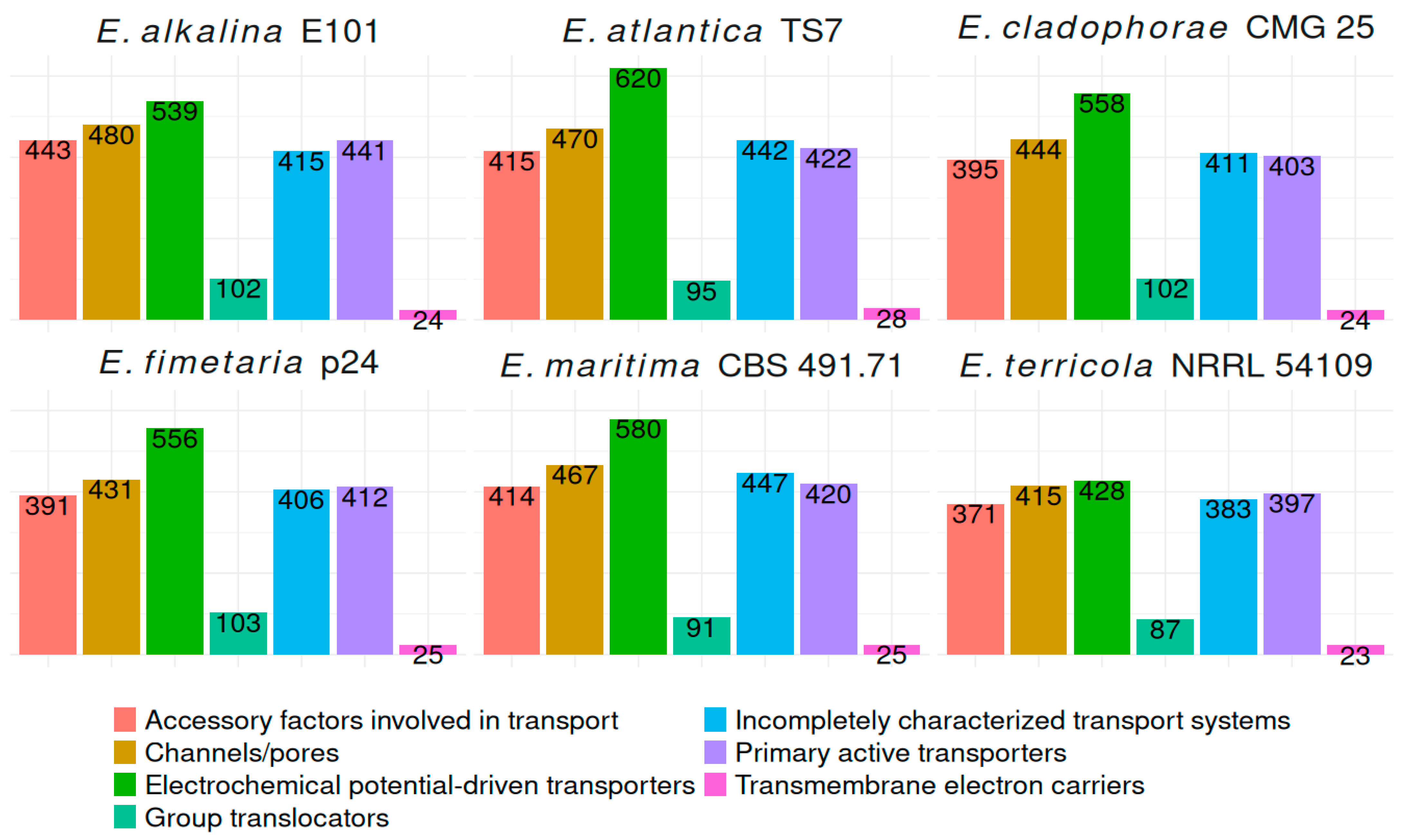
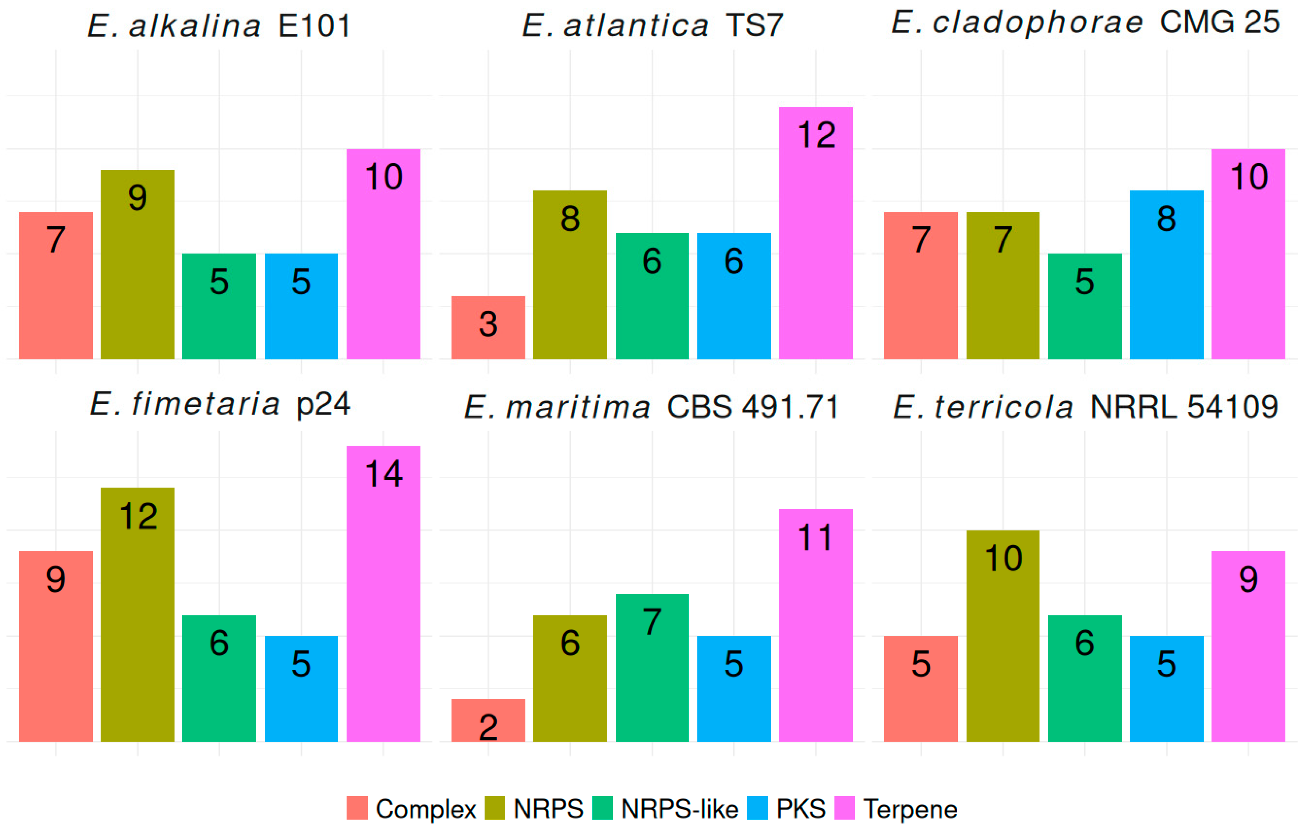
In the genomes of E. alkalina E101 and E. fimetaria p24, 2444 and 2324 transport proteins, respectively, have been annotated across all major classes, including channels/pores, electrochemical potential-driven transporters, primary active transporters, group translocators, transmembrane electron carriers, accessory factors involved in transport, and incompletely characterized transport systems. Secondary Metabolite Biosynthetic Gene Clusters (BGCs): in the genome of E. alkalina E101, 36 BGCs have been predicted, consisting of: 10 Terpene, 9 Non-Ribosomal Peptide Synthase (NRPS), 5 NRPS-like, 5 Polyketide Synthase (PKS): 4 Type I PKS (T1PKS), 1 Type III PKS (T3PKS), 7 Hybrid NRPS+T1PKS clusters. Some BGCs show similarity to those producing known secondary metabolites, such as leucostatin A/B, clavaric acid, ascochlorin, (-)-mellein, apicidin, among others. However, the majority of BGCs do not display any known similarities. In the genome of E. fimetaria p24, 46 BGCs have been discovered, comprising: 14 Terpene, 12 Non-Ribosomal Peptide Synthase (NRPS), 6 NRPS-like, 5 Polyketide Synthase (PKS), 4 Type I PKS (T1PKS), 1 Type III PKS (T3PKS), 7 Hybrid NRPS+T1PKS clusters. Several BGCs demonstrate similarity to those responsible for known secondary metabolites, including ascochlorin, helvolic acid, emerimicin, cytochalazines, etc. However, the majority of BGCs lack any known similarity. The whole genome sequence of the strains has been deposited in the NCBI database under the following accession numbers PRJNA1374271 for E. fimetaria p24 and PRJNA1374265 for E. alkalina E101.
The analysis of the genomes of E. alkalina E101 and E. fimetaria p24 using the BUSCO database (Hypocreales odb12, protein mode) indicates sufficient genome completeness: the proportion of single-copy genes is 96.4% for E. alkalina E101 and 96.1% for E. fimetaria p24. This result is quite good considering the absence of RNA-seq data and the use of a non-optimal model for Augustus.
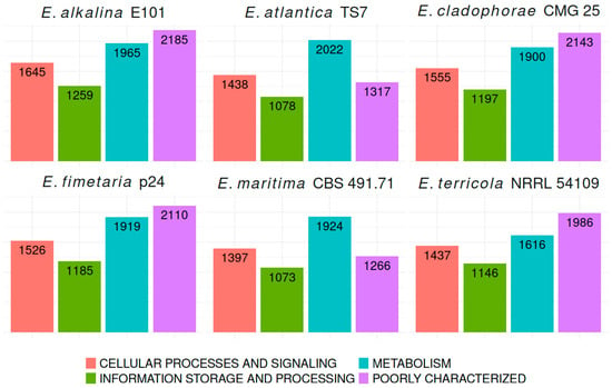
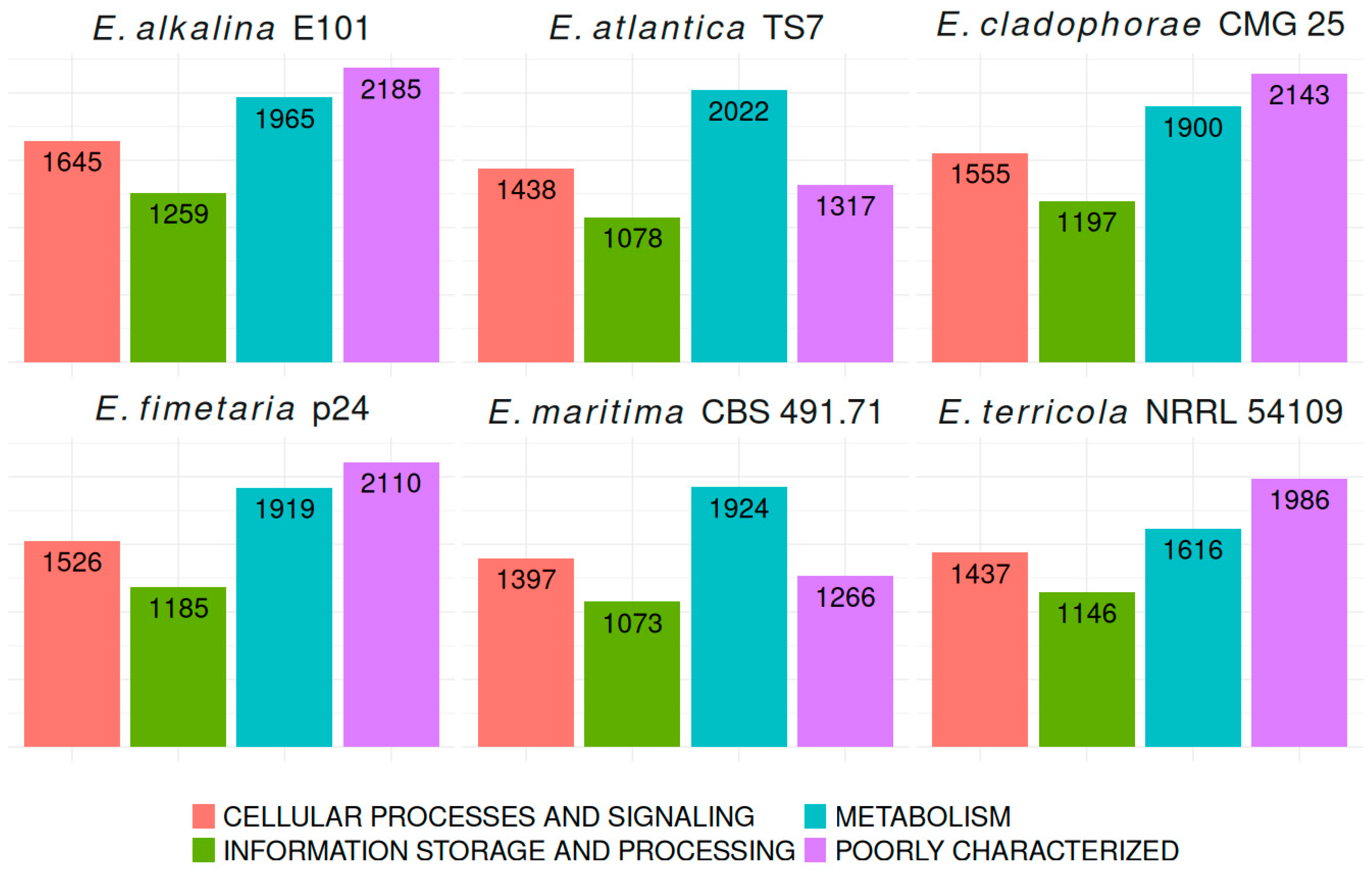
To compare the functional annotation across different species within the genus Emericellopsis, we present the number of annotated genes categorized into respective KOG groups for four genomes: E. atlantica TS7, E. maritima CBS 491.71, E. cladophorae MUM 19.33, and E. terricola NRRL 54109 (Figure 2). These comparisons allow one to understand similarities and differences in gene function distribution among closely related fungal species.
Figure 2.
Comparative KOG features for Emericellopsis genomes.
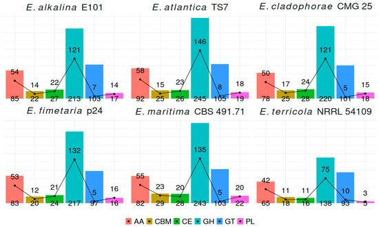
To ensure comparable results of enzyme annotation involved in carbohydrate metabolism, transport proteins, and biosynthetic gene clusters (BGCs), six genomes of Emericellopsis species: E. alkalina E101, E. fimetaria p24, E. terricola NRRL 54109, E. cladophorae CMG 25, E. atlantica TS7, and E. maritima CBS 491.71 have been analyzed. Figure 3 illustrates the overall distribution of carbohydrate-metabolizing enzymes and the proportion of secreted enzymes among the studied Emericellopsis species. It highlights considerable inter-species differences reflecting diverse ecological niches and trophic preferences of these fungi.
Figure 3.
Gene clusters encoding carbohydrate-active enzymes (CAZymes) in Emericellopsis genomes (bars) and CAZymes with signal peptide (black line). AA—Auxiliary activity, CBM—carbohydrate-binding module, CE—carbohydrate esterase, GH—glycosyl hydrolase, GT—glycosyl transferase, PL—polysaccharide lyase.
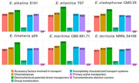
A summary of the quantity of transport proteins belonging to different classes in the analyzed Emericellopsis genomes is presented in Figure 4.
Figure 4.
Transporter classes in Emericellopsis genomes.
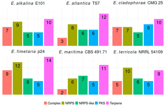
The analyzed genomes of Emericellopsis species differed little in the number of predicted BGCs (Figure 5).
Figure 5.
BGC in Emericellopsis genomes. Complex—complex BGC, NRPS—non-rybosomal peptide synthase, NRPS-like non-ribosomal peptide synthase like, PKS—polyketide synthase, Terpene—terpene synthase.
Only several BCGs showed homologies with known clusters, which are putatively responsible for producing compounds such as a leucinostatin, ascochlorin, emerimicins, clavaric acid.
Based on antiSMASH prediction, we found that Emericellopsis alkalina E101 contains one predicted cluster showing similarity to the leucinostatin cluster from Purpureocillium lilacinum (Figure 6), and we suggest that this BGC is responsible for the synthesis of emericellipsins, nonribosomal peptides belonging to the peptaibol group.
Figure 6.
Comparison of the BGC of leucinostatin from Purpureocillium lilacinum PLBJ 1 and the putative BGC of emericellipsins from Emericellopsis alkalina E101.
Importantly, the BGCs discovered in the genomes of E. alkalina E101 and E. fimetaria p24 encompass all genes necessary for ascochlorin synthesis (Supplementary Figure S2). Two biosynthetic gene clusters (BGCs) resembling those associated with emerimicins from E. tubakii PF have been identified in the genome of E. fimetaria p24 (Supplementary Figure S3).
The strain E. fimetaria p24 exhibits strong experimental antimicrobial activity against Gram-positive and Gram-negative bacteria as well as mould fungi (Supplementary Table S3). These could be established molecules like ascochlorin or helvolic acid, whose corresponding biosynthetic pathways have been identified within its genome, or novel metabolites originating from other biosynthetic gene clusters without any database homologues. Meanwhile, another strain, E. alkalina E101, is capable of producing emericellipsins—a group of peptaibols possessing strong antifungal properties [49,50]. Considering their proven antimicrobial potential combined with extensive biosynthetic capacity encoded by numerous gene clusters in their genomes, these two strains warrant further investigation to explore their potential as sources of new antimicrobial compounds.
4. Discussion
Genomics plays an important role in understanding the antimicrobial activity, biotechnological potential, and ecological roles of fungi. At present, genomic resources for the genus Emericellopsis are scarce, and genome information is available in NCBI for only four species. In this study, the whole genome of two more species E. alkalina and E. fimetaria has been sequenced and assembled. There were no significant differences in genome size, GC content, gene cluster type and number compared with other fungi of the same genus that had been reported. We have annotated and analyzed the genes encoding proteins involved with substrate specificity of fungi. Overall, the number of CAZymes in the genomes of Emericellopsis shows minor variations among different species, with few exceptions. The sequencing coverage is quite low; however, the results of genome completeness assessment allow us to consider these assemblies suitable for functional annotation and subsequent cautious interpretation of its outcomes.
The genome of the obligate alkaliphilic fungus Sodiomyces alkalinus CBS 110278, inhabiting in similar saline alkaline lakes to those where E. alkalina E101, exhibits a considerably lower number of CAZymes with the exception of class AA enzymes. It has been shown that S. alkalinus employs feeding strategies distinct from those of E. alkalina within the same biotope [48,51,52]. Adaptation strategies to extreme conditions such as high values pH and high concentrations of Na+ ions for both E. alkalina and S. alkalinus may be similar. In comparison with other species of Emericellopsis, E. alkalina exhibits an increased number of transporter proteins associated with adaptation to high pH and sodium ion concentration, comparable to their amount in S. alkalinus (Supplementary Table S2).
The variety of glycosyl-hydrolase enzymes with different substrate specificities in the genomes of E. alkalina E101 and E. fimetaria p24, along with a large number of enzymes presumably hydrolyzing plant cell wall polysaccharides (cellulose, xylan, pectin), suggests a typically saprotrophic lifestyle for these species. In the case of E. fimetaria, this assumption generally aligns with the substrate preference of the species. However, for E. alkalina E101, one would expect a greater utilization of chitin as a carbon source, given that typical habitats of this species are heavily populated by brine shrimp Artemia salina [53]. Nevertheless, no increase in the number of chitinase-encoding genes is observed in its genome. To determine whether Emericellopsis alkalina E101 can utilize alternative carbon sources such as chitin and sulfated polysaccharides from unicellular algae, additional experiments are needed aimed at assessing enzymatic activity towards these substrates, along with RNA-seq data to evaluate expression levels of corresponding enzymes under different conditions. In the genome of E. alkalina E101, there is no increase observed in the number of known fungal “pH sensors,” specifically PalH/Rim21 [54], compared to other species within the genus Emericellopsis. However, an increased number of Na+/H+ antiporters is noted, including homologs of yeast vacuolar/endosomal sodium tolerance protein NHX1, which plays a critical role in tolerating high concentrations of Na+ [55]. Additionally, several other electrochemical potential-driven transporters (EPT) are present. These EPT proteins play crucial roles in adaptation to environmental conditions. Specifically, Na+/H+ antiporters and P-type ATPases facilitate removal of sodium ions from cells [56], while H+-ATPases and V-ATPases regulate intracellular pH [57]. Furthermore, the genome contains an elevated number of genes encoding proteins homologous to SOS2 (Salt Overly Sensitive-2) from Arabidopsis thaliana, where SOS2 activates Na+/H+ antiporter function in response to excess Na+ ions [58]. The experimental characterization of many EPT functions remains incomplete, limiting conclusions about mechanisms underlying halotolerance and pH tolerance in microfungi like E. alkalina. This lack of detailed information necessitates further research to fully understand how this fungus adapts to changes in external Na+ concentration and pH.
The slightly larger number of BGCs in the genome of E. fimetaria p24 compared to other species may represent an adaptive strategy to its characteristic habitat—the dung of herbivorous animals. This nutrient-rich environment hosts a diverse microbial community, leading to intense competition for nutrients. Consequently, a higher production of antimicrobial secondary metabolites could confer a competitive advantage to E. fimetaria. Experimental evidence supports this hypothesis. Studies conducted by Singh and Webster revealed that E. fimetaria (originally referred to as Stilbella) inhibits the growth of coprophilic fungi and bacteria both in culture and in substrates. They demonstrated that antagonistic interactions occur primarily through the release of substances with antimicrobial and antifungal properties rather than direct hyphal contact [59]. Thus, the enhanced capacity for producing secondary metabolites likely enhances the fitness of E. fimetaria in its ecologically challenging habitat.
One of the proposed biosynthetic gene clusters (BGCs) in the genome of E. alkalina E101 is hypothesized to encode the biosynthesis pathway for emericellipsins—a class of peptaibols exhibiting potent antifungal and cytotoxic activities (Figure 6). Emericellipsins A-E consist of nine amino acids, including uncommon residues such as 2-amino-6-hydroxy-4-methyl-8-oxodecanoic acid (AHMOD) and β-alanine [9,60,61]. Leucinostatins, another class of nonribosomal lipopeptides structurally similar to peptaibols, incorporate rare components like AHMOD. Their biosynthesis requires polyketide synthase, aminotransferase, and cytochrome-P450 enzymes [62].
Both E. alkalina E101 and E. fimetaria p24 BGCs resemble those associated with ascochlorin (ilicicolin D) biosynthesis. Ascochlorin, a terpenoid compound, has originally been known for their antibacterial, antifungal, and cytotoxic properties [63,64,65]. Iliciolins, including ascochlorin (ilicicolin D), exert their effects by inhibiting fungal cytochrome bc1-reductase [66], making them attractive candidates for developing novel antifungal agents. The biosynthetic pathway of ascochlorin via orsellinic acid, as described in Acremonium egyptiacum, involves several key enzymes: polyketide synthase, prenyltransferase, reductase, halogenase, epoxidase, terpene cyclase, and dehydrogenase. Moreover, a transcription-regulating protein is also present within the BGC. Intermediate products in this pathway include ilicicolins A, B, and C, which themselves exhibit antimicrobial activity [67]. Importantly, the BGCs discovered in the genomes of E. alkalina E101 and E. fimetaria p24 encompass all genes necessary for ascochlorin synthesis.
We have found that two BGCs of E. fimetaria p24 exhibit similarity to the BGC of emerimicins from E. tubakii [68]. One of the detected BGCs includes two nonribosomal peptide synthetases (NRPSs): one with 15 domains and an acyltransferase domain, another with only two domains. The second BGC comprises an NRPS with 16 domains and an acyltransferase domain. Based on this observation, we propose that one BGC produces peptaibols antiamoebins consisting of 16 amino acid residues, previously reported in various strains of E. fimetaria [18]. Emerimicins belong to the family of nonribosomal peptide antibiotics, and possess broad-spectrum anti-microbial activity against Gram-positive bacteria, fungi, and protozoans [69,70]. These compounds consist of either 15 (all except for IIA and IIB) or 16 amino acid residues. The second BGC, featuring two NRPSs, might produce either. Furthermore, E. fimetaria p24 produces several compounds exhibiting antimicrobial activity against both Gram-positive and Gram-negative bacteria, as well as fungi [71].
BGCs that do not match any known clusters may either provide biosynthesis pathways for novel secondary metabolites or remain inactive or inducible only under specific conditions. Based solely on genomic BGC prediction results, we cannot draw definitive conclusions regarding their functions. No genes have been identified in predicted BGCs that could be unequivocally interpreted as responsible for self-resistance against their possible products. This might be due to limitations in available databases and annotation algorithms, as well as alternative producer tolerance mechanisms.
5. Conclusions
This study presents the genomes of E. alkalina E101 (ex-type) and E. fimetaria p24, isolated from extreme biotopes like saline alkaline lake sediments characterized by high pH and NaCl concentration. Overall, although the precise mechanisms that allow these fungi to thrive in their natural biotopes remain unclear, the set of enzymes involved in carbohydrate degradation points towards generalist strategies promoting colonization of diverse ecological niches. Similar profiles of transport proteins, particularly those from the group of electrochemical potential-driven transporters, may indicate similar adaptation mechanisms of Emericellopsis species to elevated concentrations of NaCl.
Clearly, the conclusions about substrate specificity and adaptation mechanisms to extreme conditions cannot be made solely based on genome comparison data, and further research is necessary. The abundant presence of poorly studied biosynthetic gene clusters (BGCs) in the genomes of E. alkalina E101 and E. fimetaria p24 might suggest their biotechnological potential. Further studies of these fungi will advance our understanding of the evolution and ecology of extremotolerant fungi, while exploring BGCs and their possible products could lead to the discovery of new secondary metabolites with antimicrobial activity.
Supplementary Materials
The following supporting information can be downloaded at: https://www.mdpi.com/article/10.3390/jof12050316/s1, Figure S1: Phylogenetic tree of the Emericellopsis genus included E. microspora CBS 380.62 (syn. E. salmosynnemata); Table S1: Genbank accession numbers for sequences used in the phylogenetic analysis fungi of the Emericellopsis genus; Table S2: Comparison for E. alkalina CBS 127350 (=E101) and S. alkalinus CBS 110278 genomes; Table S3: Antimicrobial activity of E. fimetaria p24; Figure S2: Comparison of BGC of ascochlorin from Acremonium egyptiacum F-1392, E. alkalina E101 and E. fimetaria p24; Figure S3: Comparison of BGCs from the E. fimetaria p24 genome similar to the BGC of emerimicins from the E. tubakii PF genome.
Author Contributions
Conceptualization, V.V.S. and M.L.G.; methodology, V.V.S., N.N.M. and O.A.K.; validation, V.V.S., N.N.M., K.V.M. and V.S.S.; formal analysis, V.V.S., N.N.M. and O.A.K.; investigation, K.V.M., V.V.S., N.N.M., K.V.M. and O.A.K.; writing—original draft preparation V.V.S., N.N.M. and M.L.G.; writing—review and editing, V.V.S., N.N.M., M.L.G., E.N.B. and V.S.S.; funding acquisition, N.N.M. and V.S.S. All authors have read and agreed to the published version of the manuscript.
Funding
The study of Markelova N.N., Georgieva M.L. was supported by the Russian Science Foundation (grant No. 25-24-00291; The studies of Malysheva K.V., Sokolov V.V., Kolpakova O.A. and Sadykova V.S. have been carried out in the framework of State Assignment for the Gause Institute of New Antibiotics.
Institutional Review Board Statement
Not applicable.
Informed Consent Statement
Not applicable.
Data Availability Statement
All sequence data are available in NCBI GenBank following the accession numbers in the manuscript.
Conflicts of Interest
The authors declare no conflict of interest.
References
- Van Beyma Thoe Kingma, F.H. Beschreibung einiger neuer Pilzarten aus dem Centraalbureau voor Schimmelcultures, Baarn (Nederland): VI. Mitteilung. Antonie van Leeuwenhoek 1940, 6, 263–290. [Google Scholar] [CrossRef] [Scilit]
- Hou, L.W.; Giraldo, A.; Groenewald, J.Z.; Rämä, T.; Summerbell, R.C.; Huang, G.Z.; Cai, L.; Crous, P.W. Redisposition of Acremonium-like Fungi in Hypocreales. Stud. Mycol. 2023, 105, 23–203. [Google Scholar] [CrossRef] [Scilit] [PubMed]
- Zhao, L.; Groenewald, J.Z.; Hou, L.W.; Summerbell, R.C.; Crous, P.W. Bionectriaceae: A Poorly Known Family of Hypocrealean Fungi with Major Commercial Potential. Stud. Mycol. 2025, 111, 115–198. [Google Scholar] [CrossRef] [Scilit] [PubMed]
- Grum-Grzhimaylo, A.A.; Georgieva, M.L.; Debets, A.J.M.; Bilanenko, E.N. Are Alkalitolerant Fungi of the Emericellopsis Lineage (Bionectriaceae) of Marine Origin? IMA Fungus 2013, 4, 213–228. [Google Scholar] [CrossRef] [Scilit]
- Bovio, E.; Garzoli, L.; Poli, A.; Prigione, V.; Firsova, D.; McCormack, G.P.; Varese, G.C. The Culturable Mycobiota Associated with Three Atlantic Sponges, Including Two New Species: Thelebolus balaustiformis and T. spongiae. Fungal Syst. Evol. 2018, 1, 141–167. [Google Scholar] [CrossRef] [Scilit]
- Marchese, P.; Garzoli, L.; Young, R.; Allcock, L.; Barry, F.; Tuohy, M.; Murphy, M. Fungi Populate Deep-sea Coral Gardens as Well as Marine Sediments in the Irish Atlantic Ocean. Environ. Microbiol. 2021, 23, 4168–4184. [Google Scholar] [CrossRef] [Scilit] [PubMed]
- Ameen, F.; AlNAdhari, S.; Al-Homaidan, A.A. Marine Fungi Showing Multifunctional Activity against Human Pathogenic Microbes and Cancer. PLoS ONE 2022, 17, e0276926. [Google Scholar] [CrossRef] [Scilit]
- Danilova, O.A.; Ianutsevich, E.A.; Bondarenko, S.A.; Georgieva, M.L.; Vikchizhanina, D.A.; Groza, N.V.; Bilanenko, E.N.; Tereshina, V.M. Osmolytes and Membrane Lipids in the Adaptation of Micromycete Emericellopsis alkalina to Ambient pH and Sodium Chloride. Fungal Biol. 2020, 124, 884–891. [Google Scholar] [CrossRef] [Scilit]
- Rogozhin, E.; Sadykova, V.; Baranova, A.; Vasilchenko, A.; Lushpa, V.; Mineev, K.; Georgieva, M.; Kul’ko, A.; Krasheninnikov, M.; Lyundup, A.; et al. A Novel Lipopeptaibol Emericellipsin A with Antimicrobial and Antitumor Activity Produced by the Extremophilic Fungus Emericellopsis alkalina. Molecules 2018, 23, 2785. [Google Scholar] [CrossRef] [Scilit]
- Persoon, C.H. Observationes Mycologicae. Seu Descriptiones Tam Novorum, Quam Notabilium Fungorum; Apud Wolfium et Gesenium: Lipsiae, Germany, 1800; Pars 2. [Google Scholar]
- Lehr, N.-A.; Meffert, A.; Antelo, L.; Sterner, O.; Anke, H.; Weber, R.W.S. Antiamoebins, Myrocin B and the Basis of Antifungal Antibiosis in the Coprophilous Fungus Stilbella erythrocephala (Syn. S. fimetaria): Antiamoebins, Myrocin B and the Basis of Antifungal Antibiosis in S. erythrocephala. FEMS Microbiol. Ecol. 2006, 55, 105–112. [Google Scholar] [CrossRef] [Scilit]
- Kildgaard, S.; Mansson, M.; Dosen, I.; Klitgaard, A.; Frisvad, J.; Larsen, T.; Nielsen, K. Accurate Dereplication of Bioactive Secondary Metabolites from Marine-Derived Fungi by UHPLC-DAD-QTOFMS and a MS/HRMS Library. Mar. Drugs 2014, 12, 3681–3705. [Google Scholar] [CrossRef] [Scilit]
- Ismail, M.A.; Moubasher, A.H.; Mohamed, A.R.; Al-Bedak, O.A. Extremophilic Fungi and Chemical Analysis of Hypersaline, Alkaline Lakes of Wadi-El-Natrun, Egypt. Int. J. Tech. Res. Sci. 2017, 1, 345–363. [Google Scholar]
- Georgieva, M.L.; Bilanenko, E.N.; Ponizovskaya, V.B.; Kokaeva, L.Y.; Georgiev, A.A.; Efimenko, T.A.; Markelova, N.N.; Kuvarina, A.E.; Sadykova, V.S. Haloalkalitolerant Fungi from Sediments of the Big Tambukan Saline Lake (Northern Caucasus): Diversity and Antimicrobial Potential. Microorganisms 2023, 11, 2587. [Google Scholar] [CrossRef] [Scilit]
- Brückner, H.; Nuber, K.; Reinecke, C. Chromatographic Detection of Bioactive Aib-Peptides in Molds of the Genus Stilbella. Fresenius’ Z. Anal. Chem. 1989, 333, 777–778. [Google Scholar] [CrossRef] [Scilit]
- Jaworski, A.; Brückner, H. New Sequences and New Fungal Producers of Peptaibol Antibiotics Antiamoebins. J. Pept. Sci. 2000, 6, 149–167. [Google Scholar] [CrossRef] [Scilit]
- Kildgaard, S.; Subko, K.; Phillips, E.; Goidts, V.; De La Cruz, M.; Díaz, C.; Gotfredsen, C.; Andersen, B.; Frisvad, J.; Nielsen, K.; et al. A Dereplication and Bioguided Discovery Approach to Reveal New Compounds from a Marine-Derived Fungus Stilbella fimetaria. Mar. Drugs 2017, 15, 253. [Google Scholar] [CrossRef] [Scilit]
- Hagestad, O.C.; Hou, L.; Andersen, J.H.; Hansen, E.H.; Altermark, B.; Li, C.; Kuhnert, E.; Cox, R.J.; Crous, P.W.; Spatafora, J.W.; et al. Genomic Characterization of Three Marine Fungi, Including Emericellopsis atlantica Sp. Nov. with Signatures of a Generalist Lifestyle and Marine Biomass Degradation. IMA Fungus 2021, 12, 21. [Google Scholar] [CrossRef] [Scilit] [PubMed]
- Gonçalves, M.F.M.; Hilário, S.; Van De Peer, Y.; Esteves, A.C.; Alves, A. Genomic and Metabolomic Analyses of the Marine Fungus Emericellopsis cladophorae: Insights into Saltwater Adaptability Mechanisms and Its Biosynthetic Potential. J. Fungi 2021, 8, 31. [Google Scholar] [CrossRef] [Scilit] [PubMed]
- Doval, R.L.; Müller, A.; Brown, D.W.; Johnson, D.; Quandt, C.A.; Barry, K.; Clum, A.; Hundley, H.; LaButti, K.; Lipzen, A.; et al. A Comparison of the Biotechnological Potential of Marine and Terrestrial Species of Two Orders of Sordariomycete Fungi. Mar. Biotechnol. 2025, 27, 103. [Google Scholar] [CrossRef] [Scilit]
- Mikheenko, A.; Saveliev, V.; Hirsch, P.; Gurevich, A. WebQUAST: Online Evaluation of Genome Assemblies. Nucleic Acids Res. 2023, 51, W601–W606. [Google Scholar] [CrossRef] [Scilit] [PubMed]
- Chen, S. Ultrafast One-pass FASTQ Data Preprocessing, Quality Control, and Deduplication Using Fastp. iMeta 2023, 2, e107. [Google Scholar] [CrossRef] [Scilit] [PubMed]
- Wick, R.R.; Judd, L.M.; Gorrie, C.L.; Holt, K.E. Unicycler: Resolving Bacterial Genome Assemblies from Short and Long Sequencing Reads. PLoS Comput. Biol. 2017, 13, e1005595. [Google Scholar] [CrossRef] [Scilit] [PubMed]
- Hoff, K.J.; Stanke, M. Predicting Genes in Single Genomes with AUGUSTUS. Curr. Protoc. Bioinform. 2019, 65, e57. [Google Scholar] [CrossRef] [Scilit] [PubMed]
- Simão, F.A.; Waterhouse, R.M.; Ioannidis, P.; Kriventseva, E.V.; Zdobnov, E.M. BUSCO: Assessing Genome Assembly and Annotation Completeness with Single-Copy Orthologs. Bioinformatics 2015, 31, 3210–3212. [Google Scholar] [CrossRef] [Scilit]
- Emms, D.M.; Kelly, S. OrthoFinder: Phylogenetic Orthology Inference for Comparative Genomics. Genome Biol. 2019, 20, 238. [Google Scholar] [CrossRef] [Scilit] [PubMed]
- Paysan-Lafosse, T.; Blum, M.; Chuguransky, S.; Grego, T.; Pinto, B.L.; Salazar, G.A.; Bileschi, M.L.; Bork, P.; Bridge, A.; Colwell, L.; et al. InterPro in 2022. Nucleic Acids Res. 2023, 51, D418–D427. [Google Scholar] [CrossRef] [Scilit]
- Jones, P.; Binns, D.; Chang, H.-Y.; Fraser, M.; Li, W.; McAnulla, C.; McWilliam, H.; Maslen, J.; Mitchell, A.; Nuka, G.; et al. InterProScan 5: Genome-Scale Protein Function Classification. Bioinformatics 2014, 30, 1236–1240. [Google Scholar] [CrossRef] [Scilit]
- Cantalapiedra, C.P.; Hernández-Plaza, A.; Letunic, I.; Bork, P.; Huerta-Cepas, J. eggNOG-Mapper v2: Functional Annotation, Orthology Assignments, and Domain Prediction at the Metagenomic Scale. Mol. Biol. Evol. 2021, 38, 5825–5829. [Google Scholar] [CrossRef] [Scilit]
- Huang, L.; Zhang, H.; Wu, P.; Entwistle, S.; Li, X.; Yohe, T.; Yi, H.; Yang, Z.; Yin, Y. dbCAN-Seq: A Database of Carbohydrate-Active Enzyme (CAZyme) Sequence and Annotation. Nucleic Acids Res. 2018, 46, D516–D521. [Google Scholar] [CrossRef] [Scilit]
- Zhang, H.; Yohe, T.; Huang, L.; Entwistle, S.; Wu, P.; Yang, Z.; Busk, P.K.; Xu, Y.; Yin, Y. dbCAN2: A Meta Server for Automated Carbohydrate-Active Enzyme Annotation. Nucleic Acids Res. 2018, 46, W95–W101. [Google Scholar] [CrossRef] [Scilit]
- Zheng, J.; Ge, Q.; Yan, Y.; Zhang, X.; Huang, L.; Yin, Y. dbCAN3: Automated Carbohydrate-Active Enzyme and Substrate Annotation. Nucleic Acids Res. 2023, 51, W115–W121. [Google Scholar] [CrossRef] [Scilit] [PubMed]
- Barbeyron, T.; Brillet-Guéguen, L.; Carré, W.; Carrière, C.; Caron, C.; Czjzek, M.; Hoebeke, M.; Michel, G. Matching the Diversity of Sulfated Biomolecules: Creation of a Classification Database for Sulfatases Reflecting Their Substrate Specificity. PLoS ONE 2016, 11, e0164846. [Google Scholar] [CrossRef] [Scilit]
- Rawlings, N.D.; Barrett, A.J.; Thomas, P.D.; Huang, X.; Bateman, A.; Finn, R.D. The MEROPS Database of Proteolytic Enzymes, Their Substrates and Inhibitors in 2017 and a Comparison with Peptidases in the PANTHER Database. Nucleic Acids Res. 2018, 46, D624–D632. [Google Scholar] [CrossRef] [Scilit]
- Altschul, S.F.; Gish, W.; Miller, W.; Myers, E.W.; Lipman, D.J. Basic Local Alignment Search Tool. J. Mol. Biol. 1990, 215, 403–410. [Google Scholar] [CrossRef]
- Saier, M.H. TCDB: The Transporter Classification Database for Membrane Transport Protein Analyses and Information. Nucleic Acids Res. 2006, 34, D181–D186. [Google Scholar] [CrossRef] [Scilit]
- Saier, M.H.; Reddy, V.S.; Moreno-Hagelsieb, G.; Hendargo, K.J.; Zhang, Y.; Iddamsetty, V.; Lam, K.J.K.; Tian, N.; Russum, S.; Wang, J.; et al. The Transporter Classification Database (TCDB): 2021 Update. Nucleic Acids Res. 2021, 49, D461–D467. [Google Scholar] [CrossRef] [Scilit]
- Eddy, S.R. Accelerated Profile HMM Searches. PLoS Comput. Biol. 2011, 7, e1002195. [Google Scholar] [CrossRef] [Scilit] [PubMed]
- Teufel, F.; Almagro Armenteros, J.J.; Johansen, A.R.; Gíslason, M.H.; Pihl, S.I.; Tsirigos, K.D.; Winther, O.; Brunak, S.; Von Heijne, G.; Nielsen, H. SignalP 6.0 Predicts All Five Types of Signal Peptides Using Protein Language Models. Nat. Biotechnol. 2022, 40, 1023–1025. [Google Scholar] [CrossRef] [Scilit]
- R Core Team R: A Language and Environment for Statistical Computing 2024. Available online: https://www.r-project.org (accessed on 28 January 2025).
- Blin, K.; Shaw, S.; Vader, L.; Szenei, J.; Reitz, Z.L.; Augustijn, H.E.; Cediel-Becerra, J.D.D.; de Crécy-Lagard, V.; Koetsier, R.A.; Williams, S.E.; et al. antiSMASH 8.0: Extended Gene Cluster Detection Capabilities and Analyses of Chemistry, Enzymology, and Regulation. Nucleic Acids Res. 2025, 53, W32–W38. [Google Scholar] [CrossRef] [Scilit]
- Katoh, K.; Standley, D.M. MAFFT Multiple Sequence Alignment Software Version 7: Improvements in Performance and Usability. Mol. Biol. Evol. 2013, 30, 772–780. [Google Scholar] [CrossRef] [Scilit] [PubMed]
- Okonechnikov, K.; Golosova, O.; Fursov, M.; The UGENE team. Unipro UGENE: A Unified Bioinformatics Toolkit. Bioinformatics 2012, 28, 1166–1167. [Google Scholar] [CrossRef] [Scilit]
- Altekar, G.; Dwarkadas, S.; Huelsenbeck, J.P.; Ronquist, F. Parallel Metropolis Coupled Markov Chain Monte Carlo for Bayesian Phylogenetic Inference. Bioinformatics 2004, 20, 407–415. [Google Scholar] [CrossRef] [Scilit]
- Ronquist, F.; Teslenko, M.; Van Der Mark, P.; Ayres, D.L.; Darling, A.; Höhna, S.; Larget, B.; Liu, L.; Suchard, M.A.; Huelsenbeck, J.P. MrBayes 3.2: Efficient Bayesian Phylogenetic Inference and Model Choice Across a Large Model Space. Syst. Biol. 2012, 61, 539–542. [Google Scholar] [CrossRef] [Scilit]
- Ayres, D.L.; Darling, A.; Zwickl, D.J.; Beerli, P.; Holder, M.T.; Lewis, P.O.; Huelsenbeck, J.P.; Ronquist, F.; Swofford, D.L.; Cummings, M.P.; et al. BEAGLE: An Application Programming Interface and High-Performance Computing Library for Statistical Phylogenetics. Syst. Biol. 2012, 61, 170–173. [Google Scholar] [CrossRef] [Scilit]
- Grum-Grzhimaylo, A.A.; Falkoski, D.L.; Van Den Heuvel, J.; Valero-Jiménez, C.A.; Min, B.; Choi, I.; Lipzen, A.; Daum, C.G.; Aanen, D.K.; Tsang, A.; et al. The Obligate Alkalophilic Soda-lake Fungus Sodiomyces alkalinus Has Shifted to a Protein Diet. Mol. Ecol. 2018, 27, 4808–4819. [Google Scholar] [CrossRef] [Scilit] [PubMed]
- Grum-Grzhimaylo, A.A.; Debets, A.J.M.; Van Diepeningen, A.D.; Georgieva, M.L.; Bilanenko, E.N. Sodiomyces alkalinus, a New Holomorphic Alkaliphilic Ascomycete within the Plectosphaerellaceae. Persoonia-Mol. Phylogeny Evol. Fungi 2013, 31, 147–158. [Google Scholar] [CrossRef] [Scilit]
- Gavryushina, I.A.; Georgieva, M.L.; Kuvarina, A.E.; Sadykova, V.S. Peptaibols as Potential Antifungal and Anticancer Antibiotics: Current and Foreseeable Development (Review). Appl. Biochem. Microbiol. 2021, 57, 556–563. [Google Scholar] [CrossRef] [Scilit]
- Sadykova, V.S.; Gavryushina, I.A.; Kuvarina, A.E.; Markelova, N.N.; Sedykh, N.G.; Georgieva, M.L.; Barashkova, A.C.; Rogozhin, E.A. Antimicrobic Activity of the Lipopeptide Emericellipsin A Isolated from Emericellopsis alkalina against Biofilm-Forming Bacteria. Appl. Biochem. Microbiol. 2020, 56, 292–297. [Google Scholar] [CrossRef] [Scilit]
- Kuvarina, A.E.; Georgieva, M.L.; Rogozhin, E.A.; Kulko, A.B.; Gavryushina, I.A.; Sadykova, V.S. Antimicrobial Potential of the Alkalophilic Fungus Sodiomyces alkalinus and Selection of Strains–Producers of New Antimicotic Compound. Appl. Biochem. Microbiol. 2021, 57, 86–93. [Google Scholar] [CrossRef] [Scilit]
- Kuvarina, A.E.; Rogozhin, E.A.; Sykonnikov, M.A.; Timofeeva, A.V.; Serebryakova, M.V.; Fedorova, N.V.; Kokaeva, L.Y.; Efimenko, T.A.; Georgieva, M.L.; Sadykova, V.S. Isolation and Characterization of a Novel Hydrophobin, Sa-HFB1, with Antifungal Activity from an Alkaliphilic Fungus, Sodiomyces alkalinus. J. Fungi 2022, 8, 659. [Google Scholar] [CrossRef] [Scilit]
- Golovatyuk, L.; Kanapatskiy, T.; Samylina, O.; Pimenov, N.; Nazarova, L.; Kallistova, A. Invertebrate Assemblages in Some Saline and Soda Lakes of the Kulunda Steppe: First Regional Assessment and Ecological Implications. Water 2025, 17, 2330. [Google Scholar] [CrossRef] [Scilit]
- Obara, K.; Yamamoto, H.; Kihara, A. Membrane Protein Rim21 Plays a Central Role in Sensing Ambient pH in Saccharomyces cerevisiae. J. Biol. Chem. 2012, 287, 38473–38481. [Google Scholar] [CrossRef] [Scilit]
- Mukherjee, S.; Kallay, L.; Brett, C.L.; Rao, R. Mutational Analysis of the Intramembranous H10 Loop of Yeast Nhx1 Reveals a Critical Role in Ion Homoeostasis and Vesicle Trafficking. Biochem. J. 2006, 398, 97–105. [Google Scholar] [CrossRef] [Scilit] [PubMed]
- Ruiz, A.; Ariño, J. Function and Regulation of the Saccharomyces cerevisiae ENA Sodium ATPase System. Eukaryot. Cell 2007, 6, 2175–2183. [Google Scholar] [CrossRef] [Scilit]
- Kane, P.M. Proton Transport and pH Control in Fungi. In Yeast Membrane Transport; Ramos, J., Sychrová, H., Kschischo, M., Eds.; Advances in Experimental Medicine and Biology; Springer International Publishing: Cham, Switzerland, 2016; Volume 892, pp. 33–68. ISBN 978-3-319-25302-2. [Google Scholar]
- Liu, J.; Ishitani, M.; Halfter, U.; Kim, C.-S.; Zhu, J.-K. The Arabidopsis thaliana SOS2 Gene Encodes a Protein Kinase That Is Required for Salt Tolerance. Proc. Natl. Acad. Sci. USA 2000, 97, 3730–3734. [Google Scholar] [CrossRef]
- Singh, N.; Webster, J. Antagonism between Stilbella erythrocephala and Other Coprophilous Fungi. Trans. Br. Mycol. Soc. 1973, 61, 487–495. [Google Scholar] [CrossRef] [Scilit]
- Kuvarina, A.E.; Gavryushina, I.A.; Sykonnikov, M.A.; Efimenko, T.A.; Markelova, N.N.; Bilanenko, E.N.; Bondarenko, S.A.; Kokaeva, L.Y.; Timofeeva, A.V.; Serebryakova, M.V.; et al. Exploring Peptaibol’s Profile, Antifungal, and Antitumor Activity of Emericellipsin A of Emericellopsis Species from Soda and Saline Soils. Molecules 2022, 27, 1736. [Google Scholar] [CrossRef] [Scilit] [PubMed]
- Kuvarina, A.E.; Gavryushina, I.A.; Kulko, A.B.; Ivanov, I.A.; Rogozhin, E.A.; Georgieva, M.L.; Sadykova, V.S. The Emericellipsins A–E from an Alkalophilic Fungus Emericellopsis alkalina Show Potent Activity against Multidrug-Resistant Pathogenic Fungi. J. Fungi 2021, 7, 153. [Google Scholar] [CrossRef] [Scilit]
- Wang, G.; Liu, Z.; Lin, R.; Li, E.; Mao, Z.; Ling, J.; Yang, Y.; Yin, W.-B.; Xie, B. Biosynthesis of Antibiotic Leucinostatins in Bio-Control Fungus Purpureocillium lilacinum and Their Inhibition on Phytophthora Revealed by Genome Mining. PLoS Pathog. 2016, 12, e1005685. [Google Scholar] [CrossRef] [Scilit] [PubMed]
- Hayakawa, S.; Minato, H.; Katagiri, K. The Ilicicolins, Antibiotics from Cylindrocladium ilicicola. J. Antibiot. 1971, 24, 653–654. [Google Scholar] [CrossRef] [Scilit]
- Che, Y.; Swenson, D.C.; Gloer, J.B.; Koster, B.; Malloch, D. Pseudodestruxins A and B: New Cyclic Depsipeptides from the Coprophilous Fungus Nigrosabulum globosum. J. Nat. Prod. 2001, 64, 555–558. [Google Scholar] [CrossRef] [Scilit] [PubMed]
- Subko, K.; Kildgaard, S.; Vicente, F.; Reyes, F.; Genilloud, O.; Larsen, T.O. Bioactive Ascochlorin Analogues from the Marine-Derived Fungus Stilbella fimetaria. Mar. Drugs 2021, 19, 46. [Google Scholar] [CrossRef] [Scilit] [PubMed]
- Singh, S.B.; Liu, W.; Li, X.; Chen, T.; Shafiee, A.; Dreikorn, S.; Hornak, V.; Meinz, M.; Onishi, J.C. Structure–Activity Relationship of Cytochrome Bc1 Reductase Inhibitor Broad Spectrum Antifungal Ilicicolin H. Bioorg. Med. Chem. Lett. 2013, 23, 3018–3022. [Google Scholar] [CrossRef] [Scilit] [PubMed]
- Araki, Y.; Awakawa, T.; Matsuzaki, M.; Cho, R.; Matsuda, Y.; Hoshino, S.; Shinohara, Y.; Yamamoto, M.; Kido, Y.; Inaoka, D.K.; et al. Complete Biosynthetic Pathways of Ascofuranone and Ascochlorin in Acremonium egyptiacum. Proc. Natl. Acad. Sci. USA 2019, 116, 8269–8274. [Google Scholar] [CrossRef] [Scilit]
- Wu, G.; Dentinger, B.T.M.; Nielson, J.R.; Peterson, R.T.; Winter, J.M. Emerimicins V–X, 15-Residue Peptaibols Discovered from an Acremonium sp. through Integrated Genomic and Chemical Approaches. J. Nat. Prod. 2021, 84, 1113–1126. [Google Scholar] [CrossRef] [Scilit]
- Argoudelis, A.D.; Johnson, L.E. Emerimicins II, III And IV, Antibiotics Produced By Emericellopsis microspora In Media Supplemented with Trans-4-N-Propyl-L-Proline. J. Antibiot. 1974, 27, 274–282. [Google Scholar] [CrossRef] [Scilit]
- Inostroza, A.; Lara, L.; Paz, C.; Perez, A.; Galleguillos, F.; Hernandez, V.; Becerra, J.; González-Rocha, G.; Silva, M. Antibiotic Activity of Emerimicin IV Isolated from Emericellopsis minima from Talcahuano Bay, Chile. Nat. Prod. Res. 2018, 32, 1361–1364. [Google Scholar] [CrossRef] [Scilit]
- Sokolov, V.V.; Markelova, N.N.; Georgieva, M.L.; Sadykova, V.S. First Data on The Antimicrobial Activity of Emericellopsis fimetaria Fungi Isolated from Bottom Sediments of the Saline Lake Bolshoy Tambukan (North Caucasus, Russia). Microbiology 2026, 95, 473–476. [Google Scholar] [CrossRef] [Scilit]



Disclaimer/Publisher’s Note: The statements, opinions and data contained in all publications are solely those of the individual author(s) and contributor(s) and not of MDPI and/or the editor(s). MDPI and/or the editor(s) disclaim responsibility for any injury to people or property resulting from any ideas, methods, instructions or products referred to in the content. |
© 2026 by the authors. Licensee MDPI, Basel, Switzerland. This article is an open access article distributed under the terms and conditions of the Creative Commons Attribution (CC BY) license.