


Abstract
(1) Background: Potentially arthropod-pathogenic and plant-associated Metarhizium fungi are of high interest for basic research, biological pest control and plant growth promotion. Unambiguous species delineation enabling the taxonomic assignment of new isolates and the identification of new Metarhizium species is of crucial importance for both research and application. Recently, the new species Metarhizium hybridum and Metarhizium parapingshaense were introduced on the basis of phylogenomic studies. (2) Methods: Neighbor- joining and Bayesian inference-based phylogenetic reconstruction of ribosomal intergenic spacer (rIGS) sequences were used to critically evaluate new species introductions. A species-discriminating diagnostic PCR tool for Metarhizium was adapted to M. hybridum and M. parapingshaense. GenBank database mining was performed to identify cryptic descriptions of the new species. (3) Results: The introduction of M. hybridum and M. parapingshaense was corroborated by rIGS sequence comparison. Data mining revealed cryptic first descriptions of M. hybridum from Canada, China, Colombia, Costa Rica, Cuba, Honduras, Mexico, New Zealand, the USA and the Philippines, and of M. parapingshaense from China, India, Japan, the Philippines and South Korea. (4) Conclusions: Results support the reliability of rIGS as a single taxonomic marker for species-level identification of Metarhizium fungi. Species-discriminating diagnostic PCR was successfully adapted to enable the sequencing-independent identification of the confirmed new species M. hybridum and M. parapingshaense.
Keywords:
Metarhizium hybridum; Metarhizium parapingshaense; Metarhizium anisopliae sensu lato; Metarhizium pingshaense sensu lato; PARB clade; ribosomal intergenic spacer marker (rIGS-ID800); single-marker phylogeny; sequencing-independent species identification; diagnostic PCR; cryptic species descriptions 1. Introduction
The taxonomic genus Metarhizium Sorokin (Hypocreales; Clavicipitaceae) comprises natural fungal pathogens of numerous economically relevant insect hosts and is among the fungi most widely investigated for and applied in biological control. The biocontrol potential of Metarhizium fungi has been successfully exploited to develop microbial insecticides [1,2]. In addition to its role as an entomopathogen, multiple studies have documented beneficial plant–fungus interactions in some Metarhizium species, including rhizosphere and endophytic colonization that can enhance plant nutrient acquisition and growth, improve resistance to biotic and abiotic stressors, and facilitate the mobilization of soil nutrients such as phosphorus and trace metals, thereby functioning as a plant growth promoter and bioinoculant in agroecosystems [3]. Review and empirical studies also report increased biomass, root elongation, and physiological performance in several crops following Metarhizium inoculation, supporting its dual use as both a biocontrol agent and a plant growth-promoting symbiont [4]. Elucidation of lineage diversification has revealed that broad-host-range clades derived from plant-associated ancestors exhibit marked ecological plasticity, enabling both insect pathogenicity and plant associations [5]. Consequently, precise taxonomic resolution is fundamental not only for regulatory and applied biocontrol purposes, but also for the strategic selection and deployment of isolates as multifunctional agents with plant growth-promoting potential.
Traditionally, species assignment within the genus Metarhizium has been based on morphological and morphometric traits such as colony morphologies and the dimensions of conidia, conidiophores and phialides. During the past decades, these morphological criteria have been complemented by the use of genetic markers such as the Internal Transcribed Spacer (ITS) sequence of the fungal rRNA operon [6], a Multilocus Sequence Analysis (MLSA) scheme comprising the genes encoding translation elongation factor 1 alpha (EF1A) and the RNA polymerase II subunits 1 and 2 (RPB1 and RPB2, respectively) and the intron-rich 5′-region of the translation elongation factor 1 alpha gene, referred to as “5TEF” [7,8]. Moreover, a nuclear intergenic region termed MzIGS3 or DUF895 [9] has been successfully employed in both species delineation and diversity studies [4,10,11], and the intergenic spacer sequence of the ribosomal RNA operon (rIGS) has been evaluated as a molecular taxonomic marker for Metarhizium fungi [12,13,14].
Molecular taxonomy has led to the discovery of cryptic or hidden species, i.e., monophyletic groups of organisms that are morphologically or phenotypically indistinguishable, but genetically diverse from a known species [15]. In the case of Metarhizium fungi that are characterized by only subtle morphological traits, systematic studies employing genetic markers led to the redefinition of genus boundaries and species delineations [16,17,18] and to the introduction of new Metarhizium species [19,20,21,22,23]. In particular, the reorganized genus Metarhizium was found to comprise a tight cluster, informally termed the “PARB clade” [18], of the five species Metarhizium anisopliae, M. brunneum, M. humberi, M. pingshaense and M. robertsii that are of high relevance for both fundamental research and insect biocontrol.
Recently, a rIGS sequence-based species-discriminating diagnostic PCR protocol for the sequencing-independent species-level identification has been established [13] and successfully applied to introduce the new PARB clade species, Metarhizium caribense [24]. Contemporaneously, an integrative taxonomic study by Kobmoo et al. (2024) combining phylogenomics with phenotypic data from morphometrics, metabolomics and insect virulence bioassays, led to the delineation of two further PARB clade species, Metarhizium hybridum and Metarhizium parapingshaense, from M. anisopliae and M. pingshaense, respectively [25]. The study described three M. parapingshaense strains from Thailand and the Solomon Islands, as well as a single M. hybridum strain from Brazil; subsequently, M. hybridum was reported from Argentina, too [26].
It was the purpose of the present study to validate the functionality of the rIGS sequence as a single taxonomic marker for species delineation within the reorganized PARB clade, to extend the species-discriminating PCR diagnostics to the new taxa M. hybridum and M. parapingshaense, and to identify possible cryptic descriptions of these new species.
Throughout this publication, “M. anisopliae sensu stricto” will be referred to in the usual way, i.e., as the taxonomic species defined by its nomenclatural type strain ARSEF 7487. A recent proposal to rename this taxon “Metarhizium neoanisopliae” [25] will not be adopted here as broader community acceptance is awaited. Moreover, the designation “Metarhizium anisopliae sensu lato” will be used to refer to a sub-structure of the PARB clade comprising the species M. anisopliae sensu stricto together with M. humberi and M. hybridum. Analogously, “M. pingshaense sensu lato” will designate a PARB sub-clade made up of the three species M. pingshaense sensu stricto, M. caribense and M. parapingshaense. If not otherwise stated, the species designations “M. anisopliae” and “M. pingshaense” will always refer to M. anisopliae sensu stricto and M. pingshaense sensu stricto, respectively.
2. Materials and Methods
2.1. DNA Extraction, PCR Amplification and DNA Sequence Determination
For DNA extraction, Metarhizium fungi investigated in this study (Supplementary Table S1) were grown for up to 5 days at 24 °C in 10 mL YPG liquid medium (2 g/L yeast extract, 10 g/L peptone, 20 g/L glucose) containing 25 µg/mL of tetracycline. Approximately 200–300 mg of fungal biomass were transferred to a 2 mL screw-capped microcentrifuge tube containing Lysing Matrix C (MP Biomedicals, Irvine, CA, USA). Samples were frozen at −20 °C for at least 3 h and processed for 60 s at intermediate speed in a Minilys homogenizer (Bertin technologies, Montigny-le-Bretonneux, France). DNA was extracted from homogenized samples using the DNeasy Plant Mini kit (Qiagen, Venlo, The Netherlands) according to the standard protocol provided by the manufacturer, starting with the addition of 400 µL AP1 buffer. DNA was eluted from the column in 100 µL AE buffer (10 mM Tris-Cl, 0.5 mM EDTA, pH = 9.0) and stored at −20 °C. DNA concentrations were determined using a NanoDrop One spectrophotometer (Thermo Scientific, Waltham, MA, USA).
PCR amplifications were run in a T-One thermocycler (Biometra, Göttingen, Germany) using 0.025 U/µL GoTaq polymerase (Promega, Fitchburg, MA, USA) with dNTP and oligonucleotide primer concentrations of 200 µM and 500 nM, respectively.
Preparative PCR amplifications for DNA sequence determination were performed as 50 µL reactions using a generalized PCR protocol. An initial denaturation step of 95 °C for 2 min was followed by 35 cycles consisting of a 30 s denaturation step at 95 °C, a 30 s annealing step and an elongation step at 72 °C. Primer pair-specific annealing temperatures and amplicon-specific elongation times were indicated in the Supplementary Table S2. The program ended with a 5 min final elongation step at 72 °C. Using this protocol, both the complete rIGS sequence and the rIGS-ID800 marker were amplified using primer pairs Ma-28S4f/Ma-18S4r and Migs1-F1/Migs850-R1, respectively (Supplementary Table S2). Formation of single PCR products of expected apparent size was routinely controlled through horizontal electrophoresis in 1x TAE buffer (40 mM TRIS, 20 mM acetic acid, 1 mM EDTA, pH 8.3) of a 5 µL sample using 1% agarose gels stained with 5 µL/100 mL Roti GelStain (Carl Roth, Karlsruhe, Germany). PCR products were purified using the Qiaquick PCR purification kit (Qiagen, Venlo, The Netherlands) according to the standard protocol provided by the manufacturer. Purified PCR products were eluted for direct sequencing in 50 µL of EB buffer (10 mM Tris·Cl, pH 8.5) and stored at −20 °C.
Sanger sequencing of rIGS-ID800 PCR products using primers Migs1-F1 and Migs850-R1, as well as Oxford Nanopore Technology sequencing of complete amplified rIGS regions, was performed by an external provider (MicroSynth-SeqLab, Gießen, Germany). rIGS-ID800 raw sequence data were combined into a consensus sequence for each fungal specimen using version 11 of the MEGA software package [27]. Sequences determined were submitted to the GenBank database under the accession numbers indicated in the Supplementary Table S1.
Diagnostic PCR amplifications for sequencing-independent species discrimination were performed as 20 µL reactions containing a working concentration of 100 pg/µL of DNA template. The diagnostic PCR program consisted of one initial denaturation step of 95 °C for 2 min, 35 cycles of denaturation at 95 °C for 30 s, annealing at 60 °C for 30 s, and elongation at 72 °C for 45 s, followed by a 2 min final elongation step at 72 °C [13].
2.2. Data Mining
Whole-length rIGS sequences of M. parapingshaense strains BCC 37941 and BCC 96582 were assembled from published SRA data (accession number PRJNA1111679), running a targeted BlastN search for reads mapping the complete rIGS sequence of M. parapingshaense reference strain ARSEF 4342. Identified reads were assembled using the Clustal X function of the MEGA 11 software package. Potential insertions or deletions with respect to the query sequence were corroborated by SRA data searches with joint flanking sequences.
A total of 46 assembled genome sequences assigned to the genus Metarhizium in the GenBank database (https://www.ncbi.nlm.nih.gov/datasets/genome/?taxon=5529, accessed on 3 February 2026) were searched by the genomic Blast option for cryptic descriptions of M. hybridum or M. parapingshaense using as query complete rIGS sequences of the specific type strains ARSEF 549 and BCC 37941. Results were cross-checked by analogous searches with 5TEF marker sequences of ARSEF 549 and the M. parapingshaense reference strain ARSEF 4342 (Supplementary Table S1).
Homologous single-marker entries to the GenBank database were searched for using the BlastN software tool (https://blast.ncbi.nlm.nih.gov/Blast.cgi?PROGRAM=blastn&PAGE_TYPE=BlastSearch&LINK_LOC=blasthome, accessed on 2 February 2026) [28,29]. Searches for cryptic descriptions of the new species M. hybridum or M. parapingshaense were performed using the rIGS-ID800 sequences of the type strains ARSEF 549 and BCC 37941 as query. In a second step, results were complemented by BlastN searches using the DUF895, 5TEF, EF1A and RPB2 marker sequences of strains M. hybridum ARSEF 549 and M. parapingshaense ARSEF 4342 as query (Supplementary Table S1). GenBank entries displaying a minimum of 90% sequence coverage with respect to the corresponding query sequence were retained for downstream analyses.
GenBank entries retained from both genomic and single-entry searches were aligned with respective reference sequences to reconstruct NJ phylogenies. Fungal strains or isolates clustering in the tree topology with the M. hybridum or the M. parapingshaense reference strains under unambiguous delineation from the further species of, respectively, M. anisopliae sensu lato and M. pingshaense sensu lato, were considered cryptic descriptions of the two species.
2.3. Phylogenetic Reconstruction
Nucleotide sequence alignments were generated using the CLUSTAL W function [30] as implemented in MEGA 11. Then, p-distance matrix-based neighbor-joining (NJ) phylogenies were reconstructed under pairwise deletion of alignment gaps and missing data. Tree topology confidence limits were explored in non-parametric bootstrap analyses over 1000 pseudo-replicates. Moreover, Bayesian phylogenetic hypotheses were explored using version 3.2.7 of the MrBayes software tool [31] under a GTR + I model assuming a gamma-shaped distribution of rates across sites and allowing for eight rate categories. Prior probabilities were estimated from the data set. Two runs comprising each one cold and three heated MCMC chains were performed in parallel over 1,000,000 generations, sampling every 5000 generations. A standard deviation of split frequencies below 0.01 was applied as a convergence criterion. Trees from the first 25% of generations were discarded as burn-in prior to the generation of a 50% majority rule consensus tree.
3. Results
3.1. rIGS-Based Phylogeny for M. hybridum and M. parapingshaense
Complete rIGS sequences were determined for those M. hybridum, M. anisopliae, M. parapingshaense and M. pingshaense strains that were compared at the whole genome level by Kobmoo et al. (2024) [25] when introducing M. hybridum and M. parapingshaense. In particular, complete rIGS sequences were assembled from WGS sequence read data deposited in the GenBank database for strains M. parapingshaense BCC 37941-T and BCC 96582, M. hybridum ARSEF 549-T, and M. anisopliae ARSEF 2080, giving rise to continuous rIGS sequences comprising 1555 bp, 1543 bp, 1708 bp and 1750 bp, respectively. Moreover, for strains ARSEF 549 and ARSEF 2080, rIGS sequence assemblies were confirmed by PCR amplification and de novo sequencing of the rIGS region. GenBank accession numbers of complete rIGS sequences were indicated in the Supplementary Table S1.
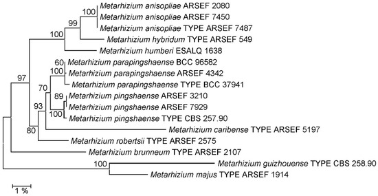
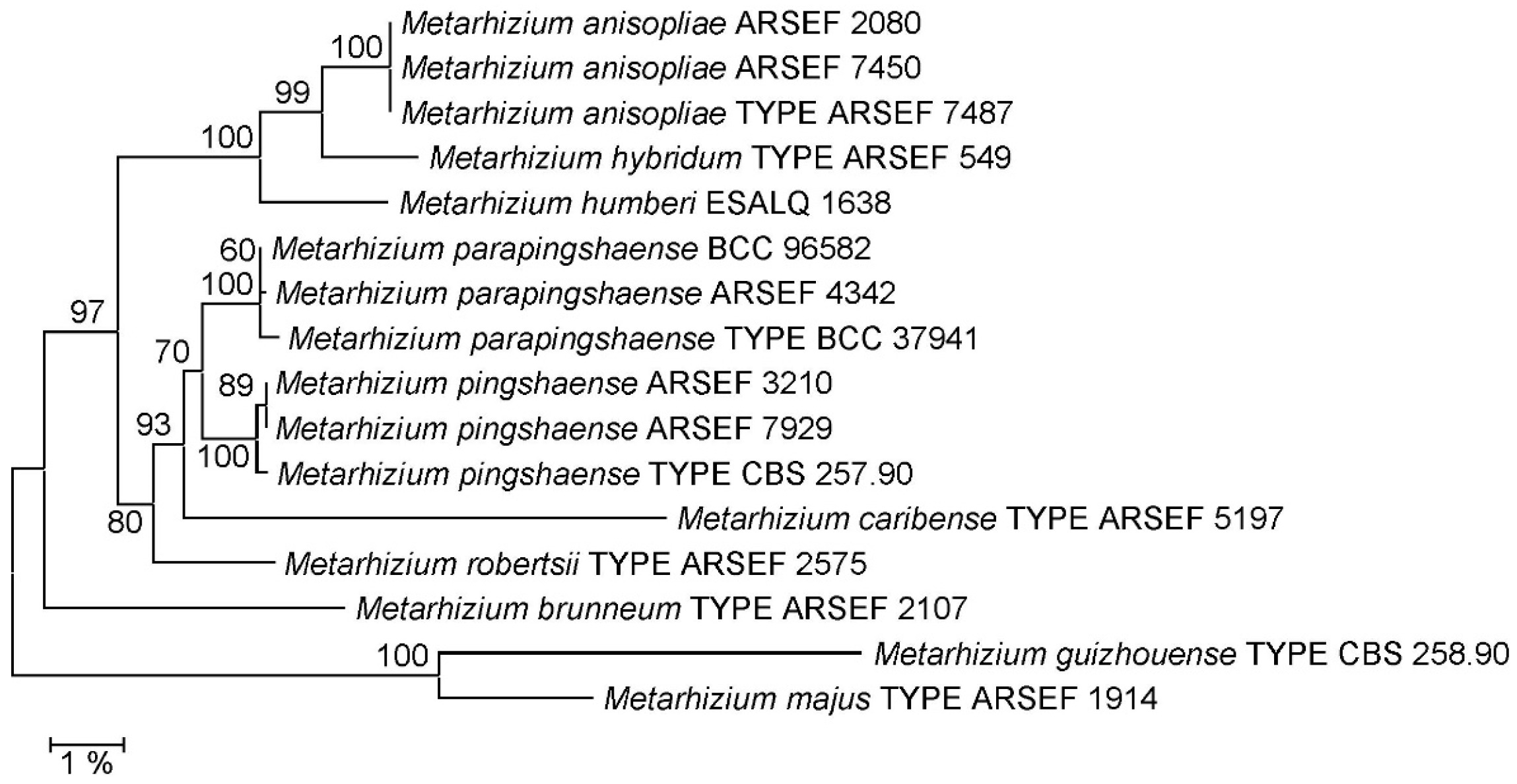
Comparison to the complete rIGS sequences of nomenclatural type and reference strains representing all Metarhizium species currently described in the PARB clade (Supplementary Table S1) gave rise to trees of identical topology irrespective of the method—neighbor-joining (Figure 1) or Bayesian inference (Supplementary Figure S1)—used for phylogenetic reconstruction. With respect to species delineation within the PARB clade, this tree topology was identical to the topological structure of the PARB clade in the phylogeny generated by Kobmoo et al. (2024) [25] from a concatenated matrix of 237 genes, with the relevant difference that species M. humberi and M. caribense were not represented in this whole genome tree. Importantly, delineations of the new species M. hybridum from M. anisopliae and M. parapingshaense from M. pingshaense as proposed by Kobmoo et al. (2024) [25] received 100% bootstrap support or posterior probabilities of 1.0 in the rIGS-based NJ and BI phylogenies, respectively. Moreover, M. hybridum and M. parapingshaense were delineated from their respective second neighboring species, M. humberi and M. caribense, by 100% bootstrap support or 1.0 posterior probability.
Figure 1.
Neighbor-joining (NJ) phylogeny of Metarhizium fungi as reconstructed from complete ribosomal intergenic spacer (rIGS) sequences. Terminal branches are labeled by genus, species and strain designations; “TYPE” denotes the nomenclatural type strain of a species. Numbers on branches indicate bootstrap support percentages. The size bar corresponds to 1% sequence divergence. The orthologous sequences from the M. majus and M. guizhouense type strains were used as outgroup.
3.2. Cryptic Descriptions of M. hybridum and M. parapingshaense
Analysis of the 46 assembled Metarhizium whole genome sequences published in the GenBank database revealed one further M. hybridum genome—namely that of strain E6 currently assigned to M. anisopliae—in addition to the type strain ARSEF 549 genome. No assembled M. parapingshaense genomes were identified.
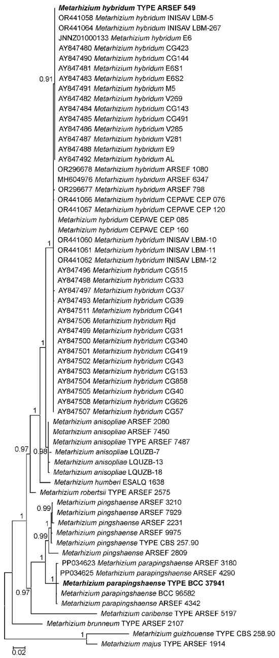
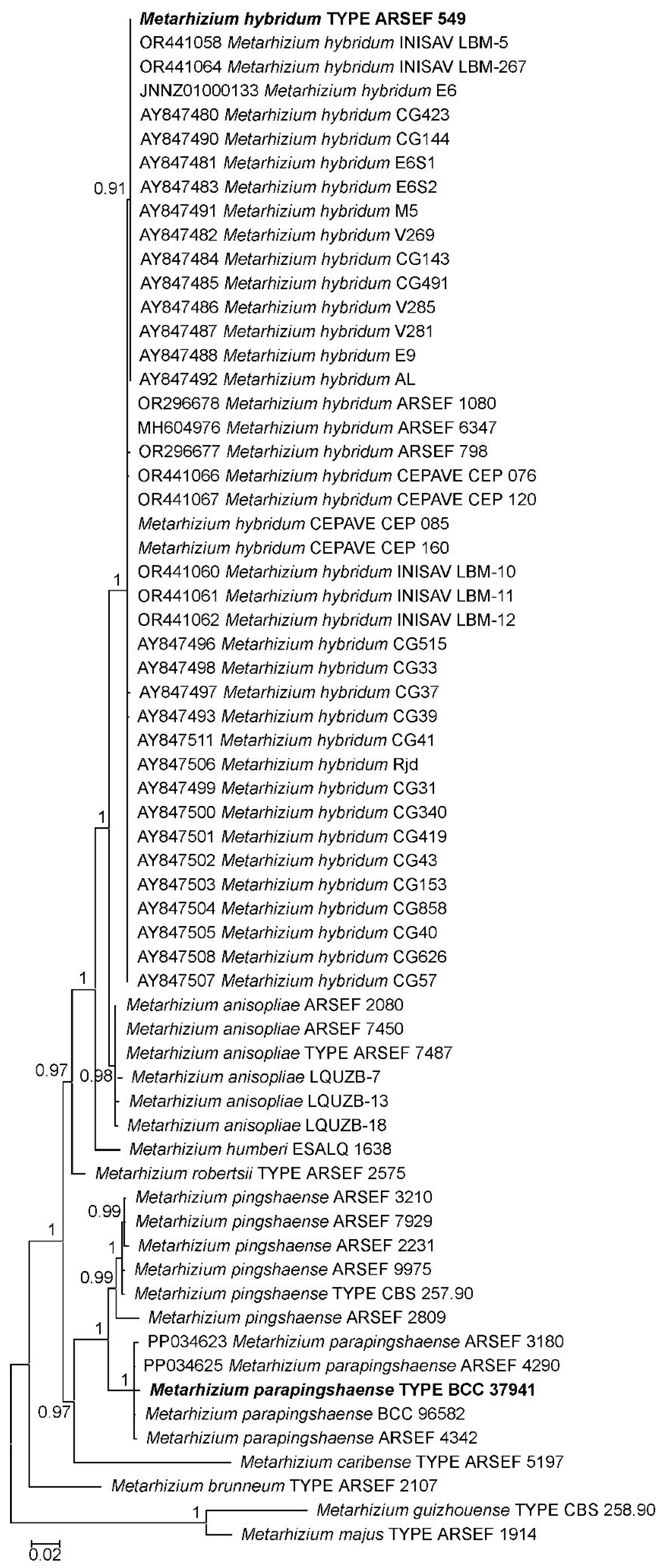
Searches for rIGS-ID800 homologous single GenBank database entries revealed 37 cryptic descriptions of M. hybridum and two of M. parapingshaense (Figure 2, Supplementary Figure S2). Extension of the GenBank search to markers DUF895, 5TEF, EF1A, and RPB2 identified 77 further strains of M. hybridum and 28 of M. parapingshaense (Supplementary Table S3). Bootstrap support values for M. hybridum and M. parapingshaense clades varied strongly for the different marker–species combinations, being >95% with rIGSI-D800, DUF895 and RPB2, but only app. 65% with 5TEF and EF1A for M. hybridum, as well as >95% with 5TEF and DUF895, but only app. 85% with rIGS-ID800 and EF1A for M. parapingshaense (Supplementary Figures S2–S6).
Figure 2.
Bayesian inference (BI)-based phylogeny of Metarhizium fungi as reconstructed from rIGS-ID800 marker sequences. Terminal branches are labeled by genus, species and strain designations; “TYPE” denotes the nomenclatural type strain of a species. M. hybridum and M. parapingshaense type strains are displayed in bold face. GenBank accession numbers are indicated for cryptic descriptions identified in the GenBank database. Numbers on branches indicate posterior probability (pp) values. The size bar indicates the number of expected substitutions per site. The orthologous sequences from the M. majus and M. guizhouense type strains were used as outgroup.
Whereas most M. hybridum strains had previously been identified as M. anisopliae or Metarhizium sp., strains of M. parapingshaense had almost exclusively been assigned to M. pingshaense (Table 1). With respect to geographic origins, cryptic description data appeared severely biased towards the Americas for M. hybridum (104/115) and Asia/Oceania for M. parapingshaense (30/30). While M. hybridum was mainly isolated from associations with insects (77/115), soil appeared to be the most frequent isolation source for M. parapingshaense (23/30).
Table 1.
Comprehensive overview of cryptic descriptions of Metarhizium hybridum and Metarhizium parapingshaense identified in the GenBank database; detailed data, including GenBank accession numbers, were organized in Supplementary Table S3. Numbers in brackets indicate the number of strains or isolates identified.
3.3. Species-Discriminating Diagnostic PCR for M. hybridum and M. parapingshaense
Building on the previous establishment of a species-discriminating PCR tool for the Metarhizium PARB clade [13], species-discriminating primer pairs for the delineation of M. hybridum and M. anisopliae, as well as M. parapingshaense and M. pingshaense were designed against alignments of complete rIGS sequences (Supplementary Figures S7 and S8) and optimized for functionality under pre-established PCR parameters. When the discriminative power of these primer pairs—termed mani-ID, mhyb-ID, mppi-ID and mpin-ID (Table 2) was tested in diagnostic PCR reactions with genomic DNA of reference strains representing the Metarhizium PARB clade species M. anisopliae, M. brunneum, M. caribense, M. hybridum, M. parapingshaense, M. pingshaense and M. robertsii, PCR products were uniquely generated from cognate DNA templates and were of expected apparent length (Supplementary Figure S9). Due to the unavailability of M. humberi DNA, discriminative power with respect to this species was not tested experimentally. However, in silico analyses comparing diagnostic primer pairs with the unique available M. humberi rIGS sequence from the ESALQ 1638 genome led to the prediction that no product would be amplified at least from this reference strain.
Table 2.
Species-discriminating diagnostic PCR primer pairs used in this study.
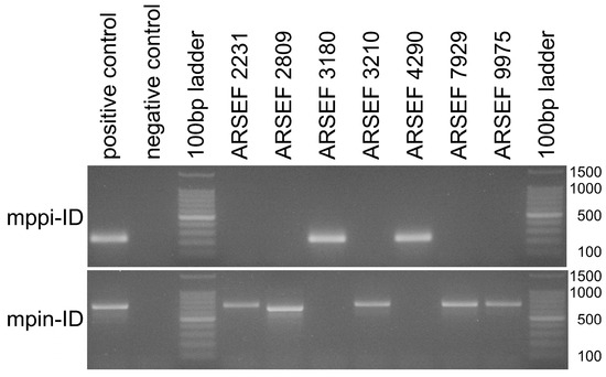
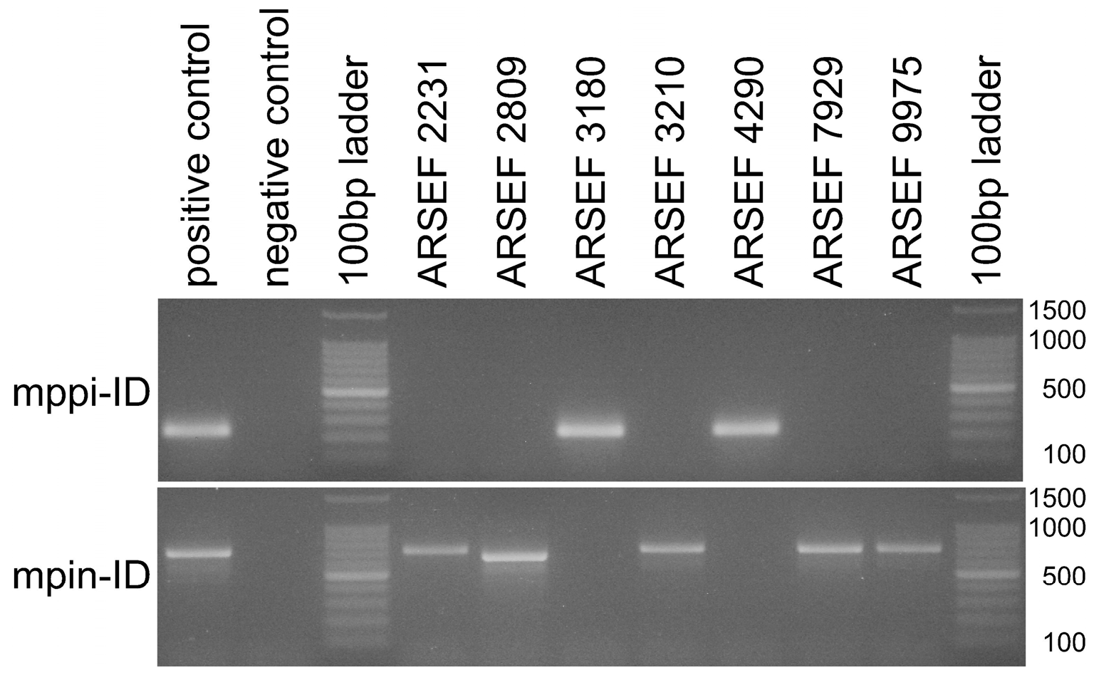
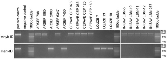
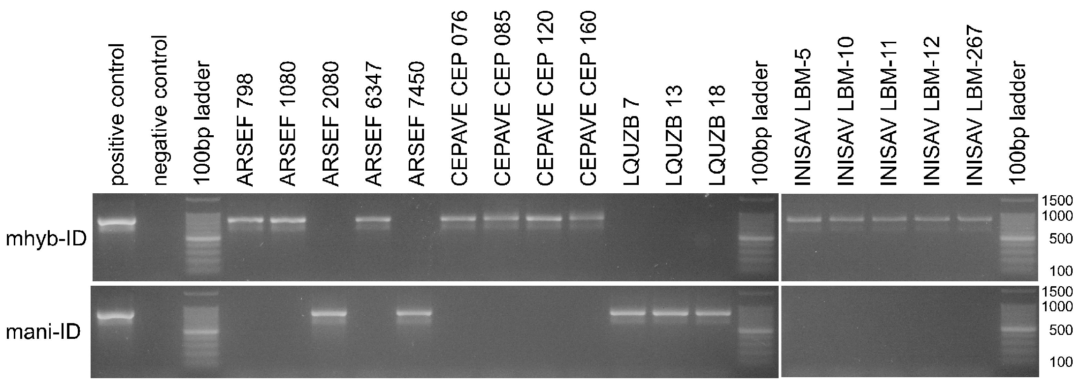
The functionality and species-discriminative power of designed diagnostic primers were further validated with two sets of 17 and seven Metarhizium strains and isolates that were, respectively, supposed to be closely related to M. anisopliae sensu lato or M. pingshaense sensu lato. More exactly, a set of twelve strains of globally distributed geographic origins obtained from the ARSEF collection was complemented with four Argentine strains from the CEPAVE and five Cuban strains from the INISAV culture collections, as well as three isolates (designated LQUZB) stemming from Uzbekistan (Supplementary Table S1). When the extracted genomic DNA of these fungi was probed with the diagnostic primer pairs mani-ID and mhyb-ID for M. anisopliae sensu lato as well as mppi-ID and mpin-ID for M. pingshaense sensu lato, exactly one single reaction per template was found positive (Figure 3 and Figure 4), motivating the assignment of each strain to one of the respective four Metarhizium species. From a total of 24 fungal strains tested, five were thus found assignable to M. anisopliae, twelve to M. hybridum, two to M. parapingshaense and five to M. pingshaense. PCR amplification and determination of the rIGS-ID800 marker sequence fully corroborated these diagnostic PCR-based species-level assignments (Figure 2).
Figure 3.
Agarose gel electrophoresis of diagnostic PCRs using species-discriminating primer pairs mpin-ID and mppi-ID specific for M. pingshaense and M. parapingshaense, respectively, as indicated at the left margin. The length (in bp) of the main signals in the size standard is indicated at the right margin. Lane labels on top of the gel pictures designate the Metarhizium strain or isolate; “positive control” denotes the primer pair-specific cognate type strain; “negative control” indicates the no template control; “100 bp ladder” denotes the size standard.
Figure 4.
Agarose gel electrophoresis of diagnostic PCRs using species-discriminating primer pairs mani-I and mhyb-ID specific for M. anisopliae and M. hybridum, respectively, as indicated at the left margin. The length (in bp) of the main signals in the size standard is indicated at the right margin. Lane labels on top of the gel pictures designate the Metarhizium strain or isolate; “positive control” denotes the primer pair-specific cognate type strain; “negative control” indicates the no template control; “100 bp ladder” denotes the size standard.
4. Discussion
As part of an integrative taxonomic study combining phylogenomics, morphometrics, metabolomics and virulence data, Kobmoo et al. (2024) had introduced two new taxa in the Metarhizium PARB clade [25]: phylogenetic reconstruction based on a concatenation of 237 selected fungal genes had delineated (i) the new species M. hybridum from M. anisopliae sensu stricto in M. anisopliae sensu lato and (ii) the new species M. parapingshaense from M. pingshaense sensu stricto in M. pingshaense sensu lato. In the present study, these species delineations were corroborated, receiving optimal support in both the NJ and BI phylogenies (Figure 1, Supplementary Figure S1) reconstructed from the complete ribosomal intergenic spacer sequences of the same fungal strains compared in the study cited.
This is a meaningful result as two further Metarhizium species that had previously been delineated within M. anisopliae sensu lato and M. pingshaense sensu lato, namely M. humberi [22] and M. caribense [24], were not considered in the description of M. hybridum and M. parapingshaense. In the present study, these taxa were represented in the analysis, and the above results confirm that (i) M. hybridum is a new species with respect not only to M. anisopliae sensu stricto, but also to M. humberi, and that (ii) M. parapingshaense is correctly delineated not only from M. pingshaense sensu stricto, but also from M. caribense.
In turn, the reproducibility of new species delineations that were introduced from an integrative geno- and phenotypic approach, in the rIGS-based phylogenetic reconstruction, lends strong support to the rIGS-based single-marker approach at this taxonomic level. It has been shown previously that the rIGS marker provides an excellent resolution of sub-clade structures within the PARB clade [12,13]. The identity of phylogenetic tree topologies generated from the single rIGS marker with those based on an integrative phylogenomic approach that is further backed by phenotypic traits strongly supports the idea that rIGS-based topological distinctions and sub-clade delineations are not only technically solid, but also phylogenetically meaningful. In conclusion, the observed congruence with a phylogenomic dataset validates the phylogenetic signal within the rIGS marker, thereby justifying its use as a surrogate for more resource-intensive methods for species delineation within the Metarhizium PARB clade.
Mining of GenBank data revealed 115 cryptic descriptions of M. hybridum and 30 of M. parapingshaense. Expectedly, most identified M. hybridum and M. parapingshaense strains had previously been assigned to M. anisopliae and M. pingshaense, respectively, or had only been identified at the genus level. Analysis of related publications revealed that the delineation of M. hybridum from M. anisopliae had been anticipated in several studies without being explicitly claimed [4,10,32]. Interestingly, Metarhizium anisopliae strain E6, i.e., a prominent model for biocontrol, molecular entomopathogenicity, genomics and secretomics studies [33,34,35,36,37,38], was identified as M. hybridum; knowledge gathered using strain E6 should therefore be attributed to this new species.
With respect to geographic origins, revealed data sets were strongly biased towards the Western hemisphere for M. hybridum and to East Asia and Oceania for M. parapingshaense. These apparent distributions are fully in line with the fact that all previous explicit descriptions of M. hybridum were from Brazil and Argentina, as well as from Thailand and the Solomon Islands for M. parapingshaense. However, this geographic bias is not necessarily meaningful with respect to the global distribution of the new taxa, as results from several extensive regional studies from China, Brazil or Mexico that address fungal diversity in particular regionally important ecosystems dominate the dataset. Nevertheless, the data presented formally constitute the first descriptions of the new species from Canada, China, Colombia, Costa Rica, Cuba, Honduras, Mexico, New Zealand, the Philippines and the USA in the case of M. hybridum, as well as from China, India, Japan, the Philippines and South Korea for M. parapingshaense.
A similar bias as for geographic origin data was observed with respect to the most frequent substrate of isolation for M. hybridum (insects) and M. parapingshaense (soil), which could potentially be indicative of a predominantly entomopathogenic lifestyle of M. hybridum and a saprophytic lifestyle of M. parapingshaense. However, as for geographic origin data, the currently available data set does by no means allow us to rule out a sampling artifact.
The species M. hybridum has been described as characterized by genetic mixed ancestry, presumably resulting from a genomic hybridization event involving M. anisopliae sensu stricto and a second unidentified PARB clade species [25]. It might be tempting to speculate that the second hybridization partner could have been M. humberi. This species had not been considered in the study introducing M. hybridum, and the currently known apparent geographic distributions of both M. humberi (Brazil, Mexico) and M. hybridum strongly coincide. Moreover, at least in the rIGS-based phylogenetic trees, M. hybridum is organized between M. anisopliae and M. humberi as would be consistent with a scenario of recombination between different intergenic spacer elements coexisting in the same genome during the early post-hybridization stage prior to purifying genome reduction with paralogous rRNA operon homogenization. It goes without saying that this intriguing but highly speculative hypothesis will require further substantiation from comparative genomics studies.
The Metarhizium PARB clade consists of a tight cluster of species comprising numerous Metarhizium strains of high relevance for both fundamental research in entomopathogenicity and microbial control of insect pests. Fast and reliable species-level assignment of fungal isolates is highly solicited across this group of fungi. Morphological and microscopic identification have clear limits with respect to the discrimination of PARB clade species, and species-level identification relies on genetic marker sequence determination. However, as reasonably fast, reliable, and inexpensive DNA sequencing may not be readily available for laboratories in many parts of the world, a sequencing-independent molecular approach can be expected to meet the needs of numerous researchers in the field.
Using the intergenic spacer (rIGS) sequence of the ribosomal RNA operon clusters of the Metarhizium genome as a target sequence, a species-discriminating diagnostic PCR tool had previously been established and demonstrated to discriminate between PARB clade species. The rationale behind this tool and the corresponding application strategy have been discussed previously [13]. The new diagnostic primer pairs designed and validated in the present study extend the existing approach, enabling the sequencing-independent identification of the newly delineated species M. anisopliae, M. hybridum, M. pingshaense and M. parapingshaense.
With respect to species identification within M. anisopliae sensu lato, the diagnostic PCR tool, as it currently stands, is subject to the highly significant caveat that—due to the inaccessibility of M. humberi reference strains—functionality of primer pairs for the delineation of M. anisopliae or M. hybridum from M. humberi cannot be tested experimentally, but has to rely on in silico analyses of published sequence data. This means that in diagnostic practice, there is a risk of generating false positives for M. anisopliae sensu stricto or M. hybridum from a M. humberi template DNA. Therefore, the correctness of PCR-based identification in M. anisopliae sensu lato should be confirmed by rIGS-ID800 sequence determination, as has been done in the present study. This is, of course, only a second-best solution for a diagnostic tool designed to enable sequencing-independent species-level identification.
5. Conclusions
Phylogenetic reconstruction based on ribosomal intergenic spacer (rIGS) sequences was employed to corroborate the previous introduction of the two new Metarhizium species, M. hybridum and M. parapingshaense, in demonstrating that these were not only correctly delineated from M. anisopliae sensu stricto and M. pingshaense sensu stricto, but also from all further species described in M. anisopliae sensu lato and M. pingshaense sensu lato, respectively. In turn, this confirmation of new species delineations derived from studies integrating whole genomes with phenotypic data lends strong support to the reliability of rIGS as a single-marker for species differentiation, at least in the Metarhizium PARB clade. A rIGS-based species-discriminating diagnostic PCR tool enabling sequencing-independent species identification has been functionally extended to these new Metarhizium species.
Supplementary Materials
The following supporting information can be downloaded at: https://www.mdpi.com/article/10.3390/jof12040272/s1, Figure S1: Bayesian Inference (BI) based phylogeny of Metarhizium fungi as reconstructed from complete rIGS sequences. Figure S2: Neighbor-Joining (NJ) phylogeny of Metarhizium fungi as reconstructed from rIGSID800 marker sequences. Figure S3: Neighbor-Joining (NJ) phylogeny of Metarhizium fungi as reconstructed from 5TEF marker sequences. Figure S4: Neighbor-Joining (NJ) phylogeny of Metarhizium fungi as reconstructed from DUF895 marker sequences. Figure S5: Neighbor-Joining (NJ) phylogeny of Metarhizium fungi as reconstructed from EF1A marker sequences. Figure S6: Neighbor-Joining (NJ) phylogeny of Metarhizium fungi as reconstructed from RPB2 marker sequences. Figure S7: Alignment of ribosomal intergenic spacer (rIGS) sequences used for the design of species-discriminating primers for Metarhizium parapingshaense and Metarhizium pingshaense. Figure S8: Alignment of ribosomal intergenic spacer (rIGS) sequences used for the design of species-discriminating primers for Metarhizium hybridum and Metarhizium anisopliae. Figure S9: Agarose gel electrophoresis of diagnostic PCRs using species-discriminating primer pairs. Table S1: Metarhizium strains and marker sequences referred to in this study. Table S2: Preparative PCR primers used and reaction-specific parameters applied in this study. Table S3: Cryptic descriptions of M. hybridum and M. parapingshaense identified in the GenBank database.
Author Contributions
Conceptualization, A.L.; methodology, C.S.; validation, C.S., H.B.G. and A.L.; investigation, C.S., H.B.G., Y.B.R., S.B. and R.G.M.; data curation, C.S. and A.L.; writing—original draft preparation, A.L.; writing—review and editing, C.S., Y.B.R., R.G.M., S.B., A.C.G. and A.L.; project administration, C.S., Y.B.R., A.C.G., C.C.L.L. and A.L.; funding acquisition, Y.B.R., C.C.L.L. and A.L. All authors have read and agreed to the published version of the manuscript.
Funding
This research has received financial support from the German Ministry of Education and Research (BMBF/PT Jülich) under research grants 031B0766, project BioEconomy International—FUNGI 4 FOOD, and 031B1130, project BioEconomy International—FUNGI 4 VectorControl, as well as from the Cuban research project: Empleo de nuevos aislados de los entomopatógenos Beauveria y Metarhizium spp. para el desarrollo de bioproductos para el control de plagas agrícolas financed from Ministerio de Ciencia Tecnología y Medio Ambiente (CITMA), under research grant PSAV P131LH003047. R.G.M. has been granted a researcher mobility scholarship from CONICET, Argentina, and a research scholarship from the German Academic Exchange Service (DAAD) under funding ID 91877683. S.B. acknowledges receipt of a research scholarship from the German Academic Exchange Service (DAAD) under funding ID 57588359.
Institutional Review Board Statement
Not applicable.
Informed Consent Statement
Not applicable.
Data Availability Statement
Sequence data analyzed in this study are publicly available from the GenBank database (https://www.ncbi.nlm.nih.gov, accessed on 18 February 2026) under nucleotide sequence accession numbers listed in Supplementary Tables S2 and S3 to this study.
Acknowledgments
The authors owe Kathryn E. Bushley from the ARSEF collection and Natalia Scelsio from the CEPAVE collection a debt of gratitude for sustainable support of the studies.
Conflicts of Interest
The authors declare no conflict of interest.
References
- Zimmermann, G. Review on safety of the entomopathogenic fungus Metarhizium anisopliae. Biocontrol Sci. Technol. 2007, 17, 879–920. [Google Scholar] [CrossRef] [Scilit]
- Aw, K.M.S.; Hue, S.M. Mode of Infection of Metarhizium spp. Fungus and their Potential as Biological Control Agents. J. Fungi 2017, 3, 30. [Google Scholar] [CrossRef] [Scilit] [PubMed]
- Tang, X.; Wang, X.; Cheng, X.; Wang, X.; Fang, W. Metarhizium fungi as plant symbionts. New Plant Prot. 2025, 2, e23. [Google Scholar] [CrossRef] [Scilit]
- Riguetti Zanardo Botelho, A.B.; Alves-Pereira, A.; Colonhez Prado, R.; Zucchi, M.; Delalibera, I., Jr. Metarhizium species in soil from Brazilian biomes: A study of diversity, distribution, and association with natural and agricultural environments. Fungal Ecol. 2019, 41, 289–300. [Google Scholar] [CrossRef] [Scilit]
- Zhang, Q.; Chen, X.; Xu, C.; Zhao, H.; Zhang, X.; Zeng, G.; Qian, Y.; Liu, R.; Guo, N.; Mi, W.; et al. Horizontal gene transfer allowed the emergence of broad host range entomopathogens. Proc. Natl. Acad. Sci. USA 2019, 116, 7982–7989. [Google Scholar] [CrossRef] [Scilit] [PubMed]
- Driver, F.; Milner, R.J.; Trueman, J.W.H. A taxonomic revision of Metarhizium based on a phylogenetic analysis of rDNA sequence data. Mycol. Res. 2000, 104, 134–150. [Google Scholar] [CrossRef] [Scilit]
- Sung, G.H.; Sung, J.M.; Hywel-Jones, N.L.; Spatafora, J.W. A multi-gene phylogeny of Clavicipitaceae (Ascomycota, Fungi): Identification of localized incongruence using a combinational bootstrap approach. Mol. Phylogenet. Evol. 2007, 44, 1204–1223. [Google Scholar] [CrossRef] [Scilit]
- Bischoff, J.F.; Rehner, S.A.; Humber, R.A. A multilocus phylogeny of the Metarhizium anisopliae lineage. Mycologia 2009, 101, 512–530. [Google Scholar] [CrossRef] [Scilit]
- Kepler, R.M.; Rehner, S.A. Genome-assisted development of nuclear intergenic sequence markers for entomopathogenic fungi of the Metarhizium anisopliae species complex. Mol. Ecol. Resour. 2013, 13, 210–217. [Google Scholar] [CrossRef] [Scilit]
- Rezende, J.M.; Zanardo, A.B.R.; da Silva Lopes, M.; Delalibera, I., Jr.; Rehner, S.A. Phylogenetic diversity of Brazilian Metarhizium associated with sugarcane agriculture. BioControl 2015, 60, 495–505. [Google Scholar] [CrossRef] [Scilit]
- Brunner-Mendoza, C.; Reyes-Montes, M.d.R.; Moonjely, S.; Bidochka, M.J.; Toriello, C. A review on the genus Metarhizium as an entomopathogenic microbial biocontrol agent with emphasis on its use and utility in Mexico. Biocontrol Sci. Technol. 2019, 29, 83–102. [Google Scholar] [CrossRef] [Scilit]
- Pantou, M.P.; Mavridou, A.; Typas, M.A. IGS sequence variation, group-I introns and the complete nuclear ribosomal DNA of the entomopathogenic fungus Metarhizium: Excellent tools for isolate detection and phylogenetic analysis. Fung. Genet. Biol. 2003, 38, 159–174. [Google Scholar] [CrossRef] [Scilit] [PubMed]
- Schuster, C.; Baró Robaina, Y.; Ben Gharsa, H.; Bobushova, S.; Manfrino, R.G.; Gutierrez, A.C.; López Lastra, C.C.; Doolotkeldieva, T.; Leclerque, A. Species Discrimination within the Metarhizium PARB Clade: Ribosomal Intergenic Spacer (rIGS)-Based Diagnostic PCR and Single Marker Taxonomy. J. Fungi. 2023, 9, 996. [Google Scholar] [CrossRef] [Scilit]
- Yanaka-Schäfer, F.Y.; Dall’Onder, L.P.; Panichi, M.C.; Mendes, R.G.; Fagundes, N.J.R.; Bandinelli, J.B.; Bogo, M.R. Sequence analysis of the rDNA intergenic spacer of Metarhizium strains isolated in Brazil. Genet. Mol. Biol. 2008, 31, 116–121. [Google Scholar] [CrossRef] [Scilit]
- Cheng, R.; Luo, A.; Orr, M.; Ge, D.; Hou, Z.; Qu, Y.; Guo, B.; Zhang, F.; Sha, Z.; Zhao, Z.; et al. Cryptic diversity begets challenges and opportunities in biodiversity research. Integr. Zool. 2025, 20, 33–49. [Google Scholar] [CrossRef] [Scilit]
- Sung, G.H.; Hywel-Jones, N.L.; Sung, J.M.; Luangsa-Ard, J.J.; Shrestha, B.; Spatafora, J.W. Phylogenetic classification of Cordyceps and the clavicipitaceous fungi. Stud. Mycol. 2007, 57, 5–59. [Google Scholar] [CrossRef] [Scilit]
- Kepler, R.M.; Humber, R.A.; Bischoff, J.F.; Rehner, S.A. Clarification of generic and species boundaries for Metarhizium and related fungi through multigene phylogenetics. Mycologia 2014, 106, 811–829. [Google Scholar] [CrossRef] [Scilit]
- Rehner, S.A.; Kepler, R.M. Species limits, phylogeography and reproductive mode in the Metarhizium anisopliae complex. J. Invertebr. Pathol. 2017, 148, 60–66. [Google Scholar] [CrossRef] [Scilit] [PubMed]
- Montalva, C.; Collier, K.; Rocha, L.F.; Inglis, P.W.; Lopes, R.B.; Luz, C.; Humber, R.A. A natural fungal infection of a sylvatic cockroach with Metarhizium blattodeae sp. nov.; a member of the M. flavoviride species complex. Fungal Biol. 2016, 120, 655–665. [Google Scholar] [CrossRef] [Scilit] [PubMed]
- Lopes, R.B.; Souza, D.A.; Rocha, L.F.N.; Montalva, C.; Luz, C.; Humber, R.A.; Faria, M. Metarhizium alvesii sp. nov.: A new member of the Metarhizium anisopliae species complex. J. Invertebr. Pathol. 2018, 151, 165–168. [Google Scholar] [CrossRef] [Scilit]
- Gutierrez, A.C.; Leclerque, A.; Manfrino, R.G.; Luz, C.; Ferrari, W.A.O.; Barneche, J.; García, J.J.; López Lastra, C.C. Natural occurrence in Argentina of a new fungal pathogen of cockroaches, Metarhizium argentinense sp. nov. Fungal Biol. 2019, 123, 364–372. [Google Scholar] [CrossRef] [Scilit]
- Luz, C.; Rocha, L.F.N.; Montalva, C.; Souza, D.A.; Botelho, A.B.R.Z.; Lopes, R.B.; Faria, M.; Delalibera, I., Jr. Metarhizium humberi sp. nov. (Hypocreales: Clavicipitaceae), a new member of the PARB clade in the Metarhizium anisopliae complex from Latin America. J. Invertebr. Pathol. 2019, 166, 107216. [Google Scholar] [CrossRef] [Scilit]
- Bischoff, J.F.; Rehner, S.A.; Humber, R.A. Metarhizium frigidum sp. nov.: A cryptic species of M. anisopliae and a member of the M. flavoviride complex. Mycologia 2006, 98, 737–745. [Google Scholar] [CrossRef] [PubMed]
- Baró Robaina, Y.; Schuster, C.; Castañeda-Ruiz, R.F.; Gato Cárdenas, Y.; Márquez Gutiérrez, M.E.; Ponce de la Cal, A.; Leclerque, A. Metarhizium caribense sp. nov., a Novel Species of Entomopathogenic Metarhizium Fungi Associated with Weevils Impairing Coffee, Sugar Cane and Sweet Potato Cultivation. J. Fungi 2024, 10, 612. [Google Scholar] [CrossRef] [Scilit]
- Kobmoo, N.; Mongkolsamrit, S.; Khonsanit, A.; Cedeño-Sanchez, M.; Arnamnart, N.; Noisripoom, W.; Kwantong, P.; Sonthirod, C.; Pootakham, W.; Amnuaykanjanasin, A.; et al. Integrative taxonomy of Metarhizium anisopliae species complex, based on phylogenomics combined with morphometrics, metabolomics, and virulence data. IMA Fungus 2024, 15, 30. [Google Scholar] [CrossRef] [Scilit]
- Lozano, F.; Manfrino, R.G.; Leclerque, A.; Schuster, C.; Rivas-Franco, F.; Gutierrez, A.C. Argentine Metarhizium spp. isolates: Molecular identification, blastospore production, and its pathogenicity against Blattella germanica adults. J. Invertebr. Pathol. 2026, 214, 108471. [Google Scholar] [CrossRef] [Scilit]
- Tamura, K.; Stecher, G.; Kumar, S. MEGA11: Molecular Evolutionary Genetics Analysis Version 11. Mol. Biol. Evol. 2021, 38, 3022–3027. [Google Scholar] [CrossRef] [Scilit] [PubMed]
- Altschul, S.F.; Madden, T.L.; Schäffer, A.A.; Zhang, J.; Zhang, Z.; Miller, W.; Lipman, D.J. Grapped BLAST and PSI-BLAST: A new generation of protein database search programs. Nucleic Acids Res. 1997, 25, 3389–3402. [Google Scholar] [CrossRef] [Scilit] [PubMed]
- Zhang, Z.; Schwartz, S.; Wagner, L.; Miller, W. A greedy algorithm for aligning DNA sequences. J. Comput. Biol. 2000, 7, 203–214. [Google Scholar] [CrossRef] [Scilit]
- Thompson, J.D.; Higgins, D.G.; Gibson, T.J. ClustalW: Improving the sensitivity of progressive multiple sequence alignment through sequence weighting, positions-specific gap penalties and weight matrix choice. Nucleic Acids Res. 1994, 22, 4673–4680. [Google Scholar] [CrossRef] [Scilit]
- Ronquist, F.; Teslenko, M.; van der Mark, P.; Ayres, D.; Darling, A.; Höhna, S.; Larget, B.; Liu, L.; Suchard, M.A.; Huelsenbeck, J.P. MrBayes 3.2: Efficient Bayesian phylogenetic inference and model choice across a large model space. Systematic Biol. 2012, 61, 539–542. [Google Scholar] [CrossRef] [Scilit]
- Pedroso, S.K.B.; Santos, A.C.S.; Alves, J.E.R.; Tiago, P.V. A Novel Species of the Metarhizium anisopliae Complex from Brazil. GenBank Database Entry, Accession Number PQ156565. 2024. Available online: https://www.ncbi.nlm.nih.gov/datasets/genome/?taxon=5529 (accessed on 2 February 2026).
- da Silva, M.V.; Santi, L.; Staats, C.C.; da Costa, A.M.; Colodel, E.M.; Driemeier, D.; Vainstein, M.H.; Schrank, A. Cuticle-induced endo/exoacting chitinase CHIT30 from Metarhizium anisopliae is encoded by an ortholog of the chi3 gene. Res. Microbiol. 2005, 156, 382–392. [Google Scholar] [CrossRef] [Scilit] [PubMed]
- Nakazato, L.; Dutra, V.; Broetto, L.; Staats, C.C.; Vainstein, M.H.; Schrank, A. Development of an expression vector for Metarhizium anisopliae based on the tef-1alpha homologous promoter. Appl. Microbiol. Biotechnol. 2006, 72, 521–528. [Google Scholar] [CrossRef] [Scilit] [PubMed]
- Anhalt, F.A.; Azevedo, J.L.; Sugayama, R.L.; Specht, A.; Barros, N.M. Potential of Metarhizium anisopliae (Metsch.) Sorokin (Ascomycetes, Hypocreales) in the control of Bonagota salubricola (Meyrick) (Lepidoptera, Tortricidae) and its compatibility with chemical insecticides. Braz. J. Biol. 2010, 70, 931–936. [Google Scholar] [CrossRef] [Scilit]
- Beys-da-Silva, W.O.; Santi, L.; Berger, M.; Guimarães, J.A.; Schrank, A.; Vainstein, M.H. Susceptibility of Loxosceles sp. to the arthropod pathogenic fungus Metarhizium anisopliae: Potential biocontrol of the brown spider. Trans. R. Soc. Trop. Med. Hyg. 2013, 107, 59–61. [Google Scholar] [CrossRef] [Scilit] [PubMed]
- Staats, C.C.; Junges, A.; Guedes, R.L.; Thompson, C.E.; de Morais, G.L.; Boldo, J.T.; de Almeida, L.G.; Andreis, F.C.; Gerber, A.L.; Sbaraini, N.; et al. Comparative genome analysis of entomopathogenic fungi reveals a complex set of secreted proteins. BMC Genom. 2014, 15, 822. [Google Scholar] [CrossRef] [Scilit]
- Saciloto-de-Oliveira, L.R.; Broetto, L.; Alves, C.I.; da Rosa, R.L.; Calegari Alves, Y.P.; da Silva, R.C.; Berger, M.; Macedo, A.J.; Dalberto, P.F.; Bizarro, C.V.; et al. Metarhizium anisopliae E6 secretome reveals molecular players in host specificity and toxicity linked to cattle tick infection. Fungal Biol. 2023, 127, 1136–1145. [Google Scholar] [CrossRef] [Scilit]

Disclaimer/Publisher’s Note: The statements, opinions and data contained in all publications are solely those of the individual author(s) and contributor(s) and not of MDPI and/or the editor(s). MDPI and/or the editor(s) disclaim responsibility for any injury to people or property resulting from any ideas, methods, instructions or products referred to in the content. |
© 2026 by the authors. Licensee MDPI, Basel, Switzerland. This article is an open access article distributed under the terms and conditions of the Creative Commons Attribution (CC BY) license.