


Abstract
Grifola frondosa is a rare fungus valued for its nutritional and medicinal properties; however, its bacterial spot disease has been largely overlooked. Thus, this study systematically investigated, isolated, and identified the pathogen and evaluated control strategies for bacterial spot disease affecting G. frondosa cultivation in Qingyuan County, Zhejiang Province. Through integrated morphological, physiological and biochemical analysis, and multi-locus phylogenetic analyses (16S rRNA, gyrB), Priestia aryabhattai was identified as the causal pathogen. This pathogen exhibited host specificity, infecting only G. frondosa and Pleurotus ostreatus, inducing primordial growth arrest and causing spots on the stipe of mature fruiting bodies. Control assessments revealed significant antimicrobial efficacy for four chemical agents, benziothiazolinone, copper sulfate, ethylicin and tetramycin, three plant extracts, garlic, leek and onion, and two biocontrol strains, Chlorophyllum molybdites and Aspergillus fumigatus. Scanning electron microscopy (SEM) demonstrated that these treatments caused ultrastructural damage to the pathogen’s cells, including membrane shrinkage, depression, and perforation. These findings establish key pathogenic characteristics and provide a scientific foundation for integrated disease management, supporting sustainable G. frondosa cultivation.
1. Introduction
China’s edible fungi industry commenced a period of substantial growth in 1978, when it accounted for merely 5.7% of global production (approximately 58,000 metric tons). By 2013, its output had surged to 31.697 million metric tons, representing over 70% of the world’s total. This share further increased to surpass 80% by 2020 [1,2]. Within China’s agricultural sector, the industry is consistently ranked as the fifth-largest by output value, establishing itself as a key pillar within crop cultivation after grain, oil crops, vegetables, and fruits [1,3]. Since the turn of the 21st century, China has accelerated its global expansion by capitalizing on its advanced industrialized production capabilities. In 2017, the nation accounted for 42% of global factory-based output, thereby cementing its dominant position in the international market [1]. Grifola frondosa (Dicks.) Gray, a rare fungus known for its nutritional and medicinal properties, possesses cultivation potential and has been successfully cultivated on an industrial scale [1]. Beyond rich concentrations of high-quality proteins, essential vitamins, and trace elements, G. frondosa polysaccharides exhibit pharmacological activities including antitumor and immunomodulatory effects, positioning it as a resource for functional foods and pharmaceuticals [4]. Thus, G. frondosa is widely cultivated in China, such as Hebei, Yunnan, Zhejiang, and other provinces, with Qingyuan County in Zhejiang Province maintaining the highest production capacity nationwide [5].
As the cultivation scale of G. frondosa has expanded, the incidence of diseases has significantly increased, severely affecting both yield and quality [6,7,8]. For example, in Qingyuan County, Zhejiang Province, the primary production region, pathogenic diseases have emerged as a major constraint to the development of the industry. Bacterial diseases, in particular, are highly detrimental, causing substantial economic losses [9]. For instance, bacterial diseases pose serious threats to the cultivation of G. frondosa. In Guangdong Province, China, infection by Bacillus cereus causes severe decay of the primordia, characterized by foul odor, extensive whitish mucus, and bubble formation, leading to stunted growth or complete failure of fruiting body development, which significantly reduces yield [7]. Concurrently, bacterial spot disease, primarily caused by Pseudomonas spp., results in yellowish to brownish rust spots on the pileus. These spots rapidly expand into sunken brown lesions with a putrid odor, causing desiccation, malformation, and cracking of the fruiting bodies, thereby severely compromising product quality and market value [8]. Likewise, the cultivation of G. frondosa in Qingyuan County is seriously threatened by bacterial pathogens. This is compellingly evidenced by a recent first report from Qingyuan County, Zhejiang Province, where Burkholderia gladioli was identified as the causal agent of a bacterial blotch disease, with field incidence rates reaching approximately 40% and causing significant quality and yield losses [10]. However, compared to fungal diseases, research on bacterial diseases lags far behind.
Due to its simplicity and rapid efficacy, chemical control remains the dominant approach, with major agents including antibiotics, copper-based compounds, and azole fungicides [11]. These chemicals act through multiple mechanisms: oxidative damage disrupts cellular structures, altered membrane permeability induces leakage of cellular contents, and metabolic inhibition interferes with biosynthetic pathways. For example, benzothiazole derivatives employ a multi-target strategy, concurrently inhibiting key processes including cell wall biosynthesis (e.g., DprE1), cell division (FtsZ), and DNA replication (GyrB/ParE). This synergistic mechanism underlies their potent activity against drug-resistant pathogens like MRSA and VRE [12].
However, chemical control methods exhibit several critical limitations that undermine their effectiveness and sustainability. These include inefficiency in utilization, persistent environmental contamination, development of pathogen resistance, and inherent toxicity, all of which pose significant risks to both ecosystem and human health while conflicting with sustainable agricultural practices. A case study from Lishui City demonstrates these concerns, where edible mushrooms contained dithiocarbamate residues at 0.165 mg/kg (13.8% detection rate), indicating potential chronic health hazards for consumers [13]. Compounding this issue, excessive application of fungicides like carbendazim has rapidly selected for resistant pathogen strains. Most alarmingly, 33.67% of China’s edible mushroom exports fail to meet international residue standards, directly threatening the industry’s long-term viability [13]. These empirical observations collectively demonstrate the fundamental deficiencies of chemical control approaches regarding their operational efficiency, safety profile, and environmental sustainability.
In contrast, biological control offers distinct advantages through its dual functionality in disease suppression and plant growth promotion. This approach employs three primary classes of biocontrol agents: (i) phytochemicals [14] such as allicin, (ii) microbial antagonists [15] including Streptomyces Waksman and Henrici species and Trichoderma Pers. fungi, and (iii) fungal-derived secondary metabolites like saponins and sesquiterpenes [16]. For instance, the microbial antagonist Trichoderma harzianum Rifai not only provides effective disease control through direct mycoparasitism and enzymatic degradation of pathogens but also promotes plant growth, achieving over 100% yield increase in tomato. This efficacy can be amplified through genetic engineering or integration with chemical methods, showcasing the adaptable and synergistic potential of biological control [17]. Collectively, these biological agents exert antimicrobial effects through multiple mechanisms—including membrane disruption and metabolic interference—while stimulating plant development. This integrated approach combines high efficacy with environmental safety and minimal resistance development, establishing a sustainable paradigm for crop protection systems.
This study first isolates, identifies and reports P. aryabhattai as the causal pathogen of bacterial spot disease in G. frondosa by integrating morphological studies, phylogenetic analysis, physiological and biochemical characteristics, while also elucidating its host specificity. In addition, chemical and biological control measures were evaluated, screening four high-efficiency chemical agents, three botanical extracts, and two biocontrol strains.
2. Materials and Methods
2.1. Pathogenic Bacteria Isolation
Symptomatic fruiting bodies of G. frondosa displaying characteristic bacterial blotch symptoms were collected from Qingyuan County Edible Fungi Research Center. The collection was conducted during a disease survey carried out between October and November of 2023. Samples were immediately placed in sterile containers, transported to the laboratory on ice, and processed within 24 h. The fruiting bodies were initially surface-sterilized with 75% ethanol, followed by three rinses with sterile distilled water, and then transferred to sterile Petri dishes [18]. Diseased tissue samples were aseptically excised from the lesion margins, inoculated onto Nutrient Agar (NA) plates (peptone 10 g; beef extract 3 g; sodium chloride 5g; agar 15g; distilled water 1000 mL; pH 7.1–7.5), and incubated at 28 °C in complete darkness. Following incubation, distinct colonies were purified through successive streak plating on fresh NA medium until axenic cultures were obtained. Then, the purified strains were stored at 4 °C for future use [19].
2.2. Morphological Observation
For pathogen identification, the purified strain was used for observation. Gram staining was performed to characterize the isolates, along with microscopic examination of specialized cellular structures, including flagella, capsules, and endospores, using specific reagents [20].
For biocontrol fungi, the identification was referred to the Atlas of Chinese Macrofungal Resources [21]. Dried specimens were rehydrated in 94% ethanol and subsequently mounted in 3% potassium hydroxide (KOH) for examination. Structures such as basidiospores, basidia, and cheilocystidia were observed.
The biocontrol microorganisms were streaked onto Gauze’s Synthetic Medium No. 1 (soluble starch 20 g, KNO3 1 g, NaCl 0.5 g, K2HPO4·3H2O 0.5 g, MgSO4·7H2O 0.5 g, FeSO4·7H2O 0.01 g, distilled water 1000 mL, pH 7.4–7.6) and incubated at 28 °C for 7–14 d. During this period, its growth characteristics, including colony morphology and production of diffusible pigments, were observed. Then, the micro-characteristics, such as aerial hyphae, spore filaments, and spores, were examined.
2.3. Physiological and Biochemical Analysis
To gain a further understanding of the pathogen’s growth characteristics, the following physiological and biochemical experiments were performed: sugar fermentation, nitrate reduction, gelatin liquefaction, starch hydrolysis, citrate utilization, hydrogen sulfide production, motility (stab inoculation), urease activity, indole production, methyl red test, oxidation-fermentation (O/F) test, and Voges–Proskauer (V-P) test [22].
2.4. PCR and Phylogenetic Analysis
Genomic DNA was extracted using the TIANamp Micro DNA kit (Tiangen Biotech (Beijing) Co., Ltd., Beijing, China), and the genome extraction quality was determined by 1% agarose gel electrophoresis.
Pathogenic bacteria were characterized using 16S rRNA (16S-27F and 16S-1492R) and gyrB (gyrB-F and gyrB-R); microorganisms were identified using ITS (ITS1 and ITS4) [23], BenA (Bt2a and Bt2b) [23], and CaM (CMD5 and CMD6) [23]; and macrofungi were identified using ITS (ITS1 and ITS4), tef1-ɑ (EF-983F and EF-1567R) [24], and nLSU (LROR and LR5) [25]. PCR amplifying system (25 µL): 9 µL dd H2O, 12.5 µL 2 × Es Taq MasterMix (Dye), 1 µL forward primer, 1 µL reverse primer and 1.5 µL genomic DNA. PCR reaction conditions: 94 °C pre-denaturation for 5 min, followed by 94 °C denaturation for 30 s, 52 °C (nLSU, tef1-ɑ), 55 °C (ITS, benA, CaM), 56 °C (16S), and 58 °C (gyrB), annealing for 45 s, 72 °C extension for 1 min, a total of 35 cycles, and finally 72 °C extension for 10 min. The PCR amplification products were confirmed by 1% agarose gel electrophoresis and then sent to Beijing Qingke Biotechnology Co., Ltd. Beijing, China, for sequencing.
The newly obtained sequences were submitted to NCBI GenBank https://www.ncbi.nlm.nih.gov/genbank/. (accessed on 2 September 2025) for BLAST analysis, the same and similar species were selected and downloaded, and the multiple sequence comparison was performed by BioEdit 7.2.5 [26]. The phylogenetic analysis was performed using both maximum likelihood analysis (ML) and Bayesian inference (BI). ML was performed through RAxML 8.0.26 [27] and BI was performed using MrBayes 3.2 [28]. And the sequences used in this study are listed in Table 1, Table 2 and Table 3.
Table 1.
Species and sequences used in phylogenetic analyses of pathogenic bacteria. Sequences produced in this study are in bold.
Table 2.
Species and sequences used in phylogenetic analyses of macrofungi. Sequences produced in this study are in bold.
Table 3.
Species and sequences used in phylogenetic analyses of microorganism. Sequences produced in this study are in bold.
2.5. Pathogenicity Tests
The surfaces of both original and differentiated G. frondosa fruiting bodies were mechanically wounded using sterile needles. The pathogen suspension was then inoculated at the wound sites and incubated at 25 °C under controlled humidity. Sterile water served as the negative control. Each treatment was performed in triplicate (three independent fungal bags), and fruiting body formation was regularly monitored and documented. The pathogen was then reisolated from the infected fruiting bodies and subjected to morphological characterization and phylogenetic analysis to fulfill Koch’s postulates [29].
2.6. Host Range Test
The widely cultivated mushrooms Pleurotus eryngii (DC.) Quél., Hericium erinaceus (Bull.) Pers., Pleurotus ostreatus (Jacq.) P. Kumm., F. filiformis (Z.W. Ge, X.B. Liu & Zhu L. Yang) P.M. Wang, Y.C. Dai, E. Horak & Zhu L. Yang, and Hymenopellis raphanipes (Berk.) R.H. Petersen were selected for the host range test. Mechanical wounding was applied to the pileus and stipes of test mushrooms at both primordial formation and fruiting body development stages using sterile needles after disinfection. Then, the pathogen suspension was uniformly sprayed onto the inoculation sites until it reached a dripping state. Sterile distilled water served as the negative control.
2.7. Effects of Chemical–and Bio–Compounds on Pathogen
Six chemical compounds and six biological pesticides were selected for testing (Table 4). All compounds were serially diluted with sterile distilled water to generate concentration gradients of 125, 250, 500, 1000, and 2000 mg/L [30] for further experiments.
Table 4.
Reagents and commercial sources used in this study.
Then, the Oxford cup method [31] was employed to evaluate the inhibitory effect of the agents on pathogens. A volume of 200 μL of each agent, at varying concentrations, was added into an Oxford cup on an NA plate inoculated with the pathogen and incubated at 28 °C for 2–3 d. Each concentration was evaluated in triplicate, with sterile distilled water serving as the negative control. The inhibition zone was then measured using cross-section method. The inhibition rate, virulence regression equation, and median effective concentration value (EC50) [32] were calculated through SPSS Statistics 27.
2.8. Effectiveness of Plant Extracts in Controlling Pathogens
Fresh garlic (Allium sativum L.), onion (Allium cepa L.), leek (Allium porrum L.), chrysanthemum (Chrysanthemum morifolium Ramat.), dandelion (Taraxacum mongolicum Hand.-Mazz.), and peony (Paeonia lactiflora Pall.) were selected, and the extractions were referred to the reference [33,34,35,36,37], then stored at 4 °C. Then, the Oxford cup method was employed to evaluate the inhibitory effect of the extractions on pathogens. A volume of 200 μL of each extraction was added into an Oxford cup on an NA plate inoculated with the pathogen, followed by incubation at 28 °C for 2–3 d. The inhibition zone was then measured using the cross-section method. All treatments in the assay were performed in triplicate, with sterile distilled water serving as the negative control.
2.9. Research on the Effectiveness of Biological Control Agents
A co-culture assay was employed for primary screening. In this assay, agar plates were first spread with 100 μL of a pathogenic fungal suspension. After drying, a 5 mm plug of a candidate biocontrol microorganism was inoculated at the center of the plate. The plates were incubated at 28 °C for 2–3 d, and isolates that formed a clear inhibition zone were selected as potential antagonists. After, based on the result of the co-culture assay, the fermentation broth was tested for its bacteriostatic activity using the Oxford cup method, with each process repeated three times.
2.10. Effects of Different Chemicals, Plant Extracts, and Biocontrol Microbiology Treatments on Pathogen
Bacterial samples were collected from the inhibition zone using a flame-sterilized toothpick and transferred onto carbon-coated adhesive tabs mounted on aluminium stubs. The samples were then sputter-coated with a 5 nm gold–palladium layer for 30 s using an ion sputter coater. Subsequently, the specimens were examined under a Zeiss EVO LS10 scanning electron microscope (SEM, Carl Zeiss AG, Oberkochen, Germany) at an accelerating voltage of 5 kV, and digital images were captured at 10,000× magnification.
3. Results
3.1. Pathogen Identification
3.1.1. Morphological Characteristics of the Pathogen
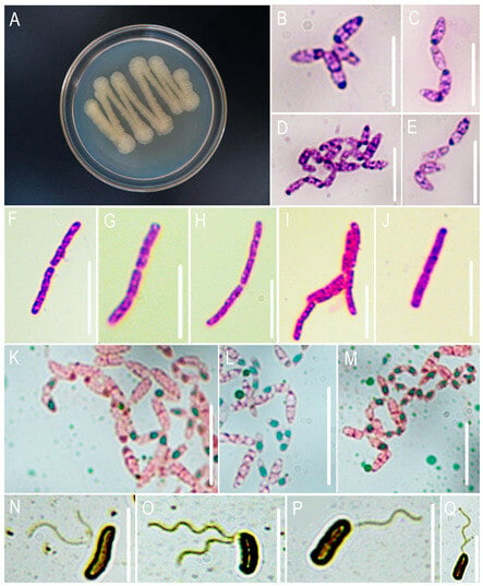
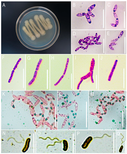
Colonies appeared pale yellow with moist surfaces on NA, flat, and margins smooth (Figure 1A), Gram-positive staining (Figure 1F–J), capsules absent (Figure 1B–E), flagella present (Figure 1N–Q), endospores round to oval (Figure 1K–M), typically terminal or subterminal, without significant sporangium swelling. These characteristics match the description of Priestia aryabhattai.
Figure 1.
Micro-morphological and special staining identification of the pathogenic bacteria. (A) Bacterial lawn; (B–E) capsules; (F–J) Gram-staining results; (K–M) endospores; (N–Q) flagella. Bars: 5 μm.
3.1.2. Physiological and Biochemical Characteristics of the Pathogen
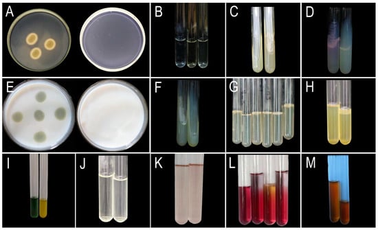
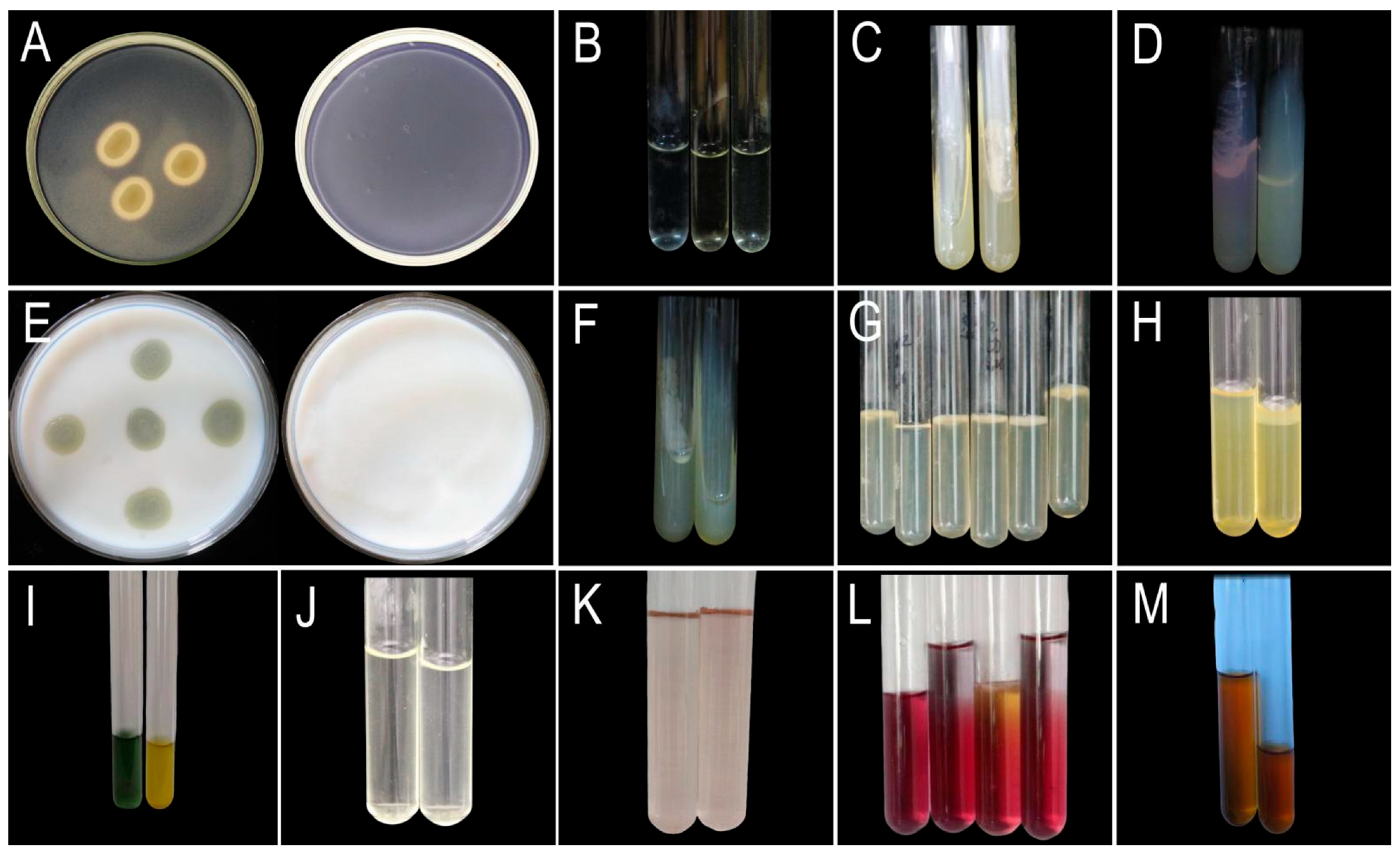
The results (Figure 2, Table 5) demonstrated that the pathogen exhibits nitrate reductase activity, gelatinase production, and starch/casein hydrolysis capabilities. Additionally, the pathogen was urease-positive, capable of utilizing mannitol, and exhibited oxidative metabolism. However, it was unable to utilize citrate as the sole carbon source, did not produce hydrogen sulfide, lacked tryptophanase activity, and failed to produce organic acids or neutral end products from glucose metabolism.
Figure 2.
Physiological and biochemical characterization of pathogenic bacteria. (A) Starch hydrolysis (left: experimental group; right: negative control). (B) Nitrate reduction test (left: negative control; middle: experimental; right: experimental + zinc dust). (C) Citrate utilization test (left: negative control; right: experimental). (D) Urea hydrolysis (left: negative control; right: experimental). (E) Casein hydrolysis (left: experimental; right: negative control). (F) Hydrogen sulfide production (left: negative control; right: experimental). (G) Motility stab test (tubes 1, 3–6: experimental; tube 2: negative control). (H) Gelatin liquefaction (tubes 1–2: experimental; tube 3: negative control). (I) Mannitol fermentation (left: negative control; right: experimental). (J) Voges–Proskauer test (left: negative control; right: experimental). (K) Indole production test (left: negative control; right: experimental). (L) Oxidation-Fermentation (O/F) test (Tube 1: Glycerol-free control; Tube 2: Glycerol-supplemented control; Tube 3: Glycerol-free experimental; Tube 4: Glycerol-supplemented experimental). (M) Methyl red test (left: control; right: experimental).
Table 5.
Physiological and biochemical identification results.
3.1.3. Phylogenetic Analysis
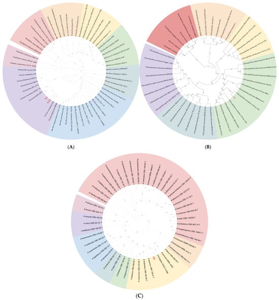
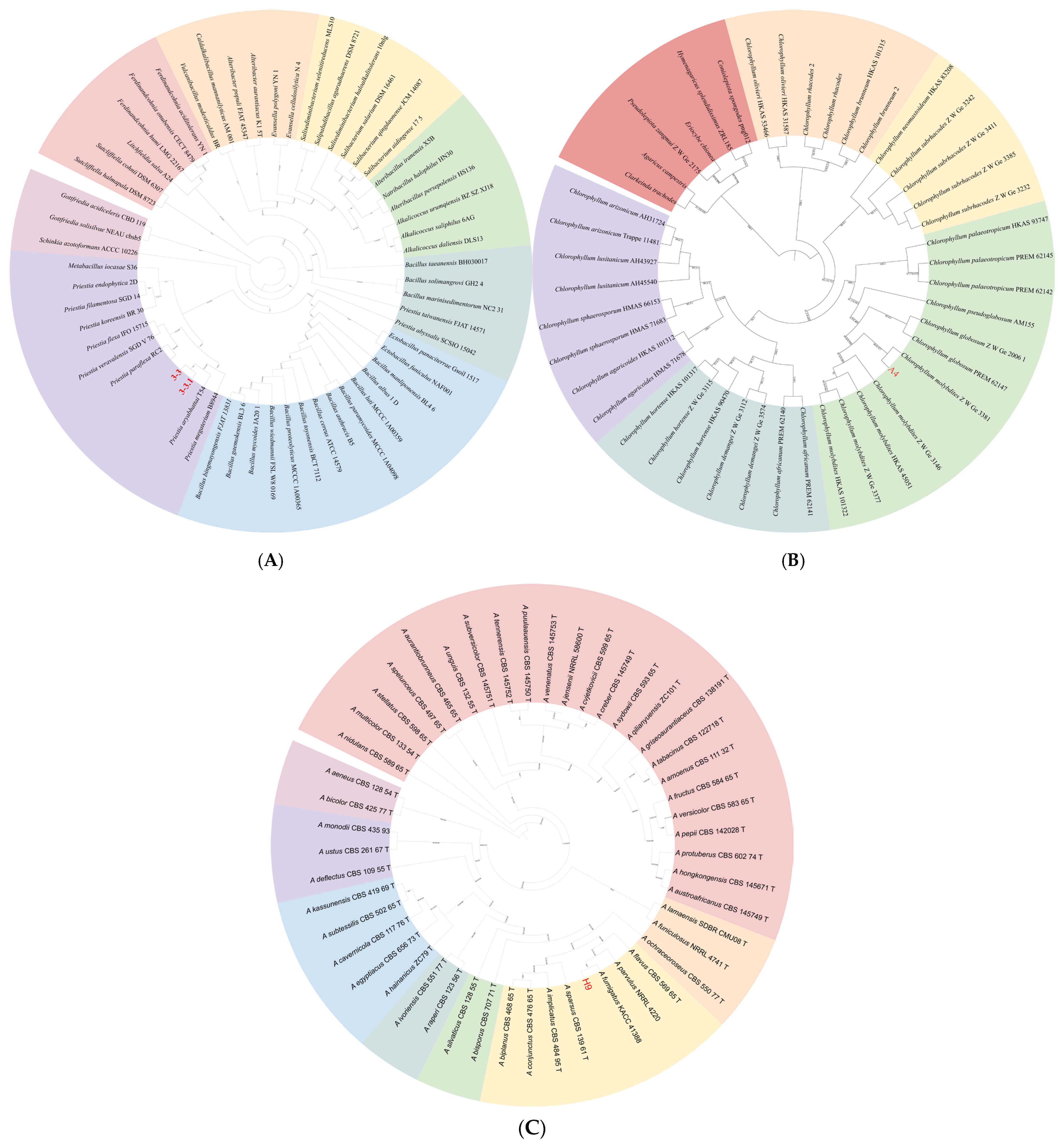
The sequenced data were uploaded to NCBI, and the BLAST results revealed a high similarity with P. aryabhattai. Phylogenetic analysis, incorporating 16S rRNA and gyrB loci, revealed that our sequences clustered within the P. aryabhattai clades (Figure 3A) with strong support.
Figure 3.
Phylogenetic reconstruction of three biological groups. (A) Pathogen inferred from Maximum likelihood (ML) and Bayesian inference (BI) analyses of the combined 16S rRNA and gyrB sequence dataset. (B) Macrofungi inferred from ML and BI analyses of the combined ITS, nLSU and tef1-α dataset. (C) Microorganism inferred from ML and BI analyses of the combined ITS, BenA and CaM dataset. Bootstrap support values (BS) ≥ 80% from ML analysis and Bayesian posterior probabilities (PPs) ≥ 0.90 are shown at the nodes (BS/PP). Color-coded branches represent major taxonomic clades.
Combined with morphological and biochemical characteristics, along with phylogenetic analysis, the isolate was determined to be P. aryabhattai.
3.2. Pathogenicity Tests
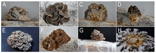
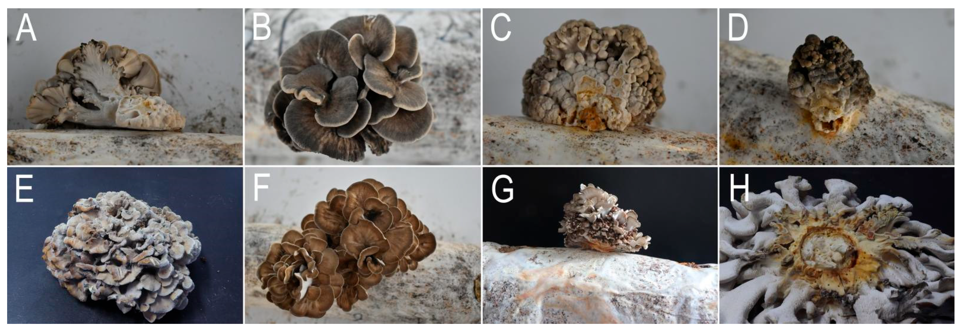
Pathogenicity assays revealed distinct symptom progression based on the timing of infection: infection during primordia formation resulted in complete growth arrest, post-differentiation infections produced basal stipe lesions without involvement of the cap, exhibiting symptom patterns identical to those of field-collected diseased specimens. Control specimens treated with sterile water remained asymptomatic throughout the observation period (Figure 4). The reisolation of the pathogen yielded the same result, thus fulfilling Koch’s postulates.
Figure 4.
Results of Koch’s postulates verification. (A,B) Control group; (C,D) G. frondosa infected at primordial stage; (E,F) stipe of G. frondosa infected post-differentiation; (G,H) pileus of G. frondosa infected post-differentiation.
3.3. Host Range Test of the Pathogen
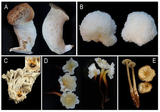
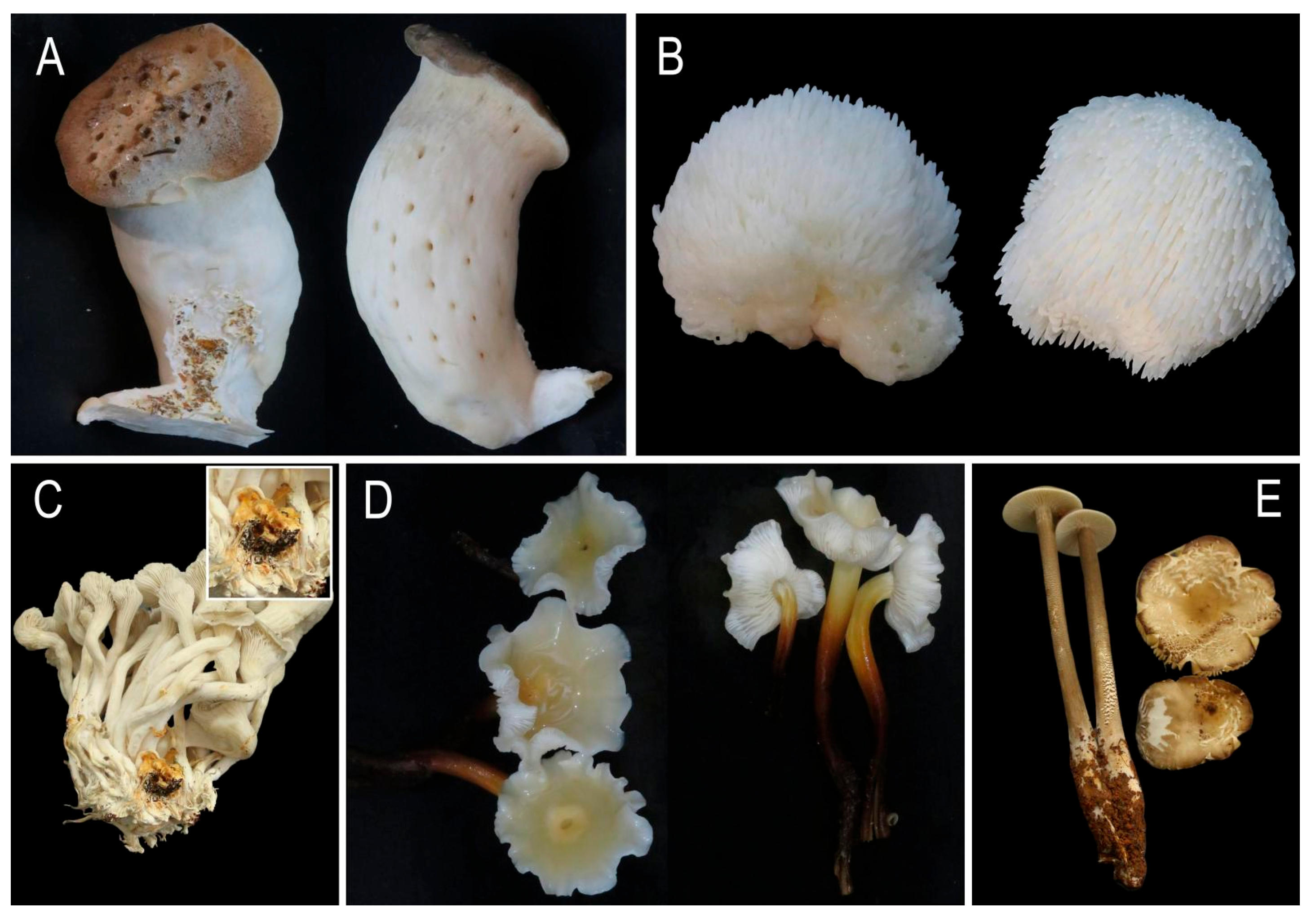
Host range assays revealed differential pathogenicity among the tested mushrooms. Only P. ostreatus caused complete growth arrest during primordia formation and exhibited spots on the stipe of mature fruiting bodies. No significant symptoms were observed in P. eryngii, H. erinaceus, F. filiformis, and H. raphanipes (Figure 5).
Figure 5.
Host range of pathogen in the tested mushrooms. (A) P. eryngii; (B) H. erinaceus; (C) P. ostreatus; (D) F. filiformis; (E) H. raphanipes.
3.4. Bacteriostatic Effect of Different Chemical and Biological Agents
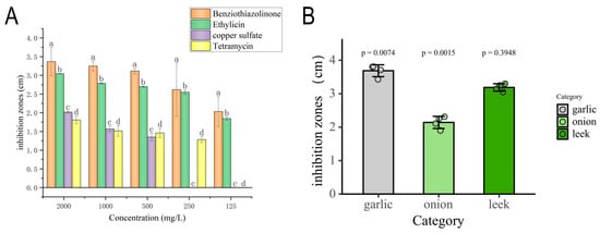
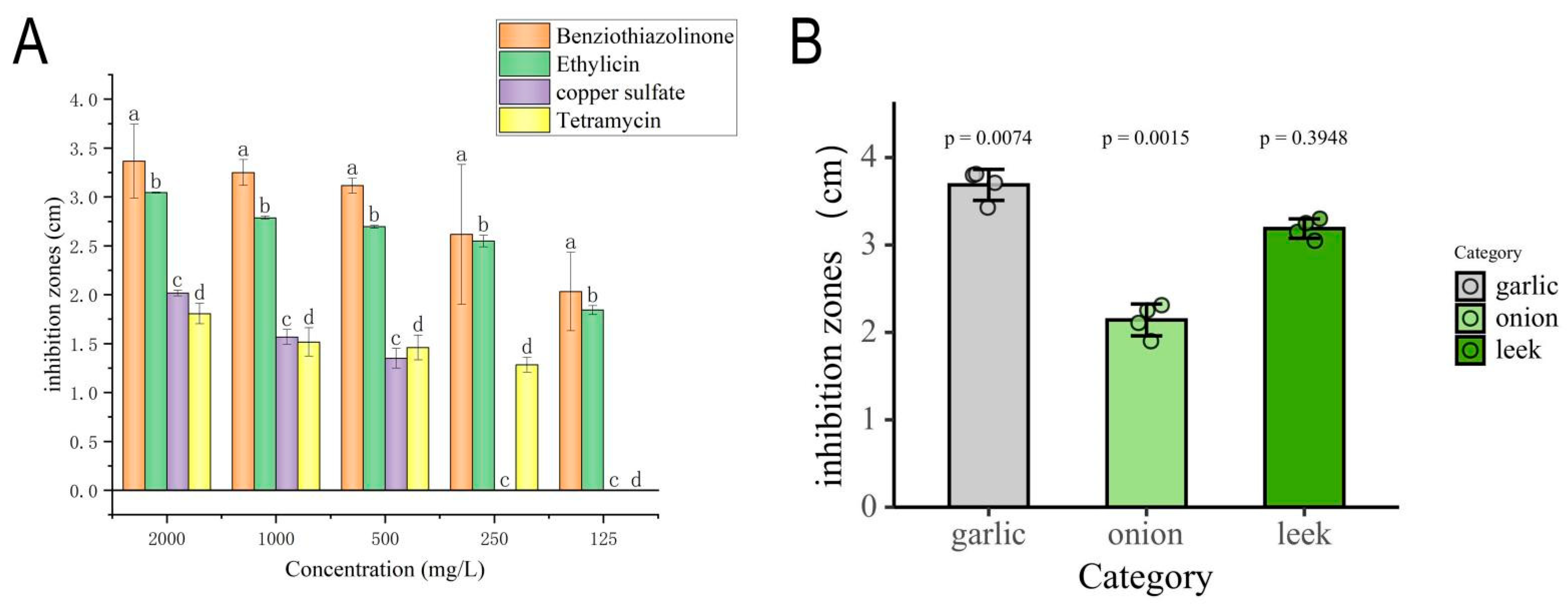
The results showed that, among the six tested chemical agents, two agents, benziothiazolinone and copper sulfate, showed significant effects (Figure 6B,C and Figure 7A). Benzisothiazolinone demonstrated the most potent antibacterial activity, producing an inhibition zone with a diameter of 25.7 ± 1.7 mm. Copper sulfate was the second most effective agent, forming an inhibition zone measuring 12.3 ± 0.3 mm. In contrast, the remaining chemical agents exhibited no detectable antibacterial effects.
Figure 6.
Oxford cup assay screening of antimicrobial agents against target pathogens. (A) CK. (B–E) Chemical agents and biological pesticides: (B) copper sulfate, (C) benziothiazolinone, (D) tetramycin, (E) ethylicin. (F–H) Plant extracts: (F) garlic, (G) leek, (H) onion. (I–K) Microbial strains: (I) H6, (J) H9, (K) T2. (L–N) Macrofungal strains: (L) CK, (M) A3, (N) A4.
Figure 7.
Dose–response profiling of antimicrobial candidates against P. aryabhattai. (A) Chemical agents and biological pesticides: Inhibition zones of benziothiazolinone (a), ethylicin (b), copper sulfate (c), and tetramycin (d) against target pathogens at concentrations (125–2000 mg/L). Letters indicate significant differences (p < 0.05, ANOVA). (B) Plant extracts: Comparative efficacy of garlic, onion, and leek extracts with p-values from t-tests. Individual circles show data points from biological replicates (n = 3); error bars represent standard deviation.
Among the six biological agents tested, only ethylicin and tetramycin inhibited pathogen growth (Figure 6C,D). Ethylicin was the most effective, producing an inhibition zone of 22.5 ± 0.4 mm. Tetramycin demonstrated the second strongest efficacy, with a zone diameter of 9.6 ± 1.5 mm. In contrast, the remaining agents exhibited no significant antibacterial activity.
Toxicity regression models (Table 6) revealed copper sulfate exhibited the narrowest acute toxicity range, while Ethylicin showed the broadest. Pathogen sensitivity was confirmed by EC50 values <5 mg/L for all effective agents, indicating strong antimicrobial activity.
Table 6.
Virulence regression curves and EC50 values of four effective reagents.
3.5. The Effectiveness of Plant Extracts in Controlling Pathogens
Antimicrobial assays demonstrated significant growth inhibition of the pathogen by the following tested extracts (Figure 6F–H and Figure 7B). Garlic extract exhibited the strongest antibacterial activity, producing a mean inhibition zone of 34.3 ± 2.1 mm, followed by leek (32.5 ± 2.5 mm) and onion (21.2 ± 1.8 mm). In contrast, the remaining plant extracts exhibited no significant antibacterial activity.
3.6. Biocontrol Microorganism Screening
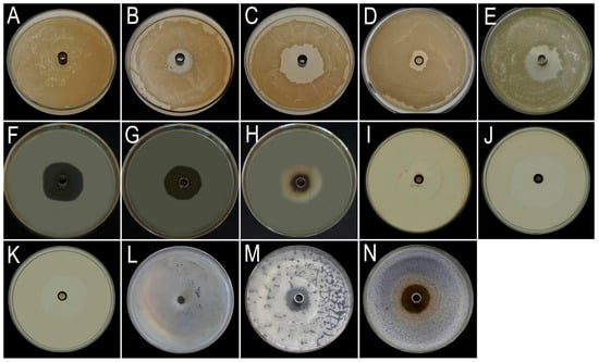
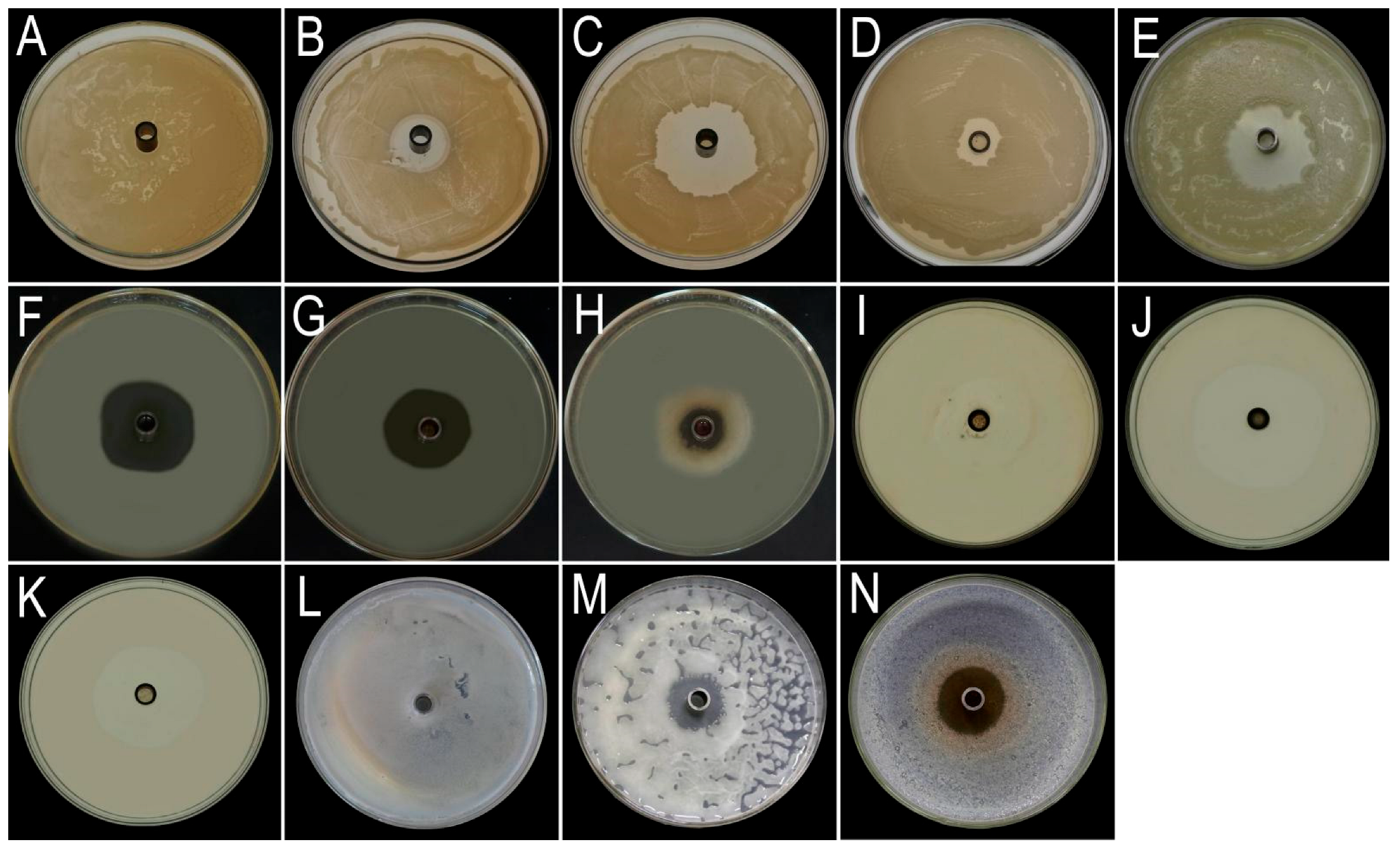
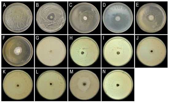
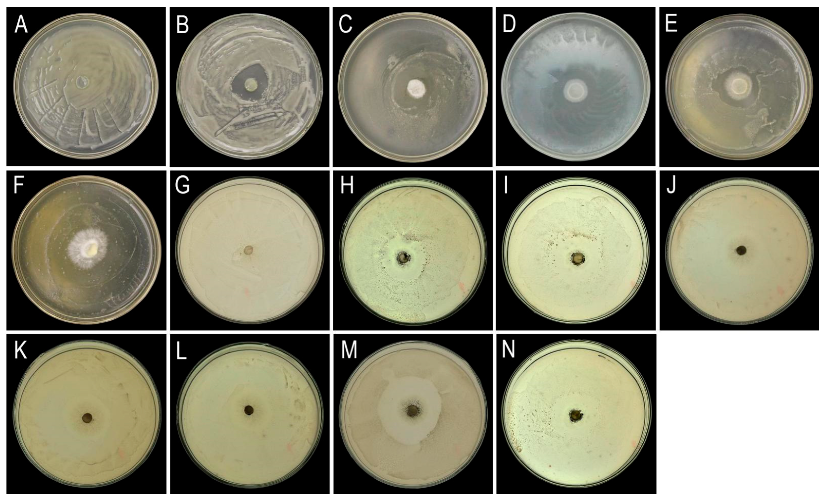
Primary screening using co-culture methods identified five macrofungal strains (A1–A5) and seven microbial strains (B6, B12, H6, H7, H9, T2, T5) (Figure 8) with inhibitory effects on the pathogen growth. Then, secondary screening via Oxford cup assays revealed two macrofungal strains and three microbial strains exhibiting strong inhibitory effects. Among these, macrofungal strain A4 and microbial strain H9 demonstrated the strongest inhibitory activity, with inhibition zones measuring 33.6 ± 0.5 mm and 45.2 ± 0.9 mm, respectively.
Figure 8.
Results of the co-culture screening assay. Macrofungal strains (A–F): (A) CK; (B) A1; (C) A2; (D) A3; (E) A4; (F) A5. Microorganism strains: (G–N): (G) CK; (H) B6; (I) B12; (J) H6; (K) H7; (L) H9; (M) T2; (N) T5.
3.7. Identification of Biocontrol Microorganisms
3.7.1. Identification of Biocontrol Macrofungi A4
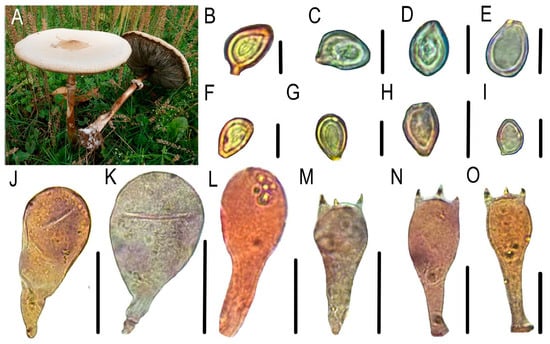
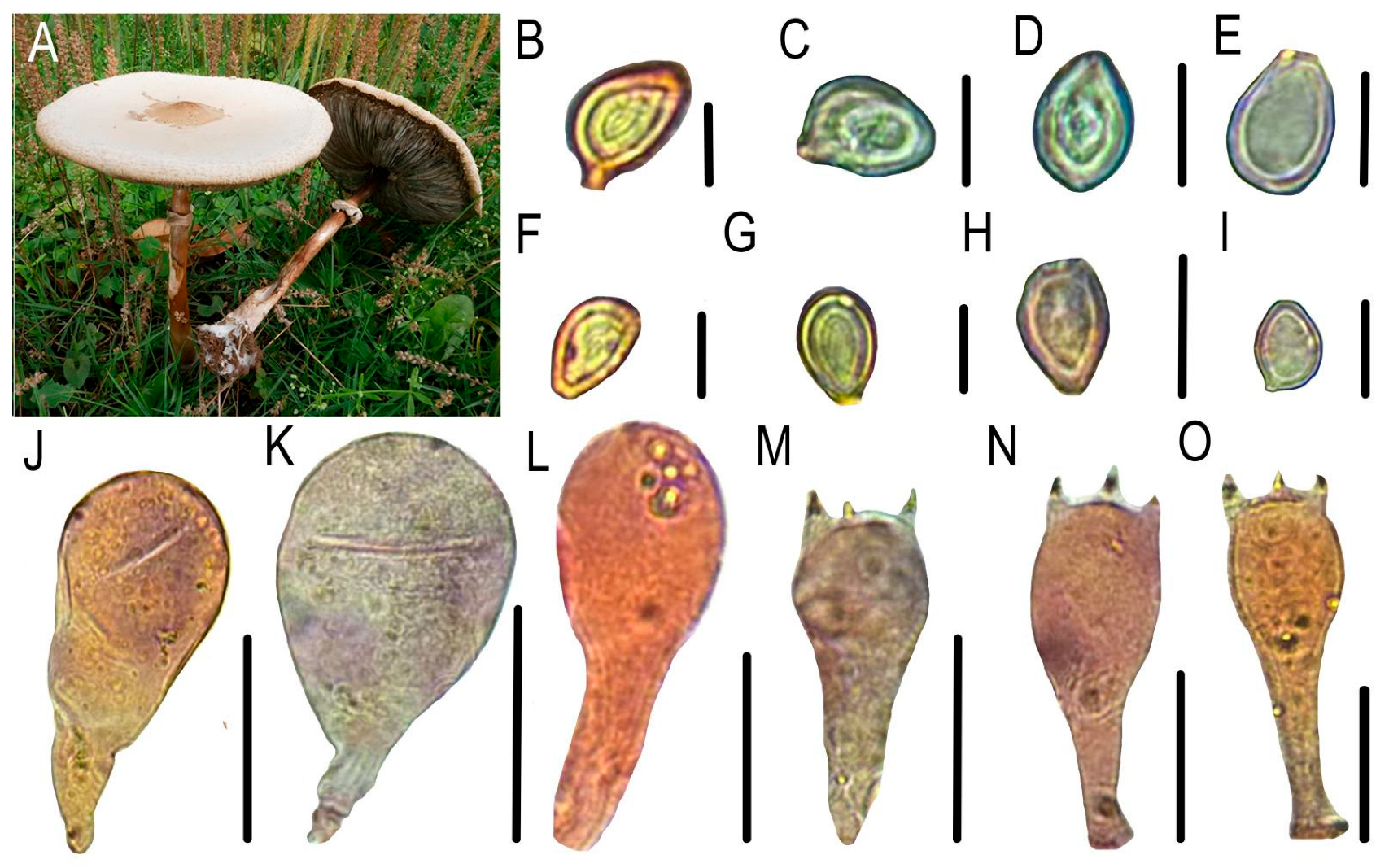
Basidiomata medium to large, pileus convex to planate, adorned with brown scales, lamellae free, closed, greenish, stipe smooth, hollow, cylindrical, enlarged at base, annulus persistent, single-layered, up-part, context white, fleshy, changes to red when injured. Basidiospores ellipsoid, light green, thick-walled, (8.5)9–10(10.5) × 6.5–8 μm. Basidia clavate, 27–36 × (9.5)11–14(16) μm, hyaline, thin-walled, two or four sterigmata (Figure 9). These characteristics match the description of Chlorophyllum molybdites.
Figure 9.
Morphological characteristics of C. molybdites. (A) Habitat, (B–I) basidiospores, (J–L) cheilocystidia, (M–O) basidia. Bars: 10 μm.
BLAST analysis revealed that the sequences of A4 exhibited 100% similarity to C. molybdites. Furthermore, phylogenetic analysis based on the combined ITS, tef1-ɑ, and nLSU fragments clustered A4 within the C. molybdites clade, with strong support (Figure 3B). Combined with the results of morphological studies and phylogenetic analysis, it was identified as C. molybdites.
3.7.2. Identification of Antagonistic Microbial Strain H9
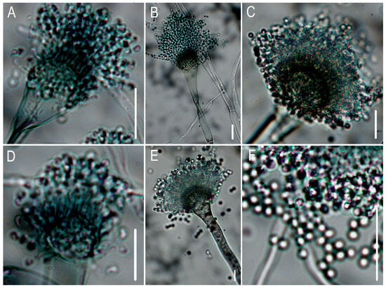
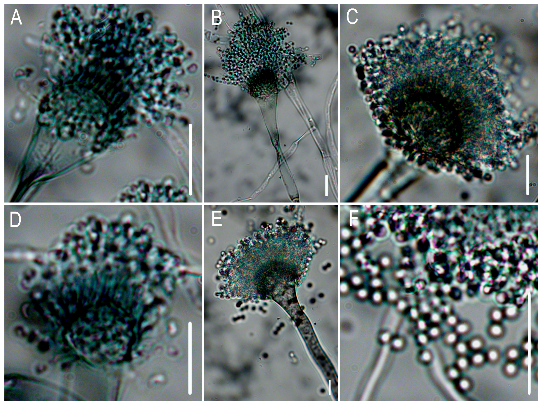
Following 7 d of incubation on Czapek–Dox medium, microscopic examination identified foot cells. Conidial heads were predominantly columnar (44.30–72.02 μm × 36.92–42.06 μm) with characteristic grayish-green pigmentation. Conidiophores appeared smooth-walled, hyaline to pale brown, and cylindrical (4.58–10.05 μm; 150–250 μm). Vesicles were consistently flask-shaped (10.87–32 μm diameter). Conidia arising from phialides on the vesicles were subhyaline, subglobose to broadly ellipsoidal (1.84–2.05 μm diameter), with finely echinulate surfaces (Figure 10). It matches the descriptions of Aspergillus fumigatus.
Figure 10.
Morphological characterization of Aspergillus fumigatus. (A–E) Conidiophores (F) conidia. Bars: 10 μm.
The BLAST results revealed a high similarity with A. fumigatus. Phylogenetic analysis, incorporating ITS, benA and CaM loci, revealed that our sequences clustered within the A. fumigatus clade (Figure 3C) with strong support. Combined with the results of morphological identification and phylogenetic analysis, it was identified as A. fumgatus.
3.8. The Influence of Different Agents on Pathogens
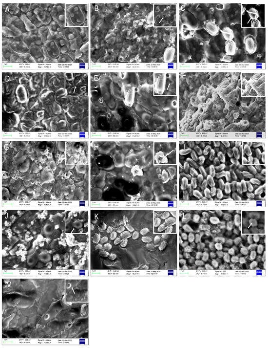
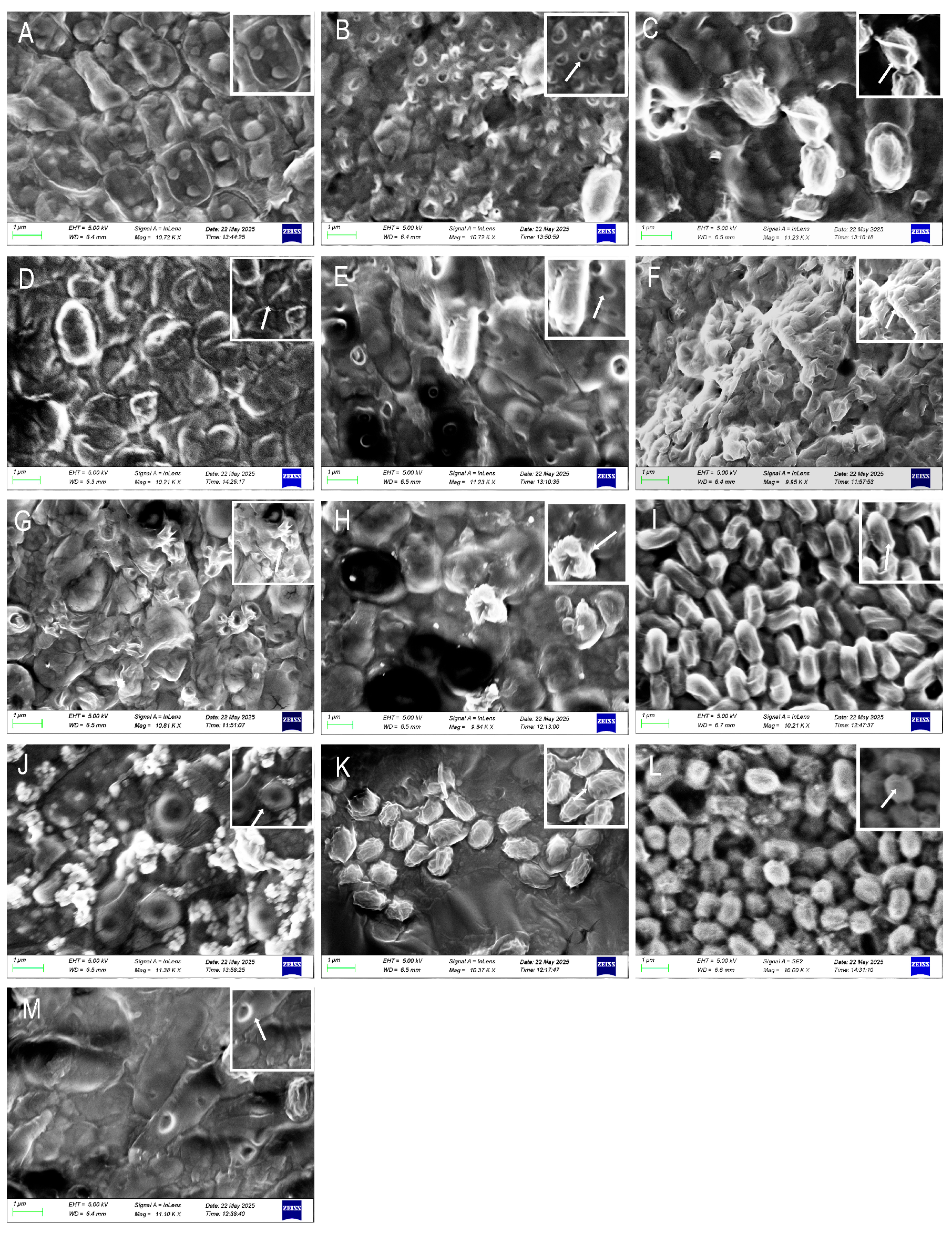
SEM analysis revealed distinct, agent-specific morphological alterations in pathogen cells following exposure to antimicrobial treatments. Cells treated with benziothiazolinone or tetramycin exhibited pronounced surface wrinkling and perforations, a phenotype that was also produced by metabolites from strains H9 and H6 (Figure 11C,D,I–M). Conversely, copper sulfate and ethylicin induced significant membrane depression and extensive perforation, a damage pattern similarly elicited by metabolites from strains A4 and A3 (Figure 11B,E,F–H). These ultrastructural changes, including membrane indentation and pore formation, were dose-dependent. In contrast, untreated control cells maintained a typical, intact cellular architecture with no signs of disruption (Figure 11A).
Figure 11.
Ultrastructural alterations in microbial cells induced by diverse antimicrobial treatments (A) CK; (B) copper sulfate; (C) benziothiazolinone; (D) tetramycin; (E) ethylicin; (F) garlic; (G) leek; (H) onion; (I) A3; (J) A4; (K) H6; (L) H9; (M) T2.
4. Discussion
Bacterial spot disease poses a significant challenge in the cultivation of G. frondosa, severely compromising its yield and quality. This study investigated bacterial spot disease of G. frondosa from Qingyuan County, Zhejiang Province, China, isolating and identifying the pathogen, determining its host range, and evaluating chemical and biological control measures. Through Koch’s postulates, this study identified P. aryabhattai as the causative pathogen of G. frondosa bacterial spot disease. The pathogen displays a narrow host range, primarily infecting G. frondosa and P. ostreatus under our experimental conditions, where it induces growth arrest at the primordia stage and exhibits spots on the stipe of mature fruiting bodies. Notably, it exhibited no detectable pathogenicity toward other tested edible fungi, including H. erinaceus, F. filiformis and H. raphanipes. This study further explored chemical and biological control strategies, identifying one microbial strain and one macrofungal species with significant antagonistic activity against P. aryabhattai. Notably, A. fumigatus (H9) and C. molybdites (A4) demonstrated the most potent inhibition. Among the three tested plant extracts, garlic extract showed the strongest antimicrobial activity. Additionally, benziothiazolinone and ethylicin were identified as effective chemical control agents for disease management. SEM revealed that the aforementioned treatments resulted in significant structural damage to the cell surface of P. aryabhattai, including shriveling, depressions, and perforations.
4.1. The Influence of Cell Wall on Pathogen Host Specificity
Host range determination revealed that P. aryabhattai is highly pathogenic to P. ostreatus (Figure 5C) and G. frondosa (Figure 4), while it remains non-pathogenic or shows negligible symptoms toward other tested edible mushroom species (Figure 5). This host specificity may stem from distinct pathogen-host cell wall interactions. Grifola frondosa, P. ostreatus, P. eryngii, H. erinaceus, F. filiformis and H. raphanipes exhibit substantial variations in their cell wall polysaccharide composition and structure, which likely determine differential susceptibility to P. aryabhattai infection.
Both G. frondosa and P. ostreatus possess similar β-(1,3)-glucan main chains with β-(1,6)-branching structures [4]. These soluble polysaccharides may serve as specific recognition sites or enzymatic targets for pathogenic bacteria, potentially explaining their shared susceptibility to P. aryabhattai infection. The mycelium of G. frondosa contains abundant low-molecular-weight (13–140 kDa) β-(1,3)-glucans with high-density β-(1,6)-branches, which significantly enhance pathogenic adhesion and enzymatic degradation efficiency [38]. P. ostreatus shares the same glucan backbone structure; its medium molecular weight polysaccharides and protective hydrophobic protein layers provide partial physical resistance [39], resulting in comparatively milder infection symptoms.
Notably, although P. ostreatus and P. eryngii are taxonomically related, their cell wall polysaccharides demonstrate fundamental structural differences. While P. ostreatus retains a characteristic β-(1,3)-glucan core structure, P. eryngii uniquely substitutes this with an α-(1,3)-galactan backbone featuring 3-O-methylation modifications. This structural reorganization confers enhanced spatial stability through steric hindrance effects, providing a mechanistic basis for the observed resistance to P. aryabhattai colonization [40,41].
Furthermore, structural analyses reveal distinct cell wall compositions among resistant species: H. erinaceus synthesizes β-glucans with a β-(1→6)-linked backbone (85.7% abundance), incorporating β-(1→3)-linked branch points (14.3%). These β-(1→3) linkages interconnect multiple β-(1→6) chains, forming a branch-on-branch topological network. Enzymatic digestion confirmed that oligosaccharides containing β-(1→3) bonds (e.g., dp5f: G1→3G1→6G1→3G1→6G) resist degradation due to steric hindrance, conferring structural rigidity [42], while F. filiformis synthesizes α-configured glucans featuring a predominantly α-(1→4)-glycosidic backbone, interspersed with α-(1→6)-linked branching points (average: one branch per 12 glycosyl units). These α-(1→6) bonds serve as topological nodes for interchain cross-linking, organizing multiple α-(1→4) chains into a sparsely branched network. Enzymatic digestion assays confirmed that oligosaccharide domains containing α-(1→6) branches—particularly those terminating in galactose or fucose residues—resist degradation via steric hindrance, thereby imparting conformational stability to the macromolecular structure [43]. Meanwhile, the cell wall of H. raphanipes consists primarily of a β-glucan backbone (72.69%), modified with arabinose (1.09%) and other functional groups. This structure forms a physical barrier via β-glycosidic linkages, where arabinose residues may facilitate cross-linking stabilization [44]. Experimental evidence validated that the polysaccharide inhibits specific binding interactions with signalling pathway proteins [45]—such as suppression of β-catenin nuclear translocation—suggesting a resistance mechanism against enzymatic degradation by pathogens.
Therefore, we speculate that P. aryabhattai may achieve infection by selectively degrading the cell wall polysaccharides of G. frondosa and P. ostreatus through glucanase targeting β-1,3 bonds, while the different cell walls of P. eryngii, H. erinaceus, and F. filiformis resist pathogen invasion through physical barriers and biochemical resistance.
4.2. Structural Disruption of the Cell Wall: A Unifying Antibacterial Mechanism for Diverse Control Agents
This study systematically evaluated various disease control strategies, including chemical bactericides, biological control agents, plant-derived antimicrobials, and antagonistic microorganisms. Screening assays identified benzothiazolinone and ethylicin as highly effective compounds, demonstrating potent antibacterial activity against P. aryabhattai. Additionally, of SEM results revealed significant structural damage to the surface of the treated pathogen cells, including shrivelling, depressions, and perforations (Figure 11). The cell wall, which acts as a key barrier in maintaining cell shape and resisting osmotic pressure, is essential for cell integrity. Any loss of this integrity leads to cell rupture, leakage of cellular contents, and ultimately, the inhibition or death of pathogens.
Mechanistic investigations revealed that both agents exert their antimicrobial effects primarily by compromising the bacterial cell wall: benziothiazolinone inhibits peptidoglycan synthesis, while ethylicin disrupts membrane-associated lipoproteins. Among these compounds, ethylicin exerts its potent oxidative effects primarily through active sulfur derivatives produced during decomposition (e.g., ethyl thiosulfinate). These derivatives target sulfur-containing functional groups in microbial cell walls and membranes, disrupting the synthesis and cross-linking of cell wall components or directly degrading their integrity, leading to structural deformation and eventual collapse. Furthermore, upon penetrating the cell membrane, ethylicin disrupts the functionality of membrane-associated lipids and proteins, increasing permeability and inducing protoplasmic leakage. Collectively, these mechanisms inhibit pathogenic growth [46].
The core antibacterial mechanism of benziothiazolinone (BIT) lies in its role as a multi-target metabolic inhibitor, as evidenced by its diverse effects on cellular structures and functions. BIT rapidly penetrates the bacterial cell membrane and enters the cytoplasm, where it irreversibly binds to the active sites of various key enzymes—particularly thiol-containing dehydrogenases. This is corroborated by recent findings in fungi, where BIT significantly inhibits NADH production and disrupts mitochondrial integrity, leading to fragmentation [47]. This binding leads to the fatal disruption of cellular energy metabolism and respiratory processes, as directly evidenced by a sharp decrease in intracellular ATP levels [47], ultimately causing physiological dysfunction due to energy deprivation. Such internal biochemical disturbances trigger secondary effects, including the failure of systems responsible for maintaining membrane integrity. Direct evidence from studies on the bacterium Xanthomonas oryzae pv. oryzae confirms that BIT compromises the structural organization and barrier function of the cell membrane, resulting in cell wrinkling, distortion, and the leakage of intracellular contents, as clearly observed via scanning and transmission electron microscopy [48]. Furthermore, these primary effects cascade into the inhibition of vital processes such as DNA replication, biofilm formation, and motility [48], underscoring its multi-target nature.
Notably, conventional copper-based compounds exhibited limited efficacy in this screening assay, suggesting the presence of copper ion resistance in the pathogen. This finding highlights the importance of novel mechanisms of action demonstrated by thiabendazole and allicin, which specifically target cell wall degradation. As a result, these compounds offer a promising strategy to overcome resistance to conventional chemical treatments [12].
Plant-derived extracts from garlic, leek, and onion exhibit significant antibacterial activity. SEM observations revealed distinct morphological changes in pathogenic bacteria, including surface indentations and perforation-like structures (Figure 11F–H). These antimicrobial effects are likely mediated by the lipophilic and chemically reactive organosulfur compounds present in these extracts, particularly allicin [49] and its structural analogs (e.g., methyl allyl trisulfide and propyl sulfide), which display potent bioactive properties.
These compounds integrate into the phospholipid bilayer of the cell membrane through lipophilic groups, reducing membrane fluidity and causing localized collapse. As the concentration increases, they further disrupt the spatial conformation of membrane protein complexes (such as pore proteins and transport proteins), forming irreversible pores, leading to leakage of intracellular ions, such as K+ and Ca2+, and the outflow of macromolecular substances [50]. This results in osmotic imbalance and membrane potential depolarization. After penetrating the cell membrane, active sulfur groups (such as thiosulfate esters) covalently bind to essential enzymes containing sulfhydryl groups (-SH), forming disulfide bonds [49]. This irreversible protein denaturation inhibits DNA replication, repair, and antioxidant functions, ultimately leading to the disruption of cellular structure.
During the screening of biocontrol microorganisms, highly antagonistic strains of C. molybdites and A. fumigatus were isolated. SEM analysis demonstrated that treatment with these microbial antagonists caused significant morphological changes in P. aryabhattai, including localized invaginations and irregular surface corrugations of the cellular envelope (Figure 11J,L). Among these, Aspergillus fumigatus mediates antibacterial effects via secretion of the anionic defensin AfusinC. This molecule specifically binds lipid II, a key peptidoglycan precursor, inhibiting cell wall biosynthesis and inducing osmotic lysis through loss of structural integrity. Concentration-dependent bactericidal activity against Gram-positive bacteria (e.g., Micrococcus luteus and Staphylococcus aureus) was observed. At 4 × MIC (16 μg/mL), 48 h exposure reduced bacterial loads by ≥3 log10 CFU/mL, with MBC values 1−4 × MIC, indicating direct lytic action. AfusinC’s function requires its conserved CSαβ motif (disulfide-stabilized α-helices/β-sheets), where cationic residues (e.g., Lys-32) electrostatically target membrane components. The defensin’s anionic character (net charge: −1.5) enables Gram-positive selectivity, while Gram-negative bacteria remain resistant due to outer membrane barriers. This cell wall synthesis-targeting mechanism contrasts with nonspecific membrane disruption by cationic antimicrobial peptides [51]. In contrast, C. molybdites exerts its antibacterial effect through the secretion of toxins. Saponin toxins interact with phospholipids through hydrophobic interactions, leading to the localized collapse of the cell wall’s network structure. Additionally, sesquiterpene toxins disrupt the lipid bilayer, forming transmembrane pores that result in membrane potential collapse and osmotic imbalance [16,52].
Grifola frondosa, a valuable edible and medicinal macrofungus, holds significant economic potential owing to its distinctive nutritional composition and pharmacological properties. This study systematically identifies the bacterial pathogen responsible for spot disease in G. frondosa cultivation. Additionally, we have screened multiple highly effective biocontrol agents, laying the groundwork for sustainable disease management in G. frondosa cultivation. Moving forward, it is essential to conduct field trials to evaluate whether these biocontrol agents remain effective in controlling G. frondosa under varying environmental conditions. Furthermore, an in-depth investigation of the antibacterial mechanisms in different control materials is required, alongside the use of response surface methodology to optimize the fermentation of selected antagonistic bacteria. The development of microbial pesticides, the exploration of synergistic chemical and biological control strategies, and the establishment of an integrated pest management system that is both environmentally friendly and economically viable will promote the sustainable growth of G. frondosa industry.
Author Contributions
J.-T.G.: Conceptualization, Data curation, Visualization, Writing—original draft, and Writing—review and editing. N.R.: Data curation and Software. J.-Z.L.: Investigation and Software. Y.-Y.L.: Data curation. S.-Y.T.: Validation and Investigation. X.-R.Y.: Conceptualization and Methodology. J.-X.C.: Conceptualization and Software. J.-Q.W.: Funding acquisition, review and editing. B.Z.: Conceptualization, Project administration, Resources, and Writing—review and editing; Y.L.: Conceptualization, Project administration, Resources, and Writing—review and editing; J.-J.H.: Conceptualization, Project administration, Resources, and Writing—review and editing. All authors have read and agreed to the published version of the manuscript.
Funding
This work was supported by Zhejiang Provincial Postdoctoral Research Project Selection Fund (ZJ2025047), National Natural Science Foundation of China (32400011), National Undergraduate Training Program on Innovation and Entrepreneurship (202510345022), Zhejiang Normal University Doctoral Initiation Fund (31970020), Creation of Novel Germplasm Resources for Characteristic Edible Fungi in Central Zhejiang (Y202456465).
Institutional Review Board Statement
Not applicable.
Informed Consent Statement
Not applicable.
Data Availability Statement
The gene accession numbers referenced in this study are publicly available in the online databases: NCBI GenBank https://www.ncbi.nlm.nih.gov/genbank/. (accessed on 2 September 2025). The reference numbers of the sequences produced in this study are PX225872, PX226006, PX242658, PX225874, PX242659, PX242660, PX225877, PX242661, PX247483 and PX264259.
Acknowledgments
The authors would express their thanks to Xue-Fei Li (Jilin Agricultural University) and Lei Yue (Zhejiang Normal University) for their kind help during our experiment.
Conflicts of Interest
The authors declare no conflict of interest.
References
- Li, C.T.; Tan, Q.; Bian, Y.B.; Xie, B.G.; Liu, Z.Q.; Li, Y. The status and prospection of edible mushroom industry in China. J. Fungal Res. 2019, 17, 1–10+12. [Google Scholar] [CrossRef]
- Zhang, J.X.; Chen, Q.; Huang, C.Y.; Gao, W.; Qu, J.B. History, current situation and trend of edible mushroom industry development. Mycosystema 2015, 34, 524–540. [Google Scholar] [CrossRef]
- Li, Y. Development Trends of the Edible Fungus Industry in China. Edible Med. Mushrooms 2011, 19, 1–5. [Google Scholar]
- Wu, J.Y.; Siu, K.C.; Geng, P. Bioactive ingredients and medicinal values of Grifola frondosa (Maitake). Foods 2021, 10, 95. [Google Scholar] [CrossRef] [Scilit]
- Dong, H.R.; Yu, H.L.; Jiang, N.; Li, Y.; Zhou, F.; Shang, X.D.; Tan, Q. The current status and trends of the development of industrialized production of edible mushrooms in China. Edible Med. Mushrooms 2024, 32, 1–9. [Google Scholar]
- Yu, Q.L. Biological Control of Pathogen on Grifola frondosa. Master’s Thesis, Jilin Agricultural University, Changchun, China, 2017. [Google Scholar]
- Fang, B.Y.; Ke, Y.; Chen, K.Z. Isolation and Identification of Bacterial Strains Infecting Grifola frondosa Primordia. Edible Fungi China 2015, 37, 56–58. [Google Scholar]
- Lv, F.X. Integrated Management of Diseases and Pests in Grifola frondosa Cultivation. J. Mudanjiang Norm. Univ. (Nat. Sci. Ed.) 2010, 03, 22–23. [Google Scholar] [CrossRef]
- Qin, W.T.; Wang, S.X.; Rong, C.B.; Song, Z.J.; Liu, Y. Occurrence and Management of Edible Fungus Diseases in China. Edible Fungi China 2020, 39, 1–7. [Google Scholar] [CrossRef]
- Shen, Z.; Rong, N.; Wang, J.; Jiang, D.; Zhang, Z. First report of bacterial blotch disease caused by Burkholderia gladioli in Grifola frondosa from China. Physiol. Mol. Plant Pathol. 2024, 134, 102458. [Google Scholar] [CrossRef] [Scilit]
- Shi, Z.L. Major Types, Epidemiological Patterns, and Integrated Management of Bacterial Diseases in Winter Protected-Crop Vegetables. Agric. Henan 2024, 21, 42–43. [Google Scholar] [CrossRef]
- Gjorgjieva, M.; Tomašič, T.; Kikelj, D.; Mašič, L.P. Benzothiazole-based compounds in antibacterial drug discovery. Curr. Med. Chem. 2018, 25, 5218–5236. [Google Scholar] [CrossRef] [Scilit]
- Li, Q.; Li, W.Y.; Xu, J.Q.; Ji, L. Investigation of pesticide residues in edible fungi in Lishui City. Chin. J. Health Lab. Technol. 2021, 31, 245–248. [Google Scholar]
- Xiao, J.; Bai, W. Bioactive phytochemicals. Crit. Rev. Food Sci. Nutr. 2019, 59, 827–829. [Google Scholar] [CrossRef] [Scilit] [PubMed]
- Heydari, A.; Pessarakli, M. A review on biological control of fungal plant pathogens using microbial antagonists. J. Biol. Sci. 2010, 10, 273–290. [Google Scholar] [CrossRef] [Scilit]
- Keller, N.P. Fungal secondary metabolism: Regulation, function and drug discovery. Nat. Rev. Microbiol. 2019, 17, 167–180. [Google Scholar] [CrossRef] [Scilit] [PubMed]
- Chet, I.; Inbar, J. Biological control of fungal pathogens. Appl. Biochem. Biotechnol. 1994, 48, 37–43. [Google Scholar] [CrossRef] [Scilit]
- Li, X.F.; Sossah, F.L.; Tuo, Y.L.; Hu, J.J.; Wei, Q.; Li, S.; Rong, N.; Wiafe-Kwagyan, M.; Li, C.T.; Zhang, B.; et al. Characterization and fungicide sensitivity of Trichoderma species causing green mold of Ganoderma sichuanense in China. Front. Microbiol. 2023, 14, 1264699. [Google Scholar] [CrossRef] [Scilit]
- Goszczynska, T.; Botha, W.J.; Venter, S.N.; Coutinho, T.A. Isolation and identification of the causal agent of brown stalk rot, a new disease of maize in South Africa. Plant Dis. 2007, 91, 711–718. [Google Scholar] [CrossRef] [Scilit]
- Gerhardt, P.; Murray, R.G.E.; Wood, W.A.; Krieg, N.R. (Eds.) Methods for General and Molecular Bacteriology; American Society for Microbiology: Washington, DC, USA, 1994. [Google Scholar]
- Li, Y.; Li, T.H.; Yang, Z.L.; Bau, T.; Dai, Y.C. Atlas of Chinese Macrofungal Resources; Central China Farmer’s Publishing House: Zhengzhou, China, 2015. [Google Scholar]
- Vos, P.; Garrity, G.M.; Jones, D.; Krieg, N.R.; Ludwig, W.; Rainey, F.A.; Schleifer, K.-H.; Whitman, W.B. Bergey’s Manual of Systematic Bacteriology. Volume 3: The Firmicutes; Springer Science & Business Media: New York, NY, USA, 2011. [Google Scholar]
- Visagie, C.M.; Houbraken, J.; Frisvad, J.C.; Hong, S.B.; Klaassen, C.H.W.; Perrone, G.; Seifert, K.A.; Varga, J.; Yaguchi, T.; Samson, R.A. Identification and nomenclature of the genus Penicillium. Stud. Mycol. 2014, 78, 343–371. [Google Scholar] [CrossRef] [Scilit] [PubMed]
- Rehner, S.A.; Buckley, E. A Beauveria phylogeny inferred from nuclear ITS and EF1-α sequences: Evidence for cryptic diversification and links to Cordyceps teleomorphs. Mycologia 2005, 97, 84–98. [Google Scholar] [CrossRef] [Scilit]
- White, T.J.B.T. Analysis of phylogenetic relationships by amplification and direct sequencing of ribosomal RNA genes. PCR Protoc. A Guide Methods Appl. 1990, 38, 315–322. [Google Scholar]
- Hall, T.A. BioEdit: A User-Friendly Biological Sequence Alignment Editor and Analysis Program for Windows 95/98/NT. Nucleic Acids Symp. Ser. 1999, 41, 95–98. [Google Scholar]
- Stamatakis, A. RAxML version 8: A tool for phylogenetic analysis and post-analysis of large phylogenies. Bioinformatics 2014, 30, 1312–1313. [Google Scholar] [CrossRef] [Scilit] [PubMed]
- Ronquist, F.; Teslenko, M.; Van Der Mark, P.; Ayres, D.L.; Darling, A.; Höhna, S.; Larget, B.; Liu, L.; Suchard, M.A.; Huelsenbeck, J.P. MrBayes 3.2: Efficient Bayesian phylogenetic inference and model choice across a large model space. Syst. Biol. 2012, 61, 539–542. [Google Scholar] [CrossRef] [Scilit] [PubMed]
- Bhunjun, C.S.; Phillips, A.J.L.; Jayawardena, R.S.; Promputtha, I.; Hyde, K.D. Importance of molecular data to identify fungal plant pathogens and guidelines for pathogenicity testing based on Koch’s postulates. Pathogens 2021, 10, 1096. [Google Scholar] [CrossRef] [Scilit]
- Zhang, J.; Meng, W.S.; Sun, Y.J.; Xu, W.; Zhang, J.H. Screening of Fungicides for the Control of Bacterial Blight in Soybean. Agrochemicals 2021, 60, 687–690. [Google Scholar] [CrossRef]
- Vincent, J.G.; Vincent, H.W.; Morton, J. Filter paper disc modification of the oxford cup penicillin determination. Proc. Soc. Exp. Biol. Med. 1944, 55, 162–164. [Google Scholar] [CrossRef] [Scilit]
- Sebaugh, J.L. Guidelines for accurate EC50/IC50 estimation. Pharm. Stat. 2011, 10, 128–134. [Google Scholar] [CrossRef] [Scilit]
- Tan, B.; Huang, K.N.; Yang, J.L.; Shen, Y. Application of garlic and onion extract in controlling common pathogenic bacteria of Lentinus edodes. Guizhou Sci. 2023, 41, 43–48. [Google Scholar]
- Li, L.; Jin, L.M.; Lu, C.Y. Optimization of Allicin Extraction Process from Red Garlic by Using Response Surface Methodology. Mod. Food 2023, 29, 208–212. [Google Scholar] [CrossRef]
- Gao, H.; Wang, S.Q.; Kong, X.Y.; Guo, L.Y. Optimization of Ultrasonic-Assisted Extractionof Total Flavonoids in Chrysanthemum. Liquor.-Mak. Sci. Technol. 2019, 4, 65–68. [Google Scholar] [CrossRef]
- Sun, Q.Y.; Dong, Z.Y.; Li, Y.Z. Study on optimization of extraction processand antioxidant activity of total flavonoids from dandelion. Feed Res. 2024, 47, 91–95. [Google Scholar] [CrossRef]
- Ruan, J.Z. Study on Antibacterial Activity Against Putrefactive Bacteria in Meats and Effective Components of Herbal Medicine in Zhejiang. Master’s Thesis, Hangzhou Normal University, Hangzhou, China, 2015. [Google Scholar]
- Wu, M.J.; Cheng, T.L.; Cheng, S.Y.; Lian, T.W.; Wang, L.; Chiou, S.Y. Immunomodulatory properties of Grifola frondosa in submerged culture. J. Agric. Food Chem. 2006, 54, 2906–2914. [Google Scholar] [CrossRef] [Scilit]
- Pérez-Bassart, Z.; Bäuerl, C.; Fabra, M.J.; Martínez-Abad, A.; Collado, M.C.; López-Rubio, A. Composition, structural properties and immunomodulatory activity of several aqueous Pleurotus β-glucan-rich extracts. Int. J. Biol. Macromol. 2023, 253, 127255. [Google Scholar] [CrossRef] [Scilit]
- Synytsya, A.; Míčková, K.; Synytsya, A.; Jablonský, I.; Spěváček, J.; Erban, V.; Kováříková, E.; Čopíková, J. Glucans from fruit bodies of cultivated mushrooms Pleurotus ostreatus and Pleurotus eryngii: Structure and potential prebiotic activity. Carbohydr. Polym. 2009, 76, 548–556. [Google Scholar] [CrossRef] [Scilit]
- Avni, S.; Ezove, N.; Hanani, H.; Yadid, I.; Karpovsky, M.; Hayby, H.; Gover, O.; Hadar, Y.; Schwartz, B.; Danay, O. Olive mill waste enhances α-glucan content in the edible mushroom Pleurotus eryngii. Int. J. Mol. Sci. 2017, 18, 1564. [Google Scholar] [CrossRef] [Scilit]
- Xie, B.Y.; Yi, L.; Zhu, Y.T.; Chang, X.M.; Hao, J.; Pang, L.; Ouyang, Y.L.; Yuan, S.; Zhang, Z.Q. Structural elucidation of a branch-on-branch β-glucan from Hericium erinaceus with A HPAEC-PAD-MS system. Carbohydr. Polym. 2021, 251, 117080. [Google Scholar] [CrossRef] [Scilit]
- Ye, S.Y.; Gao, Y.; Hu, X.Y.; Cai, J.Y.; Sun, S.W.; Jiang, J.H. Research progress and future development potential of Flammulina velutipes polysaccharides in the preparation process, structure analysis, biology, and pharmacology: A review. Int. J. Biol. Macromol. 2024, 267, 131467. [Google Scholar] [CrossRef] [Scilit] [PubMed]
- Wei, Q.; Zhong, X.R.; Haruna, M.H.; Liu, S.R.; Zhou, F.F.; Chen, M.X. Evaluation of different agricultural wastes for the production of polysaccharides from Oudemansiella raphanipes and its antioxidant properties. Food Sci. Nutr. 2022, 10, 3444–3452. [Google Scholar] [CrossRef] [Scilit] [PubMed]
- Alitongbieke, G.; Zhang, X.R.; Zhu, F.K.; Wu, Q.C.; Lin, Z.C.; Li, X.M.; Xue, Y.; Lai, X.B.; Feng, J.X.; Huang, R.J.; et al. Glucan from Oudemansiella raphanipes suppresses breast cancer proliferation and metastasis by regulating macrophage polarization and the WNT/β-catenin signaling pathway. J. Cancer 2024, 15, 1169. [Google Scholar] [CrossRef] [Scilit]
- Su, X.Z.; Chang, C.L.; Gao, C.Y.; He, Y.T.; Tian, R.Z.; Wang, S.L.; Feng, H.; Huang, L.L. Control efficacy and action mechanism of ethylicin on apple Valsa canker. J. Plant Prot. 2024, 51, 863–874. [Google Scholar] [CrossRef]
- Chen, X.; Liu, C.; Liu, W.; Yang, J.; Gu, C.; Sun, Y.; Cao, H.; Chen, Y. Study on the Mechanism of Benziothiazolinone and Its Synergistic Effect with Tebuconazole in Inhibiting Fusarium asiaticum and DON Production. J. Agric. Food Chem. 2025, 73, 4947–4957. [Google Scholar] [CrossRef] [Scilit] [PubMed]
- Chen, X.; Pang, C.; Liu, X.; Sun, J.; Jin, L.; Sun, Y.; Chen, Y. Investigation of the antibacterial activity of benziothiazolinone against Xanthomonas oryzae pv. oryzae. Pestic. Biochem. Physiol. 2024, 199, 105768. [Google Scholar] [CrossRef] [Scilit] [PubMed]
- Borlinghaus, J.; Albrecht, F.; Gruhlke, M.; Nwachukwu, I.; Slusarenko, A. Allicin: Chemistry and biological properties. Molecules 2014, 19, 12591–12618. [Google Scholar] [CrossRef] [Scilit]
- Wang, R.Z.; Zhang, H.; Chen, X.J.; Liu, S.Y.; Wang, B.; Huang, N.Q.; Xiang, T.T.; Qiu, X.M.; Wang, J.J. Inhibitory Effect of Allicin on Klebsiella pneumoniae and Its Mechanism Analysis. Microbiol. China 2025, 52, 4260–4272. [Google Scholar] [CrossRef]
- Contreras, G.; Braun, M.S.; Schäfer, H.; Wink, M. Recombinant AfusinC, an anionic fungal CSαβ defensin from Aspergillus fumigatus, exhibits antimicrobial activity against gram-positive bacteria. PLoS ONE 2018, 13, e0205509. [Google Scholar] [CrossRef] [Scilit]
- Wu, J.; Ohura, T.; Ogura, R.; Wang, J.; Choi, J.-H.; Kobori, H.; D’Alessandro-Gabazza, C.N.; Toda, M.; Yasuma, T.; Gabazza, E.C. Bioactive compounds from the mushroom-forming fungus Chlorophyllum molybdites. Antibiotics 2023, 12, 596. [Google Scholar] [CrossRef] [Scilit]

Disclaimer/Publisher’s Note: The statements, opinions and data contained in all publications are solely those of the individual author(s) and contributor(s) and not of MDPI and/or the editor(s). MDPI and/or the editor(s) disclaim responsibility for any injury to people or property resulting from any ideas, methods, instructions or products referred to in the content. |
© 2025 by the authors. Licensee MDPI, Basel, Switzerland. This article is an open access article distributed under the terms and conditions of the Creative Commons Attribution (CC BY) license (https://creativecommons.org/licenses/by/4.0/).