


Abstract
(1) Background: Left ventricular assist device (LVAD) implantation is a valuable alternative as a bridge to transplant but also as a destination therapy in ineligible patients. Right ventricular failure (RVF) is a major cause of short- and long-term mortality post-LVAD. We aimed to validate echocardiographic and hemodynamic parameters predictive of RVF and adverse outcomes post-LVAD; (2) Methods: We screened a population of patients with end-stage heart failure selected for LVAD implantation according to SIENA protocol and standard international indications, including right heart catheterization (RHC). Individuals were followed up for 1 year with different time points for the development of RVF (primary endpoint) or mortality and hospitalization (secondary endpoint); (3) Results: The population included 29 patients with a mean age of 63 ± 7 years with a mean ejection fraction of 23 ± 4%, mostly due to ischemic etiology. All the patients had a SIENA protocol score of 0–1 before LVAD, and none met the primary endpoint. Regarding the secondary endpoint, among all the tested clinical, laboratory, echo, and RHC indices, only a central venous pressure/wedge pressure (CVP/PCWP) ratio > 0.63 was significantly associated with adverse outcomes (ß = 2.99, p = 0.026); (4) Conclusions: Excluding a pre-implantation RV dysfunction according to SIENA protocol significantly reduces the risk of post-LVAD RVF. The CVP/PCWP ratio may be an additional prognostic marker for mortality and rehospitalization in LVAD patients.
1. Introduction
Patients with advanced heart failure (AdHF) comprise approximately 11% of the total heart failure population, with some variability across heart failure phenotypes. These patients face a significant mortality rate, ranging from 25% to 75% within the first year [1]. Additionally, their quality of life is severely compromised as they are classified in the highest NYHA functional classes (III or IV) and experience persistent and severe symptoms [2]. Heart transplantation (HTx) is the primary treatment when conventional medical therapy is inadequate and in the absence of contraindications. However, due to limited organ availability, an average of 10–15% of patients on the transplant waiting list die each year in European countries [3].
For patients with isolated left ventricular (LV) failure, long-term mechanical circulatory support (MCS) is often indicated to prolong survival and enhance quality of life. Indications for MCS vary, including bridge to HTx, bridge to candidacy (e.g., cases of pulmonary hypertension or recent malignancy), or destination therapy (DT), especially for those patients with AdHF and advanced age. The MOMENTUM 3 trial showed a five-year survival rate of 58.4% in the United States, with similar rates reported in Europe [4]. Right ventricular failure (RVF) remains a leading cause of mortality over time [5]. It occurs with an estimated prevalence of 8–10% in patients with continuous-flow LVADs [6] and an incidence of approximately 25% [7].
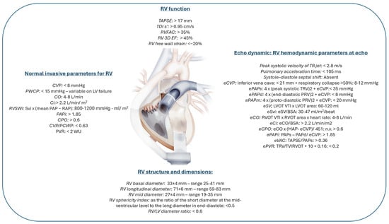
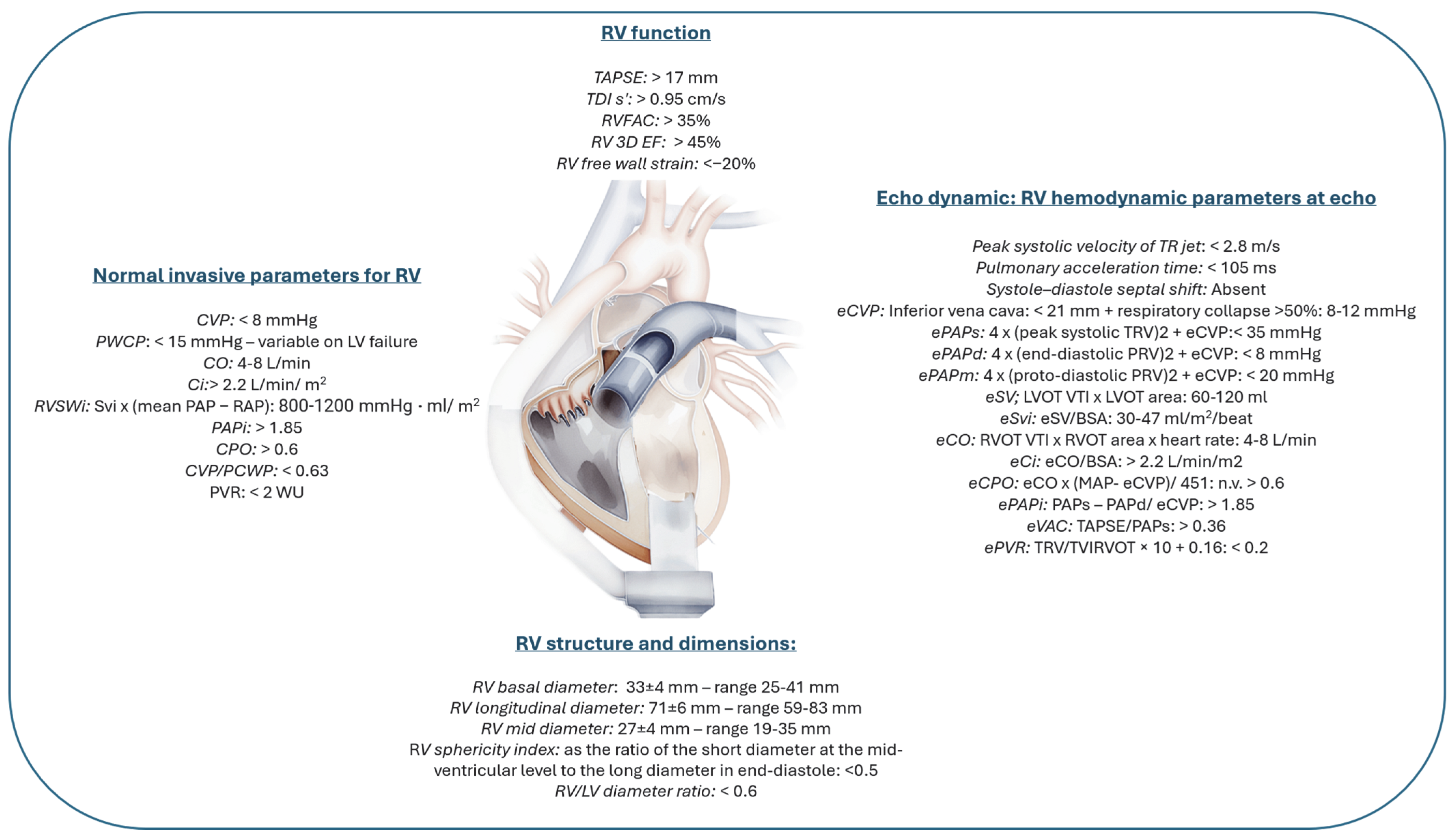
The latest consensus document on echocardiographic assessment of LVAD candidates and recipients highlights critical aspects of RV evaluation through a multiparametric approach [5]. Many AdHF patients present challenges in clinical assessment due to the variability of phenotypes, which are often difficult to categorize. Medical therapy, MCS, mechanical ventilation, arrhythmias, and rapidly changing clinical statuses can substantially affect imaging evaluations, necessitating a multiparametric approach for each case. Transthoracic echocardiography (TTE) plays a pivotal role in pre-implantation selection, offering information on right and left ventricular dimensions and function, valvular anatomy, intracavitary pressures, and hemodynamic parameters, including cardiac output, pulmonary resistance, ventricular interdependence, and ventricular–arterial coupling [8,9]. In RV assessment, it is crucial to evaluate adaptation to elevated afterload due to left-sided heart disease, characterized by initial contractility elevation, compensatory hypertrophy, and eventual dilation [10]. However, this adaptation may be absent in acute cases with abrupt afterload increases and pressure elevation [11]. Key TTE parameters include TAPSE, lateral annulus TDI, fractional area change, sphericity index, RV/LV diameter ratio, PAPs, inferior vena cava diameter with respiratory variation, global longitudinal RV strain, systole-diastole septal shift, TAPSE/PAPs, and peak systolic velocity of TR/VTI RVOT (Figure 1). Due to RV’s complex structure, 3D echocardiography and transesophageal echocardiography (TOE) should be incorporated into pre-implantation evaluations [5,12]. All pre-LVAD patients should undergo hemodynamic assessment through right heart catheterization or continuous Swan–Ganz catheter monitoring. These invasive measures provide a comprehensive RV functional profile, including stroke work, pressure evaluation, RV-pulmonary circulation coupling, pulmonary artery resistance, and right-to-left filling pressure [11,13] (Figure 1).
Figure 1.
Invasive and non-invasive parameters for RV geometry and function are described in the normal range. All the parameters preceded by an “e” are echo derived [5,11,12,14,15,16]. BSA: body surface area; Ci: cardiac index; CO: cardiac output; CPO: cardiac power output; CVP: central venous pressure; LVOT VTI: left ventricular outflow tract velocity-time integral; PAPi: pulmonary artery pulsatility index; PVR: pulmonary vascular resistance; PCWP: pulmonary capillary wedge pressure; RV 3D EF: right ventricular 3D ejection fraction; RVFAC: right ventricular fractional area change; RVSWi: right ventricular stroke work index; SV: stroke volume; TAPSE: tricuspid annular plane systolic excursion; TDI: tissular doppler imaging; eVAC: ventricular–arterial coupling.
The purpose of this study was to identify the invasive and non-invasive predictive parameters of RVF development after LVAD implantation with a follow-up of 12 months. For prognostic and therapeutic evaluation, we used the “SIENA protocol,” a validated tool incorporating four RV function and geometry indices: Tricuspid Annular Plane Systolic Excursion (TAPSE), Right Ventricular Fractional Area Change (RVFAC), Right Ventricular Sphericity Index (RVSI), and longitudinal strain of the right ventricular free wall (Free-Wall RVLS) using speckle tracking [14].
2. Materials and Methods
This single-center study collected data on patients with AdHF, as defined by the Heart Failure Association–European Society of Cardiology (HFA-ESC) criteria, who met eligibility for LVAD implantation and had the parameters available to calculate the SIENA score. Patients lacking informed consent, complete pre-operative echocardiographic examination, or with absent follow-up data were excluded.
The primary endpoint was the identification of RVF following LVAD implantation, defined by persistent signs or symptoms of RV failure, elevated central venous pressure (CVP ≥ 18 mmHg) with reduced cardiac index (<2.3 L/min/m2) in the absence of elevated pulmonary capillary wedge pressure (PCWP < 18 mmHg), or the presence of cardiac tamponade, ventricular arrhythmias, pneumothorax, the need for right ventricular assist device (RVAD) implantation, or prolonged use of inhaled nitric oxide (≥48 h) or inotropic therapy (≥14 days).
Secondary endpoints included individual and composite outcomes encompassing all-cause mortality, cardiovascular mortality (including deaths due to acute heart failure, acute myocardial infarction, ventricular tachycardia or fibrillation, sudden cardiac arrest, and stroke), and HF-related rehospitalization.
2.1. Baseline and Follow-Up Data
Baseline data collection included comprehensive medical history, cardiovascular risk factors, medical treatments, the origin and type of cardiomyopathy (ischemic or non-ischemic), myocardial infarction subtype, prior cardiac surgeries, paroxysmal or chronic atrial fibrillation, and the presence of devices such as pacemakers or implantable cardioverter defibrillators. Functional status and quality of life were assessed using the Minnesota Living with Heart Failure Questionnaire, Interagency Registry for Mechanically Assisted Circulatory Support (INTERMACS) classification, and the six-minute walk test. Vital signs, HF signs and symptoms, medical therapies, and blood analyses were documented. Post-LVAD and length of ICU stay were recorded, and outpatient follow-ups were conducted at 1, 3, 6, 9, and 12 months, with the same data collected as at baseline. All patients signed informed consent.
2.2. Echocardiographic Exam
Echocardiographic assessments (both standard and advanced, including 3D and speckle-tracking echocardiography) were performed on high-quality machines using a 2.5 MHz transducer (GE Healthcare, Milwaukee, WI, USA). Transthoracic echocardiography followed AHA and ESC-EACVI guidelines [15], and speckle-tracking analysis was completed offline using semi-automated 2D software (V:204) (EchoPac, GE, Milwaukee, Madison, WI, USA). All patients received a score according to the SIENA protocol.
2.3. Right Heart Catheterization
Right heart catheterization (RHC) was conducted within four weeks of echocardiography, prior to LVAD implantation, with measurements taken at end-expiration following standardized protocols [16]. Hemodynamic parameters included mean, systolic, and diastolic pulmonary artery pressures (mPAP, sPAP, dPAP), right atrial pressure (RAP), and average PCWP. Calculated parameters included pulmonary vascular resistance, the RAP/PCWP ratio, pulmonary artery pulsatility index (PAPi), and right ventricular stroke work index (RVSWI). Cardiac output and cardiac index were measured via thermodilution and the indirect Fick method.
2.4. Statistical Analysis
Potential outcome predictors were analyzed using univariate and multivariate analyses, focusing on right ventricular failure, cardiovascular and all-cause mortality, rehospitalization, and bleeding events post-LVAD implantation. Data analysis was performed with IBM SPSS 25.0 (1989–2017, LEAD Technologies Inc., Charlotte, NC, USA). The Kolmogorov-Smirnov test assessed the distribution of continuous variables: normally distributed variables are presented as mean ± standard deviation, non-normally distributed variables as median (interquartile range), and categorical variables as frequency (percentage). Cox univariate analysis was also conducted. A p value < 0.05 was considered statistically significant.
3. Results
Twenty-nine patients were selected at our center from May 2013 to February 2022. In this cohort, 93% were male, with a mean age of 63 ± 7 years and an average BMI of 26.38 ± 5.67. Cardiovascular risk factors, including hypertension, diabetes, dyslipidaemia, and smoking, were present in 58%, 47%, 63%, and 65% of patients, respectively. Coronary artery disease (CAD) was reported in 29% of cases. The whole population of patients was classified as NYHA class III-IV, while INTERMACS profiles 3, 4, and 5 included 40%, 46%, and 14% of the population, respectively. Ischemic etiology accounted for 58% of the heart failure cases, and 44% of patients had cardiac resynchronization therapy (CRT) devices.
After selecting the patients according to the SIENA protocol and international guidelines, none of the patients developed RVF.
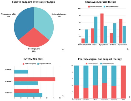
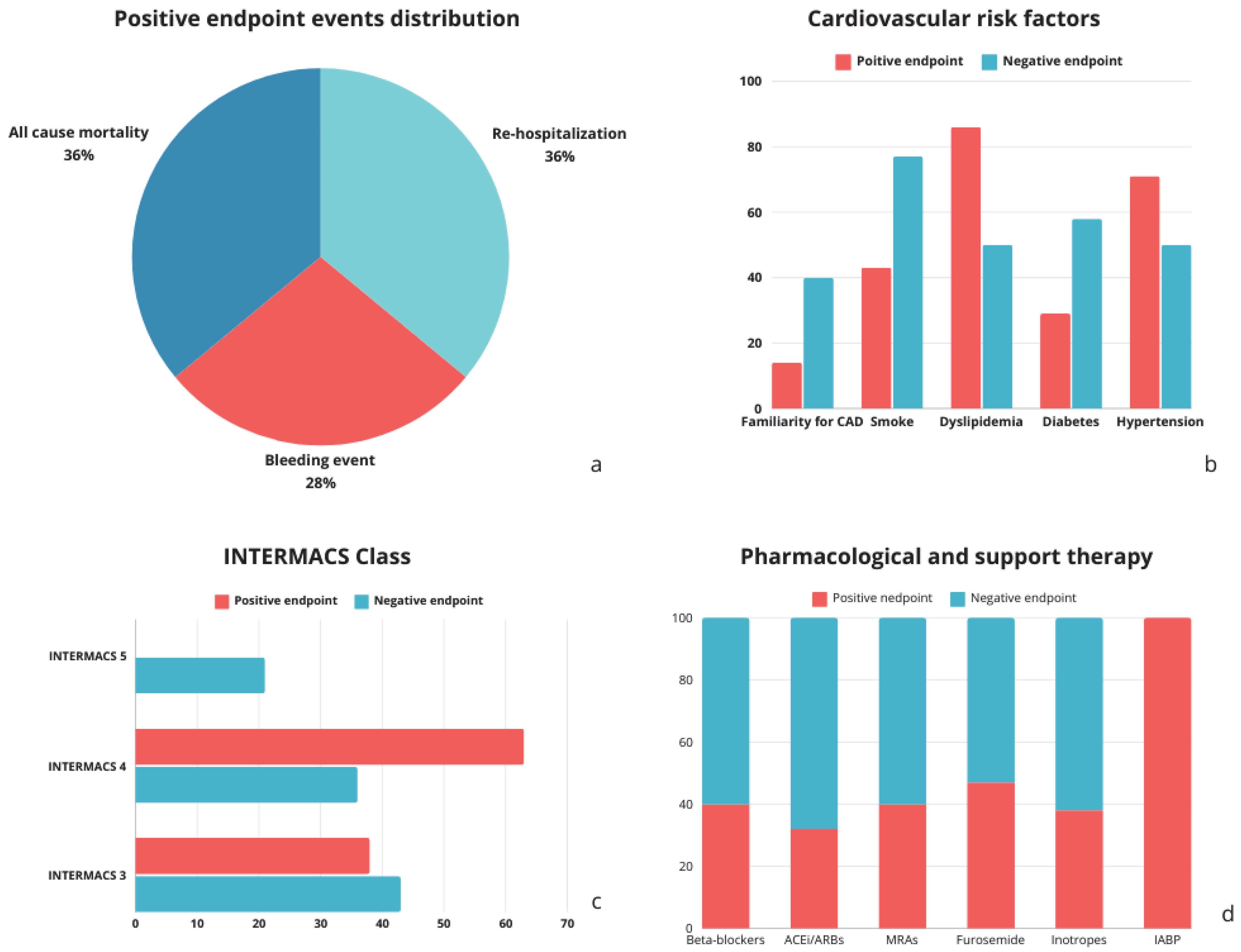
The cohort was then divided into two groups based on the occurrence of the study’s secondary composite endpoint. The “no endpoint” group included 18 patients, while 11 patients experienced adverse cardiovascular outcomes. Of these, 4 required rehospitalization, 3 experienced bleeding events, and 4 died from all causes (Figure 2a). An analysis of risk factors associated with adverse cardiovascular events revealed no significant differences in hypertension, diabetes, dyslipidaemia, smoking, or family history of CAD when comparing patients with and without adverse events, as shown in Figure 2b. Differences between groups by INTERMACS profile are displayed in Figure 2c.
Figure 2.
(a) Adverse effects in the positive endpoint group; (b) Cardiovascular risk factors in the negative and positive endpoint groups; (c) INTERMACS Class distribution in the negative and positive endpoint groups; (d) Medical therapy distribution in the negative and positive endpoint groups. ACEi/ARB, angiotensin converting enzyme inhibitor/angiotensin II receptor blockers; BB, beta blockers; CAD: coronary artery disease; IABP, intra-aortic balloon pump; MRAs, mineralocorticoid receptor antagonists.
Anthropometric and baseline characteristics, stratified by endpoint occurrence, are summarized in Table 1. No statistically significant differences were observed between the groups. Blood chemistry data, evaluated pre- and post-LVAD implantation, also showed no statistically significant differences between the two groups (Table 2). Regarding pharmacologic treatments, there were no statistically significant differences based on endpoint occurrence, despite variations in primary medications (Figure 2d and Table 3). Data from standard and advanced echocardiographic evaluations, including speckle-tracking echocardiography, are listed in Table 4. Cardiac catheterization data are summarized in Table 5, with results provided for the total population and stratified by endpoint occurrence. No significant differences in hemodynamic parameters were observed between patients with and without adverse events. Univariate analysis of composite event predictors, conducted using Cox regression with significance set at p < 0.05, revealed a statistically significant correlation only for the CVP/PCWP ratio (p = 0.026), as shown in Table 6.
Table 1.
Medical history and anthropometrical data of the population.
Table 2.
Biochemistry data of the population.
Table 3.
Medical therapy of the population.
Table 4.
Main standard and advanced echocardiographic parameters with speckle-tracking technique.
Table 5.
Hemodynamic parameters data.
Table 6.
Predictors of the composite event with confidence intervals and p values.
4. Discussion
Long-term MCS, such as LVADs, was first developed in the 1960s as a response to the global shortage of donor organs for patients with AdHF. Initially designed to support the heart and circulation during prolonged waiting periods for transplantation, the application of LVADs has broadened in recent years to include patients who are ineligible for transplant due to factors such as advanced age or relative contraindications. The American Heart Association (AHA) and European Society of Cardiology (ESC) guidelines recommend long-term mechanical support as a Class IIa indication for advanced heart failure patients, preferably those with INTERMACS Class 3–5 profiles [2,17]. Selecting optimal LVAD candidates is challenging due to the diversity of patient phenotypes. One of the primary considerations before implantation is RV function, as RV failure significantly worsens prognosis [6]. The MOMENTUM 3 trial reported that the cumulative incidence of RVF in HeartMate III recipients was approximately 35% two years post-implant. Patients without RVF had a mortality rate of 53% at one year compared to 71% in patients with RVF, and 45% versus 58% at two years [18]. The prognosis and quality of life for patients following long-term MCS implantation depend significantly on their pre-surgical clinical and instrumental profile. Thus, a systematic assessment of patients with end-stage heart failure, both before and after implantation, may help identify optimal clinical, laboratory, invasive, and echocardiographic parameters predictive of RVF post-LVAD implantation. Prior studies identified the factors associated with RVF risk, such as inotropic support requirement, high INTERMACS classification, and biomarkers [19,20]. Hemodynamic parameters obtained via right heart catheterization are closely associated with RVF prediction, including increased pulmonary vascular resistance, the central venous pressure (CVP) to pulmonary capillary wedge pressure (PCWP) ratio [21], pulmonary artery pulsatility index (PAPi), and RVSWi [22]. Additionally, advanced echocardiographic parameters, such as right ventricular longitudinal deformation assessed by speckle-tracking echocardiography (STE), are associated with invasive RV functional measures [23] and have shown prognostic value in patients undergoing LVAD implantation [24,25]. Despite these advances, existing scores demonstrate only moderate predictive power for these patients [26].
In our single-center study, we conducted a rigorous pre-implant analysis of invasive and non-invasive data (including echocardiographic SIENA protocol) and followed patients for 12 months post-implant. RVF development was closely monitored as the primary endpoint. During the 12-month follow-up, none of our patients developed RVF. For secondary outcomes, including all-cause mortality, hemorrhagic events, and rehospitalizations, the only statistically significant predictor was the CVP/PCWP ratio (OR: 19.896; p = 0.026), underscoring the prognostic importance of cardiac catheterization prior to LVAD implantation. Consistent with International Society for Heart and Lung Transplantation guidelines, invasive assessments should be conducted for all potential candidates [13]. In acute cases, the placement of a Swan–Ganz catheter allows for detailed evaluation of the patient’s pulmonary status, optimizing medical and congestive management before implantation and determining whether elevated pulmonary vascular resistance is reversible with vasodilators. This assessment also aids in evaluating RV function and cardiac output, which are essential for both pre- and post-operative management [27]. Among the 11 patients who experienced adverse events, 36% required rehospitalization, 27% had hemorrhagic events, and 36% died. Hemorrhagic complications are common in this patient population due to the delicate balance between pro-thrombotic and hemorrhagic factors and the requirement for anticoagulation therapy [28]. Rehospitalizations were also frequent, often driven by infection risks and probably due to the advanced age of the patient population, with a mean age of 63 ± 7.4 years. Given the impact of RVF on post-operative morbidity and mortality, our follow-up aimed to identify predictors of adverse events from clinical, anthropometric, biochemical, echocardiographic, and catheterization data. Patient selection for LVAD at our center involves a comprehensive assessment to ensure that candidates will benefit from long-term support [14]. As detailed by Morgan et al., a careful selection process, emphasizing parameters such as CVP, right ventricular stroke work index (RVSWI), echocardiographic RV contractility, and clinical symptoms of RV failure, is essential for reducing RVF [29]. Several studies, including those by Fukamani et al., Karavana et al., and Matthews et al., have identified RVSWI, pre-operative pulmonary artery pressure (PAP), bilirubin levels, and RV contractility indices as significant predictors of RVF [30,31,32]. For instance, Fukamani et al. noted that low pre-operative PAP and RVSWI, indicative of poor RV contractility, increased the risk of RVF in HeartMate implantation patients [30]. Similarly, Kormos et al. identified a CVP/PCWP ratio >0.63 (OR: 2.3, p = 0.009), ventilatory support requirement, and elevated blood urea nitrogen (BUN) as independent predictors of RVF post-LVAD [33]. In our population, none of the patients had pre-operative bilirubin or PAP levels similar to those associated with RVF in previous studies. From the European Registry of Patients with Mechanical Circulatory Support (EUROMACS) registry, predictors of early mortality in LVAD patients include elevated creatinine, bilirubin, low hemoglobin, and advanced INTERMACS profiles [21]. In our cohort, however, factors such as creatinine, hemoglobin, and INTERMACS profile 3 did not reach statistical significance, likely due to the different mortality causes in our study compared to multi-organ failure and sepsis in the EUROMACS population. Additionally, the recent literature underscores elevated NT-proBNP as a potential predictor of RVF and adverse events in LVAD patients, though our analysis found no significant association with rehospitalizations [34]. Risk scores, such as the HeartMate Risk Score (HMRS) [35] and others focused on NT-proBNP, CVP, and BUN, offer valuable insights into early mortality and RVF risks, although many of these parameters were not statistically significant in our cohort, possibly due to sample size and comorbidity variability. Pharmacologic treatment studies, such as those by Brinkley et al., highlight the protective effects of ACE inhibitors and mineralocorticoid receptor antagonists (MRAs) against cardiovascular mortality and gastrointestinal bleeding [36], although these therapies were not statistically significant in our cohort. Furthermore, studies from the University of Siena and other centers highlight the prognostic value of advanced RV function parameters, such as PAPi and Free-Wall RVLS, with Free-Wall RVLS emerging as a key predictor of RVF in several cohorts [22,37,38]. However, in our study, this measure did not show a similar association, consistent with the absence of RVF development in our population.
Our secondary objective of identifying the predictors of all-cause mortality, bleeding events, and rehospitalizations found the CVP/PCWP ratio to be the only hemodynamic parameter with statistical significance (p = 0.026). This aligns with EUROMACS findings linking elevated CVP/PCWP ratios and other hemodynamic indicators to early mortality risk [21]. The relationship between CVP and PCWP reflects an imbalance between right and left filling pressures. An elevation of CVP disproportionate to the elevation of PCWP, in cases of CVP elevated above normal, is representative of RV dysfunction, as in normal conditions CVP is significantly lower than PCWP [39]. Sabashnikov et al. also demonstrated that increased CVP and advanced age are critical post-LVAD mortality predictors [40]. Although age > 45 years and CVP > 18 mmHg were significant in their study, Adamson et al. noted no outcome difference based on age > 70 years, challenging age as an absolute LVAD contraindication [41]. Elevated CVP, however, remains a robust predictor of post-operative RVF and adverse outcomes, as confirmed by Mehra et al., who associated a CVP/PCWP ratio >0.6 with increased mortality [42].
Overall, our findings underscore the prognostic significance of pre-operative hemodynamic assessments, particularly the CVP/PCWP ratio, in optimizing outcomes and mitigating the risk of RVF post-LVAD implantation. These insights support the critical role of both invasive and non-invasive measures in selecting and managing LVAD candidates, with particular attention to RV function and hemodynamic stability as predictors of post-operative success.
Limitations
The limitations of our study align with those frequently observed in the existing literature, as most case series on post-LVAD right ventricular failure (RVF) involve relatively small sample sizes. The lack of a consistent, universally accepted definition of RVF limits standardization and complicates the assessment of outcomes in this patient population. Furthermore, the small sample size heightens the risk of confounding variables and reduces the generalizability of findings to a broader population. As an observational, monocentric study, our results should be interpreted cautiously due to limited statistical power, an increased risk of Type II errors, and a greater likelihood of selection bias. Future research with larger, prospective studies is essential to validate these conclusions.
5. Conclusions
This study highlights the importance of a comprehensive pre-operative assessment for patients with advanced heart failure who are candidates for long-term MCS through LVADs. In particular, a systematic echocardiographic assessment of potential candidates increases the possibility of avoiding RVF after implantation. Moreover, our findings underscore that the CVP/PCWP ratio can be a significant predictor of adverse outcomes, including rehospitalization, hemorrhagic events, and mortality, aligning with other studies that emphasize the need for precise hemodynamic evaluation prior to LVAD implantation. By utilizing a combination of invasive and non-invasive parameters, healthcare providers can optimize patient selection and improve post-operative outcomes, specifically by focusing on RV function. The study underscores the role of individualized risk assessments and the potential of predictive scores, such as the CVP/PCWP ratio, in informing treatment strategies that enhance the longevity and quality of life for LVAD patients.
Author Contributions
Conceptualization, G.E.M. and M.C.; methodology, M.B.; software, E.E.D.; validation, S.B. and S.V.; formal analysis, G.E.M.; investigation, N.G., S.F., E.E.D., M.F., F.D. and L.C.; resources, S.F. and M.C.P.; data curation, M.M. and F.L.; writing—original draft preparation, G.E.M. and M.B.; writing—review and editing, S.B. and M.M.; visualization, E.E.D.; supervision, F.L.; project administration, G.E.M. and M.C. All authors have read and agreed to the published version of the manuscript.
Funding
This research received no external funding.
Institutional Review Board Statement
The study was conducted in accordance with the Declaration of Helsinki and approved by the Ethics Committee of AREA VASTA SUD EST (protocol code 22178, date of approval 16 May 2022) for studies involving humans.
Informed Consent Statement
Informed consent was obtained from all subjects involved in the study.
Data Availability Statement
The data presented in this study are available on request from the corresponding author. The data are not publicly available due to privacy and ethical restrictions.
Conflicts of Interest
The authors declare no conflicts of interest.
Abbreviations
The following abbreviations are used in this manuscript:
| ACE | Multidisciplinary Digital Publishing Institute |
| AdHF | advanced heart failure |
| AHA | American Heart Association |
| BMI | body mass index |
| BUN | blood urea nitrogen |
| CAD | coronary artery disease |
| CRT | cardiac resynchronization therapy |
| CVP | central venous pressure |
| dPAP | diastolic pulmonary artery pressure |
| DT | destination therapy |
| ESC | European Society of Cardiology |
| EACVI | European Association of Cardiovascular Imaging |
| EUROMACS | European Registry of Patients with Mechanical Circulatory Support |
| Free-Wall RVLS | free-wall right ventricular longitudinal strain |
| HF | heart failure |
| HMRS | HeartMate Risk Score |
| HTx | heart transplantation |
| IBM | International Business Machines |
| ICU | intensive care unit |
| INTERMACS | Interagency Registry for Mechanically Assisted Circulatory Support |
| LV | left ventricle/left ventricular |
| LVAD | left ventricular assist device |
| MCS | mechanical circulatory support |
| mmHg | millimeters of mercury |
| MRA | mineralocorticoid receptor antagonist |
| mPAP | mean pulmonary artery pressure |
| NT-proBNP | N-terminal pro-B-type natriuretic peptide |
| NYHA | New York Heart Association |
| OR | odds ratio |
| PAP | pulmonary artery pressure |
| PAPi | pulmonary artery pulsatility index |
| PAPs | systolic pulmonary artery pressure |
| PCWP | pulmonary capillary wedge pressure |
| RHC | right heart catheterization |
| RV | right ventricle/right ventricular |
| RVAD | right ventricular assist device |
| RVF | right ventricular failure |
| RVFAC | right ventricular fractional area change |
| RVLS | right ventricular longitudinal strain |
| RVOT | right ventricular outflow tract |
| RVSI | right ventricular sphericity index |
| RVSWI | right ventricular stroke work index |
| SPSS | Statistical Package for the Social Sciences |
| STE | speckle-tracking echocardiography |
| TAPSE | tricuspid annular plane systolic excursion |
| TDI | tissue Doppler imaging |
| TOE | transesophageal echocardiography |
| TR | tricuspid regurgitation |
| TTE | transthoracic echocardiography |
| VTI | velocity-time integral |
References
- Subramaniam, A.V.; Weston, S.A.; Killian, J.M.; Schulte, P.J.; Roger, V.L.; Redfield, M.M.; Blecker, S.B.; Dunlay, S.M. Development of Advanced Heart Failure: A Population-Based Study. Circ. Heart Fail. 2022, 15, e009218. [Google Scholar] [CrossRef] [Scilit]
- McDonagh, T.A.; Metra, M.; Adamo, M.; Gardner, R.S.; Baumbach, A.; Böhm, M.; Burri, H.; Butler, J.; Čelutkienė, J.; Chioncel, O.; et al. 2021 ESC Guidelines for the diagnosis and treatment of acute and chronic heart failure. Eur. Heart J. 2021, 42, 3599–3726. [Google Scholar] [CrossRef] [Scilit]
- Eurotransplant Website. Available online: https://www.eurotransplant.org/statistics/ (accessed on 1 December 2025).
- Numan, L.; Schramm, R.; Oerlemans, M.I.F.J.; van der Kaaij, N.P.; Aarts, E.; Ramjankhan, F.Z.; Oppelaar, A.M.; Morshuis, M.; Guenther, S.P.W.; Zimpfer, D.; et al. Survival after HeartMate 3 left ventricular assist device implantation: Real-world data from Europe. ESC Heart Fail. 2023, 10, 2754–2756. [Google Scholar] [CrossRef] [Scilit]
- Cameli, M.; Aboumarie, H.S.; Pastore, M.C.; Caliskan, K.; Cikes, M.; Garbi, M.; Lim, H.S.; Muraru, D.; Mandoli, G.E.; Pergola, V.; et al. Multimodality imaging for the evaluation and management of patients with long-term (durable) left ventricular assist devices. Eur. Heart J. Cardiovasc. Imaging 2024, 25, e217–e240. [Google Scholar] [CrossRef] [Scilit]
- Kapelios, C.J.; Lund, L.H.; Wever-Pinzon, O.; Selzman, C.H.; Myers, S.L.; Cantor, R.S.; Stehlik, J.; Chamogeorgakis, T.; McKellar, S.H.; Koliopoulou, A.; et al. Right Heart Failure Following Left Ventricular Device Implantation: Natural History, Risk Factors, and Outcomes: An Analysis of the STS INTERMACS Database. Circ. Heart Fail. 2022, 15, e008706. [Google Scholar]
- Bellavia, D.; Iacovoni, A.; Scardulla, C.; Moja, L.; Pilato, M.; Kushwaha, S.S.; Senni, M.; Clemenza, F.; Agnese, V.; Falletta, C.; et al. Prediction of right ventricular failure after ventricular assist device implant: Systematic review and meta-analysis of observational studies. Eur. J. Heart Fail. 2017, 19, 926–946. [Google Scholar] [PubMed]
- Tello, K.; Wan, J.; Dalmer, A.; Vanderpool, R.; Ghofrani, H.A.; Naeije, R.; Roller, F.; Mohajerani, E.; Seeger, W.; Herberg, U.; et al. Validation of the Tricuspid Annular Plane Systolic Excursion/Systolic Pulmonary Artery Pressure Ratio for the Assessment of Right Ventricular-Arterial Coupling in Severe Pulmonary Hypertension. Circ. Cardiovasc. Imaging 2019, 12, e009047. [Google Scholar] [CrossRef] [Scilit] [PubMed]
- Guazzi, M.; Bandera, F.; Pelissero, G.; Castelvecchio, S.; Menicanti, L.; Ghio, S.; Temporelli, P.L.; Arena, R. Tricuspid annular plane systolic excursion and pulmonary arterial systolic pressure relationship in heart failure: An index of right ventricular contractile function and prognosis. Am. J. Physiol. Heart Circ. Physiol. 2013, 305, H1373–H1381. [Google Scholar] [CrossRef] [Scilit]
- Konstam, M.A.; Kiernan, M.S.; Bernstein, D.; Bozkurt, B.; Jacob, M.; Kapur, N.K.; Kociol, R.D.; Lewis, E.F.; Mehra, M.R.; Pagani, F.D.; et al. Evaluation and Management of Right-Sided Heart Failure: A Scientific Statement From the American Heart Association. Circulation 2018, 137, e578–e622. [Google Scholar] [PubMed]
- Arrigo, M.; Price, S.; Harjola, V.P.; Huber, L.C.; Schaubroeck, H.A.I.; Vieillard-Baron, A.; Mebazaa, A.; Masip, J. Diagnosis and treatment of right ventricular failure secondary to acutely increased right ventricular afterload (acute cor pulmonale): A clinical consensus statement of the Association for Acute CardioVascular Care of the European Society of Cardiology. Eur. Heart J. Acute Cardiovasc. Care 2024, 13, 304–312. [Google Scholar] [CrossRef] [Scilit]
- Frea, S.; Gravinese, C.; Boretto, P.; De Lio, G.; Bocchino, P.P.; Angelini, F.; Cingolani, M.; Gallone, G.; Montefusco, A.; Valente, E.; et al. Comprehensive non-invasive haemodynamic assessment in acute decompensated heart failure-related cardiogenic shock: A step towards echodynamics. Eur. Heart J. Acute Cardiovasc. Care 2024, 13, 646–655. [Google Scholar]
- Saeed, D.; Feldman, D.; Banayosy, A.E.; Birks, E.; Blume, E.; Cowger, J.; Hayward, C.; Jorde, U.; Kremer, J.; MacGowan, G.; et al. The 2023 International Society for Heart and Lung Transplantation Guidelines for Mechanical Circulatory Support: A 10-Year Update. J. Heart Lung Transplant. 2023, 42, e1–e222. [Google Scholar]
- Cameli, M.; Loiacono, F.; Sparla, S.; Solari, M.; Iardino, E.; Mandoli, G.E.; Bernazzali, S.; Maccherini, M.; Mondillo, S. Systematic Left Ventricular Assist Device Implant Eligibility with Non-Invasive Assessment: The SIENA Protocol. J. Cardiovasc. Ultrasound 2017, 25, 39–46. [Google Scholar] [CrossRef] [Scilit] [PubMed]
- Lang, R.M.; Badano, L.P.; Mor-Avi, V.; Afilalo, J.; Armstrong, A.; Ernande, L.; Flachskampf, F.A.; Foster, E.; Goldstein, S.A.; Kuznetsova, T.; et al. Recommendations for cardiac chamber quantification by echocardiography in adults: An update from the American Society of Echocardiography and the European Association of Cardiovascular Imaging. J. Am. Soc. Echocardiogr. 2015, 28, 233–271. [Google Scholar] [CrossRef] [Scilit]
- Rajagopalan, N.; Borlaug, B.A.; Bailey, A.L.; Eckman, P.M.; Guglin, M.; Hall, S.; Montgomery, M.; Ramani, G.; Khazanie, P. Practical Guidance for Hemodynamic Assessment by Right Heart Catheterization in Management of Heart Failure. JACC Heart Fail. 2024, 12, 1141–1156. [Google Scholar] [CrossRef] [Scilit] [PubMed]
- Heidenreich, P.A.; Bozkurt, B.; Aguilar, D.; Allen, L.A.; Byun, J.J.; Colvin, M.M.; Deswal, A.; Drazner, M.H.; Dunlay, S.M.; Evers, L.R.; et al. 2022 AHA/ACC/HFSA Guideline for the Management of Heart Failure: A Report of the American College of Cardiology/American Heart Association Joint Committee on Clinical Practice Guidelines. J. Am. Coll. Cardiol. 2022, 79, e263–e421. [Google Scholar]
- Soliman, O.I.I.; Akin, S.; Muslem, R.; Boersma, E.; Manintveld, O.C.; Krabatsch, T.; Gummert, J.F.; de By, T.M.M.H.; Bogers, A.J.J.C.; Zijlstra, F.; et al. Derivation and Validation of a Novel Right-Sided Heart Failure Model After Implantation of Continuous Flow Left Ventricular Assist Devices: The EUROMACS (European Registry for Patients with Mechanical Circulatory Support) Right-Sided Heart Failure Risk Score. Circulation 2018, 137, 891–906. [Google Scholar] [PubMed]
- Kirklin, J.K.; Pagani, F.D.; Kormos, R.L.; Stevenson, L.W.; Blume, E.D.; Myers, S.L.; Miller, M.A.; Baldwin, J.T.; Young, J.B.; Naftel, D.C. Eighth annual INTERMACS report: Special focus on framing the impact of adverse events. J. Heart Lung Transplant. 2017, 36, 1080–1086. [Google Scholar] [CrossRef] [Scilit]
- Yalcin, Y.C.; Muslem, R.; Veen, K.M.; Soliman, O.I.; Manintveld, O.C.; Darwish Murad, S.; Kilic, A.; Constantinescu, A.A.; Brugts, J.J.; Alkhunaizi, F.; et al. Impact of preoperative liver dysfunction on outcomes in patients with left ventricular assist devices. Eur. J. Cardiothorac. Surg. 2020, 57, 920–928. [Google Scholar] [CrossRef] [Scilit]
- Akin, S.; Soliman, O.; de By, T.M.M.H.; Muslem, R.; Tijssen, J.G.P.; Schoenrath, F.; Meyns, B.; Gummert, J.F.; Mohacsi, P.; Caliskan, K.; et al. Causes and predictors of early mortality in patients treated with left ventricular assist device implantation in the European Registry of Mechanical Circulatory Support (EUROMACS). Intensive Care Med. 2020, 46, 1349–1360. [Google Scholar]
- Stricagnoli, M.; Sciaccaluga, C.; Mandoli, G.E.; Rizzo, L.; Sisti, N.; Aboumarie, H.S.; Benfari, G.; Maritan, L.; Tsioulpas, C.; Bernazzali, S.; et al. Clinical, echocardiographic and hemodynamic predictors of right heart failure after LVAD placement. Int. J. Cardiovasc. Imaging 2022, 38, 561–570. [Google Scholar] [CrossRef] [Scilit]
- Cameli, M.; Lisi, M.; Righini, F.M.; Tsioulpas, C.; Bernazzali, S.; Maccherini, M.; Sani, G.; Ballo, P.; Galderisi, M.; Mondillo, S. Right ventricular longitudinal strain correlates well with right ventricular stroke work index in patients with advanced heart failure referred for heart transplantation. J. Card. Fail. 2012, 18, 208–215. [Google Scholar] [CrossRef] [Scilit] [PubMed]
- Cameli, M.; Righini, F.M.; Lisi, M.; Bennati, E.; Navarri, R.; Lunghetti, S.; Padeletti, M.; Cameli, P.; Tsioulpas, C.; Bernazzali, S.; et al. Comparison of right versus left ventricular strain analysis as a predictor of outcome in patients with systolic heart failure referred for heart transplantation. Am. J. Cardiol. 2013, 112, 1778–1784. [Google Scholar] [CrossRef] [Scilit]
- Cameli, M.; Lisi, M.; Righini, F.M.; Focardi, M.; Lunghetti, S.; Bernazzali, S.; Marchetti, L.; Biagioli, B.; Galderisi, M.; Maccherini, M.; et al. Speckle tracking echocardiography as a new technique to evaluate right ventricular function in patients with left ventricular assist device therapy. J. Heart Lung Transplant. 2013, 32, 424–430. [Google Scholar] [PubMed]
- Kalogeropoulos, A.P.; Kelkar, A.; Weinberger, J.F.; Morris, A.A.; Georgiopoulou, V.V.; Markham, D.W.; Butler, J.; Vega, J.D.; Smith, A.L. Validation of clinical scores for right ventricular failure prediction after implantation of continuous-flow left ventricular assist devices. J. Heart Lung Transplant. 2015, 34, 1595–1603. [Google Scholar]
- Craig, M.L. Management of right ventricular failure in the era of ventricular assist device therapy. Curr. Heart Fail. Rep. 2011, 8, 65–71. [Google Scholar]
- Hollis, I.B.; Jennings, D.L.; Krim, S.; Ton, V.K.; Ducharme, A.; Cowger, J.; Looby, M.; Eulert-Green, J.J.; Bansal, N.; Horn, E.; et al. An ISHLT consensus statement on strategies to prevent and manage hemocompatibility related adverse events in patients with a durable, continuous-flow ventricular assist device. J. Heart Lung Transplant. 2024, 43, 1199–1234. [Google Scholar]
- Morgan, J.A.; Paone, G.; Nemeh, H.W.; Murthy, R.; Williams, C.T.; Lanfear, D.E.; Tita, C.; Brewer, R.J. Impact of continuous-flow left ventricular assist device support on right ventricular function. J. Heart Lung Transplant. 2013, 32, 398–403. [Google Scholar]
- Fukamachi, K.; McCarthy, P.M.; Smedira, N.G.; Vargo, R.L.; Starling, R.C.; Young, J.B. Preoperative risk factors for right ventricular failure after implantable left ventricular assist device insertion. Ann. Thorac. Surg. 1999, 68, 2181–2184. [Google Scholar] [CrossRef] [Scilit] [PubMed]
- Kavarana, M.N.; Pessin-Minsley, M.S.; Urtecho, J.; Catanese, K.A.; Flannery, M.; Oz, M.C.; Naka, Y. Right ventricular dysfunction and organ failure in left ventricular assist device recipients: A continuing problem. Ann. Thorac. Surg. 2002, 73, 745–750. [Google Scholar] [CrossRef] [Scilit]
- Matthews, J.C.; Koelling, T.M.; Pagani, F.D.; Aaronson, K.D. The right ventricular failure risk score: A pre-operative tool for assessing the risk of right ventricular failure in left ventricular assist device candidates. J. Am. Coll. Cardiol. 2008, 51, 2163–2172. [Google Scholar] [CrossRef] [Scilit]
- Kormos, R.L.; Teuteberg, J.J.; Pagani, F.D.; Russell, S.D.; John, R.; Miller, L.W.; Massey, T.; Milano, C.A.; Moazami, N.; Sundareswaran, K.S.; et al. Right ventricular failure in patients with the HeartMate II continuous-flow left ventricular assist device: Incidence, risk factors, and effect on outcomes. J. Thorac. Cardiovasc. Surg. 2010, 139, 1316–1324. [Google Scholar]
- Janssen, E.; Jukema, J.W.; Beeres, S.L.M.A.; Schalij, M.J.; Tops, L.F. Prognostic Value of Natriuretic Peptides for All-Cause Mortality, Right Ventricular Failure, Major Adverse Events, and Myocardial Recovery in Advanced Heart Failure Patients Receiving a Left Ventricular Assist Device: A Systematic Review. Front. Cardiovasc. Med. 2021, 8, 699492. [Google Scholar] [CrossRef] [Scilit] [PubMed]
- Milam, A.J.; Ramakrishna, H. Predicting Survival After HeartMate 3 Left Ventricular Assist Device Implantation-Progress Continues. J. Cardiothorac. Vasc. Anesth. 2023, 37, 1347–1348. [Google Scholar] [CrossRef] [Scilit]
- Brinkley, D.M., Jr.; Wang, L.; Yu, C.; Grandin, E.W.; Kiernan, M.S. Impact of renin-angiotensin-aldosterone system inhibition on morbidity and mortality during long-term continuous-flow left ventricular assist device support: An IMACS report. J. Heart Lung Transplant. 2021, 40, 1605–1613. [Google Scholar] [CrossRef] [Scilit]
- Grant, A.D.; Smedira, N.G.; Starling, R.C.; Marwick, T.H. Independent and incremental role of quantitative right ventricular evaluation for the prediction of right ventricular failure after left ventricular assist device implantation. J. Am. Coll. Cardiol. 2012, 60, 521–528. [Google Scholar] [CrossRef] [Scilit]
- Kato, T.S.; Jiang, J.; Schulze, P.C.; Jorde, U.; Uriel, N.; Kitada, S.; Takayama, H.; Naka, Y.; Mancini, D.; Gillam, L.; et al. Serial echocardiography using tissue Doppler and speckle tracking imaging to monitor right ventricular failure before and after left ventricular assist device surgery. JACC Heart Fail. 2013, 1, 216–222. [Google Scholar] [CrossRef] [Scilit]
- Saxena, A.; Garan, A.R.; Kapur, N.K.; O’Neill, W.W.; Lindenfeld, J.; Pinney, S.P.; Uriel, N.; Burkhoff, D.; Kern, M. Value of Hemodynamic Monitoring in Patients With Cardiogenic Shock Undergoing Mechanical Circulatory Support. Circulation 2020, 141, 1184–1197. [Google Scholar] [CrossRef] [Scilit] [PubMed]
- Sabashnikov, A.; Mohite, P.N.; Zych, B.; García, D.; Popov, A.F.; Weymann, A.; Patil, N.P.; Hards, R.; Capoccia, M.; Wahlers, T.; et al. Outcomes and predictors of early mortality after continuous-flow left ventricular assist device implantation as a bridge to transplantation. ASAIO J. 2014, 60, 162–169. [Google Scholar] [CrossRef] [Scilit] [PubMed]
- Adamson, R.M.; Stahovich, M.; Chillcott, S.; Baradarian, S.; Chammas, J.; Jaski, B.; Hoagland, P.; Dembitsky, W. Clinical strategies and outcomes in advanced heart failure patients older than 70 years of age receiving the HeartMate II left ventricular assist device: A community hospital experience. J. Am. Coll. Cardiol. 2011, 57, 2487–2495. [Google Scholar] [CrossRef] [Scilit]
- Mehra, M.R.; Nayak, A.; Morris, A.A.; Lanfear, D.E.; Nemeh, H.; Desai, S.; Bansal, A.; Guerrero-Miranda, C.; Hall, S.; Cleveland, J.C., Jr.; et al. Prediction of Survival After Implantation of a Fully Magnetically Levitated Left Ventricular Assist Device. JACC Heart Fail. 2022, 10, 948–959. [Google Scholar] [CrossRef] [Scilit]

Disclaimer/Publisher’s Note: The statements, opinions and data contained in all publications are solely those of the individual author(s) and contributor(s) and not of MDPI and/or the editor(s). MDPI and/or the editor(s) disclaim responsibility for any injury to people or property resulting from any ideas, methods, instructions or products referred to in the content. |
© 2026 by the authors. Licensee MDPI, Basel, Switzerland. This article is an open access article distributed under the terms and conditions of the Creative Commons Attribution (CC BY) license.