


Abstract
Cardiac fibrosis is usually described as a problem of excessive extracellular matrix production, yet collagen abundance alone does not explain why some fibrotic matrices become unusually persistent, stiff and resistant to regression. Fibrillar collagens undergo an extensive sequence of post-translational modifications before they enter the extracellular matrix. Among these, O-linked glycosylation of hydroxylysine residues is unusually specific to collagen and collagen-like domains. Hydroxylysines generated by lysyl hydroxylases can be galactosylated by collagen galactosyltransferases COLGALT1 and COLGALT2 and, at selected sites, further glucosylated by lysyl hydroxylase 3/PLOD3. Recent structural work has begun to resolve how these enzymes recognise procollagen and coordinate sequential lysine processing. Functional studies outside the heart further indicate that altered collagen glycosylation can change secretion, fibril architecture and matrix stability. In contrast, direct causal evidence in myocardial fibrosis remains limited. This gap is important because pressure overload, ischaemia, cardiomyopathy and congenital heart disease all induce fibroblast activation and extensive collagen remodelling, but current antifibrotic strategies rarely target collagen maturation itself. In this review, we integrate current knowledge of collagen hydroxylysine glycosylation with cardiac fibroblast biology and myocardial extracellular matrix remodelling. We discuss the PLOD–COLGALT–LH3 enzymatic machinery, the potential consequences of glycosylation for collagen folding, secretion, fibrillogenesis, cross-linking and receptor signalling, and the evidence linking these processes to fibrotic disease. We then consider therapeutic strategies based on enzyme modulation, disruption of enzyme–substrate interactions, fibroblast-selective delivery and glycopeptide biomarkers. We argue that collagen glycosylation is best viewed not as an isolated biochemical decoration but as a candidate control point connecting intracellular collagen processing with extracellular matrix persistence. Establishing this concept in the heart will require site-resolved glycoproteomics, cell-specific genetic models and validation in human myocardial tissue.
1. Cardiac Fibrosis Is a Collagen-Processing Problem as Well as a Collagen-Production Problem
Fibrosis is a final common pathway in many forms of heart disease. Pressure overload, myocardial infarction, cardiomyopathy, metabolic stress and congenital haemodynamic lesions all activate cardiac fibroblasts and alter the composition, organisation and mechanical properties of the myocardial extracellular matrix (ECM) [1,2]. Type I and type III collagens dominate the fibrillar network of the adult myocardium. When their production and turnover become uncoupled, the interstitium expands, myocardial compliance falls and electrical as well as mechanical coupling is disturbed [1,2]. These changes are clinically relevant: myocardial fibrosis tracks with ventricular dysfunction, arrhythmia risk and adverse outcomes across a broad range of cardiac disorders [1].
Most mechanistic frameworks place transcriptional activation of fibroblasts at the centre of this process [3,4]. That view is correct but incomplete. A newly synthesised collagen molecule does not enter the ECM directly after translation. It is hydroxylated, glycosylated, folded into a triple helix, trafficked through the secretory pathway, cleaved extracellularly, assembled into fibrils and cross-linked. The final matrix therefore depends not only on how much collagen is produced, but also on how collagen is processed. This distinction matters because post-translational processing can alter secretion, fibril architecture, solubility, receptor engagement and susceptibility to degradation [2,5,6].
Collagen hydroxylysine O-glycosylation is particularly interesting in this context. Unlike the broad and heterogeneous glycosylation landscape of cell-surface and secreted proteins, collagen O-glycosylation uses a small set of dedicated enzymes and a restricted substrate: hydroxylysine embedded in collagenous sequences [5,6]. The pathway is conserved, yet its tissue-specific regulation and disease functions remain incompletely understood. Recent structural studies have now visualised key components of this machinery, including the PLOD3–COLGALT1 enzyme complex and the substrate-recognition mechanism of GLT25D1/COLGALT1 [7,8]. These advances make it timely to revisit an old biochemical modification from a cardiac fibrosis perspective. The sequential reactions of collagen hydroxylysine glycosylation in the ER lumen are illustrated in Figure 1.
Figure 1.
Collagen biosynthesis and hydroxylysine O-glycosylation. Lysine residues in nascent procollagen are hydroxylated by PLOD enzymes. COLGALT1 and COLGALT2 add galactose to selected hydroxylysines, and LH3/PLOD3 can add glucose to generate glucosylgalactosyl-hydroxylysine. Although the scheme highlights LH3/PLOD3 as the best-characterised collagen glucosyltransferase, this activity has also been reported for LH1/PLOD1 and LH2b/PLOD2. These intracellular events precede triple-helix folding, secretion, fibril assembly and extracellular cross-linking.
The central premise of this review is that collagen glycosylation could serve as a molecular bridge between intracellular procollagen processing and extracellular matrix persistence. The premise is not yet proven in the heart. It is supported by converging observations: collagen-modification experiments address collagen handling and fibril organisation [9,10,11]; gene-perturbation studies suggest fibrotic phenotypes in liver models without establishing catalytic dependence [12,13]; and cardiac studies address matrix maturation and cross-linking separately [14]. Any effect on persistence must be balanced against extracellular proteolysis and cellular clearance, which glycosylation may also facilitate (Section 3.5). The value of this framework is that it generates testable questions and identifies intervention points that differ from conventional attempts to suppress fibroblast activation globally. The review considers pressure overload, ischaemic injury and cardiomyopathy/HFpEF across the heart, while giving particular attention to CHD-associated RV remodelling as a clinically accessible setting for mechanistic investigation.
Potential cardiac interventions include enzyme modulation, blockade of enzyme–procollagen recognition and fibroblast-directed delivery, with glycopeptides as candidate pharmacodynamic readouts. These concepts require a staged test: establish selective target engagement and glycoform changes in primary cardiac fibroblasts, assess matrix and contractile function in engineered cardiac tissues, and then test benefit and repair safety in disease-appropriate animal models. A reduction in collagen abundance alone would not establish a glycosylation-mediated therapeutic effect.
Box 1 | Terminology matters: glycosylation is not glycation.
Collagen hydroxylysine O-glycosylation is an enzyme-directed modification in which galactose, and in some cases an additional glucose, is attached to hydroxylysine during collagen biosynthesis. It should not be conflated with advanced glycation end-product formation, which is a non-enzymatic reaction of reducing sugars with proteins and is prominent in ageing and diabetes. It is also distinct from O-GlcNAcylation, a dynamic intracellular modification of serine/threonine residues that regulates signalling and metabolism. All three processes can influence the cardiovascular system, but they involve different substrates, enzymes and biological questions [5,6,14].
Literature Identification and Selection
This narrative review combines the cited literature with a targeted revision search of PubMed/MEDLINE, Web of Science and Europe PMC for English-language publications available through 31 October 2026. Search terms combined collagen, hydroxylysine and glycosylation with COLGALT1, COLGALT2, GLT25D1, GLT25D2, PLOD3 or lysyl hydroxylase 3; complementary searches addressed cardiac/myocardial fibrosis, congenital heart disease, pulmonary artery banding, collagen degradation, hydroxylysine glycosides and LH2/PLOD2 inhibitors. Reference lists and publisher records were checked to identify foundational studies and verify citations. Selection prioritised primary biochemical, structural, genetic, cellular, animal and human studies relevant to collagen modification, turnover or cardiac fibrosis; selected reviews provided context.
2. The Collagen Glycosylation Pathway
2.1. Hydroxylysine Provides the Substrate for O-Glycosylation
Collagen contains a distinctive repeating Gly-X-Y motif, and its post-translational chemistry is unusually dense. Proline and lysine residues are hydroxylated in the endoplasmic reticulum (ER), and selected hydroxylysines can subsequently be glycosylated [5]. Lysyl hydroxylases are encoded by PLOD1, PLOD2 and PLOD3. Although substrate preferences overlap, their biological roles are not interchangeable. PLOD2-dependent telopeptide hydroxylation favours hydroxyallysine-derived cross-links [15,16]. Helical lysine hydroxylation and the multifunctional LH3 domain architecture are addressed separately by biochemical and structural studies [5,17]. In practice, the pattern of lysyl hydroxylation sets the substrate landscape on which collagen glycosylation is built [5,6].
This sequence has two implications for fibrosis. First, glycosylation cannot be interpreted independently of lysyl hydroxylation. An apparent change in glycosylated hydroxylysine may reflect altered glycosyltransferase activity, altered availability of hydroxylysine, or both. Second, telopeptide hydroxylation and helical glycosylation can converge functionally at the level of fibril stability and cross-linking. A site-resolved analytical strategy is therefore preferable to bulk measurements of “total glycosylation” [18,19]. At a defined helical site, glycan occupancy is the fraction of hydroxylysines carrying Gal or Glc-Gal; it should be reported alongside the total lysyl-hydroxylation fraction and the Gal/Glc-Gal distribution. This prevents loss of Hyl substrate from being misread as a glycosyltransferase effect; telopeptide hydroxylation must be measured separately [15,16,18,19].
2.2. COLGALT1 and COLGALT2 Initiate Collagen Galactosylation
COLGALT1 (GLT25D1) and COLGALT2 (GLT25D2) catalyse transfer of galactose from UDP-galactose to hydroxylysine, producing galactosyl-hydroxylysine [5,6]. The enzymes reside in the ER and act before complete collagen maturation. Their identification resolved a long-standing question in collagen biochemistry, but the two isoforms remain unevenly characterised. Early tissue surveys reported broader COLGALT1 expression and a narrower COLGALT2 distribution [6]; this bulk-tissue evidence does not establish cardiac cell selectivity. Whether this difference translates into tissue-specific or disease-inducible functions is a major unresolved question. An ENU-induced COLGALT1 mouse mutant showed skeletal and muscle defects with altered collagen IV handling [20]. Human biallelic COLGALT1 variants cause cerebral small-vessel disease; impaired enzyme activity and COL4A1 secretion were supported by functional and rescue assays [21]. These specific phenotypes establish developmental and vascular liabilities, rather than a demonstrated requirement for cardiac fibrosis.
Structural biology has recently moved the field beyond enzyme annotation. In 2025, cryo-EM analysis of a complex containing PLOD3/LH3 and GLT25D1/COLGALT1 revealed a higher-order assembly that physically coordinates lysine hydroxylation, galactosylation and glucosylation [7]. In 2026, structures of human GLT25D1 in apo and substrate-bound states defined a bi-lobed architecture, oligomerisation states and recognition of a hydroxylysine-containing collagen motif [8]. These studies establish a framework for rational inhibitor or interface-blocker design. Importantly, however, they primarily define COLGALT1 biology. Extrapolation to COLGALT2 should be made cautiously until its own substrate repertoire, structure and tissue-specific regulatory mechanisms are resolved. Independent structural work has also defined the dimeric architecture, metal coordination and catalytic GT2 domain of human COLGALT1 [22].
2.3. LH/PLOD Glucosyltransferase Activities and Cellular Compartments
PLOD3 encodes lysyl hydroxylase 3 (LH3), a multifunctional enzyme with lysyl-hydroxylase and glycosyltransferase activities. After galactose has been attached to hydroxylysine, LH3 can add glucose to generate glucosylgalactosyl-hydroxylysine [23]. This combination of activities makes PLOD3 a particularly interesting node: it participates in generating the glycosylation substrate and in extending the glycan. Structural studies of the PLOD3–COLGALT1 complex suggest that sequential modification may be spatially coordinated rather than occurring as completely independent reactions [7]. Full-length LH3 structures provide a complementary framework for distinguishing its hydroxylase and glycosyltransferase domains [17].
LH3 is the best-characterised source of galactosylhydroxylysyl glucosyltransferase (GGT) activity, but this activity has also been reported for LH1/PLOD1 and LH2/PLOD2. The three paralogues share an N-terminal glycosyltransferase domain, a central accessory domain and a C-terminal lysyl-hydroxylase domain [17,24]. Guo et al. detected GGT activity of LH1 and LH2b using Gal-Hyl and deglucosylated collagen IV as acceptors. LH2b showed markedly greater activity than LH2a, linked to its exon-13A-encoded accessory-domain loop and enhanced UDP-glucose binding [24]. Activity is nevertheless isoform- and assay-dependent: UDP-release assays can detect uncoupled donor hydrolysis, so direct Glc-Gal-Hyl product measurements are important for attribution [25]. These findings broaden the family-level framework without establishing equivalent catalytic efficiencies or relative contributions to myocardial collagen glucosylation.
The established glucosyltransferase activity was demonstrated by Heikkinen et al. [23]. LH3 has also been reported to galactosylate hydroxylysine in vitro [26], although another biochemical study detected only trace galactosyltransferase activity and questioned its biological significance [27]. These findings should not be equated with a major in vivo galactosylation role: COLGALT1/2 remain the principal collagen galactosyltransferases defined by direct enzyme assays [6]. PLOD3 perturbation consequently requires separation of hydroxylase and glycosyltransferase effects, ideally through activity-selective rescue.
The canonical pathway in Figure 1 is intracellular, but LH3 activity is not confined to the ER. Salo et al. detected secreted and cell-surface-associated LH3, serum LH3, and extracellular glycosyltransferase activity on accessible substrates [28]. This demonstrates an extracellular enzyme pool, not routine modification of mature, cross-linked myocardial fibrils. Substrate accessibility, donor-sugar availability and cardiac activity remain to be established. Whole-gene PLOD3 manipulation may alter both compartments; compartment-restricted rescue or extracellular enzyme inhibition would help separate their contributions.
The functional meaning of mono- versus disaccharide occupancy is not yet fully resolved. Different collagen types, tissues and sites show different degrees of glycosylation, implying that stoichiometry and positional context matter [9,29,30]. A useful analogy is phosphorylation: the existence of the modification is less informative than which site is modified, to what extent, and under which physiological state. Collagen glycoproteomics should therefore focus on occupancy at defined hydroxylysine sites rather than only total glycan abundance. Studies in osteoblasts and collagen IV-producing cells demonstrate that LH3-dependent modification and secretion phenotypes vary with collagen type and experimental context [9,29,31].
2.4. Deglycosylation and Collagen Turnover
Protein-glucosylgalactosylhydroxylysine glucosidase (PGGHG), encoded by ATHL1, removes glucose from glucosylgalactosyl-hydroxylysine. Its 2026 structure explains substrate recognition but does not demonstrate post-synthetic editing of mature cardiac collagen fibrils [32]. Turnover of free hydroxylysine glycosides or degradation-derived glycopeptides should be considered alongside modification of intact collagen. The physiological substrates, compartment and accessibility of cross-linked matrix remain unresolved. The principal enzymes, their reactions and cellular compartments are summarised in Table 1.
Table 1.
Principal enzymes that shape collagen hydroxylysine glycosylation.
2.5. Collagen Types and Collagen-Like Substrates
Cardiac collagen is not a single substrate. Types I/III form the principal interstitial fibrillar network, type IV forms basement-membrane networks, and type VI contributes to the pericellular matrix [2]. Their hydroxylysine abundance, glycan occupancy and assembly differ; types IV/VI are highly glycosylated substrates [30], so a glycosylation profile derived from fibrillar type I should not be assumed for them. In matched LH3-null cell experiments, glucosylation was affected in collagens I, III and IV, whereas secretion was impaired selectively for type IV [29]. Other LH3-deficient models showed impaired type IV/VI secretion and type VI tetramer formation [30]. By contrast, a Colgalt1-mutant fibroblast model showed collagen IV accumulation without reduced collagen VI secretion [20]. The phenotype therefore depends on the enzyme, collagen type and model.
These data argue against transferring a type I bone or tendon glycosylation map directly to myocardial types III, IV or VI. Highly glycosylated basement-membrane collagen IV also has a demonstrated glycan-sensitive uptake route [33]. Cardiac studies need chain- and site-resolved measurements of hydroxylation, Gal-Hyl and Glc-Gal-Hyl in each collagen type, together with its abundance and extracellular distribution. Direct comparative glycoform maps across these major cardiac collagens remain insufficient to infer a shared effect on secretion, degradation or fibrosis.
Collagen-like domains broaden the substrate and safety questions. Chemical analysis established hydroxylysine-linked glycans in the collagen-like region of C1q [34]; this does not by itself assign the responsible COLGALT/LH3 isoenzyme. For adiponectin, LH3 deficiency altered lysine modifications and oligomer formation [35], while COLGALT1 knockdown reduced secretion of high-molecular-weight adiponectin [36]. Thus, evidence for non-collagen substrates is substrate- and enzyme-specific. Effects on endocrine or complement pathways must be considered without assuming that every family member modifies C1q or adiponectin equivalently in vivo.
3. Functional Roles of Collagen Glycosylation
The most important conceptual shift in this field is to move from asking whether collagen is glycosylated to asking what a defined glycoform does. Existing evidence supports several non-exclusive functions, but the strength of evidence differs across mechanisms. Direct biochemical data are strongest for effects on collagen maturation and fibril organisation, while links to cardiac receptor signalling and matrix mechanics remain more inferential [5]. These potential effects and their differing levels of evidence are illustrated in Figure 2.
Figure 2.
Potential consequences of collagen hydroxylysine glycosylation. (A), folding and quality control; (B), trafficking and secretion; (C), fibril architecture; (D), cross-linking; (E), protease susceptibility; (F), receptor signalling. Circular markers indicate evidence sources: blue, human cardiac; purple, animal cardiac; turquoise, non-cardiac experiments; amber, in silico. In (D), the solid arrow denotes the hydroxylation/Hyl-availability route and the dashed arrow a proposed glycan-occupancy effect. Question marks denote unresolved glycan-specific mechanisms.
The evidence sources in Figure 2 should be interpreted separately from cardiac causal validation. Folding and secretion (A/B) draw mainly on non-cardiac collagen-handling studies [11,29,30]. Fibril architecture (C) combines non-cardiac experiments [9,10] with complementary molecular simulations [37]. The non-cardiac marker in D refers to hydroxylation chemistry and glycoform associations [15,16,38]; these do not establish an independent glycan effect on myocardial cross-linking or persistence. No panel currently has direct human or animal cardiac causal evidence for the glycosylation-specific mechanism shown. The glycan-occupancy route in D requires testing at matched hydroxylation (Section 3.3); glycan-specific effects on proteolysis and integrin/DDR signalling remain unresolved (Section 3.4 and Section 3.5). The direct non-cardiac Endo180 uptake evidence discussed in Section 3.5 concerns a separate clearance mechanism.
3.1. Folding, ER Quality Control and Secretion
Collagen folding is unusually demanding because three large procollagen chains must undergo extensive modification before a stable triple helix forms. Classic biochemical studies establish sequential glycosylation and its intracellular localisation [39,40]. Evidence for ER chaperone dependence is separate: Hsp47 deletion impairs procollagen triple-helix formation [41]. Enzyme-perturbation studies show that altered modification can affect intracellular accumulation or secretion in a collagen- and model-dependent manner [11,29]. TANGO1 and its partner cTAGE5 provide experimentally established components of collagen export at ER exit sites, offering relevant controls when evaluating a glycosylation-dependent secretion phenotype [42,43].
Published liver studies provide expression and gene-perturbation evidence rather than a selective test of GLT25D1 catalytic activity. In activated hepatic stellate cells and bile-duct-ligation models, GLT25D1 manipulation was associated with changes in cell activation, collagen deposition and type I collagen glycopeptide sites [12]. In CCl4-treated Glt25d1+/− mice, increased collagen-I/III transcripts coexisted with reduced collagen-III protein and a trend toward lower collagen-I/ECM deposition [13]. The authors proposed impaired secretion from these discordant readouts. Catalytically inactive controls and rescue are needed to attribute the fibrotic phenotype specifically to galactosyltransferase activity.
Baumann and Hennet [11] provide a counterpoint to a uniform secretion model: GLT25D1-null SaOS-2 cells accumulated type I collagen in the ER, while pulse–chase experiments showed largely preserved secretion rates for types I/V. Triple-helical conformation and thermal stability were retained, although refolding was faster [11]. Both hepatic and osteosarcoma systems include type I collagen; the liver work also assessed type III, and the osteosarcoma secretion assay included type V [11,13]. Differences in cell state, partial knockdown/heterozygosity versus clonal knockout, and compensatory COLGALT2 induction are plausible contributors, not demonstrated explanations. Intracellular abundance, export kinetics and deposited matrix must be measured separately. Wang et al. [12] also found altered fibril diameter without significantly altered insoluble collagen. Matched cell-type comparisons and catalytic rescue are needed before proposing a shared glycosylation–fibrosis mechanism [11,12,13].
3.2. Fibrillogenesis and Matrix Architecture
Once secreted, collagen molecules align and assemble into fibrils with tissue-specific diameter, spacing and orientation. Glycosylation can influence this process by changing local steric and hydrophilic properties along the collagen helix. Direct osteoblast experiments link altered modification to type I collagen fibril properties [9,10]. After GLT25D1 perturbation in hepatic models, mass spectrometry identified altered type I collagen glycopeptide sites, while electron microscopy showed differences in fibril diameter; these paired readouts do not by themselves establish a glycan-mediated mechanism [12]. GLT25D1 or LH3 suppression in osteoblasts altered type I collagen fibril diameter and cross-link maturation, while molecular simulations support local effects of hydroxylysine glycosylation on collagen conformation and solvation [9,10,37].
This is particularly relevant to the heart, where the orientation of collagen fibres is as important as total collagen content. A thin, organised collagen network transmits force and preserves chamber geometry, whereas excess interstitial collagen and abnormal cross-linking increase passive stiffness [44]. Human valve-disease studies further show that ECM composition and histological collagen burden do not always correlate simply with haemodynamic severity, underscoring the need to characterise matrix quality as well as quantity [45].
3.3. Cross-Linking, Insolubility and Resistance to Regression
Extracellular cross-linking locks collagen fibrils into a mechanically stable network. Lysyl oxidases, transglutaminases and non-enzymatic glycation all contribute to myocardial collagen cross-linking [14]. Two routes can change the cross-link profile. PLOD2-dependent telopeptide hydroxylation alters whether lysyl oxidases generate allysine or hydroxyallysine, providing a hydroxylation route that need not involve a primary change in glycosyltransferase activity [15,16]. A change in glycan occupancy at an already hydroxylated helical site could instead alter its participation in cross-links or fibril packing at matched hydroxylation. A perturbation affecting both processes cannot be assigned to either route from cross-link abundance alone. Experiments in neonatal rat ventricular fibroblasts support a contribution of transglutaminases to insoluble collagen accumulation, while human hypertrophic cardiomyopathy studies associate collagen cross-linking with ventricular relaxation and atrial remodelling [46,47].
Whether glycosylation directly increases or decreases cross-link formation appears to depend on site and context. A glycan could shield a hydroxylysine, alter local conformation or change fibril packing, any of which could influence access by cross-linking enzymes. This complexity argues against a simple “more glycosylation equals more fibrosis” model. The proposed link between altered glycans, cross-link maturation and matrix persistence therefore requires evidence that hydroxylation is unchanged. In bovine bone type I collagen, direct peptide-level measurements showed that divalent cross-links at the α1/2-87 locus carried both Gal and Glc-Gal, whereas mature trivalent cross-links were predominantly Gal-modified [38]. This directly establishes different glycoform distributions among cross-link species; it does not by itself establish that changing glycan occupancy causes cross-link maturation or matrix persistence.
Perturbation experiments provide complementary non-cardiac evidence. In osteoblast cultures, LH3 suppression shifted type I collagen from Glc-Gal-Hyl towards Gal-Hyl with essentially unchanged total hydroxylysine [31]. Subsequent site-resolved analysis found a glycoform shift at helical α1-87, where unhydroxylated lysine was not detected, together with altered cross-link abundance and glycosylated cross-link forms [9]. These findings support a glycosylation contribution, but whole-enzyme suppression does not exclude changes at other sites or isolate a cardiac mechanism.
3.4. Cell–Matrix Signalling: A Plausible Bridge to Integrin-Dependent Fibrosis
Collagen is not merely a structural material. Collagen-binding integrins participate in cardiac matrix signalling [48], whereas Leitinger directly characterised collagen recognition by discoidin-domain receptors DDR1/DDR2 [49]. Cardiac fibroblasts express several collagen-binding integrins, and integrin signalling couples matrix composition and stiffness to focal adhesion kinase (FAK), cytoskeletal tension and myofibroblast activation [48]. In pressure-overloaded hearts, targeting integrin pathways can attenuate fibrosis and remodelling, illustrating the therapeutic relevance of this axis [50].
A key unanswered question is whether collagen glycosylation changes receptor binding directly. This possibility is biologically credible because glycan occupancy modifies the chemical surface of collagen and can alter fibril architecture, but it has not been established for cardiac fibroblast integrins. A second possibility is indirect: glycosylation changes fibril packing and matrix stiffness, which then changes integrin clustering and mechanotransduction. Distinguishing direct ligand effects from indirect mechanical effects will require purified glycoform-defined collagen, receptor-binding assays and engineered matrices with controlled stiffness.
3.5. Collagen Degradation and Cellular Clearance
Matrix persistence depends on degradation as well as synthesis, secretion and cross-linking. Extracellular matrix metalloproteinases (MMPs), including fibrillar collagenases, initiate collagen cleavage; internalised collagen can then undergo lysosomal degradation by cysteine cathepsins. Cathepsin K can also cleave triple-helical collagen under appropriate conditions [51]. Extracellular cleavage and intracellular turnover are distinct steps. The blocked tryptic cleavage observed at glycosylated hydroxylysine in analytical digests [9] concerns protease specificity and should be distinguished from cleavage by physiological collagenases.
Evidence from non-cardiac systems shows that glycosylation can promote cellular clearance. Jürgensen et al. showed that the active lectin domain of urokinase plasminogen activator receptor-associated protein (uPARAP)/Endo180 interacts with glycosylated collagens and modulates uptake of highly glycosylated collagen IV [33]. This receptor-mediated uptake leads to lysosomal turnover, and receptor use differs between mesenchymal cells and macrophages [51].
For cardiac collagens, the effects of defined glycan occupancy on physiological proteolysis (Figure 2E) and preferential cellular uptake remain unresolved. Discriminating experiments should compare sequence-matched collagen glycoforms with controlled hydroxylation, cross-links and fibril architecture. Purified MMP and cathepsin assays should quantify cleavage separately from receptor binding and uptake. In primary cardiac cells, uPARAP/Endo180 perturbation, lysosomal inhibition and pulse–chase measurements could distinguish internalisation from intracellular degradation. Following pre-existing labelled matrix while measuring new synthesis independently would test net clearance. These experiments should compare collagens I/III with IV/VI.
4. Evidence Linking Collagen Glycosylation to Cardiac Fibrosis
The cardiac field currently faces an asymmetry of evidence. Myocardial fibrosis and collagen remodelling are established across cardiac disease settings [1,2,52]. For hydroxylysine glycosylation, direct enzyme structures [7,8,22] and non-cardiac perturbation studies [11,12,13] provide distinct evidence; they do not establish a myocardial causal mechanism. A useful review should therefore resist filling the gap with assumptions. The more productive approach is to define where collagen glycosylation could fit into established cardiac disease pathways and to identify the experiments needed to test that fit.
4.1. Ventricular Pressure Overload and Pulmonary Artery Banding
Pressure-overload fibrosis is relevant to both ventricles. In the left ventricle, experimental afterload elevation activates resident fibroblast lineages and promotes collagen accumulation; targeting scleraxis or integrin signalling modifies this fibrotic response [3,50,52]. Human aortic valve-disease tissue also demonstrates myocardial collagen remodelling [45]. These acquired left-sided conditions complement congenital RV loading lesions and provide a broader setting in which to investigate collagen processing.
Pulmonary artery banding (PAB) increases right ventricular (RV) afterload and models the pressure-loading component of congenital pulmonary stenosis and right ventricular outflow tract (RVOT) obstruction. Experimental studies demonstrate progressive RV hypertrophy, fibroblast activation and interstitial collagen accumulation following PAB [53,54]. Importantly, increased total collagen can precede the accumulation of insoluble fibrillar collagen and the elevation of RV end-diastolic pressure, suggesting that collagen processing and matrix incorporation contribute to the transition from hypertrophy to fibrotic remodelling [54]. PAB-induced fibrosis has also been associated with regional integrin-β1 and TGF-β/Smad signalling, connecting mechanical stress with fibroblast activation [53,55]. Neonatal PAB models also show molecular responses that differ from adult pressure-overload programmes, making developmental stage an important consideration in CHD-oriented experiments [56].
Experiments can test whether COLGALT1/COLGALT2 or LH3/PLOD3 activity alters procollagen secretion, fibril organisation, matrix accumulation and ventricular stiffness by combining fibroblast-selective enzyme manipulation with site-resolved collagen glycoproteomics, collagen solubility and cross-link measurements, fibril imaging and RV functional assessment. Matching pressure overload between groups would separate effects on collagen processing from differences in haemodynamic burden. Paired loading and unloading experiments provide a framework for testing whether altered glycosylation affects matrix residence after relief of obstruction [57].
In rabbits with reversible pulmonary artery banding, Fujioka et al. found that RV unloading improved biventricular geometry and function despite collagen volume fractions remaining similar to those under sustained banding [58]. Functional recovery can therefore occur without parallel fibrosis regression. Across the loading and unloading studies, site-specific hydroxylysine glycosylation was not measured, leaving its contribution to matrix persistence untested. Fibrosis, collagen chemistry and ventricular function should consequently be assessed as separate outcomes.
4.2. Congenital Heart Disease Within the Spectrum of Myocardial Fibrosis
Across congenital heart disease (CHD) and acquired cardiac conditions, fibrosis reflects the interaction of haemodynamic stress, developmental state and matrix turnover [1,45,59,60]. Pressure and volume loading can affect either ventricle, whereas ischaemic and cardiomyopathic injury introduce additional reparative or metabolic influences. Within this wider cardiac framework, CHD involving RVOT obstruction offers an opportunity to investigate collagen maturation during cardiac growth and to obtain myocardial tissue during clinically indicated surgery. In tetralogy of Fallot (TOF), RV pressure loading may coexist with hypoxaemia, depending on the haemodynamic anatomy [59,61]. Histopathological studies of surgically resected TOF RVOT myocardium document fibrosis and its clinical or haemodynamic associations [61,62]. Quantitative analysis by Wülfers et al. specifically demonstrates marked variability within and between samples [63]. These observations establish a myocardial fibrosis substrate in congenital outflow obstruction and highlight the importance of representative sampling when relating collagen composition to disease severity.
After TOF repair, residual obstruction and pulmonary regurgitation create mixed pressure and volume loading. PVR biopsy studies associate a higher RV-to-left ventricular systolic pressure ratio with greater RV fibrosis and link histological fibrosis to later adverse cardiac events [64,65]. These observations support accounting for both loading components when interpreting myocardial collagen. Native interstitial fibrosis should also be distinguished from surgical scar [60,66,67].
Within this clinical framework, collagen glycosylation represents a candidate mechanism linking chronic RV loading to changes in matrix organisation and persistence. Whether specific hydroxylysine glycoforms influence myocardial stiffness or recovery after relief of congenital obstruction remains unresolved. RVOT tissue obtained during clinically indicated TOF repair offers an opportunity to examine this question by integrating glycoproteomics with quantitative histology, collagen architecture and haemodynamic measurements. Analyses should account for age, oxygen saturation, obstruction severity, previous interventions and sampling location [61,63]. Such studies could determine whether collagen glycosylation adds mechanistic information beyond collagen abundance in CHD-associated myocardial fibrosis. The proposed role of collagen glycosylation in CHD-associated RV fibrosis is illustrated in Figure 3.
Figure 3.
Collagen glycosylation in CHD-associated RV fibrosis. Solid arrows indicate established cardiac loading–fibroblast–matrix relationships; dashed A-to-B and B-to-C arrows indicate proposed regulation of modifying enzymes and glycoform-mediated matrix effects, respectively. Neither dashed link has direct causal validation in cardiac fibrosis (Section 4.1, Section 4.2 and Section 4.3). CHD, congenital heart disease; RV, right ventricle; RVOT, right ventricular outflow tract; PAB, pulmonary artery banding.
In Figure 3, RVOT obstruction and PAB illustrate how cardiac loading promotes fibroblast activation and collagen-rich matrix accumulation [53,54,55,61,62,63]. The solid arrows summarise these established cardiac relationships, without establishing that they are mediated by hydroxylysine glycosylation. The dashed A-to-B link proposes disease-associated regulation of modifying enzymes, and the dashed B-to-C link proposes glycoform-mediated effects on fibrotic matrix.
4.3. Ischaemic Injury and Replacement Fibrosis
After myocardial infarction, collagen deposition is initially protective because it prevents ventricular rupture. Fibroblasts then coordinate scar maturation through collagen synthesis, alignment and cross-linking. In this setting, indiscriminate inhibition of collagen production can be harmful [4,68]. Temporal studies could test whether collagen glycosylation modifies scar quality while preserving early wound stabilisation and limiting late adverse stiffening. Stage-specific fibroblast states and collagen V-dependent control of scar size provide experimental examples of how matrix composition and maturation can influence repair beyond total collagen abundance [69,70].
Glycosylation-related measurements have been reported after myocardial injury. In 24 patients with acute myocardial infarction, Anttinen et al. followed serum galactosylhydroxylysyl glucosyltransferase activity for 18 days. An early peak accompanied acute injury, followed by a renewed rise from approximately day 5, interpreted as accompanying collagen scar formation [71]. This assay measures circulating enzyme activity in the hydroxylysine-glycosylation pathway.
More direct glycoform evidence comes from site-resolved mass spectrometry during zebrafish heart regeneration after ventricular amputation. Sarohi et al. reported that the Glc-Gal-Hyl form at COL1A1a K1017 increased from 2.74 ± 0.7% in control cardiac ECM to 15.47 ± 0.3% at 30 days after amputation [72]. This demonstrates dynamic, site-specific collagen hydroxylysine glycosylation in an injured heart.
The evidence is limited to circulating enzyme activity of uncertain tissue origin and descriptive glycoform changes during zebrafish regeneration, without causal validation in mammalian ischaemic scar persistence or resolution. Infarct inflammation, hypoxia, cellular metabolism and nucleotide-sugar availability should be assessed alongside enzyme expression to distinguish enzyme regulation from substrate effects.
4.4. Cardiomyopathy, HFpEF and Chronic Matrix Remodelling
Dilated and hypertrophic cardiomyopathies, as well as HFpEF, share a high burden of interstitial fibrosis despite different initiating insults [44,47,73]. Recent ECM-focused reviews emphasise that collagen turnover products and matrix composition can provide information beyond conventional ventricular function [1]. For collagen glycosylation, these diseases may be useful human discovery cohorts because tissue and circulating samples can be integrated with imaging measures such as extracellular volume and late gadolinium enhancement [74].
Human HFpEF data also permit examination of the glycosylation machinery. Ye et al. analysed left ventricular (LV) biopsies from 16 patients with HFpEF and 14 referent controls undergoing coronary bypass surgery. Their ECM expression panel reported HFpEF-to-control fold changes of 1.13 for COLGALT1, 0.90 for COLGALT2 and 1.02 for PLOD3 [75]. These data describe transcript-level expression of the relevant enzymes.
The key limitation is that enzyme transcripts, collagen abundance, cross-linking and glycation do not establish glycan occupancy or a causal glycosylation effect on matrix stiffness [44,47]. Given the scarcity of longitudinal human tissue, experimental systems should reproduce disease-associated glycoforms and test their effects on collagen handling and matrix function, with site-resolved glycoproteomics and controls for fibroblast state. The available evidence and remaining uncertainties are summarised in Table 2.
Table 2.
Evidence levels and measurement scope in collagen glycosylation and cardiac fibrosis studies.
5. Evidence for the Enzyme Family and the COLGALT2 Hypothesis
COLGALT2 is discussed here as a hypothesis-generating candidate, not an established regulator or preferred therapeutic target in cardiac fibrosis. COLGALT1 and PLOD3/LH3 have stronger direct support from human enzyme structures [7,17,22], collagen-modification experiments [9,11,29], and defined developmental or human genetic phenotypes [20,21,76]. Their better-defined biology makes both essential comparators in cardiac experiments. The rationale for investigating COLGALT2 is the possibility of distinct regulation or substrate use, which remains to be demonstrated in the heart.
The restricted-expression rationale originates from the primary survey by Schegg et al. [6]. Northern blotting across ten human tissues detected broad COLGALT1 expression, whereas the lower COLGALT2 signal was detected in brain and skeletal muscle; a 75-tissue/cell-type RNA array supported this narrower distribution. This historical bulk-RNA evidence does not establish induction in cardiac fibroblasts or a safe therapeutic window. Such a window is a conditional hypothesis requiring disease-specific expression, substrate and normal-tissue safety data.
Human COLGALT1 and COLGALT2 share approximately 63% amino-acid identity and both galactosylate collagen in vitro [6]. Compensation has experimental support outside the heart: in osteosarcoma cells, GLT25D1/COLGALT1 inactivation induced GLT25D2/COLGALT2 expression [11]. This does not establish complete functional redundancy or compensation at cardiac collagen sites. Isoform-selective inhibition requires recombinant-enzyme counter-screening and comparison of single and combined perturbations, with catalytic rescue, collagen-site measurements and monitoring of the remaining isoform. Sequence similarity may complicate selectivity, while distinct substrate recognition could still permit differentiation.
Relevant public resources include the human healthy-heart atlas of Litviňuková et al. [77], the dilated/hypertrophic cardiomyopathy atlas of Chaffin et al. [73], mouse interstitial and fibroblast-enriched populations from Farbehi et al. [78], and the integrated cardiac fibroblast-state resource of Patrick et al. [79]. Table 3 provides descriptive three-enzyme summaries from the Human Protein Atlas (HPA) heart-muscle cluster panel [80,81,82] and the deposited Farbehi total interstitial population (TIP) matrix. HPA detects COLGALT2 in fibroblasts, cardiomyocytes, macrophages and pericytes, arguing against assuming fibroblast exclusivity. The disease atlases provide complementary contexts, but are not presented here as a newly computed donor-level differential-expression analysis.
Table 3.
Descriptive public cardiac expression summaries for COLGALT1, COLGALT2 and PLOD3. HPA nCPM values (A) and TIP detection percentages (B) are different measures from separate datasets and must not be compared numerically. (A). Human HPA heart-muscle clusters (nCPM). (B). Mouse TIP: cells and detection percentages in sham/day 3/day 7 after myocardial infarction.
The expression summaries support comparing the three enzymes rather than prioritising COLGALT2 from presumed tissue restriction. Transcript detection and its variation across populations can nominate experiments, but require protein localisation, catalytic measurements and disease-appropriate validation. The mouse injury groups are separate pooled conditions, and sparse detection, cell recovery and sequencing depth limit interpretation.
Three questions should guide comparative enzyme studies. First, which cardiac cell types express COLGALT1, COLGALT2 and PLOD3 at baseline and after injury? Bulk myocardium cannot resolve whether a signal comes from fibroblasts, cardiomyocytes, vascular cells or infiltrating populations. Single-cell or single-nucleus transcriptomics should be paired with protein-level validation because glycosyltransferase abundance may not track mRNA quantitatively [77,78,79]. Second, which collagen chains and hydroxylysine sites are preferred substrates? Family-level enzymology cannot substitute for isoform-specific substrate maps. Third, does perturbing one enzyme, or a combination of enzymes, alter collagen deposition independently of changes in fibroblast activation? A definitive answer requires catalytic mutants, rescue experiments and direct measurement of glycoform occupancy.
The distinction between expression and enzymatic activity is especially important. Up-regulation of a glycosyltransferase does not prove increased glycosylation if hydroxylysine substrate availability, UDP-sugar supply or enzyme-complex assembly is limiting. Conversely, glycosylation may change without a large transcriptional change if substrate flux is altered. Multi-omic studies should therefore combine transcriptomics or proteomics with targeted collagen glycoproteomics [18,83,84].
6. Crosstalk with Canonical Fibrotic Signalling
6.1. TGF-β–SMAD Signalling
TGF-β signalling is a dominant driver of fibroblast activation and collagen gene transcription [85]. In liver models, expression and knockdown/overexpression data associate GLT25D1 abundance with stellate-cell activation, TGF-β1/SMAD3 signalling and collagen glycosylation, while catalytic dependence remains untested [12]. The direction of causality is not fully settled. TGF-β may induce a collagen-processing programme that includes GLT25D1, while altered collagen matrix can in turn reinforce TGF-β activation through mechanical and integrin-dependent mechanisms [86,87]. A similar feed-forward loop is plausible in cardiac fibroblasts but should be demonstrated rather than assumed.
6.2. Integrins, FAK and Mechanotransduction
Integrins provide a direct route by which a modified collagen matrix could influence cell state. Cardiac fibroblasts and myofibroblasts alter integrin expression during disease, and integrin-linked focal adhesions transmit matrix stiffness into intracellular signalling [48]. Pressure-overload experiments demonstrate that integrin-targeted strategies can reduce fibrosis [50]. These observations justify testing whether collagen glycoforms differ in affinity for collagen-binding integrins or in their ability to support focal-adhesion maturation.
A proposed feedback model connects matrix mechanics to continued fibroblast activation. Dupont et al. identified YAP/TAZ as mediators of cell responses to matrix rigidity and cell geometry [88]; this is general mechanotransduction evidence rather than a collagen-glycosylation experiment. Wipff et al. showed that myofibroblast contraction can activate latent TGF-β1 from extracellular matrix [86]. Together, these studies provide mechanisms through which a stiff matrix could sustain profibrotic signalling. Whether defined cardiac collagen glycoforms initiate or maintain this loop remains a hypothesis requiring controlled-stiffness matrices, YAP/TAZ and TGF-β readouts, and glycoform-specific perturbations.
6.3. ER Stress, Metabolism and Hypoxia
Collagen synthesis imposes a heavy secretory burden. Activated fibroblasts expand their ER and increase amino-acid, ATP and nucleotide-sugar demand. ER stress, altered redox state and hypoxia can therefore influence collagen processing even when collagen transcript abundance is unchanged. This is highly relevant to cyanotic congenital heart disease and to infarct tissue, where oxygen availability and cellular metabolism are perturbed. Future experiments should measure UDP-galactose/UDP-glucose pools and ER-stress markers alongside glycoform occupancy to avoid attributing metabolic effects solely to glycosyltransferase expression. Non-cardiac fibroblast studies demonstrate that glutamine-dependent glycine/proline biosynthesis supports collagen production and that IRE1α-mediated ER stress promotes myofibroblast activation; their contribution to cardiac collagen glycoforms remains to be tested [89,90].
Similar constraints extend across acquired and congenital cardiac disease. Ischaemic myocardium experiences limited oxygen and nutrient availability, while pressure-overloaded ventricles sustain secretory and metabolic demands during remodelling [3,4,52]. In mouse and rat cardiac fibroblasts, ascorbate deprivation increased intracellular procollagens and ER-resident collagen-modifying proteins, including hydroxylysyl galactosyltransferases; re-addition of ascorbate initially increased procollagen secretion [91]. This cardiac-cell evidence connects nutrient conditions to collagen-processing machinery, although glycosylation-site occupancy was not measured. Cyanotic CHD and pressure-loaded RV tissue remain informative applications, alongside infarct and left ventricular disease models, for testing whether these changes alter hydroxylysine glycoforms.
7. Therapeutic Opportunities
All proposed collagen-glycosylation-targeted interventions remain conceptual in cardiac fibrosis: none has established cardiac proof of concept linking target engagement, a defined glycoform change and therapeutic benefit. Therapeutic enthusiasm should be balanced by a basic fact: collagen is essential. Systemic suppression of collagen maturation could impair vascular integrity, wound healing, bone and connective tissues [21,68,76,92]. The therapeutic problem is therefore not how to abolish collagen glycosylation, but how to modulate a disease-associated collagen-processing programme with sufficient cell, isoform and temporal selectivity. The four proposed translational routes are outlined in Figure 4.
Figure 4.
Therapeutic opportunities in collagen glycosylation. (A) Four translational routes are conceivable: direct enzyme modulation; (B) blockade of enzyme–substrate recognition; (C) fibroblast-selective delivery of RNA or genetic tools; (D) development of circulating glycopeptide biomarkers. Each route has a distinct safety or validation checkpoint because collagen processing is essential for the integrity of normal connective tissues and collagen IV-containing basement membranes. These glycosylation-targeted strategies remain conceptual in cardiac fibrosis; related delivery platforms do not constitute target-specific cardiac proof of concept.
7.1. Enzyme Inhibition and Catalytic Modulation
The most direct strategy is to inhibit a collagen glycosylation enzyme. Recent structural information for GLT25D1 and the PLOD3–GLT25D1 complex provides druggable surfaces and mechanistic detail that were previously lacking [7,8]. Small molecules could target donor-sugar binding, catalytic residues or oligomeric interfaces. The major challenge is selectivity. The skeletal/muscle phenotype of Colgalt1-mutant mice [20] and cerebral small-vessel disease associated with biallelic human variants [21] identify concrete COLGALT1 safety concerns; PLOD3 has multiple catalytic activities. COLGALT2-selective inhibition remains a hypothesis requiring disease-relevant expression, differential substrate use and benefit beyond COLGALT1/LH3 perturbation, together with counter-screening for isoform selectivity and compensatory responses. Human PLOD3-associated connective tissue disease and LH3-dependent basement-membrane phenotypes illustrate the need to assess safety alongside catalytic selectivity [30,76,92].
Isoform selectivity also does not ensure substrate selectivity. The adiponectin findings identify potential on-target effects beyond structural collagens, including altered assembly and secretion of metabolically active oligomers [35,36]. Safety testing should include adiponectin forms and metabolic function, collagen IV/VI basement-membrane and pericellular integrity, and C1q-dependent complement function as a precautionary endpoint rather than an established inhibitor phenotype. Compartment-specific exposure and extracellular LH3 activity should be assessed alongside intracellular target engagement [28].
Related hydroxylase pharmacology provides an experimental precedent. Lee et al. described 1,3-diketone compounds 12 and 13 with submicromolar LH2 inhibition and biochemical selectivity over LH1/LH3, together with LH2-dependent effects on lung-cancer-cell migration [93]. These findings concern lysyl hydroxylation and a non-cardiac model. They do not establish selective inhibition of galactose/glucose transfer or efficacy in myocardial fibrosis. Cardiac testing would require target engagement, isoenzyme counter-screening, separate hydroxylation and glycoform readouts, and assessment of normal collagen-dependent tissues.
7.2. Blocking Enzyme–Substrate Recognition
A second approach is to disrupt a pathological enzyme–procollagen interaction rather than completely suppress catalytic activity. The substrate-recognition information emerging from structural studies makes this increasingly plausible [8]. In principle, a peptide, peptidomimetic or biologic could block modification of selected collagen motifs while preserving broader collagen homeostasis. This strategy is attractive conceptually, but it depends on proving that a small number of disease-relevant glycosylation sites have disproportionate functional importance.
7.3. Fibroblast-Selective Delivery
RNA interference, antisense oligonucleotides or gene-editing approaches could be more selective than systemic small-molecule inhibition if they can be delivered to activated cardiac fibroblasts. Fibroblast-targeted nanoparticles, receptor-directed conjugates or cell-selective viral vectors are possible routes. The heart poses a delivery challenge because cardiomyocytes dominate tissue mass and many vector systems preferentially transduce them. Therapeutic studies should therefore document cell-type exposure directly rather than infer it from whole-heart target knockdown. Related proof-of-principle studies include siRNA delivery to hepatic stellate cells using vitamin A-coupled liposomes, FAP-directed engineered T cells for cardiac fibrosis, and T-cell-targeted lipid nanoparticles that transiently generate antifibrotic CAR T cells [94,95,96]. These platforms support cellular selectivity, but do not establish delivery of a glycosyltransferase-targeting payload to cardiac fibroblasts.
7.4. Glycopeptide Biomarkers
Collagen fragments are already being explored as circulating indicators of matrix turnover in heart failure and cardiomyopathy [97,98]. Glycopeptide-resolved assays could add a new dimension by reporting collagen processing rather than total synthesis or degradation. Candidate readouts include site-specific Gal-Hyl and Glc-Gal-Hyl peptides, or ratios that reflect glycosylation occupancy. The analytical barrier is substantial: collagen is highly cross-linked, repetitive and chemically modified, and circulating peptides may originate from multiple organs. A clinically useful biomarker will therefore require rigorous assay standardisation and evidence of cardiac enrichment or disease-specific kinetics [84,98]. The rationale, potential advantages and development hurdles of these candidate strategies are summarised in Table 4.
Table 4.
Candidate therapeutic strategies and development hurdles.
Hydroxylysine glycosides have a substantial biomarker history. Segrest and Cunningham measured urinary Gal-Hyl and Glc-Gal-Hyl and related their ratio to collagen turnover and tissue composition [99]. Krane et al. subsequently compared urinary glycoside patterns in bone disease and extensive burns, supporting tissue-source inference under those conditions [100]. The historical Glc-Gal-Hyl/Gal-Hyl ratio, or its reciprocal if explicitly defined, measures free urinary degradation products rather than peptide-bound, site-resolved myocardial glycoforms. Its application to cardiac fibrosis is not established. Contributions from bone, skin and other collagenous tissues, together with age and renal handling, complicate cardiac attribution. A proposed cardiac assay therefore needs independent tissue/chain/site assignment and clinical validation; a systemic glycoside ratio alone cannot identify myocardial collagen breakdown.
Circulating LH3 requires a separate interpretation from collagen glycopeptides. Because active LH3 can occur extracellularly and in serum [28], a plasma enzyme-activity signal does not identify its tissue of origin, the compartment in which a collagen glycan was added, or modification of mature myocardial matrix. Tissue/plasma glycoform comparisons, collagen-type and site assignment, and controls for peptide clearance are needed before interpreting either enzyme activity or glycopeptide abundance as a cardiac-specific pharmacodynamic readout.
8. Experimental Roadmap for Establishing Causality in the Heart
A cardiac research programme, with CHD-associated RV fibrosis as a priority application, should move through four levels of evidence. The first is descriptive: define where COLGALT1, COLGALT2 and PLOD3 are expressed in normal and fibrotic myocardium across ventricular regions at single-cell resolution, then confirm protein localisation [56,77,78,79]. The second is biochemical: identify site-specific collagen glycoforms by LC–MS/MS and determine whether pressure overload or myocardial injury changes glycosylation occupancy [19,83,84]. The third is functional: manipulate the relevant enzyme in primary cardiac fibroblasts, including RV-derived cells, and assess collagen secretion, gel contraction, matrix assembly, fibril ultrastructure and receptor signalling. The fourth is causal in vivo: use fibroblast-selective genetic or targeted delivery systems in disease-appropriate pressure-overload or injury models, with PAB providing a focused test under increased RV afterload, followed by rescue experiments with catalytically active versus inactive constructs. Comparable pressure loading across experimental groups is essential to distinguish effects on collagen processing from differences in haemodynamic burden.
To link the therapeutic concepts in Section 7 to experimental tests, enzyme modulation and recognition blockade should first be evaluated with purified COLGALT1/2 and LH/PLOD panels, then with matched perturbations in primary human cardiac fibroblasts. Catalytic-dead and, where biochemically validated, separation-of-function rescue constructs should distinguish target-specific effects from altered fibroblast state. Site-resolved glycoproteomics must quantify glycan occupancy alongside hydroxylation [7,8,19,25]. Fibroblast-deposited matrices and engineered cardiac tissues could then test collagen secretion, assembly, degradation, passive stiffness and cardiomyocyte performance. Delivery studies should verify which cardiac cell types receive the payload; the cardiac CAR-T precedents support cellular targeting, without validating delivery of glycosyltransferase-directed cargo to fibroblasts [95,96].
An intervention that changes a defined glycoform in vitro could next be tested by inducible fibroblast-restricted perturbation or verified targeted delivery in left ventricular pressure overload, PAB-induced RV loading or infarct repair, selected for the intended indication [52,54,68]. Comparisons should match haemodynamic burden and measure collagen turnover and ventricular function alongside tissue glycoforms. Post-infarction studies should distinguish early scar stabilisation from later adverse stiffening and include rupture and repair safety, rather than infer benefit from less collagen [68]. Paired myocardial and plasma glycopeptides could be assessed as candidate pharmacodynamic readouts, with chain/site assignment and non-cardiac source controls. Preservation of collagen IV-containing basement membranes should be assessed in parallel [30].
The two cross-link routes can be separated using a factorial design in primary cardiac fibroblasts: perturb COLGALT1/2-dependent galactosylation or LH3-dependent glucose extension, PLOD2-dependent telopeptide hydroxylation, and both. For PLOD3, use biochemically validated separation-of-function rescue constructs because total depletion is not glycosylation-specific. Measure helical and telopeptide hydroxylation, site-specific glycan occupancy and Gal/Glc-Gal composition, LOX-family activity and collagen-normalised cross-link species. An orthogonal cell-free assay should compare collagen or cross-linking peptides matched for sequence and hydroxylation but differing in defined glycans under identical oxidation and maturation conditions. A glycan effect requires a difference at matched hydroxylation; a PLOD2 effect at matched glycoforms supports the hydroxylation route, whereas an interaction supports combined control. Claims about persistence additionally require degradation or pulse–chase measurements, separately from collagen abundance [9,18,19,38].
Human validation should not be postponed until the end. Myocardial tissue from valve surgery, congenital heart surgery, transplantation or ventricular assist-device procedures can be used to test whether experimental glycoforms are present in human disease. Where tissue is limited, glycopeptide profiling can be integrated with CMR extracellular volume, late gadolinium enhancement, diastolic function and circulating collagen-turnover markers [74,98,101]. This design would connect molecular mechanism to clinically recognisable fibrosis rather than treating human samples as a final confirmatory stain.
Two technical issues deserve particular attention. First, immunoblotting is poorly suited to resolving glycosylation occupancy at defined hydroxylysines. Site-resolved mass spectrometry is the preferred readout [18,83,102]. Second, immunofluorescent colocalisation does not establish protein interaction or enzyme–substrate competition. Proximity ligation, reciprocal endogenous co-immunoprecipitation, purified-protein binding and structure-guided mutants are stronger approaches when direct interaction is part of the hypothesis [22,103]. The priority research areas and corresponding experimental approaches are summarised in Table 5. Proximity ligation reports molecular proximity and should be complemented by orthogonal binding assays when a direct physical interaction is claimed.
Table 5.
Priority research areas for the next phase of the field.
Fibrosis can change the collagen I:III ratio and the proportions of basement-membrane or pericellular collagens, so a bulk glycosylation difference need not represent altered site occupancy. Quantification should first assign each peptide to a collagen chain/type, estimate chain abundance using suitable unique peptides, and report glycoforms at the same site within that chain. Glycan occupancy should be expressed relative to the total hydroxylysine-containing forms at that site, with lysyl hydroxylation and the Gal-Hyl/Glc-Gal-Hyl distribution reported separately. Appropriate standards are needed to account for differences in ionisation and fragmentation between glycoforms [18,19,84,102].
Recovery is a separate source of bias: cross-linked, insoluble collagen may be under-extracted, while glycosylated hydroxylysine can alter tryptic cleavage and peptide detectability [9]. Studies should assess soluble and insoluble fractions with a collagen mass balance, use recovery controls introduced before extraction where feasible, and compare validated complementary digestion conditions. Isotope-labelled collagen can assess processing losses, whereas peptide standards added after digestion mainly control the analytical step [19,84]. A soluble labelled spike may not reproduce the extraction of mature cross-linked fibrils. Orthogonal total-collagen or bulk glycoside measurements can test recovery consistency but cannot replace site-resolved analysis. Unrecovered material and protocol-dependent glycoform yields should be reported; absence of a detected glycopeptide is not proof of absent occupancy.
9. Future Directions: From a Collagen “Code” to a Clinically Useful Mechanism
The next stage of collagen glycosylation research will probably be defined by resolution rather than by discovery of another enzyme. We already know the core reaction sequence. What is missing is a map of which sites are modified, in which cell states, at which disease stages and with what functional consequences. This is analogous to the maturation of other post-translational-modification fields, where site occupancy and context eventually proved more informative than total modification abundance.
Cardiac studies are well positioned to contribute because the heart offers sharply contrasting fibrotic settings: reactive interstitial fibrosis during pressure overload, replacement fibrosis after infarction, mixed fibrosis in cardiomyopathy and developmentally conditioned remodelling in congenital heart disease. If each setting produces a different collagen glycosylation signature, those patterns may explain why fibrotic matrices differ in reversibility and mechanical behaviour even when total collagen content is similar.
Technically, the field should integrate glycoproteomics with single-cell biology rather than treating them as separate platforms. Single-cell data can identify disease-associated fibroblast states and candidate glycosylation enzymes; proteomics can determine whether those states actually generate distinct collagen glycoforms [18,73,78,79]. Spatial methods and imaging mass spectrometry may ultimately connect fibroblast state, collagen modification and local matrix architecture within the same tissue region [104].
A final priority is therapeutic discipline. It will be tempting to interpret any reduction in collagen after glycosyltransferase knockdown as evidence for an antifibrotic target. That conclusion is premature unless collagen quality, ventricular mechanics and safety in collagen-dependent tissues are assessed [68,92]. The most attractive target will not necessarily be the enzyme with the largest effect on collagen abundance, but the node that selectively disrupts pathological matrix persistence while preserving physiological matrix repair.
10. Conclusions
Collagen hydroxylysine glycosylation sits at an underexplored point in the biology of cardiac fibrosis. During canonical biosynthesis, it links intracellular collagen processing to extracellular assembly; extracellular LH3 activity adds a compartment-specific question that remains unresolved in the heart. The enzymatic pathway is now sufficiently defined—and its structural biology sufficiently mature—to support rigorous mechanistic work. Cardiac causal evidence remains limited. COLGALT1 and LH3 provide the better-characterised mechanistic comparators, while COLGALT2 remains an exploratory candidate whose added value requires direct comparison. The decisive studies will be those that connect site-specific collagen glycoforms to cardiac fibroblast state, matrix architecture, ventricular mechanics and human disease. If that connection is established, collagen glycosylation could become both a mechanistic biomarker of matrix quality and a new class of antifibrotic target.
Author Contributions
Conceptualisation, Q.W. and L.L.; methodology, Q.W.; investigation, C.H.; resources, S.L. and G.X.; writing, L.L. and C.H.; supervision, G.L.; project administration, Q.W.; funding acquisition, Q.W. All authors have read and agreed to the published version of the manuscript.
Funding
This study was funded by Beijing Anzhen Hospital High-Level Research Special Program-RCT Project (2024AZB1012); Noncommunicable Chronic Diseases—National Science and Technology Major Project (2023ZD0514400); the Capital’s Funds for Health Improvement and Research (CRF-2024-1-2062); the Science and Technology Innovation Fund—Major Science and Technology Innovation Research and Development Special Project (KCZD202202).
Institutional Review Board Statement
Not applicable.
Informed Consent Statement
Not applicable.
Data Availability Statement
All data in this study are available.
Conflicts of Interest
The authors declare no conflicts of interest.
Abbreviations
| ATHL1 | Acid trehalase-like protein 1 |
| ATP | Adenosine triphosphate |
| CAR | Chimeric antigen receptor |
| CHD | Congenital heart disease |
| CMR | Cardiovascular magnetic resonance |
| COLGALT1 | Collagen beta(1-O)galactosyltransferase 1 |
| COLGALT2 | Collagen beta(1-O)galactosyltransferase 2 |
| cryo-EM | Cryo-electron microscopy |
| cTAGE5 | Cutaneous T-cell lymphoma-associated antigen 5 |
| DDR | Discoidin domain receptor |
| ECM | Extracellular matrix |
| EM | Electron microscopy |
| ER | Endoplasmic reticulum |
| FAK | Focal adhesion kinase |
| FAP | Fibroblast activation protein |
| Gal | Galactose |
| Gal-Hyl | Galactosyl-hydroxylysine |
| Glc | Glucose |
| Glc-Gal-Hyl | Glucosylgalactosyl-hydroxylysine |
| GGT | Galactosylhydroxylysyl glucosyltransferase |
| GLT25D1 | Glycosyltransferase 25 domain-containing protein 1 |
| GLT25D2 | Glycosyltransferase 25 domain-containing protein 2 |
| Gly | Glycine |
| GT2 | Glycosyltransferase family 2 |
| HFpEF | Heart failure with preserved ejection fraction |
| Hyl | Hydroxylysine |
| IRE1α | Inositol-requiring enzyme 1 alpha |
| LC–MS/MS | Liquid chromatography–tandem mass spectrometry |
| LH1 | Lysyl hydroxylase 1 |
| LH2 | Lysyl hydroxylase 2 |
| LH3 | Lysyl hydroxylase 3 |
| Lys | Lysine |
| MS | Mass spectrometry |
| O-GlcNAcylation | O-linked N-acetylglucosaminylation |
| PAB | Pulmonary artery banding |
| PGGHG | Protein-glucosylgalactosylhydroxylysine glucosidase |
| PLOD1 | Procollagen-lysine,2-oxoglutarate 5-dioxygenase 1 |
| PLOD2 | Procollagen-lysine,2-oxoglutarate 5-dioxygenase 2 |
| PLOD3 | Procollagen-lysine,2-oxoglutarate 5-dioxygenase 3 |
| PVR | Pulmonary valve replacement |
| RNA | Ribonucleic acid |
| RV | Right ventricle/right ventricular |
| RVOT | Right ventricular outflow tract |
| scRNA-seq | Single-cell RNA sequencing |
| SHG | Second-harmonic generation |
| siRNA | Small interfering RNA |
| SMAD | SMAD family signalling proteins |
| SMAD3 | SMAD family member 3 |
| snRNA-seq | Single-nucleus RNA sequencing |
| Src | Proto-oncogene tyrosine-protein kinase Src |
| TANGO1 | Transport and Golgi organisation protein 1 |
| TGF-β | Transforming growth factor beta |
| TGF-β1 | Transforming growth factor beta 1 |
| TOF | Tetralogy of Fallot |
| UDP | Uridine diphosphate |
| UDP-Gal | Uridine diphosphate galactose |
| UDP-Glc | Uridine diphosphate glucose |
| HPA | Human Protein Atlas |
| nCPM | Normalised counts per million |
| TIP | Total interstitial population |
| MMP | Matrix metalloproteinase |
| uPARAP | Urokinase plasminogen activator receptor-associated protein |
| YAP | Yes-associated protein |
| TAZ | Transcriptional coactivator with PDZ-binding motif |
References
- Lunde, I.G.; Rypdal, K.B.; Van Linthout, S.; Diez, J.; González, A. Myocardial fibrosis from the perspective of the extracellular matrix: Mechanisms to clinical impact. Matrix Biol. 2024, 134, 1–22. [Google Scholar] [CrossRef] [Scilit] [PubMed]
- Chakraborty, S.; Dutta, A.; Roy, A.; Joshi, A.; Basak, T. The theatrics of collagens in the myocardium: The supreme architect of the fibrotic heart. Am. J. Physiol. Cell Physiol. 2025, 328, C1893–C1920. [Google Scholar] [CrossRef] [Scilit] [PubMed]
- Moore-Morris, T.; Guimarães-Camboa, N.; Banerjee, I.; Zambon, A.C.; Kisseleva, T.; Velayoudon, A.; Stallcup, W.B.; Gu, Y.; Dalton, N.D.; Cedenilla, M.; et al. Resident fibroblast lineages mediate pressure overload-induced cardiac fibrosis. J. Clin. Investig. 2014, 124, 2921–2934. [Google Scholar] [CrossRef] [Scilit] [PubMed]
- Kanisicak, O.; Khalil, H.; Ivey, M.J.; Karch, J.; Maliken, B.D.; Correll, R.N.; Brody, M.J.; Lin, S.-C.J.; Aronow, B.J.; Tallquist, M.D.; et al. Genetic lineage tracing defines myofibroblast origin and function in the injured heart. Nat. Commun. 2016, 7, 12260. [Google Scholar] [CrossRef] [Scilit] [PubMed]
- Yamauchi, M.; Sricholpech, M. Lysine post-translational modifications of collagen. Essays Biochem. 2012, 52, 113–133. [Google Scholar] [CrossRef] [Scilit] [PubMed]
- Schegg, B.; Hülsmeier, A.J.; Rutschmann, C.; Maag, C.; Hennet, T. Core glycosylation of collagen is initiated by two beta(1-O)galactosyltransferases. Mol. Cell. Biol. 2009, 29, 943–952. [Google Scholar] [CrossRef] [Scilit] [PubMed]
- Peng, J.; Li, W.; Yao, D.; Xia, Y.; Wang, Q.; Cai, Y.; Li, S.; Cao, M.; Shen, Y.; Ma, P.; et al. The structural basis for the human procollagen lysine hydroxylation and dual-glycosylation. Nat. Commun. 2025, 16, 2436. [Google Scholar] [CrossRef] [Scilit] [PubMed]
- Sun, H.; Zhang, M.; Shi, Y.; Zhang, D.; Hua, Z.; Yang, P.; Yang, Y.; Li, J.; Chen, J.; Wang, C.; et al. Molecular basis of collagen galactosylation by GLT25D1. Nat. Commun. 2026, 17, 2426. [Google Scholar] [CrossRef] [Scilit] [PubMed]
- Sricholpech, M.; Perdivara, I.; Yokoyama, M.; Nagaoka, H.; Terajima, M.; Tomer, K.B.; Yamauchi, M. Lysyl hydroxylase 3-mediated glucosylation in type I collagen: Molecular loci and biological significance. J. Biol. Chem. 2012, 287, 22998–23009. [Google Scholar] [CrossRef] [Scilit] [PubMed]
- Terajima, M.; Taga, Y.; Sricholpech, M.; Kayashima, Y.; Sumida, N.; Maeda, N.; Hattori, S.; Yamauchi, M. Role of Glycosyltransferase 25 Domain 1 in Type I Collagen Glycosylation and Molecular Phenotypes. Biochemistry 2019, 58, 5040–5051. [Google Scholar] [CrossRef] [Scilit] [PubMed]
- Baumann, S.; Hennet, T. Collagen Accumulation in Osteosarcoma Cells lacking GLT25D1 Collagen Galactosyltransferase. J. Biol. Chem. 2016, 291, 18514–18524. [Google Scholar] [CrossRef] [Scilit] [PubMed]
- Wang, S.; He, L.; Xiao, F.; Gao, M.; Wei, H.; Yang, J.; Shu, Y.; Zhang, F.; Ye, X.; Li, P.; et al. Upregulation of GLT25D1 in Hepatic Stellate Cells Promotes Liver Fibrosis via the TGF-β1/SMAD3 Pathway In Vivo and In Vitro. J. Clin. Transl. Hepatol. 2023, 11, 1–14. [Google Scholar] [CrossRef] [Scilit] [PubMed]
- He, L.; Ye, X.; Gao, M.; Yang, J.; Ma, J.; Xiao, F.; Wei, H. Down-regulation of GLT25D1 inhibited collagen secretion and involved in liver fibrogenesis. Gene 2020, 729, 144233. [Google Scholar] [CrossRef] [Scilit] [PubMed]
- Neff, L.S.; Bradshaw, A.D. Cross your heart? Collagen cross-links in cardiac health and disease. Cell. Signal. 2021, 79, 109889. [Google Scholar] [CrossRef] [Scilit] [PubMed]
- van der Slot, A.J.; Zuurmond, A.M.; Bardoel, A.F.; Wijmenga, C.; Pruijs, H.E.; Sillence, D.O.; Brinckmann, J.; Abraham, D.J.; Black, C.M.; Verzijl, N.; et al. Identification of PLOD2 as telopeptide lysyl hydroxylase, an important enzyme in fibrosis. J. Biol. Chem. 2003, 278, 40967–40972. [Google Scholar] [CrossRef] [Scilit] [PubMed]
- van der Slot, A.J.; Zuurmond, A.M.; van den Bogaerdt, A.J.; Ulrich, M.M.; Middelkoop, E.; Boers, W.; Karel Ronday, H.; DeGroot, J.; Huizinga, T.W.; Bank, R.A. Increased formation of pyridinoline cross-links due to higher telopeptide lysyl hydroxylase levels is a general fibrotic phenomenon. Matrix Biol. 2004, 23, 251–257. [Google Scholar] [CrossRef] [Scilit] [PubMed]
- Scietti, L.; Chiapparino, A.; De Giorgi, F.; Fumagalli, M.; Khoriauli, L.; Nergadze, S.; Basu, S.; Olieric, V.; Cucca, L.; Banushi, B.; et al. Molecular architecture of the multifunctional collagen lysyl hydroxylase and glycosyltransferase LH3. Nat. Commun. 2018, 9, 3163, Correction in Nat. Commun. 2018, 9, 3912. https://doi.org/10.1038/s41467-018-06481-x. [Google Scholar] [CrossRef] [Scilit] [PubMed]
- Yamauchi, M.; Sricholpech, M.; Terajima, M.; Tomer, K.B.; Perdivara, I. Glycosylation of Type I Collagen. Methods Mol. Biol. 2019, 1934, 127–144. [Google Scholar] [CrossRef] [Scilit] [PubMed]
- Taga, Y.; Kusubata, M.; Ogawa-Goto, K.; Hattori, S. Site-specific quantitative analysis of overglycosylation of collagen in osteogenesis imperfecta using hydrazide chemistry and SILAC. J. Proteome Res. 2013, 12, 2225–2232. [Google Scholar] [CrossRef] [Scilit] [PubMed]
- Geister, K.A.; Lopez-Jimenez, A.J.; Houghtaling, S.; Ho, T.H.; Vanacore, R.; Beier, D.R. Loss of function of Colgalt1 disrupts collagen post-translational modification and causes musculoskeletal defects. Dis. Models Mech. 2019, 12, dmm037176. [Google Scholar] [CrossRef] [Scilit] [PubMed]
- Miyatake, S.; Schneeberger, S.; Koyama, N.; Yokochi, K.; Ohmura, K.; Shiina, M.; Mori, H.; Koshimizu, E.; Imagawa, E.; Uchiyama, Y.; et al. Biallelic COLGALT1 variants are associated with cerebral small vessel disease. Ann. Neurol. 2018, 84, 843–853. [Google Scholar] [CrossRef] [Scilit] [PubMed]
- De Marco, M.; Rai, S.R.; Scietti, L.; Mattoteia, D.; Liberi, S.; Moroni, E.; Pinnola, A.; Vetrano, A.; Iacobucci, C.; Santambrogio, C.; et al. Molecular structure and enzymatic mechanism of the human collagen hydroxylysine galactosyltransferase GLT25D1/COLGALT1. Nat. Commun. 2025, 16, 3624. [Google Scholar] [CrossRef] [Scilit] [PubMed]
- Heikkinen, J.; Risteli, M.; Wang, C.; Latvala, J.; Rossi, M.; Valtavaara, M.; Myllylä, R. Lysyl hydroxylase 3 is a multifunctional protein possessing collagen glucosyltransferase activity. J. Biol. Chem. 2000, 275, 36158–36163. [Google Scholar] [CrossRef] [Scilit] [PubMed]
- Guo, H.F.; Bota-Rabassedas, N.; Terajima, M.; Rodriguez, B.L.; Gibbons, D.L.; Chen, Y.; Banerjee, P.; Tsai, C.L.; Tan, X.; Liu, X.; et al. A collagen glucosyltransferase drives lung adenocarcinoma progression in mice. Commun. Biol. 2021, 4, 482. [Google Scholar] [CrossRef] [Scilit] [PubMed]
- Mattoteia, D.; Chiapparino, A.; Fumagalli, M.; De Marco, M.; De Giorgi, F.; Negro, L.; Pinnola, A.; Faravelli, S.; Roscioli, T.; Scietti, L.; et al. Identification of Regulatory Molecular “Hot Spots” for LH/PLOD Collagen Glycosyltransferase Activity. Int. J. Mol. Sci. 2023, 24, 11213. [Google Scholar] [CrossRef] [Scilit] [PubMed]
- Wang, C.; Luosujärvi, H.; Heikkinen, J.; Risteli, M.; Uitto, L.; Myllylä, R. The third activity for lysyl hydroxylase 3: Galactosylation of hydroxylysyl residues in collagens in vitro. Matrix Biol. 2002, 21, 559–566. [Google Scholar] [CrossRef] [Scilit] [PubMed]
- Rautavuoma, K.; Takaluoma, K.; Passoja, K.; Pirskanen, A.; Kvist, A.P.; Kivirikko, K.I.; Myllyharju, J. Characterization of three fragments that constitute the monomers of the human lysyl hydroxylase isoenzymes 1-3. The 30-kDa N-terminal fragment is not required for lysyl hydroxylase activity. J. Biol. Chem. 2002, 277, 23084–23091. [Google Scholar] [CrossRef] [Scilit] [PubMed]
- Salo, A.M.; Wang, C.; Sipilä, L.; Sormunen, R.; Vapola, M.; Kervinen, P.; Ruotsalainen, H.; Heikkinen, J.; Myllylä, R. Lysyl hydroxylase 3 (LH3) modifies proteins in the extracellular space, a novel mechanism for matrix remodeling. J. Cell. Physiol. 2006, 207, 644–653. [Google Scholar] [CrossRef] [Scilit] [PubMed]
- Ishikawa, Y.; Taga, Y.; Coste, T.; Tufa, S.F.; Keene, D.R.; Mizuno, K.; Tournier-Lasserve, E.; Gould, D.B. Lysyl hydroxylase 3-mediated post-translational modifications are required for proper biosynthesis of collagen α1α1α2(IV). J. Biol. Chem. 2022, 298, 102713. [Google Scholar] [CrossRef] [Scilit] [PubMed]
- Sipilä, L.; Ruotsalainen, H.; Sormunen, R.; Baker, N.L.; Lamandé, S.R.; Vapola, M.; Wang, C.; Sado, Y.; Aszodi, A.; Myllylä, R. Secretion and assembly of type IV and VI collagens depend on glycosylation of hydroxylysines. J. Biol. Chem. 2007, 282, 33381–33388. [Google Scholar] [CrossRef] [Scilit] [PubMed]
- Sricholpech, M.; Perdivara, I.; Nagaoka, H.; Yokoyama, M.; Tomer, K.B.; Yamauchi, M. Lysyl hydroxylase 3 glucosylates galactosylhydroxylysine residues in type I collagen in osteoblast culture. J. Biol. Chem. 2011, 286, 8846–8856. [Google Scholar] [CrossRef] [Scilit] [PubMed]
- Casas-Flórez, D.; Ortega-García, R.; Sanz-Benito, P.; Monterroso, B.; Sanz-Aparicio, J.; González, B. The structure of human glucosidase PGGHG reveals a very specific active site accessible through a flat surface for collagen approximation. Int. J. Biol. Macromol. 2026, 345, 150556. [Google Scholar] [CrossRef] [Scilit] [PubMed]
- Jürgensen, H.J.; Madsen, D.H.; Ingvarsen, S.; Melander, M.C.; Gårdsvoll, H.; Patthy, L.; Engelholm, L.H.; Behrendt, N. A novel functional role of collagen glycosylation: Interaction with the endocytic collagen receptor uPARAP/Endo180. J. Biol. Chem. 2011, 286, 32736–32748. [Google Scholar] [CrossRef] [Scilit] [PubMed]
- Shinkai, H.; Yonemasu, K. Hydroxylysine-linked glycosides of human complement subcomponent C1q and various collagens. Biochem. J. 1979, 177, 847–852. [Google Scholar] [CrossRef] [Scilit] [PubMed]
- Ruotsalainen, H.; Risteli, M.; Wang, C.; Wang, Y.; Karppinen, M.; Bergmann, U.; Kvist, A.P.; Pospiech, H.; Herzig, K.H.; Myllylä, R. The activities of lysyl hydroxylase 3 (LH3) regulate the amount and oligomerization status of adiponectin. PLoS ONE 2012, 7, e50045. [Google Scholar] [CrossRef] [Scilit] [PubMed]
- Webster, J.A.; Yang, Z.; Kim, Y.H.; Loo, D.; Mosa, R.M.; Li, H.; Chen, C. Collagen beta (1-O) galactosyltransferase 1 (GLT25D1) is required for the secretion of high molecular weight adiponectin and affects lipid accumulation. Biosci. Rep. 2017, 37, BSR20170105. [Google Scholar] [CrossRef] [Scilit] [PubMed]
- Tang, M.; Wang, X.; Gandhi, N.S.; Foley, B.L.; Burrage, K.; Woods, R.J.; Gu, Y. Effect of hydroxylysine-O-glycosylation on the structure of type I collagen molecule: A computational study. Glycobiology 2020, 30, 830–843. [Google Scholar] [CrossRef] [Scilit] [PubMed]
- Terajima, M.; Perdivara, I.; Sricholpech, M.; Deguchi, Y.; Pleshko, N.; Tomer, K.B.; Yamauchi, M. Glycosylation and cross-linking in bone type I collagen. J. Biol. Chem. 2014, 289, 22636–22647. [Google Scholar] [CrossRef] [Scilit] [PubMed]
- Blumenkrantz, N.; Rosenbloom, J.; Prockop, D.J. Sequential steps in the synthesis of hydroxylysine and the glycosylation of hydroxylysine during the biosynthesis of collagen. Biochim. Biophys. Acta 1969, 192, 81–89. [Google Scholar] [CrossRef] [Scilit] [PubMed]
- Harwood, R.; Grant, M.E.; Jackson, D.S. Studies on the glycosylation of hydroxylysine residues during collagen biosynthesis and the subcellular localization of collagen galactosyltransferase and collagen glucosyltransferase in tendon and cartilage cells. Biochem. J. 1975, 152, 291–302. [Google Scholar] [CrossRef] [Scilit] [PubMed]
- Nagai, N.; Hosokawa, M.; Itohara, S.; Adachi, E.; Matsushita, T.; Hosokawa, N.; Nagata, K. Embryonic lethality of molecular chaperone hsp47 knockout mice is associated with defects in collagen biosynthesis. J. Cell Biol. 2000, 150, 1499–1506. [Google Scholar] [CrossRef] [Scilit] [PubMed]
- Wilson, D.G.; Phamluong, K.; Li, L.; Sun, M.; Cao, T.C.; Liu, P.S.; Modrusan, Z.; Sandoval, W.N.; Rangell, L.; Carano, R.A.; et al. Global defects in collagen secretion in a Mia3/TANGO1 knockout mouse. J. Cell Biol. 2011, 193, 935–951, Correction in J. Cell Biol. 2011, 194, 347. https://doi.org/10.1083/jcb.2010071621942c. [Google Scholar] [CrossRef] [Scilit] [PubMed]
- Saito, K.; Yamashiro, K.; Ichikawa, Y.; Erlmann, P.; Kontani, K.; Malhotra, V.; Katada, T. cTAGE5 mediates collagen secretion through interaction with TANGO1 at endoplasmic reticulum exit sites. Mol. Biol. Cell 2011, 22, 2301–2308. [Google Scholar] [CrossRef] [Scilit] [PubMed]
- Zile, M.R.; Baicu, C.F.; Ikonomidis, J.S.; Stroud, R.E.; Nietert, P.J.; Bradshaw, A.D.; Slater, R.; Palmer, B.M.; Van Buren, P.; Meyer, M.; et al. Myocardial stiffness in patients with heart failure and a preserved ejection fraction: Contributions of collagen and titin. Circulation 2015, 131, 1247–1259. [Google Scholar] [CrossRef] [Scilit] [PubMed]
- Sedmera, D.; Kvasilova, A.; Eckhardt, A.; Kacer, P.; Penicka, M.; Kocka, M.; Schindler, D.; Kaban, R.; Kockova, R. Fibrosis and expression of extracellular matrix proteins in human interventricular septum in aortic valve stenosis and regurgitation. Histochem. Cell Biol. 2024, 161, 367–379. [Google Scholar] [CrossRef] [Scilit] [PubMed]
- Al-U’datt, D.G.F.; Tranchant, C.C.; Al-Husein, B.; Hiram, R.; Al-Dwairi, A.; AlQudah, M.; Al-Shboul, O.; Jaradat, S.; Alqbelat, J.; Almajwal, A. Involvement and possible role of transglutaminases 1 and 2 in mediating fibrotic signalling, collagen cross-linking and cell proliferation in neonatal rat ventricular fibroblasts. PLoS ONE 2023, 18, e0281320. [Google Scholar] [CrossRef] [Scilit] [PubMed]
- Bi, X.; Song, Y.; Song, Y.; Yuan, J.; Cui, J.; Zhao, S.; Qiao, S. Collagen Cross-Linking Is Associated with Cardiac Remodeling in Hypertrophic Obstructive Cardiomyopathy. J. Am. Heart Assoc. 2021, 10, e017752. [Google Scholar] [CrossRef] [Scilit] [PubMed]
- Meagher, P.B.; Lee, X.A.; Lee, J.; Visram, A.; Friedberg, M.K.; Connelly, K.A. Cardiac Fibrosis: Key Role of Integrins in Cardiac Homeostasis and Remodeling. Cells 2021, 10, 770. [Google Scholar] [CrossRef] [Scilit] [PubMed]
- Leitinger, B. Molecular analysis of collagen binding by the human discoidin domain receptors, DDR1 and DDR2. Identification of collagen binding sites in DDR2. J. Biol. Chem. 2003, 278, 16761–16769. [Google Scholar] [CrossRef] [Scilit] [PubMed]
- Njegić, A.; Laid, L.; Zi, M.; Maniati, E.; Wang, J.; Chelu, A.; Wisniewski, L.; Hunter, J.; Prehar, S.; Stafford, N.; et al. Treatment with αvβ3-integrin-specific 29P attenuates pressure-overload induced cardiac remodelling after transverse aortic constriction in mice. J. Mol. Cell. Cardiol. Plus 2024, 8, 100069. [Google Scholar] [CrossRef] [Scilit] [PubMed]
- Madsen, D.H.; Ingvarsen, S.; Jürgensen, H.J.; Melander, M.C.; Kjøller, L.; Moyer, A.; Honoré, C.; Madsen, C.A.; Garred, P.; Burgdorf, S.; et al. The non-phagocytic route of collagen uptake: A distinct degradation pathway. J. Biol. Chem. 2011, 286, 26996–27010. [Google Scholar] [CrossRef] [Scilit] [PubMed]
- Nagalingam, R.S.; Chattopadhyaya, S.; Al-Hattab, D.S.; Cheung, D.Y.C.; Schwartz, L.Y.; Jana, S.; Aroutiounova, N.; Ledingham, D.A.; Moffatt, T.L.; Landry, N.M.; et al. Scleraxis and fibrosis in the pressure-overloaded heart. Eur. Heart J. 2022, 43, 4739–4750. [Google Scholar] [CrossRef] [Scilit] [PubMed]
- Friedberg, M.K.; Cho, M.Y.; Li, J.; Assad, R.S.; Sun, M.; Rohailla, S.; Honjo, O.; Apitz, C.; Redington, A.N. Adverse biventricular remodeling in isolated right ventricular hypertension is mediated by increased transforming growth factor-β1 signaling and is abrogated by angiotensin receptor blockade. Am. J. Respir. Cell Mol. Biol. 2013, 49, 1019–1028. [Google Scholar] [CrossRef] [Scilit] [PubMed]
- Baicu, C.F.; Li, J.; Zhang, Y.; Kasiganesan, H.; Cooper, G.; Zile, M.R.; Bradshaw, A.D. Time course of right ventricular pressure-overload induced myocardial fibrosis: Relationship to changes in fibroblast postsynthetic procollagen processing. Am. J. Physiol. Heart Circ. Physiol. 2012, 303, H1128–H1134. [Google Scholar] [CrossRef] [Scilit] [PubMed]
- Sun, M.; Ishii, R.; Okumura, K.; Krauszman, A.; Breitling, S.; Gomez, O.; Hinek, A.; Boo, S.; Hinz, B.; Connelly, K.A.; et al. Experimental Right Ventricular Hypertension Induces Regional β1-Integrin-Mediated Transduction of Hypertrophic and Profibrotic Right and Left Ventricular Signaling. J. Am. Heart Assoc. 2018, 7, e007928. [Google Scholar] [CrossRef] [Scilit] [PubMed]
- Xiao, Y.; Xue, Y.; Li, D.; Ye, L.; Wang, Z.; Zheng, S.; Ruan, P.; Chen, H.; Hong, H. The unique hypertrophic and fibrotic features of neonatal right ventricle in response to pressure overload. Sci. Rep. 2025, 15, 17525. [Google Scholar] [CrossRef] [Scilit] [PubMed]
- Boehm, M.; Tian, X.; Mao, Y.; Ichimura, K.; Dufva, M.J.; Ali, K.; Ali, K.; Dannewitz Prosseda, S.; Shi, Y.; Kuramoto, K.; et al. Delineating the molecular and histological events that govern right ventricular recovery using a novel mouse model of pulmonary artery de-banding. Cardiovasc. Res. 2020, 116, 1700–1709. [Google Scholar] [CrossRef] [Scilit] [PubMed]
- Fujioka, T.; Akazawa, Y.; Ide, H.; Karur, G.R.; Bannan, B.; Grosse-Wortmann, L.; Sun, M.; Hui, W.; Slorach, C.; Honjo, O.; et al. Reversal of right ventricular pressure loading improves biventricular function independent of fibrosis in a rabbit model of pulmonary artery banding. J. Physiol. 2022, 600, 3689–3703. [Google Scholar] [CrossRef] [Scilit] [PubMed]
- Gordon, B.; González-Fernández, V.; Dos-Subirà, L. Myocardial fibrosis in congenital heart disease. Front. Pediatr. 2022, 10, 965204. [Google Scholar] [CrossRef] [Scilit] [PubMed]
- DiLorenzo, M.P.; Grosse-Wortmann, L. Myocardial Fibrosis in Congenital Heart Disease and the Role of MRI. Radiol. Cardiothorac. Imaging 2023, 5, e220255. [Google Scholar] [CrossRef] [Scilit] [PubMed]
- Chowdhury, U.K.; Sathia, S.; Ray, R.; Singh, R.; Pradeep, K.K.; Venugopal, P. Histopathology of the right ventricular outflow tract and its relationship to clinical outcomes and arrhythmias in patients with tetralogy of Fallot. J. Thorac. Cardiovasc. Surg. 2006, 132, 270–277. [Google Scholar] [CrossRef] [Scilit] [PubMed]
- Chowdhury, U.K.; Jha, A.; Ray, R.; Kalaivani, M.; Hasija, S.; Kumari, L.; Chauhan, A. Histopathology of the right ventricular outflow tract and the relation to hemodynamics in patients with repaired tetralogy of Fallot. J. Thorac. Cardiovasc. Surg. 2019, 158, 1173–1183.e5. [Google Scholar] [CrossRef] [Scilit] [PubMed]
- Wülfers, E.M.; Greiner, J.; Giese, M.; Madl, J.; Kroll, J.; Stiller, B.; Kohl, P.; Rog-Zielinska, E.A.; Fürniss, H.E. Quantitative collagen assessment in right ventricular myectomies from patients with tetralogy of Fallot. Europace 2021, 23, i38–i47. [Google Scholar] [CrossRef] [Scilit] [PubMed]
- Kido, T.; Ueno, T.; Taira, M.; Ozawa, H.; Toda, K.; Kuratani, T.; Sawa, Y. Clinical Predictors of Right Ventricular Myocardial Fibrosis in Patients with Repaired Tetralogy of Fallot. Circ. J. 2018, 82, 1149–1154. [Google Scholar] [CrossRef] [Scilit] [PubMed]
- Yuan, J.; Su, Z.; Wang, G.; Yang, K.; Zhang, B.; Ma, K.; Zhang, S.; Yang, Y.; Feng, Z.; Mao, F.; et al. Biopsy-detected myocardial fibrosis predicts adverse cardiac events after pulmonary valve replacement in patients with repaired tetralogy of Fallot. Interdiscip. Cardiovasc. Thorac. Surg. 2023, 36, ivad023. [Google Scholar] [CrossRef] [Scilit] [PubMed]
- Ghonim, S.; Ernst, S.; Keegan, J.; Giannakidis, A.; Spadotto, V.; Voges, I.; Smith, G.C.; Boutsikou, M.; Montanaro, C.; Wong, T.; et al. Three-Dimensional Late Gadolinium Enhancement Cardiovascular Magnetic Resonance Predicts Inducibility of Ventricular Tachycardia in Adults with Repaired Tetralogy of Fallot. Circ. Arrhythm. Electrophysiol. 2020, 13, e008321. [Google Scholar] [CrossRef] [Scilit] [PubMed]
- Riesenkampff, E.; Luining, W.; Seed, M.; Chungsomprasong, P.; Manlhiot, C.; Elders, B.; McCrindle, B.W.; Yoo, S.J.; Grosse-Wortmann, L. Increased left ventricular myocardial extracellular volume is associated with longer cardiopulmonary bypass times, biventricular enlargement and reduced exercise tolerance in children after repair of Tetralogy of Fallot. J. Cardiovasc. Magn. Reson. 2016, 18, 75. [Google Scholar] [CrossRef] [Scilit] [PubMed]
- Khalil, H.; Kanisicak, O.; Vagnozzi, R.J.; Johansen, A.K.; Maliken, B.D.; Prasad, V.; Boyer, J.G.; Brody, M.J.; Schips, T.; Kilian, K.K.; et al. Cell-specific ablation of Hsp47 defines the collagen-producing cells in the injured heart. JCI Insight 2019, 4, e128722. [Google Scholar] [CrossRef] [Scilit] [PubMed]
- Fu, X.; Khalil, H.; Kanisicak, O.; Boyer, J.G.; Vagnozzi, R.J.; Maliken, B.D.; Sargent, M.A.; Prasad, V.; Valiente-Alandi, I.; Blaxall, B.C.; et al. Specialized fibroblast differentiated states underlie scar formation in the infarcted mouse heart. J. Clin. Investig. 2018, 128, 2127–2143. [Google Scholar] [CrossRef] [Scilit] [PubMed]
- Yokota, T.; McCourt, J.; Ma, F.; Ren, S.; Li, S.; Kim, T.H.; Kurmangaliyev, Y.Z.; Nasiri, R.; Ahadian, S.; Nguyen, T.; et al. Type V Collagen in Scar Tissue Regulates the Size of Scar after Heart Injury. Cell 2020, 182, 545–562.e23. [Google Scholar] [CrossRef] [Scilit] [PubMed]
- Anttinen, H.; Järvensivu, P.M.; Savolainen, E.R. Serum galactosylhydroxylysyl glucosyltransferase in acute myocardial infarction and during subsequent collagen scar formation. Eur. J. Clin. Investig. 1981, 11, 375–379. [Google Scholar] [CrossRef] [Scilit] [PubMed]
- Sarohi, V.; Srivastava, S.; Basak, T. Comprehensive Mapping and Dynamics of Site-Specific Prolyl-Hydroxylation, Lysyl-Hydroxylation and Lysyl O-Glycosylation of Collagens Deposited in ECM During Zebrafish Heart Regeneration. Front. Mol. Biosci. 2022, 9, 892763. [Google Scholar] [CrossRef] [Scilit] [PubMed]
- Chaffin, M.; Papangeli, I.; Simonson, B.; Akkad, A.D.; Hill, M.C.; Arduini, A.; Fleming, S.J.; Melanson, M.; Hayat, S.; Kost-Alimova, M.; et al. Single-nucleus profiling of human dilated and hypertrophic cardiomyopathy. Nature 2022, 608, 174–180. [Google Scholar] [CrossRef] [Scilit] [PubMed]
- Messroghli, D.R.; Moon, J.C.; Ferreira, V.M.; Grosse-Wortmann, L.; He, T.; Kellman, P.; Mascherbauer, J.; Nezafat, R.; Salerno, M.; Schelbert, E.B.; et al. Clinical recommendations for cardiovascular magnetic resonance mapping of T1, T2, T2* and extracellular volume: A consensus statement by the Society for Cardiovascular Magnetic Resonance (SCMR) endorsed by the European Association for Cardiovascular Imaging (EACVI). J. Cardiovasc. Magn. Reson. 2017, 19, 75. [Google Scholar] [CrossRef] [Scilit] [PubMed]
- Ye, B.; Bradshaw, A.D.; Abrahante, J.E.; Dragon, J.A.; Häußler, T.N.; Bell, S.P.; Hirashima, F.; LeWinter, M.; Zile, M.R.; Meyer, M. Left Ventricular Gene Expression in Heart Failure with Preserved Ejection Fraction—Profibrotic and Proinflammatory Pathways and Genes. Circ. Heart Fail. 2023, 16, e010395. [Google Scholar] [CrossRef] [Scilit] [PubMed]
- Ruotsalainen, H.; Sipilä, L.; Vapola, M.; Sormunen, R.; Salo, A.M.; Uitto, L.; Mercer, D.K.; Robins, S.P.; Risteli, M.; Aszodi, A.; et al. Glycosylation catalyzed by lysyl hydroxylase 3 is essential for basement membranes. J. Cell Sci. 2006, 119, 625–635. [Google Scholar] [CrossRef] [Scilit] [PubMed]
- Litviňuková, M.; Talavera-López, C.; Maatz, H.; Reichart, D.; Worth, C.L.; Lindberg, E.L.; Kanda, M.; Polanski, K.; Heinig, M.; Lee, M.; et al. Cells of the adult human heart. Nature 2020, 588, 466–472. [Google Scholar] [CrossRef] [Scilit] [PubMed]
- Farbehi, N.; Patrick, R.; Dorison, A.; Xaymardan, M.; Janbandhu, V.; Wystub-Lis, K.; Ho, J.W.; Nordon, R.E.; Harvey, R.P. Single-cell expression profiling reveals dynamic flux of cardiac stromal, vascular and immune cells in health and injury. eLife 2019, 8, e43882. [Google Scholar] [CrossRef] [Scilit] [PubMed]
- Patrick, R.; Janbandhu, V.; Tallapragada, V.; Tan, S.S.M.; McKinna, E.E.; Contreras, O.; Ghazanfar, S.; Humphreys, D.T.; Murray, N.J.; Tran, Y.T.H.; et al. Integration mapping of cardiac fibroblast single-cell transcriptomes elucidates cellular principles of fibrosis in diverse pathologies. Sci. Adv. 2024, 10, eadk8501. [Google Scholar] [CrossRef] [Scilit] [PubMed]
- Human Protein Atlas. Heart-Muscle Single-Cell Expression Summary for COLGALT1. Available online: https://www.proteinatlas.org/ENSG00000130309-COLGALT1/single+cell/heart+muscle (accessed on 30 October 2026).
- Human Protein Atlas. Heart-Muscle Single-Cell Expression Summary for COLGALT2. Available online: https://www.proteinatlas.org/ENSG00000198756-COLGALT2/single+cell/heart+muscle (accessed on 30 October 2026).
- Human Protein Atlas. Heart-Muscle Single-Cell Expression Summary for PLOD3. Available online: https://www.proteinatlas.org/ENSG00000106397-PLOD3/single+cell/heart+muscle (accessed on 30 October 2026).
- Zhang, Y.; Yu, C.Y.; Song, E.; Li, S.C.; Mechref, Y.; Tang, H.; Liu, X. Identification of Glycopeptides with Multiple Hydroxylysine O-Glycosylation Sites by Tandem Mass Spectrometry. J. Proteome Res. 2015, 14, 5099–5108. [Google Scholar] [CrossRef] [Scilit] [PubMed]
- Taga, Y.; Kusubata, M.; Ogawa-Goto, K.; Hattori, S. Stable isotope-labeled collagen: A novel and versatile tool for quantitative collagen analyses using mass spectrometry. J. Proteome Res. 2014, 13, 3671–3678. [Google Scholar] [CrossRef] [Scilit] [PubMed]
- Khalil, H.; Kanisicak, O.; Prasad, V.; Correll, R.N.; Fu, X.; Schips, T.; Vagnozzi, R.J.; Liu, R.; Huynh, T.; Lee, S.J.; et al. Fibroblast-specific TGF-β-Smad2/3 signaling underlies cardiac fibrosis. J. Clin. Investig. 2017, 127, 3770–3783. [Google Scholar] [CrossRef] [Scilit] [PubMed]
- Wipff, P.J.; Rifkin, D.B.; Meister, J.J.; Hinz, B. Myofibroblast contraction activates latent TGF-beta1 from the extracellular matrix. J. Cell Biol. 2007, 179, 1311–1323. [Google Scholar] [CrossRef] [Scilit] [PubMed]
- Henderson, N.C.; Arnold, T.D.; Katamura, Y.; Giacomini, M.M.; Rodriguez, J.D.; McCarty, J.H.; Pellicoro, A.; Raschperger, E.; Betsholtz, C.; Ruminski, P.G.; et al. Targeting of αv integrin identifies a core molecular pathway that regulates fibrosis in several organs. Nat. Med. 2013, 19, 1617–1624. [Google Scholar] [CrossRef] [Scilit] [PubMed]
- Dupont, S.; Morsut, L.; Aragona, M.; Enzo, E.; Giulitti, S.; Cordenonsi, M.; Zanconato, F.; Le Digabel, J.; Forcato, M.; Bicciato, S.; et al. Role of YAP/TAZ in mechanotransduction. Nature 2011, 474, 179–183. [Google Scholar] [CrossRef] [Scilit] [PubMed]
- Hamanaka, R.B.; O’Leary, E.M.; Witt, L.J.; Tian, Y.; Gökalp, G.A.; Meliton, A.Y.; Dulin, N.O.; Mutlu, G.M. Glutamine Metabolism Is Required for Collagen Protein Synthesis in Lung Fibroblasts. Am. J. Respir. Cell Mol. Biol. 2019, 61, 597–606. [Google Scholar] [CrossRef] [Scilit] [PubMed]
- Heindryckx, F.; Binet, F.; Ponticos, M.; Rombouts, K.; Lau, J.; Kreuger, J.; Gerwins, P. Endoplasmic reticulum stress enhances fibrosis through IRE1α-mediated degradation of miR-150 and XBP-1 splicing. EMBO Mol. Med. 2016, 8, 729–744. [Google Scholar] [CrossRef] [Scilit] [PubMed]
- Cowling, R.T.; Park, J.I.; Sotimehin, A.E.; Greenberg, B.H. Ascorbate starvation alters endoplasmic reticulum-resident enzymes in cardiac fibroblasts, priming them for increased procollagen secretion. J. Mol. Cell. Cardiol. 2017, 113, 1–8. [Google Scholar] [CrossRef] [Scilit] [PubMed]
- Salo, A.M.; Cox, H.; Farndon, P.; Moss, C.; Grindulis, H.; Risteli, M.; Robins, S.P.; Myllylä, R. A connective tissue disorder caused by mutations of the lysyl hydroxylase 3 gene. Am. J. Hum. Genet. 2008, 83, 495–503. [Google Scholar] [CrossRef] [Scilit] [PubMed]
- Lee, J.; Guo, H.F.; Wang, S.; Maghsoud, Y.; Vázquez-Montelongo, E.A.; Jing, Z.; Sammons, R.M.; Cho, E.J.; Ren, P.; Cisneros, G.A.; et al. Unleashing the Potential of 1,3-Diketone Analogues as Selective LH2 Inhibitors. ACS Med. Chem. Lett. 2023, 14, 1396–1403. [Google Scholar] [CrossRef] [Scilit] [PubMed]
- Sato, Y.; Murase, K.; Kato, J.; Kobune, M.; Sato, T.; Kawano, Y.; Takimoto, R.; Takada, K.; Miyanishi, K.; Matsunaga, T.; et al. Resolution of liver cirrhosis using vitamin A-coupled liposomes to deliver siRNA against a collagen-specific chaperone. Nat. Biotechnol. 2008, 26, 431–442. [Google Scholar] [CrossRef] [Scilit] [PubMed]
- Aghajanian, H.; Kimura, T.; Rurik, J.G.; Hancock, A.S.; Leibowitz, M.S.; Li, L.; Scholler, J.; Monslow, J.; Lo, A.; Han, W.; et al. Targeting cardiac fibrosis with engineered T cells. Nature 2019, 573, 430–433, Correction in Nature 2019, 576, E2. https://doi.org/10.1038/s41586-019-1761-7. [Google Scholar] [CrossRef] [Scilit] [PubMed]
- Rurik, J.G.; Tombácz, I.; Yadegari, A.; Méndez Fernández, P.O.; Shewale, S.V.; Li, L.; Kimura, T.; Soliman, O.Y.; Papp, T.E.; Tam, Y.K.; et al. CAR T cells produced in vivo to treat cardiac injury. Science 2022, 375, 91–96. [Google Scholar] [CrossRef] [Scilit] [PubMed]
- López, B.; Ravassa, S.; González, A.; Zubillaga, E.; Bonavila, C.; Bergés, M.; Echegaray, K.; Beaumont, J.; Moreno, M.U.; San José, G.; et al. Myocardial Collagen Cross-Linking Is Associated with Heart Failure Hospitalization in Patients with Hypertensive Heart Failure. J. Am. Coll. Cardiol. 2016, 67, 251–260. [Google Scholar] [CrossRef] [Scilit] [PubMed]
- DiLorenzo, M.P.; DeCost, G.; Mai, A.D.; Hughes, N.; Goldmuntz, E.; Jones, A.; Fogel, M.A.; Mercer-Rosa, L. Comparison of serum biomarkers of myocardial fibrosis with cardiac magnetic resonance in patients operated for tetralogy of Fallot. Int. J. Cardiol. 2022, 358, 27–33. [Google Scholar] [CrossRef] [Scilit] [PubMed]
- Segrest, J.P.; Cunningham, L.W. Variations in human urinary O-hydroxylysyl glycoside levels and their relationship to collagen metabolism. J. Clin. Investig. 1970, 49, 1497–1509. [Google Scholar] [CrossRef] [Scilit] [PubMed]
- Krane, S.M.; Kantrowitz, F.G.; Byrne, M.; Pinnell, S.R.; Singer, F.R. Urinary excretion of hydroxylysine and its glycosides as an index of collagen degradation. J. Clin. Investig. 1977, 59, 819–827. [Google Scholar] [CrossRef] [Scilit] [PubMed]
- Hanneman, K.; Crean, A.M.; Wintersperger, B.J.; Thavendiranathan, P.; Nguyen, E.T.; Kayedpour, C.; Wald, R.M. The relationship between cardiovascular magnetic resonance imaging measurement of extracellular volume fraction and clinical outcomes in adults with repaired tetralogy of Fallot. Eur. Heart J. Cardiovasc. Imaging 2018, 19, 777–784. [Google Scholar] [CrossRef] [Scilit] [PubMed]
- Perdivara, I.; Perera, L.; Sricholpech, M.; Terajima, M.; Pleshko, N.; Yamauchi, M.; Tomer, K.B. Unusual fragmentation pathways in collagen glycopeptides. J. Am. Soc. Mass Spectrom. 2013, 24, 1072–1081. [Google Scholar] [CrossRef] [Scilit] [PubMed]
- Söderberg, O.; Gullberg, M.; Jarvius, M.; Ridderstråle, K.; Leuchowius, K.J.; Jarvius, J.; Wester, K.; Hydbring, P.; Bahram, F.; Larsson, L.G.; et al. Direct observation of individual endogenous protein complexes in situ by proximity ligation. Nat. Methods 2006, 3, 995–1000. [Google Scholar] [CrossRef] [Scilit] [PubMed]
- Kuppe, C.; Ramirez Flores, R.O.; Li, Z.; Hayat, S.; Levinson, R.T.; Liao, X.; Hannani, M.T.; Tanevski, J.; Wünnemann, F.; Nagai, J.S.; et al. Spatial multi-omic map of human myocardial infarction. Nature 2022, 608, 766–777. [Google Scholar] [CrossRef] [Scilit] [PubMed]
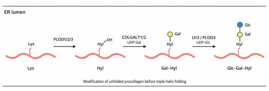
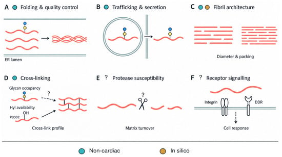
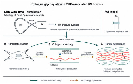
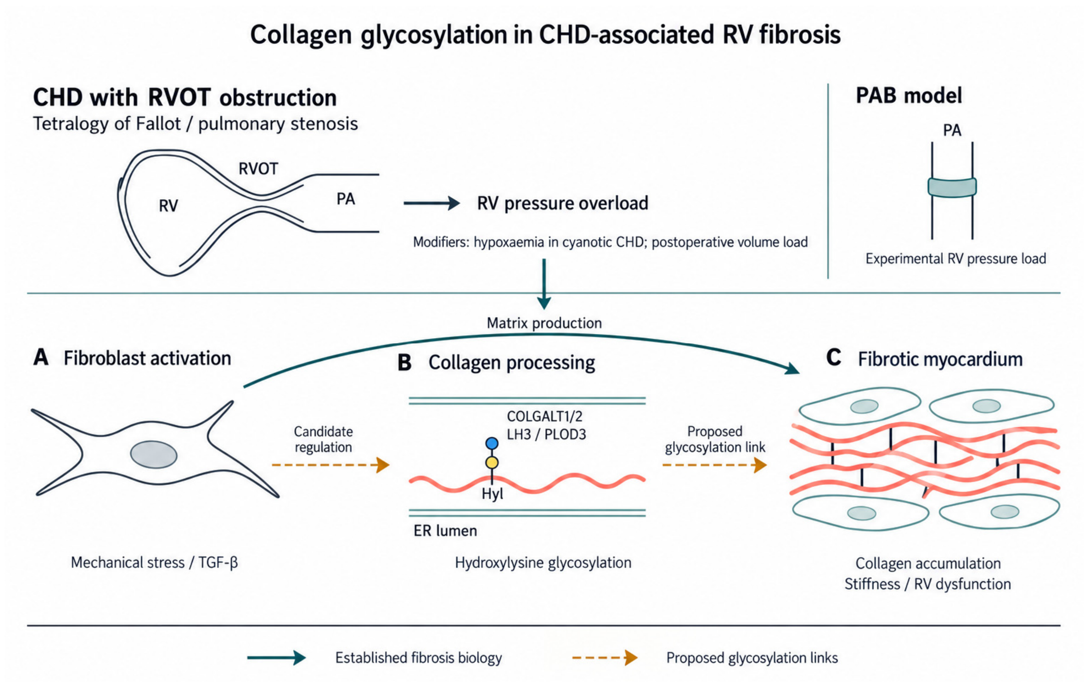
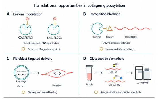
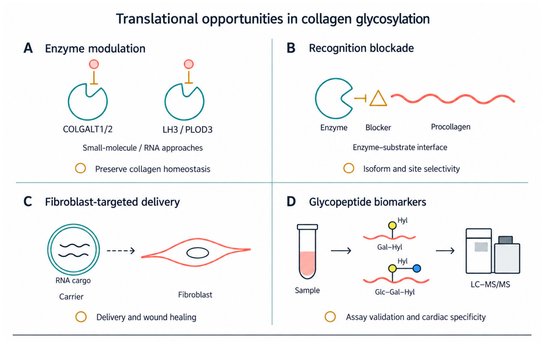

Disclaimer/Publisher’s Note: The statements, opinions and data contained in all publications are solely those of the individual author(s) and contributor(s) and not of MDPI and/or the editor(s). MDPI and/or the editor(s) disclaim responsibility for any injury to people or property resulting from any ideas, methods, instructions or products referred to in the content. |
© 2026 by the authors. Licensee MDPI, Basel, Switzerland. This article is an open access article distributed under the terms and conditions of the Creative Commons Attribution (CC BY) license.