


Simple Summary
Bacterial diseases are a major challenge in fish farming, and the frequent use of antibiotics to control these infections can contribute to the development of antimicrobial resistance. Plants containing naturally active compounds may provide new options for developing safer approaches to disease control. In this study, extracts prepared from the cladodes and aerial parts of butcher’s broom (Ruscus aculeatus) using ethanol or methanol were screened for their chemical composition and their ability to inhibit five important disease-causing bacteria of farmed fish. Chemical analysis showed that the extracts differed considerably in their major natural compounds. Their antibacterial effects also varied depending on the plant part, extraction solvent, bacterial species, and amount tested. Several extracts produced marked inhibition of bacterial growth, with the strongest effects observed against three of the tested fish pathogens, whereas one species was considerably less susceptible. These findings suggest that butcher’s broom warrants further investigation as a potential source of natural antimicrobial agents against bacterial diseases in farmed fish, with the long-term potential to contribute to reduced reliance on antibiotics in aquaculture.
Abstract
The growing need for sustainable alternatives to antibiotics in aquaculture has increased interest in plant-derived antimicrobial compounds. This study characterized ethanol and methanol extracts from the cladodes and aerial parts of butcher’s broom (Ruscus aculeatus L.) by GC–MS and screened, as a preliminary in vitro assessment, their antibacterial activity against Aeromonas hydrophila, Lactococcus garvieae, Vibrio anguillarum, Edwardsiella tarda, and Yersinia ruckeri. The extracts showed distinct chemical profiles. Phytol showed the highest relative chromatographic abundance in the ethanol cladode extract (25.16% of the total ion chromatogram peak area), whereas n-hexadecanoic acid dominated the ethanol aerial parts extract (42.08%). In the methanol cladode extract, the most abundant peak was a co-eluting group (2,2-dimethylbutane/1,2-dimethylaziridine/oxalic acid isobutyl pentyl ester; 22.21%), followed by methyl 9,12,15-octadecatrienoate (14.64%) and phytol (7.86%), while the methanol aerial parts extract was characterized by a 4H-pyran-4-one derivative (16.41%). Antibacterial activity varied with extract, pathogen, and dose, with maximum inhibition zones of 25.0 ± 1.0 mm against A. hydrophila, 24.0 ± 1.0 mm against Y. ruckeri, and 23.7 ± 0.6 mm against V. anguillarum. The E. tarda isolate was the least susceptible. These preliminary findings provide the first evidence of antibacterial activity of R. aculeatus extracts against fish pathogens and support further investigation of this species as a potential source of antimicrobial phytochemicals for aquaculture.
1. Introduction
Aquaculture has emerged as one of the fastest-growing food-production sectors worldwide and is expected to make an increasingly important contribution to meeting the global demand for high-quality animal protein [1]. Nevertheless, the continued intensification of aquaculture production has created conditions that facilitate the emergence and transmission of infectious diseases. Bacterial infections, in particular, remain a major constraint on sustainable fish production because they can cause substantial mortality, impair growth and production performance, and result in considerable economic losses. Among the bacterial pathogens frequently associated with freshwater and marine aquaculture are the Gram-negative species Aeromonas hydrophila, Vibrio anguillarum, Yersinia ruckeri, and Edwardsiella tarda, together with the Gram-positive pathogen Lactococcus garvieae [2].
Antibiotics have traditionally represented one of the principal approaches to the control of bacterial diseases in cultured fish. However, their extensive or inappropriate use has contributed to the emergence and dissemination of antimicrobial-resistant bacteria and resistance determinants within aquatic environments [3]. This growing concern, together with the potential accumulation of antimicrobial residues and their effects on non-target organisms, has intensified the search for safer and more sustainable approaches to disease management in aquaculture [4]. In this context, naturally derived antimicrobial compounds have attracted particular interest because many are biodegradable and may exert lower environmental impacts than conventional synthetic chemotherapeutics [5]. Plant-derived phytochemicals are therefore increasingly being investigated as potential alternatives or complementary agents for controlling bacterial infections in aquatic animals [6].
Medicinal plants have long been recognized as a rich source of structurally diverse secondary metabolites, and plant-derived compounds such as terpenoids, alkaloids and flavonoids constitute the predominant part of the phytochemicals reported to possess antimicrobial activity against bacteria, fungi, viruses and protozoa [7]. This broad spectrum of biological activity has stimulated increasing interest in phytotherapeutic approaches in veterinary medicine, including aquaculture [8,9]. Experimental studies have already demonstrated that extracts from several medicinal plants can inhibit important bacterial fish pathogens. For example, ethanolic plant extracts have shown antibacterial activity against Y. ruckeri, L. garvieae, and V. anguillarum in disc-diffusion and microdilution assays [10]. Similarly, screening of 36 plant species from Türkiye revealed marked differences in susceptibility among fish pathogens, with V. anguillarum being among the most sensitive organisms tested [11]. Collectively, these findings support the continued exploration of medicinal plants as sources of candidate antimicrobial compounds for aquaculture applications [12].
Butcher’s broom, Ruscus aculeatus L., is a perennial evergreen subshrub belonging to the family Asparagaceae, subfamily Nolinoideae (formerly Ruscaceae), within the order Asparagales. A distinctive botanical feature of the species is that photosynthesis is performed predominantly by flattened, leaf-like stem structures known as cladodes rather than by true leaves [13,14]. The species is naturally distributed throughout much of the Mediterranean Basin and western Europe, where it is commonly associated with dry, shaded woodland and scrub habitats and exhibits considerable tolerance to the combined effects of shade and drought [13,15].
The pharmacological properties of R. aculeatus are largely associated with its rich secondary-metabolite profile. Particular attention has been given to steroidal saponins, especially ruscogenin- and neoruscogenin-related compounds, which occur predominantly in the rhizome but are also present in aerial parts [16,17]. In addition to steroidal saponins, the species contains phenolic compounds, flavonoids, sterols, triterpenes, coumarins, and other secondary metabolites that may contribute to its biological activity [18]. Saponins, in particular, are known to exhibit a wide range of biological effects, including antioxidant, hypolipidemic, and antimicrobial activities [19], while chemically related steroidal saponins have also been characterized in other members of the genus, including Ruscus hyrcanus [20].
A growing body of evidence supports the pharmacological potential of R. aculeatus. Extracts of the species have demonstrated antioxidant, anti-urease, and anticholinesterase activities [21], as well as antinociceptive and anti-inflammatory effects [22]. Antimicrobial activity has also been reported, with certain isolated compounds demonstrating inhibitory effects that are comparable to, or in some cases exceed, those of standard reference drugs [23]. Hydroethanolic and aqueous extracts of R. aculeatus have been shown to inhibit several clinically relevant microorganisms [18]. Likewise, ethyl acetate and chloroform extracts of the related species Ruscus hypophyllum displayed activity against both susceptible and multidrug-resistant bacterial strains, with reported minimum inhibitory concentrations (MIC) ranging from 0.125 to 0.5 mg/mL [24]. More recently, secondary metabolites obtained from the branches and cladodes of R. aculeatus have been evaluated against bacterial and fungal clinical isolates, with the observed activity largely confined to Bacillus subtilis ATCC 6633 [25], while methanolic extracts of plants collected in southern Türkiye have also shown measurable antimicrobial activity against Staphylococcus aureus and Candida albicans [26]. These observations further support the presence of biologically active antimicrobial constituents within the species.
Despite these findings, research on the antimicrobial properties of R. aculeatus has thus far focused almost exclusively on microorganisms of human clinical relevance [18,23,24,25]. To the best of our knowledge, its antibacterial activity against major bacterial pathogens of fish has not yet been investigated. Furthermore, phytochemical characterization of R. aculeatus has predominantly relied on HPLC-, LC–MS-, and NMR-based analytical approaches, whereas GC–MS profiling of its extracts remains comparatively limited [27]. Evaluating extracts obtained using solvents of differing polarity and from different plant fractions may therefore provide additional insight into both their chemical composition and their antibacterial potential.
Accordingly, the present study aimed to characterize the chemical composition of ethanol and methanol extracts prepared from the cladodes and aerial parts of R. aculeatus using GC–MS and to evaluate, as a preliminary screening, their in vitro antibacterial activity against five economically important bacterial fish pathogens. By integrating chemical profiling with antibacterial screening, this study seeks to provide an initial assessment of R. aculeatus as a potential source of naturally derived antimicrobial compounds for future applications in aquaculture health management.
2. Materials and Methods
2.1. Plant Material and Extract Preparation
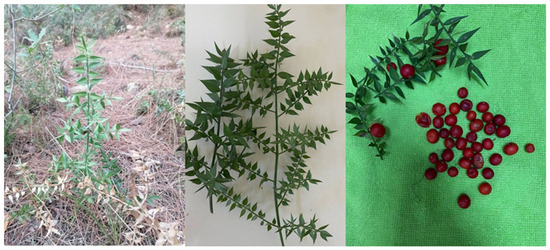
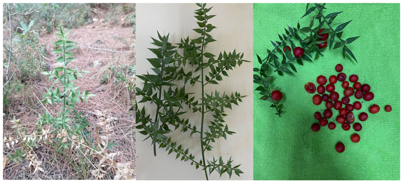
Plant material was collected along the lower slopes of Zorkun Plateau Road (Osmaniye, Türkiye), where R. aculeatus species occurs naturally at altitudes up to approximately 500 m (Figure 1). The plants were transported to the Fish Diseases Laboratory of the Faculty of Fisheries, Cukurova University, Türkiye, washed under running water and air-dried at room temperature.
Figure 1.
Photograph of the R. aculeatus plant taken at the Zorkun plateau (Osmaniye, Türkiye).
A portion of the dried plants was separated into cladodes, whereas the remaining material, consisting of cladodes, stems, and fruits (excluding rhizome and roots), was processed as aerial parts. Ten grams of cladodes and aerial parts material were weighed out and placed into glass bottles with screw caps, to which 100 mL of ethanol (96%) and 100 mL of methanol (≥99.9%) were added (1:10, w/v), and the mixture was subjected to maceration; as a result of this process, four extraction groups were obtained: ethanol-extracted cladodes (EC), ethanol-extracted aerial parts (EA), methanol-extracted cladodes (MC) and methanol-extracted aerial parts (MA).
The bottles were kept in the dark at room temperature (22 ± 2 °C) for 7 days. The resulting mixtures were then filtered through Whatman No.4 filter paper (Whatman International Ltd., Maidstone, UK), and the filtrate was concentrated under reduced pressure using a rotary evaporator (Büchi Laboratory Equipment, Postfach, Switzerland). The solvent was removed to dryness and 100 mg of the dry residue of each extract was weighed and dissolved in 10 mL of the corresponding extraction solvent (96% ethanol or ≥99.9% methanol), giving a 10 mg/mL stock solution for each of the four extracts. Disc loadings of 100, 250 and 500 µg of dry extract per disc were obtained by applying 10, 25 and 50 µL of these stock solutions, respectively. The stock solutions were transferred to 10-mL amber glass vials and stored at +4 °C until analysis. Two millilitres of each stock solution were reserved for GC–MS characterization, and 3 mL were used for the disc diffusion assay.
2.2. Chemical Characterization by GC–MS
The extracts were transported under cold-chain conditions to the Central Research Laboratory of Cukurova University (Adana, Türkiye). Chemical constituents were determined using a gas chromatograph coupled to a mass spectrometer (7890B GC and 7010B MS; Agilent Technologies, Santa Clara, CA, USA) equipped with a DB-Wax capillary column (60 m × 0.25 mm i.d. × 0.25 µm film thickness; J&W Scientific, Folsom, CA, USA). One microliter of each sample, filtered through a 0.45 µm membrane filter, was injected into the capillary column at an injector temperature of 250 °C with a split ratio of 1:20. Helium (99.999% purity) was used as the carrier gas at a flow rate of 2 mL/min, operated in constant flow mode. The oven temperature was held at 40 °C for 4 min, increased to 90 °C at 3 °C/min, then to 130 °C at 4 °C/min and held for 4 min, and finally raised to 240 °C at 5 °C/min and held for 8 min. The ion source, quadrupole and MS transfer line temperatures were maintained at 240 °C, 150 °C and 280 °C, respectively. Mass spectra were acquired in electron impact mode at an electron energy of 70 eV over a mass range of 30–600 m/z in full-scan (MS1) mode; multiple reaction monitoring was not employed.
Constituents were tentatively identified by comparing their mass spectra with those of the NIST14 mass spectral library (NIST/EPA/NIH, version 2014). No internal standard, external calibration or response-factor correction was applied, and no absolute quantification was performed. The proportion of each constituent was calculated by area normalization of the total ion chromatogram, that is, the integrated peak area of each compound was divided by the summed area of all integrated peaks and expressed as a percentage. These values therefore represent relative chromatographic abundance rather than absolute concentrations.
2.3. Bacterial Strains and Antibacterial Assay
Five bacterial fish pathogens (A. hydrophila SY-AH2, L. garvieae SY-LG1, V. anguillarum SY-L24, E. tarda SY-ED1 and Y. ruckeri E42) previously isolated from diseased fish during field studies were used (Table 1). These strains were obtained from culture collection of the Faculty of Marine Sciences and Technology, Çanakkale Onsekiz Mart University, Türkiye. Species identity was confirmed by 16S rRNA gene sequencing of the isolates; the corresponding GenBank accession numbers are given in Table 1. The phenotypic and biochemical characteristics of the five isolates were determined in this study by conventional tests (Gram staining, cell morphology, motility, oxidase, catalase, oxidation/fermentation of glucose, indole production, H2S production, ornithine and arginine decarboxylase, mannitol and sucrose fermentation, and growth in the absence of NaCl) and were interpreted according to Buller [28]; the results are given in Table 2. Stock cultures were plated on tryptic soy agar and subsequently grown in tryptic soy broth for 24 h in a shaking incubator. The cultures were then centrifuged at 5000 rpm for 10 min, washed three times with sterile 0.9% NaCl, and each bacterial inoculum was adjusted to a 0.5 McFarland using a densitometer (DEN-1, BioSan, Warren, MI, USA), and the resulting densities were verified by viable counts for each of the five isolates (mean 1.2 × 10⁸ CFU/mL; range 1.1–1.3 × 10⁸ CFU/mL).
Table 1.
Bacterial fish pathogen isolates used in this study, their host species of isolation and GenBank 16S rRNA gene accession numbers *.
Table 2.
Phenotypic and biochemical characteristics of the five tested isolates.
The antibacterial activity of the extracts was determined by the Kirby–Bauer disc diffusion method [30]. Sterile blank paper discs (Bioanalyse® Dublin, Ireland; 6 mm diameter) were loaded with 100, 250 or 500 µg of dry extract per disc, applied as 10, 25 or 50 µL of the corresponding 10 mg/mL stock solution, in a laminar flow cabinet, allowed to dry completely, and stored at +4 °C in sterile Petri dishes until use. Negative-control discs impregnated with equal volumes of the corresponding extraction solvents (96% ethanol or ≥99.9% methanol), dried under the same conditions, and otherwise processed identically were included in each assay; no inhibition zones developed around these solvent-control discs against any of the five isolates, indicating that, after drying, residual solvent had no adverse effect on bacterial growth. Each bacterial suspension was spread onto Mueller–Hinton agar plates and allowed to dry, after which the extract-impregnated discs were placed on the agar surface at regular intervals. For comparison, a panel of ten commercial antibiotic discs (ampicillin 10 µg, imipenem 10 µg, enrofloxacin 5 µg, levofloxacin 5 µg, tetracycline 30 µg, oxytetracycline 30 µg, chloramphenicol 30 µg, streptomycin 10 µg, sulfamethoxazole-trimethoprim 25 µg and trimethoprim 5 µg (Oxoid, Basingstoke, Hampshire, UK)) were tested against the same isolates under identical conditions. The plates were incubated at 24 °C, and inhibition zone diameters were measured with a digital caliper after 48 h. For the halophilic V. anguillarum isolate (SY-L24), the assay was performed on Mueller–Hinton agar supplemented with NaCl (1.5% w/v) for its salinity requirement, and the extract-impregnated discs were placed on this salt-adjusted medium; the antibiotic discs and the corresponding solvent-control discs were likewise tested on the salt-supplemented plates and produced no inhibition zones. All five isolates produced confluent and homogeneous lawns under the assay conditions (24 °C, 48 h), allowing unambiguous zone measurement. Incubation at 24 °C was chosen to match the growth optima of the fish pathogens tested; because these conditions differ from those under which clinical interpretive breakpoints for the antibiotic discs were established, zone diameters were compared quantitatively and were not classified as susceptible or resistant. All assays, for both extracts and antibiotics, were performed in triplicate, with each replicate consisting of an independently grown bacterial culture plated on a separate agar plate.
2.4. Statistical Analysis
Inhibition zone diameters were recorded for all 60 extract–dose–isolate combinations of the design (4 extracts × 3 disc loadings × 5 isolates, n = 3). A combination was classified as inactive when no zone was detected in any replicate; because the disc itself was 6 mm in diameter, these observations lie at the detection floor of the assay and were entered into the analyses as measurements at that floor (zone diameter = 6 mm) rather than treated as missing values. The primary analysis was therefore performed on the complete 4 × 3 design of each isolate; because cells at the floor have zero within-cell variance, it was evaluated both by two-way ANOVA (extract x dose) and by the assumption-free Kruskal–Wallis test across the 12 extract–dose combinations (Table A5), and the Tukey comparisons shown in Table A1 were derived from this model. For comparison with the presentation customary for screening data, the same models were also fitted to the active combinations only; those results are reported in Table A3 and are conditional on activity, the extract and dose levels entering each model being specified in this study. Normality of the residuals was checked with the Shapiro–Wilk test and homogeneity of variances with the Brown–Forsythe (median-centred Levene) test. For each isolate with more than one active extract, inhibition zone diameters of the active combinations were analyzed by two-way analysis of variance (ANOVA; extract × dose, type II sums of squares) followed by Tukey’s HSD test for pairwise comparisons; because the Shapiro–Wilk test indicated mild departures from normality for A. hydrophila (p = 0.040) and V. anguillarum (p = 0.007), the results were additionally confirmed with the non-parametric Kruskal–Wallis test, applied to the active extract–dose combinations treated as a single factor, which yielded the same conclusions (Table A3). For the E. tarda isolate, only one extract–dose combination was active, and no inferential statistics were applied. The EC extract was likewise active against the A. hydrophila isolate at a single dose level only (500 µg per disc), so neither a regression nor a two-dose comparison was possible for this combination. Dose-dependence was additionally quantified by regressing zone diameter on log2-transformed dose for each extract–isolate combination with at least three active dose levels; combinations with only two active dose levels were summarized descriptively, because two points determine a linear slope mathematically and provide no information on the shape of the dose–response relationship. Each regression was therefore based on three dose levels (nine observations), as reported in Table A4; for combinations with only two active dose levels, the mean difference in zone diameter between the two doses is reported instead. Extract and antibiotic zone diameters (both n = 3) were compared by Welch’s t-tests at the highest disc loading (500 µg of dry extract per disc), with the Holm correction applied across all extract–antibiotic comparisons within each isolate; given the differences in diffusion behaviour, disc loading and formulation between crude extracts and purified antibiotics, these comparisons describe relative performance under the assay conditions rather than equivalence of potency. The level of significance was set at p < 0.05. Statistical analyses were performed with IBM SPSS Statistics (version 30; IBM Corp., Armonk, NY, USA); the Holm correction for the extract–antibiotic comparisons was applied to the SPSS-derived p values in a spreadsheet.
3. Results
3.1. Chemical Characterization of the Extracts
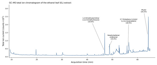
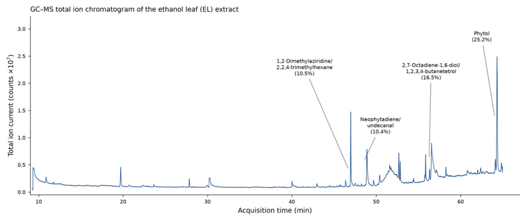
The chemical composition of the four extracts, as tentatively determined by GC–MS, varied considerably with both the solvent and the plant part used. The major constituents (peak area ≥ 5%) of each extract are presented in Table 3, Table 4, Table 5 and Table 6 and Figure 2, Figure 3, Figure 4 and Figure 5. All values are expressed as the relative chromatographic abundance of each compound, obtained by area normalization of the total ion chromatogram.
Table 3.
Major chemical constituents (GC–MS; peak area ≥ 5%) of the EC extract of R. aculeatus. Identifications are tentative (NIST14 library matching); co-eluting alternatives are separated by a slash. Compounds are listed in decreasing order of relative peak area *.
Table 4.
Major chemical constituents (GC–MS; peak area ≥ 5%) of the EA extract of R. aculeatus. Identifications are tentative (NIST14 library matching); co-eluting alternatives are separated by a slash. Compounds are listed in decreasing order of relative peak area *.
Table 5.
Major chemical constituents (GC–MS; peak area ≥ 5%) of the MC extract of R. aculeatus. Identifications are tentative (NIST14 library matching); co-eluting alternatives are separated by a slash. Compounds are listed in decreasing order of relative peak area *.
Table 6.
Major chemical constituents (GC–MS; peak area ≥ 5%) of the MA extract of R. aculeatus. Identifications are tentative (NIST14 library matching); co-eluting alternatives are separated by a slash. Compounds are listed in decreasing order of relative peak area *.
Figure 2.
GC–MS total ion chromatogram of the EC extract of R. aculeatus. Major constituents (peak area ≥ 5%; Table 3) are annotated at their retention times with their relative peak areas.
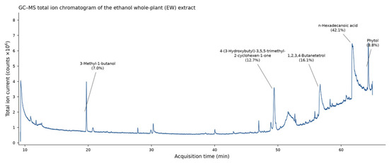
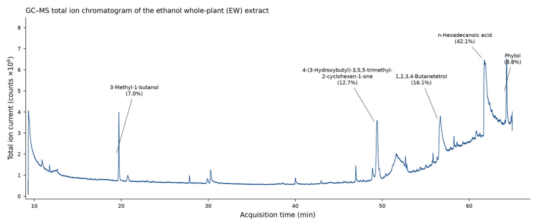
Figure 3.
GC–MS total ion chromatogram of the EA extract of R. aculeatus. Major constituents (peak area ≥ 5%; Table 4) are annotated at their retention times with their relative peak areas.
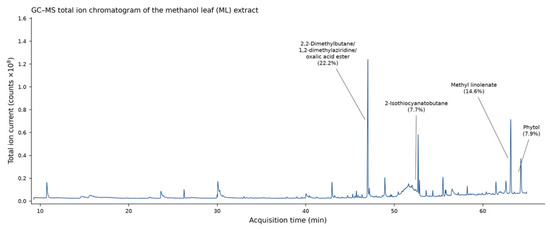
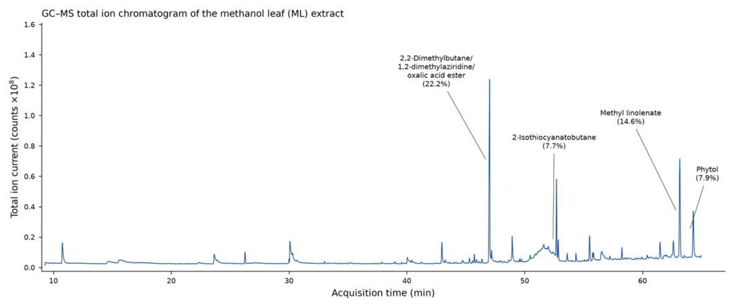
Figure 4.
GC–MS total ion chromatogram of the MC extract of R. aculeatus. Major constituents (peak area ≥ 5%; Table 5) are annotated at their retention times with their relative peak areas.
Figure 5.
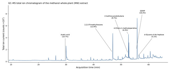
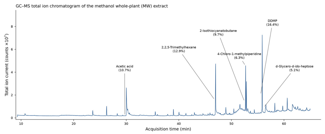
GC–MS total ion chromatogram of the MA extract of R. aculeatus. Major constituents (peak area ≥ 5%; Table 6) are annotated at their retention times with their relative peak areas.
A total of 32 compounds were tentatively identified in the EC extract, of which four exceeded 5% of the total peak area and together accounted for 62.6% of the chromatogram (Table 3, Figure 2). The diterpene alcohol phytol was the single most abundant constituent (25.16%), followed by the co-eluting pairs 2,7-octadiene-1,6-diol, 2,6-dimethyl/1,2,3,4-butanetetrol (16.52%), 1,2-dimethylaziridine/2,2,4-trimethylhexane (10.51%) and neophytadiene/undecanal (10.36%). The remaining 28 minor constituents each represented less than 5% of the total peak area (cumulatively 37.5%).
The EA extract showed the least complex profile, with 17 compounds detected, five of which exceeded the 5% threshold and jointly represented 86.5% of the total peak area (Table 4, Figure 3). n-Hexadecanoic acid (palmitic acid) clearly dominated the profile (42.08%), followed by 1,2,3,4-butanetetrol (16.05%), the megastigmane-type ketone 4-(3-hydroxybutyl)-3,5,5-trimethyl-2-cyclohexen-1-one (12.66%), phytol (8.77%) and 3-methyl-1-butanol (6.98%). The predominance of a saturated fatty acid distinguishes this extract from the cladode extracts, in which terpenoid constituents prevailed.
In the MC extract, 33 compounds were detected, four of which exceeded 5% of the total peak area (52.4% cumulatively) (Table 5, Figure 4). The most abundant peak corresponded to the co-eluting group 2,2-dimethylbutane/1,2-dimethylaziridine/oxalic acid isobutyl pentyl ester (22.21%), followed by methyl (Z,Z,Z)-9,12,15-octadecatrienoate (methyl linolenate; 14.64%), phytol (7.86%) and 2-isothiocyanatobutane (7.65%).
The MA extract was the most complex, with 38 detected compounds, six of which exceeded the 5% threshold and together accounted for 61.2% of the total peak area (Table 6, Figure 5). The pyranone derivative 2,3-dihydro-3,5-dihydroxy-6-methyl-4H-pyran-4-one (DDMP; 16.41%) was the principal constituent, followed by 2,2,5-trimethylhexane (12.92%), acetic acid (10.70%), 2-isothiocyanatobutane (9.71%), 4-chloro-1-methylpiperidine (6.33%) and d-glycero-d-ido-heptose (5.10%).
3.2. Antibacterial Activity
The inhibition zone diameters produced by the four extracts against the five fish pathogen isolates after 48 h of incubation are shown in Table A1, and those of the ten reference antibiotics in Table A2, representative plates are shown in Figure S1. No inhibition zones were observed around the solvent-control discs. Among the antibiotics, enrofloxacin, imipenem and levofloxacin were active against all five isolates (22.0–32.0 mm), whereas the L. garvieae, Y. ruckeri and E. tarda isolates showed no measurable zones with six to seven of the ten antibiotics tested under the present assay conditions, including oxytetracycline, sulfamethoxazole-trimethoprim and trimethoprim (Table A2). Of the 60 extract–dose-isolate combinations tested, 38 (63%) produced a measurable inhibition zone: 9 of 15 for the EC extract, 7 of 15 for EA, and 11 of 15 for each of MC and MA. The number of active combinations increased with disc loading (8 of 20 at 100 µg, 14 of 20 at 250 µg and 16 of 20 at 500 µg per disc) and differed markedly among isolates (L. garvieae 11 of 12, V. anguillarum and Y. ruckeri 10 of 12 each, A. hydrophila 6 of 12, E. tarda 1 of 12). For the four isolates with more than one active extract, two-way ANOVA on the complete 4 × 3 design indicated significant effects of extract, dose and their interaction (all p < 0.001), and the Kruskal–Wallis test across the 12 extract–dose combinations gave the same result (H = 33.3–34.9, df = 11, p < 0.001; Table A5); given the significant interactions, the pairwise Tukey comparisons reported in Table A1 were interpreted as simple effects, that is, among extracts within each dose and among doses within each extract. When the analysis was restricted to the active combinations, the same effects remained significant (extract and dose p < 0.001, interaction p <= 0.005; Table A3), but the relative standing of the extracts differed: against A. hydrophila the EC extract had the highest mean among the active cells (22.7 mm at its single active dose), whereas over the complete design the MC extract ranked first (18.0 versus 11.6 mm), and against L. garvieae the EA extract fell from the second to the fourth rank. Where inhibition occurred, zone diameters generally increased with dose: of the eight extract–isolate combinations with three active dose levels, seven showed significant positive dose–response slopes in the log-dose regressions (Table A4). The steepest slope was obtained for the MC extract against A. hydrophila (6.2 mm per doubling of dose, 95% CI 5.5–6.8). Among the six combinations with only two active dose levels, the largest descriptive difference was observed for the MC extract against V. anguillarum (9.0 mm between the 250 and 500 µg loadings); for these combinations only the pairwise Tukey comparison given in Table A1 is reported and no dose–response slope was estimated, because two dose levels do not allow the shape of the dose–response relationship to be assessed. In contrast, the MC extract against Y. ruckeri was active already at 100 µg per disc with no further significant increase across doses (slope 0.3 mm per doubling, p = 0.22; Table A4).
Against A. hydrophila, the MC extract was the most effective, producing inhibition zones of 10.7 ± 0.6, 18.3 ± 0.6 and 25.0 ± 1.0 mm at 100, 250 and 500 µg of dry extract per disc, respectively, whereas the EA extract produced no inhibition at any dose; at 500 µg, the MC extract zone was significantly larger than those of chloramphenicol and oxytetracycline (Welch’s t-test, Holm-corrected p < 0.01) and did not differ significantly from those of ampicillin, tetracycline, imipenem or streptomycin. Against V. anguillarum, the EA extract was the most active at all doses (17.7–23.7 mm), and the MC extract reached a comparable zone diameter (23.7 ± 0.6 mm) at 500 µg per disc; at this dose, both extracts produced significantly larger zones than tetracycline (18.0 ± 1.0 mm) and chloramphenicol (14.0 ± 1.0 mm) (Holm-corrected p < 0.05), did not differ significantly from ampicillin (16.0 ± 2.0 mm), and remained below the zones of enrofloxacin, imipenem and levofloxacin (26.0–28.0 mm). Against Y. ruckeri, the largest inhibition zone was obtained with the MA extract at 500 µg per disc (24.0 ± 1.0 mm), followed by the MC extract (20.7–21.3 mm), which was active even at the lowest dose tested; the MA extract zone did not differ significantly from that of ampicillin (23.0 ± 1.0 mm). Against L. garvieae, the MC and MA extracts produced the largest zones at 500 µg per disc (18.7 ± 0.6 mm); although these values remained below those of the three active antibiotics (22.0–31.7 mm), this isolate showed no zone with the remaining seven antibiotics of the panel, including ampicillin and tetracycline, to which the extracts were accordingly the only inhibitory agents besides enrofloxacin, imipenem and levofloxacin. The E. tarda isolate was the least susceptible to the extracts, being inhibited only by the EC extract at 500 µg per disc (12.3 ± 0.6 mm), whereas no inhibition zone was observed for the other extracts at any dose.
4. Discussion
In the present study, we investigated, to our knowledge for the first time, the antibacterial activity of R. aculeatus extracts against bacterial fish pathogens. All four extracts exhibited measurable, dose-dependent activity against at least one of the five pathogens tested, and four of the five pathogens (A. hydrophila, V. anguillarum, Y. ruckeri and L. garvieae) were inhibited by at least two extracts. These results suggest that the antibacterial activity of R. aculeatus previously reported against human clinical isolates may also extend to bacterial pathogens of farmed fish; however, as a single field isolate per species was tested, this conclusion requires confirmation with additional strains.
Our findings are consistent with previous studies on the antimicrobial potential of the genus Ruscus. Hadžifejzović et al. [23] reported that extracts and isolated compounds of R. aculeatus and R. hypoglossum exhibited antibacterial and especially antifungal activity, which in some cases exceeded that of standard drugs. Similarly, Edziri et al. [24] found that ethyl acetate and chloroform extracts of R. hypophyllum were active against multidrug-resistant bacteria, with MIC between 0.125 and 0.5 mg/mL, and Rodrigues et al. [18] demonstrated that hydroethanolic and aqueous extracts of R. aculeatus inhibited several multiresistant clinical strains. The activity observed in the present study against Gram-negative fish pathogens such as A. hydrophila, V. anguillarum and Y. ruckeri, as well as against the Gram-positive L. garvieae, therefore complements the existing evidence obtained with clinically relevant bacteria. The inhibition zones observed in this study were within the range reported for ethanolic plant extracts against the same fish pathogens in disc diffusion assays [10,11] and for other natural products such as propolis against A. hydrophila and Y. ruckeri [31], although differences in extract preparation and disc loading preclude direct quantitative comparison.
The differences in activity among the four extracts may be related to their chemical composition, which varied markedly with the solvent and the plant part used. The major constituents identified in this study included phytol, n-hexadecanoic acid, 9,12,15-octadecatrienoic acid methyl ester, 2-isothiocyanatobutane and 2,3-dihydro-3,5-dihydroxy-6-methyl-4H-pyran-4-one. Plant-derived secondary metabolites, including fatty acids, terpenoids and sulfur-containing compounds, are widely recognized as natural antimicrobial agents [5], and saponins, which constitute the characteristic bioactive fraction of Ruscus species [19,32], may also have contributed to the observed activity, although they are not amenable to detection by the GC–MS approach used. It should be noted, however, that saponins can also act as anti-nutritional factors, binding minerals and interfering with nutrient absorption [19]; this dual character would need to be considered if R. aculeatus preparations were to be developed as feed additives. Further studies employing complementary analytical techniques will be required to identify the compounds responsible for the antibacterial effects.
When compared with the reference antibiotics, the extracts generally produced smaller inhibition zones than the most active agents of the panel (enrofloxacin, imipenem and levofloxacin), which is expected considering that crude extracts were tested against purified compounds applied at standardized doses. Nevertheless, with all comparisons based on triplicate assays, the EA and MC extracts at 500 µg per disc produced significantly larger zones than tetracycline and chloramphenicol against V. anguillarum, and the MC extract zone against A. hydrophila exceeded those of chloramphenicol and oxytetracycline; moreover, the extracts inhibited the L. garvieae isolate, for which seven of the ten antibiotics tested, including ampicillin and tetracycline, produced no measurable zone. It should be noted, however, that disc diffusion comparisons between crude extracts and antibiotic discs are influenced by differences in diffusion behaviour, disc loading and formulation; these comparisons therefore describe relative performance under the assay conditions rather than equivalence of potency and require confirmation by the determination of MIC. The susceptibility patterns observed for the isolates themselves—particularly the absence of measurable zones for the L. garvieae, Y. ruckeri and E. tarda isolates with six or more of the antibiotics tested under the present assay conditions, underline the relevance of searching for alternative antimicrobial agents in aquaculture.
The use of natural products as antimicrobial agents has attracted increasing attention in food and aquaculture systems, both as alternatives to synthetic preservatives and in response to the spread of antibiotic resistance [5,33,34,35]. Formulation approaches such as combinations with biopolymers or nanoparticle carriers have been reported to enhance the efficacy of plant-derived antimicrobials and feed additives in fish [36,37,38,39,40,41,42].
Some limitations of the present study should be acknowledged. Only one field isolate per pathogen species was tested, and susceptibility to plant extracts may vary among strains of the same species; the present results should therefore not be generalized to the species level without confirmation. The antibacterial activity was evaluated only in vitro and at the level of inhibition zone diameters; MIC and minimum bactericidal concentrations (MBC) were not determined, and no quality-control reference strain was included. Because no zone could be measured for the 22 inactive extract–dose–isolate combinations, the inferential comparisons of zone diameters are conditional on activity; an analysis of the complete design, with these combinations at the detection floor, is reported in Table A5 and supported the effects described above. In addition, the gravimetric extraction yield of each solvent–plant part combination was not recorded, so the extraction efficiency of the four preparations cannot be compared; the antibacterial assays were nevertheless standardized on an equal dry-extract mass basis (100, 250 and 500 µg per disc). Because disc diffusion is influenced by the molecular size, polarity and agar diffusivity of the extract constituents, inhibition-zone diameters alone do not allow the intrinsic antimicrobial potency of the four extracts to be compared directly; the present results should therefore be regarded as a preliminary screening. Species assignments of the isolates were based on 16S rRNA gene sequencing, which has limited discriminatory power at the species level within some genera, notably Aeromonas, Vibrio and Edwardsiella; confirmation by multilocus or genome-based approaches would strengthen the identifications. In addition, the rhizome, which is regarded as the pharmacologically most important organ of R. aculeatus, was deliberately excluded to avoid destructive harvesting of natural populations; the antibacterial potential of rhizome extracts against fish pathogens therefore remains to be evaluated. Finally, cytotoxicity and in vivo efficacy remain to be assessed. Further studies should therefore include the determination of MIC and MBC values, cytotoxicity assays, in vivo challenge trials, and, if the extracts are to be administered as feed additives, an evaluation of their effects on the intestinal microbiota of fish.
5. Conclusions
In the present study, ethanol and methanol extracts obtained from the cladodes and the aerial parts of R. aculeatus exhibited in vitro antibacterial activity in this preliminary screening study against bacterial fish pathogens, with dose-dependent responses in most of the extract–pathogen combinations in which activity occurred. Because a single field isolate per pathogen species was tested, all comparisons among pathogens reflect the responses of the specific isolates examined and should not be generalized to the species level. The EC extract was the only extract that inhibited all five isolates, whereas the MC extract produced the largest inhibition zones against A. hydrophila and, together with the EA extract, against V. anguillarum; the E. tarda isolate was the least susceptible. To our knowledge, this is the first report of antibacterial activity of R. aculeatus extracts against bacterial fish pathogens. These preliminary in vitro findings suggest that R. aculeatus, a medicinal plant occurring naturally in Türkiye, deserves further evaluation as a potential source of plant-derived antimicrobial agents for aquaculture; however, the determination of MIC and MBC values, cytotoxicity tests, testing of additional isolates per species, and in vivo challenge trials will be required before any practical application can be considered.
Supplementary Materials
The following supporting information can be downloaded at: https://www.mdpi.com/article/10.3390/vetsci13090945/s1, Figure S1: Disc-diffusion assay plates of Ruscus aculeatus extracts against five bacterial fish pathogens.
Author Contributions
Conceptualization, İ.D., H.B., M.E.B. and F.S.S.; methodology, İ.D., M.E.B. and F.S.S.; validation, İ.D., H.B., M.E.B. and F.S.S.; formal analysis, İ.D., H.B. and M.E.B.; investigation, H.B.; İ.D.; resources, İ.D. and M.E.B.; data curation, H.B., İ.D. and M.E.B.; writing—original draft preparation, H.B. and İ.D.; writing—review and editing, İ.D., M.E.B. and F.S.S.; visualization, İ.D. and F.S.S.; supervision, İ.D.; project administration, İ.D.; funding acquisition, İ.D. All authors have read and agreed to the published version of the manuscript.
Funding
This study was supported by the Cukurova University, Scientific Research Projects Coordination Unit (Project No: FYL-2023-15846), Türkiye. This article is based on the MSc thesis of H. Bikzad.
Institutional Review Board Statement
Not applicable.
Informed Consent Statement
Not applicable.
Data Availability Statement
The original contributions presented in this study are included in the article/Supplementary Material. Further inquiries can be directed to the corresponding author.
Acknowledgments
The authors thank the Central Research Laboratory of Cukurova University for performing the GC–MS analyses and Sevdan Yılmaz (Department of Aquaculture, Faculty of Marine Sciences and Technology, Çanakkale Onsekiz Mart University, Çanakkale, Türkiye for kindly providing the bacterial fish pathogen isolates used in this study. This article is based on the MSc thesis of Hasibullah Bikzad, completed at the Institute of Natural and Applied Sciences, Cukurova University.
Conflicts of Interest
Faik Sertel Secer owned the company Aquaterra Co., Ltd., a company operating in Ankara University Technopolis. The company had no role in the design of the study; in the collection, analysis, or interpretation of data; in the writing of the manuscript; or in the decision to publish the results. The authors declare no conflicts of interest.
Abbreviations
The following abbreviations are used in this manuscript:
| EC | Ethanol-extracted cladodes |
| EA | Ethanol-extracted aerial parts |
| MC | Methanol-extracted cladodes |
| MA | Methanol-extracted aerial parts |
| MBC | Minimum bactericidal concentrations |
| MIC | Minimum inhibitory concentrations |
| HSD | Honestly significant difference (Tukey’s test) |
| GC–MS | Gas chromatography–mass spectrometry |
| NIST | National Institute of Standards and Technology (NIST14 mass spectral library) |
| RT | Retention time |
| CI | Confidence interval |
| rRNA | Ribosomal RNA |
Appendix A
Table A1.
Inhibition zone diameters (mm) produced by R. aculeatus extracts against five bacterial fish pathogen isolates after 48 h of incubation at 24 °C. Disc loadings of 100, 250 and 500 µg of dry extract per disc were applied as 10, 25 and 50 µL of a 10 mg/mL extract stock solution.
Table A2.
Inhibition zone diameters (mm) produced by ten reference antibiotic discs against the five bacterial fish pathogen isolates after 48 h of incubation at 24 °C.
Table A3.
Structure and results of the per-isolate two-way ANOVA models (extract × dose, type II sums of squares) fitted to the active extract–dose combinations. Doses of 100, 250 and 500 µg of dry extract per disc were applied as 10, 25 and 50 µL of a 10 mg/mL extract stock solution.
Table A4.
Log2-dose regressions of inhibition zone diameter for extract–isolate combinations with three active dose levels, and descriptive dose differences for combinations with two active dose levels. Doses of 100, 250 and 500 µg of dry extract per disc were applied as 10, 25 and 50 µL of a 10 mg/mL extract stock solution.
Table A5.
Per-isolate analysis of the complete 4 × 3 design of each isolate, with the inactive extract–dose combinations entered at the detection floor of the assay (zone diameter = 6 mm). Doses of 100, 250 and 500 µg of dry extract per disc were applied as 10, 25 and 50 µL of a 10 mg/mL extract stock solution.
References
- FAO. The State of World Fisheries and Aquaculture 2026—Blue Transformation: Turning Vision into Impact; Food and Agriculture Organization of the United Nations: Rome, Italy, 2026. [Google Scholar]
- Milijasevic, M.; Veskovic-Moracanin, S.; Babic Milijasevic, J.; Petrovic, J.; Nastasijevic, I. Antimicrobial Resistance in Aquaculture: Risk Mitigation within the One Health Context. Foods 2024, 13, 2448. [Google Scholar] [CrossRef] [Scilit] [PubMed]
- Cabello, F.C.; Godfrey, H.P.; Buschmann, A.H.; Dölz, H.J. Aquaculture as yet Another Environmental Gateway to the Development and Globalisation of Antimicrobial Resistance. Lancet Infect. Dis. 2016, 16, e127–e133. [Google Scholar] [CrossRef] [Scilit] [PubMed]
- Kolygas, M.N.; Kostou, V.; Pappas, I.S.; Karagouni, E.; Toubanaki, D.K.; Bakopoulos, V.; Kotzamanis, Y.P.; Nathanailides, C.; Athanassopoulou, F. In Vitro Antibacterial Activity of Essential Oils from Medicinal Plants against Major Fish Pathogens in Mediterranean Aquaculture. Front. Aquac. 2025, 4, 1665877. [Google Scholar] [CrossRef] [Scilit]
- Gyawali, R.; Ibrahim, S.A. Natural Products as Antimicrobial Agents. Food Control 2014, 46, 412–429. [Google Scholar] [CrossRef] [Scilit]
- Rahimi, N.N.M.N.N.; Natrah, I.; Loh, J.-Y.; Ranzil, F.K.E.; Gina, M.; Lim, S.-H.E.; Lai, K.-S.; Chong, C.-M. Phytocompounds as an Alternative Antimicrobial Approach in Aquaculture. Antibiotics 2022, 11, 469. [Google Scholar] [CrossRef] [Scilit] [PubMed]
- Huang, W.; Wang, Y.; Tian, W.; Cui, X.; Tu, P.; Li, J.; Shi, S.; Liu, X. Biosynthesis Investigations of Terpenoid, Alkaloid, and Flavonoid Antimicrobial Agents Derived from Medicinal Plants. Antibiotics 2022, 11, 1380. [Google Scholar] [CrossRef] [Scilit] [PubMed]
- Tamminen, L.-M.; Emanuelson, U.; Blanco-Penedo, I. Systematic Review of Phytotherapeutic Treatments for Different Farm Animals Under European Conditions. Front. Vet. Sci. 2018, 5, 140. [Google Scholar] [CrossRef] [Scilit] [PubMed]
- Ng, J.J.Y.; Yusoff, N.A.H.; Elias, N.A.; Norhan, N.A.S.; Harun, N.A.; Abdullah, F.; Ishak, A.N.; Hassan, M. Phytotherapy Use for Disease Control in Aquaculture: A Review of the Last 5 Years. Aquac. Int. 2024, 32, 2687–2712. [Google Scholar] [CrossRef] [Scilit]
- Bulfon, C.; Volpatti, D.; Galeotti, M. In Vitro Antibacterial Activity of Plant Ethanolic Extracts Against Fish Pathogens. J. World Aquac. Soc. 2014, 45, 545–557. [Google Scholar] [CrossRef] [Scilit]
- Turker, H.; Yıldırım, A.B. Screening for Antibacterial Activity of Some Turkish Plants Against Fish Pathogens: A Possible Alternative in the Treatment of Bacterial Infections. Biotechnol. Biotechnol. Equip. 2015, 29, 281–288. [Google Scholar] [CrossRef] [Scilit] [PubMed]
- Bulfon, C.; Volpatti, D.; Galeotti, M. Current Research on the Use of Plant-Derived Products in Farmed Fish. Aquac. Res. 2015, 46, 513–551. [Google Scholar] [CrossRef] [Scilit]
- Thomas, P.A.; Mukassabi, T.A. Biological Flora of the British Isles: Ruscus aculeatus. J. Ecol. 2014, 102, 1083–1100. [Google Scholar] [CrossRef] [Scilit]
- Pandey, A.K.; Kasana, S. Plant Systematics; CRC Press: Boca Raton, FL, USA, 2021. [Google Scholar]
- Pivovaroff, A.; Sharifi, R.; Scoffoni, C.; Sack, L.; Rundel, P.W. Making the Best of the Worst of Times: Traits Underlying Combined Shade and Drought Tolerance of Ruscus aculeatus and R. microglossum (Asparagaceae). Funct. Plant Biol. 2014, 41, 11–24. [Google Scholar] [CrossRef] [Scilit] [PubMed]
- Masullo, M.; Pizza, C.; Piacente, S. Ruscus Genus: A Rich Source of Bioactive Steroidal Saponins. Planta Medica 2016, 82, 1513–1524. [Google Scholar] [CrossRef] [Scilit] [PubMed]
- Khojasteh, A.; Sanchez-Muñoz, R.; Moyano, E.; Bonfill, M.; Cusido, R.M.; Eibl, R.; Palazon, J. Biotechnological Production of Ruscogenins in Plant Cell and Organ Cultures of Ruscus aculeatus. Plant Physiol. Biochem. 2019, 141, 133–141. [Google Scholar] [CrossRef] [Scilit] [PubMed]
- Rodrigues, J.P.B.; Fernandes, Â.; Dias, M.I.; Pereira, C.; Pires, T.C.S.P.; Calhelha, R.C.; Carvalho, A.M.; Ferreira, I.C.F.R.; Barros, L. Phenolic Compounds and Bioactive Properties of Ruscus aculeatus L. (Asparagaceae): The Pharmacological Potential of an Underexploited Subshrub. Molecules 2021, 26, 1882. [Google Scholar] [CrossRef] [Scilit] [PubMed]
- Sharma, K.; Kaur, R.; Kumar, S.; Saini, R.K.; Sharma, S.; Pawde, S.V.; Kumar, V. Saponins: A Concise Review on Food Related Aspects, Applications and Health Implications. Food Chem. Adv. 2023, 2, 100191. [Google Scholar] [CrossRef] [Scilit]
- Nazemiyeh, H.; Zengin, G.; Mehrad, H.; Farhoudi, M.; Bahadori, M.B. LC-MS/MS-Based Steroidal Saponins Profiling and Biological Activities of Ruscus hyrcanus Woronow. Eur. J. Integr. Med. 2020, 40, 101245. [Google Scholar] [CrossRef] [Scilit]
- Taşkın, T.; Güler, E.; Şahin, T.; Bulut, G. Enzyme Inhibitory and Antioxidant Activities of Different Extracts from Ruscus aculeatus L. Acta Pharm. Sci. 2020, 58, 497–507. [Google Scholar] [CrossRef] [Scilit]
- Daniari, T.; Ramezani, M.; Pakpour, B. Anti-Nociceptive and Anti-Inflammatory Effect of Ruscus aculeatus L. Hydroalcoholic Extract in Male NMRI Mice. J. Gorgan Univ. Med. Sci. 2020, 21, 47–52. [Google Scholar]
- Hadžifejzović, N.; Kukić-Marković, J.; Petrović, S.; Soković, M.; Glamočlija, J.; Stojković, D.; Nahrstedt, A. Bioactivity of the Extracts and Compounds of Ruscus aculeatus L. and Ruscus hypoglossum L. Ind. Crops Prod. 2013, 49, 407–411. [Google Scholar] [CrossRef] [Scilit]
- Edziri, H.; Haddad, O.; Saidana, D.; Chouchen, S.; Skhiri, F.; Mastouri, M.; Flamini, G. Ruscus hypophyllum L. Extracts: Chemical Composition, Antioxidant, Anticoagulant, and Antimicrobial Activity Against a Wide Range of Sensitive and Multi-Resistant Bacteria. Environ. Sci. Pollut. Res. 2020, 27, 17063–17071. [Google Scholar] [CrossRef] [Scilit] [PubMed]
- Soydinç, A.; Aktürk, R.; Biryol, S.; Usta, M. Investigation of Antimicrobial Effects of Essential Oil and Secondary Metabolite Extraction from Ruscus aculeatus L. (Asparagaceae). Int. J. Second. Metab. 2025, 12, 832–846. [Google Scholar] [CrossRef] [Scilit]
- Göycıncık, S.; Dolap, H. A Study on Antioxidant and Antimicrobial Activities of Phlomis amanica, Echinops viscosus and Ruscus aculeatus from Hatay, Türkiye. Int. J. Second. Metab. 2025, 12, 886–897. [Google Scholar] [CrossRef] [Scilit]
- Pacuła, W.; Sowa, I.; Feldo, M.; Graczyk, F.; Patryn, R.; Wójciak, M. Current Insights into the Phytochemistry and Pharmacological Properties of Ruscus aculeatus. Molecules 2025, 30, 4417. [Google Scholar] [CrossRef] [Scilit] [PubMed]
- Buller, N.B. Bacteria and Fungi from Fish and Other Aquatic Animals: A Practical Identification Manual; CABI: Wallingford, UK, 2004. [Google Scholar]
- Yilmaz, S.; Sova, M.; Ergün, S. Antimicrobial Activity of trans-cinnamic Acid and Commonly Used Antibiotics against Important Fish Pathogens and Nonpathogenic Isolates. J. Appl. Microbiol. 2018, 125, 1714–1727. [Google Scholar] [CrossRef] [Scilit] [PubMed]
- Bauer, A.W.; Kirby, W.M.M.; Sherris, J.C.; Turck, M. Antibiotic Susceptibility Testing by a Standardized Single Disk Method. Am. J. Clin. Pathol. 1966, 45, 493–496. [Google Scholar] [CrossRef] [Scilit]
- Tukmechi, A.; Ownagh, A.; Mohebbat, A. In Vitro Antibacterial Activities of Ethanol Extract of Iranian Propolis (EEIP) against Fish Pathogenic Bacteria (Aeromonas hydrophila, Yersinia ruckeri, Streptococcus iniae). Braz. J. Microbiol. 2010, 41, 1086–1092. [Google Scholar] [CrossRef] [Scilit]
- Güvenç, A.; Şatır, E.; Coşkun, M. Determination of Ruscogenin in Turkish Ruscus L. Species by UPLC. Chromatographia 2007, 66, 141–145. [Google Scholar] [CrossRef] [Scilit]
- Castro-Enríquez, D.; Miranda, J.M.; Trigo, M.; Rodríguez-Félix, F.; Aubourg, S.P.; Barros-Velázquez, J. Antioxidant and Antimicrobial Effect of Biodegradable Films Containing Pitaya (Stenocereus thurberi) Extracts during the Refrigerated Storage of Fish. Antioxidants 2023, 12, 544. [Google Scholar] [CrossRef] [Scilit] [PubMed]
- Han, A.; Hwang, J.-H.; Lee, S.-Y. Antimicrobial Activities of Asian Plant Extracts against Pathogenic and Spoilage Bacteria. Food Sci. Biotechnol. 2022, 32, 229–238. [Google Scholar] [CrossRef] [Scilit] [PubMed]
- Tagrida, M.; Nilsuwan, K.; Gulzar, S.; Prodpran, T.; Benjakul, S. Fish Gelatin/Chitosan Blend Films Incorporated with Betel (Piper betle L.) Leaf Ethanolic Extracts: Characteristics, Antioxidant and Antimicrobial Properties. Food Hydrocoll. 2023, 137, 108316. [Google Scholar] [CrossRef] [Scilit]
- Thangapandiyan, S.; Monika, S. Green Synthesized Zinc Oxide Nanoparticles as Feed Additives to Improve Growth, Biochemical, and Hematological Parameters in Freshwater Fish Labeo rohita. Biol. Trace Elem. Res. 2019, 195, 636–647. [Google Scholar] [CrossRef] [Scilit] [PubMed]
- Abd El-Naby, A.S.; Al-Sagheer, A.A.; Negm, S.S.; Naiel, M.A.E. Dietary Combination of Chitosan Nanoparticle and Thymol Affects Feed Utilization, Digestive Enzymes, Antioxidant Status, and Intestinal Morphology of Oreochromis niloticus. Aquaculture 2020, 515, 734577. [Google Scholar] [CrossRef] [Scilit]
- Kumaran, S.; Perianaika Anahas, A.M.; Prasannabalaji, N.; Karthiga, M.; Bharathi, S.; Rajasekar, T.; Joseph, J.; Prasad, S.G.; Pandian, S.; Pugazhvendan, S.R.; et al. Chitin Derivatives of NAG and Chitosan Nanoparticles from Marine Disposal Yards and Their Use for Economically Feasible Fish Feed Development. Chemosphere 2021, 281, 130746. [Google Scholar] [CrossRef] [Scilit] [PubMed]
- Çiçek, S.; Özoğul, F. Effects of Selenium Nanoparticles on Growth Performance, Hematological, Serum Biochemical Parameters, and Antioxidant Status in Fish. Anim. Feed Sci. Technol. 2021, 281, 115099. [Google Scholar] [CrossRef] [Scilit]
- El-Naggar, M.; Medhat, F.; Taha, A. Applications of Chitosan and Chitosan Nanoparticles in Fish Aquaculture. Egypt. J. Aquat. Biol. Fish. 2022, 26, 23–43. [Google Scholar] [CrossRef] [Scilit]
- Aksoy, A.; Alazragi, R.; Alabdali, A.Y.M.; Aljazzar, R.; El Sadi, S.; Alostaz, M.; El Hindi, M. Antibacterial Activity of Metallic-Core Gold and Silver Nanoparticles against Some Animal Pathogens. Ann. Anim. Sci. 2023, 23, 473–479. [Google Scholar] [CrossRef] [Scilit]
- Bhatia, N.; Kumari, A.; Chauhan, N.; Thakur, N.; Sharma, R. Duchesnea indica Plant Extract Mediated Synthesis of Copper Oxide Nanomaterials for Antimicrobial Activity and Free-Radical Scavenging Assay. Biocatal. Agric. Biotechnol. 2023, 47, 102574. [Google Scholar] [CrossRef] [Scilit]

Disclaimer/Publisher’s Note: The statements, opinions and data contained in all publications are solely those of the individual author(s) and contributor(s) and not of MDPI and/or the editor(s). MDPI and/or the editor(s) disclaim responsibility for any injury to people or property resulting from any ideas, methods, instructions or products referred to in the content. |
© 2026 by the authors. Licensee MDPI, Basel, Switzerland. This article is an open access article distributed under the terms and conditions of the Creative Commons Attribution (CC BY) license.