


Simple Summary
Anaplasma platys is a zoonotic pathogen affecting canines and humans. Although the hard ticks of Rhipicephalus sanguineus sensu lato largely infest dogs in Southern Taiwan, there is no evidence confirming A. platys infection in these ticks. We conducted a field survey for screening the Anaplasma infection in R. sanguineus s. l. ticks by nested-PCR assay. Results reveal infection rates of 0.8%, 1.0% and 1.4% in nymphs, males and females, respectively. Monthly prevalence was observed from July to October. Phylogenetic analysis demonstrating the detected Anaplasma spp. from Taiwan was genetically affiliated to the A. platys species, with a 100% sequence homology verified from different countries. Our results provide the first molecular evidence identifying the A. platys detected in R. sanguineus s. l. ticks in Southern Taiwan and highlight the potential threat for canine and human infections in Southern Taiwan.
Abstract
Anaplasma platys was recognized as a zoonotic pathogen for canine and human infections. However, there is no confirmed evidence demonstrating the existence of A. platys within hard ticks (Rhipicephalus sanguineus sensu lato) infesting dogs in Southern Taiwan. We conducted a field survey for the detection of Anaplasma infection by screening the R. sanguineus s. l. ticks collected from dogs. Molecular detection was performed via a nested-PCR assay targeting the 16S rRNA gene of Anaplasma. Phylogenetic analyses were performed via neighbor-joining against maximum parsimony methods. The genetic relatedness was analyzed via pairwise distance calculation, using the Kimura 2-parameter model. The A. platys infection was detected in various life stages of R. sanguineus s. l. ticks, with an infection rate of 0.8%, 1.0% and 1.4% in nymphs, males and females, respectively. Monthly prevalence was observed from July to October. The genetic identity of detected Anaplasma strains from Taiwan was phylogenetically affiliated to the A. platys species with a 100% sequence homology verified from different countries. This study reveals the first molecular evidence of a zoonotic pathogen, A. platys, detected in R. sanguineus s. l. ticks from Southern Taiwan. Further investigation focused on the vectorial capacity of R. sanguineus s. l. ticks for transmitting A. platys will be beneficial to reveal the public health importance regarding the potential risk for canine and human infection in Southern Taiwan.
1. Introduction
The hard tick Rhipicephalus sanguineus has been observed as the most common tick species infesting dogs [1]. This tick species has three life-cycle stages (i.e., larval, nymph and adult), with each stage engorging blood from the infested host. The geographical distribution of R. sanguineus has been recorded throughout the tropical and subtropical countries [2,3]. Current studies based on the mitochondrial 16S ribosomal DNA have revealed the existence of at least two separate groups (tropical vs. temperate lineage) of R. sanguineus ticks [4,5,6] and are currently treated as R. linnaei and R. sanguineus sensu stricto, respectively. In our previous study, the R. sanguineus ticks collected from Southern Taiwan were genetically affiliated with the tropical lineage of R. sanguineus sensu lato [7], and this species is currently treated as R. linnaei [8]. In addition, the R. sanguineus complex was recognized as the main vector tick for various zoonotic pathogens, including Babesia gibsoni, B. vogeli, Hepatozoon canis, Rickettsia conorii, R. massiliae, R. rickettsia, Ehrlichia canis and Anaplasma platys that are transmitted among canine hosts [9,10,11,12,13,14]. In Taiwan, this tick species was observed as the dominant tick species infesting dogs in northern Taiwan [15]. Although R. sanguineus has been detected with various zoonotic pathogens, including Rickettsia, Babesia and Ehrlichia in Taiwan [7,16,17,18,19], there is no confirmed evidence demonstrating the existence of A. platys within R. sanguineus ticks collected from Southern Taiwan.
The genus Anaplasma is an intracellular bacterium that includes A. phagocytophilum, a species infecting numerous mammals and humans; the ruminant pathogens including A. centrale, A. ovis, and A. bovis; and the canine pathogen, A. platys [20,21]. In general, the hard ticks of Ixodidae served as common vectors and reservoir hosts for the agent of Anaplasma spp. [19,21,22,23,24,25,26]. In previous reports, A. platys has been recognized as the etiological agent for zoonotic infection of dogs in various countries [13,27,28,29,30], and human infections with A. platys have also been described [31,32]. Although a serosurvey of Anaplasma and Ehrlichia infections in dogs has been investigated in Central Taiwan [33], there is no report confirming the vector ticks responsible for the transmission of A. platys. Thus, a molecular survey for A. platys in R. sanguineus ticks is essential for evaluating the possible risk regarding A. platys infections for canine and human populations in Southern Taiwan.
Currently, there is no report providing confirmed evidence of A. platys in hard ticks of R. sanguineus s. l. in Southern Taiwan. Accordingly, the aims of this study were designed to screen the existence of Anaplasma spp. in these hard ticks parasitizing dogs of Southern Taiwan and to determine the genospecies of Anaplasma spp. within these ticks. In addition, phylogenetic analysis was also performed to display the genetic relationship of Anaplasma spp. detected in these ticks in Southern Taiwan.
2. Materials and Methods
2.1. Tick Collection and Species Identification
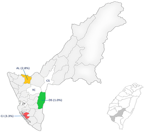
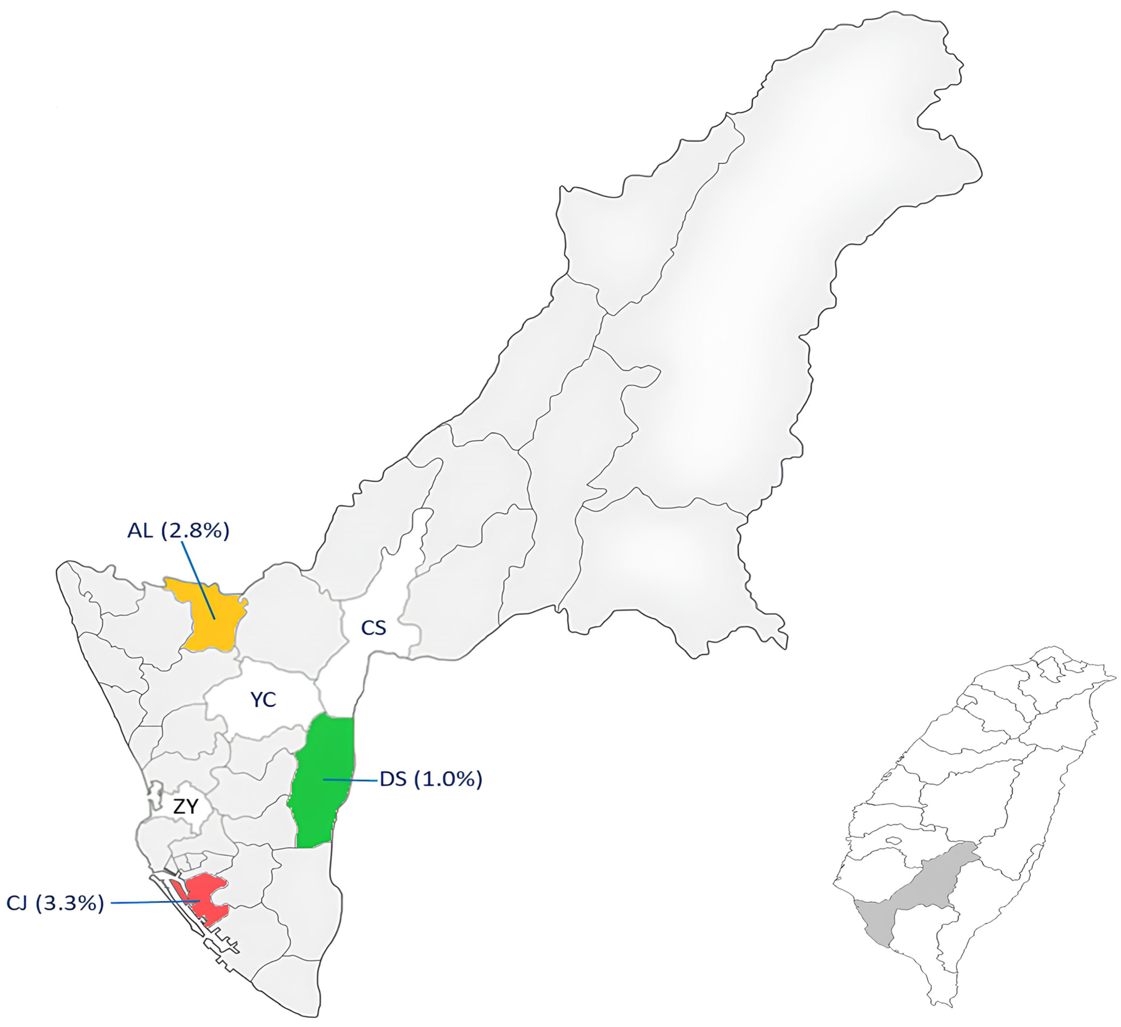
Tick specimens infesting stray dogs that appeared to be healthy were collected monthly for one year from six districts of Kaohsiung City, including Da-Shu (DS), Yan-Chao (YC), Zuo-Ying (ZY), Cian-Jhen (CJ), Ci-Shan (CS) and A-Lian (AL), located in Southern Taiwan (Figure 1). To avoid interference from blood, only flat (unengorged) ticks were selected for molecular screening of Anaplasma spp. infection. A total of 1512 ticks (504 females, 503 males and 505 nymphs) were collected, and the collected ticks were cleaned using sonication in distilled water and subsequently stored in 75% ethanol in glass vials. The tick specimens were identified to the species level according to the pictorial keys of Rhipicephalus, as described previously [34,35]. In general, a Nikon stereo-microscope (SMZ 1500, Tokyo, Japan) was used to observe the external structures of these ticks and to photograph the features for species identification, as described previously [18].
Figure 1.
Map of Taiwan showing the tick collection from six districts of Kaohsiung City and the percentage of Anaplasma platys infection detected in ticks from DS (Da-Shu), AL (A-Lian), CJ (Cian-Jhen), Zuo-Ying (ZY), Yan-Chao (YC) and Ci-Shan (CS) districts of Kaohsiung City, Taiwan.
2.2. DNA Extraction from Tick Specimens
Total genomic DNA was extracted from each collected tick. In general, tick specimens were cleaned by 3–5 min sonication with 75% ethanol solution and then washed twice in sterile distilled water. Thereafter, each tick specimen was homogenized with 180 μL lysing buffer solution by a TissueLyser II apparatus (catalogue no. 85300, Qiagen, Hilden, Germany). The resulting homogenate was centrifuged at room temperature, and the supernatant fluid was further processed by a commercial kit (catalogue no. 69506, DNeasy Blood & Tissue Kit, Qiagen, Taipei, Taiwan). The DNA-containing filtrated fluid was collected and quantitated with a spectrophotometer (Epoch, Biotek, Winooski, VA, USA). All the quantitated DNA specimens were preserved in a deep freezer (−80 °C) until further analysis, as described previously [17].
2.3. Molecular Detection of Anaplasma by Nested Polymerase Chain Reaction (nPCR)
The nPCR assays were conducted to screen DNA samples extracted from individual tick specimens by targeting the 16S rRNA gene of Anaplasma. The PCR amplification used two primer sets, including the primer set of ECC/ECB for the initial amplification and the primer set of EPLAT5/EPLAT3 targeting the species-specific DNA fragment of A. platys, which was used for the secondary amplification, producing a DNA product approximately 359 bp in length, as described previously [36,37]. The Taq polymerase and PCR reagents were commercially purchased and used as instructed (Takara Shuzo Co., Ltd., Tokyo, Japan). In general, each PCR reaction contains a 25 μL reaction mixture that includes 3 μL DNA template, 2 μL dNTP mixture (10 mM each), 2.5 μL 10× PCR buffer (Mg2+), 1.5 μL forward and reverse primers, 1 unit of Taq DNA polymerase, and the remaining volume was replenished with ddH2O, as described previously [38]. In parallel with each reaction, the reaction mixture was supplemented with adequate amounts of sterile distilled water and used as a negative control.
The entire PCR process was performed by a Veriti thermocycler (Applied Biosystems, Taipei, Taiwan) under the following conditions: initial denaturation at 94 °C for 3 min, followed by 35 cycles with the specific conditions of denaturation at 94 °C for 1 min, annealing at 55 °C for 2 min and extension at 72 °C for 2 min. For secondary PCR assay, the same initial denaturation was performed and followed by 35 cycles with the same denaturation and annealing conditions, except the extension condition (72 °C for 90 s), followed by a final extension step (72 °C for 2 min). All amplified PCR products were examined by electrophoresing on 1.5% agarose gels after staining with ethidium bromide and observed under ultraviolet light. A standard DNA marker (100 bp ladder, GeneRuler, Thermo Scientific, Taipei, Taiwan) was used to measure the molecular size of the amplified PCR products.
2.4. Gene Sequencing and Phylogenetic Analysis
For gene sequencing, each selected sample (approximately 10 μL) appearing with clear band on agarose gel was prepared and submitted for sequencing (Mission Biotech Co., Ltd., Taipei, Taiwan). Briefly, the same primer sets and conditions of nested-PCR were used for the sequencing reaction with an ABI Prism 377-96 DNA Sequencer (Applied Biosystems, Foster City, CA, USA) using the Big Dye Terminator Cycle Sequencing Kit, as described previously [37]. The resulting sequences were edited and aligned with software by BioEdit (V5.3) and CLUSTAL W (version X), respectively [39].
For phylogenetic analysis, the aligned 16S rRNA gene sequences from 6 Anaplasma spp. of Taiwan were compared by analyzing with other 17 strains of Anaplasma spp., Rickettsia spp. and E. coli verified from various geographical and biological origins documented in GenBank (Table 1). The phylogenetic analyses were performed with neighbor-joining (NJ) and maximum parsimony (MP) methods by estimating the entire alignment using MEGA X software package [40]. The genetic distance values were also analyzed by the Kimura two-parameter model to demonstrate the inter- and intra-species variations [41]. The phylogenetic trees were constructed and the reliability of the construction was evaluated with 1000 bootstrap replications, as described previously [42].
Table 1.
Phylogenetic analysis of Anaplasma strains of Taiwan with other bacterial strains documented in GenBank based on 16S rRNA gene.
2.5. Nucleotide Sequences Registration for Anaplasma Detected in Ticks of Southern Taiwan
The nucleotide sequences of 16S rRNA gene of 6 Anaplasma spp. detected in hard ticks of R. sanguineus s. l. from Southern Taiwan were submitted to GenBank and assigned the following accession numbers: PX285921, PX307904, PX307910, PX307909, PX307921 and PX285925, as indicated in Table 1. In addition, the 16S rRNA gene sequences from 17 strains (fourteen Anaplasma spp. and three outgroup spp.) were also included for comparison (Table 1).
2.6. Statistical Analysis
The detection rate of A. platys infection in different life stages of ticks and various districts of Kaohsiung City was analyzed by Chi-square test (SPSS 20 software).
3. Results
3.1. Molecular Detection of Anaplasma platys in Dog Ticks from Southern Taiwan
The nested PCR assay targeting the 16S rRNA gene was used to detect the existence of A. platys in R. sanguineus ticks. A. platys was detected in R. sanguineus s. l. ticks collected from three districts of Kaohsiung City (Table 2). The highest geographical prevalence of A. platys was detected in the Cian-Jhen district (3.3%), followed by A-Lian (2.8%) and Da-Shu (1.0%) (Table 2). Based on the life-cycle stage of ticks, the A. platys infection was detected in females, males and nymphs with infection rates of 1.4%, 1.0% and 0.8%, respectively (Table 2). The detection rate of A. platys infection was significantly higher in the Cian-Jhen district (Chi-square test, p < 0.05) (Table 2). However, the detection rate of A. platys infection in females is insignificantly higher than that in males or nymphs (Chi-square test, p > 0.05) (Table 2).
Table 2.
Molecular detection of Anaplasma platys in Rhipicephalus sanguineus s. l. ticks infesting dogs from various districts of Kaohsiung City in Southern Taiwan by nested-PCR assay.
3.2. Monthly Prevalence of A. platys Infection in Dog Ticks from Southern Taiwan
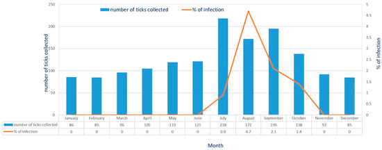
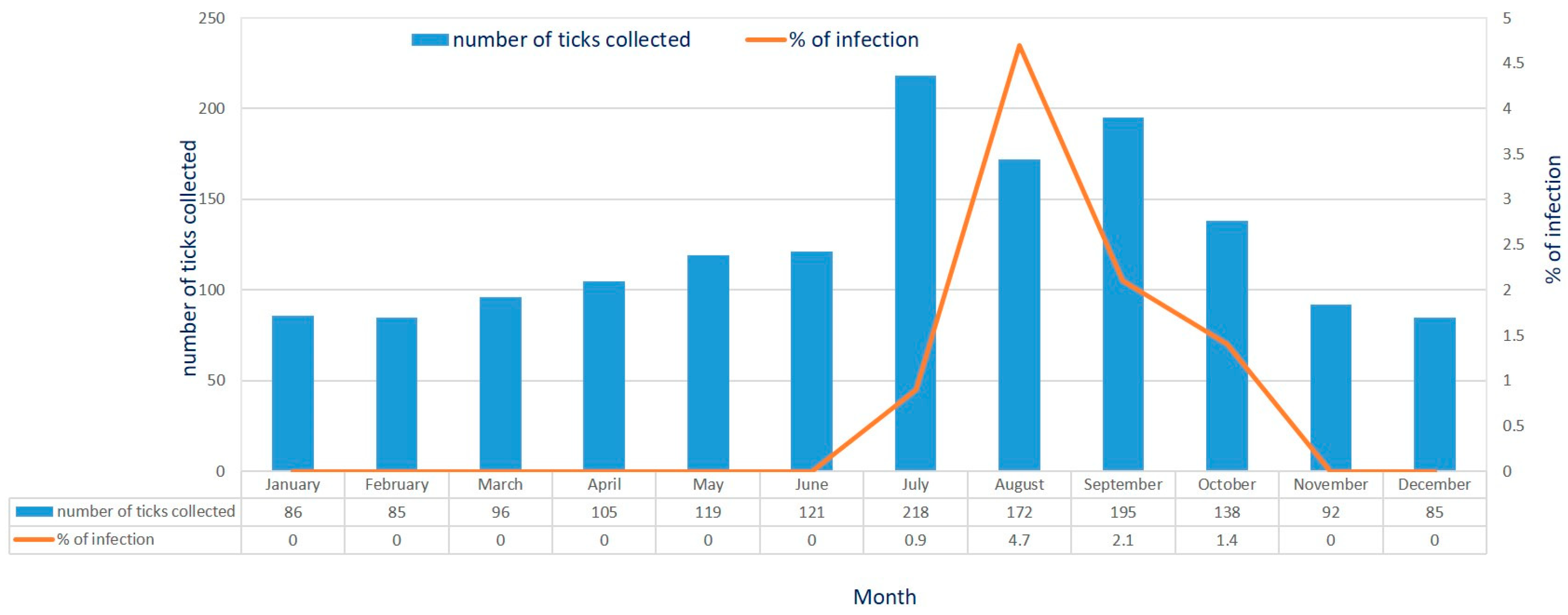
The detection rate of A. platys infection was measured from July to October and dramatically increased during the Summer (July–August) and Autumn (September–October) seasons. The highest detection rate was in August, with a detection rate of 4.7%, followed by detection rates of 2.1%, 1.4% and 0.9% in September, October and July, respectively (Figure 2).
Figure 2.
Monthly collection of Rhipicephalus sanguineus s. l. ticks from dogs and percentage of infection of Anaplasma platys in these ticks from Southern Taiwan.
3.3. Genetic Identity of Anaplasma spp. Detected in Dog Ticks of Southern Taiwan
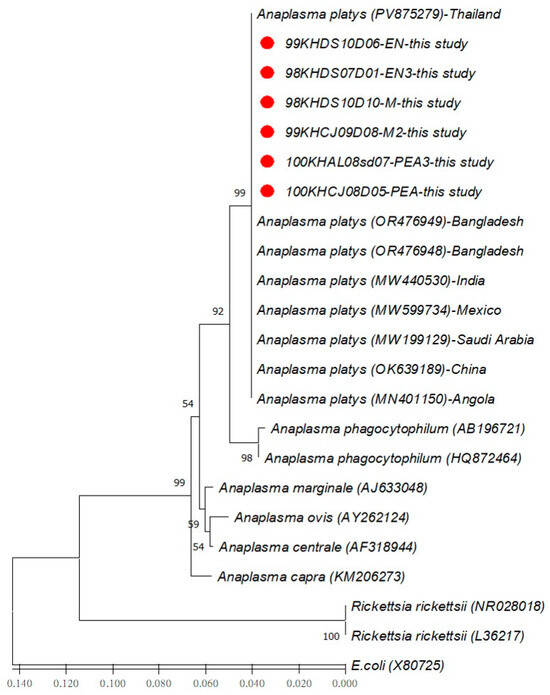
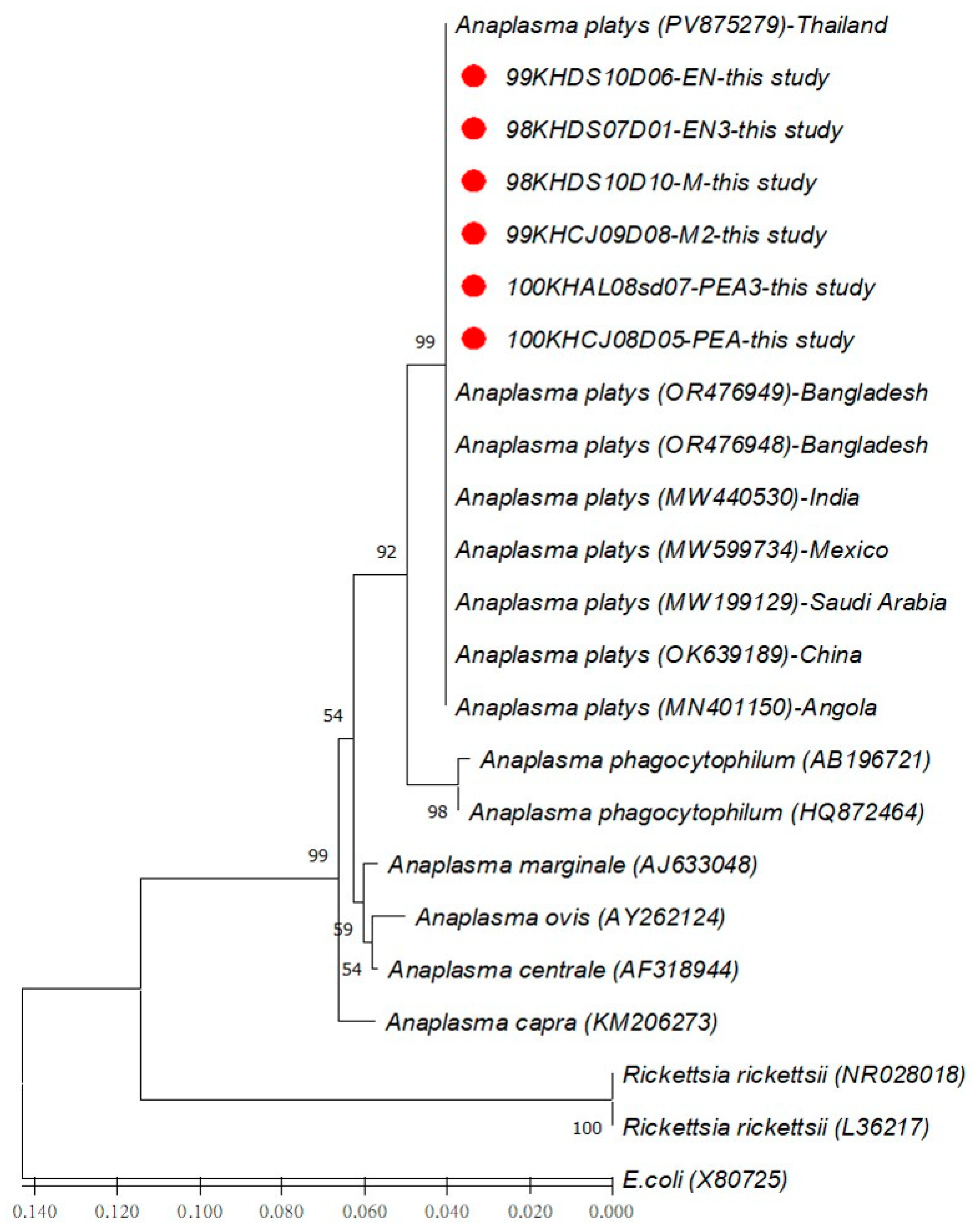
To identify the genetic identity of Anaplasma spp. detected in R. sanguineus s. l. ticks of Southern Taiwan, 16S rRNA gene sequences of six Anaplasma strains from Taiwan were compared with fourteen other Anaplasma spp. and three outgroup strains verified from various geographical and biological origins. Results indicated that the genetic relatedness of all Anaplasma spp. detected in R. sanguineus s. l. ticks of Taiwan was affiliated to the genospecies of A. platys detected in China (GenBank no: OK639189) and Thailand (GenBank no: PV875279) with a 100% sequence similarity (Table 3). Furthermore, inter- and intra-species analysis based on the genetic distance (GD) values of the 16S rRNA gene revealed no distance (GD = 0.00) within the Anaplasma strains of Taiwan, as compared with a high level of genetic divergence with the other species of Anaplasma (GD > 0.02) and outgroup species (GD > 0.16) (Table 3).
Table 3.
Intra- and Inter-species analysis of genetic distance values a based on the 16S rRNA gene sequences between Anaplasma strains detected in Rhipicephalus sanguineus s. l. ticks of Taiwan and other strains of Anaplasma, Rickettsia and E. coli documented in GenBank.
3.4. Phylogenetic Affiliation of Anaplasma spp. Detected in Dog Ticks of Southern Taiwan
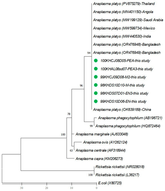
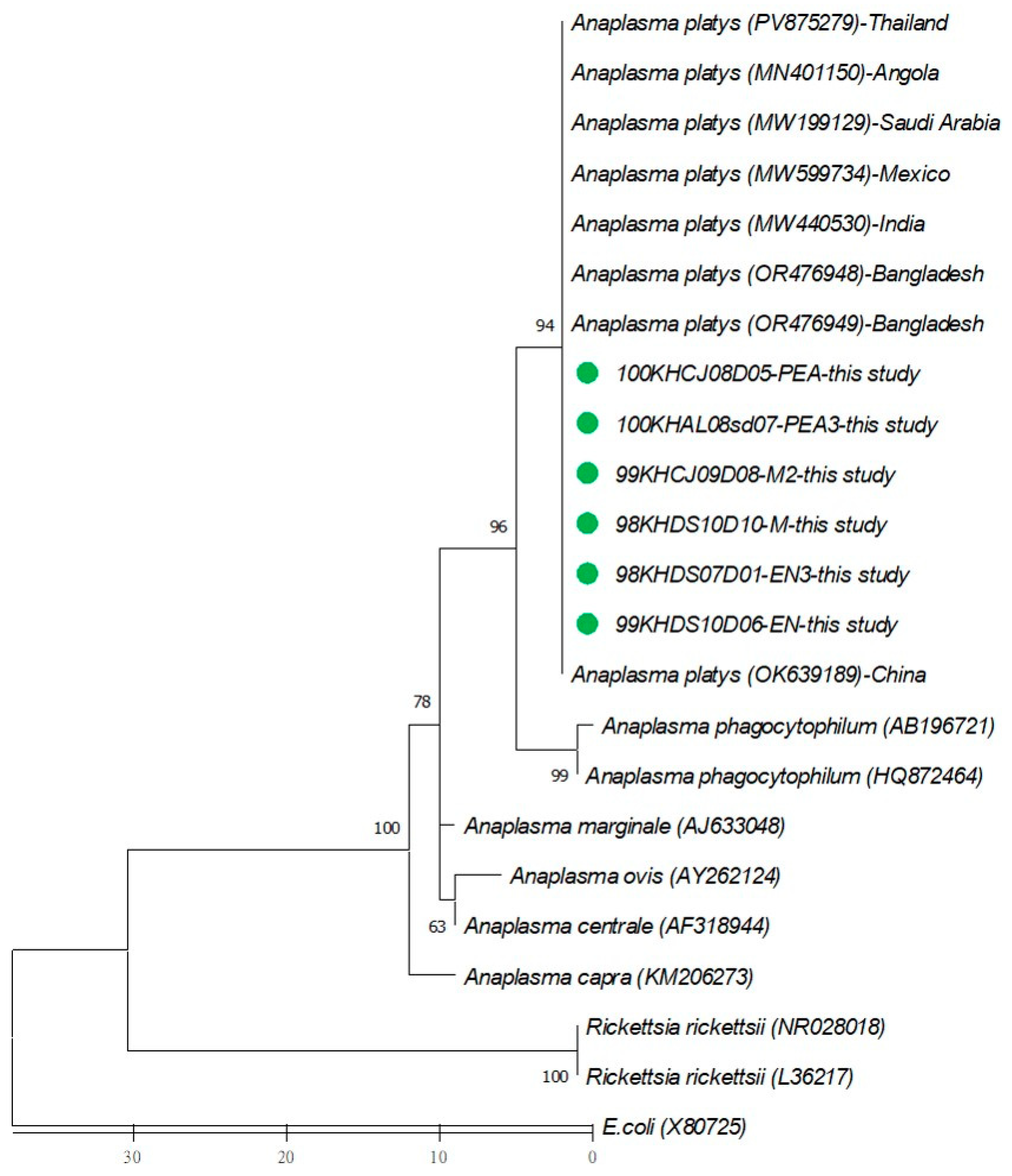
The phylogenetic relationships among 20 species of Anaplasma and three outgroup species were analyzed by constructing the phylogenic trees using neighbor-joining (NJ) and maximum parsimony (MP) methods. Results indicated five major clades of Anaplasma spp. displayed in both NJ (Figure 3) and MP (Figure 4) trees, which can be easily distinguished between different Anaplasma species and outgroup species. Briefly, a monophyletic clade constituted by all Anaplasma species detected in this study was genetically affiliated to the genospecies of A. platys identified from various countries. These results demonstrate a high genetic homology (100% sequence similarity) within the A. platys strains detected in R. sanguineus ticks of Southern Taiwan, but a higher genetic divergence (high sequence variation) distinguished them from other groups of Anaplasma species identified from different geographical and biological origins (Figure 3 and Figure 4).
Figure 3.
Phylogenetic tree constructed by neighbor-joining (NJ) method based on the 16S rRNA gene. The aligned sequences of six Anaplasma strains (indicated as ●) detected in Rhipicephalus sanguineus s. l. ticks from Kaohsiung City of Taiwan were analyzed with fourteen other Anaplasma strains and three outgroup strains verified from various geographical and biological origins. The tree was analyzed using 1000 bootstrap replicates, and the numbers at the nodes display the percentages of reliability. Branch length is drawn proportional to the estimated sequence divergence.
Figure 4.
Phylogenetic tree constructed by maximum parsimony (MP) method based on the 16S rRNA gene. The aligned sequences of six Anaplasma strains (indicated as ●) detected in Rhipicephalus sanguineus s. l. ticks from Kaohsiung City of Taiwan were analyzed with fourteen other Anaplasma strains and three outgroup strains verified from various geographical and biological origins. The tree was analyzed using 1000 bootstrap replicates, and the numbers at the nodes display the percentages of reliability. Branch length is drawn proportional to the estimated sequence divergence.
4. Discussion
Our results demonstrate the primary prevalence and genetic characterization of Anaplasma species detected in hard ticks (R. sanguineus s. l.) from Southern Taiwan. The genetic analysis reveals that these Anaplasma species are phylogenetically affiliated to the genospecies of A. platys, which was first identified in hard ticks (R. sanguineus s. l.) of Southern Taiwan. In previous reports, A. platys was first verified as an etiological agent for the cyclic thrombocytopenia of canine and is described to infect the host’s platelets [43,44]. This canine infection was reported throughout the world and was characterized by mild clinical symptoms in infected dogs [27,28,45,46,47]. However, canine infection with severe clinical symptoms has mainly been reported in Europe [48,49,50]. Although the R. sanguineus s. l. tick is incriminated as the major tick species responsible for the zoonotic transmission of A. platys among canines, this zoonotic pathogen (A. platys) has also been identified in various hard ticks, such as Ixodes persulcatus, Haemaphysalis longicornis and Dermacentor auratus [45,51]. Nevertheless, our study provides the first molecular evidence identifying A. platys in hard ticks (R. sanguineus s. l.) in Southern Taiwan.
The monthly variation in A. platys infection in hard ticks (R. sanguineus s. l.) of Southern Taiwan remains uncertain. Our results reveal the prevalence of A. platys infection in hard ticks (R. sanguineus s. l.) detected from July to October, and the highest detection rate was observed in August (Figure 1). During the hot months, this observation is coincident with the monthly abundance of the tick population described in our previous investigation [15]. In addition, it has also been reported that the R. sanguineus s. l. ticks exposed to high temperatures attach more rapidly to animals and humans [52,53]. Indeed, the warmer climate during the long summer season can induce the attaching movement of R. sanguineus s. l. ticks for approaching animal hosts in the natural environment [54]. Thus, the abundance of tick populations in various months was highly correlated with the monthly prevalence of A. platys detected in hard ticks (R. sanguineus s. l.) of Southern Taiwan.
Molecular identification of the tick-borne pathogens by analyzing the sequences of the specific target gene makes it possible to measure the genetic variation and determine the genetic identity among Anaplasma strains. Indeed, previous investigations on Anaplasma spp. have revealed the validity of the 16S rRNA gene for identifying A. platys in ticks and dogs [13,27,28,49,55,56]. In addition, genetic comparison of the 16S rRNA gene sequences between the A. platys strains from dogs in different areas constitutes a separate cluster on the phylogenetic tree with a high homology [46,47,49]. Thus, molecular characterization according to the 16S rRNA gene of Anaplasma spp. provides a reliable tool for discriminating the genetic variation and determining the genetic identity of Anaplasma species within R. sanguineus ticks.
The phylogenetic relationship among Anaplasma spp. in hard ticks (R. sanguineus s. l.) can be distinguished by analyzing the sequence similarity based on the 16S rRNA genes of Anaplasma. In our investigation, genetic identification based on the phylogenetic analysis of 16S rRNA genes of Anaplasma indicated that all Anaplasma spp. detected in hard ticks (R. sanguineus s. l.) from Southern Taiwan display an obvious affiliation with the genospecies of A. platys (Figure 3 and Figure 4). These A. platys strains detected in hard ticks (R. sanguineus s. l.) of Southern Taiwan are highly associated with the A. platys strain verified from a dog from Thailand (GenBank no. PV875279) and an R. sanguineus tick from China (GenBank no. OK639189), evinced by 100% sequence identity (Table 3). This genetic discrimination is strongly supported by the phylogenic trees constructed by either NJ or MP analysis (Figure 3 and Figure 4). Thus, our results reveal the genetic identity of Anaplasma spp. detected in hard ticks (R. sanguineus s. l.) of Southern Taiwan as a monophyletic group affiliated with A. platys, a zoonotic pathogen for canine anaplasmosis.
The prevalence of A. platys infection in collected ticks may correlate with environmental and biological factors. In our results, the prevalence of A. platys infection was observed significantly higher in the Cian-Jhen (CJ) district (Table 2). Indeed, there are large municipal parks in this district, and these parks gather a high population of stray dogs that may contribute to the high abundance of ticks and high prevalence of A. platys infection in collected ticks. In addition, the prevalence of A. platys infection had been detected in 19.6% of R. sanguineus s. l. ticks and 15.4% of accompanying dog blood collected from Nantou County in central Taiwan [57]. These results are significantly higher than our detection in R. sanguineus s. l. ticks (0.8–1.4%) from Kaohsiung City of Southern Taiwan (Table 2). The variation in A. platys infection in R. sanguineus s. l. ticks between Nantou County and Kaohsiung City may be due to the geographical variation between the mountainous area of Nantou County and urbanized Kaohsiung City. Indeed, the mountainous area of Nantou County provides higher feeding activity for R. sanguineus s. l. ticks on canine hosts. Further investigation focused on the biting frequency of R. sanguineus s. l. ticks on canine hosts may help to illustrate this puzzle.
The actual mechanisms for the maintenance of A. platys in R. sanguineus ticks remain controversial. Results in this study reveal the detection of A. platys in nymph and male ticks of R. sanguineus, and the higher prevalence of A. platys in male ticks may be inherited from infected nymphs. Indeed, the previous study has reported that the transstadial transmission of A. platys infection was observed from infected nymphs to the adult stage of R. sanguineus ticks [11]. In addition, the transovarial transmission of A. platys infection through several generations was also observed in a laboratory colony of R. sanguineus ticks [58]. Furthermore, the co-feeding mechanism may also account for another mechanism responsible for the transmission of A. platys infection from an infected tick to a new tick by feeding closely on the same host [59]. Thus, the possibility of biological transmission of A. platys within R. sanguineus ticks needs to be further identified.
5. Conclusions
Our results reveal the initial detection of A. platys in hard ticks (R. sanguineus s. l.) infesting dogs in Southern Taiwan. According to the phylogenetic analyses, the genetic identity of A. platys was first identified in R. sanguineus s. l. ticks of Southern Taiwan. Human infections with A. platys had been reported in patients exposed to R. sanguineus s. l. ticks [31] and accompanied dog blood [32]. Further study focused on the prevalence of A. platys infection in accompanied canine hosts will benefit the evaluation of the transmission risk of A. platys infection among canines and illustrate the potential threat for human infection in Southern Taiwan.
Author Contributions
Conceptualization: L.-L.C. and C.-M.S.; Methodology: L.-L.C. and C.-M.S.; Analysis: L.-L.C.; Investigation: L.-L.C.; Supervision: C.-M.S.; Funding Acquisition: C.-M.S.; Writing: C.-M.S. All authors have read and agreed to the published version of the manuscript.
Funding
This work was supported in part by grants from the Kaohsiung Medical University (NPUST-KMU-115-P002) and National Science and Technology Council (NSTC 114-2314-B-037-031), Taiwan, Republic of China.
Institutional Review Board Statement
The collection of ticks from dogs was assisted by veterinary practitioners and approved by the Institutional Animal Care and Use Committee (IACUC) of Kaohsiung Medical University (IACUC-106142), approval date 4 April 2023.
Data Availability Statement
The raw data supporting the conclusions of this article will be made available by the authors on request.
Acknowledgments
We would like to appreciate the generous help with tick collection by veterinary practitioners and pet clinics of Kaohsiung City, Taiwan.
Conflicts of Interest
The authors declare that there are no conflicts of interest in this manuscript.
References
- Dantas-Torres, F. Biology and ecology of the brown dog tick, Rhipicephalus sanguineus. Parasit. Vectors 2010, 3, 26. [Google Scholar] [CrossRef] [Scilit] [PubMed]
- Walker, J.B.; Keirans, J.E.; Horak, I.H. The Genus Rhipicephalus (Acari, Ixodidae): A Guide to the Brown Ticks of the World; Cambridge University Press: Cambridge, UK, 2000. [Google Scholar]
- Guglielmone, A.A.; Robbins, R.G.; Apanaskevich, D.A.; Petney, T.N.; Estrada-Peria, A.; Horak, I. The Hard Ticks of the World; Springer: Dordrecht, The Netherlands, 2014. [Google Scholar]
- Szabo, M.P.; Mangold, A.J.; Joao, C.F.; Bechara, G.H.; Guglielmone, A.A. Biological and DNA evidence of two dissimilar populations of the Rhipicephalus sanguineus tick group (Acari: Ixodidae) in South America. Vet. Parasitol. 2005, 130, 131–140. [Google Scholar] [CrossRef] [Scilit] [PubMed]
- Moraes-Filho, J.; Marcili, A.; Nieri-Bastos, F.A.; Richtzenhain, L.J.; Labruna, M.B. Genetic analysis of ticks belonging to the Rhipicephalus sanguineus group in Latin America. Acta Trop. 2011, 117, 51–55. [Google Scholar] [CrossRef] [Scilit] [PubMed]
- Zemtsova, G.E.; Apanaskevich, D.A.; Reeves, W.K.; Hahn, M.; Snellgrove, A.; Levin, M.L. Phylogeographical of Rhipicephalus sanguineus sensu lato and its relationships with climatic factors. Exp. Appl. Acarol. 2016, 69, 191–203. [Google Scholar] [CrossRef] [Scilit] [PubMed]
- Chao, L.L.; Shih, C.M. Molecular analysis of Rhipicephalus sanguineus (Acari: Ixodidae), an incriminated vector tick for Babesia vogeli in Taiwan. Exp. Appl. Acarol. 2016, 70, 469–481. [Google Scholar] [CrossRef] [Scilit] [PubMed]
- Slapeta, J.; Chandra, S.; Halliday, B. The “tropical lineage” of the brown dog tick Rhipicephalus sanguineus sensu lato identified as Rhipicephalus linnaei (Audouin, 1826). Int. J. Parasitol. 2021, 51, 431–436. [Google Scholar] [PubMed]
- Otranto, D.; Dantas-Torres, F.; Breitschwerdt, E.B. Managing canine vector-borne diseases of zoonotic concern: Part one. Trends Parasitol. 2009, 25, 157–163. [Google Scholar] [CrossRef] [Scilit] [PubMed]
- Dantas-Torres, F.; Otranto, D. Further thoughts on the taxonomy and vector role of Rhipicephalus sanguineus group ticks. Vet. Parasitol. 2015, 208, 9–13. [Google Scholar] [CrossRef] [Scilit] [PubMed]
- Aktas, M.; Ozubek, S. Molecular evidence for transstadial transmission of Anaplasma platys by Rhipicephalus sanguineus sensu lato under field condition. Med. Vet. Entomol. 2018, 32, 78–83. [Google Scholar] [PubMed]
- Ipek, N.D.S.; Ozubek, S.; Aktas, M. Molecular evidence for transstadial transmission of Ehrlichia canis by Rhipicephalus sanguineus sensu lato under field conditions. J. Med. Entomol. 2018, 55, 440–444. [Google Scholar] [PubMed]
- Laatamna, A.; Strube, C.; Bakkes, D.K.; Schaper, S.; Aziza, F.Z.; Ben Chelef, H.; Amrane, N.E.H.; Bedraoui, R.; Dobler, G.; Chitimia-Dobler, L. Molecular detection of tick-borne pathogens in Rhipicephalus sanguineus sensu stricto collected from dogs in the steppe and high plateau regions of Algeria. Acta Trop. 2022, 234, 106582. [Google Scholar] [CrossRef] [Scilit] [PubMed]
- Mohanta, U.K.; Marguerite, M.P.; Ji, S.; Ma, Z.; Li, H.; El-Sayed, S.A.E.; Amer, M.M.; Chikufenji, B.; Do, T.T.; Ceylan, O.; et al. Molecular survey of canine tick-borne pathogens in ticks and stray dogs in Dhaka city, Bangladesh. Parasitol. Int. 2024, 100, 102860. [Google Scholar] [CrossRef] [Scilit] [PubMed]
- Chao, L.L.; Hsieh, C.K.; Ho, T.Y.; Shih, C.M. First zootiological survey of hard ticks (Acari: Ixodidae) infesting dogs in northern Taiwan. Exp. Appl. Acarol. 2019, 77, 105–115. [Google Scholar] [PubMed]
- Shih, C.M.; Chao, L.L. Molecular Detection and Genetic Identification of Rickettsia Infection in Rhipicephalus sanguineus (Acari: Ixodidae) ticks collected from Southern Taiwan. Exp. Appl. Acarol. 2021, 85, 291–304. [Google Scholar] [CrossRef] [Scilit] [PubMed]
- Chao, L.L.; Esmeralda, E.; Robinson, M.; Liang, Y.F.; Shih, C.M. First detection and molecular identification of a pathogenic spotted fever group Rickettsia, R. massiliae, from Rhipicephalus haemaphysaloides ticks infesting dogs in southern Taiwan. Acta Trop. 2022, 236, 106666. [Google Scholar] [CrossRef] [Scilit] [PubMed]
- Chao, L.L.; Liao, H.T.; Ho, T.Y.; Shih, C.M. First detection and molecular identification of Babesia gibsoni from Rhipicephalus sanguineus ticks. Acta Trop. 2017, 166, 356–362. [Google Scholar] [CrossRef] [Scilit] [PubMed]
- Shih, C.M.; Ko, P.Y.; Chao, L.L. Molecular Survey and Genetic Analysis of Ehrlichia canis in Rhipicephalus sanguineus Ticks Infesting Dogs in Northern Taiwan. Microorganisms 2025, 13, 1372. [Google Scholar] [CrossRef] [Scilit] [PubMed]
- Rikihisa, Y. New findings on members of the family Anaplasmataceae of veterinary importance. Ann. N. Y. Acad. Sci. 2006, 1078, 438–445. [Google Scholar] [CrossRef] [Scilit] [PubMed]
- Rar, V.; Golovljova, I. Anaplasma, Ehrlichia and “Candidatus Neoehrlichia” bacteria: Pathogenicity, biodiversity, and molecular genetic characteristics, a review. Infect. Genet. Evol. 2011, 11, 1842–1861. [Google Scholar] [CrossRef] [Scilit] [PubMed]
- Potgieter, F.; van Rensburg, L. Tick transmission of Anaplasma centrale. Onderstepoort J. Vet. Res. 1987, 54, 5–7. [Google Scholar] [PubMed]
- Stich, R.W.; Kocan, K.M.; Palmer, G.H.; Ewing, S.A.; Hair, J.A.; Barron, S.J. Transstadial and attempted transovarial transmission of Anaplasma marginale by Dermacentor variabilis. Am. J. Vet. Res. 1989, 50, 1377–1380. [Google Scholar] [CrossRef] [Scilit] [PubMed]
- Telford, S.R., III; Dawson, J.E.; Katavolos, P.; Warner, C.K.; Kolbert, C.P.; Persing, D.H. Perpetuation of the agent of human granulocytic ehrlichiosis in a deer tick-rodent cycle. Proc. Natl. Acad. Sci. USA 1996, 11, 6209–6214. [Google Scholar] [CrossRef] [Scilit]
- Bakken, J.S.; Dumler, J.S. Human granulocytic ehrlichiosis. Clin. Infect. Dis. 2000, 31, 554–560. [Google Scholar] [CrossRef] [Scilit] [PubMed]
- Burkot, T.R.; Maupin, G.O.; Schneider, B.S.; Denatale, C.; Happ, C.M.; Rutherford, J.S.; Zeidner, N.S. Use of s sentinel host system to study the questing behavior of Ixodes spinipalpis and its role in the transmission of Borrelia bissettii, human granulocytic ehrlichisis and Babesia microti. Am. J. Trop. Med. Hyg. 2001, 65, 293–299. [Google Scholar] [CrossRef] [Scilit] [PubMed]
- Ybanez, A.P.; Ybanez, R.H.D.; Yokoyama, N.; Inokuma, H. Multiple infections of Anaplasma platys variants in Philippine dogs. Vet. World 2016, 9, 1456–1460. [Google Scholar] [CrossRef] [Scilit] [PubMed]
- Pesapane, R.; Foley, J.; Thomas, R.; Castro, L.R. Molecular detection and characterization of Anaplasma platys and Ehrlichia canis in dogs from northern Colombia. Vet. Microbiol. 2019, 233, 184–189. [Google Scholar] [CrossRef] [Scilit] [PubMed]
- Sarker, B.R.; Mitpasa, T.; Macotpet, A.; Bupata, P.A. First report on molecular prevalence and identification of Anaplasma platys in dogs in Khon Kaen, Thailand. Vet. World 2021, 14, 2613–2619. [Google Scholar] [CrossRef] [Scilit] [PubMed]
- Arif, E.D.; Khan, K.M.H.; Ai-Sada, I.H.A.; Al-Barzinji, Y.M.S. Molecular detection of Anaplasma platys in Pshdar Kurdish shepherd dogs of sulaymaniyah province, Iraq. Vet. World 2024, 17, 2797–2801. [Google Scholar] [CrossRef] [Scilit] [PubMed]
- Arraga-Alvarado, C.M.; Qurollo, B.A.; Parra, O.C.; Berrueta, M.A.; Hegarty, B.C.; Breitschwerdt, E.B. Case report: Evidence of Anaplasma platys infection in two women from Venezuela. Am. J. Trop. Med. Hyg. 2014, 91, 1161–1165. [Google Scholar] [CrossRef] [Scilit] [PubMed]
- Breitschwerdt, E.B.; Hegarty, B.C.; Qurollo, B.A.; Saito, T.B.; Maggi, R.G.; Blanton, L.S.; Bouyer, D.H. Intravascular persistence of Anaplasma platys, Ehrlichia chaffeensis, and Ehrlichia ewingii DNA in the blood of a dog and two family members. Parasit. Vectors 2014, 7, 298. [Google Scholar] [CrossRef] [Scilit] [PubMed]
- Yuasa, Y.; Hsu, T.H.; Chou, C.C.; Huang, C.C.; Huang, W.C.; Chang, C.C. The comparison of spatial variation and risk factors between mosquito-borne and tick-borne diseases: Seroepidemiology of Ehrlichia canis, Anaplasma species, and Dirofilaria immitis in dogs. Comp. Immunol. Microbiol. Infect. Dis. 2012, 35, 599–606. [Google Scholar] [CrossRef] [Scilit] [PubMed]
- Teng, K.F.; Jiang, Z.J. Economic Insect Fauna of China Fasc 39 Acari: Ixodidae; Science Press: Beijing, China, 1991. (In Chinese) [Google Scholar]
- Shih, C.M.; Chao, L.L. A Catalog of Ixodidae Ticks of Taiwan; Department of Parasitology and Tropical Medicine, National Defense Medical Center: Taipei, Taiwan, 2011. [Google Scholar]
- Mathew, J.S.; Ewing, S.A.; Murphy, G.L.; Kocan, K.C.; Cortvet, R.E.; Fox, J.C. Characterization of a new isolate of Ehrlichia platys (order Rickettsiales) using electron microscopy and polymerase chain reaction. Vet. Parasitol. 1997, 68, 1–10. [Google Scholar] [CrossRef] [Scilit] [PubMed]
- Murphy, G.L.; Ewing, S.A.; Whitwirth, L.C.; Fox, J.C.; Kocan, A.A. A molecular and serological survey of Ehrlichia canis, E. chafeensis, and E. ewingi in dogs and ticks from Oklahoma. Vet. Parasitol. 1998, 79, 325–339. [Google Scholar] [CrossRef] [Scilit] [PubMed]
- Chao, L.L.; Robinson, M.; Liang, Y.F.; Shih, C.M. First detection and molecular identification of Rickettsia massiliae, a human pathogen, in Rhipicephalus sanguineus ticks collected from Southern Taiwan. PLoS Negl. Trop. Dis. 2022, 16, e0010917. [Google Scholar] [CrossRef] [Scilit] [PubMed]
- Thompson, J.D.; Higgins, D.G.; Gibson, T.J. CLUSTAL W: Improving the sensitivity of progressive multiple sequence alignment through sequence weighting, position-specific gap penalties and weight matrix choice. Nuc. Acids Res. 1994, 22, 4673–4680. [Google Scholar] [CrossRef] [Scilit]
- Kumar, S.; Stecher, G.; Li, M.; Knyaz, C.; Tamura, K. MEGA X: Molecular evolutionary genetics analysis across computing platforms. Mol. Biol. Evol. 2018, 35, 1547–1549. [Google Scholar] [CrossRef] [Scilit] [PubMed]
- Kimura, M. A simple method for estimating evolutionary rate of base substitutions through comparative studies of nucleotide sequences. J. Mol. Evol. 1980, 16, 111–120. [Google Scholar] [CrossRef] [Scilit] [PubMed]
- Felsenstein, J. Confidence limits on phylogenies: An approach using the bootstrap. Evolution 1985, 52, 1119–1134. [Google Scholar]
- Harvey, J.W.; Simpson, C.P.; Gaskin, J. Cyclic thrombocytopenia induced by a Rickettsia-like agent in dogs. J. Infect. Dis. 1978, 137, 182–188. [Google Scholar] [CrossRef] [Scilit] [PubMed]
- Dumler, J.S.; Barbet, A.F.; Bekker, C.P.; Dasch, G.A.; Palmer, G.H.; Ray, S.C.; Rikihisa, Y.; Rurangirwa, F.R. Reorganization of genera in the families Rickettsiaceae and Anaplasmaceae in the order Rickettsiales. Unification of some species of Ehrlichia with Anaplasma, Cowdria with Ehrlichia and Ehrlichia with Neorickettsia, descriptions of six new species combinations and designation of Ehrlichia equi and HGE agent as subjective synonyms of Ehrlichia phagocytophila. Int. J. Syst. Evol. Microbiol. 2001, 51, 2145–2165. [Google Scholar] [CrossRef] [Scilit] [PubMed]
- Kim, C.M.; Yi, Y.H.; Yu, D.H.; Lee, M.J.; Cho, M.R.; Desai, A.R.; Shringi, S.; Klein, T.A.; Kim, H.C.; Song, J.W.; et al. Tick-borne rickettsial pathogens in ticks and small mammals in Korea. Appl. Environ. Microbiol. 2006, 72, 5766–5776. [Google Scholar] [CrossRef] [Scilit] [PubMed]
- Pinyoowong, D.; Jittapalapong, S.; Sukasawat, F.; Stich, R.W.; Thamchaipenet, A. Molecular characterization of Thai Ehrlichia canis and Anaplasma platys strains detected in dogs. Infect. Genet. Evol. 2008, 8, 433–438. [Google Scholar] [CrossRef] [Scilit] [PubMed]
- Almazán, C.; González-Alvarez, V.H.; Fernández de Mera, I.G.; Cabezas-Cruz, A.; Rodriguez-Martinez, R.; de la Fuente, J. Molecular identification and characterization of Anaplasma platys and Ehrlichia canis in dogs in Mexico. Ticks Tick. Borne Dis. 2016, 7, 276–283. [Google Scholar] [CrossRef] [Scilit] [PubMed]
- Beaufils, J.P.; Inokuma, H.; Martin-Granel, J.; Jumelle, P.; Barbault-Jumelle, M.; Brouqui, P. Anaplasma platys (Ehrlichia platys) infection in a dog in France: Description of the case and characterization of the agent. Rev. Med. Vet. 2002, 153, 85–90. [Google Scholar]
- Aguirre, E.; Tesouro, M.A.; Ruiz, L.; Amusategui, I.; Sainz, A. Genetic characterization of Anaplasma (Ehrlichia) platys in dogs in Spain. J. Vet. Med. Ser. B Infect. Dis. Vet. Public Health 2006, 53, 197–200. [Google Scholar] [CrossRef] [Scilit]
- Cardoso, L.; Tuna, J.; Vieira, L.; Yisaschar-Mekuzas, Y.; Baneth, G. Molecular detection of Anaplasma platys and Ehrlichia canis in dogs from the North of Portugal. Vet. J. 2010, 183, 232–233. [Google Scholar] [CrossRef] [Scilit] [PubMed]
- Parola, P.; Cornet, J.P.; Sanogo, Y.O.; Miller, R.S.; Thien, H.V.; Conzalez, J.P.; Raoult, D.; Telford, S.R., III; Wongsrichanalai, C. Detection of Ehrlichia spp., Anaplasma spp., Rickettsia spp. and other eubacteria in ticks from the Thai-Myanmar border and Vietnam. J. Clin. Microbiol. 2003, 41, 1600–1608. [Google Scholar] [CrossRef] [Scilit] [PubMed]
- Parola, P.; Socolovschi, C.; Jeanjean, L.; Bitam, I.; Fournier, P.E.; Sotto, A.; Labauge, P.; Raoult, D. Warmer weather linked to tick attack and emergence of severe rickettsioses. PLoS Negl. Trop. Dis. 2008, 2, e338. [Google Scholar] [CrossRef] [Scilit] [PubMed]
- Socolovschi, C.; Raoult, D.; Parola, P. Influence of temperature on the attachment of Rhipicephalus sanguineus ticks on rabbits. Clin. Microbiol. Infect. 2009, 15, 326–327. [Google Scholar] [CrossRef] [Scilit] [PubMed]
- Gray, J.S.; Dautel, H.; Estrade-Peria, A.; Kahl, O.; Lindgren, E. Effects of climate change on ticks and tick-borne disease in Europe. Interdiscip. Perspect. Infect. Dis. 2009, 2009, 598232. [Google Scholar] [CrossRef] [Scilit]
- Inokuma, H.; Raoult, D.; Brouqui, P. Detection of Ehrlichia platys DNA in brown dog ticks (Rhipicephalus sanguineus) in Okinawa Island, Japan. J. Clin. Microbiol. 2000, 38, 4219–4221. [Google Scholar] [CrossRef] [Scilit] [PubMed]
- Arraga-Alvarado, C.M.; Palmar, M.; Parra, O.; Salas, P. Ehrlichia platys (Anaplasma platys) in dogs from Maracaibo, Venezuela: An ultrastructural study of experimental and natural infections. Vet. Path. 2003, 40, 149–156. [Google Scholar] [CrossRef] [Scilit]
- Yuasa, Y.; Tsai, Y.L.; Chang, C.C.; Hsu, T.H.; Chou, C.C. The prevalence of Anaplasma platys and a potential novel Anaplasma species exceed that of Ehrlichia canis in asymptomatic dogs and Rhipicephalus sanguineus in Taiwan. J. Vet. Med. Sci. 2017, 79, 1494–1502. [Google Scholar] [CrossRef] [Scilit] [PubMed]
- Snellgrove, A.N.; Krapiunaya, I.; Ford, S.L.; Stanley, H.M.; Wickson, A.G.; Hartzer, K.L.; Levin, M.L. Vector competence of Rhipicephalus sanguineus sensu lato for Anaplasma platys. Ticks Tick Borne Dis. 2020, 11, 101517. [Google Scholar] [CrossRef] [Scilit] [PubMed]
- Otranto, D.; Dantas-Torres, F.; Giannelli, A.; Latrofa, M.S.; Cascio, A.; Cazzin, S.; Ravagnan, S.; Montarsi, F.; Zanzani, S.A.; Manredi, M.T.; et al. Ticks infesting humans in Italy and associated pathogens. Parasit. Vectors 2014, 7, 328. [Google Scholar] [CrossRef] [Scilit] [PubMed]

Disclaimer/Publisher’s Note: The statements, opinions and data contained in all publications are solely those of the individual author(s) and contributor(s) and not of MDPI and/or the editor(s). MDPI and/or the editor(s) disclaim responsibility for any injury to people or property resulting from any ideas, methods, instructions or products referred to in the content. |
© 2026 by the authors. Licensee MDPI, Basel, Switzerland. This article is an open access article distributed under the terms and conditions of the Creative Commons Attribution (CC BY) license.