


Simple Summary
Specific pathogen-free (SPF) chickens are essential for the reliability and reproducibility of scientific research data. Among the pathogens that must be excluded, five respiratory bacterial species are of particular importance. Currently, the lack of a unified testing method—with existing assays covering anywhere from two to four targets—makes the process time-consuming and cost-inefficient. In this study, we developed a new method capable of simultaneously detecting all five bacteria within a single reaction system. The assay functions by detecting specific target genes unique to each bacterium. Our results demonstrated that this method exhibits high sensitivity, specificity, and repeatability. It can accurately identify the target bacteria and shows no cross-reactivity with other common bacterial species. When we tested samples from SPF chickens, none of the five bacteria were detected, as expected. This method provides a faster, simpler, and more efficient tool for routine monitoring of SPF chickens, thereby contributing to improved quality control and more reliable research data.
Abstract
A multiplex liquid array assay based on Luminex xTAG technology was developed for the first time for the simultaneous detection of five respiratory bacterial pathogens in Specific Pathogen-Free (SPF) chickens—Mycoplasma gallisepticum (MG), Mycoplasma synoviae (MS), Pasteurella multocida (Pm), Avibacterium paragallinarum (Apg), and Mycobacterium avium (Mav)—filling the gap that these five pathogens could not be simultaneously detected previously. Specific primers were designed targeting conserved genes of each pathogen, with a TAG sequence attached to the 5′ end of each forward primer and biotin to the 5′ end of each reverse primer. After multiplex PCR amplification, the products were hybridized with MagPlex-TAG microspheres and analyzed using the Luminex system. The method was validated for sensitivity, specificity, and repeatability, and tested with simulated positive samples (a mixture of genomic DNA from the five target pathogens) and SPF chicken samples. The LOD was 10 copies/μL for Pm and 100 copies/μL for the other four pathogens. No cross-reactivity was observed with 18 non-target bacterial species. Intra- and Inter-assay coefficients of variation (CV) ranged from 0.8% to 12.2% and 2.3% to 14.4%, respectively, both below 15%. All simulated mixed positive samples yielded results consistent with expectations, and none of the 114 nasopharyngeal swab samples from SPF chickens tested positive for any of the five targets. The developed assay offers high throughput, high sensitivity, high specificity, and good repeatability, providing a robust tool for routine quality control in SPF chickens.
1. Introduction
Laboratory animals are indispensable resources in life science research, and their microbiological quality control directly affects the accuracy and reproducibility of scientific data. Specific pathogen-free (SPF) chickens serve as important animal models in various fields of life science research. According to the Chinese national standard GB/T 17999.1-2008 [1] and the Chinese Pharmacopoeia (2025 Edition, Volume IV) [2], SPF chickens must be verified as free from five respiratory bacterial pathogens: Mycoplasma gallisepticum (MG), Mycoplasma synoviae (MS), Pasteurella multocida (Pm), Avibacterium paragallinarum (Apg), and Mycobacterium avium (Mav).
These five pathogens pose significant threats to poultry health and are of particular concern in SPF chickens. MG causes chronic respiratory disease, reduced egg production, and poor feed conversion [3]; MS causes infectious synovitis (joint swelling and lameness) and respiratory disease [4]; Pm causes fowl cholera, which can lead to acute septicemia and high mortality [5]; Apg causes infectious coryza, characterized by nasal discharge, facial swelling, and decreased egg production [6]; and Mav causes avian tuberculosis, a chronic wasting disease leading to emaciation and reduced productivity [7]. Importantly, MG and MS are vertically transmitted through eggs, making their exclusion critical for SPF chickens [8]. Furthermore, the presence of any of these pathogens can interfere with experimental outcomes by inducing immune responses, altering baseline physiological parameters, or confounding challenge studies, thereby compromising data reproducibility and reliability [9].
Traditionally, the detection of these five pathogens has relied on culture isolation and biochemical identification. However, these pathogens have diverse growth requirements and are generally slow-growing, making conventional methods time-consuming (often taking several weeks) and operationally cumbersome, which cannot meet the timeliness requirements for routine health monitoring of SPF chickens.
With the development of molecular biology techniques, PCR-based detection methods have gradually become important tools for pathogen detection due to their rapidity and high sensitivity, and their application formats have evolved from single-target PCR to multiplex PCR and then to real-time fluorescence quantitative PCR (qPCR). Single-target PCR can achieve species-specific detection but requires separate reactions for each pathogen, resulting in low efficiency; conventional multiplex PCR can simultaneously detect multiple targets in a single reaction but is limited by primer compatibility issues and gel-based endpoint detection; qPCR significantly improves sensitivity and quantification capability, yet its multiplexing capacity is constrained by the number of available fluorescence channels, typically allowing only 4–6 targets per reaction. For SPF chicken respiratory pathogens, researchers have developed various molecular detection methods, including single-plex or multiplex qPCR [10,11,12] and conventional multiplex PCR [13,14]; commercial kits are also available. However, these methods still fall short of meeting the requirement for simultaneous detection of all five respiratory bacterial pathogens (MG, MS, Pm, Apg, and Mav) in a single reaction, and remain inadequate in terms of detection throughput, cost-effectiveness, and target coverage completeness.
Luminex xTAG liquid array technology, based on fluorescently coded microspheres, offers a theoretical multiplexing capacity of up to 500 targets, conferring a distinct advantage in multiplex pathogen detection. This platform has been successfully applied for multiplex detection in swine [15], cattle [16], and poultry viral pathogens [17,18], and also offers advantages, including enhanced specificity through dual recognition, expandable panel design, low sample volume requirements, and automated readout [19], making it particularly suitable for high-throughput routine surveillance. However, to our knowledge, no study has yet applied this technology for the simultaneous detection of all five respiratory bacterial pathogens in SPF chickens. Unlike previous Luminex assays for poultry that covered only a subset of targets or focused on viral pathogens, the present study specifically designed and optimized primer pairs for the five target pathogens, overcoming technical challenges arising from genomic differences (e.g., the GC content of Mav is approximately 69% [20], whereas that of Mycoplasma species is only 28–33% [21]).
In this study, we aimed to establish a multiplex liquid array assay based on Luminex xTAG technology for the simultaneous detection of MG, MS, Pm, Apg, and Mav within a single reaction system. This approach was intended to overcome the limitations of existing methods—including insufficient multiplexing capacity, procedural complexity, and incomplete coverage of the nationally required pathogens—and to provide an efficient and reliable monitoring tool for quality control in SPF chickens.
2. Materials and Methods
2.1. Bacterial Strains, DNA and Test Samples
MG (CVCC 352, China Veterinary Culture Collection Center, Beijing, China), MS (ATCC 25204, American Type Culture Collection, Manassas, VA, USA), Apg (ATCC 29545), and Pm (ATCC 43137) were preserved in our laboratory. Genomic DNA of Mav (CMCC 95001, China Medical Culture Collection, Beijing, China), Mycobacterium tuberculosis (CMCC 93009), and Mycobacterium bovis (CMCC 95055) were provided by the Division of Tuberculosis Vaccines and Allergen Products, National Institutes for Drug Control (NIDC, Beijing, China). A total of 18 additional non-target bacterial strains, including Mycoplasma pneumoniae and Mycoplasma pulmonis, were also stored in our laboratory for specificity testing. The complete list of these strains is provided in Section 2.9 (Specificity Test). A total of 114 nasopharyngeal swab samples were collected from SPF chickens originating from three SPF chicken breeding facilities located in Beijing and Zhejiang Province, China (one in Beijing and two in Zhejiang).
2.2. Design and Synthesis of Primers and Probes for qPCR
qPCR was performed to quantify MG DNA using primers targeting the mgc2 gene, while MS DNA was quantified using qPCR with primers specific to the highly conserved vlh2 gene, following the protocol originally reported by Raviv et al. [11] with minor modifications. For DNA quantification of other bacterial species, qPCR assays targeting the 16S rRNA gene were conducted as described by Nadkarni et al. [22], with slight modifications (Table 1).
Table 1.
Primers and probes used for qPCR quantification.
All qPCR reactions were set up in a total volume of 20 μL using the GoTaq® Probe qPCR Master Mix (Promega, Madison, WI, USA). Each reaction mixture contained 10 μL of master mix, 0.4 μL of each 10 μM forward/reverse primer pair, 0.4 μL of probe, 6.8 μL of nuclease-free water, and 2 μL of template DNA. Amplification was performed on an ABI 7500 Fast Real-Time PCR System (Thermo Fisher Scientific, Waltham, MA, USA). The thermal cycling program was as follows: initial denaturation at 95 °C for 2 min; 40 cycles of 95 °C for 15 s and 60 °C for 60 s, with fluorescence signals recorded at the end of each extension step. All primers and probes were synthesized by Sangon Biotech (Shanghai, China).
2.3. Design and Synthesis of Multiplex Primers and TAG-Conjugated Probes
Specific primers for multiplex PCR were designed based on the available sequences of target genes: the mgc2 gene of MG (GenBank: LS991952.1) [11], the VY93_RS02250 gene of MS (GenBank: NZ_CP011096.1) [23], the kmt1 gene of Pm (GenBank: CP008918.1) [24], the hagA gene of Apg (GenBank: CP050316.1) [25], and the IS1245 gene of Mav (GenBank: L33879.1) [26]. Primer pairs were designed using Primer 6.0 software (PREMIER Biosoft), with the amplicon size controlled between 100 and 200 bp. The compatibility between the designed primers and the TAG sequences (complementary to the anti-TAG sequences on MagPlex-TAG microspheres) was analyzed using the Multiple Primer Analyzer tool (Thermo Fisher Scientific) to assign the appropriate microsphere numbers. A Spacer18 (iSp18) was added between the 5′ end of each forward primer and the TAG sequence, and a biotin label was attached to the 5′ end of each reverse primer [27]. After screening and validation, the final primer and TAG sequences were determined and are listed in Table 2. All primers and probes were synthesized by Sangon Biotech. The detailed primer screening and validation procedures are described in the Supplementary Materials.
Table 2.
Primers and TAG sequences for the multiplex liquid array detection of five SPF chicken respiratory bacterial pathogens.
2.4. Synthesis of Plasmid Standards
Recombinant plasmids containing the target gene fragments of MG (mgc2), MS (VY93_RS02250), Pm (kmt1), Apg (hagA), and Mav (IS1245) were constructed using the pUC57 vector. The resulting plasmids were designated as pUC57-mgc2, pUC57-VY93, pUC57-kmt1, pUC57-hagA, and pUC57-IS1245. All recombinant plasmids were synthesized by Sangon Biotech.
2.5. Genomic DNA Extraction and Quantification
Genomic DNA was extracted from target pathogens using a magnetic bead-based bacterial genomic DNA extraction kit (TIANGEN Biotech, Beijing, China) supplemented with lysozyme solution (TIANGEN) for enzymatic digestion. The extracted DNA was then quantified by qPCR using target-specific primers and probes (Table 1), and the absolute copy number was determined based on the cycle threshold (Ct) values. The genomic DNA concentration was adjusted to approximately 104 copies/μL with TE buffer (Solarbio, Beijing, China) for subsequent experiments (Table 3).
Table 3.
Concentrations of genomic DNA from the five target pathogens used in this study.
2.6. Multiplex PCR Amplification
Genomic DNA extracted from each of the five target pathogens was used as a positive template for single-target multiplex PCR amplification. The reaction was performed in a total volume of 30 μL, containing 15 μL of 2× Multiplex PCR Plus Master Mix (QIAGEN, Hilden, Germany), 1.5 μL of each forward and reverse primer (final concentration 5 μM each), and 1 μL of template DNA. The volume was adjusted to 30 μL with nuclease-free water.
The thermal cycling program was as follows: initial denaturation at 95 °C for 5 min; 35 cycles of 95 °C for 30 s, 57 °C for 90 s, and 72 °C for 30 s; and a final extension at 68 °C for 10 min.
Amplification products were verified by agarose gel electrophoresis [28]. A 5 μL aliquot of each PCR product was mixed with 1 μL of 6× loading buffer, loaded onto a 1.0% agarose gel, and electrophoresed at a constant voltage of 100 V for 40 min in 1× TAE buffer. A DNA molecular ladder marker, positive controls (genomic DNA of the five target pathogens), and a negative control (nuclease-free water) were included in each run. Gels were visualized and documented using a gel documentation system. Subsequently, the amplified products were subjected to Sanger sequencing to confirm their identity. The sequencing results were aligned and compared against the reference sequences in the GenBank database using Snapgene (version 6.0.2) analysis.
2.7. Hybridization and Detection of Multiplex PCR Products
The hybridization reaction was prepared in a total volume of 100 μL, consisting of 75 μL of SAPE working solution (diluted to 10 μg/mL in 1× TMAC buffer containing 0.1% BSA), 20 μL of microsphere mix (prepared by adding 1 μL of each of the five MagPlex-TAG microspheres to 15 μL of 1× TMAC buffer), and 5 μL of the multiplex PCR product. The mixture was thoroughly vortexed and incubated at 45 °C for 30 min in a metal bath.
Detection was performed using the Luminex liquid array system with xPONENT 3.1 software (Luminex Corporation, Austin, TX, USA). Results were reported as median fluorescence intensity (MFI) for each microsphere set. MagPlex microspheres were selected as the bead type, with a sample volume of 50 μL per well and a minimum of 100 microspheres counted per target.
2.8. Sensitivity Test
The sensitivity of the established multiplex liquid array assay was evaluated using both single-plasmid templates and mixed-plasmid templates [29]. The five recombinant plasmid standards (pUC57-mgc2, pUC57-VY93, pUC57-kmt1, pUC57-hagA, and pUC57-IS1245) were separately diluted in ten-fold serial dilutions to generate six concentrations ranging from 1 × 10⁵ copies/μL to 1 copy/μL. Each dilution of the single-plasmid templates was tested individually to assess the detection capability for each target. Nuclease-free water was used as the negative control. To evaluate the performance of the multiplex method, the five plasmid standards were mixed in equal proportions and serially diluted ten-fold, yielding six concentrations with each plasmid ranging from 1 × 10⁶ copies/μL to 10 copies/μL. The mixed-plasmid dilutions were then tested using the established multiplex liquid array assay. Results were reported as MFI, and the detection sensitivity for each target was comprehensively assessed under both single-target and multiplex reaction conditions.
2.9. Specificity Test
Specificity testing was performed using genomic DNA from the five target pathogens and 18 non-target bacteria [30]. The non-target strains were selected based on their phylogenetic relationships: (1) closely related species (12 species) from the same genus or family, including Mycoplasma pulmonis, M. pneumoniae, M. orale, M. neurolyticum, M. arthritidis (same genus as MG and MS), Pasteurella aerogenes (same genus as Pm), Avibacterium avium and A. gallinarum (same genus as Apg), Haemophilus haemolyticus, H. influenzae (same family as Apg), Mycobacterium tuberculosis, and M. bovis (same genus as Mav); (2) phylogenetically related species (3 species) from the same order, including Rodentibacter pneumotropicus, R. heylii, and Actinobacillus muris (Pasteurellales); and (3) other control strains (3 species), including Bordetella bronchiseptica, Streptococcus pneumoniae, and Streptococcus hemolyticus. All non-target strains were tested using the established multiplex liquid array assay, and the results were interpreted based on MFI values to determine specificity for the target pathogens and the absence of cross-reactivity with non-target strains.
2.10. Repeatability Test
To determine the repeatability of the established multiplex method, mixed-plasmid DNA at three concentration levels—1 × 10⁶ copies/μL, 1 × 10⁴ copies/μL, and 1 × 10² copies/μL—was used as the template, with nuclease-free water as the negative control. Both intra- and inter-assay repeatability were assessed by performing three replicate tests within the same run and on three different days, respectively. The stability and repeatability of the method were evaluated by calculating the coefficient of variation (CV) of the MFI values for each concentration.
2.11. Detection of Simulated Positive Samples
Genomic DNA of the five target pathogens, extracted and quantified as described in Section 2.6, was used as simulated positive samples. These DNA templates were mixed in various combinations to generate 16 different mixtures [31], covering single infections, dual infections, triple infections, four-plex infections, and five-plex infections (specific combinations are detailed in Table 4). Each mixed sample was tested using the established multiplex liquid array assay following the amplification and hybridization procedures described above. Results were interpreted based on MFI values to evaluate the capability of the method to correctly identify single and mixed infections.
Table 4.
Different combinations of the five target pathogen DNA.
2.12. Sample Detection
To evaluate the practical applicability of the established method, genomic DNA extracted from 114 SPF chicken nasopharyngeal swab samples (as described in Section 2.5) was tested using the developed multiplex liquid array assay. Positive controls (a mixture of genomic DNA from the five target pathogens) and negative controls (nuclease-free water) were included in each run. Multiplex PCR amplification and liquid array hybridization were performed under the optimized conditions described above, and results were interpreted based on MFI values. For comparison, the same set of swab samples was also tested using a previously reported real-time PCR methods [11,32,33,34], and the results were compared with those obtained by the developed method.
2.13. Verification of Primer and Platform Adaptability
Three independent verification experiments were performed using the established multiplex liquid array assay to assess the adaptability of previously reported primers on the Luminex platform and their compatibility with the primers designed in this study. Detailed primer sequences are provided in the Supplementary Materials (Table S1).
Briefly, previously reported qPCR primers for MG, Pm, and Apg [10] were modified according to the primer design strategy of the present study (i.e., addition of an iSp18 and TAG sequence to the 5′ end of each forward primer, and biotin labeling at the 5′ end of each reverse primer), and tested in a triplex format on the Luminex platform. Subsequently, these modified primers were combined with the MS and Mav primers designed in this study to assess their compatibility in a multiplex system. In addition, previously reported liquid array primers for MG and MS from the Guangdong Provincial Local Standard DB44/T 2338-2021 [35] were modified by replacing their original TAG sequences with those used in this study, and tested in combination with the Pm, Apg, and Mav primers of the present study. Finally, an alternative pair of Mav primers (Mav-F2/R2) was designed based on the IS1245 gene sequence to replace the original Mav primer pair, and its performance in the multiplex system was evaluated. All tests were performed using ten-fold serially diluted mixtures of genomic DNA from the five target pathogens.
2.14. Statistical Analysis
For analytical validation experiments (sensitivity, specificity, and repeatability), all tests were performed in at least three independent replicates. For repeatability assessment, data were expressed as mean ± standard deviation (SD) of MFI values, and the CV was calculated as (SD/mean) × 100, with a CV below 15% considered acceptable. Intra-assay repeatability was evaluated by testing three replicates within the same run, while inter-assay repeatability was assessed by performing the assay on three different days. For sample positivity determination, a sample was considered positive when the MFI value of the corresponding microsphere set was ≥3-fold that of the negative control and simultaneously >300; otherwise, it was considered negative. For simulated positive samples, descriptive comparison was used to evaluate concordance between expected and observed results. For clinical sample detection (n = 114), Cohen’s kappa coefficient (κ) and overall agreement percentage were calculated to compare the multiplex method with qPCR results. All statistical analyses were performed using GraphPad Prism 8.0 software (GraphPad Software, San Diego, CA, USA) and WPS Excel (version 12.1, Kingsoft Office).
3. Results
3.1. Multiplex PCR Amplification Results
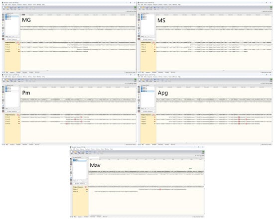
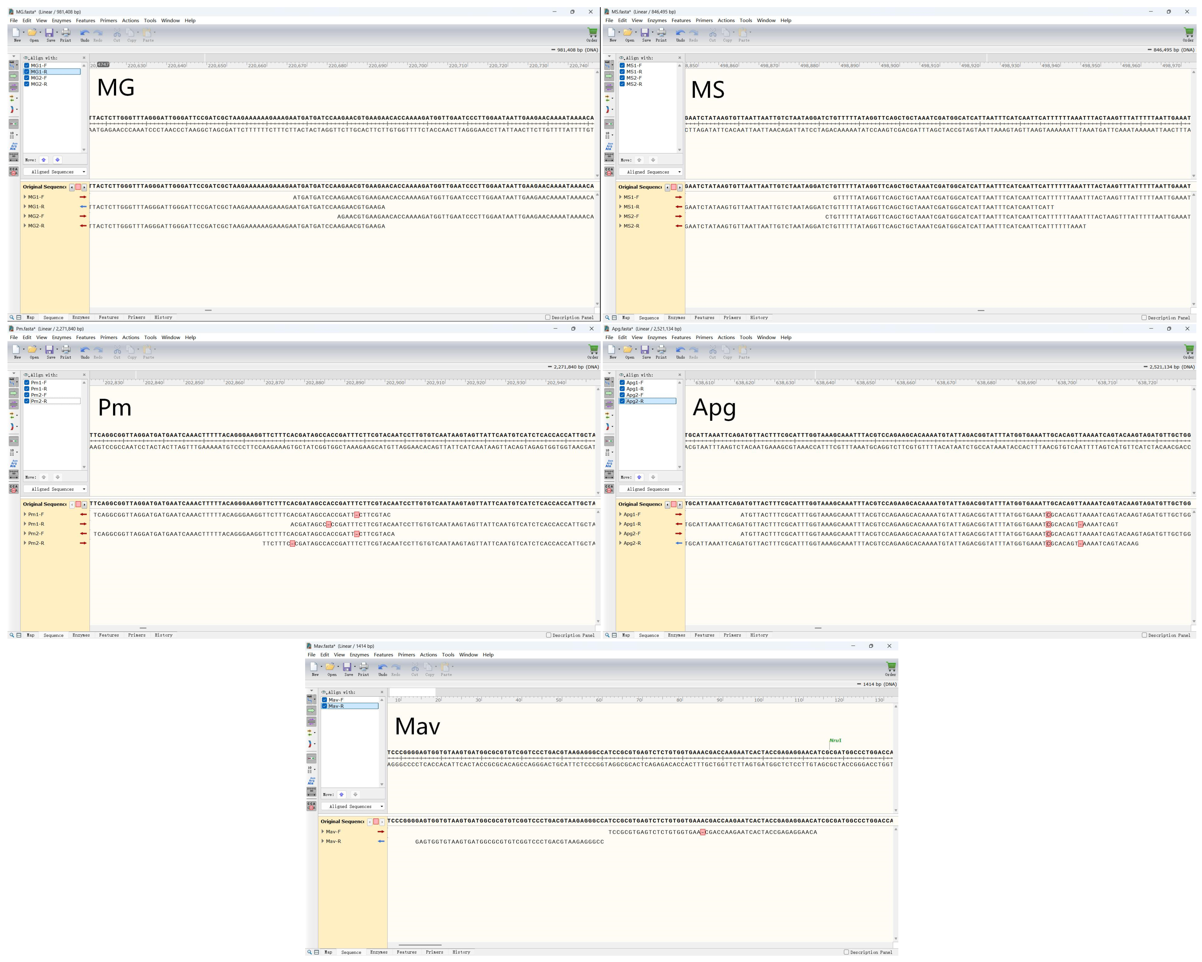
Multiplex PCR amplification was performed using genomic DNA of the five target pathogens as templates, and the products were analyzed by 1% agarose gel electrophoresis. The results showed specific bands at the expected sizes for each target, with no non-specific amplification observed, and no bands were detected in the negative control. To confirm that the amplified fragments corresponded to the correct target sequences, the PCR products from the standard strains were sequenced. The sequencing alignment results confirmed that the amplified fragments from each primer pair showed >98% sequence identity with the reference sequences of the corresponding target genes, with the observed minor nucleotide variations being within the expected range of natural genetic diversity. The sequencing alignment results are presented in Figure 1, while the original gel images are available in the Supplementary Materials (Figure S1). These results demonstrate that the designed primers exhibit good amplification specificity in the multiplex PCR system.
Figure 1.
Sanger sequencing confirmation of amplified products. Representative sequence alignments showing >98% identity of the amplified products to the reference sequences of the corresponding target genes for each pathogen.
3.2. Sensitivity Tests Results
The sensitivity of the established multiplex liquid array assay was evaluated using both single-plasmid and mixed-plasmid templates. The five recombinant plasmid standards were serially diluted ten-fold to concentrations ranging from 1 × 10⁵ copies/μL to 1 copy/μL for individual template testing. Additionally, the five plasmids were mixed in equal proportions and serially diluted ten-fold to concentrations ranging from 1 × 10⁶ copies/μL to 10 copies/μL for mixed template testing. Nuclease-free water was used as the negative control, and results were interpreted based on MFI values.
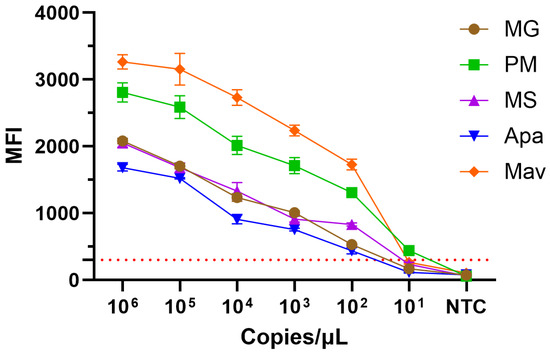
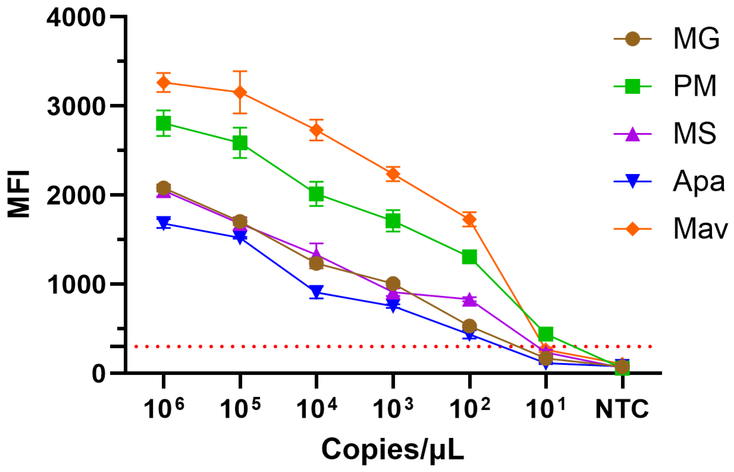
For single-template detection, the limit of detection (LOD) for all five targets was 10 copies/μL (Figure 2). For mixed-template detection, the LOD for Pm reached 10 copies/μL (MFI = 437), while the LODs for MG, MS, Apg, and Mav were 100 copies/μL each (Table 5).
Figure 2.
Sensitivity test Results (single plasmids, MFI values), the red dotted line represents the approximate positive cutoff line at 300 MFI.
Table 5.
Sensitivity test Results (mixed plasmids, MFI values).
3.3. Specificity Test Results
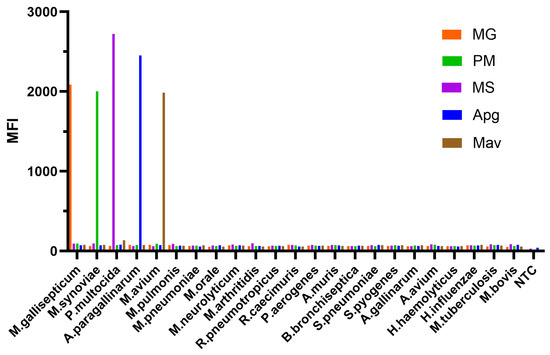
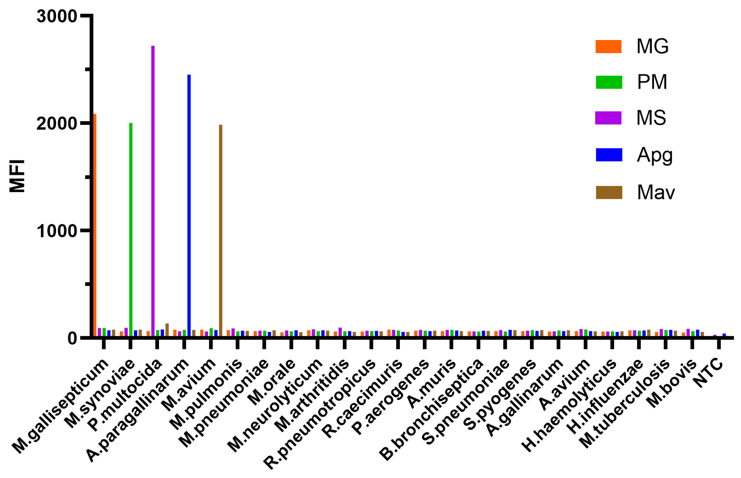
The specificity of the established method was evaluated by testing genomic DNA from the five target pathogens and 18 non-target bacterial species. As shown in Figure 3, the MFI values for the five target pathogens were significantly higher than those of the negative controls: MG, 2085.5; MS, 2003; Pm, 2721; Apg, 2452.5; and Mav, 1985. In contrast, all non-target bacterial species tested negative, with no significant difference from the negative control. These results demonstrate that the developed assay exhibits excellent specificity for the five target pathogens, with no cross-reactivity observed against the 18 non-target species tested, including Mycoplasma pneumoniae, Mycoplasma pulmonis, Mycobacterium tuberculosis, and Mycobacterium bovis. The detailed MFI values for all tested strains are provided in Table S2 in the Supplementary Materials.
Figure 3.
Specificity test results of the multiplex liquid array assay for the five target pathogens and 18 non-target bacterial species. MFI values for the five target pathogens (MG, MS, Pm, Apg, and Mav) were significantly higher than those of the negative controls, while all non-target species tested negative, demonstrating no cross-reactivity.
3.4. Repeatability Test Results
Intra-assay and inter-assay repeatability were evaluated using mixed plasmid templates at three concentration levels: 1 × 10⁶ copies/μL, 1 × 10⁴ copies/μL, and 1 × 10² copies/μL. As shown in Table 6, the CVs for all targets at all concentrations were below 15%, with intra-assay CV values ranging from 0.8% to 12.2% and inter-assay CV values ranging from 2.3% to 14.4%. These results indicate that the developed method exhibits excellent repeatability and stability.
Table 6.
Intra- and inter-assay repeatability test results of the multiplex method.
3.5. Detection Results of Simulated Positive Samples
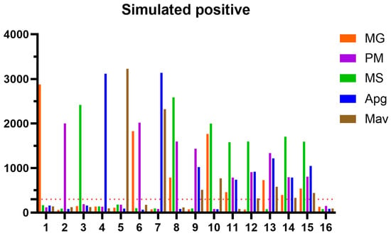
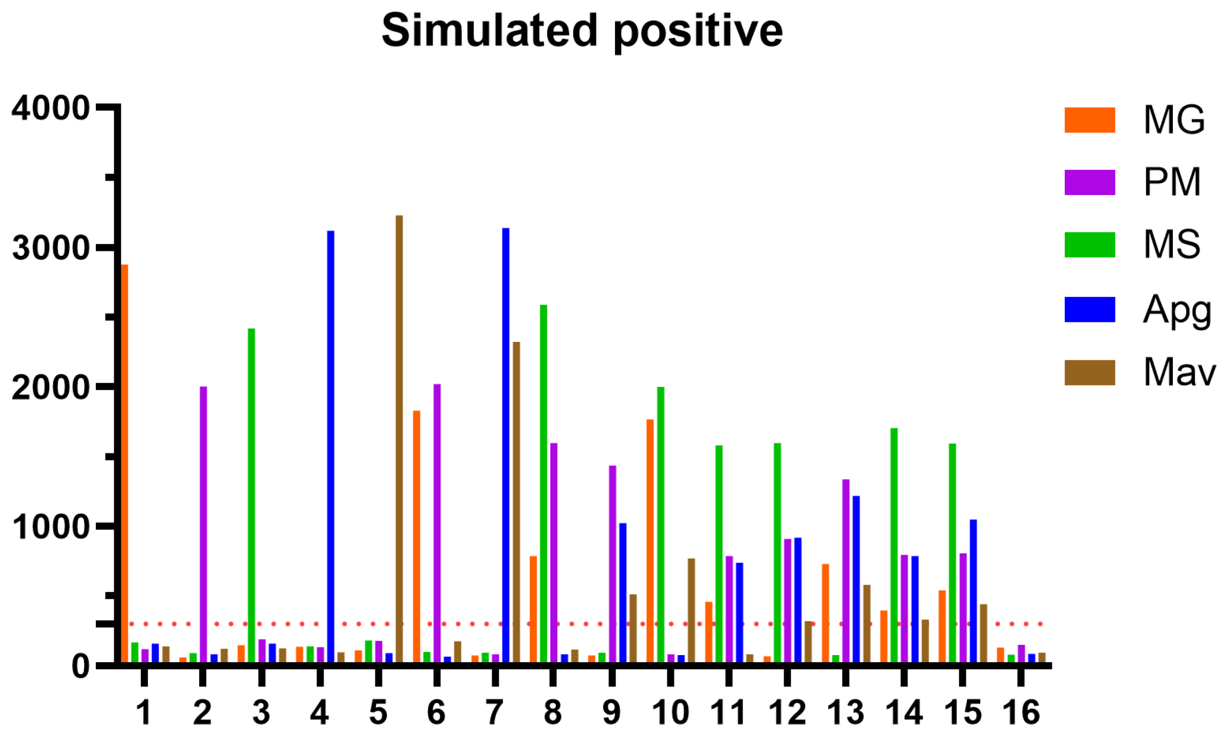
Simulated detection was performed using 16 different combinations of the five pathogen genomic DNA templates, ranging from single to five-plex infections. As shown in Table 7, the detection results for all combinations were completely consistent with the expected combinations. The MFI values for all positive targets were significantly higher than those of the negative controls (≥300 and ≥3-fold that of the negative control MFI), while the MFI values for all negative targets remained below the threshold. Notably, as the number of mixed targets increased, the MFI values of some targets showed varying degrees of reduction; however, these reductions did not affect the final qualitative result. These results demonstrate that the developed method can accurately identify both single and mixed infections in samples (Figure 4).
Table 7.
Results of the multiplex simulation test (partial, MFI values).
Figure 4.
Results of Simulated Positive Samples. Each bar denotes the MFI of simulated positive samples, and the red dotted line represents the approximate positive cutoff line at 300 MFI.
3.6. Sample Detection Results
The developed method was applied to detect genomic DNA extracted from 114 SPF chicken nasopharyngeal swab samples. All samples tested negative based on the MFI threshold criteria, while the positive controls were valid, indicating that the method can be effectively used for routine microbiological monitoring of SPF chickens (partial MFI values are provided in the Table S3). In comparison, qPCR detection using the previously reported method revealed a positive rate of 20.2% (23/114) for MG, with Ct values of 35.53 ± 0.85. Based on the standard curve equation, the nucleic acid concentration in the positive samples was calculated to be 1.3 ± 0.3 copies/μL. The other four target pathogens were also negative by qPCR, with no corresponding nucleic acids detected (Table 8).
Table 8.
Comparison of detection results between the developed multiplex liquid array assay and qPCR for 114 SPF chicken nasopharyngeal swab samples (n = 114).
3.7. Verification of Primer and Platform Adaptability Results
To evaluate the adaptability of previously reported primers on the Luminex platform and their compatibility with the primers designed in this study, three independent verification experiments were conducted. Detailed results, including MFI values for each microsphere set, are provided in the Supplementary Materials (Tables S4–S7).
The previously reported qPCR primers for MG, Pm, and Apg [10] were modified and tested on the Luminex platform. However, severe non-specific background signals and cross-reactivity were observed in the negative controls, indicating that these primers cannot be directly adapted to the Luminex platform simply by adding TAG sequences (Table S4).
When the modified qPCR primers were combined with the MS and Mav primers of the present study, high background signals persisted for Pm and Mav in the negative controls, resulting in false-positive results and demonstrating poor compatibility between the two primer sets (Table S5).
Similarly, previously reported liquid array primers for MG and MS from the Guangdong Provincial Local Standard DB44/T 2338-2021 [35], when modified and combined with the Pm, Apg, and Mav primers of the present study, exhibited significant non-specific signal elevation in the negative controls, leading to false-positive results and failure to construct a functional multiplex system (Table S6).
Finally, an alternative Mav primer pair (Mav-F2/R2) designed based on the IS1245 gene sequence was tested in combination with the other four primer pairs. Abnormally elevated MFI values were observed for multiple targets in the negative controls, with particularly high non-specific signals for Mav, rendering the alternative primers unsuitable for practical detection applications (Table S7).
Collectively, these results confirm that previously reported primers—whether originally designed for qPCR or liquid array platforms—cannot be effectively adapted to the Luminex-based multiplex liquid array assay of the present study through simple modification, underscoring the specificity and robustness of the primer sets developed in this work.
4. Discussion
In this study, a multiplex liquid array nucleic acid detection method based on Luminex xTAG technology was successfully established, enabling the simultaneous detection of all five SPF chicken respiratory bacterial pathogens mandated by the Chinese national standard GB/T 17999.1-2008 [1] and the Chinese Pharmacopoeia (2025 Edition) [2] in a single reaction system. This method fills the application gap of Luminex xTAG technology for the simultaneous detection of these five pathogens in SPF chickens, providing a comprehensive technical solution for SPF chicken quality control.
The Luminex xTAG liquid array technology, based on fluorescently coded microspheres and flow-based fluorescent detection, offers a theoretical multiplexing capacity of up to 500 targets [36]. Compared with traditional detection methods, this technology demonstrates significant advantages not only in multiplexing capability but also in detection throughput, sample consumption, and automation [19]. The multiplex liquid array assay established in this study fully exploits these technological features, integrating the five respiratory bacterial pathogens mandated by Chinese national standards into a single reaction system, thereby overcoming the limitation of qPCR methods that are typically restricted to 4–6 targets per reaction due to fluorescence channel constraints. Notably, Wang et al. [10] recently reported a four-plex qPCR method for quails enabling simultaneous detection of Pm, Apg, MG, and MS; however, their target panel still did not include Mav, which may be constrained by the multiplexing capacity of the qPCR platform. The present study further expands the target coverage on this basis, demonstrating the unique advantages of the Luminex platform in multiplex detection.
The sensitivity results showed that Pm could be detected at 10 copies/μL, while the other four pathogens had a LOD of 100 copies/μL, which meets the requirements for routine surveillance of SPF chickens [37,38]. The repeatability results showed that both intra- and inter-assay CV were below 15%, indicating good stability and reproducibility of the method. In the specificity tests, none of the 18 non-target bacterial species showed cross-reactivity, confirming the rigor of the primer design and the high specificity of the method. Notably, the primers for MG and MS target the mgc2 and VY93_RS02250 genes [39,40,41], respectively, which have been previously validated for their species-specificity; similarly, the primers for Pm and Apg target the kmt1 and hagA genes [42,43,44], respectively, both of which are conserved specific regions validated in the literature.
The primer screening and optimization process in this study provides important methodological insights. Verification experiments demonstrated that directly “transplanting” previously reported qPCR primers or liquid array primers from existing standards into the Luminex system of the present study—by simply adding TAG sequences and biotin labels—resulted in severe non-specific background signals and false-positive outcomes. This phenomenon suggests that different detection platforms (qPCR vs. liquid array) impose substantially different performance requirements on primers, and simple primer modification does not guarantee platform compatibility. This further underscores the necessity of the substantial effort invested in de novo primer design and systematic screening in the present study. The hybridization reaction conditions, microsphere surface chemistry, and signal readout mechanisms of the Luminex platform are fundamentally different from those of qPCR, requiring that platform-specific factors be taken into consideration during primer design, rather than simply adopting existing primer sequences.
The five target pathogens exhibit significant differences in genomic characteristics, which poses additional challenges for establishing a unified multiplex method. The GC content of Mav is approximately 69% [20,45], whereas that of Mycoplasma species is only 28–33% [21]. This substantial difference in genomic composition directly affects the coordination of primer annealing temperatures and the balance of amplification efficiency. Through strict control of amplicon lengths (101–191 bp) and optimization of the annealing temperature (57 °C), the primer design in this study successfully achieved balanced amplification of all five pathogens in a single reaction system, providing a referable strategy for handling mixed targets with markedly different genomic features.
From the perspective of practical application, the method established in this study has undergone preliminary validation with 114 nasopharyngeal swab samples from SPF chickens, all of which tested negative, consistent with the expected microbiological status of SPF chickens. Mixed infections with avian respiratory pathogens are extremely common in clinical settings, and such co-infections often lead to more severe clinical manifestations than single infections [46]. The flexible panel design of the Luminex platform allows for the addition or removal of targets as monitoring needs evolve, providing a technical foundation for future expansion of this method to include simultaneous detection of more SPF chicken pathogens, such as viral pathogens. Furthermore, MG and MS are vertically transmitted through eggs, making their exclusion status particularly critical for maintaining SPF chicken quality [8,47]. The high sensitivity and specificity of this method enable accurate detection at early stages of infection, which is of great significance for the prevention and control of vertically transmitted pathogens.
However, several limitations of this method need to be clearly acknowledged. In comparative sensitivity testing, the LOD of the multiplex liquid array assay for MG was 100 copies/μL, which is higher than that reported for single-plex quantitative PCR (approximately 1 copy/μL). When validating with 114 SPF chicken samples, qPCR detected 23 MG-positive samples with a Ct value of 35.53 ± 0.85, corresponding to approximately 1.3 ± 0.3 copies/μL; in contrast, the present method yielded negative results for all samples, indicating inferior detection capability for samples with extremely low bacterial loads compared to qPCR. This is likely attributable to the fact that the Luminex platform requires a two-step procedure (multiplex PCR amplification followed by hybridization reaction), and its signal accumulation efficiency is lower than that of single-plex qPCR. Nevertheless, it must be clearly emphasized that although the sensitivity of this method is slightly lower than that of single-target qPCR, the LOD of 100 copies/μL is fully adequate for routine monitoring of SPF chickens—because the core objective of SPF quality control is to exclude the presence of target pathogens, rather than to pursue the absolute lowest detection threshold. This LOD is consistent with industry-recognized standards: commercial MG detection kits typically define Ct ≤ 35 as positive (MG Nucleic Acid Detection Kit, PCR-Fluorescence Probe Method, TSTOV0096), and some international brands claiming LODs of 500 copies/μL (e.g., InviScreen® Mycoplasma (Mg/Ms) Detection Kit, No.6016003200), and published qPCR studies have reported LODs of 102–101 copies/μL from artificially contaminated swab samples [48]. In practical applications, the bacterial loads in naturally infected cases typically range from 103 to 108 copies/μL [37,49], far exceeding the LOD of the present method. Therefore, from a practical application perspective, this method, characterized by high throughput, cost-effectiveness, and multiplexing capability, offers greater value for routine monitoring than the pursuit of extremely high sensitivity; these advantages are particularly noteworthy in large-scale SPF chicken quality monitoring programs. In addition, the risk of missing samples with extremely low loads could be further reduced by employing magnetic bead-based nucleic acid pre-concentration or pre-enrichment.
Furthermore, it is necessary to clarify the complementary role of this method relative to qPCR: the present assay is specifically designed for routine monitoring as a high-throughput screening tool, whereas qPCR is more suitable for confirmatory testing of samples with ambiguous signals or low target levels. This complementary positioning provides a clear basis for the rational selection of the two methods in practical applications. Although the current detection panel covers five major respiratory bacterial pathogens, future expansion may be needed to address emerging pathogens or evolving testing requirements; at the same time, the long-term applicability of this method for large-scale sample testing in routine production environments and different poultry farm settings still requires further validation through continuous data accumulation. It should be particularly noted that all samples tested in this study were derived from SPF chickens and were negative for all five target pathogens; therefore, the diagnostic performance of this method in naturally infected or suspected samples has not yet been fully validated. As a preliminary study focused on method establishment and analytical validation, the present results have demonstrated the technical feasibility of this detection method for SPF chicken quality monitoring. Future evaluation using commercial chicken flocks or outbreak field samples is needed to comprehensively determine its sensitivity and specificity.
5. Conclusions
In this study, a multiplex liquid array detection method based on Luminex xTAG technology was successfully established. This method enables the simultaneous detection of all five SPF chicken respiratory bacterial pathogens mandated by the Chinese national standard GB/T 17999.1-2008 [1] and the Chinese Pharmacopoeia (2025 Edition) [2]—MG, MS, Pm, Apg, and Mav—in a single reaction system. The developed assay offers several advantages, including high throughput, high sensitivity (10 copies/μL for Pm and 100 copies/μL for the other four targets), high specificity (no cross-reactivity with 18 non-target bacterial species), and good repeatability (CV < 15%). The detection results for both simulated and routine samples were consistent with expectations, confirming the reliability and practical applicability of the method. Collectively, this method provides an efficient and convenient monitoring tool for SPF chicken quality control, with significant potential for broad application and translation (Table S8).
6. Patents
A patent application has been submitted for this research under application number 202610846363X.
Supplementary Materials
The following supporting information can be downloaded at: https://www.mdpi.com/article/10.3390/vetsci13080753/s1, Text: the detailed primer screening and validation procedures, Table S1: primer sequences used for adaptability verification, Figure S1: original gel electrophoresis images, Table S2: specificity test results, Tables S3–S7: primer and platform adaptability verification data, and Table S8: a summary of analytical performance metrics.
Author Contributions
Conceptualization, J.X.; methodology, Q.S., R.F. and J.X.; software, Q.S.; validation, Y.F. and Q.G.; formal analysis, Q.S.; investigation, Q.S. and S.Y. and J.X.; resources, Q.S., Y.F. and F.D.; data curation, Q.G.; writing—original draft preparation, Q.S.; writing—review and editing, J.X.; visualization, X.Z.; supervision, C.L.; project administration, J.X.; funding acquisition, J.X. All authors have read and agreed to the published version of the manuscript.
Funding
This work was supported by the Training Fund for academic leaders of NIDC (2023X5).
Institutional Review Board Statement
All procedures involving the collection and handling of animal samples were conducted in strict accordance with the guidelines established by the Institutional Animal Care and Use Committee (IACUC) of National Institutes for Drug Control (NIDC). Ethical approval was obtained prior to the initiation of the study. The approval number is NIDC Animal (Welfare Review) No. 2021 (A) 007, with an approval date of 7 April 2026.
Informed Consent Statement
Not applicable.
Data Availability Statement
The original contributions presented in this study are included in the article. Further inquiries can be directed to the corresponding author.
Conflicts of Interest
The authors declare no conflicts of interest.
References
- GB/T 17999.1-2008; SPF Chicken—Microbiological Surveillance—Part 1: General Rules for the Microbiological Surveillance for SPF Chicken. China Standard Press: Beijing, China; Standardization Administration of China: Beijing, China, 2008.
- Chinese Pharmacopoeia Commission. Pharmacopoeia of the People’s Republic of China, Volume IV—General Chapter 3601, 2025 ed.; China Medical Science Press: Beijing, China, 2025; p. 475. [Google Scholar]
- Miller, J.M.; Ozyck, R.G.; Pagano, P.L.; Hernandez, E.F.; Davis, M.E.; Karam, A.Q.; Malek, J.B.; Mara, A.B.; Tulman, E.R.; Szczepanek, S.M.; et al. Rationally designed Mycoplasma gallisepticum vaccine using a recombinant subunit approach. npj Vaccines 2024, 9, 178. [Google Scholar] [CrossRef] [Scilit] [PubMed]
- El-Gazzar, M. Mycoplasma synoviae infection in poultry. In Merck Veterinary Manual; Merck & Co., Inc.: Rahway, NJ, USA, 2025; Revised/Modified September 2025; Available online: https://www.merckvetmanual.com/poultry/mycoplasmosis/mycoplasma-synoviae-infection-in-poultry (accessed on 20 July 2026).
- Lighty, M. Fowl cholera. In Merck Veterinary Manual; Merck & Co., Inc.: Rahway, NJ, USA, 2024; Available online: https://www.merckvetmanual.com/poultry/fowl-cholera/fowl-cholera (accessed on 25 July 2026).
- Wang, Y.; Si, M.; Qiu, J.; Chong, X.; Yang, B.; Chu, M.; Liang, Y.; Cheng, W.; Zhang, H.; Chen, X.; et al. Prevalence, risk factors, and regional insights of infectious coryza among poultry populations in China during 1993–2024: A meta-analysis. J. Vet. Med. Sci. 2026, 88, 201–209. [Google Scholar] [CrossRef] [Scilit] [PubMed]
- Puspitasari, Y.; Khairullah, A.R.; Raharjo, H.M.; Tyasningsih, W.; Kurniasih, D.A.A.; Moses, I.B.; Wardhani, B.W.K.; Ekawasti, F.; Pratama, B.P.; Rehman, S.; et al. Current perspectives on avian tuberculosis in domestic and wild birds: Molecular diagnostics, genomic surveillance, and vaccination challenges. Open Vet. J. 2026, 16, 2562–2580. [Google Scholar] [CrossRef] [Scilit] [PubMed]
- Yadav, J.P.; Tomar, P.; Singh, Y.; Khurana, S.K. Insights on Mycoplasma gallisepticum and Mycoplasma synoviae infection in poultry: A systematic review. Anim. Biotechnol. 2022, 33, 1711–1720. [Google Scholar] [PubMed]
- Huang, Q.; Yang, X.; Zhao, X.; Han, X.; Sun, S.; Xu, C.; Cui, N.; Lu, M. Co-infection of H9N2 subtype avian influenza virus and QX genotype live attenuated infectious bronchitis virus increase the pathogenicity in SPF chickens. Vet. Microbiol. 2024, 295, 110163. [Google Scholar] [CrossRef] [Scilit] [PubMed]
- Wang, H.; Xue, L.; Wang, L.; Liu, Y.; Chen, J.; Sun, Y.; An, T.; Li, C.; Chen, H.; Yu, C.; et al. The quadruplex fluorescent quantitative PCR method for the simultaneous detection of respiratory diseases in quail: Pasteurella multocida, Avibacterium paragallinarum, Mycoplasma gallisepticum, and Mycoplasma synoviae. Front. Microbiol. 2025, 16, 1605356. [Google Scholar] [CrossRef] [Scilit] [PubMed]
- Raviv, Z.; Kleven, S.H. The development of diagnostic real-time TaqMan PCRs for the four pathogenic avian mycoplasmas. Avian Dis. 2009, 53, 103–107. [Google Scholar] [CrossRef] [Scilit] [PubMed]
- Xu, B.; Wang, S.; Yao, W.; Ni, B.; Yuan, T.; Liu, B.; Yuan, L.; Wei, Y.; Ma, S.; Lyu, L.; et al. New molecular diagnostic targets for Avibacterium paragallinarum and a set of single-plex and multiplex qPCR methods for the rapid differential diagnosis of Mycoplasma gallisepticum, Mycoplasma synoviae, and Avibacterium paragallinarum. Poult. Sci. 2025, 104, 105665. [Google Scholar] [CrossRef] [Scilit] [PubMed]
- Wei, B.; Cha, S.Y.; Kang, M.; Park, I.-J.; Moon, O.-K.; Park, C.-K.; Jang, H.-K. Development and application of a multiplex PCR assay for rapid detection of 4 major bacterial pathogens in ducks. Poult. Sci. 2013, 92, 1164–1170. [Google Scholar] [CrossRef] [Scilit] [PubMed]
- Gopinath, K.; Singh, S. Multiplex PCR assay for simultaneous detection and differentiation of Mycobacterium tuberculosis, Mycobacterium avium complexes and other Mycobacterial species directly from clinical specimens. J. Appl. Microbiol. 2009, 107, 425–435. [Google Scholar] [CrossRef] [Scilit] [PubMed]
- Shi, Y.; Li, B.; Tao, J.; Cheng, J.; Liu, H. The Complex Co-infections of Multiple Porcine Diarrhea Viruses in Local Area Based on the Luminex xTAG Multiplex Detection Method. Front. Vet. Sci. 2021, 8, 602866. [Google Scholar] [CrossRef] [Scilit] [PubMed]
- Anderson, S.; Wakeley, P.; Wibberley, G.; Webster, K.; Sawyer, J. Development and evaluation of a Luminex multiplex serology assay to detect antibodies to bovine herpes virus 1, parainfluenza 3 virus, bovine viral diarrhoea virus, and bovine respiratory syncytial virus, with comparison to existing ELISA detection methods. J. Immunol. Methods 2011, 366, 79–88. [Google Scholar] [CrossRef] [Scilit] [PubMed]
- Cong, F.; Zhu, Y.; Wang, J.; Lian, Y.; Liu, X.; Xiao, L.; Huang, R.; Zhang, Y.; Chen, M.; Guo, P. A multiplex xTAG assay for the simultaneous detection of five chicken immunosuppressive viruses. BMC Vet. Res. 2018, 14, 347. [Google Scholar] [CrossRef] [Scilit] [PubMed]
- Laamiri, N.; Fällgren, P.; Zohari, S.; Ben Ali, J.; Ghram, A.; Leijon, M.; Hmila, I. Accurate Detection of Avian Respiratory Viruses by Use of Multiplex PCR-Based Luminex Suspension Microarray Assay. J. Clin. Microbiol. 2016, 54, 2716–2725. [Google Scholar] [CrossRef] [Scilit] [PubMed]
- van der Steen, M. xMAP® Technology Overview [EB/OL]. Diasorin & Luminex Support. 2025. Available online: https://support.diasorin.com/Blog/xmap-multiplexing/#top (accessed on 20 July 2026).
- Kumar, A.; Kaur, J. Primer Based Approach for PCR Amplification of High GC Content Gene: Mycobacterium Gene as a Model. Mol. Biol. Int. 2014, 2014, 937308. [Google Scholar] [CrossRef] [Scilit] [PubMed]
- Brennan, P.C.; Fritz, T.E.; Flynn, R.J. Role of Pasteurella pneumotropica and Mycoplasma pulmonis in murine pneumonia. J. Bacteriol. 1969, 97, 337–349. [Google Scholar] [CrossRef] [Scilit] [PubMed]
- Nadkarni, M.A.; Martin, F.E.; Jacques, N.A.; Hunter, N. Determination of bacterial load by real-time PCR using a broad-range (universal) probe and primers set. Microbiology 2002, 148, 257–266. [Google Scholar] [CrossRef] [Scilit] [PubMed]
- Xu, B.; Chen, X.; Lu, F.; Sun, Y.; Sun, H.; Zhang, J.; Shen, L.; Pan, Q.; Liu, C.; Zhang, X. Comparative Genomics of Mycoplasma synoviae and New Targets for Molecular Diagnostics. Front. Vet. Sci. 2021, 8, 640067. [Google Scholar] [CrossRef] [Scilit] [PubMed]
- Townsend, K.M.; Frost, A.J.; Lee, C.W.; Papadimitriou, J.M.; Dawkins, H.J.S. Development of PCR assays for species- and type-specific identification of Pasteurella multocida isolates. J. Clin. Microbiol. 1998, 36, 1096–1100. [Google Scholar] [CrossRef] [Scilit] [PubMed]
- Jeong, O.M.; Kang, M.S.; Blackall, P.J.; Jeon, B.-W.; Kim, J.-H.; Jeong, J.; Lee, H.-J.; Kim, D.-W.; Kwon, Y.-K.; Kim, J.-H. Genotypic divergence of Avibacterium paragallinarum isolates with different growth requirements for nicotinamide adenine dinucleotide. Avian Pathol. 2020, 49, 153–160. [Google Scholar] [CrossRef] [Scilit] [PubMed]
- Slana, I.; Kaevska, M.; Kralik, P.; Horvathova, A.; Pavlik, I. Distribution of Mycobacterium avium subsp. avium and M. a. hominissuis in artificially infected pigs studied by culture and IS901 and IS1245 quantitative real time PCR. Vet. Microbiol. 2010, 144, 437–443. [Google Scholar] [CrossRef] [Scilit] [PubMed]
- Babady, N.E.; Mead, P.; Stiles, J.; Brennan, C.; Li, H.; Shuptar, S.; Stratton, C.W.; Tang, Y.-W.; Kamboj, M. Comparison of the Luminex® xTAG® RVP Fast Assay and the Idaho Technology FilmArray RP assay for detection of respiratory viruses in pediatric patients at a cancer hospital. J. Clin. Microbiol. 2012, 50, 2282–2288. [Google Scholar] [CrossRef] [Scilit] [PubMed]
- Sambrook, J.; Russell, D.W. Molecular Cloning: A Laboratory Manual, 3rd ed.; Cold Spring Harbor Laboratory Press: Woodbury, NY, USA, 2001. [Google Scholar]
- Xiao, L.; Wang, Y.; Kang, R.; Wu, X.; Lin, H.; Ye, Y.; Yu, J.; Ye, J.; Xie, J.; Cao, Y.; et al. Development and application of a novel Bio-Plex suspension array system for high-throughput multiplexed nucleic acid detection of seven respiratory and reproductive pathogens in swine. J. Virol. Methods 2018, 261, 104–111. [Google Scholar] [CrossRef] [Scilit] [PubMed]
- Shrinet, G.; Chhabra, R.; Sharma, A.; Batra, K.; Talukdar, S.J.; Maan, S. High throughput Luminex beads based multiplex assay for identification of six major bacterial pathogens of mastitis in dairy animals. Front. Cell. Infect. Microbiol. 2023, 13, 1125562. [Google Scholar] [CrossRef] [Scilit] [PubMed]
- Battaglia, A.; Schweighardt, A.J.; Wallace, M.M. Pathogen detection using a liquid array technology. J. Forensic Sci. 2011, 56, 760–765. [Google Scholar] [CrossRef] [Scilit] [PubMed]
- Kuchipudi, S.V.; Yon, M.; Surendran Nair, M.; Byukusenge, M.; Barry, R.M.; Nissly, R.H.; Williams, J.; Pierre, T.; Mathews, T.; Walner-Pendleton, E.; et al. A Highly Sensitive and Specific Probe-Based Real-Time PCR for the Detection of Avibacterium paragallinarum in Clinical Samples from Poultry. Front. Vet. Sci. 2021, 8, 609126. [Google Scholar] [CrossRef] [Scilit] [PubMed]
- Li, K.; Zhang, Y.; Luo, T.; Li, C.; Yu, H.; Wang, W.; Zhang, H.; Chen, H.; Xia, C.; Gao, C. Development of a Triplex qPCR Assay Based on the TaqMan Probe for the Detection of Haemophilus parasuis, Streptococcus suis Serotype 2 and Pasteurella multocida. Microorganisms 2024, 12, 2017. [Google Scholar] [CrossRef] [Scilit] [PubMed]
- Sevilla, I.A.; Molina, E.; Elguezabal, N.; Li, C.; Yu, H.; Wang, W.; Zhang, H.; Chen, H.; Xia, C.; Gao, C. Detection of mycobacteria, Mycobacterium avium subspecies, and Mycobacterium tuberculosis complex by a novel tetraplex real-time PCR assay. J. Clin. Microbiol. 2015, 53, 930–940. [Google Scholar] [CrossRef] [Scilit] [PubMed]
- DB44/T 2338-2021; Laboratory Animals—Qualitative Analysis of Pathogen Nucleic Acids with Suspension Array Method. Guangdong Provincial Administration for Market Regulation: Guangzhou, China, 2021.
- Periyannan Rajeswari, P.K.; Soderberg, L.M.; Yacoub, A.; Leijon, M.; Svahn, H.A.; Joensson, H.N. Multiple pathogen biomarker detection using an encoded bead array in droplet PCR. J. Microbiol. Methods 2017, 139, 22–28. [Google Scholar] [CrossRef] [Scilit] [PubMed]
- Bao, J.; Wu, Z.; Ishfaq, M.; Miao, Y.; Li, R.; Clifton, A.; Ding, L.; Li, J. Comparison of Experimental Infection of Normal and Immunosuppressed Chickens with Mycoplasma gallisepticum. J. Comp. Pathol. 2020, 175, 5–12. [Google Scholar] [CrossRef] [Scilit] [PubMed]
- Lin, L.; Chen, X.-L.; Wu, S.-H.; Cai, X.; Jiang, B.; You, W.; Zheng, M. Simultaneous Detection and Differentiation of Four Eimeria Species in Chickens (E. tenella, E. maxima, E. necatrix, and E. acervulina) Using a Multiplex TaqMan-MGB qPCR Assay. Animals 2025, 15, 2792. [Google Scholar] [CrossRef] [Scilit] [PubMed]
- Al-baqir, A.; Hassanin, O.; Al-Rasheed, M.; Ahmed, M.S.; Mohamed, M.H.A.; El Sayed, M.S.; Megahed, M.; El-Demerdash, A.; Hashem, Y.; Eid, A. Mycoplasmosis in Poultry: An Evaluation of Diagnostic Schemes and Molecular Analysis of Egyptian Mycoplasma gallisepticum Strains. Pathogens 2023, 12, 1131. [Google Scholar] [CrossRef] [Scilit] [PubMed]
- Zhou, L.; Wang, A.; Song, F.; Wu, J.; Yu, C.; Wei, S.; Yang, S.; Zhang, R.; Jiang, S.; Zhu, Y. Development of droplet digital PCR (ddPCR) for the detection and quantification of Mycoplasma gallisepticum in duck flocks. Microb. Pathog. 2025, 209, 108123. [Google Scholar] [CrossRef] [Scilit] [PubMed]
- Hnatow, L.L.; Keeler, C.L.; Tessmer, L.L.; Czymmek, K.; Dohms, J.E. Characterization of MGC2, a Mycoplasma gallisepticum cytadhesin with homology to the Mycoplasma pneumoniae 30-kilodalton protein P30 and Mycoplasma genitalium P32. Infect. Immun. 1998, 66, 3436–3442. [Google Scholar] [CrossRef] [Scilit] [PubMed]
- Zhao, M.; Wu, W.; Song, W.; Yang, H.; Liu, H.; Xie, R.; Huang, X.; Huang, J.; Hua, L.; Chen, H.; et al. A quadruplex real-time fluorescent quantitative PCR detection method for identification and serotyping of Pasteurella multocida. Microb. Pathog. 2025, 207, 107889. [Google Scholar] [CrossRef] [Scilit] [PubMed]
- Pan, Y.; Yu, Q.; Wang, Q.; Li, Q.; Tian, W.; Xin, L.; Hu, X.; Xiao, H.; Liu, Y.; Zhu, L.R.D.; et al. Establishment and epidemiological investigation of a dual fluorescent qPCR assay for Pasteurella multocida and Salmonella in yaks in the Tibetan Autonomous Prefecture of Garzê, China. Front. Cell. Infect. Microbiol. 2025, 15, 1599817. [Google Scholar] [CrossRef] [Scilit] [PubMed]
- Anjaneya Singh, S.D.; Dhama, K.; Wani, M.; Gowthaman, V.; Chawak, M. Molecular Characterization of Avibacterium paragallinarum Isolated from Poultry Flocks of India. Asian J. Anim. Vet. Adv. 2014, 9, 440–451. [Google Scholar] [CrossRef] [Scilit]
- Šlosárek, M. DNA base composition and adansonian analysis of mycobacteria. Folia Microbiol. 1970, 15, 431–436. [Google Scholar] [CrossRef] [Scilit]
- Liu, H.; Pan, S.; Wang, C.; Yang, W.; Wei, X.; He, Y.; Xu, T.; Shi, K.; Si, H. Review of respiratory syndromes in poultry: Pathogens, prevention, and control measures. Vet. Res. 2025, 56, 101. [Google Scholar] [CrossRef] [Scilit] [PubMed]
- Wang, M.J.; Ma, W.J.; Pan, Y.; Chen, J.X.; Zhang, H.; Xia, C.Y.; Wang, Y. Analysis of Major Vertically Transmissible Pathogens and Their Detection Standards in SPF Chickens. Lab. Anim. Comp. Med. 2024, 44, 305–312. [Google Scholar]
- Müştak, İ.; Kolukirik, M. Development of Diagnostic Quantitative Real-Time PCR for Mycoplasma gallisepticum and Mycoplasma synoviae. Etlik Vet. Mikrobiyol. Derg. 2021, 31, 40–44. [Google Scholar] [CrossRef] [Scilit]
- Guo, M.; Liu, D.; Chen, X.; Wu, Y.; Zhang, X. Pathogenicity and innate response to Avibacterium paragallinarum in chickens. Poult. Sci. 2022, 101, 101523. [Google Scholar] [CrossRef] [Scilit] [PubMed]

Disclaimer/Publisher’s Note: The statements, opinions and data contained in all publications are solely those of the individual author(s) and contributor(s) and not of MDPI and/or the editor(s). MDPI and/or the editor(s) disclaim responsibility for any injury to people or property resulting from any ideas, methods, instructions or products referred to in the content. |
© 2026 by the authors. Licensee MDPI, Basel, Switzerland. This article is an open access article distributed under the terms and conditions of the Creative Commons Attribution (CC BY) license.