


Simple Summary
Tibial fractures in heavy beef cattle are often considered difficult to treat because of the animals’ size, high costs for the farmer and poor prognosis. This study describes the use of a full limb cast combined with double transfixation pinning to repair tibial fractures in 11 beef cattle weighing between 260 and 480 kg under field conditions. The technique consisted of placing pins above and below the fracture site and stabilizing the entire limb with a fiberglass cast. Most animals recovered successfully: over 70% completed their production cycle and reached slaughter weight. Although mild to moderate residual lameness was observed, no animals showed severe functional impairment or carcass devaluation. The results suggest that full limb double TPC is a practical, cost-effective and reliable option for treating tibial fractures in heavy cattle directly on farms, helping to avoid premature slaughter and economic losses for farmers.
Abstract
Tibial fractures in beef cattle are a significant clinical challenge, particularly in animals weighing over 300 kg, where surgical treatment is often discouraged due to poor prognosis. This study evaluated the use of full limb double transfixation pin casting (TPC) in field settings for treating tibial fractures in 11 beef cattle weighing between 260 and 480 kg. All animals received the same surgical approach, involving pin placement both proximal and distal to the fracture site, followed by application of a fiberglass cast encompassing the entire limb. The short-term outcome was positive in 90.9% of cases, with two animals not surviving to cast removal. A long-term positive outcome—defined as completing the production cycle and reaching slaughter weight—was achieved in 72.7% of cases. All surviving animals exhibited some residual lameness, but none experienced functional limitations or carcass devaluation. These results support the feasibility and effectiveness of a full limb cast with double transfixation pinning for tibial fracture repair in heavy cattle under field conditions, offering a cost-effective and practical alternative to internal fixation or Thomas splint casts. Further studies are encouraged to confirm these findings and better assess outcomes in open fracture cases.
Keywords:
bovine; tibial fracture; cast; beef cattle; tibia; transfixation pin cast; fracture repair; field 1. Introduction
Long-bone limb fractures represent a relatively common issue in beef cattle and can result in a significant economic loss for farmers. Fractures most frequently involve the metacarpus and metatarsus (about 50%), while other long-bone fractures are encountered in lower percentages: femur (15%), tibia (12%), radius and ulna (7%) and humerus (5%). Pelvic and phalangeal fractures are rare [1]. A successful outcome can depend on many different factors: age and weight of the patient, fracture localization within the bone, type of fracture and treatment choice. Tibial fractures are typically caused by trauma and are usually comminuted and localized in the tibial diaphysis [2,3]. The treatment can be challenging, especially in heavy and double muscled animals, so the farmer must be motivated by cost-effectiveness, by the animal’s economic or genetic value and by a high probability of positive outcome. Treatment is often successful for calves and lightweight animals, but it is normally not recommended for animals weighing over 300 kg where the short-term outcome is often poor [4,5,6]. Several treatment options are reported in the literature for tibial fracture reduction. In some sporadic cases, stall confinement and external coaptation using Thomas splint or cast material may be sufficient [2,4,5], but most cases require surgical repair, considering that casts cannot adequately immobilize fractures proximal to the distal tibial physis [7]. Different kinds of external skeletal fixation (ESF) methods have been described on bovine patients in [7,8]. Transfixation pin casting (TPC) is a type of ESF that involves placing pins above and eventually below the fracture site in combination with casting materials to enclose the limb and create an external support structure. When pins are placed both proximally and distally to the fracture and the foot is included in the cast, a full limb double TPC is performed. Some cases could potentially require internal fixation alone or combined with ESF [9], but high level of sterility, proper surgery room, operator skill and higher costs are required, making TPC more suitable for fracture repair in field settings.
The aim of this study was to describe the diagnostic investigation, surgical technique, outcome and follow-up of eleven beef cattle patients weighing over 260 kg and presenting with tibial fracture, treated with full limb double TPC in field settings. Additionally, the aim was to determine whether the treatment can be recommended to farmers seeking a satisfactory outcome.
2. Materials and Methods
Medical records of 11 bovine patients with tibial fracture treated by the same veterinary surgeon were reviewed. The cases were presented from April 2020 to December 2024. Patients were included in the study if a tibial fracture was confirmed by radiological examination and no previous treatment had been attempted. All animals were treated with a full limb cast with the double pinning (full limb double TPC) surgical technique in field settings. Collected data included: signalment (breed, age, sex and weight), radiographic examination findings, anesthesiologic protocol, surgical procedure details, post-operatory medical treatment and short-, middle- and long-term outcome. All animals were injured upon arrival at the fattening facility; weight was estimated by dividing the net weight of the cattle truck by the number of transported animals. All transported animals were coming from the same breeding facility and were part of the same age group.
Complete physical examination was performed in all animals included in the study, and diagnosis of tibial fracture was established by palpation of the affected limb and radiographic examination performed under sedation with xylazine 0.2 mg/kg intramuscularly. Additional information was collected, including whether the fracture was open or closed, the site of fracture within the bone and fracture configuration. Animals presenting tibial plateau fracture, joint involvement or segmental fracture with no contact between proximal and distal fragment were not included in the study.
2.1. Anesthesiologic Protocol, Surgical Technique and Post-Operative Management
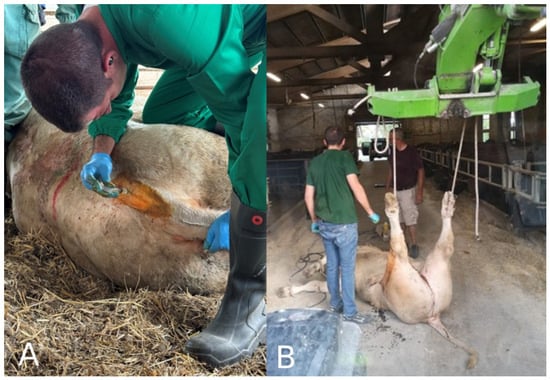
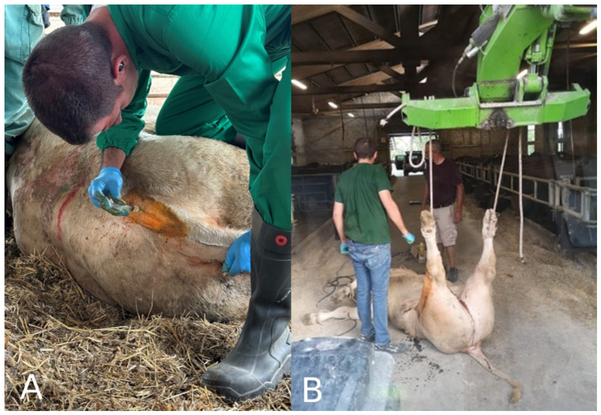
The surgical procedure was performed under general anesthesia induced with ketamine hydrochloride (3 mg/kg IV) administered through an intravenous jugular catheter and maintained with boluses of ketamine hydrochloride (1 mg/kg IV). Sacrococcygeal epidural anesthesia was administered using procaine 2% (10 mL) (Figure 1A). The animal was placed in dorsal recumbency with the fractured limb kept in vertical position using a rope coiled around the foot and secured to a tractor arm. The same method was used to achieve fracture fragments’ alignment when deemed necessary due to the fracture’s configuration (Figure 1B). In case of open fractures, the fracture site was cleaned with sterile saline.
Figure 1.
(A) After palpating the depression between the sacrum and first coccygeal vertebra, moving the tail up and down to better assess the correct space for needle insertion, the site is aseptically prepared and a sacrococcygeal epidural anesthesia is performed using 10 mL of procaine 2%. (B) Correct patient positioning to perform surgery; if necessary, traction can be exercised to achieve fracture fragments’ alignment.
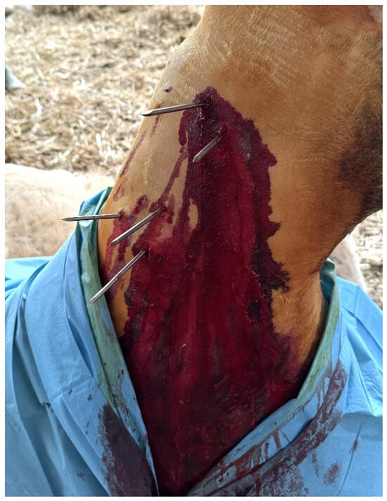
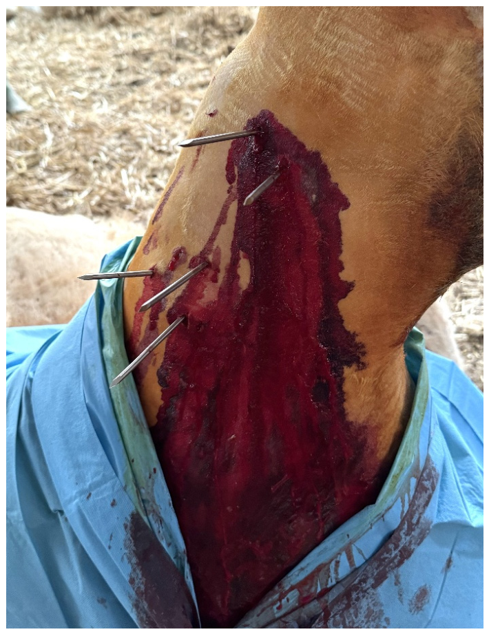
Sterile preparation of the affected limb included trichotomy, a surgical scrub of the crural area and sterile draping of the leg proximally and distally to the surgical site. The pin diameter was chosen based on a core diameter equal to 20% of the tibial diaphyseal diameter [10], ranging from 5 mm to 6 mm. Two to three smooth transcortical pins were placed both proximal and distal to the fracture site, with an inter-pin distance of about 4 cm, all of them applied to tibial bone (Figure 2). A 1 cm stab incision was made in the skin on either the lateral or medial aspect of the limb at the intended pin insertion site. During drilling and pin placement, sterile saline was continuously applied as lavage to cool and lubricate both the drill bit and the pin. Each pin was inserted through both cortices, and drilling was continued until skin tension was observed on the opposite side of the limb. At that point, a skin incision over the emerging pin tip was made, allowing the pin to be advanced further, fully engaging both cortices. This process was repeated for all pins. Pins were positioned at a slightly divergent angle—estimated at 30° within the transverse plane—to reduce the risk of transcortical pin fractures (Figure 2) [10,11].
Figure 2.
In this case (case 9), three smooth transcortical pins were applied proximally to the fracture site, while two were applied distally. Pins are placed with an inter-pin distance of about 4 cm at a slightly divergent angle estimated at 30° within the transverse plane.
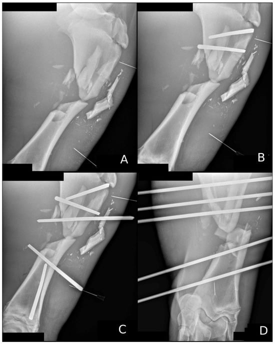
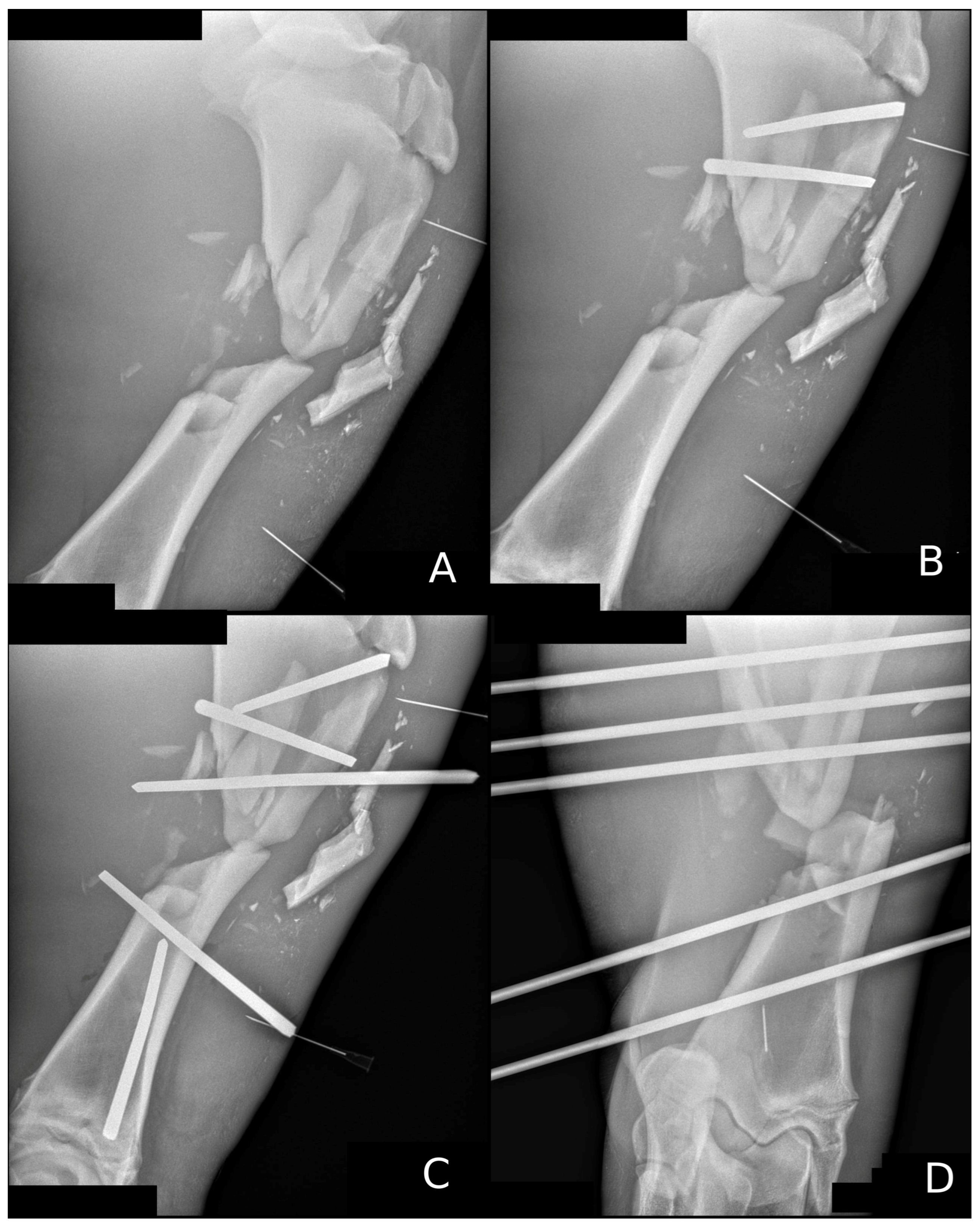
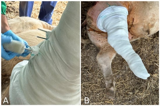
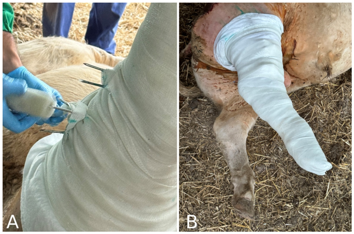
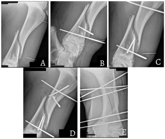
In case of open fracture, soft tissues were debrided by surgical excision, and when possible, closure of the wound was accomplished. Intraoperative radiographs were taken during surgery to choose pins placement with accuracy (Figure 3). A fiberglass cast was applied in the tibial and metatarsal area over an abundant cotton bandage, creating multiple loops around the exposed pins; the pins were then cut to expose only 3 cm on each side of the leg; and the cast was completed, including the foot (Figure 4). A total of 10 to 15 cast rolls were used for each animal depending on body weight, and post-operative medical treatment consisted of 12,000 IU of benzylpenicillin intramuscularly for seven days and flunixin meglumine 2.2 mg/kg IV for three days, starting the day after surgery. The patients were confined in a stall until cast removal. A radiographical control was scheduled 4 weeks post-surgery, while cast and pins removal was planned 6 weeks after surgery.
Figure 3.
(A) Severely comminuted fracture of case 8; after surgical preparation, needles are positioned as markers to determine the pins’ positioning proximally and distally to the fracture. (B) Consecutive radiographs are obtained after the placement of each pin to ensure appropriate spacing and inclination with respect to previously inserted pins; needles may also serve as radiographic markers in this step. (C) Lateromedial radiograph of the final result, with three pins positioned above and two below the fracture site; note the different inclination of pins. (D) Dorso-palmar radiograph of the final result.
Figure 4.
(A) A fiberglass cast was applied in the tibial and metatarsal area, creating multiple loops around the exposed pins to keep tension, avoiding any connecting device between pins. (B) Final result of full limb double transfixation pinning and casting; some cotton bandaging is used as padding over the pins’ stumps before applying the last covering layers of the cast.
2.2. Evaluation of Outcome
The patients were considered to have a short-term positive outcome if no major complications were reported during surgery, during recovery from anesthesia or within 24 h from surgery. A middle-term outcome was considered positive if they survived to the time of cast and pins removal, while a long-term positive outcome was recorded when the animals were able to complete the production cycle and reach the intended slaughter weight. Eventual residual lameness was recorded by the vet during subsequent visits to the farm and graded by locomotion score [12].
3. Results
Eleven patients were included in the study, aged between 8 to 13 months. Weight was estimated to range between 260 kg and 480 kg (Table 1). Five animals (45.4%) were beef cattle crossbreds, four animals were Limousine (36.4%) and two animals were Charolaise (18.2%). Nine animals were males (81.9%), while only two were females (18.1%).
Table 1.
Signalment, fracture characteristics and surgical variables in 11 bovine patients treated with double transfixation pin casting (TPC) for tibial fracture. Data include breed, sex, age, body weight, interval between fracture and surgery, fracture type, number of pins placed relative to the fracture line, duration of surgery and TPC removal time.
All animals had unilateral tibial fracture; upon radiographical diagnosis with lateromedial and caudocranial projections, the fracture configuration was recorded: seven (63.6%) animals presented a comminuted diaphyseal fracture, two (18.2%) presented a transverse diaphyseal fracture and two (18.2%) presented an oblique diaphyseal fracture, one of the two being an open fracture classified as IIIA Gustilo fracture [13].
Surgery was performed from 8 h to 72 h after injury in eight cases 24 h post-injury. In ten animals, three pins were inserted proximal to the fracture and two pins distal to the fracture; only one case (case 4) was treated with two instead of three proximal pins because the fracture was located in the proximal diaphysis, resulting in insufficient healthy bone for the placement of a third pin. Surgical time was about 60 min, and the recovery from anesthesia was uneventful in all 11 cases.
The outcome is reported in Table 2. The short-term outcome was positive for 10 animals (90.9%); one case required surgical revision one day after the first surgery due to pin failure that occurred as a consequence of the animal’s particularly excitable temperament, but middle- and long-term outcomes were positive after surgical revision. The middle-term outcome was positive for nine animals (81.8%), while two animals died before cast removal. One of them had an open fracture and was euthanized five days post-surgery due to septic polyarthritis and prolonged recumbency, while another case died two weeks post-surgery for an unrelated respiratory disease. Out of the remaining nine cases, only one was slaughtered two months before expected slaughtering time due to reduced weight gain, while 72.7% of animals (eight) had a positive long-term outcome, reaching the end of the production cycle.
Table 2.
Post-operative outcomes in 11 bovine patients included in the study. Results are reported for short-term (first 24 h), middle-term (pins removal) and long-term (end of production cycle) follow-up, with residual lameness scores and relevant clinical comments.
Residual lameness was evaluated: all animals had residual lameness after full limb double TPC removal and six of them had a grade 2 lameness, while three animals had grade 3 lameness; none of the animals exhibited any limitations to their routine activities. None of the slaughtered animals had a reduction in dressing percentage or carcass devaluation.
4. Discussion
Transfixation pinning and casting (TPC) aims to provide a stable and well-tolerated method to facilitate early return to weight bearing and function following surgery, while preserving joint mobility when possible. The TPC technique described in our study combines two external skeletal fixation strategies: a double pin configuration (proximal and distal to the fracture) traditionally employed with partial limb casting from the stifle to tarsus area and mainly used in calves to prevent atrophy of the flexor and extensor musculotendinous structures; and a full limb cast, generally associated with a single-pin configuration (proximal to the fracture) and described in adult patients [7,8]. The tibia is subjected to significant biomechanical forces due to the reciprocal apparatus of the hindlimb making reduction and stabilization of tibial fractures particularly challenging. With each stride, the proximal and distal ends of the tibia experience differing rotational forces, creating considerable torsional stress across the bone [14]. For this reason, our approach combined proximal and distal pinning with a full limb cast. This hybrid configuration appeared to confer additional mechanical advantage, countering the high torsional forces typically acting on the bovine tibia. While hanging limb casts often avoid distal pin insertion, they may not offer sufficient rotational control in diaphyseal fractures of the tibia, leading to delayed or non-healing [6,15].
Casts used on the hindlimb need to be particularly thick to reduce stress on the hock and to sufficiently support the patient’s weight; in our cases, 10 to 15 fiberglass layers were applied, as also described by Anderson and St. Jean [16].
The described full limb double TPC technique does not require the use of connecting rods, clamps, or threaded pins, often used in ESF systems [2,17,18,19] The tension between pins is kept by loops of cast bandage wrapped around pins.
Internal fixation would permit the achievement of proper anatomical alignment and stable fixation thanks to direct compression plates that offer enhanced fracture stability under high loading forces [20,21]. However, internal fixation requires proper surgical conditions, surgical skills and long anesthesia; post-operative care is more demanding, and the costs are inevitably higher. For this reason, TPC proved to be a better choice in bovine patients treated in field settings. The described TPC method can be carried out in a minimally invasive manner, allowing closed reduction, which shortens surgical time, promoting a quicker recovery of limb function by minimizing soft tissue trauma and preserving neurovascular integrity [16,22]. However, inadequate anatomical alignment achieved with TPC may occasionally result in limb shortening or angulation [3,22] which, if not severe, does not affect the animal’s ability to perform its vital functions and complete the production cycle. Consistent with observations by Lozier et al. [6], residual lameness was observed in all animals following cast and pin removal, and it ranged from mild to moderate, but all of them remained functional for routine activities and did not show evidence of significant impairment affecting productivity. The Thomas splint technique proved to be effective and practical for field use, particularly in young or lightweight cattle, but animals weighing 250 kg or more had episodes of failure caused by decubitus and slipping of the splint [5]; additionally, this simple and cost-effective technique requires frequent veterinary monitoring and splint repositioning, making the recovery process time- and energy-consuming for both the veterinarian and the owner.
Despite the relatively large body weight of the animals included in the study, the majority of cases (72.7%) achieved a successful long-term outcome, being able to complete the production cycle and achieve slaughter weight. These results are encouraging, especially considering that surgical treatment of long-bone fractures in heavier cattle is generally discouraged due to the risk of complications and poor prognosis; weight alone should not be considered an absolute contraindication for surgical repair as frequently reported in the literature [6].
The combination of a relatively simple surgical technique, limited requirement for specialized surgical facilities and favorable outcomes make full limb double TPC a valuable option for practitioners, offering a balance between mechanical stability, cost-effectiveness and feasibility in field settings.
Possible complications of TPC reported in the literature include transcortical pin fracture, disuse osteopenia, pin tract osteitis, cast sores, sequestra formation, decreased range of motion, limb shortening and angulation, and contralateral or ipsilateral femur physeal fracture [6,15]. Consistent with what is described in other studies, in our cases, pin tract osteitis was detected as the most frequent complication [6,23,24,25,26]. However, no pin loosening was noted, confirming the full limb TPC structure permitted good stabilization of ESF despite the radiographically evident osteitis.
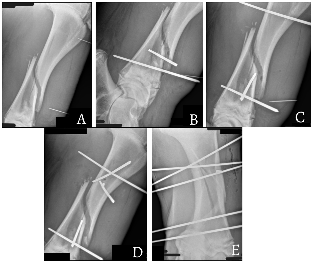
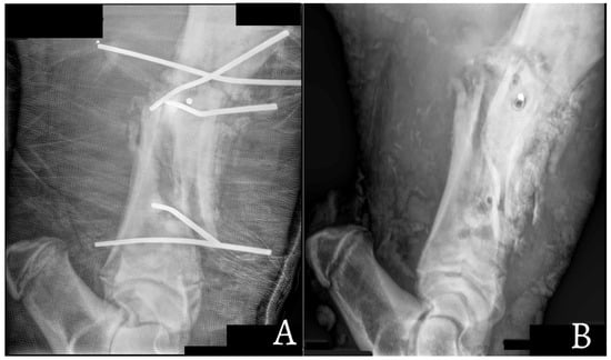
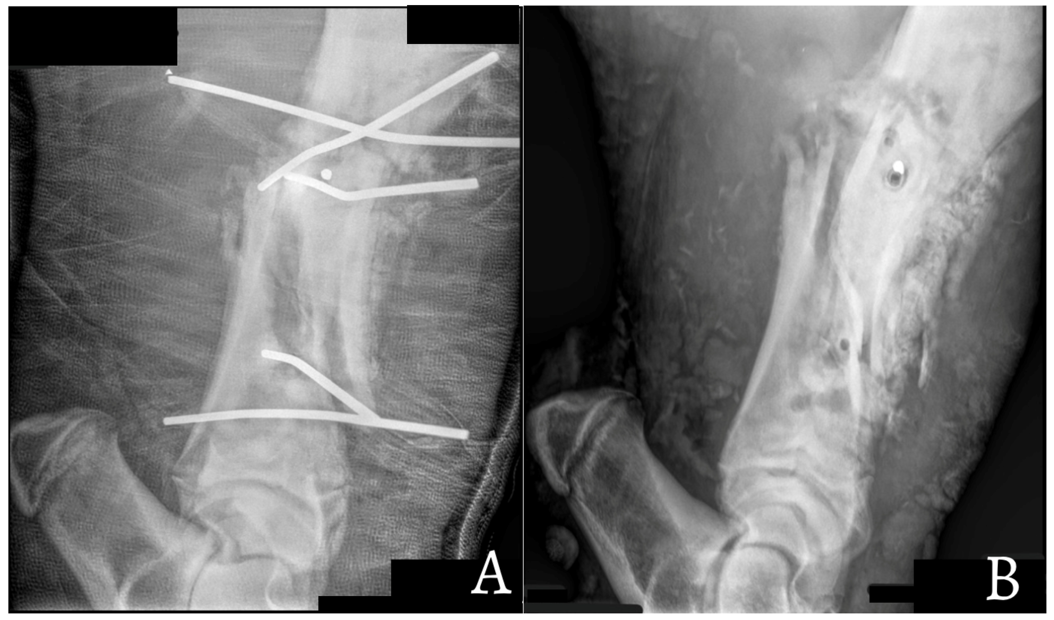
Follow-up radiographs obtained prior to cast removal confirmed progressive fracture healing and satisfactory callus formation in all surviving animals. In case 9, a small fragment of one transfixation pin fractured during pin insertion and remained embedded within the bone. The retained fragment did not interfere with fracture healing, implant stability or limb function. Figure 5 and Figure 6 present the pre-operative, intraoperative, and follow-up radiographs of case 9. The follow-up radiographs show the retained pin fragment together with mild pin tract osteitis; however, both findings were clinically insignificant, and the case achieved a successful outcome.
Figure 5.
Oblique diaphyseal fracture of case 9. (A–C) The radiographic sequence illustrates the pin insertion procedure, as described in the legend of Figure 3. In panels (D,E), a small, retained fragment of a transfixation pin, fractured during an unsuccessful insertion attempt, is visible. The retained fragment did not result in any clinical complications or interfere with fracture healing.
Figure 6.
(A) Follow-up radiograph of case 9 obtained 4 weeks after transfixation pinning and casting (TPC), showing mild pin tract osteitis surrounding the transfixation pins and abundant periosteal callus formation, indicating satisfactory fracture healing. (B) Radiograph obtained after cast and pin removal, 6 weeks post-surgery. The retained pin fragment remains embedded within the bone without causing any clinical complications. Abundant callus formation is evident, providing adequate fracture stability and preservation of limb function.
Open fractures continue to represent a significant clinical challenge. Our single case of a Gustilo type IIIA fracture became open during sedation process, but nevertheless resulted in fatal septic complications, mirroring findings from both bovine and human literature that categorize such injuries as high-risk for infection and poor healing [9,27]. Its unfavorable outcome was due to septic polyarthritis, probably derived from septic emboli coming from fracture site infection, thus supporting the need for cautious case selection and decision analysis [9].
In our study, the majority of the animals included were male (9 out of 11). Animal temperament appears to be an important factor influencing the risk of tibial fractures. Groups consisting of male cattle of the same age often display increased levels of aggression and dominance-related behaviors. Notably, individuals that sustained fractures were frequently those exhibiting more aggressive behavioral patterns. The energy required for long bones to fracture is extremely high; for this reason, fractures to tibia, radio, ulna or femur are frequently comminuted in bovine patients [3,9] who, on the other hand, represent good orthopedic patients as they experience less complications compared with horses [28,29,30,31].
5. Conclusions
This study demonstrated that the application of full limb double TPC for tibial fracture repair in bovine patients weighing over 260 kg, within a field surgery context, was highly effective. Despite the challenges due to patients’ size, the technique proved robust and well tolerated, allowing for positive short-, middle- and long-term outcomes in the large majority of treated animals, reaching the end of the production cycle. When the fracture configuration gives the possibility and the farmer is motivated by cost-effectiveness of the treatment, full limb double TPC of the tibia can be the best treatment option in field conditions to avoid premature slaughter of beef cattle. More cases should be included in further studies to assess the effectiveness of the treatment on a wider population, improve surgical technique and better assess the feasibility and outcome for patients with open tibial fractures.
Author Contributions
Conceptualization, A.S. and A.B.; methodology, A.B.; validation, M.V.R.; formal analysis, M.V.R. and P.D.; investigation, A.B.; resources, A.B.; data curation, M.V.R. and E.M.; writing—original draft preparation, M.V.R.; writing—review and editing, P.D., E.M., R.R. and A.S.; visualization, M.V.R. and E.M.; supervision, A.S.; All authors have read and agreed to the published version of the manuscript.
Funding
This research received no external funding.
Institutional Review Board Statement
Ethical review and approval were waived for this study due to the retrospective analysis of bovine patients treated in field settings; no action other than patient treatment was performed.
Informed Consent Statement
Not applicable.
Data Availability Statement
The original contributions presented in this study are included in the article. Further inquiries can be directed to the corresponding author.
Acknowledgments
During the preparation of this manuscript, the authors used ChatGPT-5 to polish the language, improve scientific soundness, and support the preparation of the work for the purposes of language editing and sentence structure. The authors reviewed and edited the output as needed and take full responsibility for the content of this publication.
Conflicts of Interest
The authors declare no conflicts of interest.
References
- Ferguson, J.G. Management and repair of bovine fractures. Compend. Contin. Educ. Pract. Vet. 1982, 4, 128–136. [Google Scholar]
- Martens, A.; Steenhaut, M.; De Cupere, C.; Gasthuys, F.; De Moor, A.; Verschooten, F. Conservative and surgical treatment of tibial fractures in cattle. Vet. Rec. 1998, 143, 12–16. [Google Scholar] [CrossRef] [Scilit] [PubMed]
- Mulon, P.Y. Management of long bone fractures in cattle. Practice 2013, 35, 265–271. [Google Scholar] [CrossRef] [Scilit]
- Adams, S.B.; Fessler, J.F. Treatment of radial-ulnar and tibial fractures in cattle, using a modified Thomas splint-cast combination. J. Am. Vet. Med. Assoc. 1983, 183, 430–433. [Google Scholar] [CrossRef] [Scilit]
- Gangl, M.; Grulke, S.; Serteyn, D.; Touati, K. Retrospective study of 99 cases of bone fractures in cattle treated by external coaptation or confinement. Vet. Rec. 2006, 158, 264–268. [Google Scholar] [CrossRef] [Scilit] [PubMed]
- Lozier, J.W.; Niehaus, A.J.; Muir, A.; Lakritz, J. Short-and long-term success of transfixation pin casts used to stabilize long bone fractures in ruminants. Can. Vet. J. 2018, 59, 635. [Google Scholar] [PubMed]
- Vogel, S.R.; Anderson, D.E. External skeletal fixation of fractures in cattle. Vet. Clin. N. Am. Food Anim. Pract. 2014, 30, 127–142. [Google Scholar] [CrossRef] [Scilit] [PubMed]
- Anderson, D.E.; St-Jean, G. External skeletal fixation in ruminants. Vet. Clin. N. Am. Food Anim. Pract. 1996, 12, 117–152. [Google Scholar] [CrossRef] [Scilit] [PubMed]
- St-Jean, G.; Anderson, D.E. Decision analysis for fracture management in cattle. Vet. Clin. N. Am. Food Anim. Pract. 2014, 30, 1–10. [Google Scholar] [CrossRef] [Scilit] [PubMed]
- Auer, J.A.; Stick, J.A. Principles of fracture healing. In Equine Surgery, 4th ed.; Elsevier/Saunders: St. Louis, MO, USA, 2012; pp. 1047–1054, Chapter 76. [Google Scholar]
- McClure, S.R.; Watkins, J.P.; Ashman, R.B. In vitro comparison of the effect of parallel and divergent transfixation pins on breaking strength of equine third metacarpal bones. Am. J. Vet. Res. 1994, 55, 1327–1330. [Google Scholar] [CrossRef] [Scilit] [PubMed]
- Sprecher, D.E.A.; Hostetler, D.E.; Kaneene, J.B. A lameness scoring system that uses posture and gait to predict dairy cattle reproductive performance. Theriogenology 1997, 47, 1179–1187. [Google Scholar] [CrossRef] [Scilit] [PubMed]
- Gustilo, R.B.; Mendoza, R.M.; Williams, D.N. Problems in the management of type III (severe) open fractures: A new classification of type III open fractures. J. Trauma Inj. Infect. Crit. Care 1984, 24, 742–746. [Google Scholar]
- Bowers, K.; Weinhandl, J.T.; Anderson, D.E. A review of equine tibial fractures. Equine Vet. J. 2023, 55, 171–181. [Google Scholar] [CrossRef] [Scilit] [PubMed]
- Hatate, K.; Tsuzuki, N.; Kajihara, A.; Devkota, B.; Takehana, K.; Sato, R.; Iino, K.; Aoki, T.; Takahashi, E.; Yamagishi, N. Influence of transfixation pinning and casting (TPC) configurations on treatment outcomes in bovine tibial fracture. J. Vet. Med. Sci. 2019, 81, 857–862. [Google Scholar] [CrossRef] [Scilit] [PubMed]
- Anderson, D.E.; St-Jean, G. Management of fractures in field settings. Vet. Clin. N. Am. Food Anim. Pract. 2008, 24, 567–582. [Google Scholar] [CrossRef] [Scilit] [PubMed]
- Kumar, R.; Tiwary, R.; Singh, G.D.; Kumar, M. External skeletal fixation for long bone fracture management-A clinical study in 12 bovine calves. Intas Polivet 2016, 17, 1–3. [Google Scholar]
- Dwivedi, D.K. Management of long bone fractures by external coaptation and external skeletal fixators in Bovines. In Compendium Virtual Training Programme on Diagnosis and Management of Common Surgical Affections of Bovines; Indian Council of Agricultural Research: New Delhi, India, 2020; p. 23. [Google Scholar]
- Palsania, S.K.; Bishnoi, P.; Sharma, M.L.; Patwa, R.; Kumar, R.; Kumari, A. Clinical use of transarticular linear external skeletal fixation system for the management of metacarpal and radius-ulna fractures near carpal joint in cattle. Vet. Pract. 2022, 23, 343–349. [Google Scholar]
- Verschooten, F.; De Moor, A.; Desmet, P.; Steenhaut, M. Surgical treatment of tibial fractures in cattle. Vet. Rec. 1972, 90, 24–28. [Google Scholar] [CrossRef] [Scilit] [PubMed]
- Nuss, K. Plates, pins, and interlocking nails. Vet. Clin. N. Am. Food Anim. Pract. 2014, 30, 91–126. [Google Scholar] [CrossRef] [Scilit] [PubMed]
- Itagaki, K.; Kim, D.; Sasaki, K.; Devkota, B.; Yamagishi, N. Clinical characteristics of limb fractures in 25 calves and growing cattle. Jpn. J. Large Anim. Clin. 2011, 2, 197–204. [Google Scholar] [CrossRef] [Scilit]
- Hamilton, G.F.; Tulleners, E.P. Transfixation pinning of proximal tibial fractures in calves. J. Am. Vet. Med. Assoc. 1980, 176, 725–727. [Google Scholar] [CrossRef] [Scilit] [PubMed]
- Kaneps, A.J.; Schmotzer, W.B.; Huber, M.J.; Riebold, T.W.; Watrous, B.J.; Arnold, J.S. Fracture repair with transfixation pins and fiberglass cast in llamas and small ruminants. J. Am. Vet. Med. Assoc. 1989, 195, 1257–1261. [Google Scholar] [CrossRef] [Scilit] [PubMed]
- St-Jean, G.; Clem, M.F.; DeBowes, R.M. Transfixation pinning and casting of tibial fractures in calves: Five cases (1985–1989). J. Am. Vet. Med. Assoc. 1991, 198, 139–143. [Google Scholar] [PubMed]
- Kofler, J.; Wetchy, G.; Schoffmann, G. Transfixation pinning and casting of a comminuted metacarpal fracture in a 870 kg bull. Vet. Surg. 2014, 43, 1014–1019. [Google Scholar] [CrossRef] [Scilit] [PubMed]
- Ahern, B.J.; Richardson, D.W.; Boston, R.C.; Schaer, T.P. Orthopedic infections in equine long bone fractures and arthrodeses treated by internal fixation: 192 cases (1990–2006). Vet. Surg. 2010, 39, 588–593. [Google Scholar] [CrossRef] [Scilit] [PubMed]
- Crawford, W.H.; Fretz, P.B. Long Bone Fractures in Large Animals A Retrospective Study. Vet. Surg. 1985, 14, 295–302. [Google Scholar] [CrossRef] [Scilit]
- Joyce, J.; Baxter, G.M.; Sarrafian, T.L.; Stashak, T.S.; Trotter, G.; Frisbie, D. Use of transfixation pin casts to treat adult horses with comminuted phalangeal fractures: 20 cases (1993–2003). J. Am. Vet. Med. Assoc. 2006, 229, 725–730. [Google Scholar] [CrossRef] [Scilit] [PubMed]
- Lescun, T.B.; McClure, S.R.; Ward, M.P.; Downs, C.; Wilson, D.A.; Adams, S.B.; Hawkins, J.F.; Reinertson, E.L. Evaluation of transfixation casting for treatment of third metacarpal, third metatarsal, and phalangeal fractures in horses: 37 cases (1994–2004). J. Am. Vet. Med. Assoc. 2007, 230, 1340–1349. [Google Scholar] [CrossRef] [Scilit] [PubMed]
- Virgin, J.E.; Goodrich, L.R.; Baxter, G.M.; Rao, S. Incidence of support limb laminitis in horses treated with half limb, full limb or transfixation pin casts: A retrospective study of 113 horses (2000–2009). Equine Vet. J. 2011, 40, 7–11. [Google Scholar] [CrossRef] [Scilit] [PubMed]

Disclaimer/Publisher’s Note: The statements, opinions and data contained in all publications are solely those of the individual author(s) and contributor(s) and not of MDPI and/or the editor(s). MDPI and/or the editor(s) disclaim responsibility for any injury to people or property resulting from any ideas, methods, instructions or products referred to in the content. |
© 2026 by the authors. Licensee MDPI, Basel, Switzerland. This article is an open access article distributed under the terms and conditions of the Creative Commons Attribution (CC BY) license.